Title Page Critical Care for Providers Without Critical Care Training Searchable Just-In-Time Learning Resource Now What…? UPHS resources by the Critical Care Committee Cameron Baston, MD MSCE Emily Gordon, MD Caitlin Clancy, MD Janae Heath, MD MSCE Concept Attributed to University of Washington PCCM division Imitation is the highest form of flattery V4.2.20

Welcome message from author
This document is posted to help you gain knowledge. Please leave a comment to let me know what you think about it! Share it to your friends and learn new things together.
Transcript
Title Page
Critical Care for Providers Without Critical Care Training Searchable Just-In-Time Learning Resource
Now What…?
UPHS resources by the Critical Care Committee
Cameron Baston, MD MSCE
Emily Gordon, MD Caitlin Clancy, MD
Janae Heath, MD MSCE
Concept Attributed to University of Washington PCCM division Imitation is the highest form of flattery
V4.2.20
Table of Contents
Respiratory ● My non-intubated patient is hypoxemic… now what? ● My patient just got intubated… now what? ● I just got blood gas results… now what? ● My intubated patient is more hypoxemic… now what? ● My intubated patient has ARDS… now what? ● My intubated patient is agitated… now what? ● The Peak Pressure alarm is going off… now what? ● The RT says my patient has AutoPEEP… now what? ● The RT says my patient is breath stacking… now what? ● I think my patient might be ready to be extubated… now what?
Hemodynamics
● My patient is hypotensive… now what? ● The pressor requirements are rising… now what? ● My patient is tachycardic… now what? ● My patient is in atrial fibrillation (or flutter)… now what? ● My patient has an abnormal central venous saturation… now what?
Renal
● My patient’s Cr is rising and/or urine output is decreasing… now what? ● My patient’s K+ is too high… now what?
Other
● My patient has a fever… now what? ● My patient has altered mental status… now what? ● My patient has delirium… now what? ● My patient is in the ICU...now what?

My non-intubated patient is hypoxemic… Now what? Step 1: Make sure the data is accurate
● Ensure that the pulse oximeter HR matches the patient’s HR. Ensure the waveform is reliable. ● If SpO2 is not trustworthy, get an arterial blood gas to measure the PaO2. ● Goal SpO2 is 88-97%, Goal PaO2 is 60-90
Step 2: Supplement Oxygen Delivery ● See the most up to date respiratory decompensation pathway on Sharepoint ● Increase supplemental oxygen via Nasal Cannula ● IF COVID+ or PUI, Call for Rapid Response when Supplemental O2 reaches 6 L NC ● If SpO2 still below goal, ask for Non-rebreather or High Flow Nasal Cannula, and call for Rapid
Response. If COVID+ or PUI, place surgical mask on patient Step 3: Initial Evaluation
● IF COVID+ or PUI, Call for Rapid Response when Supplemental O2 reaches 6 L NC ● ABG ● Basic labs: CBC, BMP, consider BNP, Troponin if heart failure in differential ● Stat portable chest radiograph ● POCUS (if able) – See educational materials below ● EKG if indicated
Step 4: Decide if patient needs ventilatory support ● The decision to intubate is not based on specific lab thresholds, but must include clinical factors
such as work of breathing, mental status, and hemodynamics ● If patient not protecting their airway, call for intubation ● If arterial pH<7.35 and PaCO2 is > 45, patient needs ventilatory support (invasive or non-invasive)
o If COVID negative, consider trial of BIPAP o If COVID positive or PUI, call for intubation
● If arterial pH>7.35, or PaCO2 < 45, patient may need only oxygenation support o If COVID negative, High Flow Nasal Cannula o If COVID positive or PUI, call for intubation
Step 5: Address potentially reversible causes based on patient clinical state Problem Intervention Pulmonary Edema (Heart failure, volume overload) Diuresis with IV furosemide Large Pleural Effusion Diuresis, consider thoracentesis Pneumonia Antibiotics Mucus Plug (lobar collapse) Chest PT COPD / Asthma exacerbation Inhaled bronchodilators / Steroids Concern for Pulmonary embolism Chest CT with contrast, consider anticoagulation Learn More: ● ACP Critical Care Video Shorts: Oxygenation, Mechanical Ventilation (Indications, NIPPV Overview,
Delivering NIPPV) ● SCCM Critical Care Video Learning Modules: Diagnosis and Management of Acute Respiratory
Failure ● POCUS: , Introduction, U/S training videos from Sonosite (scroll down), POCUS in COVID discussion,
ACP modules (make a free account)

My patient just got intubated… now what? Note: Some sites have an Epic post-intubation orderset, please familiarize yourself with it Step 1: Choose ventilator settings
● Mode: Volume Assist Control (AC/VC) ● Tidal Volume: 6 cc/kg of ideal-body weight (determined based on patient height) ● Rate: Discuss with RT, If serum bicarb <20, aim for MV >12 ● FiO2: 1.0 ● PEEP: 5 cm H2O. In COVID+/PUI, initial PEEP should be at least 10 ● Check ventilator mechanics (plateau pressure) immediately while patient is paralyzed
Step 2: Ask Nursing to place Orogastric tube Step 3: Choose sedation and analgesia
● If patient paralyzed for intubation, give additional dose of midazolam 2mg after intubation ● Analgesia: Fentanyl infusion, start at rate of 25 ● Sedative if needed: Propofol infusion, titrate to RASS of 0-1 ● In COVID+/PUI, sedate with midazolam infusion, titrate to RASS of -2 to -3
Step 4: Decide if patient needs central access
● If patient on escalating doses of vasopressors, or has insufficient access, answer is yes ● Call for help if needed to place CVC ● In COVID+/PUI, plan to place central access (CVC/arterial line) on same side for ease of
proning Step 5: Get Stat portable CXR to confirm placement of ETT, OG tube, CVC
● Ensure ETT above carina and below clavicles, goal ~4 cm above carina Step 6: Check a blood gas 10-15 minutes after intubation
● See I just got blood gas results… now what? Step 7: Titrate FiO2 to SpO2
● See I just got blood gas results… now what?, or My intubated patient is more hypoxemic… now what?
Step 8: Ensure that the Pennchart Post-intubation order-set has been completed Learn More: ● ACP Critical Care Video Shorts: Mechanical Ventilation (MV modes, Volume versus Pressure Targets,
Sedation) ● SCCM Critical Care Video Learning Modules: Mechanical Ventilation 1, Mechanical Ventilation 2 ● Penn PCCM Mechanical Ventilation review video: Link forthcoming
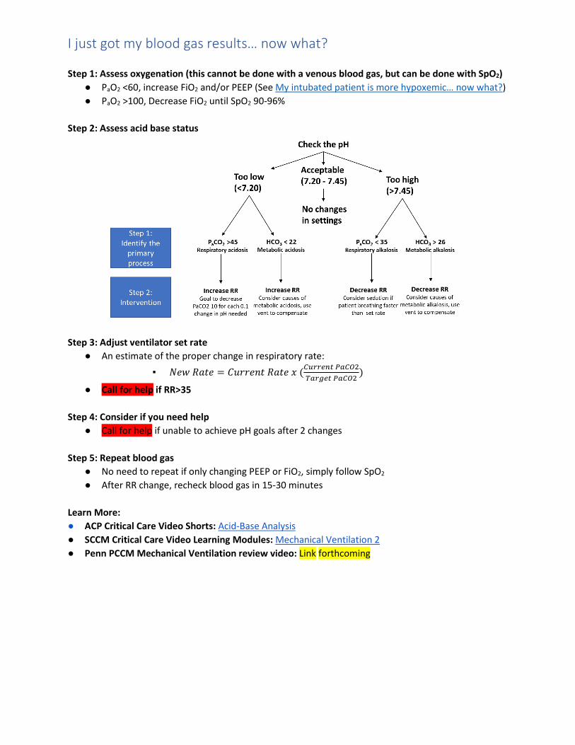
I just got my blood gas results… now what? Step 1: Assess oxygenation (this cannot be done with a venous blood gas, but can be done with SpO2)
● PaO2 <60, increase FiO2 and/or PEEP (See My intubated patient is more hypoxemic… now what?) ● PaO2 >100, Decrease FiO2 until SpO2 90-96%
Step 2: Assess acid base status
Step 3: Adjust ventilator set rate
● An estimate of the proper change in respiratory rate: ▪ 𝑁𝑁𝑁𝑁𝑁𝑁 𝑅𝑅𝑅𝑅𝑅𝑅𝑁𝑁 = 𝐶𝐶𝐶𝐶𝐶𝐶𝐶𝐶𝑁𝑁𝐶𝐶𝑅𝑅 𝑅𝑅𝑅𝑅𝑅𝑅𝑁𝑁 𝑥𝑥 (𝐶𝐶𝐶𝐶𝐶𝐶𝐶𝐶𝐶𝐶𝐶𝐶𝐶𝐶 𝑃𝑃𝑃𝑃𝐶𝐶𝑃𝑃2
𝑇𝑇𝑃𝑃𝐶𝐶𝑇𝑇𝐶𝐶𝐶𝐶 𝑃𝑃𝑃𝑃𝐶𝐶𝑃𝑃2)
● Call for help if RR>35 Step 4: Consider if you need help
● Call for help if unable to achieve pH goals after 2 changes Step 5: Repeat blood gas
● No need to repeat if only changing PEEP or FiO2, simply follow SpO2 ● After RR change, recheck blood gas in 15-30 minutes
Learn More: ● ACP Critical Care Video Shorts: Acid-Base Analysis ● SCCM Critical Care Video Learning Modules: Mechanical Ventilation 2 ● Penn PCCM Mechanical Ventilation review video: Link forthcoming

My intubated patient is more hypoxemic… now what? Step 1: Check equipment, consider calling the respiratory therapist for help
● Ensure the following: Patient remains connected to the ventilator circuit and circuit is intact ● No inadvertent changes FIO2, PEEP or other settings ● Endotracheal tube is patent and remains in correct position ● SpO2 waveform is reliable (HR matches tele HR at least)
Step 2: Check if the ventilator pressure alarm is sounding ● Look for the The Peak Pressure alarm is going off… now what?
Step 3: Examine the patient and evaluate their interaction with the ventilator ● Look at the vent waveforms
o Is the patient breathing irregularly, or taking double breaths? Ask the RT for help o If patient is asynchronous with the ventilator, ask nursing to increase sedation o If patient is agitated and having repeated peak pressure alarms, ask nursing to increase
sedation ● Listen for bilateral breath sounds
o If breath sounds are asymmetric, consider pneumothorax or lung collapse and evaluate accordingly with CXR or POCUS (if able, See resources at bottom of this page)
● Suction the patient to clear any mucus in the endotracheal tube or central airways Step 4: Consider diagnostic studies
● Arterial Blood Gas (venous blood gases cannot be used to assess oxygenation) ● Chest radiograph ● POCUS (if able, See resources at bottom of this page)
Step 5: Adjust the ventilator to improve oxygenation (increase the FiO2 or PEEP) ● The two parameters on the ventilator that address oxygenation are FIO2 and PEEP ● For COVID + /PUI patients use high PEEP-FIO2 ladder:
FiO2 0.3 0.3 0.3 0.4 0.4 0.5 0.5 0.5-0.8 >.8 PEEP 10 12 14 14 16 16 18 20 22-24
● Expect a slow rise in oxygen saturation with increased PEEP ● If PEEP causes paradoxical worsening of oxygenation return to previous PEEP ● If PEEP causes hypotension return to previous PEEP
Step 6: Treat reversible causes of hypoxemia if present ● If chest radiograph reveals lobar or whole lung collapse, start chest physiotherapy ● If worsening bilateral infiltrates on chest radiograph, consider diuresis or new diagnosis of ARDS ● Check hemoglobin and transfuse red blood cells if HgB <7 g/dL ● If patient has a central line, check ScvO2 and Call for help if < 60% (See My patient has an
abnormal central venous saturation… now what?) ● Call for help! if these maneuvers do not resolve the situation
Learn More: ● ACP Critical Care Video Shorts: Mechanical Ventilation (Respiratory Mechanics, Intrinsic PEEP,
Refractory Hypoxemia) ● SCCM Critical Care Video Learning Modules: Mechanical Ventilation 2 ● POCUS: Lung Ultrasound basics, Lung ultrasound part 2, ACP Lung Ultrasound module (make free
account)
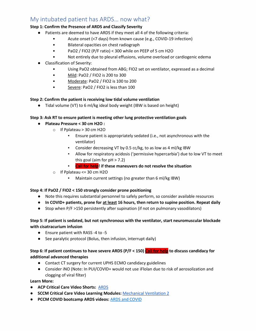
My intubated patient has ARDS… now what? Step 1: Confirm the Presence of ARDS and Classify Severity
● Patients are deemed to have ARDS if they meet all 4 of the following criteria: • Acute onset (<7 days) from known cause (e.g., COVID-19 infection) • Bilateral opacities on chest radiograph • PaO2 / FIO2 (P/F ratio) < 300 while on PEEP of 5 cm H2O • Not entirely due to pleural effusions, volume overload or cardiogenic edema
● Classification of Severity: • Using PaO2 obtained from ABG; FIO2 set on ventilator, expressed as a decimal • Mild: PaO2 / FIO2 is 200 to 300 • Moderate: PaO2 / FIO2 is 100 to 200 • Severe: PaO2 / FIO2 is less than 100
Step 2: Confirm the patient is receiving low tidal volume ventilation
● Tidal volume (VT) to 6 ml/kg ideal body weight (IBW is based on height) Step 3: Ask RT to ensure patient is meeting other lung protective ventilation goals
● Plateau Pressure < 30 cm H2O : o If Pplateau > 30 cm H2O
▪ Ensure patient is appropriately sedated (i.e., not asynchronous with the ventilator)
▪ Consider decreasing VT by 0.5 cc/kg, to as low as 4 ml/kg IBW ▪ Allow for respiratory acidosis (‘permissive hypercarbia’) due to low VT to meet
this goal (aim for pH > 7.2) ▪ Call for help! if these maneuvers do not resolve the situation
o If Pplateau <= 30 cm H2O ▪ Maintain current settings (no greater than 6 ml/kg IBW)
Step 4: If PaO2 / FIO2 < 150 strongly consider prone positioning
● Note this requires substantial personnel to safely perform, so consider available resources ● In COVID+ patients, prone for at least 16 hours, then return to supine position. Repeat daily ● Stop when P/F >150 persistently after supination (if not on pulmonary vasodilators)
Step 5: If patient is sedated, but not synchronous with the ventilator, start neuromuscular blockade with cisatracurium infusion
● Ensure patient with RASS -4 to -5 ● See paralytic protocol (Bolus, then infusion, interrupt daily)
Step 6: If patient continues to have severe ARDS (P/F < 150) Call for help to discuss candidacy for additional advanced therapies
● Contact CT surgery for current UPHS ECMO candidacy guidelines ● Consider iNO (Note: In PUI/COVID+ would not use iFlolan due to risk of aerosolization and
clogging of viral filter) Learn More: ● ACP Critical Care Video Shorts: ARDS ● SCCM Critical Care Video Learning Modules: Mechanical Ventilation 2 ● PCCM COVID bootcamp ARDS videos: ARDS and COVID
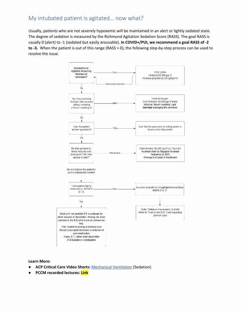
My intubated patient is agitated… now what? Usually, patients who are not severely hypoxemic will be maintained in an alert or lightly sedated state. The degree of sedation is measured by the Richmond Agitation Sedation Score (RASS). The goal RASS is usually 0 (alert) to -1 (sedated but easily arousable). In COVID+/PUI, we recommend a goal RASS of -2 to -3. When the patient is out of this range (RASS > 0), the following step-by-step process can be used to resolve the issue.
Learn More: ● ACP Critical Care Video Shorts: Mechanical Ventilation (Sedation) ● PCCM recorded lectures: Link
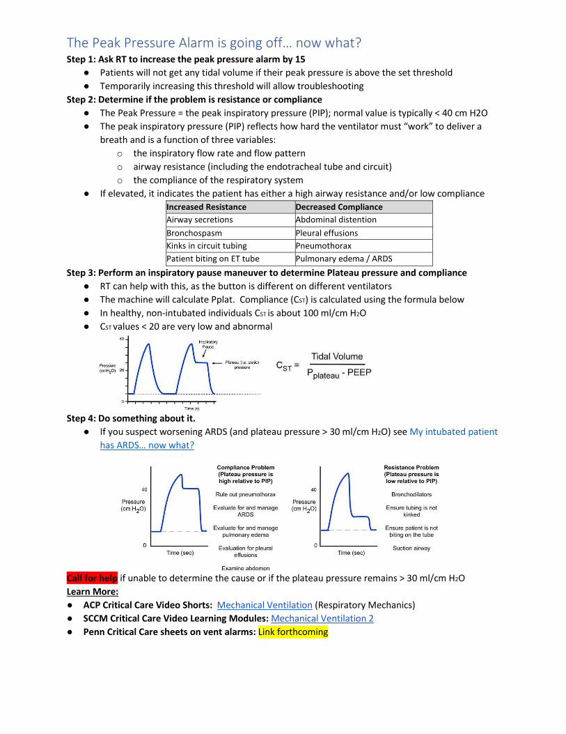
The Peak Pressure Alarm is going off… now what? Step 1: Ask RT to increase the peak pressure alarm by 15
● Patients will not get any tidal volume if their peak pressure is above the set threshold ● Temporarily increasing this threshold will allow troubleshooting
Step 2: Determine if the problem is resistance or compliance ● The Peak Pressure = the peak inspiratory pressure (PIP); normal value is typically < 40 cm H2O ● The peak inspiratory pressure (PIP) reflects how hard the ventilator must “work” to deliver a
breath and is a function of three variables: o the inspiratory flow rate and flow pattern o airway resistance (including the endotracheal tube and circuit) o the compliance of the respiratory system
● If elevated, it indicates the patient has either a high airway resistance and/or low compliance Increased Resistance Decreased Compliance Airway secretions Abdominal distention Bronchospasm Pleural effusions Kinks in circuit tubing Pneumothorax Patient biting on ET tube Pulmonary edema / ARDS
Step 3: Perform an inspiratory pause maneuver to determine Plateau pressure and compliance ● RT can help with this, as the button is different on different ventilators ● The machine will calculate Pplat. Compliance (CST) is calculated using the formula below ● In healthy, non-intubated individuals CST is about 100 ml/cm H2O ● CST values < 20 are very low and abnormal
Step 4: Do something about it. ● If you suspect worsening ARDS (and plateau pressure > 30 ml/cm H2O) see My intubated patient
has ARDS… now what?
Call for help if unable to determine the cause or if the plateau pressure remains > 30 ml/cm H2O Learn More: ● ACP Critical Care Video Shorts: Mechanical Ventilation (Respiratory Mechanics) ● SCCM Critical Care Video Learning Modules: Mechanical Ventilation 2 ● Penn Critical Care sheets on vent alarms: Link forthcoming

The RT says my patient has AutoPEEP… now what? What Is AutoPEEP and Why Is It a Problem? Under normal circumstances, the entire delivered tidal volume is expired during exhalation. If expiratory time is insufficient (see below), some portion of the previously delivered breath may remain in the lungs at the time the next breath is delivered. If this happens on a repeated basis, the lungs become hyperinflated. This can lead to increased intrathoracic pressure which decreases venous return and impairs cardiac output. In severe cases, people become hypotensive and can even go into pulseless electrical activity. It tends to occur in several populations:
● Patients with obstructive lung disease (e.g., COPD, asthma) ● Patients requiring a very high respiratory rate to compensate for severe metabolic acidosis ● Patients who are spontaneously breathing at a very high rate
Step 1: Check the patient’s hemodynamics
● Note: Severe autoPEEP can cause hypotension or cardiac arrest ● If hypotensive due to autoPEEP, disconnect the patient from the ventilator to allow for full
exhalation of hyperinflated lungs (the loss of cardiac output is a greater risk than a transient cessation of ventilation)
Step 2: Look at the ventilator to see if you can see signs of incomplete exhalation or perform an expiratory pause maneuver with the assistance of RT
Step 3: Address the cause of the autoPEEP ● The goal is to improve time of exhalation (prolong to I:E time):
o Decrease respiratory rate (only effective if patient is not overbreathing the set RR) o Increase the inspiratory flow rate; change to a square wave flow pattern o Decrease Tidal volume o Increase the applied PEEP on the ventilator
● Ensure there is no breath stacking or ventilator dyssynchrony leading to autoPEEP o Increase sedation
● Treat the underlying condition o Initiate bronchodilators and consider steroids for patients with asthma and COPD
● If none of the above work, Call for help, consider initiating neuromuscular blockade Learn More: ● ACP Critical Care Video Shorts: Mechanical Ventilation (Intrinsic PEEP) ● SCCM Critical Care Video Learning Modules: Mechanical Ventilation 1
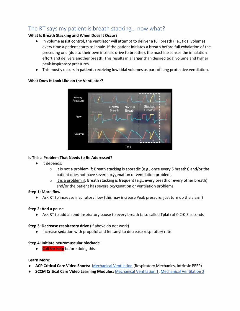
The RT says my patient is breath stacking… now what? What Is Breath Stacking and When Does It Occur?
● In volume assist control, the ventilator will attempt to deliver a full breath (i.e., tidal volume) every time a patient starts to inhale. If the patient initiates a breath before full exhalation of the preceding one (due to their own intrinsic drive to breathe), the machine senses the inhalation effort and delivers another breath. This results in a larger than desired tidal volume and higher peak inspiratory pressures.
● This mostly occurs in patients receiving low tidal volumes as part of lung protective ventilation. What Does It Look Like on the Ventilator?
Is This a Problem That Needs to Be Addressed?
● It depends: o It is not a problem if: Breath stacking is sporadic (e.g., once every 5 breaths) and/or the
patient does not have severe oxygenation or ventilation problems o It is a problem if: Breath stacking is frequent (e.g., every breath or every other breath)
and/or the patient has severe oxygenation or ventilation problems Step 1: More flow
● Ask RT to increase inspiratory flow (this may increase Peak pressure, just turn up the alarm) Step 2: Add a pause
● Ask RT to add an end-inspiratory pause to every breath (also called Tplat) of 0.2-0.3 seconds Step 3: Decrease respiratory drive (If above do not work)
● Increase sedation with propofol and fentanyl to decrease respiratory rate Step 4: Initiate neuromuscular blockade
● Call for help before doing this Learn More: ● ACP Critical Care Video Shorts: Mechanical Ventilation (Respiratory Mechanics, Intrinsic PEEP) ● SCCM Critical Care Video Learning Modules: Mechanical Ventilation 1, Mechanical Ventilation 2

I think my patient might be ready to be extubated… now what? Step 1: Ask RT to do a Spontaneous Breathing Trial
● At our institution, a spontaneous breathing trial is a breathing test performed on minimal ventilator settings, which are the following settings in UPHS:
o Pressure Support of 7 with 5 of PEEP (PSV 7/5) ● To qualify, the patient must be on 40% FiO2 or less, and less than 8 of PEEP ● Prior to proceeding, ensure that the patient’s minute ventilation is < 15 L/min
Step 2: Ensure that the patient is simultaneously scheduled for a spontaneous awakening trial ● A spontaneous awakening trial is a test during which a patient's sedative medications are
discontinued ● Talk to nursing about appropriate sedation dose
Step 3: Assess the patient during the SBT
Step 4: Assess secretions
● Ensure patient is requiring suction Q3h or less Step 5: Assess Mental Status and Ability to Protect Airway
● Normal mental status: Extubate patient ● Altered mental status: Extubate patient if they have:
o A good cough o A gag reflex o Withdrawal to noxious stimuli
Learn More: ● ACP Critical Care Video Shorts: Mechanical Ventilation (Liberation)

My patient is hypotensive… now what? Step 1: Assess the patient
● Physical examination for warm/cool extremities, neck veins, capillary refill ● Laboratory assessment of end-organ function, including BMP, LFTs, and lactate ● POCUS for Cardiac function, lung assessment, neck veins (if able, See resources below)
Step 2: Think about etiology
● If central venous catheter (CVC) is present, check venous blood gas for ScvO2 (See My patient has an abnormal central venous saturation… now what?)
● If Sepsis a consideration (and it almost always is) consider blood cultures, urine cultures, respiratory cultures, CXR
Step 3: Assess for volume tolerance (additional volume will not hurt) and responsiveness (additional volume will improve cardiac output)
● Consider: comorbidities (e.g., heart failure, chronic kidney disease), severity of hypoxemia, cardiac function, volume status
● If patient will both tolerate volume and is likely to be volume responsive, move to Step 4 below ● If tolerance and/or responsiveness unlikely, skip to Step 5 below ● If uncertain:
o Call for help! o Use advanced tools if available (e.g., ultrasound, pulse pressure variation)
Step 4: Give fluid bolus and reassess
● Give 500 mL Lactated Ringer’s IV and reassess ● Consider additional 500 mL boluses if blood pressure rises or urine output increases ● For COVID positive and ARDS patients, limit initial resuscitation to 1-2L before calling for help
Step 5: Start norepinephrine
● Start at 5 mcg/min. Titrate to mean arterial pressure (MAP) > 65 mmHg ● Norepinephrine can be started through a peripheral IV above the AC, if escalating will need CVC
Learn More: ● ACP Critical Care Video Shorts: Sepsis, and Vasopressors ● SCCM Critical Care Video Learning Modules: Evaluation of the ill patient and Shock ● Penn PCCM Critical Care: COVID critical care update video ● POCUS: RUSH exam, ACP Cardiac Exam, U/S training videos from Sonosite

The pressor requirements are rising… now what? Step 1: Call for help! Step 2: Ensure your understanding matches the situation
● Are the blood pressure measurements accurate? ● Do I have the correct diagnosis? ● Do I have source control? (e.g., is there a surgical infection that requires drainage or
debridement, does the patient have a parapneumonic effusion?) ● Is my antimicrobial coverage appropriate?
Step 3: Start vasopressin at 0.04 units/minutes if norepinephrine > 10 mcg/min
● This requires a central venous catheter Step 4: Consider placing arterial line, if not already present
● In PUI/COVID+ patients, please place CVC and arterial line on same side for ease of proning Step 5: Look for development of new etiologies of shock
● Evaluate for sepsis-induced, viral, or primary cardiomyopathy with bedside ultrasound or formal echocardiogram, electrocardiogram, and ScvO2 (provided the patient has a central line)
● This is a known finding in COVID+ patients ● POCUS learning below or My patient has an abnormal central venous saturation… now what? ● If not confident in POCUS capability, contact cardiology about TTE/POCUS workflow
o If echocardiogram consistent with cardiomyopathy or ScvO2 < 60% ▪ Call for help and consider inotrope (dobutamine 2.5 mcg/kg/min) ▪ Consider repleting calcium to normal (ionized calcium > 1.0. mmol/L)
● If the etiology of shock remains unclear or inotropic support is initiated, call for help and consider placement of a PA catheter to guide management
Step 6: Strongly consider stress-dose steroids (hydrocortisone 50 mg q8 hours IV)
● While steroids are not used empirically in COVID, they can be considered in shock Step 6: Re-evaluate fluid tolerance and responsiveness as in previous sheet
● Consider judicious 500 mL boluses in non COVID patients ● See My patient is hypotensive… now what?
Step 7: Evaluate acid-base status
● Consider continuous renal replacement therapy or bicarbonate infusion if the patient has a severe metabolic acidosis and pH <7.1 despite vent adjustments
● See I just got my blood gas results… now what? Step 8: If vasopressor needs continue to increase
● Initiate goals of care discussion Learn More: ● ACP Critical Care Video Shorts: Sepsis, and Vasopressors ● SCCM Critical Care Video Learning Modules: Shock ● POCUS: RUSH exam, ACP Cardiac Exam, U/S training videos from Sonosite
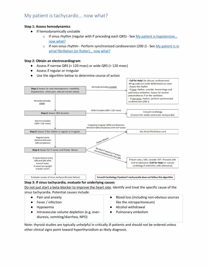
My patient is tachycardic… now what? Step 1: Assess hemodynamics
● If hemodynamically unstable o If sinus rhythm (regular with P preceding each QRS) - See My patient is hypotensive…
now what? o If non-sinus rhythm - Perform synchronized cardioversion (200 J) - See My patient is in
atrial fibrillation (or flutter)... now what? Step 2: Obtain an electrocardiogram
● Assess if narrow QRS (< 120 msec) or wide QRS (> 120 msec) ● Assess if regular or irregular ● Use the algorithm below to determine course of action
Step 3: If sinus tachycardia, evaluate for underlying causes Do not just start a beta-blocker to improve the heart rate. Identify and treat the specific cause of the sinus tachycardia. Potential causes include:
● Pain and anxiety ● Fever / infection ● Hypoxemia ● Intravascular volume depletion (e.g, over-
diuresis, vomiting/diarrhea, NPO)
● Blood loss (including non-obvious sources like the retroperitoneum)
● Alcohol withdrawal ● Pulmonary embolism
Note: thyroid studies are typically unhelpful in critically ill patients and should not be ordered unless other clinical signs point toward hyperthyroidism as likely diagnosis.
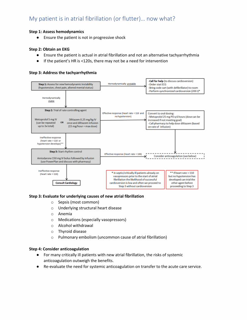
My patient is in atrial fibrillation (or flutter)… now what? Step 1: Assess hemodynamics
● Ensure the patient is not in progressive shock
Step 2: Obtain an EKG ● Ensure the patient is actual in atrial fibrillation and not an alternative tachyarrhythmia ● If the patient’s HR is <120s, there may not be a need for intervention
Step 3: Address the tachyarrhythmia
Step 3: Evaluate for underlying causes of new atrial fibrillation
o Sepsis (most common) o Underlying structural heart disease o Anemia o Medications (especially vasopressors) o Alcohol withdrawal o Thyroid disease o Pulmonary embolism (uncommon cause of atrial fibrillation)
Step 4: Consider anticoagulation
● For many critically ill patients with new atrial fibrillation, the risks of systemic anticoagulation outweigh the benefits.
● Re-evaluate the need for systemic anticoagulation on transfer to the acute care service.
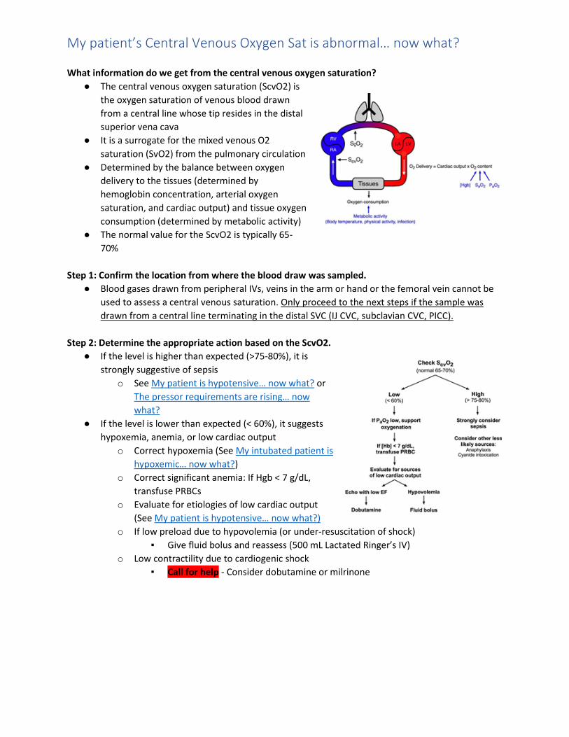
My patient’s Central Venous Oxygen Sat is abnormal… now what? What information do we get from the central venous oxygen saturation?
● The central venous oxygen saturation (ScvO2) is the oxygen saturation of venous blood drawn from a central line whose tip resides in the distal superior vena cava
● It is a surrogate for the mixed venous O2 saturation (SvO2) from the pulmonary circulation
● Determined by the balance between oxygen delivery to the tissues (determined by hemoglobin concentration, arterial oxygen saturation, and cardiac output) and tissue oxygen consumption (determined by metabolic activity)
● The normal value for the ScvO2 is typically 65-70%
Step 1: Confirm the location from where the blood draw was sampled.
● Blood gases drawn from peripheral IVs, veins in the arm or hand or the femoral vein cannot be used to assess a central venous saturation. Only proceed to the next steps if the sample was drawn from a central line terminating in the distal SVC (IJ CVC, subclavian CVC, PICC).
Step 2: Determine the appropriate action based on the ScvO2.
● If the level is higher than expected (>75-80%), it is strongly suggestive of sepsis
o See My patient is hypotensive… now what? or The pressor requirements are rising… now what?
● If the level is lower than expected (< 60%), it suggests hypoxemia, anemia, or low cardiac output
o Correct hypoxemia (See My intubated patient is hypoxemic… now what?)
o Correct significant anemia: If Hgb < 7 g/dL, transfuse PRBCs
o Evaluate for etiologies of low cardiac output (See My patient is hypotensive… now what?)
o If low preload due to hypovolemia (or under-resuscitation of shock) ▪ Give fluid bolus and reassess (500 mL Lactated Ringer’s IV)
o Low contractility due to cardiogenic shock ▪ Call for help - Consider dobutamine or milrinone

My patient’s creatinine is rising and/or urine output is decreasing… now what? Step 1: Determine if the patient meets Acute Kidney Injury criteria by asking three questions:
● In the last 24 hours: has the creatinine risen to 1.5 times the baseline? ● In the last 24 hours: has the creatinine risen by >0.3 mg/dL? ● In the last 12 hours: has the average urine output been lower than 0.5 ml/kg/hr
If “No” to all of the above, monitor serum creatinine, potassium, and magnesium daily.
If “Yes” to any of the above, the patient has acute kidney injury; Go to Step 2 below Step 2: Adjust medication doses
● Work with the pharmacist to adjust medications based on the glomerular filtration rate Step 3: Rule out post-obstructive nephropathy:
● Flush foley catheter and inspect for obstruction. ● Obtain bladder scan: If bladder is decompressed, post-obstructive nephropathy is unlikely ● If concern remains, perform POCUS or order renal ultrasound to evaluate for hydronephrosis if
able (ACP POCUS for obstructive uropathy, 5 minute sono bladder and kidney) Step 4: If no evidence of post-obstructive nephropathy, evaluate for prerenal versus intrinsic etiology
● Measure urine sodium and urine creatinine ● Measure urine BUN if patient has been receiving diuretic therapy ● Obtain urinalysis with microscopy ● Calculate fractional excretion of sodium
○ FENa = (Urine Na/Serum Na)/(Urine Cr/Serum Cr) ● If patient has been receiving diuretic therapy, calculate the fractional excretion of urea
○ FEUrea = (Urine Urea/Serum Urea)/(Urine Cr/Serum Cr)]
Step 5: If present, evaluate and treat for pre-renal nephropathy ● Prerenal etiology is likely with urine sodium < 10 and/or FENa < 1% and/or FEUrea < 35%
○ Assess for hypovolemia and cardiac dysfunction ○ Treat hypovolemia with fluid boluses of lactated ringers in most patients
■ In COVID + patients, limit fluids to 1-2 liters before calling for help ○ Treat hypotension using the approach laid out in “My patient is hypotensive…”
Step 6: Evaluate and treat for intrinsic nephropathy
● An intrinsic renal process is likely if urine sodium > 20 or FENa > 1% and/or FEUrea > 35% or urine microscopy reveals muddy brown casts, granular casts, red or white cell casts
● Avoid repeated fluid boluses ● Review medication list for nephrotoxic medications ● Monitor potassium, magnesium, calcium, phosphate
Step 7: Consult nephrology if above does not fix the problem, or if during workup patient develops
● Severe or worsening metabolic acidosis (bicarbonate < 15 mEq/L) ● Hyperkalemia unresponsive to medical interventions (> 5.5 mEq/L)
○ See My patient’s potassium is too high… now what ● The patient is anuric for 24 hours
My patient’s potassium is too high… now what? Step 1: Decide if the value is high enough to be concerning
● Normal range for serum potassium is 3.5 to 5.0 mEq/L ● Higher values incur a risk of cardiac complications ● Patients with CKD can typically tolerate greater increases in serum potassium above normal
than patients without chronic kidney disease ● Most patients do not experience problems until the potassium rises above 6.0 mEq/L ● Hemolyzed blood samples have an artificially elevated potassium value ● Note: Some sites have an Epic orderset for hyperkalemia, please familiarize yourself with it
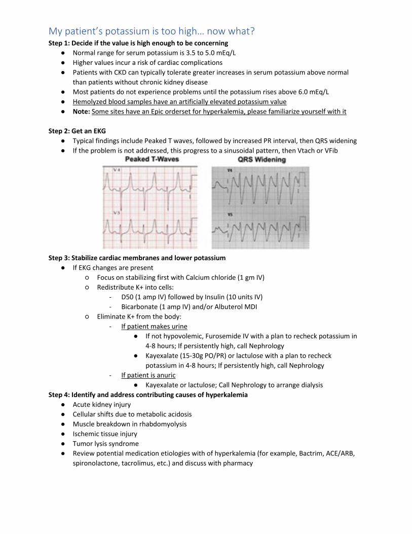
Step 2: Get an EKG
● Typical findings include Peaked T waves, followed by increased PR interval, then QRS widening ● If the problem is not addressed, this progress to a sinusoidal pattern, then Vtach or VFib
Step 3: Stabilize cardiac membranes and lower potassium
● If EKG changes are present ○ Focus on stabilizing first with Calcium chloride (1 gm IV) ○ Redistribute K+ into cells:
- D50 (1 amp IV) followed by Insulin (10 units IV) - Bicarbonate (1 amp IV) and/or Albuterol MDI
○ Eliminate K+ from the body: - If patient makes urine
● If not hypovolemic, Furosemide IV with a plan to recheck potassium in 4-8 hours; If persistently high, call Nephrology
● Kayexalate (15-30g PO/PR) or lactulose with a plan to recheck potassium in 4-8 hours; If persistently high, call Nephrology
- If patient is anuric ● Kayexalate or lactulose; Call Nephrology to arrange dialysis
Step 4: Identify and address contributing causes of hyperkalemia ● Acute kidney injury ● Cellular shifts due to metabolic acidosis ● Muscle breakdown in rhabdomyolysis ● Ischemic tissue injury ● Tumor lysis syndrome ● Review potential medication etiologies with of hyperkalemia (for example, Bactrim, ACE/ARB,
spironolactone, tacrolimus, etc.) and discuss with pharmacy

My patient has a fever… now what? Step 1: Perform a targeted history and exam to identify potential sites of infection
• Examine central lines, urinary catheter, drains for erythema, pus • Intubated patients: assess for change in amount / quality of secretions, or worsening oxygenation • Abdominal exam for focal tenderness or rigidity (pancreatitis, acalculous cholecystitis) • Assess surgical sites for purulence or erythema • Assess for recent diarrhea
Step 2: Start work-up for infection, unless performed in last 24 hours
• Blood cultures x 2 • Chest radiograph versus CT chest versus POCUS (if able, POCUS for PNA) • Endotracheal aspirate • Urinalysis with reflexive culture • Consider liver panel (cholecystitis, drug reaction) and lipase (pancreatitis) • If diarrhea, send stool for C. Diff PCR if patient has not recently received laxatives
• If specific procedures are indicated (paracentesis, thoracentesis) or if a CT scan is needed: - Call for help!
Step 3: Evaluate need for new or augmented antibiotics
● If immune compromised, hemodynamically unstable, or with new or worsening hypoxemia: ○ Start empiric antibiotics per UPHS guidelines [See abx guidelines from your
hospital, or the ILUM app] ○ if no clear source: Cefepime and vancomycin (discuss dosing with pharmacy) ○ If C Diff likely: Oral vancomycin ○ If U/A positive change foley catheter
● If already on broad spectrum antibiotics ○ Call for help with antibiotic selection
Step 4: Evaluate for non-infectious etiologies of fever:
● Review medication list for sources of drug fever (e.g., antibiotics, anti-seizure meds) ● Drug fever: rash, peripheral eosinophilia ● Transfusion-related (can check a direct antiglobulin test) ● Venous thromboembolism (consider POCUS or Duplex U/S)
Step 5: Treat the fever
● Acetaminophen 650 mg every six hours ● Avoid NSAIDS, which may contribute to worsening kidney injury
Learn More: ● ACP Critical Care Video Shorts: Sepsis (presentation) ● SCCM Critical Care Video Learning Modules: Evaluation of ill patient
My patient has altered mental status… now what? Step 1: Perform an initial assessment
● Check vital signs: assess for shock (hypotension), hypoxemia, or infection (fever) ● Check a STAT fingerstick glucose: if hypoglycemic (FSG < 70), give 1 ampule of D50 ● Perform neurologic exam:
○ If Focal findings: Call for help! Consult neurology immediately ■ Must weigh infection control risks of traveling for brain imaging
○ If history of seizures or exam concerning non-convulsive status epilepticus (roving eyes): ■ Call for help! Consult neurology for electroencephalogram (EEG)
● Perform a targeted history and exam to identify potential sites of infection ● Review medications (opiates, sedative/hypnotics), recent events/procedures
Step 2: Order some initial laboratory studies
● Arterial blood gas to rule out hypercarbia ● Serum electrolytes to assess for hypo/hypernatremia, hypercalcemia, increased BUN ● Serum ammonia level in patients with cirrhosis ● Evaluate for infection if fever and/or leukocytosis:
○ Urinalysis with reflexive culture ○ Blood cultures x 2 (peripheral and central) ○ Chest radiograph or POCUS (if able, POCUS for PNA) ○ Consider respiratory viral panel / COVID-19 (if not already checked) ○ Lumbar puncture if recommended by Neurology or ID
Step 3: Consider STAT Head CT (non-contrast)
● Indicated if focal neurologic findings, increased risk of bleeding, sudden onset severe headache, or sudden change in level of consciousness
● Must weigh benefit of imaging with infection control risk of traveling for imaging
Step 4: Do something about it
● Assess ability to protect airway. Call for help! Intubate if airway protection in question ● Suspected opioid overdose: Give Naloxone 0.4 mg IV. May repeat up to 2 mg total ● Discontinue sedating medications ● Consider ventilatory support for patients with hypercarbia not related to opiates ● Address electrolyte disturbances ● Treat identified sources of infection
Step 5: Treat Delirium as below
● See My patient has delirium… now what?
My patient has delirium… now what? How Do I Recognize Delirium:
● Multiple presentations: ○ Withdrawn state (“hypoactive delirium”) ○ Severe agitation (“hyperactive delirium”) ○ Commonly with irritability, delusions, day-night reversal, and increased sympathetic
activity ● ICU Nurses screen for delirium using a standardized bedside tool (CAM-ICU)
Step 1: Evaluate for Contributing Factors
● Delirium typically results from the combination of severe illness and the ICU environment ● Look for other contributing factors including:
○ Untreated infection ○ Hypoxemia ○ Inadequately treated pain ○ Altered day/night cycle and poor sleep ○ Medications: Top culprits are benzodiazepines, antihistamines, antiemetics
Step 2: Do something about it
● Non-pharmacologic interventions focused on orientation and the environment ○ Frequent reorientation during the course of the day ○ Encourage visitation from family (Except in COVID+) ○ Lights on in the room during the day, off at night ○ Minimize noise and stimulation at night ○ Remove catheters and physical restraints when able ○ Mobilize and do physical and occupational therapy as able ○ Ensure patient has their glasses and/or hearing aids ○ Consistency in the nursing staff as able
● Pharmacologic interventions ○ Stop benzodiazepines, antihistamines other contributing medications, particularly those
with anticholinergic side effects ○ Dexmedetomidine or propofol for sedation in intubated patients with target of alert or
mildly sedated (RASS 0 to -1) ○ Consider pharmacologic intervention for sleep (e.g. trazodone, quetiapine, melatonin,
haloperidol) if non-pharmacologic measures are ineffective ○ If Severe agitated delirium:
■ As needed haloperidol (2.5-5 mg IV in repeated doses) if severe agitated delirium poses a risk of harm to the patient or the staff
■ EKG if getting >2 doses of IV haloperidol ■ May consider scheduled oral quetiapine if repeated PRN required ■ Call for help!

My patient is in the ICU… now what? Use the FAST HUG BID - E for a safety checklist Every Day
Feeding – Review nutrition plan virtually with Clinical Nutrition Specialist for the unit ● Trickle TEN feeds can be started on most patients, unless clinically with ileus or on escalating
doses of pressors ● No evidence of benefit to early TPN (before 7 days)
Activity – Choose from Passive ROM, Active ROM, Sitting, Standing, or Walking ● If patient with RASS of 0-1, consider standing even if intubated ● Early mobilization has been linked to improved mortality ● For PUI/COVID+ patients, discuss mobilization plan with critical care attending
Sedation/Analgesia – State RASS goal and plan to minimize sedation ● Ensure that non-paralyzed patients have sedation weaned as soon as safe ● For PUI/COVID+, early RASS goal is -2-3; with improvement in oxygenation would decrease to
RASS 0-1
Thromboprophylaxis – State DVT prophylaxis plan ● All ICU patients get chemical prophylaxis unless actively bleeding ● Consider enoxaparin to decrease frequency of dosing in COVID+ patients unless
contraindications (such as extremes in BMI and/or renal dysfunction)
Hyperoxia - State if present (Sat >96% on supplemental O2) and discuss weaning ● Goal is to wean FiO2 or PEEP as soon as SpO2 stable at 97% or above
Ulcer prophylaxis – Determine if indicated and state plan ● Typically used if patient intubated and in shock ● If patient intubated and getting feeds, can probably stop ● Use H2 blocker or PPI per unit preference and bleeding risk
Glycemic control – State glucose goal (usually 120-180) and state plan ● In COVID+ patients consider less frequent glucose monitoring ● If unable to control BG with subcutaneous insulin, consider insulin gtt
Bowel activity and regimen – Review last bowel movement and bowel regimen
Indwelling devices – Review indication for every device and planned removal ● Consider removing CVC once no longer requiring pressors ● Consider removing arterial line once on low-dose pressors and cuff BP reliable ● Consider removing Foley if close monitoring of I/Os not necessary
○ Can use external condom catheter for men ○ Can use Purewick collection system for women
Drug de-escalation / antibiotic stewardship – Discuss antibiotic de-escalation
Engagement of Families – Ensure that family communication happens daily for all patients ● Consider use of tablet for virtual rounds with family members ● See the covid sharepoint for updated communication guidelines for UPHS
Learn More ACP Critical Care Video Shorts: Prevention in the ICU Vital Talks: Covid specific section Penn Family Communication Guidance: Pocket guide
Related Documents