Novel Insights Into Interactions Between Mitochondria and Xanthine Oxidase in Acute Cardiac Volume Overload James D Gladden, BS 1,5 , Blake R Zelickson, BS 1,3,4 , Chih-Chang Wei, PhD 1,2,6 , Elena Ulasova, PhD 1,3,4 , Junying Zheng, PhD 1,2 , Mustafa I. Ahmed, MD 1,2 , Yuanwen Chen, MD, PhD 1,2 , Marcas Bamman, PhD 5,6 , Scott Ballinger, PhD 1,3,4 , Victor Darley-Usmar, PhD 1,3,4 , and Louis J Dell’Italia, MD 1,2,4,6 1 UAB Center for Heart Failure Research, University of Alabama at Birmingham 2 Department of Medicine, University of Alabama at Birmingham 3 Department of Pathology, University of Alabama at Birmingham 4 Center for Free Radical Biology, University of Alabama at Birmingham 5 Department of Physiology and Biophysics, University of Alabama at Birmingham 6 Department of Veterans Affairs Medical Center, Birmingham, Alabama Abstract Xanthine oxidoreductase (XOR) is increased in the left ventricle (LV) of humans with volume overload (VO) and mitochondrial inhibition of the respiratory chain occurs in animal models of VO. Since mitochondria are both a source and target of reactive oxygen and nitrogen species, we hypothesized that activation of XOR and mitochondrial dysfunction are interdependent. To test this we used the aortocaval fistula (ACF) rat model of VO and a simulation of the stretch response in isolated adult cardiomyocytes with and without the inhibitor of XOR, allopurinol, or the mitochondrially targeted antioxidant MitoQ. XO activity was increased in cardiomyocytes from ACF vs. sham rats (24h) without an increase in XO protein. A two-fold increase in LV end- diastolic pressure/wall stress and a decrease in LV systolic elastance with ACF were improved with allopurinol (100 mg/kg) started at ACF induction. Subsarcolemmal state 3 mitochondrial respiration was significantly decreased in ACF and normalized by allopurinol. Cardiomyocytes subjected to 3 hour cyclical stretch resulted in an increase in XO activity and mitochondrial swelling, which was prevented by allopurinol or MitoQ pretreatment. These studies establish an early interplay between cardiomyocyte XO activation and bioenergetic dysfunction that may provide a new target that prevents progression to heart failure in VO. Keywords volume overload; oxidative stress; MitoQ; stretch; mitochondria; allopurinol Introduction Volume overload (VO) increases diastolic load and results in a progressive eccentric left ventricular (LV) remodeling and systolic dysfunction leading to cardiac failure (1). Currently, there is no medical therapy that halts the progression to heart failure in an isolated Address reprints: Louis J Dell’Italia, MD, UAB Center for Heart Failure Research, Division of Cardiology, 434 BMR2, 1530 3 rd Avenue South, Birmingham, AL 35294-2180, Telephone: (205) 934-3969, Fax: (205) 996-2586, [email protected]. NIH Public Access Author Manuscript Free Radic Biol Med. Author manuscript; available in PMC 2012 May 30. Published in final edited form as: Free Radic Biol Med. 2011 December 1; 51(11): 1975–1984. doi:10.1016/j.freeradbiomed.2011.08.022. NIH-PA Author Manuscript NIH-PA Author Manuscript NIH-PA Author Manuscript

Welcome message from author
This document is posted to help you gain knowledge. Please leave a comment to let me know what you think about it! Share it to your friends and learn new things together.
Transcript

Novel Insights Into Interactions Between Mitochondria andXanthine Oxidase in Acute Cardiac Volume Overload
James D Gladden, BS1,5, Blake R Zelickson, BS1,3,4, Chih-Chang Wei, PhD1,2,6, ElenaUlasova, PhD1,3,4, Junying Zheng, PhD1,2, Mustafa I. Ahmed, MD1,2, Yuanwen Chen, MD,PhD1,2, Marcas Bamman, PhD5,6, Scott Ballinger, PhD1,3,4, Victor Darley-Usmar, PhD1,3,4,and Louis J Dell’Italia, MD1,2,4,6
1UAB Center for Heart Failure Research, University of Alabama at Birmingham2Department of Medicine, University of Alabama at Birmingham3Department of Pathology, University of Alabama at Birmingham4Center for Free Radical Biology, University of Alabama at Birmingham5Department of Physiology and Biophysics, University of Alabama at Birmingham6Department of Veterans Affairs Medical Center, Birmingham, Alabama
AbstractXanthine oxidoreductase (XOR) is increased in the left ventricle (LV) of humans with volumeoverload (VO) and mitochondrial inhibition of the respiratory chain occurs in animal models ofVO. Since mitochondria are both a source and target of reactive oxygen and nitrogen species, wehypothesized that activation of XOR and mitochondrial dysfunction are interdependent. To testthis we used the aortocaval fistula (ACF) rat model of VO and a simulation of the stretch responsein isolated adult cardiomyocytes with and without the inhibitor of XOR, allopurinol, or themitochondrially targeted antioxidant MitoQ. XO activity was increased in cardiomyocytes fromACF vs. sham rats (24h) without an increase in XO protein. A two-fold increase in LV end-diastolic pressure/wall stress and a decrease in LV systolic elastance with ACF were improvedwith allopurinol (100 mg/kg) started at ACF induction. Subsarcolemmal state 3 mitochondrialrespiration was significantly decreased in ACF and normalized by allopurinol. Cardiomyocytessubjected to 3 hour cyclical stretch resulted in an increase in XO activity and mitochondrialswelling, which was prevented by allopurinol or MitoQ pretreatment. These studies establish anearly interplay between cardiomyocyte XO activation and bioenergetic dysfunction that mayprovide a new target that prevents progression to heart failure in VO.
Keywordsvolume overload; oxidative stress; MitoQ; stretch; mitochondria; allopurinol
IntroductionVolume overload (VO) increases diastolic load and results in a progressive eccentric leftventricular (LV) remodeling and systolic dysfunction leading to cardiac failure (1).Currently, there is no medical therapy that halts the progression to heart failure in an isolated
Address reprints: Louis J Dell’Italia, MD, UAB Center for Heart Failure Research, Division of Cardiology, 434 BMR2, 1530 3rd
Avenue South, Birmingham, AL 35294-2180, Telephone: (205) 934-3969, Fax: (205) 996-2586, [email protected].
NIH Public AccessAuthor ManuscriptFree Radic Biol Med. Author manuscript; available in PMC 2012 May 30.
Published in final edited form as:Free Radic Biol Med. 2011 December 1; 51(11): 1975–1984. doi:10.1016/j.freeradbiomed.2011.08.022.
NIH
-PA Author Manuscript
NIH
-PA Author Manuscript
NIH
-PA Author Manuscript

VO of aortic or mitral regurgitation (2). We have recently shown that patients with isolatedmitral regurgitation have decreased LV systolic function 6 months post-mitral valve repair,despite LV ejection fraction (EF) > 60% prior to surgery (3). Biopsies taken at the time ofmitral valve repair demonstrate significant myofibrillar loss with increased xanthineoxidoreductase (XOR), protein nitration, and lipofuscin accumulation in cardiomyocytes,consistent with increased formation of reactive oxygen and nitrogen species (ROS/RNS).Correspondingly, there is also evidence of aggregates of small mitochondria incardiomyocytes, which is generally considered a response to bioenergetic deficit in cells.
XOR exists as xanthine dehydrogenase and xanthine oxidase (XO), both of whichmetabolize purines to form uric acid (4). In its oxidase form, XOR produces superoxide andhydrogen peroxide when oxygen is used as an electron acceptor during purine metabolism.Superoxide and hydrogen peroxide can negatively impact multiple metabolic processes inthe cardiomyocyte either independently or after reaction with nitric oxide (NO) (5,6). Onekey target for the actions of ROS/RNS in the cell is the mitochondrion because of its highconcentration of reactive proteins and lipids in close proximity to multiple enzymescontaining redox active iron and copper (7,8).
Not surprisingly, increasing evidence implicates mitochondrial dysfunction in a broad rangeof cardiovascular pathologies, many of which are associated with increased ROS/RNS (9–11). Mitochondria are a major source of ROS within the cardiomyocyte, mostly due to theunivalent reduction of oxygen to superoxide at complexes I, and III (12,13). Importantly,exposure of mitochondria to ROS/RNS can lead to mtDNA damage that compromise proteinsynthesis and directly modify mitochondrial proteins (8,12). This in turn can lead toincreased mitochondrial ROS production and a vicious cycle can then be establishedbetween the generation of ROS from non-mitochondrial sources, bioenergetic dysfunction,and oxidative damage to the organelle. Based upon these concepts we hypothesize that XOactivation in VO is intimately involved in the mitochondrial dysfunction associated with thisform of hemodynamic stress.
To test these concepts we have used a model of VO by inducing an aortocaval fistula (ACF)in rats. Acutely, ACF causes a three-fold increase in LV end-diastolic pressure and asignificant increase in LV chamber diameter, consistent with an increase in LV preload (14).The increased work load associated with ACF is manifested by an increase in the pressurevolume area (PVA), suggesting increased myocardial oxygen demand (MVO2) and ATPconsumption (15) and is associated with a decrease in subsarcolemmal mitochcondrialfunction (16). Thus, in this setting, it is likely that a decrease in the bioenergetic capacity ofthe mitochondria plays a significant role in the myocardium’s ability to meet increasedhemodynamic and oxygen demand.
Previous studies have demonstrated beneficial effects of XO inhibition with allopurinol oroxypurinol in multiple animal models of myocardial disease, such as ischemia (17,18),diabetic cardiomyopathy (19), pacing-induced tachycardia in the dog (20), and in thespontaneously hypertensive rat (21). Volume overload increases the physical stress on theheart due to the elevated diastolic load that increases the stretching force on the heart wall,especially during early diastolic filling (22). Interestingly, acute stretch in the lung increasesXO activity in endothelial cells and mediates oxidative tissue damage (23). Further,cardiomyocytes display increased ROS in response to mechanical stretch (24). However, theeffect of isolated VO or its potential impact resulting from stretch has not previously beenstudied in human or animal models and its effects on bioenergetic and cardiac function areunknown. Taken together, these findings suggest that the stretching force associated withVO may induce XO activity resulting in increased oxidative stress in cardiomyocytes.
Gladden et al. Page 2
Free Radic Biol Med. Author manuscript; available in PMC 2012 May 30.
NIH
-PA Author Manuscript
NIH
-PA Author Manuscript
NIH
-PA Author Manuscript
While the mechanism of XO activation in heart failure is not well understood, it is plausiblethat mitochondrial-induced oxidative stress can lead to the activation of XO byposttranslational modification of the enzyme. XO is generated from its parent enzyme XDHeither by irreversible proteolytic cleavage or transient thiol oxidation within the XORprotein (25,26). In patients with heart failure, ROS have been shown to increase sulfhydryloxidation and have been shown to increase XO activity (27). To test the hypothesis thatoxidative stress in the mitochondrion and XO activation are linked in VO, we have utilizedthe mitochondrially-targeted antioxidant mitoubiquinone (Mito Q) (28–30). Mito Q has beenshown to protect mitochondrial function in a number of cardiovascular models includingischemia-reperfusion, endotoxemia, and cardiac hypertrophy (31–33). In the present study,we demonstrate that allopurinol attenuates the mitochondrial dysfunction associated withVO and that cyclic stretch activates XO through a mechanism dependent on mitochondrialROS.
Materials and MethodsAnimal Preparation
Sprague-Dawley rats (200–250g) at 12 weeks of age were subjected to sham and ACFsurgery as previously described in our laboratory (14,16) with and without XO inhibitorallopurinol (100 mg/kg, Sigma), which was started at the time of sham surgery or ACFinduction. Separate sets of sham and ACF rats were sacrificed 24 h after surgery for studiesof isolated cardiomyocytes (N=5 per group) and heart mitochondria (N=6 per group).Another group of sham and ACF rats were studied for in vivo hemodynamic andechocardiographic measurements prior to sacrifice and this tissue was used for proteinanalysis. To examine the effects of cardiomyocyte stretch on XO activity, 12 week oldSprague-Dawley rats (200–250g) were sacrificed and isolated cardiomyocytes were obtainedfor in vitro stretch studies. This study was approved by the University of Alabama atBirmingham Animal Resource Program.
Hemodynamics and echocardiographyEchocardiography and hemodynamics were performed prior to sacrifice using theVisualsonics imaging system (Vivo 770, Toronto, Canada) combined with simultaneoushigh-fidelity LV pressure catheter recordings (Millar Inst. Houston, TX). With the rat underisoflurane anesthesia, a high-fidelity LV pressure catheter was advanced into the LV cavityvia a right carotid cut-down. Simultaneous LV pressure and echocardiographic dimensions(wall thickness and chamber diameter) were obtained using software included in theVisualsonics system. LV volume was calculated from traced m-mode LV dimensions usingthe Teicholz formula.
Where V= volume, LVID= LV internal dimension.
LV wall stress was calculated from traced m-mode LV dimensions and simultaneous LVpressure data using the equation below.
Where LV σ= LV wall stress, LVP= LV pressure, r= LV chamber radius, LVwt= LV wallthickness.
Gladden et al. Page 3
Free Radic Biol Med. Author manuscript; available in PMC 2012 May 30.
NIH
-PA Author Manuscript
NIH
-PA Author Manuscript
NIH
-PA Author Manuscript
The LV pressure-volume data were analyzed for LV PVA and stroke work using theLabscribe2 (iWorx System Dover, NH) software package.
Isolation of heart subsarcolemmal mitochondria and activity measurementsHeart subsarcolemmal mitochondria (SSM) were isolated from LV tissue (70mg) aspreviously described in our laboratory(16,34). The pellet resulting from centrifugation of LVhomogenate at 1000×g for 5 minutes (4°C) was discarded. The supernatant was centrifugedat 6000×g for 10min and the resulting pellet was washed twice in isolation buffer and usedfor respiration measurements.
Oxygen consumption was measured using a Clark-type electrode (Hansatech Instruments,Norfolk, UK). State 2 mitochondrial respiration was initiated upon addition of glutamate/malate (5 mmol/L) as substrates. Respiratory state 3 rate of oxygen consumption (nmol O2min−1·mg protein−1) was measured in the presence of 1.5 mmol/L ADP and State 4determined after utilization of 15 µmol/L adenosine diphosphate (ADP).
Isolation of LV myocytesCardiomyocytes were isolated from Sham and ACF rats, as described in our laboratory(14,16). Briefly, hearts were perfused with perfusion buffer (120 mmol/L NaCl, 15 mmol/LKCl, 0.5 mmol/L KH2PO4, 5 mmol/L NaHCO3, 10 mmol/L HEPES, and 5 mmol/L glucose,at pH 7.0) for 5 min and digested with perfusion buffer containing 2% collagenase II(Invitrogen, Carlsbad, CA) for 30 min at 37°C. The right ventricle, atria and apex wereremoved before the perfused-heart was minced. The digestion was filtered, washed and cellswere pelleted. Only samples with purity and viability (rod-shaped) > 95% or 80%,respectively, were used.
Application of stretch to isolated adult rat cardiomyocytesCells (2×10⁶/well) were allowed to adhere to laminin coated Flexcell plates (FlexcellInternational Corp., Hillsborough, NC, USA) in DMEM medium containing 10% FBS, 2mM glutamine, 10 U/mL penicillin, and 100 mg/mL streptomycin for 2 hours before use.Cells were subjected to cyclical strain (60 cycles/min, 3h) on the Flexcell Strain apparatus(model FX-4000; Flexcell International, Hillsborough, NC, USA) at a level of distensionsufficient to promote an increment of approximately 5% in surface area at the point ofmaximal distension on the culture surface. A group of cells stretched for 3 hours were alsosubjected to either the mitochondrial ROS inhibitor Mito Q (10 nM or 50 nM) or 250 µMallopurinol. Control cells were prepared on identical culture plates but were not exposed tostretch.
Immunohistochemistry in LV myocardiumRat hearts were immersion-fixed in 10% neutral buffered formalin and paraffin-embedded.5µm sections were mounted on slides, deparaffinized in xylene and rehydrated in a gradedseries of ethanol. After blocking with 1x PBS/1% Casein, overnight incubation at 4°C witheither XO antibody (1:1000) or nitrotyrosine antibody (1:1000) Upstate Biotechnology, IncLake Placid, NY. Alexa Fluor conjugated secondary antibodies (Molecular Probes, Eugene,OR; 1:500 each) were applied to visualize XO (green) and nitrotyrosine (red) in the tissue.Nuclei were stained (blue) with DAPI (1.5µg/ml; Vector Laboratories, Burlingame, CA).Image acquisition (100× objective, 4000× video-screen magnification) was performed on aLeica DM6000 epifluorescence microscope with SimplePCI software (Compix, Inc.,Cranberry Township, PA). Images were adjusted appropriately to remove backgroundfluorescence.
Gladden et al. Page 4
Free Radic Biol Med. Author manuscript; available in PMC 2012 May 30.
NIH
-PA Author Manuscript
NIH
-PA Author Manuscript
NIH
-PA Author Manuscript

Western blotTissue lysate (30 µg protein) was separated on 4–12% Bis-Tris gradient gel (Invitrogen),transferred to a PVDF membrane, then incubated with antibody to either XO (1:1000) SantaCruz Biotechnology, Santa Cruz, CA, NAPDH oxidase p47 phox subunit (1:1000) CellSignaling Technology, Inc. Danvers, MA, INOS (1:1500) Santa Cruz Biotechnology, SantaCruz, CA, β-tubulin (1:2000) Sigma-Aldrich, St. Louis, MO, overnight at 4°C followed byincubation with HRP conjugated secondary antibodies. Membranes were incubated withChemiluminescent Substrate (Pierce, Rockford, IL) and exposed to X-ray film.Densitometry analysis was performed on XOR expression levels which were normalized totubulin in Sham vs ACF. NADPH oxidase and iNOS expression were normalized to totalprotein determined by densitometric analysis from ponceau stained membranes in allgroups. Membranes were stained with ponceau stain for 2 mins and then destained inphosphate buffered saline to remove background.
Measurement of XO activity in LV tissue and isolated cardiomyocytesXO activity was measured using high performance liquid chromatography (HPLC) withelectrochemical detection (ESA Coularray). LV free wall tissue or isolated cardiomyocyteswere homogenized in RIPA buffer. Before measuring enzymatic activity, endogenous uratewas removed by eluting the sample on a Sephadex G-25 column. Xanthine (75µM) was thenadded, and XO activity assessed by monitoring urate production. The specificity of thisdetection method for urate production by XO was verified by inhibition of urate formationfollowing allopurinol addition in duplicate samples. Activity was normalized to post columnprotein concentration determined by bicinchoninic acid (BCA) protein assay for myocardialtissue samples and isolated cells.
Transmission electron microscopy of adult rat cardiomyocytesCells were fixed in 25% glutaraldehyde overnight. Cells were then suspended in phosphatebuffered saline and carefully removed from the flexcell membrane using a cell scraper. Cellswere pelleted and mounted for transmission electron microscopy which was performed byEmLabs Inc. Birmingham, Al.
StatisticsData are expressed as mean ± SEM. XO activity data was compared using Student’s t-testfor control/stretched isolated cardiomyocytes and Sham/ACF LV tissue homogenates andisolated cardiomyocytes. A two-way ANOVA with student-Newman-Keuls post hoc testwas used for all other comparisons among sham, ACF, Sham + allopurinol and ACF +allopurinol. P< 0.05 was considered statistically significant.
ResultsMorphometric and Hemodynamic Effects of Allopurinol on Aortocaval Fistula
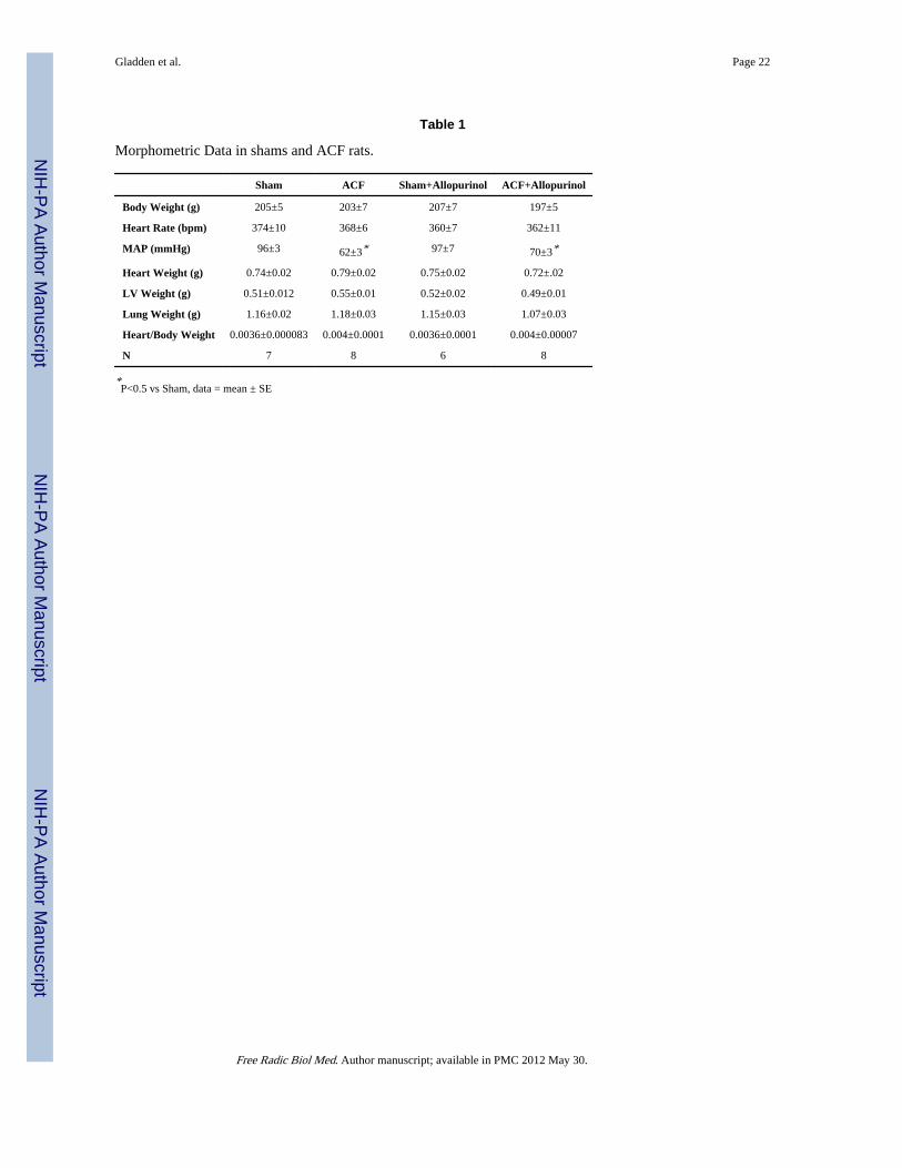
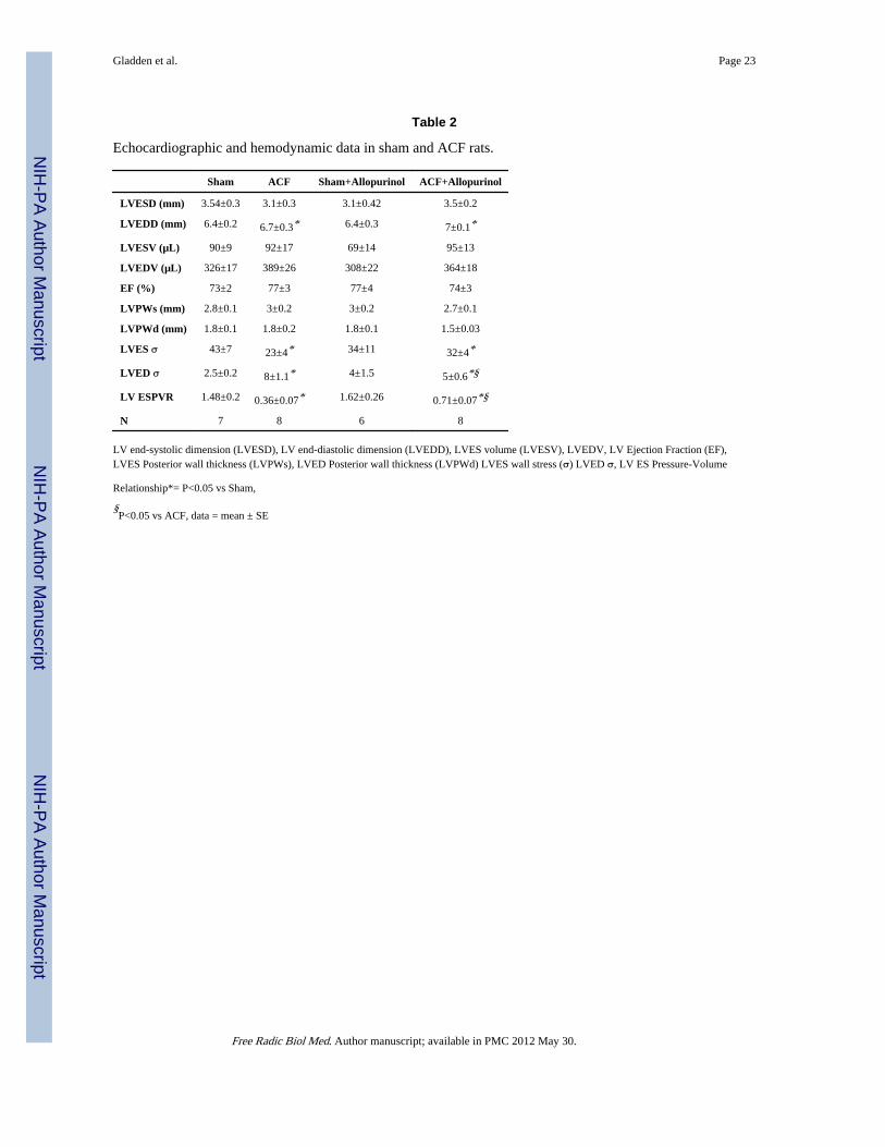
ACF increases venous return to the heart resulting in a left ventricular VO and diastolicdysfunction evidenced by increased LVEDP and LVED wall stress (σ). At 24 hours afterinduction of ACF, body weight, heart weight/body weight ratio, or heart rate did not differamong Sham, ACF, Sham + allopurinol, and ACF + allopurinol rats (Table 1). Mean arterialpressure (MAP) was decreased with ACF and was unaffected by allopurinol (Table 1). LVend-diastolic dimension (LVEDD) and LVED volume were increased in ACF vs sham ratsand were unaffected by treatment with allopurinol (Table 2). LVED pressure and LVED σincreased in ACF vs. sham rats and were normalized in ACF rats treated with allopurinol(Figure 1). LV ejection fraction did not change with ACF and was unaffected by allopurinol(Table 2).
Gladden et al. Page 5
Free Radic Biol Med. Author manuscript; available in PMC 2012 May 30.
NIH
-PA Author Manuscript
NIH
-PA Author Manuscript
NIH
-PA Author Manuscript
LV Pressure-Volume AnalysisLV high-fidelity pressure was matched to simultaneous echocardiographic dimensions togenerate LV pressure-volume loops. Data collected during a 3–5 sec inferior vena cavaocclusion yielded a set of LV pressure-volume loops at multiple LV preloads (Figure 2).Prior to occlusion, the LV pressure-volume area was increased 20% in both ACF and ACF +allopurinol vs. sham rats (P<0.05 in both cases), indicative of increased LV work load andoxygen demand. Analysis of the LV end-systolic pressure-volume relationship (ESPVR), arelatively load independent measure of LV contractile function, demonstrated a decrease inLV contractility in the ACF group vs. Sham rats (0.36±0.07 vs 1.48±0.2 mmHg/µl, P<0.05),which was significantly improved in ACF + allopurinol rats (0.71±0.07 mmHg/µl) vs ACFrats (P<0.05).
XO Activity is Increased in ACF Myocardial Tissue and Isolated MyocytesImmunohistochemistry from sham and control ACF rats demonstrated extensive XO/XDHdistribution in endothelial cells and interstitial cells (Figure 3C) and along Z lines incardiomyocytes (Figure 3A and B), which is consistent with our previous report in thehuman heart (3). To determine if XO is activated during ACF, XO activity was measured inboth LV homogenates and isolated cardiomyocytes from ACF rats (Figure 4A and B).Cardiomyocyte XO specific activity was approximately 10% of the XO activity in whole LVtissue homogenates after normalization to sample protein content (Figure 4A and B). ACFLV tissue homogenates demonstrated a modest 15% increase in XO activity compared toSham LV homogenates. Interestingly, XO activity from isolated ACF cardiomyocytes wasincreased 300% compared to sham cardiomyocytes. This suggests that ACF causes greaterXO activation in cardiomyocytes even though other cellular locations of XO such as theendothelium and interstitial cells may contain higher basal levels of the enzyme.
Allopurinol decreases protein nitration in ACF LV myocardiumWestern blot analysis of iNOS expression demonstrated no significant change in ACF vsSham (Figure 5A). However, treatment with allopurinol was associated with increased iNOSexpression in both Sham and ACF vs. untreated Sham and ACF rats (P<0.05 in both cases).Expression of the NADPH oxidase subunit p47 phox did not significantly differ among allgroups (Figure 5A, 5B). Immunohistochemical analysis for nitrotyrosine demonstratedincreased staining in ACF left ventricles compared to all other groups (Figure 5D vs 5C, 5D,and 5E). It is of interest that in the ACF group (Figure 5D) the staining was aligned alongthe Z-lines, suggesting increased nitration with elevated XO.
Effects of ACF on Mitochondrial FunctionOxygen consumption was determined in isolated subsarcolemmal mitochondria (SSM) fromall experimental groups (Figure 6). As we reported previously (16), State 3 mitochondrialoxygen respiration was decreased in ACF vs. sham rats (Figure 6). In the current study,allopurinol normalized State 3 mitochondrial respiration in the ACF + allopurinol vs. ACFrats. Allopurinol had no effect on State 2 or State 4 mitochondrial respiration butsignificantly decreased State 3 respiration in Shams. The reasons for this change are notclear but could be due to effects of allopurinol on the purine salvage pathway leading to adecreased activation of mitochondrial respiration or biogenesis.
Effects of Cardiomyocyte Stretch on XO Activity and Mitochondrial MorphologyVO causes increased myocardial stretch and since this has been reported to increase ROSformation (22), we hypothesized that this could contribute to XO activation. To test thishypothesis, isolated adult rat LV myocytes were plated on laminin-coated Flexcell platesand stretched at 1 Hz at 5% sinusoidal strain for 3 hours. Protein homogenates from
Gladden et al. Page 6
Free Radic Biol Med. Author manuscript; available in PMC 2012 May 30.
NIH
-PA Author Manuscript
NIH
-PA Author Manuscript
NIH
-PA Author Manuscript
stretched vs. unstretched cardiomyocytes demonstrated an increase in XO activity at 3 hrsafter stretch (Figure 7). Pre-treatment with Mito Q in unstretched cells and in cells subjectedto 3 hours stretch demonstrated no increase in XO activity with both 10 nM or 50 nM dosesof Mito Q. Taken together, these results suggest a cause and effect relationship betweenisolated stretch and increased cardiomyocyte XO activation that is induced bymitochondrial-derived ROS.
Effect of Mechanical Stretch on Myofibrillar and Mitochondrial StructureTransmission electron microscopy of stretched cardiomyocytes revealed a marked decreasein myofibrillar density as well as structural disruption of the Z-line (Figure 8). In addition,there was evidence of mitochondrial swelling and loss of electron density of cristae instretched cells vs. unstretched cardiomyocytes. Z-line structural integrity and mitochondrialmorphology were preserved by pre-treatment with allopurinol or Mito Q.
DiscussionInflammation and oxidative stress produce ROS/RNS through a number of enzymaticsystems including XO. It is well known that mitochondria are both targets and sources ofoxidative stress, which we have shown in VO results in inhibition of the respiratory chain.This is important since VO produces an increase in pressure-volume area (PVA) as shown inFigure 2, which is known to require higher myocardial oxygen consumption (MVO2) andATP consumption (15). We reasoned that this combination of increased energy demand andmitochondrial dysfunction increases the susceptibility of the VO heart to failure. Indeed, wehave recently established a novel interaction between bioenergetics and activation of MMPsin the cardiomyocyte of the VO heart (16). This is particularly interesting in the context ofXO since its substrates, xanthine and hypoxanthine, are elevated under increasedbioenergetic demand and bioenergetic dysfunction. In the current study, we report that VOcauses an increase in XO activity in LV tissue and cardiomyocytes without changes in totalXO protein, consistent with an oxidative post-translational activation of XO (Figure 4).
Since the activation of XO through this mechanism involves the oxidation of thiols, wereasoned that mitochondrial derived oxidants are an early event that could lead to theactivation of XO. To test this hypothesis, MitoQ is used to inhibit mitochondrially-derivedROS in the stretched cardiomyocytes. Indeed, MitoQ prevents both stretch induced-XOactivation and mitochondrial swelling and disorganization (Figure 8). These results suggestthat mitochondria, which comprise 40% of the cardiomyocyte by volume, may be animportant source of ROS and play a regulatory role in XO activation in the VOcardiomyocyte.
The current study also demonstrates loss of myofibrillar integrity in isolated cardiomyocytessubjected to cyclic stretch. We have previously shown that 24 hours of ACF results inincreased TNF-α levels (35) and ROS formation and matrix metalloproteinase (MMP)activation (16) within cardiomyocytes. Cytokines, XO, and ROS have been shown to causeMMP activation (36–38) and there is increasing evidence that cardiomyocyte MMPactivation is responsible for myosin and troponin degradation during cardiac ischemiareperfusion injury (39,40). In addition, transgenic mice expressing active MMP-2 driven bythe α-myosin heavy chain promoter exhibit breakdown of Z-band registration, lysis ofmyofilaments, and disruption of sarcomere and mitochondrial architecture (41). It is ofinterest that we have recently demonstrated extensive cardiomyocyte myofibrillar loss inassociation with increased oxidative stress in the myocardium of VO patients with chronicisolated mitral regurgitation (3). Thus, it is tempting to speculate that mitochondriallyderived ROS and XO-mediated MMP activation may play a causative role in the
Gladden et al. Page 7
Free Radic Biol Med. Author manuscript; available in PMC 2012 May 30.
NIH
-PA Author Manuscript
NIH
-PA Author Manuscript
NIH
-PA Author Manuscript

myofibrillar degeneration that has now been identified in the rat (16), dog (42,43), andhuman (3) with isolated VO.
Increased cardiomyocyte XO activity with acute ACF is also associated with decreased State3 maximal bioenergetic capacity of isolated subsarcolemmal mitochondria, which isnormalized by allopurinol (Figure 6). Mechanical stretch is associated with increasedcardiomyocyte XO activity and abnormal mitochondrial structure that is prevented byallopurinol and MitoQ. NADPH oxidase and uncoupled NOS activation have also beenidentified in cardiomyocyte stretch (44,45). The current study demonstrates XO activation ina heart failure model may be a direct response to physical stretch. Further, our in vitrostudies also implicate the mitochondria as a source of ROS by demonstrating that XOactivation and mitochondrial and cytoskeletal derangements with stretch can be preventedby MitoQ. Therefore, it is tempting to speculate that XO activation is related to ROSproduction from mitochondrial structural alterations and that allopurinol and MitoQ mayhave synergistic effects in vivo. These data do not exclude a role for NADPH oxidase andiNOS in contributing to the VO-dependent response to stretch but they do suggest that theyare required to interact with both mitochondria and XO to contribute to the pathology.
The in vivo and in vitro findings of the current study suggest that increased XO activity andmitochondrial oxidative stress are central factors in bioenergetic dysfunction in the face ofthe increased ATP requirements and MVO2 of VO. Studies by Hare and coworkers haveshown that acute administration of allopurinol decreases the MVO2 while improvingcontractile function in patients with dilated cardiomyopathy (46) and in dogs with pacingtachycardia induced heart failure (47), suggesting improved myocardial efficiency. Wefurther speculate that increased levels of ADP and AMP, particularly in the setting ofmitochondrial dysfunction, that are degraded to XO substrates hypoxanthine and xanthine,can set up a self-perpetuating cycle by which activated XO produces ROS that damagemitochondria, that in turn causes further ROS production and XO activation (Figure 9). Insupport of this argument, it is of interest that the acute stretch of VO causes relativelygreater XO activation in isolated cardiomyocytes (300%) than in the LV tissue homogenate(15%).
LV ejection fraction is preserved after 24 hours of ACF. However, the LV ESPVR, whichprovides a load independent index of LV contractility, is depressed in the acute 24 hourACF, and allopurinol improves both LV contractility and diastolic function (Table 2). It is ofinterest that increased cardiac ADP levels have been linked to diastolic dysfunction byoutcompeting ATP at the actin-myosin crossbridge site and subsequently impairing therelaxation process by delayed ADP dissociation, which is the rate limiting step in crossbridge cycling (48). Indeed, artificially altered ADP levels have been shown to directlycorrelate with increased LVEDP in the rat heart (49). The beneficial effect on LVED σ isparticularly important because wall stress is the driving force for LV hypertrophy in VO (1).Because LV mitochondrial and diastolic function are simultaneously normalized byallopurinol, it is tempting to speculate that allopurinol attenuates ROS-dependentmitochondrial damage and improves diastolic function by improving maximal ADP-stimulated respiratory capacity and decreasing buildup of ADP.
Whether allopurinol improves LV function in acute VO through its effect on mitochondrialrespiration cannot be conclusively demonstrated in the current study, since we onlymeasured the respiration of sub-sarcolemmal mitochondria respiration and notintermyofibrillar mitochondrial respiration. In addition, in vitro studies demonstrate that XOdepresses myofilament sensitivity to calcium and that it co-localizes with nitric oxidesynthase-1 in the sarcoplasmic reticulum in the mouse cardiomyocyte, which can regulateexcitation-contraction coupling as well as myofilament oxidative damage (50,51).
Gladden et al. Page 8
Free Radic Biol Med. Author manuscript; available in PMC 2012 May 30.
NIH
-PA Author Manuscript
NIH
-PA Author Manuscript
NIH
-PA Author Manuscript
Specifically, XO inhibition restores ryanodine receptor nitrosylation, reverses diastolicsarcoplasmic reticulum calcium leak, and improves cardiomyocyte contractility in thespontaneously hypertensive heart failure rat (52) and improves the maladaptive changes incalcium cycling proteins associated with LV failure in the pacing tachycardia dog model(53). In the current study, XO protein staining by immunohistochemistry is mostconcentrated at the Z-line in the cardiomyocyte, which is similar to the findings in thehuman cardiomyocyte (3). Further, in vitro stretch of cardiomyocytes results in loss ofmyofibrillar structural integrity of the Z-line concurrent with increase XO activity, both ofwhich are prevented with allopurinol or Mito Q. Thus, we cannot rule out an alternativecalcium/myofilament mediated mechanism by which allopurinol improves LV contractileperformance in this acute VO.
In summary, we have shown that acute VO increases XO activity in heart tissue and isolatedcardiomyocytes and that defects in subsarcolemmal mitochondrial respiration and LVdysfunction with VO are reversed by allopurinol. Further, cyclic stretch of cardiomyocytesincreases XO activity producing mitochondrial structural defects that are attenuated byallopurinol or MitoQ. Taken together, these studies indicate that XO activation from stretchinduced oxidative stress may be central to both bioenergetic and LV dysfunction in acuteVO.
AcknowledgmentsThis study is supported by NHLBI Grants RO1 HL54816 (LJD) and Specialized Center of Clinically OrientatedResearch in Cardiac Dysfunction P50HL077100 (LJD). BRZ was supported by National Institute of Health grants T32 HL007918.
List of Abbreviations
ACF aortocaval fistula
ATP adenosine triphosphate
ECM extracellular matrix
LV left ventricle
LVEDD LV end-diastolic dimension
LVED σ LV end-diastolic wall stress
LVEDV LV end-diastolic volume
LVESD LV end-systolic dimension
LVES σ LV end-systolic wall stress
LVESV LV end-systolic volume
MVO2 myocardial oxygen consumption
PVA pressure-volume area
SSM subsarcolemmal mitochondria
VO volume overload
XO xanthine oxidase
References1. Grossman W, Jones D, McLaurin LP. Wall stress and patterns of hypertrophy in the human left
ventricle. J. Clin. Invest. 1975; 56:56–64. [PubMed: 124746]
Gladden et al. Page 9
Free Radic Biol Med. Author manuscript; available in PMC 2012 May 30.
NIH
-PA Author Manuscript
NIH
-PA Author Manuscript
NIH
-PA Author Manuscript
2. Borer JS, Bonow RO. Contemporary approach to aortic and mitral regurgitation. Circulation. 2003;108:2432–2438. [PubMed: 14623790]
3. Ahmed MI, Gladden JD, Litovsky SH, Lloyd SG, Gupta H, Inusah S, Denney T Jr, Powell P,McGiffin DC, Dell'Italia LJ. Increased oxidative stress and cardiomyocyte myofibrillardegeneration in patients with chronic isolated mitral regurgitation and ejection fraction >60%. J.Am. Coll. Cardiol. 2010; 55:671–679. [PubMed: 20170794]
4. Pacher P, Nivorozhkin A, Szabo C. Therapeutic effects of xanthine oxidase inhibitors: renaissancehalf a century after the discovery of allopurinol. Pharmacol. Rev. 2006; 58:87–114. [PubMed:16507884]
5. Ungvari Z, Gupte SA, Recchia FA, Batkai S, Pacher P. Role of oxidative-nitrosative stress anddownstream pathways in various forms of cardiomyopathy and heart failure. Curr. Vasc. Pharmacol.2005; 3:221–229. [PubMed: 16026319]
6. Houston M, Chumley P, Radi R, Rubbo H, Freeman BA. Xanthine oxidase reaction with nitricoxide and peroxynitrite. Arch. Biochem. Biophys. 1998; 355:1–8. [PubMed: 9647660]
7. Moncada S, Higgs EA. The discovery of nitric oxide and its role in vascular biology. Br. J.Pharmacol. 2006; 147:S193–S201. [PubMed: 16402104]
8. Gutierrez J, Ballinger SW, Darley-Usmar VM, Landar A. Free radicals, mitochondria, and oxidizedlipids: the emerging role in signal transduction in vascular cells. Circ. Res. 2006; 99:924–932.[PubMed: 17068300]
9. Ramachandran A, Levonen AL, Brookes PS, Ceaser E, Shiva S, Barone MC, Darley-Usmar V.Mitochondria, nitric oxide, and cardiovascular dysfunction. Free Radic. Biol. Med. 2002; 33:1465–1474. [PubMed: 12446203]
10. Di Lisa F, Kaludercic N, Carpi A, Menabo R, Giorgio M. Mitochondria and vascular pathology.Pharmacol. Rep. 2009; 61:123–130. [PubMed: 19307700]
11. Sorescu D, Griendling KK. Reactive oxygen species, mitochondria, and NAD(P)H oxidases in thedevelopment and progression of heart failure. Congest. Heart Fail. 2002; 8:132–140. [PubMed:12045381]
12. Benzi G, Curti D, Pastoris O, Marzatico F, Villa RF, Dagani F. Sequential damage inmitochondrial complexes by peroxidative stress. Neurochem. Res. 1991; 16:1295–1302. [PubMed:1664494]
13. Fry M, Green DE. Cardiolipin requirement for electron transfer in complex I and III of themitochondrial respiratory chain. J. Biol. Chem. 1981; 256:1874–1880. [PubMed: 6257690]
14. Ryan TD, Rothstein EC, Aban I, Tallaj JA, Husain A, Lucchesi PA, Dell'Italia LJ. Left ventriculareccentric remodeling and matrix loss are mediated by bradykinin and precede cardiomyocyteelongation in rats with volume overload. J. Am. Coll. Cardiol. 2007; 49:811–821. [PubMed:17306712]
15. Nozawa T, Cheng CP, Noda T, Little WC. Relation between left ventricular oxygen consumptionand pressure-volume area in conscious dogs. Circulation. 1994; 89:810–817. [PubMed: 8313570]
16. Ulasova E, Gladden JD, Zheng J, Chen Y, Pat B, Powell P, Zhmijewski J, Ballinger S, Darley-Usmar V, Dell'Italia LJ. Extracellular matrix loss in acute volume overload causes structuralalterations and dysfunciton in cardiomyocyte subsarcolemmal mitochondria. J. Mol. Cell. Cardiol.2011; 50:147–156. [PubMed: 21059354]
17. Nishizawa J, Nakai A, Matsuda K, Komeda M, Ban T, Nagata K. Reactive oxygen species play animportant role in the activation of heat shock factor 1 in ischemic-reperfused heart. Circulation.1999; 99:934–941. [PubMed: 10027818]
18. Angelos MG, Kutala VK, Torres CA, He G, Stoner JD, Mohammad M, Kuppusamy P. Hypoxicreperfusion of the ischemic heart and oxygen radical generation. Am. J. Physiol. Heart Circ.Physiol. 2006; 290:H341–H347. [PubMed: 16126819]
19. Rajesh M, Mukhopadhyay P, Batkai S, Mukhopadhyay B, Patel V, Hasko G, Szabo C, Mabley JG,Liaudet L, Pacher P. Xanthine oxidase inhibitor allopurinol attenuates the development of diabeticcardiomyopathy. J. Cell Mol. Med. 2009; 13:2330–2341. [PubMed: 19175688]
20. Amado LC, Saliaris AP, Raju SV, Lehrke S, St John M, Xie J, Stewart G, Fitton T, Minhas KM,Brawn J, Hare JM. Xanthine oxidase inhibition ameliorates cardiovascular dysfunction in dogswith pacing-induced heart failure. J. Mol. Cell. Cardiol. 2005; 39:531–536. [PubMed: 15963530]
Gladden et al. Page 10
Free Radic Biol Med. Author manuscript; available in PMC 2012 May 30.
NIH
-PA Author Manuscript
NIH
-PA Author Manuscript
NIH
-PA Author Manuscript
21. Minhas KM, Saraiva RM, Schuleri KH, Lehrke S, Zheng M, Saliaris AP, Berry CE, Barouch LA,Vandegaer KM, Li D, Hare JM. Xanthine oxidoreductase inhibition causes reverse remodeling inrats with dilated cardiomyopathy. Circ. Res. 2006; 98:271–279. [PubMed: 16357304]
22. Ashikaga H, Covell JW, Omens JH. Diastolic dysfunction in volume-overload hypertrophy isassociated with abnormal shearing of myolaminar sheets. Am. J. Physiol. Heart Circ. Physiol.2005; 288:H2603–H2610. [PubMed: 15708954]
23. Abdulnour RE, Peng X, Finigan JH, Han EJ, Hasan EJ, Birukov KG, Reddy SP, Watkins JE III,Kayyali US, Garcia JG, Tuder RM, Hassoun PM. Mechanical stress activates xanthineoxidoreductase through MAP kinase-dependent pathways. Am. J. Physiol. Lung Cell Mol.Physiol. 2006; 291:L345–L353. [PubMed: 16632522]
24. Pimentel DR, Amin JK, Xiao L, Miller T, Viereck J, Oliver-Krasinski J, Baliga R, Wang J, SiwikDA, Singh K, Pagano P, Colucci WS, Sawyer DB. Reactive oxygen species mediate amplitude-dependent hypertrophic and apoptotic responses to mechanical stretch in cardiac myocytes. Circ.Res. 2001; 89:453–460. [PubMed: 11532907]
25. Della CE, Stirpe F. The regulation of rat-liver xanthine oxidase: Activation by proteolyticenzymes. FEBS Lett. 1968; 2:83–84. [PubMed: 11946275]
26. Stirpe F, Della CE. The regulation of rat liver xanthine oxidase. Conversion in vitro of the enzymeactivity from dehydrogenase (type D) to oxidase (type O). J. Biol. Chem. 1969; 244:3855–3863.[PubMed: 4308738]
27. Yücel D, Aydoğdu S, Cehreli S, Saydam G, Canatan H, Seneş M, Ciğdem B, Nebioğlu S.Increased oxidative stress in dilated cardiomyopathic heart failure. Clin. Chem. 1998; 44(1):148–154. [PubMed: 9550572]
28. Ross MF, Prime TA, Abakumova I, James AM, Porteous CM, Smith RA, Murphy MP. Rapid andextensive uptake and activation of hydrophobic triphenylphosphonium cations within cells.Biochem. J. 2008; 411:633–645. [PubMed: 18294140]
29. Murphy MP. Targeting lipophilic cations to mitochondria. Biochim. Biophys. Acta. 2008;1777:1028–1031. [PubMed: 18439417]
30. Lowes DA, Thottakam BM, Webster NR, Murphy MP, Galley HF. The mitochondria-targetedantioxidant MitoQ protects against organ damage in a lipopolysaccharide-peptidoglycan model ofsepsis. Free Radic. Biol. Med. 2008; 45:1559–1565. [PubMed: 18845241]
31. Adlam VJ, Harrison JC, Porteous CM, James AM, Smith RA, Murphy MP, Sammut IA. Targetingan antioxidant to mitochondria decreases cardiac ischemia-reperfusion injury. Faseb. J. 2005;19:1088–1095. [PubMed: 15985532]
32. Supinski GS, Murphy MP, Callahan LA. MitoQ administration prevents endotoxin-induced cardiacdysfunction. Am. J. Physiol. Regul. Integr. Comp. Physiol. 2009; 297:R1095–R1102. [PubMed:19657095]
33. Graham D, Huynh NN, Hamilton CA, Beattie E, Smith RA, Cocheme HM, Murphy MP,Dominiczak AF. Mitochondria-targeted antioxidant MitoQ10 improves endothelial function andattenuates cardiac hypertrophy. Hypertension. 2009; 54:322–328. [PubMed: 19581509]
34. Bailey SM, Robinson G, Pinner A, Chamlee L, Ulasova E, Pompilius M, Page G, Chhieng D, JhalaN, Landar A, Kharbanda KK, Ballinger S, Darley-Usmar V. S-adenosylmethionine preventschronic alcohol-induced mitochondrial dysfunction in the rat liver. Am. J. Physiol. Gastrointest.Liver Physiol. 2006; 291:G857–G867. [PubMed: 16825707]
35. Chen Y, Pat B, Zheng J, Cain L, Powell P, Shi K, Sabri A, Husain A, Dell'Italia LJ. Tumornecrosis factor-alpha produced in cardiomyocytes mediates a predominant myocardialinflammatory response to stretch in early volume overload. J. Mol. Cell. Cardiol. 2010; 49:70–78.[PubMed: 20045005]
36. Okamoto T, Akaike T, Nagano T, Miyajima S, Suga M, Ando M, Ichimori K, Maeda H. Activationof human neutrophil procollagenase by nitrogen dioxide and peroxynitrite: a novel mechanism forprocollagenase activation involving nitric oxide. Arch. Biochem. Biophys. 1997; 342:261–274.[PubMed: 9186487]
37. Okamoto T, Akaike T, Sawa T, Miyamoto Y, van der Vliet A, Maeda H. Activation of matrixmetalloproteinases by peroxynitrite-induced protein S-glutathiolation via disulfide S-oxideformation. J. Biol. Chem. 2001; 276:29596–29602. [PubMed: 11395496]
Gladden et al. Page 11
Free Radic Biol Med. Author manuscript; available in PMC 2012 May 30.
NIH
-PA Author Manuscript
NIH
-PA Author Manuscript
NIH
-PA Author Manuscript
38. Okamoto T, Valacchi G, Gohil K, Akaike T, van der Vliet A. S-nitrosothiols inhibit cytokine-mediated induction of matrix metalloproteinase-9 in airway epithelial cells. Am. J. Respir. CellMol. Biol. 2002; 27:463–473. [PubMed: 12356580]
39. Sawicki G, Leon H, Sawicka J, Sariahmetoglu M, Schulze CJ, Scott PG, Szczesna-Cordary D,Schulz R. Degradation of myosin light chain in isolated rat hearts subjected to ischemia-reperfusion injury: a new intracellular target for matrix metalloproteinase-2. Circulation. 2005;112:544–552. [PubMed: 16027249]
40. Wang W, Schulze CJ, Suarez-Pinzon WL, Dyck JR, Sawicki G, Schulz R. Intracellular action ofmatrix metalloproteinase-2 accounts for acute myocardial ischemia and reperfusion injury.Circulation. 2002; 106:1543–1549. [PubMed: 12234962]
41. Bergman MR, Teerlink JR, Mahimkar R, Li L, Zhu BQ, Nguyen A, Dahi S, Karliner JS, LovettDH. Cardiac matrix metalloproteinase-2 expression independently induces marked ventricularremodeling and systolic dysfunction. Am. J. Physiol. Heart Circ. Physiol. 2007; 292:H1847–H1860. [PubMed: 17158653]
42. Pat B, Chen Y, Killingsworth C, Gladden JD, Shi K, Zheng J, Powell PC, Walcott G, Ahmed MI,Gupta H, Desai R, Wei CC, Hase N, Kobayashi T, Sabri A, Granzier H, Denney T, Tillson M,Dillon AR, Husain A, Dell'Italia LJ. Chymase inhibition prevents fibronectin and myofibrillar lossand improves cardiomyocyte function and LV torsion angle in dogs with isolated mitralregurgitation. Circulation. 2010; 122:1488–1495. [PubMed: 20876440]
43. Tsutsui H, Spinale FG, Nagatsu M, Schmid PG, Ishihara K, DeFreyte G, Cooper G, Carabello BA.Effects of chronic beta-adrenergic blockade on the left ventricular and cardiocyte abnormalities ofchronic canine mitral regurgitation. J. Clin. Invest. 1994; 93:2639–2648. [PubMed: 7911128]
44. Browe DM, Baumgarten CM. Angiotensin II (AT1) receptors and NADPH oxidase regulate Cl−
current elicited by beta1 integrin stretch in rabbit ventricular myocytes. J. Gen. Physiol. 2004;124:273–287. [PubMed: 15337822]
45. Zhou C, Ziegler C, Birder LA, Stewart AF, Levitan ES. Angiotensin II and stretch activateNADPH oxidase to destabilize cardiac Kv4.3 channel mRNA. Circ. Res. 2006; 98:1040–1047.[PubMed: 16556864]
46. Cappola TP, Kass DA, Nelson GS, Berger RD, Rosas GO, Kobeissi ZA, Marbán E, Hare JM.Allopurinol improves myocardial efficiency in patients with idiopathic dilated cardiomyopathy.Circulation. 2001; 104:2407–2411. [PubMed: 11705816]
47. Ekelund UE, Harrison RW, Shokek O, Thakkar RN, Tunin RS, Senzaki H, Kass DA, Marbán E,Hare JM. Intravenous allopurinol decreases myocardial oxygen consumption and increasesmechanical efficiency in dogs with pacing-induced heart failure. Circ. Res. 1999; 85:437–445.[PubMed: 10473673]
48. Zhao Y, Kawai M. Kinetic and thermodynamic studies of the cross-bridge cycle in rabbit psoasmuscle fibers. Biophys. J. 1994; 67:1655–1668. [PubMed: 7819497]
49. Tian R, Nascimben L, Ingwall JS, Lorell BH. Failure to maintain a low ADP concentration impairsdiastolic function in hypertrophied rat hearts. Circulation. 1997; 96:1313–1319. [PubMed:9286964]
50. Perez NG, Gao WD, Marban E. Novel myofilament Ca2+-sensitizing property of xanthine oxidaseinhibitors. Circ. Res. 1998; 83:423–430. [PubMed: 9721699]
51. Khan SA, Lee K, Minhas KM, Gonzalez DR, Raju SV, Tejani AD, Li D, Berkowitz DE, Hare JM.Neuronal nitric oxide synthase negatively regulates xanthine oxidoreductase inhibition of cardiacexcitation-contraction coupling. Proc. Natl. Acad. Sci. U. S. A. 2004; 101:15944–15948.[PubMed: 15486091]
52. Gonzalez DR, Treuer AV, Castellanos J, Dulce RA, Hare JM. Impaired S-nitrosylation of theryanodine receptor caused by xanthine oxidase activity contributes to calcium leak in heart failure.J. Biol. Chem. 2010; 285:28938–28945. [PubMed: 20643651]
53. Saliaris AP, Amado LC, Minhas KM, Schuleri KH, Lehrke S, St John M, Fitton T, Barreiro C,Berry C, Zheng M, Kozielski K, Eneboe V, Brawn J, Hare JM. Chronic allopurinol administrationameliorates maladaptive alterations in Ca2+ cycling proteins and beta-adrenergichyporesponsiveness in heart failure. Am. J. Physiol. 2007; 292:H1328–H1335.
Gladden et al. Page 12
Free Radic Biol Med. Author manuscript; available in PMC 2012 May 30.
NIH
-PA Author Manuscript
NIH
-PA Author Manuscript
NIH
-PA Author Manuscript

Figure 1. Allopurinol treatment in ACF and its effect on diastolic cardiac functionSham and ACF rats were studied with and without allopurinol (Allop. 100 mg/kg) initiatedat the time of ACF induction. Simultaneous echocardiography and LV high-fidelity pressurecatheterization were obtained at 24 hours. LV end diastolic (LVED) pressure and wall stressis increased with ACF and normalized with allopurinol *=P<0.05 vs. sham, sham + Allop.,and ACF + Allop.
Gladden et al. Page 13
Free Radic Biol Med. Author manuscript; available in PMC 2012 May 30.
NIH
-PA Author Manuscript
NIH
-PA Author Manuscript
NIH
-PA Author Manuscript
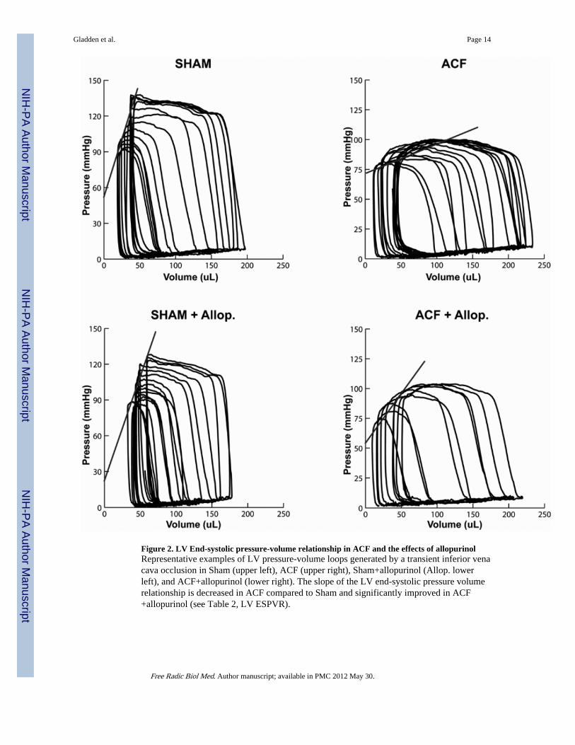
Figure 2. LV End-systolic pressure-volume relationship in ACF and the effects of allopurinolRepresentative examples of LV pressure-volume loops generated by a transient inferior venacava occlusion in Sham (upper left), ACF (upper right), Sham+allopurinol (Allop. lowerleft), and ACF+allopurinol (lower right). The slope of the LV end-systolic pressure volumerelationship is decreased in ACF compared to Sham and significantly improved in ACF+allopurinol (see Table 2, LV ESPVR).
Gladden et al. Page 14
Free Radic Biol Med. Author manuscript; available in PMC 2012 May 30.
NIH
-PA Author Manuscript
NIH
-PA Author Manuscript
NIH
-PA Author Manuscript
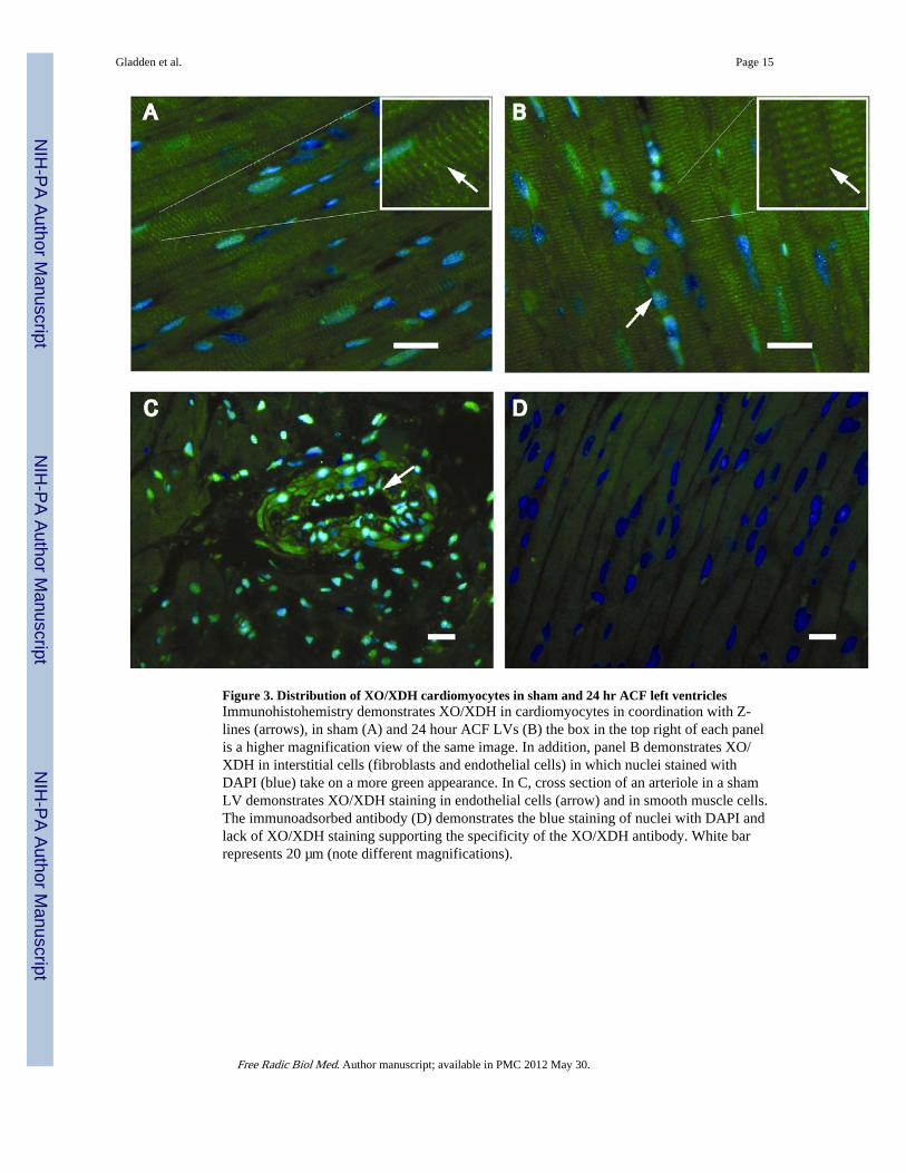
Figure 3. Distribution of XO/XDH cardiomyocytes in sham and 24 hr ACF left ventriclesImmunohistohemistry demonstrates XO/XDH in cardiomyocytes in coordination with Z-lines (arrows), in sham (A) and 24 hour ACF LVs (B) the box in the top right of each panelis a higher magnification view of the same image. In addition, panel B demonstrates XO/XDH in interstitial cells (fibroblasts and endothelial cells) in which nuclei stained withDAPI (blue) take on a more green appearance. In C, cross section of an arteriole in a shamLV demonstrates XO/XDH staining in endothelial cells (arrow) and in smooth muscle cells.The immunoadsorbed antibody (D) demonstrates the blue staining of nuclei with DAPI andlack of XO/XDH staining supporting the specificity of the XO/XDH antibody. White barrepresents 20 µm (note different magnifications).
Gladden et al. Page 15
Free Radic Biol Med. Author manuscript; available in PMC 2012 May 30.
NIH
-PA Author Manuscript
NIH
-PA Author Manuscript
NIH
-PA Author Manuscript

Figure 4. XO activity is predominantly increased cardiomyocytes in 24 hr ACFXO activity was measured in LV homogenates (A) and isolated cardiomyocytes (B) after 24hours of ACF. *=P<0.05 vs. sham (n = 4) per group. Protein levels were assessed using anXO/XDH antibody in LV tissue homogenates (sham, n=5 ACF, n=4) (C, densitometryanalysis in E) and isolated cardiomyocyte homogenates (n=3) (D, densitometry analysis inF).
Gladden et al. Page 16
Free Radic Biol Med. Author manuscript; available in PMC 2012 May 30.
NIH
-PA Author Manuscript
NIH
-PA Author Manuscript
NIH
-PA Author Manuscript
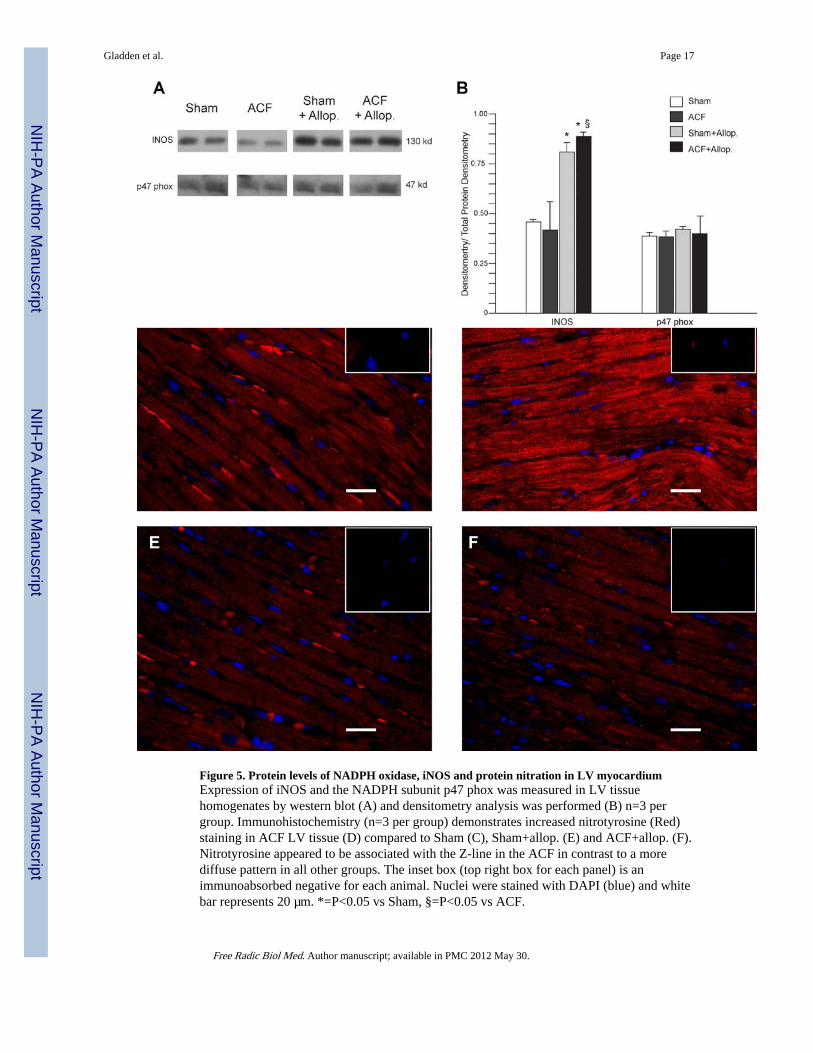
Figure 5. Protein levels of NADPH oxidase, iNOS and protein nitration in LV myocardiumExpression of iNOS and the NADPH subunit p47 phox was measured in LV tissuehomogenates by western blot (A) and densitometry analysis was performed (B) n=3 pergroup. Immunohistochemistry (n=3 per group) demonstrates increased nitrotyrosine (Red)staining in ACF LV tissue (D) compared to Sham (C), Sham+allop. (E) and ACF+allop. (F).Nitrotyrosine appeared to be associated with the Z-line in the ACF in contrast to a morediffuse pattern in all other groups. The inset box (top right box for each panel) is animmunoabsorbed negative for each animal. Nuclei were stained with DAPI (blue) and whitebar represents 20 µm. *=P<0.05 vs Sham, §=P<0.05 vs ACF.
Gladden et al. Page 17
Free Radic Biol Med. Author manuscript; available in PMC 2012 May 30.
NIH
-PA Author Manuscript
NIH
-PA Author Manuscript
NIH
-PA Author Manuscript
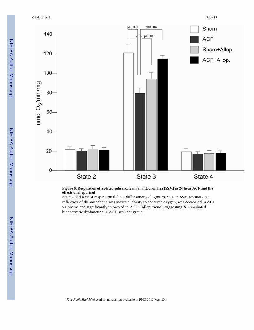
Figure 6. Respiration of isolated subsarcolemmal mitochondria (SSM) in 24 hour ACF and theeffects of allopurinolState 2 and 4 SSM respiration did not differ among all groups. State 3 SSM respiration, areflection of the mitochondria’s maximal ability to consume oxygen, was decreased in ACFvs. shams and significantly improved in ACF + allopurionol, suggesting XO-mediatedbioenergetic dysfunction in ACF. n=6 per group.
Gladden et al. Page 18
Free Radic Biol Med. Author manuscript; available in PMC 2012 May 30.
NIH
-PA Author Manuscript
NIH
-PA Author Manuscript
NIH
-PA Author Manuscript
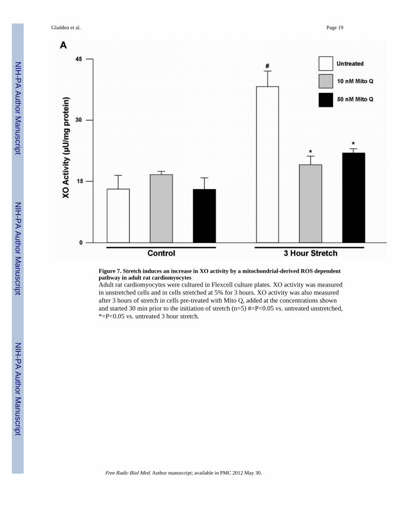
Figure 7. Stretch induces an increase in XO activity by a mitochondrial-derived ROS dependentpathway in adult rat cardiomyocytesAdult rat cardiomyocytes were cultured in Flexcell culture plates. XO activity was measuredin unstretched cells and in cells stretched at 5% for 3 hours. XO activity was also measuredafter 3 hours of stretch in cells pre-treated with Mito Q, added at the concentrations shownand started 30 min prior to the initiation of stretch (n=5) #=P<0.05 vs. untreated unstretched,*=P<0.05 vs. untreated 3 hour stretch.
Gladden et al. Page 19
Free Radic Biol Med. Author manuscript; available in PMC 2012 May 30.
NIH
-PA Author Manuscript
NIH
-PA Author Manuscript
NIH
-PA Author Manuscript
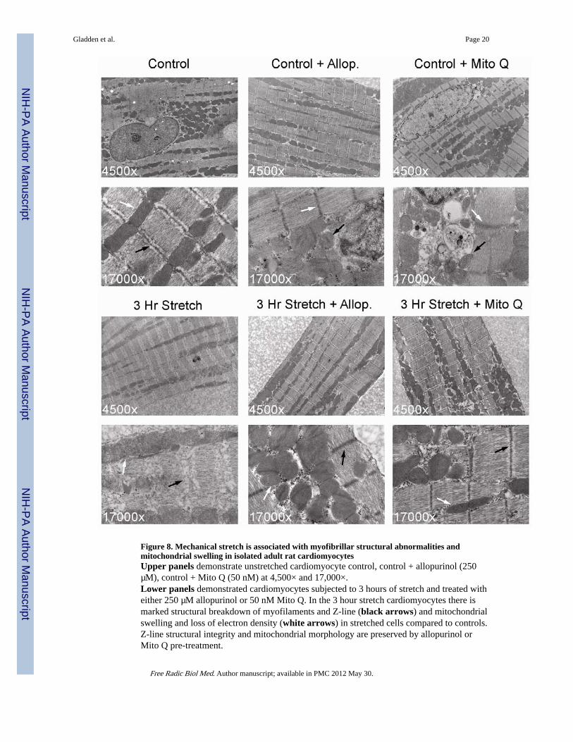
Figure 8. Mechanical stretch is associated with myofibrillar structural abnormalities andmitochondrial swelling in isolated adult rat cardiomyocytesUpper panels demonstrate unstretched cardiomyocyte control, control + allopurinol (250µM), control + Mito Q (50 nM) at 4,500× and 17,000×.Lower panels demonstrated cardiomyocytes subjected to 3 hours of stretch and treated witheither 250 µM allopurinol or 50 nM Mito Q. In the 3 hour stretch cardiomyocytes there ismarked structural breakdown of myofilaments and Z-line (black arrows) and mitochondrialswelling and loss of electron density (white arrows) in stretched cells compared to controls.Z-line structural integrity and mitochondrial morphology are preserved by allopurinol orMito Q pre-treatment.
Gladden et al. Page 20
Free Radic Biol Med. Author manuscript; available in PMC 2012 May 30.
NIH
-PA Author Manuscript
NIH
-PA Author Manuscript
NIH
-PA Author Manuscript
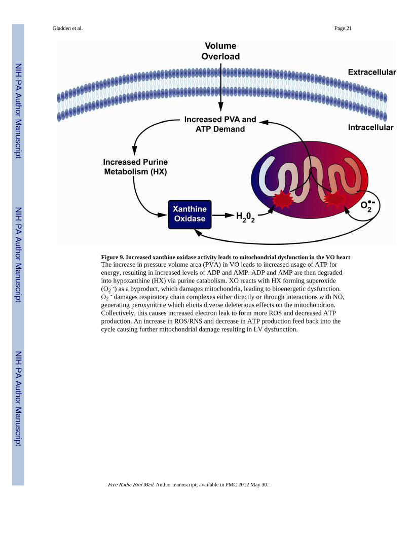
Figure 9. Increased xanthine oxidase activity leads to mitochondrial dysfunction in the VO heartThe increase in pressure volume area (PVA) in VO leads to increased usage of ATP forenergy, resulting in increased levels of ADP and AMP. ADP and AMP are then degradedinto hypoxanthine (HX) via purine catabolism. XO reacts with HX forming superoxide(O2
˙-) as a byproduct, which damages mitochondria, leading to bioenergetic dysfunction.O2
˙- damages respiratory chain complexes either directly or through interactions with NO,generating peroxynitrite which elicits diverse deleterious effects on the mitochondrion.Collectively, this causes increased electron leak to form more ROS and decreased ATPproduction. An increase in ROS/RNS and decrease in ATP production feed back into thecycle causing further mitochondrial damage resulting in LV dysfunction.
Gladden et al. Page 21
Free Radic Biol Med. Author manuscript; available in PMC 2012 May 30.
NIH
-PA Author Manuscript
NIH
-PA Author Manuscript
NIH
-PA Author Manuscript
NIH
-PA Author Manuscript
NIH
-PA Author Manuscript
NIH
-PA Author Manuscript
Gladden et al. Page 22
Table 1
Morphometric Data in shams and ACF rats.
Sham ACF Sham+Allopurinol ACF+Allopurinol
Body Weight (g) 205±5 203±7 207±7 197±5
Heart Rate (bpm) 374±10 368±6 360±7 362±11
MAP (mmHg) 96±3 62±3* 97±7 70±3*
Heart Weight (g) 0.74±0.02 0.79±0.02 0.75±0.02 0.72±.02
LV Weight (g) 0.51±0.012 0.55±0.01 0.52±0.02 0.49±0.01
Lung Weight (g) 1.16±0.02 1.18±0.03 1.15±0.03 1.07±0.03
Heart/Body Weight 0.0036±0.000083 0.004±0.0001 0.0036±0.0001 0.004±0.00007
N 7 8 6 8
*P<0.5 vs Sham, data = mean ± SE
Free Radic Biol Med. Author manuscript; available in PMC 2012 May 30.
NIH
-PA Author Manuscript
NIH
-PA Author Manuscript
NIH
-PA Author Manuscript
Gladden et al. Page 23
Table 2
Echocardiographic and hemodynamic data in sham and ACF rats.
Sham ACF Sham+Allopurinol ACF+Allopurinol
LVESD (mm) 3.54±0.3 3.1±0.3 3.1±0.42 3.5±0.2
LVEDD (mm) 6.4±0.2 6.7±0.3* 6.4±0.3 7±0.1*
LVESV (µL) 90±9 92±17 69±14 95±13
LVEDV (µL) 326±17 389±26 308±22 364±18
EF (%) 73±2 77±3 77±4 74±3
LVPWs (mm) 2.8±0.1 3±0.2 3±0.2 2.7±0.1
LVPWd (mm) 1.8±0.1 1.8±0.2 1.8±0.1 1.5±0.03
LVES σ 43±7 23±4* 34±11 32±4*
LVED σ 2.5±0.2 8±1.1* 4±1.5 5±0.6*§
LV ESPVR 1.48±0.2 0.36±0.07* 1.62±0.26 0.71±0.07*§
N 7 8 6 8
LV end-systolic dimension (LVESD), LV end-diastolic dimension (LVEDD), LVES volume (LVESV), LVEDV, LV Ejection Fraction (EF),LVES Posterior wall thickness (LVPWs), LVED Posterior wall thickness (LVPWd) LVES wall stress (σ) LVED σ, LV ES Pressure-Volume
Relationship*= P<0.05 vs Sham,
§P<0.05 vs ACF, data = mean ± SE
Free Radic Biol Med. Author manuscript; available in PMC 2012 May 30.
Related Documents