Non-enzymatically derived minor lipids found in Escherichia coli lipid extracts Teresa A. Garrett a,† , Christian R. H. Raetz b , Jennifer D. Son a,1 , Travis D. Richardson b , Craig Bartling b,2 , and Ziqiang Guan b a Department of Chemistry, Vassar College, Box 580, 124 Raymond Avenue, Poughkeepsie, NY 12604, US b Department of Biochemistry, Duke University Medical Center, P.O. Box 3711, Durham, North Carolina 27710, USA Abstract Electrospray ionization mass spectrometry is a powerful technique to analyze lipid extracts especially for the identification of new lipid metabolites. A hurdle to lipid identification is the presence of solvent contaminants that hinder the identification of low abundance species or covalently modify abundant lipid species. We have identified several non-enzymatically derived minor lipid species in lipid extracts of Escherichia coli, phosphatidylmethanol, ethyl and methyl carbamates of PE and N-succinyl PE were identified in lipid extracts of Escherichia coli. Phosphatidylmethanol (PM) was identified by exact mass measurement and collision induced dissociation tandem mass spectrometry (MS/MS). Extraction in the presence of deuterated methanol leads to a 3 atomic mass unit shift in the [M-H] - ions of PM indicating its formation during extraction. Ethyl and methyl carbamates of PE, also identified by exact mass measurement and MS/MS, are likely to be formed by phosgene, a breakdown product of chloroform. Addition of phosgene to extractions containing synthetic PE significantly increases the levels of PE-MC detected in the lipid extracts by ESI-MS. Extraction in the presence of methylene chloride significantly reduced the levels of these lipid species. N-succinyl PE is formed from reaction of succinyl-CoA with PE during extraction. Interestingly N-succinyl PE can be formed in an aqueous reaction mixture in the absence of added E. coli proteins. This work highlights the reactivity of the amine of PE and emphasizes that careful extraction controls are required to ensure that new minor lipid species identified using mass spectrometry are indeed endogenous lipid metabolites. Keywords mass spectrometry; E. coli; lipids; chloroform; phosgene; artifacts © 2011 Elsevier B.V. All rights reserved. † Corresponding author Department of Chemistry Vassar College, Box 580 124 Raymond Avenue Poughkeepsie, NY 12604 Phone: 845-437-5738 Fax: 845-437-5732 [email protected]. [email protected] (C. Raetz), [email protected] (J. Son), [email protected] (C. Bartling), [email protected] (Z. Guan).. 1 Current address: Boston University School of Medicine, 72 East Concord Street, Boston, MA 02118, USA 2 Current address: Battelle, 505 King Avenue, Columbus, OH 43201-2693 Publisher's Disclaimer: This is a PDF file of an unedited manuscript that has been accepted for publication. As a service to our customers we are providing this early version of the manuscript. The manuscript will undergo copyediting, typesetting, and review of the resulting proof before it is published in its final citable form. Please note that during the production process errors may be discovered which could affect the content, and all legal disclaimers that apply to the journal pertain. NIH Public Access Author Manuscript Biochim Biophys Acta. Author manuscript; available in PMC 2012 November 1. Published in final edited form as: Biochim Biophys Acta. 2011 November ; 1811(11): 827–837. doi:10.1016/j.bbalip.2011.08.012. NIH-PA Author Manuscript NIH-PA Author Manuscript NIH-PA Author Manuscript

Welcome message from author
This document is posted to help you gain knowledge. Please leave a comment to let me know what you think about it! Share it to your friends and learn new things together.
Transcript

Non-enzymatically derived minor lipids found in Escherichia colilipid extracts
Teresa A. Garretta,†, Christian R. H. Raetzb, Jennifer D. Sona,1, Travis D. Richardsonb, CraigBartlingb,2, and Ziqiang Guanb
aDepartment of Chemistry, Vassar College, Box 580, 124 Raymond Avenue, Poughkeepsie, NY12604, USbDepartment of Biochemistry, Duke University Medical Center, P.O. Box 3711, Durham, NorthCarolina 27710, USA
AbstractElectrospray ionization mass spectrometry is a powerful technique to analyze lipid extractsespecially for the identification of new lipid metabolites. A hurdle to lipid identification is thepresence of solvent contaminants that hinder the identification of low abundance species orcovalently modify abundant lipid species. We have identified several non-enzymatically derivedminor lipid species in lipid extracts of Escherichia coli, phosphatidylmethanol, ethyl and methylcarbamates of PE and N-succinyl PE were identified in lipid extracts of Escherichia coli.Phosphatidylmethanol (PM) was identified by exact mass measurement and collision induceddissociation tandem mass spectrometry (MS/MS). Extraction in the presence of deuteratedmethanol leads to a 3 atomic mass unit shift in the [M-H]- ions of PM indicating its formationduring extraction. Ethyl and methyl carbamates of PE, also identified by exact mass measurementand MS/MS, are likely to be formed by phosgene, a breakdown product of chloroform. Additionof phosgene to extractions containing synthetic PE significantly increases the levels of PE-MCdetected in the lipid extracts by ESI-MS. Extraction in the presence of methylene chloridesignificantly reduced the levels of these lipid species. N-succinyl PE is formed from reaction ofsuccinyl-CoA with PE during extraction. Interestingly N-succinyl PE can be formed in an aqueousreaction mixture in the absence of added E. coli proteins. This work highlights the reactivity of theamine of PE and emphasizes that careful extraction controls are required to ensure that new minorlipid species identified using mass spectrometry are indeed endogenous lipid metabolites.
Keywordsmass spectrometry; E. coli; lipids; chloroform; phosgene; artifacts
© 2011 Elsevier B.V. All rights reserved.†Corresponding author Department of Chemistry Vassar College, Box 580 124 Raymond Avenue Poughkeepsie, NY 12604 Phone:845-437-5738 Fax: 845-437-5732 [email protected]. [email protected] (C. Raetz), [email protected] (J. Son),[email protected] (C. Bartling), [email protected] (Z. Guan)..1Current address: Boston University School of Medicine, 72 East Concord Street, Boston, MA 02118, USA2Current address: Battelle, 505 King Avenue, Columbus, OH 43201-2693Publisher's Disclaimer: This is a PDF file of an unedited manuscript that has been accepted for publication. As a service to ourcustomers we are providing this early version of the manuscript. The manuscript will undergo copyediting, typesetting, and review ofthe resulting proof before it is published in its final citable form. Please note that during the production process errors may bediscovered which could affect the content, and all legal disclaimers that apply to the journal pertain.
NIH Public AccessAuthor ManuscriptBiochim Biophys Acta. Author manuscript; available in PMC 2012 November 1.
Published in final edited form as:Biochim Biophys Acta. 2011 November ; 1811(11): 827–837. doi:10.1016/j.bbalip.2011.08.012.
NIH
-PA Author Manuscript
NIH
-PA Author Manuscript
NIH
-PA Author Manuscript
1. IntroductionThe identification of new cellular metabolites can facilitate the identification of newbiochemical pathways and lead to accurate genome annotation. High-resolution massspectrometry is a superb tool for the identification of new metabolites especially lipidmetabolites. The combination of accurate mass measurements and collision-induceddissociation MS/MS analysis facilitates the identification of known and novel lipidstructures and has been used to characterize several new lipid metabolites in Gram-negativebacteria such as Escherichia coli and other organisms such as Clostridium [1-5].
In addition to being used to identify new minor lipid metabolites, ESI-MS/MS is extensivelyused to characterize the levels of the major lipid metabolites [6,7]. In nearly all of thesestudies lipids are extracted from cells or tissues using standard extraction conditions, such asBligh-Dyer or Folch extractions [8, 9]. Many of these extractions are chloroform-methanolbased. Through the course of our analysis of total lipid extracts from wild-type andphosphatidylglycerolphosphate synthase (pgsA) deficient E. coli [2,10] we have identifiedseveral minor lipid species that are formed from major lipid metabolites during extraction.In particular, phosphatidic acid (PA) and phosphatidylethanolamine (PE) derivatives havebeen identified in E. coli extracted by the method of Bligh and Dyer [8].
The structures of these non-enzymatically formed lipid derivatives were determined byaccurate mass measurement and collision-induced dissociation tandem mass spectrometry(MS/MS) using an electrospray ionization (ESI) quadrupole time-of-flight (qTOF) massspectrometer and by comparison to synthetic standards. We have identified one modificationof PA and four modifications to PE. Investigations into the origin of these novel lipidsrevealed that these lipids are not present in the cell prior to extraction but are likely derivedduring extraction particularly in the presence of chloroform. Of particular interest is the factthat several of these non-enzymatically-generated lipids are isobaric with well-characterizedlipids of the cell hampering structural identification and quantification.
2. Materials and Methods2.1. Materials
Tryptone and yeast extract were from FisherBiotech (Fairlawn, New Jersey). Glass-backedSilica Gel 60 thin layer chromatography plates (0.25-mm) and high performance thin layerchromatography (HPTLC) plates were from E. Merck; solvents were reagent grade fromMalinckrodt. CDCl3, CD3OD, 15N-NH4Cl, and phosgene (20% in toluene) were fromSigma. Other chemicals were purchased from VWR. 1-palmitoyl-2-oleoyl-sn-glycero-3-phosphoethanolamine (PE(16:0/18:1)), 1-stearoyl-2-arachidonoyl-sn-glycero-3-phosphoethanolamine (PE(18:0/20:4)), 1,2-dioleoyl-sn-glycero-3-phosphoethanolamine-N-(succinyl), and 1,2-dipalmitoyl-sn-glycero-3-phosphoethanolamine-N-ethylcarbamate werefrom Avanti Polar Lipids, Alabaster, AL. L-α 1-palmitoyl-2-lineoylphosphatidylethanolamine [palmitoyl-1-14C] ((50-62 mCi/mmol, 25 uCi/ml) was fromAmersham.
2.2. Growth of E. coliE. coli K-12 strain W3110 was cultured at 37 °C in Luria Broth (LB) consisting of 10 g ofNaCl, 5 g of yeast extract, and 10 g of tryptone per liter [11]. The cells were grownovernight in LB medium at 37 °C and then diluted into LB medium to an A600 of 0.01. Theculture was grown at 37 °C, shaking at 225 rpm until the A600 was about 1.0. Cells wereharvested by centrifugation for 20 minutes at 2600 × g and washed with phosphate bufferedsaline (PBS, 137 mM NaCl, 2.7 mM KCl, 10 mM Na2HPO4, 1.8 mM KH2PO4). pgsAdeficient E. coli [10] was grown in LB as described above except at 30 °C instead of 37 °C.
Garrett et al. Page 2
Biochim Biophys Acta. Author manuscript; available in PMC 2012 November 1.
NIH
-PA Author Manuscript
NIH
-PA Author Manuscript
NIH
-PA Author Manuscript
2.3. Extraction of E. coli total lipidsThe final cell pellet from a 1 L growth of E. coli was re-suspended in 40 mL of PBS andtransferred to a Teflon-lined centrifuge bottle. The cellular lipids were extracted using themethod of Bligh and Dyer [8] as described previously [1]. Briefly, 50 mL of chloroform and100 mL of methanol were added to the cell suspension to generate a single-phase extractionmixture of CHCl3/CH3OH/PBS (1:2:0.8, v/v/v). After incubation at room temperature for 20minutes, the mixture was centrifuged at 2600 × g for 15 minutes. The supernatant wastransferred to a clean bottle and converted to a two-phase Bligh-Dyer extraction mixture(CHCl3/CH3OH/PBS, 2:2:1.8, v/v/v) by the addition of 50 mL of chloroform and 50 mL ofPBS. The extraction mixture was centrifuged as above to resolve the phases. The upperphase, designated U1, was separated from the lower phase, designated L1, and transferred toa clean bottle. U1 was acidified with HCl (final concentration 0.2M), and washed with 100mL pre-equilibrated acidic lower phase (generated from mixing CHCl3, CH3OH, 0.2M HCl(2:2:1.8, v/v/v)). The resulting lower phase was washed with 190 mL of pre-equilibratedacidic lower phase. The extraction mixture was centrifuged as above to resolve the phasesand this lower phase, designated L2, was dried using rotary evaporation. L1 was washedwith 190 mL of pre-equilibrated neutral upper phase and the resulting lower phase driedusing rotary evaporation.
Wild type E. coli W3110 was extracted in the presence of CD3OD and CDCl3. A cell pelletfrom a 50 mL growth was re-suspended in 0.8 mL of PBS and divided into two glass tubes.To one tube 1 mL of CD3OD and 0.5 mL CDCl3 and to the second tube 1 mL of CH3OHand 0.5 mL CHCl3 to generate a single phase Bligh-Dyer extraction mixture. The tubes wereincubated at room temperature for 20 minutes and then centrifuged for 15 minutes in aclinical centrifuge. The supernatant was transferred to a clean tube and then 0.5 mL CDCl3or 0.5 mL of CHCl3 and 0.5 mL of PBS were added to generate a two-phase Bligh-Dyerextraction mixture. The resulting lower phase was transferred to a fresh tube and dried underN2. Samples were re-dissolved in 100 μL of either 2:1, CDCl3/CD3OD (v/v) or CHCl3/CH3OH (v/v) for mass spectrometry analysis. Cells were also extracted using Bligh-Dyerextraction mixtures in which the mixtures were acidified by the addition of HCl in theaqueous phase to a final concentration of 0.2 M and/or the CHCl3 was replaced withCH2Cl2.
2.4. Ion exchange chromatographyThe total lipid extracts were fractionated on DEAE-cellulose as described previously [1,12].
2.5. Mass spectrometryThe dried lipid film was re-dissolved in CHCl3/CH3OH (2:1, v/v) to a concentration of ~ 1μg/mL. This solution was directly infused into the Turbo electrospray ionization source of aQSTAR XL quadrupole time-of-flight tandem mass spectrometer (ABI/MDS-Sciex,Toronto, Canada) at 6 μL/min. Mass spectra were obtained scanning from 200 to 2000 Da innegative-ion mode with the electrospray ionization source operating at the followingsettings: nebulizer gas, 21 kPa, curtain gas, 27 kPa, ion-spray voltage, -4500 V, declusteringpotential, -55 V, focusing potential, -265 V, declustering potential 2, -15 V. The instrumentwas calibrated using PPG (Applied Biosystems). Collision induced dissociation tandemmass spectrometry (MS/MS) was performed with a collision energy of -50.0 V (laboratoryframe of reference) and N2 as the collision gas. Data acquisition, analysis and elementalcomposition calculations were performed using the Analyst QS 1.1 software. Exact massesof lipid species and product ions were obtained using CS Chem Draw Pro, version 12.0.
Normal phase liquid chromatography mass spectrometry was performed as describedpreviously [1].
Garrett et al. Page 3
Biochim Biophys Acta. Author manuscript; available in PMC 2012 November 1.
NIH
-PA Author Manuscript
NIH
-PA Author Manuscript
NIH
-PA Author Manuscript

2.6. 15N-labeling of E. coliCells were grown overnight in LB medium at 37 °C and then diluted into M9 minimal mediasupplemented with 15NH4Cl to an A600 of 0.01 [13]. The culture was grown at 37 °C,shaking at 225 rpm until the A600 was about 1.0. Cells were harvested and extracted asdescribed above.
2.7. Extractions in the presence of phosgeneA synthetic PE(18:0/20:4) (100 μg) was extracted in the presence or absence of 0.05 %phosgene. Briefly, PE(18:0/20:4) (10 mg/mL in CHCl3) was mixed with 0.5 ml CHCl3, 1mL CH3OH, 0.4 mL 1X PBS, and 5 μL of toluene or 20% phosgene in toluene andincubated at room temperature for 10 minutes. To each tube 0.5 mL CHCl3 and 0.5 mL 1XPBS was added to generate a two-phase Bligh-Dyer extraction mixture. The tube wascentrifuged to resolve the phases and the upper phase discarded. The lower phase was driedunder nitrogen.
2.8. Exchanging protons using deuterated solventsA dried lipid extract was re-dissolved in 100 μL of 2:1 CDCl3/CD3OD, v/v, dried under N2.This resuspension/drying cycle was repeated two more times and finally re-dissolved in 100μL of 2:1 CDCl3/CD3OD, v/v and analyzed using electrospray ionization massspectrometry.
2.9. Preparation of cell free extracts and membranesE. coli W3110 (50 mL) was grown in LB medium as described above. The cell pellet wasre-suspended in 3 mL of 20 mM Hepes, pH 7.4 and cells lysed in a French Pressure cell at18000 psi. The extract was centrifuged for 20 minutes at 4 °C and 3000 × g to pellet un-lysed cells. The cell-free extract was transferred to a microcentrifuge tube and stored at -80°C.
Membranes were prepared by centrifuging the cell free extract at 4 °C, 100000 × g for 1hour. The resulting membrane pellet was re-suspended by homogenization in 2 mL of 20mM Hepes pH 7.4 and the membranes pelleted by centrifugation as described above. Thefinal membrane pellet was re-suspended in 1.5 mL of 20 mM Hepes pH 7.4 and stored at -80°C. The concentration of the extract was determined using the bicinchoninic acid reagent(Thermo Scientific) with bovine serum albumin as the standard.
2.10. In vitro synthesis of N-succinyl phosphatidylethanolaminePhosphatidylethanolamine (16:0/18:1) was suspended in 20 mM Hepes buffer, pH 7.4 at afinal concentration of 2 mM. Reactions containing 20 mM Hepes buffer, pH 7.4, 0.5 mMPE, 0.2% Triton X-100, in the presence or absence of 1 mg/mL cell free extract and in thepresence or absence of 2 mM succinate, succinic anhydride, succinyl-CoA, glutaryl-CoA, ormalonyl-CoA were incubated at 30 °C for 2 hours. Five μL of the reaction was spotted to asilica gel Thin layer chromatography plate and developed in CHCl3/CH3OH/H2O/CH3COOH (25:15:4:2, v/v/v/v). The lipids were visualized by charring with 10 % sulfuricacid.
3. Results3.1. Phosphatidylmethanol
Wild type E. coli lipid extracts were fractionated on a DEAE cellulose column based oncharge [1]. A fraction eluting with CHCl3/CH3OH/30 mM NH4Ac (2:3:1, v/v/v) wasanalyzed by negative-ion electrospray ionization mass spectrometry (ESIMS). The
Garrett et al. Page 4
Biochim Biophys Acta. Author manuscript; available in PMC 2012 November 1.
NIH
-PA Author Manuscript
NIH
-PA Author Manuscript
NIH
-PA Author Manuscript

predominant ions between m/z 691 and 773 represent singly charged species [M-H]- ofphosphatidylglycerol (PG) as determined by exact mass and MS/MS analysis (Figure 1). Inaddition several peaks at m/z 631.486, 633.474, 659.502, 673.526, 685.543 and 687.546were observed (Figure 1 inset). The exact mass of these negative ions is suggestive ofphosphatidic acid (PA). However PA is expected to elute from DEAE cellulose with higherconcentrations of NH4Ac [1]. In addition, the most prominent ion at m/z 659.502corresponds by exact mass to PA with 33 cycolpropane fatty acyl chains consistent with thefatty acid composition of E. coli grown to late log phase [14]. Collision-induced dissociation(CID) MS/MS analysis of these peaks revealed fragmentation patterns inconsistent with PA.Figure 2 shows the MS/MS spectrum of another prominent ion in that mass range, at m/z687.5. While this ion mass may correspond to a PA with 37cyclopropane acyl chains, afragment ion for a cyclopropane fatty acid is not detected. Instead, product ionscorresponding to palmitate (16:0, m/z 255.235) and octadecenoate (18:1, m/z 281.254) areobserved. In addition a peak at m/z 110.985 was observed and corresponds by exact mass tomethylated phosphate (CH4O4P-, exact mass 110.9853). The ion at m/z 167.026 is 14 atomicmass units (a.m.u.) larger than the common glycerophospholipid fragment ion C3H6O5P-,(exact m/z 152.9953) [15-17] and corresponds to a methylated form of this fragment ion(C4H9O5P-). From these fragment ions we hypothesized that the ion at m/z 687.5corresponds to phosphatidic acid with a methyl group added to the phosphate (Figure 2),designated phosphatidylmethanol (PM). The other major peaks in the product ion spectra areattributable to losses of acyl chains from the precursor ion. The ions at m/z 423.277 and m/z449.300 correspond to the loss of the 18:1 and 16:0 acyl chains respectively as ketenes(RCH=CO). The MS/MS analysis of the ions at m/z 631.5, 633.5, 659.5, 673.5 and 687.5reveal similar fragmentation patterns, especially the presence of the ion at m/z 110.9 ioncorresponding to methylated phosphate.
It has been previously reported that cardiolipin synthase can catalyze the addition of analcohol via an alcoholysis reaction with cardiolipin [18]. Before pursuing the possibleenzymatic formation of PM in E. coli we investigated the possibility that PM may be formedduring extraction. Wild-type E. coli cells were extracted in the presence of deuteratedmethanol and deuterated chloroform. Figure 3 shows the ESI-MS spectrum of total lipidsextracted in the presence of non-deuterated solvents (panel A) or deuterated solvents (panelB and C). Lipid extracts were analyzed by ESI-MS after being re-dissolved in non-deuterated (Figure 3, panel A or C) or deuterated solvents (Figure 3, panel B). Because theselipid extracts were not fractionated using anion exchange as above, singly charged [M-H]-
PA and doubly-charged [M-2H]2- CL ions isobaric with the PM ions are present. The peakat m/z 659.463, corresponding to PM with 32:1 acyl chains shifts, in part to m/z of 662.482(panel B). The same result is observed when the lipids extracted in deuterated solvents werere-dissolved in non-deuterated solvents consistent with the formation of PM duringextraction and not during ionization or MS analysis. MS/MS of the ion at m/z 662.5confirms the addition of deuterated methanol to the lipid (Figure 4). The product ion at m/z110.945 is shifted to 114.003 consistent with the methylated phosphate gaining the methylgroup from the solvent during extraction. In addition the fragment ions at m/z 167.012 and423.254 gain three mass units to m/z 170.029 and 426.269 in the presence of deuteratedmethanol. These data strongly suggest that PM is being formed during solvent extraction.Extraction in presence of deuterated solvents did not alter ions of the other majorglycerophospholipids PE, PG, and CL (data not shown).
3.2. PE-EthylcarbamateLipids were extracted from E. coli lacking the phosphatidylglycerol phosphate synthase(pgsA) gene [10]. As previously reported, ESI-MS confirmed that these cells lack PG andcardiolipin (CL) [2], and the major remaining negative-ions arise from
Garrett et al. Page 5
Biochim Biophys Acta. Author manuscript; available in PMC 2012 November 1.
NIH
-PA Author Manuscript
NIH
-PA Author Manuscript
NIH
-PA Author Manuscript

phosphatidylethanolamine (PE). However, we also identified a series of ions [M-H]- with m/z of 760.477, 774.502, 788.494, 800.519, and 814.527 that correspond by exact mass tophosphatidylserine (PS) (Figure 5) with 34:1, 35:1cyclopropane, 36:1, 37:2cyclopropane,and 38:2. PS is an intermediate in the biosynthesis of PE but is not readily detected in wildtype E. coli [21]. These ions are also observed in wild-type cells; however they are obscuredby the predominant ions [M-H]- of PG.
MS/MS of one of these ions, the ion at m/z 760.5, is shown in Figure 6 and is inconsistentwith PS that in the negative mode yields a prominent product ion corresponding to theneutral loss of 87 a.m.u [17]. However, product ions indicative of a glycerophospholipid areobserved. The ion at m/z 78.951 corresponds to PO3
- and the ion at m/z 152.986 correspondsto C3H6O5P- [15]. Acyl chains corresponding to palmitate (16:0) and palmitoleate (16:1) areobserved as their carboxylic anions at m/z 255.217 and m/z 253.200, respectively. Inaddition, the product ion at m/z 524.281 corresponds to the loss of the 16:1 acyl chain as aketene (RCH=C=O).
A neutral loss of 46 a.m.u. leads to the formation of the product ion at m/z 673.481, which isinterpreted as a 16:0, 18:1 phosphatidic acid anion (C37H70O8P-, exact mass 673.4814) andis consistent with a diacylated glycerophospholipid. The 216 mass units loss, consistentlyseen in the MS/MS spectra of all of the unknown [M-H]- ions at m/z 861.506, 875.521,887.525, and 915.548 (data not shown), could correspond to the loss of a part of the headgroup of a novel diacylated glycerophospholipid.
A neutral loss of 46 a.m.u. leads to the formation of the product ion at m/z 714.489, which isinterpreted as loss of ethanol (Figure 6). The product ions at m/z 478.254 and 460.220correspond to loss of ethanol and the 16:1 acyl chain as a ketene (RCH=C=O) or a fatty acid(RCOOH), respectively.
Several ions in the low mass region are derived from the head group of this lipid. Theproduct ion at m/z 268.040 corresponds to the loss of both acyl chains from the precursorion, one as a ketene and one as a fatty acid. The product ion at m/z 222.009 is ethanol lossfrom m/z 268.040. As indicated in Figure 6, the product ion at m/z 212.017 corresponds tothe head group and is observed in MS/MS spectra of all of the unknown ions. The ions at m/z 194.008 and m/z 165.978 correspond to the loss of water and ethanol, respectively, fromthe ion at m/z 212.017.
Several lines of evidence supported a novel modification to the free amine of PE.Continuous labeling of cells in M9 minimal media supplemented with 15NH4Cl shifts theseunknown ions one mass unit consistent with the presence of one nitrogen in the molecule.These lipids were retained by DEAE cellulose and eluted with CHCl3/CH3OH/15 mMNH4Ac (2:3:1, v/v/v) consistent with a modification to the free amine of PE. Finally, theions contained only one exchangeable proton as seen by a one mass unit shift after exchangeinto deuterated solvents (data not shown).
Taken together we believe that the amine of PE is modified by the addition of -CO2CH2CH3 to form an ethyl carbamate. We propose a new PE derivative which wedesignate phosphatidylethanolamine ethylcarbamate (PE-EC).
To confirm the structure proposed above a synthetic PE-EC containing two 18:1 acyl chainswas obtained. The fragment ions obtained from the synthetic standard and the endogenousPE-EC are very similar (data not shown), strongly supporting the structure shown in Figure4 and 5.
Garrett et al. Page 6
Biochim Biophys Acta. Author manuscript; available in PMC 2012 November 1.
NIH
-PA Author Manuscript
NIH
-PA Author Manuscript
NIH
-PA Author Manuscript

Cone et al. indicated that the formation of ethyl carbamates could occur on amine containingdrugs during chloroform extraction [20]. During normal phase liquid chromatography massspectrometry (LC-MS), PE-EC elutes at ~5 minutes as compared to PE which elutes at~14.5 minutes (Figure 7), strongly suggesting that PE-EC was formed during extraction. Tofurther confirm this, we next extracted cells using a modified Bligh-Dyer extraction inwhich methylene chloride (less likely to decompose to phosgene than chloroform) replacedthe chloroform. ESI-MS of the methylene chloride extracted samples as well as thechloroform-extracted samples are shown in Figure 8. The PE-EC was not detected insamples that were extracted with methylene chloride indicating that the PE-EC was beingformed during extraction in the presence of chloroform.
We tested whether this process was indeed mediated by phosgene. A 18:0, 20:4 synthetic PEstandard (exact mass of [M-H]- is 766.5392) was extracted in the presence or absence ofadded phosgene and then analyzed by ESI-MS. Extraction of PE leads to the formation of a[M-H]- ion at m/z 824.550. This lipid corresponds to PE-MC as determined by exact massmeasurement and MS/MS analysis (Figure 9 Panel C). A neutral loss of 32 a.m.u. leads tothe formation of the product ion at m/z 792.517, which is interpreted as loss of methanol(Figure 9, panel C). This loss is analogous to the 46 a.m.u. neutral loss of ethanol as occurswith PE-EC (Figure 6). The product ion at m/z 198.018 corresponds to the head group. Theions at m/z 194.08 and m/z 165.978 correspond to the loss of water and methanol,respectively, from the ion at m/z 198.018.
The levels of PE-MC appear to be nearly equal to the levels of PE. However themodification of the amine of PE to form PE-MC likely leads to increased ionizationefficiency as compared to PE. When phosgene is present the levels of PE-MC increase(Figure 9 panel A versus panel B), indicating that phosgene is likely involved in theformation of the ethyl and methyl carbamates of PE we have detected. It is hypothesized thatthe relative levels of PE-MC and PE-EC may be dependent on the chloroform used andwhether ethanol is used as a stabilizing agent.
The proposed mechanism for the formation of the ethyl- and methyl-carbamates of PE isshown in Figure 10 (compounds 1 and 2). A second novel minor lipid was detected in theupper phase of a neutral Bligh-Dyer extraction. When this neutral upper phase (U1) isacidified and partitioned against a pre-equilibrated acidic lower phase highly acidic lipidspartition to the lower phase. In this acidified lower phase (L2) we detected a new lipid inwhich glutathione is attached to PE via a carbamate linkage (Figure 10, compound 3, datanot shown). Like PE-EC, PE-MC and PE-glutathione are not detected in lipid extractsprepared in the presence of methylene chloride instead of chloroform (data not shown).
3.3 N-succinyl phosphatidylethanolamineWild type E. coli were extracted to generate an acidic lower phase L2 (described above) [5].This acidified lower phase was analyzed using ESI-MS (Figure 11). A series of [M-H]- ionswith m/z of 786.488, 788.506, 802.526, 814.528, and 816.538 were observed which did notcorrespond to known lipids. MS/MS of the ions showed a fragmentation pattern consistentwith a glycerophospholipid with two acyl chains. Figure 12, panel A shows the MS/MSspectrum of the ion at m/z 788.5. Carboxylic anions of fatty acids, 16:1 and 16:0 areprominent (m/z 253.213 and 255.230, respectively). In addition, the ion at m/z 152.991corresponds to C3H6O5P-, a diagnostic ion for glycerophospholipids [17]. A neutral loss of100 a.m.u. yields a product ion (m/z 688.491) with the mass expected for PE with 32:1 acylchains. The fragment ion at m/z 645.452 corresponds to the loss of the head group. Thefragment ions at m/z 532.258 and 534.280 correspond to the loss of the 16:0 and 16:1 acylchains as a fatty acid (RCOO-). All of the unknown [M-H]- ions yielded similarfragmentation patterns, particularly the neutral loss of 100 a.m.u. to yield a product ion with
Garrett et al. Page 7
Biochim Biophys Acta. Author manuscript; available in PMC 2012 November 1.
NIH
-PA Author Manuscript
NIH
-PA Author Manuscript
NIH
-PA Author Manuscript

the mass of PE acylated with the prominent fatty acids product ions. The MS/MS resultallowed us to propose a metabolite of PE in which the amine is modified with succinate(Figure 12, panel A).
Synthetic carboxyacyl derivatives of phosphatidylethanolamine (PE) are commerciallyavailable and have been used to efficiently couple proteins to liposomes [21,22]. To confirmour proposed structure, synthetic N-succinyl PE was obtained (Avanti Polar Lipids,Alabaster, AL) and the MS/MS of the synthetic standard compared to the MS/MS spectra ofthe unknown ions. As shown in Figure 12, panel B the spectra show similar fragmentationpatterns with the characteristic loss of 100 a.m.u. from the precursor ion and the formationof the cognate PE. These results strongly suggest that the structure of the lipids observed isN-succinyl PE.
We attempted to establish an in vitro assay for the formation of N-succinyl PE. Wehypothesized that the amine of PE could be modified in a reaction similar to the acylationreactions of other lipids [23]. In those instances, an acyl chain is activated via covalentlinkage to CoA or ACP and serves as the acyl donor to either a hydroxyl or an amine[24-26]. We tested to see if succinate could be transferred from succinyl CoA to PE in anenzyme dependent manner. 14C-PE and succinyl CoA were combined in the presence andabsence of cell-free extract or membranes derived from wild type E. coli W3110 andincubated for 1 hour. Display of the reaction products using thin layer chromatographyrevealed the formation of a more hydrophilic product in the absence of added protein(Figure 13, lane 2, marked with *). Interestingly, in the presence of protein, either cell freeextracts or membranes, the product was decreased compared to in the absence of protein(Figure 13, lane 2 compared to lane 4 and lane 6). To establish that the product formed inthe absence of protein is indeed N-succinyl PE, synthetic, unlabeled 16:0/18:1-PE (exactmass [M-H]- is 716.5236) was reacted with succinyl CoA, the lipid isolated by Bligh-Dyerextraction and analyzed using ESI-MS. An ion 100 a.m.u. larger than the 16:0/18:1 PE wasobserved (data not shown). MS/MS of the ion at m/z 816.467 revealed a fragmentationpattern consistent with the addition of succinate to 16:0/18:1 PE (data not shown). Theseresults strongly indicate that the formation of N-succinyl PE occurs in an aqueous mixturevia reaction of PE with N-succinyl CoA and in the absence of protein.
The reaction of PE with succinyl CoA is specific. Succinic anhydride does not add to PEunder these aqueous reaction conditions. Furthermore addition of CoA does not stimulatethe formation of N-succinyl CoA in the presence of succinic anhydride (data not shown).This eliminates the possibility of residual succinic anhydride and/or CoA, which are used tosynthesize succinyl CoA [27] and may be present in low levels in commercial preparationsof succinyl-CoA, being responsible for the formation of N-succinyl PE. In addition, glutaryl,malonyl, acetoacetyl, and butyryl CoA did not react with PE. Lauryl-CoA was also non-reactive (Figure 14 and data not shown).
In theory, the formation of N-succinyl PE could occur in vivo. Estimations of the in vivoconcentrations of succinyl CoA are ~ 4 μM [28] and the free amine of the PE would bereadily available as it composes approximately 38% of the total lipid of the inner leaflet ofthe inner membrane [29]. Alternatively, the N-succinyl PE could be formed during the firststep of the lipid extraction. To distinguish between these possibilities, cells were extracted inthe presence of excess lysine. If N-succinyl PE was formed during the extraction then thelysine would compete with the PE for reaction with the free succinyl CoA in the extract. Ifthe N-succinyl PE is formed in the cell, the presence of the lysine should have no effect onthe levels of the lipid. E. coli cells were extracted with in the presence or absence of 10 mMlysine and the lipid extract analyzed with ESI-MS. When lysine was present duringextraction N-succinyl PE is absent from the lipid extract (data not shown). This strongly
Garrett et al. Page 8
Biochim Biophys Acta. Author manuscript; available in PMC 2012 November 1.
NIH
-PA Author Manuscript
NIH
-PA Author Manuscript
NIH
-PA Author Manuscript

suggests that the formation of N-succinyl PE is occurring during the extraction and is not anin vivo metabolite of the cell.
The data in Figure 13 show that in the presence of cell extracts or membranes the formationof N-succinyl PE is reduced compared to the formation in the absence of added protein. Thisis suggestive of a specific enzyme that removes the succinate group from N-succinyl PE.However attempts to enzymatically de-succinylate N-succinyl PE were unsuccessful.Perhaps the high concentration of free amines, from proteins for example, prevents theformation of N-succinyl PE in vivo.
Figure 15 shows a proposed mechanism for the formation of N-succinyl PE. Thismechanism is based on the mechanism proposed by Walsh et al for the formation ofsuccinyl phosphate [30]. The free carboxyl group of succinyl-CoA can attack the carbonylcarbon to form a tetrahedral intermediate. The amine of PE can then add to the remainingcarbonyl with concurrent loss of the CoA group. This mechanism also explains the resultsshown in Figure 14. The succinate is able to form a relatively stable six-membered ringintermediate. In contrast cyclization of malonyl-CoA would require the formation of a lessstable five-membered ring. Glutaryl-CoA would yield a seven membered ring. Perhaps theadditional methylene group in glutaryl-CoA leads to increased flexibility decreasing thelikelihood of the formation of the cyclized intermediate.
4. DiscussionHighly sensitive ESI-MS analysis of lipid mixtures reveals the remarkable complexity ofthis class of biomolecules. In addition, minor lipid species are also detectable and potentiallyrepresent new and interesting lipid metabolites whose biosynthesis and function needs to bedetermined in order to understand fully the biochemistry of even simple organisms such asbacteria. However, as our work has shown some of the minor lipid species detected by ESI-MS are artifacts of extraction in the presence of organic solvents.
We have identified several minor lipid species present in lipid extracts yet formed during theextraction process. The methyl group of PM comes from methanol of the extraction mixture[37] and does not appear to be enzyme dependent unlike the formation ofphosphatidylmannitol [18], which depends on cardiolipin synthase. It may result fromchemical methanolysis of cardiolipin or another glycerophospholipid; however this was notexplicitly tested [38]. An additional possibility is that PM is formed by E. coli phospholipaseD [39] that remains active during initial stages of the lipid extraction procedure.
Of particular interest is the reactivity of the amine of PE. PE constitutes approximately 75%of the total glycerophospholipids of the E. coli cell [32]. So far we have identified fouramine modifications to PE, ethyl and methyl carbamate modifications, N-succinylation andglutathione addition as a thiocarbamate. While the levels of these lipids are low compared tothe levels of the major glycerophospholipids, it highlights the need for caution when usingmass spectrometry to identify new lipid metabolites. In all cases one could hypothesizebiosynthetic pathways that are reasonable based on known metabolic pathways. It was onlywith thorough careful extraction controls that these lipids were identified as non-enzymatically formed artifacts.
It is not without precedent for modifications to metabolites to occur during ESIMS [40]. Themultiple evidence presented in this paper, however, points to the formation of these non-enzymatically derived lipids during extraction. First, the four a.m.u. shift observed for PMwhen the extraction is performed in the presence of deuterated solvents persists when theextract is analyzed by ESI-MS after being re-dissolved in nondeuterated solvents. If PM wasformed during ESI-MS, the solvent used to re-dissolve the lipid extract, would dictate the
Garrett et al. Page 9
Biochim Biophys Acta. Author manuscript; available in PMC 2012 November 1.
NIH
-PA Author Manuscript
NIH
-PA Author Manuscript
NIH
-PA Author Manuscript

results. Second, PE-EC elutes separately from PE during DEAE-cellulose and normal phasechromatography. If these lipids were formed from PE during ESI-MS they would co-elutewith PE. Third, PE-glutathione and N-succinyl PE are detected in the acidic lower phase L2which does not contain PE therefore, it is unlikely for them to be formed from PE duringESI-MS. Whether formed during extraction or during ESI-MS, these lipid species are notformed in the cell. This work highlights the need to couple metabolite identification withcareful biochemistry to determine the in vivo substrates and enzymes required for theirbiosynthesis.
N-succinyl PE formation, quite interestingly, is neither enzyme dependent nor extractiondependent. Indeed its formation occurs efficiently under aqueous conditions. Its formationappears to occur as the cells are solubilized during single-phase extraction. N-succinyl PEformation can be inhibited by addition of high concentrations of free amines in theextraction mixture. This suggests that despite the high concentrations of both succinyl CoAand PE in cells that N-succinyl PE is not formed in vivo perhaps due to different subcellularlocations of the two substrates. In fact our data suggest that high concentrations of proteinsas would be found in the cell may in fact protect against the formation of N-succinyl PE invivo.
Mass spectrometric analysis of lipid extracts is sure to lead to the identification of new lipidmetabolites with important functions in the cell. These discoveries will need to be coupledwith careful biochemistry to identify genuine in vivo substrates and biosynthetic enzymes toestablish their relevance.
AcknowledgmentsThis research was supported by the Large Scale Collaborative Grant GM-069338 to CRHR, which supports theLIPID MAPS mass spectrometry facility at Duke University, and by a Research Corporation for the Advancementof Science Cottrell College Science Award to TAG.
Abbreviations
DEAE diethylaminoethyl
ESI-MS electrospray ionization mass spectrometry
LC-MS liquid chromatography mass spectrometry
MS/MS tandem mass spectrometry
CID collision induced dissociation
LB Luria broth
PBS phosphate buffered saline
PE phosphatidylethanolamine
PG phosphatidylglycerol
PA phosphatidic acid
cp cyclopropane
CL cardiolipin
PM phosphatidylmethanol
PE-EC phosphatidylethanolamine-ethyl carbamate
PE-MC phosphatidylethanolamine-methyl carbamate
Garrett et al. Page 10
Biochim Biophys Acta. Author manuscript; available in PMC 2012 November 1.
NIH
-PA Author Manuscript
NIH
-PA Author Manuscript
NIH
-PA Author Manuscript

N-succinyl PE N-succinyl phosphatidylethanolamine
N-acyl PE N-acyl phosphatidylethanolamine
a.m.u. atomic mass unit
References1. Garrett TA, Raetz CRH, Richardson T, Kordestani R, Son JD, Rose RL. Identification of
phosphatidylserylglutamate: A Novel Minor Lipid in Escherichia coli. J. Lipid Res. 2009; 50:1589–1599. [PubMed: 19096047]
2. Mileykovskaya E, Ryan AC, Mo X, Lin C- Khalaf KK, Dowhan W, Garrett TA. Phosphatidic acidand N-acyl phosphatidylethanolamine form membrane domains in Escherichia coli mutant lackingcardiolipin and phosphatidylglycerol. J Biol Chem. 2009; 284:2990–3000. [PubMed: 19049984]
3. Johnston NC, Aygun-Sunar S, Guan Z, Ribeiro AA, Daldal F, Raetz CR, Goldfine H. Aphosphoethanolamine-modified glycosyl diradylglycerol in the polar lipids of Clostridium tetani. J.Lipid Res. 2010; 51:1953–1961. [PubMed: 20173213]
4. Guan Z, Li S, Smith DC, Shaw WA, Raetz CR. Identification of N-acylphosphatidylserinemolecules in eukaryotic cells. Biochemistry. 2007; 46:14500–14513. [PubMed: 18031065]
5. Guan Z, Breazeale SD, Raetz CRH. Extraction and identification by mass spectrometry ofundecaprenyl diphosphate-MurNAc-pentapeptide-GlcNAc from Escherichia coli. AnalyticalBiochemistry. 2005; 345:336–339. [PubMed: 16118008]
6. Quehenberger O, Armando AM, Brown AH, Milne SB, Myers DS, Merrill AH, Bandyopadhyay S,Jones KN, Kelly S, Shaner RL, Sullards CM, Wang E, Murphy RC, Barkley RM, Leiker TJ, RaetzCR, Guan Z, Laird GM, Six DA, Russell DW, McDonald JG, Subramaniam S, Fahy E, Dennis EA.Lipidomics reveals a remarkable diversity of lipids in human plasma. J. Lipid Res. 2010
7. Han X, Gross RW. Shotgun lipidomics: Multidimensional MS analysis of cellular lipidomes. ExpertReview of Proteomics. 2005; 2:253–264. [PubMed: 15892569]
8. Bligh EG, Dyer WJ. A rapid method of total lipid extraction and purification. Can J BiochemPhysiol. 1959; 37:911–917. [PubMed: 13671378]
9. Folch J, Lees M, Sloane Stanely GH. A simple method for the isolation and purification of totallipids from animal tissues. J. Biol. Chem. 1957; 226:497–509. [PubMed: 13428781]
10. Kikuchi S, Shibuya I, Matsumoto K. Viability of an Escherichia coli pgsA null mutant lackingdetectable phosphatidylglycerol and cardiolipin. J. Bacteriol. 2000; 182:371–376. [PubMed:10629182]
11. Miller, JR. Experiments in Molecular Genetics. Anonymous Cold Spring Harbor Laboratory; 1972.p. 433
12. Kanjilal-Kolar S, Basu SS, Kanipes MI, Guan Z, Garrett TA, Raetz CR. Expression cloning ofthree Rhizobium leguminosarum lipopolysaccharide core galacturonosyltransferases. J Biol Chem.2006; 281:12865–12878. [PubMed: 16497674]
13. Radika K, Raetz CR. Purification and properties of lipid A disaccharide synthase of Escherichiacoli. J Biol Chem. 1988; 263:14859–67. [PubMed: 3049593]
14. Wang A, Cronan Jr JE. The growth phase-dependent synthesis of cyclopropane fatty acids inEscherichia coli is the result of an rpoS[katF]-dependent promoter plus enzyme instability.Molecular Microbiology. 1994; 11:1009–1017. [PubMed: 8022273]
15. Hsu F, Turk J. Studies on phosphatidylserine by tandem quadrupole and multiple stage quadrupoleion-trap mass spectrometry with electrospray ionization: Structural characterization and thefragmentation processes. Journal of the American Society for Mass Spectrometry. 2005; 16:1510–1522. [PubMed: 16023863]
16. Hsu F, Turk J. Charge-remote and charge-driven fragmentation processes in diacylglycerophosphoethanolamine upon low-energy collisional activation: A mechanistic proposal.Journal of the American Society for Mass Spectrometry. 2000; 11:892–899. [PubMed: 11014451]
17. Pulfer M, Murphy RC. Electrospray mass spectrometry of phospholipids. Mass Spectrom Rev.2003; 22:332–364. [PubMed: 12949918]
Garrett et al. Page 11
Biochim Biophys Acta. Author manuscript; available in PMC 2012 November 1.
NIH
-PA Author Manuscript
NIH
-PA Author Manuscript
NIH
-PA Author Manuscript

18. Shibuya I, Yamagoe S, Miyazaki C. Biosynthesis of novel acidic phospholipid analogs inEscherichia coli. J. Bacteriol. 1985; 161:473–477. [PubMed: 3918012]
19. DeChavigny A, Heacock PN, Dowhan W. Sequence and inactivation of the pss gene ofEscherichia coli. Phosphatidylethanolamine may not be essential for cell viability. J Biol Chem.1991; 266:5323–5332. [PubMed: 2002065]
20. Cone EJ, Buchwald WF, Darwin WD. Analytical controls in drug metabolic studies. II. Artifactformation during chloroform extraction of drugs and metabolites with amine substituents. DrugMetab. Disposition. 1982; 10:561–567.
21. Ng K, Zhao L, Meyer JD, Rittmann-Grauer L, Manning MC. Use of circular dichroismspectroscopy in determining the conformation of a monoclonal antibody prior to its incorporationin an immunoliposome. J. Pharm. Biomed. Anal. 1997; 16:507–513. [PubMed: 9589410]
22. Kung VT, Redemann CT. Synthesis of carboxyacyl derivatives of phosphatidylethanolamine anduse as an efficient method for conjugation of protein to liposomes. BBA - Biomembranes. 1986;862:435–439. [PubMed: 3778901]
23. Zhang Y, Rock CO. Acyltransferases in bacterial glycerophospholipid synthesis. J. Lipid Res.2008; 49:1867–1874. [PubMed: 18369234]
24. Raetz CRH, Whitfield C. Lipopolysaccharide endotoxins. Annual Review of Biochemistry. 2002;71:635–700.
25. Gault CR, Obeid LM, Hannun YA. An overview of sphingolipid metabolism: from synthesis tobreakdown. Adv. Exp. Med. Biol. 2010; 688:1–23. [PubMed: 20919643]
26. Zhang Y, Rock CO. Acyltransferases in bacterial glycerophospholipid synthesis. J. Lipid Res.2008; 49:1867–1874. [PubMed: 18369234]
27. Simon EJ, Shemin D. The preparation of S-succinyl coenzyme A. J. Am. Chem. Soc. 1953;75:2520.
28. Jackowski S, Rock CO. Consequences of reduced intracellular coenzyme A content in Escherichiacoli. J. Bacteriol. 1986; 166:866–871. [PubMed: 3519582]
29. Shukla SD, Green C, Turner JM. Phosphatidylethanolamine distribution and fluidity in outer andinner membranes of the Gram-negative bacterium Erwinia carotovora. Biochem. J. 1980;188:131–135. [PubMed: 6996674]
30. Walsh CT Jr. Hildebrand JG, Spector LB. Succinyl phosphate. Its nonenzymatic hydrolysis andreaction with coenzyme A. J. Biol. Chem. 1970; 245:5699–5708. [PubMed: 5472367]
31. Rawyler AJ, Braendle RA. N-Acylphosphatidylethanolamine accumulation in potato cells uponenergy shortage caused by anoxia or respiratory inhibitors. Plant Physiol. 2001; 127:240–251.[PubMed: 11553752]
32. Merkel O, Schmid PC, Paltauf F, Schmid HHO. Presence and potential signaling function of N-acylethanolamines and their phospholipid precursors in the yeast Saccharomyces cerevisiae.Biochim Biophys Acta. 2005; 1734:215–219. [PubMed: 15878693]
33. Gillum MP, Zhang D, Zhang X, Erion DM, Jamison RA, Choi C, Dong J, Shanabrough M, DuenasHR, Frederick DW, Hsiao JJ, Horvath TL, Lo CM, Tso P, Cline GW, Shulman GI. N-acylphosphatidylethanolamine, a Gut-Derived Circulating Factor Induced by Fat Ingestion,Inhibits Food Intake. Cell. 2008; 135:813–824. [PubMed: 19041747]
34. Raetz CRH, Newman KF. Neutral lipid accumulation in the membranes of Escherichia colimutants lacking diglyceride kinase. J. Biol. Chem. 1978; 253:3882–3887. [PubMed: 206553]
35. Kilaru A, Isaac G, Tamura P, Baxter D, Duncan SR, Venables BJ, Welti R, Koulen P, ChapmanKD. Lipid profiling reveals tissue-specific differences for ethanolamide lipids in mice lacking fattyacid amide hydrolase. Lipids. 2010; 45:863–875. [PubMed: 20714818]
36. Kilaru A, Blancaflor EB, Venables BJ, Tripathy S, Mysore KS, Chapman KD. The N-acylethanolamine-mediated regulatory pathway in plants. Chem Biodivers. 2007; 4:1933–1955.[PubMed: 17712835]
37. Lu Y-H, Guan Z, Zhao J, Raetz CRH. Three phosphatidylglycerol-phosphate phosphatases in theinner membrane of Escherichia coli. J. Biol. Chem. 2011; 286:5506–5518. [PubMed: 21148555]
38. Ichihara K, Fukubayashi Y. Preparation of fatty acid methyl esters for gas-liquid chromatography.J Lipid Res. 51:635–40. [PubMed: 19759389]
Garrett et al. Page 12
Biochim Biophys Acta. Author manuscript; available in PMC 2012 November 1.
NIH
-PA Author Manuscript
NIH
-PA Author Manuscript
NIH
-PA Author Manuscript
39. Cole R, Proulx P. Phospholipase D activity of gram-negative bacteria. J. Bacteriol. 1975;124:1148–1152. [PubMed: 360]
40. Guan Z, Liesch JM. Solvation of acylium fragment ions in electrospray ionization quadrupole iontrap and Fourier transform ion cyclotron resonance mass spectrometry. J. Mass Spectrom. 36:264–276. [PubMed: 11312518]
Garrett et al. Page 13
Biochim Biophys Acta. Author manuscript; available in PMC 2012 November 1.
NIH
-PA Author Manuscript
NIH
-PA Author Manuscript
NIH
-PA Author Manuscript

HighlightsExtraction of lipids using solvents can generate artifacts
Mass spectrometry identifies lipids formed non-enzymatically during extraction
The amine of phosphatidylethanolamine is particularly reactive during chlorofomextraction
Garrett et al. Page 14
Biochim Biophys Acta. Author manuscript; available in PMC 2012 November 1.
NIH
-PA Author Manuscript
NIH
-PA Author Manuscript
NIH
-PA Author Manuscript

Figure 1.Negative-ion ESI-MS of E. coli lipids that elute from DEAE cellulose column with 30 mMNH4Ac. The prominent ions at m/z 691.487, 693.489, 705.513, 717.508, 719.462, 733.498,745.496, 747.497, 759.554, 761.558, and 773.544 correspond to the [M-H]- ions ofphosphatidylglycerol with 30:1, 30:0, 31:1cyclopropane (cp), 32:2, 32:1, 33:1cp, 34:2, 34:1,35:2cp, 35:1cp, 36:2 acyl chains, respectively. The inset shows an expansion of the regionfrom m/z 630 – 700 revealing the unknown ions.
Garrett et al. Page 15
Biochim Biophys Acta. Author manuscript; available in PMC 2012 November 1.
NIH
-PA Author Manuscript
NIH
-PA Author Manuscript
NIH
-PA Author Manuscript

Figure 2.MS/MS of the negative ion at m/z 687.5. The inset shows the proposed structure of PM andthe proposed major product ion fragments.
Garrett et al. Page 16
Biochim Biophys Acta. Author manuscript; available in PMC 2012 November 1.
NIH
-PA Author Manuscript
NIH
-PA Author Manuscript
NIH
-PA Author Manuscript

Figure 3.Negative-ion ESI-MS of total E. coli lipids extracted in the presence of nondeuterated anddeuterated chloroform and methanol. The region from m/z 656-670 for the lipid extractprepared in non-deuterated (panel A) and deuterated (panel B and C) solvents. The sampleswere re-dissolved in non-deuterated (panel A and C) or deuterated solvents and analyzed bynegative ion ESI-MS. The ions at m/z 660.462 and 662.479 correspond to the [M-H]- ions ofPE with 30:1 and 30:0 acyl chains, respectively. The ions at m/z 659.955 and 660.962correspond to [M-2H]2- CL ions with one 13C and 62:2 and 62:1 acyl chains, respectively.The [M-H]- ion of PM with 32:1 acyl chains (m/z 659.463) shifts to m/z 662.48 whenextracted in the presence of deuterated solvents.
Garrett et al. Page 17
Biochim Biophys Acta. Author manuscript; available in PMC 2012 November 1.
NIH
-PA Author Manuscript
NIH
-PA Author Manuscript
NIH
-PA Author Manuscript

Figure 4.MS/MS of the negative ions at m/z 659.5 and 662.5. Panel A shows the MS/MS of the ion atm/z 659.5 found in lipid extracts prepared in non-deuterated solvents. Panel B shows theMS/MS of the ion at m/z 662.5 found in lipid extracts prepared in deuterated solvents. Thefragment ions in boxes are extraction solvent dependent. The inset shows the proposedstructure of PM and the proposed major product ion fragments.
Garrett et al. Page 18
Biochim Biophys Acta. Author manuscript; available in PMC 2012 November 1.
NIH
-PA Author Manuscript
NIH
-PA Author Manuscript
NIH
-PA Author Manuscript
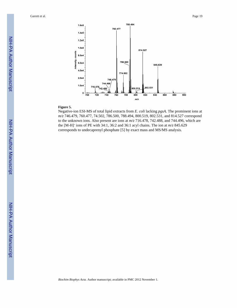
Figure 5.Negative-ion ESI-MS of total lipid extracts from E. coli lacking pgsA. The prominent ions atm/z 746.479, 760.477, 74.502, 786.500, 788.494, 800.519, 802.531, and 814.527 correspondto the unknown ions. Also present are ions at m/z 716.478, 742.488, and 744.496, which arethe [M-H]- ions of PE with 34:1, 36:2 and 36:1 acyl chains. The ion at m/z 845.629corresponds to undecaprenyl phosphate [5] by exact mass and MS/MS analysis.
Garrett et al. Page 19
Biochim Biophys Acta. Author manuscript; available in PMC 2012 November 1.
NIH
-PA Author Manuscript
NIH
-PA Author Manuscript
NIH
-PA Author Manuscript
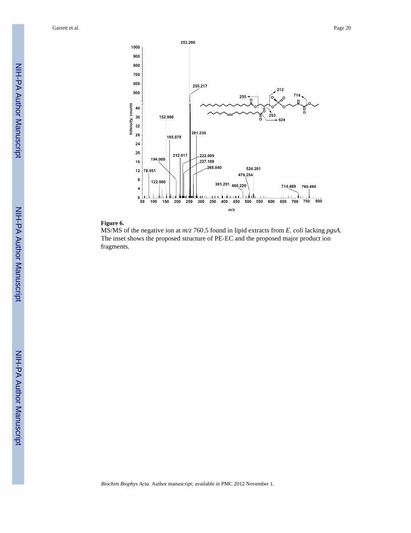
Figure 6.MS/MS of the negative ion at m/z 760.5 found in lipid extracts from E. coli lacking pgsA.The inset shows the proposed structure of PE-EC and the proposed major product ionfragments.
Garrett et al. Page 20
Biochim Biophys Acta. Author manuscript; available in PMC 2012 November 1.
NIH
-PA Author Manuscript
NIH
-PA Author Manuscript
NIH
-PA Author Manuscript
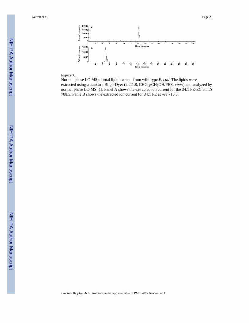
Figure 7.Normal phase LC-MS of total lipid extracts from wild-type E. coli. The lipids wereextracted using a standard Bligh-Dyer (2:2:1.8, CHCl3/CH3OH/PBS, v/v/v) and analyzed bynormal phase LC-MS [1]. Panel A shows the extracted ion current for the 34:1 PE-EC at m/z788.5. Panle B shows the extracted ion current for 34:1 PE at m/z 716.5.
Garrett et al. Page 21
Biochim Biophys Acta. Author manuscript; available in PMC 2012 November 1.
NIH
-PA Author Manuscript
NIH
-PA Author Manuscript
NIH
-PA Author Manuscript
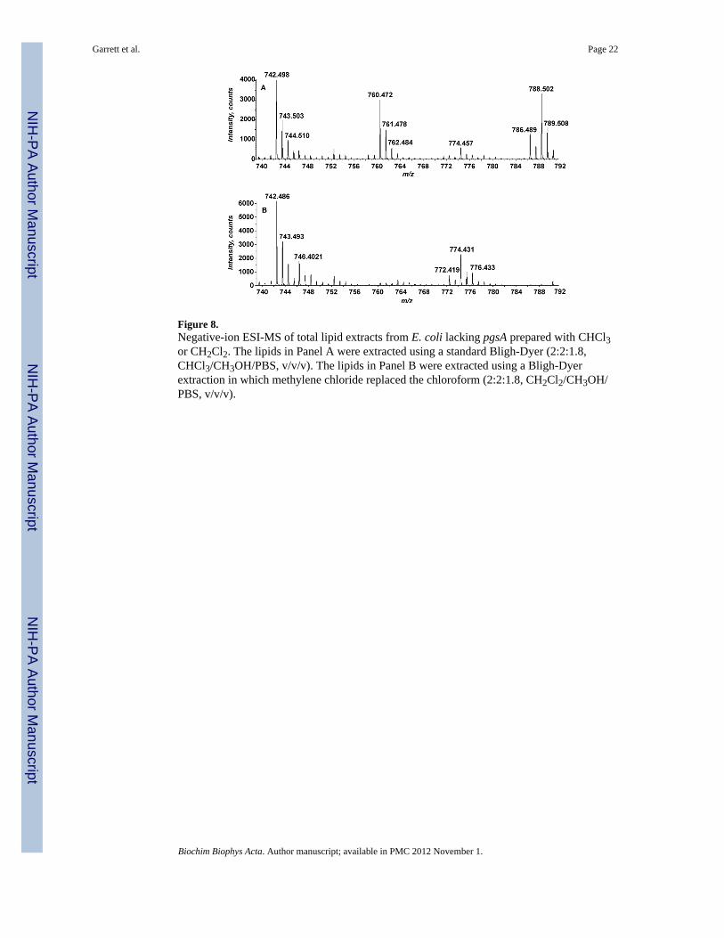
Figure 8.Negative-ion ESI-MS of total lipid extracts from E. coli lacking pgsA prepared with CHCl3or CH2Cl2. The lipids in Panel A were extracted using a standard Bligh-Dyer (2:2:1.8,CHCl3/CH3OH/PBS, v/v/v). The lipids in Panel B were extracted using a Bligh-Dyerextraction in which methylene chloride replaced the chloroform (2:2:1.8, CH2Cl2/CH3OH/PBS, v/v/v).
Garrett et al. Page 22
Biochim Biophys Acta. Author manuscript; available in PMC 2012 November 1.
NIH
-PA Author Manuscript
NIH
-PA Author Manuscript
NIH
-PA Author Manuscript
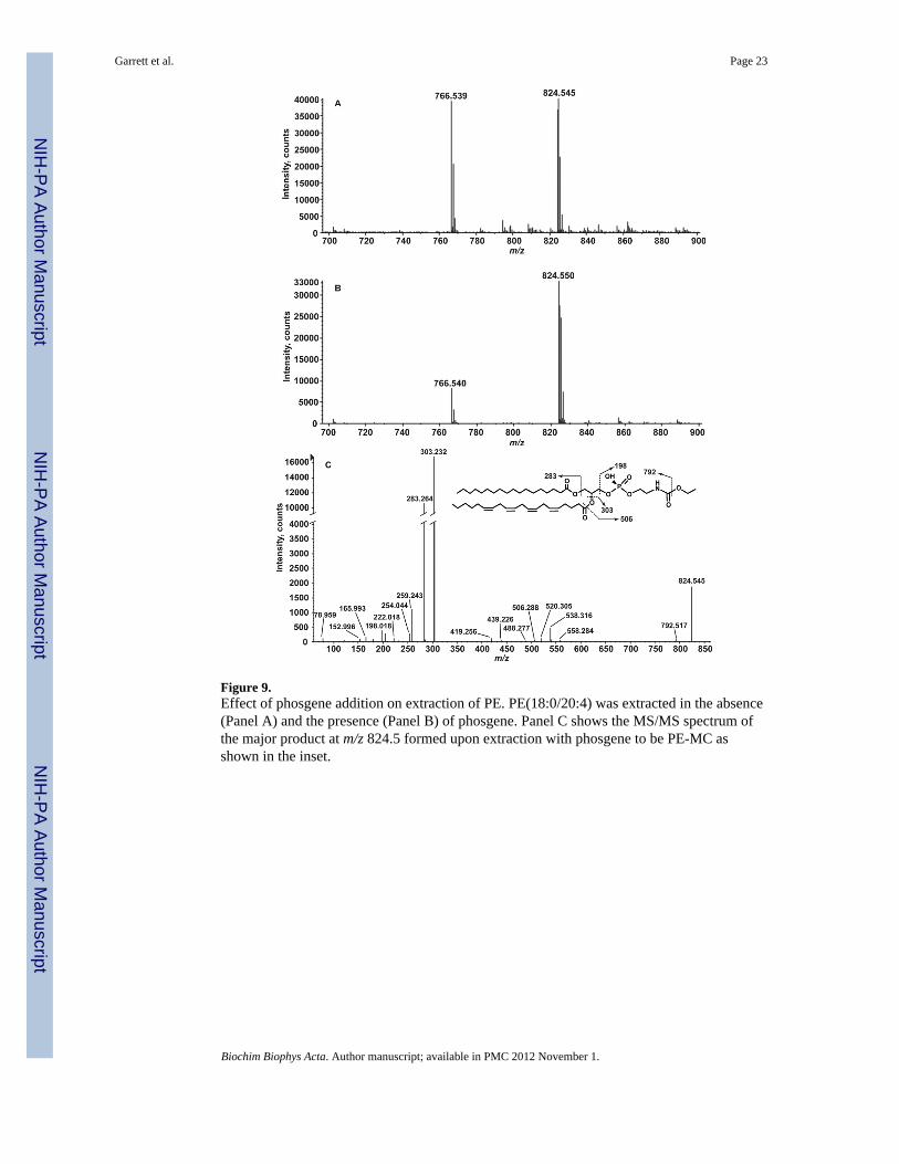
Figure 9.Effect of phosgene addition on extraction of PE. PE(18:0/20:4) was extracted in the absence(Panel A) and the presence (Panel B) of phosgene. Panel C shows the MS/MS spectrum ofthe major product at m/z 824.5 formed upon extraction with phosgene to be PE-MC asshown in the inset.
Garrett et al. Page 23
Biochim Biophys Acta. Author manuscript; available in PMC 2012 November 1.
NIH
-PA Author Manuscript
NIH
-PA Author Manuscript
NIH
-PA Author Manuscript

Figure 10.Proposed mechanism for the phosgene dependent formation of PE-EC, PE-MC, and PE-glutathione. Compound 1: PE-MC, compound 2: PE-EC, and compound 3: PE-glutathione.
Garrett et al. Page 24
Biochim Biophys Acta. Author manuscript; available in PMC 2012 November 1.
NIH
-PA Author Manuscript
NIH
-PA Author Manuscript
NIH
-PA Author Manuscript

Figure 11.Negative-ion ESI-MS of L2 E. coli lipid extracts. The ions at m/z 786.488, 788.506,802.526, 814.528 and 816.538 correspond to unknown ions.
Garrett et al. Page 25
Biochim Biophys Acta. Author manuscript; available in PMC 2012 November 1.
NIH
-PA Author Manuscript
NIH
-PA Author Manuscript
NIH
-PA Author Manuscript
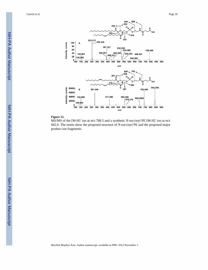
Figure 12.MS/MS of the [M-H]- ion at m/z 788.5 and a synthetic N-succinyl PE [M-H]- ion at m/z842.6. The insets show the proposed structure of N-succinyl PE and the proposed majorproduct ion fragments.
Garrett et al. Page 26
Biochim Biophys Acta. Author manuscript; available in PMC 2012 November 1.
NIH
-PA Author Manuscript
NIH
-PA Author Manuscript
NIH
-PA Author Manuscript

Figure 13.Formation of N-succinyl PE from succinyl CoA and PE in the absence of protein. 14C-PEwas incubated for 1 hour at 30 °C in the presence or absence of succinyl CoA, wild-type E.coli cell-free extracts or washed membranes. The asterisk indicates the N-succinyl-14C-PEproduct. This product co-migrates with N-succinyl PE synthetic standard.
Garrett et al. Page 27
Biochim Biophys Acta. Author manuscript; available in PMC 2012 November 1.
NIH
-PA Author Manuscript
NIH
-PA Author Manuscript
NIH
-PA Author Manuscript

Figure 14.N-succinyl PE is formed from succinyl CoA and PE but not other CoA esters or succinicanhydride. PE (0.5 mM) was incubated for 2 hours at 30 °C in a reaction containing 20 mMHepes pH 7.4, 0.2 % Triton X-100, and 2 mM of a CoA thioester as indicated; Lane 1, noCoA thioester added, Lane 2, succinyl-CoA, Lane 3, succinic anhydride, Lane 4, glutaryl-CoA, Lane 5, malonyl-CoA. Lane 6 is N-succinyl PE synthetic standard.
Garrett et al. Page 28
Biochim Biophys Acta. Author manuscript; available in PMC 2012 November 1.
NIH
-PA Author Manuscript
NIH
-PA Author Manuscript
NIH
-PA Author Manuscript

Figure 15.Proposed mechanism for the formation of N-succinyl PE.
Garrett et al. Page 29
Biochim Biophys Acta. Author manuscript; available in PMC 2012 November 1.
NIH
-PA Author Manuscript
NIH
-PA Author Manuscript
NIH
-PA Author Manuscript
Related Documents