ELSEVIER Journal of Pharmaceutical and Biomedical Analysis 15 (1997) 901-910 JOURNAL OF PHARMACEUTICAL AND BIOMEDICAL ANALYSIS NMR and HPLC-NMR spectroscopic studies of futile deacetylation in paracetamol metabolites in rat and man Andrew W. Nicholls ", R. Duncan Farrant b John P. Shockcor c, Steve E. Unger c, Ian D. Wilson d, John C. Lindon ~"*, Jeremy K. Nicholson :' '~ Department of' CTwmistry, Birkbeek College, University of London, Gordon House, 29 Gordon Square, London WCIH OPP, UK b Physical Sciences Research Unit, GlaxoWelleome Medicines Research Centre, Gunnels Wood Road, Stevenage SGI 2NY, UK Bioanalysis and Drug Metabolism, Glaxo Welh'ome Inc., 5 Moore Drit~e, Research Triangle Park~ NC 27709, USA a Department q[" Safiqy qf' Medicines, Zeneca Pharmaceuticals. Mereside, AIderley Park, Macelesfield, CTwshire SKIO 4TG, UK Received 5 August 1996: accepted 16 September 1996 Abstract HPLC-NMR spectroscopy has been used to investigate the level of deacetylation followed by reacetylation (futile deacetylation) of metabolites of paracetamol detected in human and rat urine. This has been achieved through the synthesis and administration of paracetamol isotopically labeled at the acetyl group with C2H3, 13CH3 and :3CO-13CH3. Using paracetamol-C2H3 it had been shown that in the rat the sulphate metabolite present in the urine shows 10 13% futile deacetylation depending on the dose, whereas for paracetamol-~3CO -13CH3 the corresponding value was about 8%. After solid phase extraction, it was also possible to determine the level of futile deacetylation in the glucuronide metabolite using directly-coupled HPLC-NMR. This approach was facilitated by the use of acetonitrile-d 3 as an HPLC eluent and the HPLC-NMR analyses showed that the level of futile deacetylation in the sulphate and glucuronide metabolites were equal at about 9%. The glucuronide of paracetamol-C2H3 was the predominant metabolite in man and following separation using HPLC-NMR, the level of futile deacetylation was shown to be 1% for the glucuronide and 2% for the sulphate, these values being equal within experimental error. This work demonstrates the utility of NMR and HPLC-NMR spectroscopy for isotope exchange studies. © 1997 Elsevier Science B.V. Keywords: Futile deacetylation; Paracetamol: 4-acetamidophenol; Metabolites; Rat; Human; HPLC-NMR; 2H-label- ing; :~C-labeling 1. Introduction Paracetamol (4-acetamidophenol, acetamino- phen) is one of the most widely used anti-pyretic, * Corresponding author. Tel.: + 44 171 3807527; fax: + 44 171 3807464. analgesic compounds currently available. The ma- jor metabolites of paracetamol at the therapeutic dose level are paracetamol sulphate and glu- curonide and L-cysteinyl and N-acetyl-L-cysteinyl conjugates (Fig. 1). Of these paracetamol sulphate and paracetamol glucuronide form the largest proportion and other minor metabolites 0731-7085/97/$17.00 @ 1997 Elsevier Science B.V. All rights reserved. PII $0731 -7085(96)01 950-4

Welcome message from author
This document is posted to help you gain knowledge. Please leave a comment to let me know what you think about it! Share it to your friends and learn new things together.
Transcript
ELSEVIER Journal of Pharmaceutical and Biomedical Analysis
15 (1997) 901-910
JOURNAL OF PHARMACEUTICAL AND BIOMEDICAL
ANALYSIS
NMR and HPLC-NMR spectroscopic studies of futile deacetylation in paracetamol metabolites in rat and man
Andrew W. Nicholls ", R. Duncan Far rant b John P. Shockcor c, Steve E. Unger c, Ian D. Wilson d, John C. Lindon ~"*, Jeremy K. Nicholson :'
'~ Department of' CTwmistry, Birkbeek College, University of London, Gordon House, 29 Gordon Square, London WCIH OPP, UK b Physical Sciences Research Unit, GlaxoWelleome Medicines Research Centre, Gunnels Wood Road, Stevenage SGI 2NY, UK
Bioanalysis and Drug Metabolism, Glaxo Welh'ome Inc., 5 Moore Drit~e, Research Triangle Park~ NC 27709, USA a Department q[" Safiqy qf' Medicines, Zeneca Pharmaceuticals. Mereside, AIderley Park, Macelesfield, CTwshire SKIO 4TG, UK
Received 5 August 1996: accepted 16 September 1996
Abstract
HPLC-NMR spectroscopy has been used to investigate the level of deacetylation followed by reacetylation (futile deacetylation) of metabolites of paracetamol detected in human and rat urine. This has been achieved through the synthesis and administration of paracetamol isotopically labeled at the acetyl group with C2H3, 13CH3 and :3CO-13CH3. Using paracetamol-C2H3 it had been shown that in the rat the sulphate metabolite present in the urine shows 10 13% futile deacetylation depending on the dose, whereas for paracetamol-~3CO -13CH3 the corresponding value was about 8%. After solid phase extraction, it was also possible to determine the level of futile deacetylation in the glucuronide metabolite using directly-coupled HPLC-NMR. This approach was facilitated by the use of acetonitrile-d 3 as an HPLC eluent and the HPLC-NMR analyses showed that the level of futile deacetylation in the sulphate and glucuronide metabolites were equal at about 9%. The glucuronide of paracetamol-C2H3 was the predominant metabolite in man and following separation using HPLC-NMR, the level of futile deacetylation was shown to be 1% for the glucuronide and 2% for the sulphate, these values being equal within experimental error. This work demonstrates the utility of NMR and HPLC-NMR spectroscopy for isotope exchange studies. © 1997 Elsevier Science B.V.
Keywords: Futile deacetylation; Paracetamol: 4-acetamidophenol; Metabolites; Rat; Human; HPLC-NMR; 2H-label- ing; :~C-labeling
1. Introduction
Paracetamol (4-acetamidophenol, acetamino- phen) is one o f the most widely used anti-pyretic,
* Corresponding author. Tel.: + 44 171 3807527; fax: + 44 171 3807464.
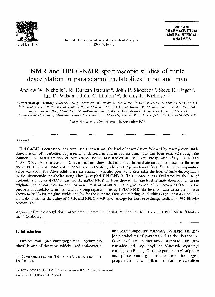
analgesic compounds currently available. The ma- jor metabolites o f paracetamol at the therapeutic dose level are paracetamol sulphate and glu- curonide and L-cysteinyl and N-acetyl-L-cysteinyl conjugates (Fig. 1). Of these paracetamol sulphate and paracetamol glucuronide form the largest p ropor t ion and other minor metabolites
0731-7085/97/$17.00 @ 1997 Elsevier Science B.V. All rights reserved. PII $0731 -7085(96)01 950-4
902 A.W. Nicholls et al./J. Pharm. Biomed. Anal. 15 (1997) 901 910
~H3
H,NtC~ O
OH 4-acetamidophenol (paracetamol)
CH3 H,N,C<- O
[ ~ 4-acetamidophenol sulphate (paracetamol sulphate)
OSO3" OH 3
/ H,N,C.~ 0
COOH H O ~ I Ho-~O
OH
CH3 H.N/C~'- O
~ S NH2 OH
COOH 3-(L-cysteinyl)-4-acetamidophenol
4-acetamidophenol glucuronide (paracetamol glucuronide)
CH3 H,N~C-.~ O
"~OH "Sx__~N- C- CH3 COOH
3-(N-acetyl- L-cysteinyl)-4-acetamidophenol
Fig. 1. The structure of paracetamol and its principal metabolites.
may also be observed, but usually only following drug overdose [1].
In humans, high doses of paracetamol can re- sult in hepatotoxicity, with occasional observa- tions of nephrotoxicity [2]. One of the proposed mechanisms of the kidney damage involves the formation of 4-aminophenol, a known nephro- toxin that causes severe and acute necrosis of the kidney proximal tubules [3]. For this to occur, paracetamol is required to undergo deacetylation, but this process has generally been considered to be only of minor importance due to the low fraction of the paracetamol dose excreted as 4- aminophenol [4]. However, it has previously been observed that 4-aminophenol may become reacetylated back to paracetamol prior to excre- tion [5]. This transacetylation or, more correctly, 'futile deacetylation' process has been shown for paracetamol (in rats and man) using GC-MS [5] and inferred by the reduced deacetylation of 4- acetaminohippurate in the presence of paraceta- mol and the reduced acetylation of 4-aminohip- purate in the presence of 4-aminophenol [6]. Studies using 14C-radiolabelling have also been
used in an effort to quantify the extent of this metabolic route for paracetamol [7]. However, until recently, no measurement of the futile deacetylation of the individual paracetamol metabolites has been made, nor has NMR been utilised in the investigation of such metabolic processes. Moreover, deficiencies in the recovery of the drug from these earlier studies has not led to an accurate measure of the level of futile deacetylation of paracetamol.
Recently it has been shown that, in the rat, there is a significant level of futile deacetylation which can be detected in the sulphate metabolite [8] and here an extension to the study is reported in which levels of futile acetylation have been determined in both rat and man for isotopically- labelled paracetamol sulphate and glucuronide metabolites. This has been achieved through the use of directly-coupled HPLC-NMR spectroscopy to separate and characterise the metabolites and to quantify the proportions of the different iso- topically labelled metabolites in the chromato- graphic peaks. Directly-coupled HPLC-NMR spectroscopy has been demonstrated to have widespread potential for the identification of drug

A.W. Nicholls et al. /J. Pharm. Biomed. Anal. 15 (1997) 901 910 903
Table 1 The percentage futile deacetylation (FD) for paracetamol sulphate and glucuronide conjugates and the percentage total recovery of all paracetamol metabolites in rat and human urine
Compound Dose (mg kg- ~)/species % FD sulphate % FD glucuronide % Total recovery in urine
Paracetamol-C-'H 3 25/rat 13.2 _+ 0.2 ND 55.6 _+ 12.3 Paracetamol-C2H3 40/rat 9.98 ± 0.86 ND 60.6 + 8.8 Paracetamol-~3CH3 40/rat 8.87 _+ 0.51 ND 97.8 _+ 3.5 Paracetamol-~3CO~3CH3 40/rat 9.80 ± (/.35 ND 75.0 _+ 3.7 Paracetamol-~COI3CH3 100/rat 7.9 + 1.8 6.61 _+ 0.1 47.4 _+ 11.3 Paracetamol-~COI3CH3 100/rat 9. I "b 5.0 "'b 47.4 _+ 11.3 Paracetamol-~COI3CH3 100/rat 9.2 .... 9.1 ..... 47.4 + 11.3 ParacetamoI-C2H~ 4.3/human 1.0 ~' 2.0" 45.6 '~ Paracetamol 4.3/human 22.6 ~'
~' Single measurement. b After 600 MHz HPLC-NMR separation using methanol/D20 elution. c After 500 MHz HPLC-NMR separation using acetonitrile-d~/D20 elution. ND, not detected.
metabolites [9,10] and can be carried out either in continuous-flow detection or in stop-flow mode. Here stop-flow HPLC-NMR spectroscopy has been used to study the level of futile deacetylation in both sulphate and glucuronide metabolites of paracetamol.
until NMR urinalysis. Prior to NMR spectro- scopic studies, 1.0 ml aliquots of each urine were freeze-dried and were studied at a four-fold con- centration.
2.3. HPLC-NMR analysis of SPEC fractions of human and rat urine
2. Experimental
2.1. Synthesis of paracetamol-C2H~ and paracetamol-13CO -13Ctt~ and methods for in vivo rat studies
These compounds were synthesised and solid phase extraction chromatography (SPEC) and metabolism studies in the rat were carried out as described previously [8]. The dose levels for the rat studies are given in Table 1.
2,2. Procedures for in vivo human studies
A male volunteer was administered orally with a 300 mg dose of paracetamol-C2H3 correspond- ing to 4.3 mg k g - 1. Urine was collected pre-dose, 0 - 2 and 2 - 4 h post-dose. For comparison, the same volunteer also received a 300 mg dose of non-deuterated authentic paracetamol and urine was collected for the same time intervals All sam- ples were weighed and the urine frozen at - 19°C
Solid-phase extraction chromatography was carried out on a 1 ml C18 Bond-Elut TM column pretreated with methanol and acidified water (pH 2). The acidified sample was loaded onto the column and eluted with 1 ml acidified water and subsequently l ml methanol under vacuum. The HPLC system comprised a Bruker LC22C pump and a Bischoff 1000 Lambda variable-wavelength UV detector (operating at 250 nm). The outlet from the UV detector was connected to the HPLC-NMR flow probe via an inert polyether(ether ketone) capillary (0.25 mm i.d.). A column oven was used to maintain a column temperature at 25°C. Data were collected using the Bruker Chromstar HPLC data system. Analy- sis was performed on a Knauer column (120 x 4.6 mm i.d.) packed with Spherisorb ODS-2, 5 ~m. The mobile phase consisted of D20-0.05M sodium dihydrogenphosphate (pH 2) (99:1, v/v) for the first 5 min followed by a linear gradient to D20-methanol-0.05M sodium dihydrogenphos- phate (pH 2) (49:50:1, v/v/v) after 30 rain with a flow rate of 1.0 ml rain 1
904 A.W. Nicholls et al./J. Pharm. Biomed. Anal. 15 (1997)901 910
HPLC-NMR spectra were acquired using a Bruker AMX-600 spectrometer equipped with a ]H flow probe (cell of 3 mm i.d. with a volume of 100 lal). ~H NMR spectra were obtained in stop- flow mode at 600.14 MHz. Spectra were acquired using the NOESYPRESAT (Bruker GmbH, Rhe- instetten, Germany) pulse sequence with double presaturation of the water and methanol signals. FIDs were collected into 64K data-points, using a 90 ° pulse with an acquisition time of 2.69 s, a recycle delay of 4.79 s and a spectral width of 12 195.2 Hz, for 64 scans. An exponential line- broadening function of 0.4 Hz was applied prior to FT.
2.4. H P L C - N M R analysis o f whole rat urine
The HPLC system comprised a Hewlett Pack- ard 1050 Series pump operating at 21°C and a variable-wavelength UV detector (operating at 210 nm). The outlet from the UV detector was connected to the HPLC-NMR flow probe via an inert polyether(ether ketone) capillary. Data were collected using the Bruker Chromstar HPLC data system. Analysis was performed on a Waters Symmetry C18 column (3 .9x150 mm i.d.) packed with Spherisorb ODS-2, 3 gm. The mobile phase consisted of D20-TFA (pH 2)/acetonitrile- d3 increasing from 100:0 at the beginning of the run to 50:50 after 30 min at 1.0 ml min ~.
HPLC-NMR spectra were acquired using a Bruker DMX-500 equipped with a 'H flow probe (cell of 3 mm i.d. with a volume of 120 gl). 'H NMR spectra were obtained in stop-flow mode at 500.13 MHz. Presaturation of the water signal was achieved by the use of a selective sinc pulse applied for six cycles of 100 ms each. FIDs were collected into 32 K data-points, using a 90 ° pulse with an acquisition time of 3.28 s, a recycle delay of 0.6 s and a spectral width of 5000 Hz, for 4096 scans. An exponential line-broadening function of 1.0 Hz was applied prior to FT and the data were zero-filled by a factor of 2.
2.5. Analysis" o f N M R spectra containing paracetamol metabolites
The regions of the ]H NMR spectrum corre-
sponding to the aromatic and acetyl signals of the paracetamol metabolites were integrated. The ex- tent of futile deacetylation was calculated by inte- gration of the CH3 acetyl signal relative to those of the aromatic H3 and H5 protons of the parac- etamol metabolite concerned.
3. Results
3.1. Direct measurement o f paracetamol-C2H3 metabolites in rat urine using N M R
Comparison of the ]H NMR spectra of the post-dose 0 8 h rat urine and pre-dose rat urine samples clearly showed the aromatic signals (AA' XX' spin system) at 67.33 and 7.47 of the parac- etamol-C2H3 sulphate. Expansion of the spectrum in the region c~1.75-2.30 revealed a number of peaks resulting from the appearance of signals from acetyl groups which were not present in the urine of the rat prior to dosing with paracetamol- C2H3 . New resonances include the cysteine acetyl group of the N-acetyl cysteinyl metabolite at gl.87 and a signal at (52.18, previously assigned to the acetyl group of paracetamol sulphate based on the chemical shift of authentic material [11]. The presence of the sulphate acetyl signal indi- cates that in vivo there is a significant degree of deacetylation of the CO.C2H3 group and reacety- lation with CO.CH 3. The mean level of futile deacetylation was dose dependent as shown in Table 1 being about 13% at the lower dose and 10% at the higher dose.
The proton aromatic resonances at cS7.37 and c~7.15 (AA' XX' spin system) indicated the pres- ence in the urine of small amounts of the parac- etamol glucuronide. The signal at d7.37 was partially overlapped with a signal from an uniden- tified endogenous metabolite, so only the signal at ~7.15 was used for quantification. No ~H reso- nances from a paracetamol glucuronide acetyl group were observed possibly because the signal was too small to be measured under the N MR acquisition conditions used. The relatively small quantity of glucuronide metabolite observed cor- responded to 6.9_+ 1.7% of the dose because of
A.W. Nicholls et al . /J . Pharm. Biomed. Anal. 15 (1997) 901-910 905
the high capacity for sulphation in the rat which dominates the metabolic profile.
There was no tH N M R evidence for the pres- ence of either 4-aminophenol or paracetamol- C2H3 itself in the urine. These results confirmed previous observations of futile deacetylation in vivo in the rat. They additionally, via the use of high field NMR, identified the futile deacetylation process in the paracetamol sulphate metabolite. However, observations of a low overall percent- age urinary recovery of the metabolites, prompted repetition of the experiment at an elevated parac- etamol-C2H3 dose level of 40 mg kg ~ which showed little change in the observed urinary re- covery (see Table 1). In addition paracetamol- 13CH3 was also dosed to determine whether or not deuterium kinetic isotope effects [12] had affected either the level of futile deacetylation or the over- all urinary recovery of the drug since kinetic isotope effects on metabolism from t3C-labelled compounds are generally small [12]. This experi- ment showed that whilst the level of futile deacetylation remained essentially unchanged at 8.9%, the recovery of metabolites in the urine increased from 55-60 to 97.8%.
3.2. N M R studies on the metabolism of paracetamol-~3CO t3CH3 in rat urine
The 600 MHz ~H NMR spectrum of rat urine taken 0 8 h after dosing with paracetamol-13CO ~3CH3 at 100 mg kg ~ has also been examined. The aromatic resonances from the paracetamol sulphate were again visible at 67.45 and 67.31 with the glucuronide conjugate aromatic reso- nances identified at 67.36 and 67.14 (the former being overlapped by an unidentified endogenous signal). A resonance at 66.90 was assigned to the parent compound and an acetyl signal at 61.87 was assigned to the N-acetyl-L-cysteinyl metabo- lite. However, no aromatic signals were observed for this metabolite due to its low concentration. Doublet of doublets signals for the acetyl groups (qcH = 129 Hz and 2JcH = 5.9 Hz) of the non- deacetylated metabolites were seen at 62.24-2.29 and 62.03 2.08.
The paracetamol sulphate acetyl signal resulting from futile deacetylation was visible as a singlet at
62.18. Integration of this peak gave a mean per- centage futile deacetylation level for the paraceta- mol sulphate of 7.9_+ 1.8%. An acetyl singlet, overlapped by the sulphate signal, was assigned to the paracetamol glucuronide at 62.17 and this was resolved via Gaussian resolution enhance- ment. Integration gave a mean percentage futile deacetylation of 6.6 _+ 0.1.
The results obtained at a dose level of 40 mg kg ~ for all of the different types of labelled paracetamol do not appear to be significantly different showing that there is no isotope effect on the futile deacetylation process. Moreover, the endogenous process causing the futile deacetyla- tion would also appear to be unaffected by the potential level of substrate available to it. The use of the doubly labelled paracetamol-~3CO-~3CH3 allowed the unusual and unlikely possibility that futile demethylation as well as deacetylation was taking place to be distinguished. However, since no acetyl NMR resonance showing the loss of the one-bond JcH and retention of the two-bond JCH was observed for futile deacetylated products, the level of any futile demethylation must be at most only a very minor process. As a result of the high dose level used in the study with paracetamol- 13 13 -CO- ~CH 3 the glucuronide conjugate was also detected and seen to undergo futile deacetylation. It could not be determined from this study whether or not there is a significant difference in the percentage futile deacetylation between the glucuronide and sulphate metabolites. Since no J3C-coupled NMR acetyl peaks were observed, the process in the glucuronide would also appear to be futile deacetylation.
3.3. H P L C - N M R studies on the metabolism of paracetamol-13CO -13CH~ in rat urine
To provide additional evidence of the futile deacetylation in both major metabolites, a sample of the 0 -8 h urine following dosing of paraceta- mol-13CO J3CH3 at 100 mg kg ~ was first purified using SPEC (acidified water and 100% methanol fractions). The paracetamol and its metabolites were observed in the acidified water fraction and this was further analysed using HPLC-NMR at 600 MHz.
906 A.W. Nicholls et al. /J. Pharm. Biomed. Anal. 15 (1997) 901-910
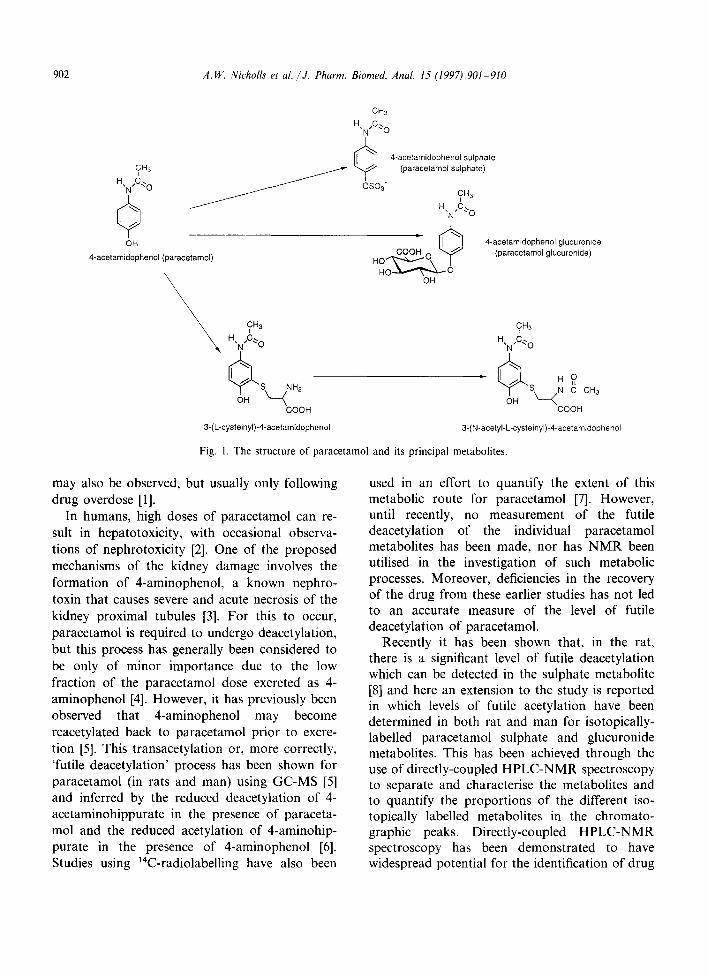
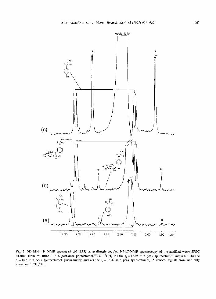
Shown in Fig. 2(a) is the 600 MHz 1H NMR spectrum obtained using directly-coupled HPLC- NMR in stop-flow mode corresponding to the UV absorbance peak at a retention time of 13.05 min. Although methanol was used as the organic modifier in the HPLC, a detectable level of ace- tonitrile also leached from the HPLC column. This caused the appearance of a peak in the acetyl region of the ~H NMR spectrum. This paraceta- mol metabolite was assigned as paracetamol sul- phate by comparison to the NMR spectrum of the whole urine. The aromatic resonances were visible at 37.45 and 7.31 whereas the doublet of doublets corresponding to the non-futile deacetylated paracetamol sulphate was centred around ~52.17. An acetyl singlet was observed at 62.17 corre- sponding to the acetyl group of the futile deacety- lated paracetamol sulphate and by integration relative to the aromatic resonances, it was deter- mined that 9.1% futile deacetylation had oc- curred. No evidence was observed for any futile demethylation in the paracetamol sulphate.
The 600 MHz ~H NMR spectrum correspond- ing to the UV absorbance peak at a retention time of 14.5 min is shown in Fig. 2(b). Again by comparison with the whole urine spectrum, this was assigned to the glucuronide metabolite. The aromatic resonances were visible at 67.35 and 7.13, whereas the acetyl resonance doublet of doublets of the non-futile deacetylated metabolite was centred around ~2.15. An acetyl singlet was observed at ~2.15 corresponding to the futile deacetylated paracetamol glucuronide. Integration of this signal relative to that of the aromatic resonances gave a futile deacetylation level of 5.0%. This figure is probably less precise than that for the sulphate because of the lower concentra- tion of the glucuronide and the consequent difficulty of NMR integration.
The 600 MHz ~H NMR spectrum of the UV absorbance peak at a retention time of 18.42 min was assigned to the parent compound and is shown in Fig. 2(c). The aromatic resonances were observed at c~7.26 and 66.90. The acetyl doublet of doublets was centred around cf2.14. No acetyl peak corresponding to futile deacetylated parac- etamol could be observed due to the large reso- nance caused by the residual acetonitrile on the
column and the low level of free paracetamol in the urine.
As an alternative to the use of methanol in the HPLC eluent, HPLC-NMR studies were also car- ried out on whole concentrated urine from rats dosed at 100 mg kg ~ using acetonitrile-d3 as the organic solvent phase. In the stop-flow mode us- ing 500 MHz IH NMR spectroscopy, spectra were obtained from both glucuronide and sulphate metabolites which gave acetyl resonances from the transacetylated products, these peaks now being observable through the use of deuterated acetoni- trile thereby removing a large solvent resonance in the same spectral region. The levels of futile deacetylation were 9.2% for the sulphate and 9.1% for the glucuronide metabolites.
3.4. N M R studies on the metabolites o f paracetamol-C2H3 present & human urine
Following the results obtained from experi- ments in the rat, studies into futile deacetylation in man were undertaken using both paracetamol and paracetamol-CZH3 . Both the 0-2 and 2-4 h urine following dosing of 300 mg of paracetamol- C2H3 contained drug metabolites. The glu- curonide conjugate was the predominant metabolite with aromatic proton resonances at c57.37 and ~7.14, whereas resonances for the sul- phate were observed at c57.46 and 67.33. The recovery of the metabolites for the total 4 h period as a percentage of the dose was 33.3 and 12.3% for the glucuronide and sulphate respec- tively. Following administration of 300 mg of paracetamol to the same subject, the correspond- ing figures were 16.4% for the glucuronide and 6.2% for the sulphate. No acetyl signals for the two metabolites were observed because of overlap from NMR resonances from endogenous com- pounds. Solid phase extraction followed by NMR spectroscopy (SPEC-NMR) of the 2-4 h urine yielded the paracetamol metabolites in the aci- dified water fraction. Although some endogenous metabolites had been removed, the spectral clarity of the acetyl region had not been sufficiently improved to accurately identify any signals result- ing from futile deacetylation. Therefore, due to the background interference in the acetyl region
A.W. Nicholls et al. /"J. Pharm. Biomed. Anal. 15 (1997) 901-910 907
Acetonitrite
"CH~
(c) L . /
rh
"CH. ---------___.._._____
" ':c"° ~ c. f HO COOH O
H ~C "O
"CH~ H ,N'~C,~ 0 CH 3
H N,~.~ 0
OSO~"
/ /
rh
-k
I . . . . I . . . . I . . . . I . . . . ~ . . . . i . . . . I . . . . t . . . . I . . . . I
2.30 2.25 2.20 2.15 2.10 2.05 2.00 1.95 ppm
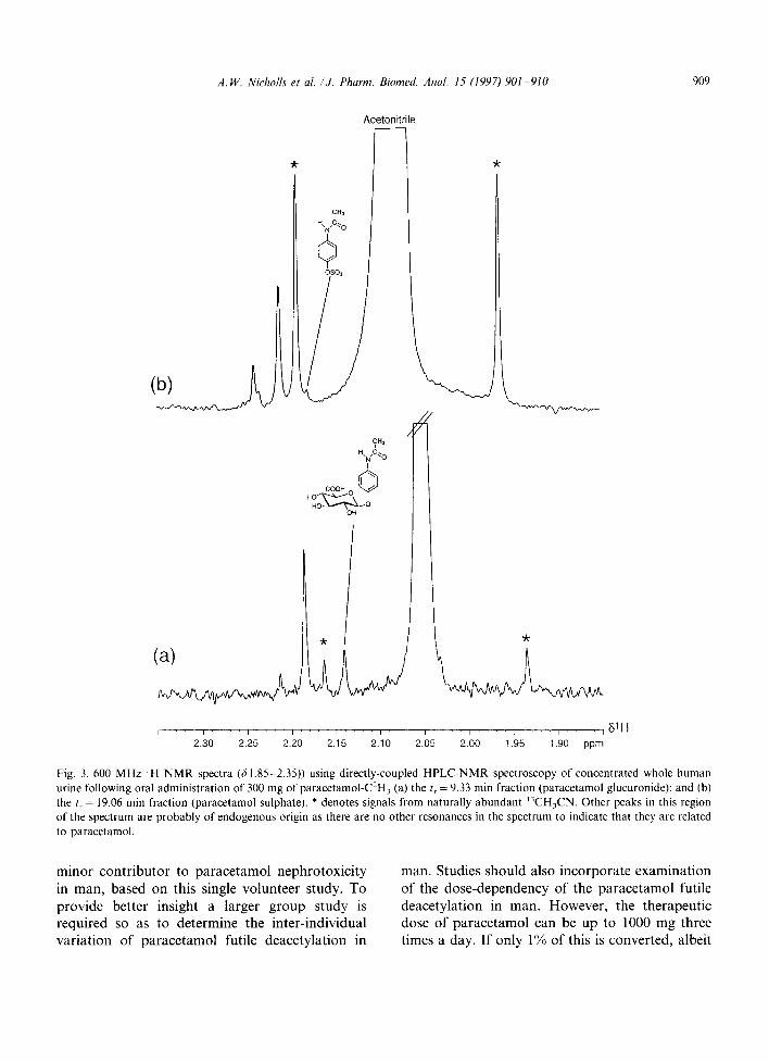
Fig. 2. 600 MHz ~H NMR spectra (51.90 2.35) using directly-coupled HPLC-NMR spectroscopy of the acidified water SPEC fraction from rat urine 0-8 h post-dose paracetamolJ3CO ~3CH3 (a) the t r= 13.05 rain peak (paracetamol sulphate); (b) the t r= 14.5 min peak (paracetamol glucuronide); and (c) the t r = 18.42 min peak (paracetamol). * denotes signals from naturally abundant 13CH3CN.
908 A.W. Nicholls et al. /J. Pharm. Biomed. Anal. 15 (1997) 901-910
and the potential low level of futile deacetylated drug, it was necessary to use HPLC-NMR spec- troscopy for quantitative purposes.
3.5. H P L C - N M R spectroscopic analysis" o f the metabolites o f paracetamol-C2H3 in human urine
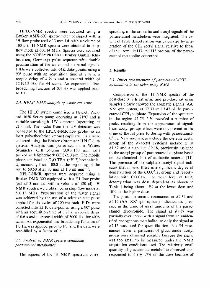
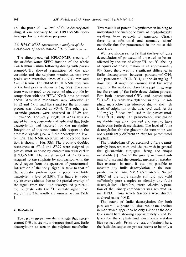
The directly-coupled HPLC-NMR spectra of the acidified-water SPEC fraction of the whole 2-4 h human urine following dosing with parac- etamol-C2H3 showed separation of the glu- curonide and the sulphate metabolites into two peaks with retention times of t = 9.33 rain and t = 19.06 min. The 600 MHz ~H NMR spectrum of the first peak is shown in Fig. 3(a). The spec- trum was assigned to paracetamol glucuronide by comparison with the HPLC-NMR data obtained above. Aromatic resonances were observed at (57.32 and /57.11 and the signal for the anomeric proton was observed at (55.09. The other glu- curonide protons were observed at (53.89 and (53.65-3.55. The acetyl singlet at (52.14 was as- signed to the glucuronide and indicated that futile deacetylation had occurred for the metabolite. Integration of this resonance with respect to the aromatic signals gave a futile deacetylation level of 1.0%. The NMR spectrum of the second frac- tion is shown in Fig. 3(b). The aromatic doublet resonances at 67.42 and (57.27 were assigned to paracetamol sulphate by comparison with earlier HPLC-NMR. The acetyl singlet at (52.13 was assigned to the sulphate by comparison with the acetyl region from the spectrum of paracetamol. Integration of the acetyl signal relative to that of the aromatic protons gave a percentage futile deacetylation level of 2.0%. This figure is proba- bly an over-estimate due to the partial overlap of the signal from the futile deacetylated paraceta- mol sulphate with the ~3C satellite signal from acetonitrile. The results are summarised in Table 1.
4. Discussion
The results given here demonstrate that parac- etamol-CZH3 in the rat undergoes significant futile deacetylation as seen in the sulphate metabolite.
This result is of potential significance in helping to understand the metabolic basis of nephrotoxicity resulting from paracetamol ingestion. Clearly there is a substantial and normally 'silent' metabolic flux for paracetamol in the rat at this dose level.
We have shown earlier [8] that the level of futile deacetylation of paracetamol appeared to be un- affected by the use of either 2H- or J3C-labelling at equivalent doses, remaining at approximately 9%. Since there was no significant difference in futile deacetylation between paracetamol-C2H3 and paracetamol-~3CO-~3CH3 at the 40 mg kg- dose level, it might be assumed that the acetyl region of the molecule plays little part in govern- ing the extent of the futile deacetylation process. For both paracetamol-C2H3 and paracetamol- 13CO-13CH3 futile deacetylation in only the sul- phate metabolite was observed due to the high levels of sulphation at the dose level used. At the 100 mg kg J dose level used in the paracetamol- 13CO.13CH3 study, the paracetamol glucuronide metabolite was also observed and seen to have undergone futile deacetylation. The level of futile deacetylation for the glucuronide metabolite was not significantly different to that for paracetamol sulphate.
The metabolism of paracetamol differs quanti- tatively between man and the rat with in general the glucuronide conjugate being the major metabolite [1]. Due to the greatly increased vol- ume of urine and the complex mixture of metabo- lites excreted in man, it was not possible to measure any futile deacetylation in the non- purified urine using NMR spectroscopy. Simple SPEC of the urine sample still did not yield sufficiently pure samples to identify any futile deacetylation. Therefore, more selective separa- tion of the urinary components was achieved us- ing HPLC, from which fractions were directly analysed using NMR.
The extent of futile deacetylation for both paracetamol sulphate and glucuronide metabolites in man would appear to be only minor at the dose levels used here showing approximately 2 and 1% levels for the sulphate and glucuronide metabo- lites respectively. From the results obtained here, the futile deacetylation process seems to be only a
A.W. Nicholls et al. /J. Pharm. Biomed. Anal. 15 (/997) 901-910 909
(a)
Acetonitrile
F "k
CH, H,N,C,> 0
+
CiH3
H.N,C~ O
COOH ~o'~ °
HO .~..---~-,..~ 0 OH
"k
<
. . . . . . , . . . . L . . , . . . . , . . . . , . . . . , . . . . , . . . . , . . . . , . . . . , D 1 H
2.30 2.25 2.20 2.15 2.10 2.05 2.00 1.95 1.90 ppm
Fig. 3. 600 MHz ~H N M R spectra (~ 1.85-2.35)) using directly-coupled H P L C - N M R spectroscopy of concentrated whole human urine following oral administration of 300 mg of paracetamol-C2H3 (a) the tr = 9,33 min fraction (paracetamol glucuronide); and (b) the t r = 19.06 min fraction (paracetamol sulphate). * denotes signals from naturally abundant '~CH3CN. Other peaks in this region of the spectrum are probably of endogenous origin as there are no other resonances in the spectrum to indicate that they are related to paracetamol.
minor contributor to paracetamol nephrotoxicity in man, based on this single volunteer study. To provide better insight a larger group study is required so as to determine the inter-individual variation of paracetamol futile deacetylation in
man. Studies should also incorporate examination of the dose-dependency of the paracetamol futile deacetylation in man. However, the therapeutic dose of paracetamol can be up to 1000 mg three times a day. If only 1% of this is converted, albeit
910 A.W. Nicholls et al. /J. Pharm. Biomed. Anal. 15 (1997)901-910
possibly transiently, into 4-aminophenol , this represents 30 mg o f this very toxic compound. Whilst most people suffer no ill effects f rom the possible presence o f this process, there may be classes o f patients where for genetic reasons, the process o f futile deacetylation produces toxic ef- fects.
This study demonstrates the use o f directly- coupled H P L C - N M R spectroscopy to study fu- tile deacetylation o f paracetamol metabolites. Even the use o f very high field N M R spec- t roscopy on whole urine samples is limited by the degree o f overlap o f resonances f rom the paracetamol metabolites and endogenous species. Nevertheless, N M R spectroscopy on whole urine can allow quantification o f the futile deacetyla- tion in paracetamol metabolites. However, addi- tional confirmation becomes possible by the use of directly-coupled H P L C - N M R spectroscopy. This has the dual advantage o f confirming that the N M R resonances from the futile deacetylated glucuronide and sulphate metabolites do indeed arise f rom these substances since they co-elute with the isotopically labelled corresponding metabolites. Quantification o f the level o f futile deacetylation is simpler in the H P L C - N M R spec- tra because o f the lack o f resonances f rom en- dogenous species. In addition, this work demonstrates that 1H H P L C - N M R spectroscopy can be used to detect and quantify metabolites even for the low levels observed for humans.
Acknowledgements
We thank the E P S R C and the Wellcome Founda t ion Ltd (now GlaxoWellcome) for a CASE studentship (to AWN) .
References
[1] G.G. Duggin, The biochemistry and pharmacology of antipyretic-analgesics, in J.H. Stewart (Ed.), Analgesic and NSAID-induced Kidney Disease, Oxford Press, Ox- ford, 1993, pp. 5 16.
[2] S.H.L. Thomas, Pharmacol. Ther., 60 (1993) 91 120. [3] C.R. Green, K.N. Ham and J.D. Tange, Br. Med. J., 1
(1969) 162-164. [4] J.F. Newton, M. Yoshimoto, J. Bernstein, G.F. Rush and
J.B. Hook, Toxicol. Appl. Pharmacol., 69 (1983) 291- 306.
[5] J.D. Baty, R.M. Lindsay, W.R. Fox and R.G. Willis, Biomed. Environ. Mass Spec., 16 (1988) 183-189.
[6] H.M. Carpenter and G.H. Mudge, J. Pharmacol. Exp. Ther., 218 (1981) 161 167.
[7] G.E. Smith and L.A. Griffiths, Xenobiotica, 6 (1976) 217-236.
[8] A.W. Nicholls, S.T. Caddick, I.D. Wilson, R.D. Farrant, J.C. Lindon and J.K. Nicholson, Biochem. Pharmacol., 49 (1995) 1155 1161.
[9] J.C. Lindon, J.K. Nicholson and I.D. Wilson, Adv. Chro- matogr., 36 (1995) 315-382.
[10] J.C. Lindon, J.K. Nicholson and I.D. Wilson, Prog. NMR Spectrosc., 29 (1996) 1 49.
[1 l] J.R. Bales, P.J. Sadler, J.K. Nicholson and J.A. Timbrell, Clin. Chem., 30 (1984) 1631 1636.
[12] P.J.H. Jones and S.T. Leatherdale, Clin. Sci., 80 (1991) 277 280.
Related Documents