TCR recognition of peptide/MHC class II complexes and superantigens Eric J. Sundberg 1,* , Lu Deng 2 , and Roy A. Mariuzza 2,* 1 Boston Biomedical Research Institute, Watertown, MA 02472, USA. 2 W.M. Keck Laboratory for Structural Biology, Center for Advanced Research in Biotechnology, University of Maryland Biotechnology Institute, Rockville, MD 20850, USA. Summary Major histocompatibility complex (MHC) class II molecules display peptides to the T cell receptor (TCR). The ability of the TCR to discriminate foreign from self peptides presented by MHC molecules is a requirement of an effective adaptive immune response. Dysregulation of this molecular recognition event often leads to a disease state. Recently, a number of structural studies have provided significant insight into several such dysregulated interactions between peptide/MHC complexes and TCR molecules. These include TCR recognition of self peptides, which results in autoimmune reactions, and of mutant self-peptides, common in the immunosurveillance of tumors, as well as the engagement of TCRs by superantigens, a family of bacterial toxins responsible for toxic shock syndrome. Keywords T cell receptor; major histocompatibility complex; superantigen; peptide antigen; T cell activity 1. TCR RECOGNITION OF PEPTIDE/MHC CLASS II COMPLEXES The immune system has evolved through the requirement to distinguish non-self pathogens from self tissues. Whereas T cell recognition of foreign peptides is essential for immune defense against invading microorganisms, recognition of self peptides may cause autoimmune disease. A third category of T cell epitopes involves self peptides resulting from mutations accumulated during aging or disease [1,2]. However, in terms of T cell recognition, the boundaries separating foreign, self, and altered self epitopes are not necessarily absolute. For example, immunity to cancer can arise from mutations in self proteins that render them visible to T cells [1,2], a process that may also induce autoimmunity [3]. While much is known about TCR recognition of foreign antigens [4], only very recently have the structural and biophysical principles governing TCR recognition of self and mutant self begun to be elucidated. This portion of the review will focus on the latter. © 2007 Elsevier Ltd. All rights reserved. * Address correspondence to EJS (Tel.: 617-658-7882; Fax: 617-972-1761; E-mail: [email protected]) or RAM (Tel.: 240-314-6243; Fax: 240-314-6255; E-mail: [email protected]). Publisher's Disclaimer: This is a PDF file of an unedited manuscript that has been accepted for publication. As a service to our customers we are providing this early version of the manuscript. The manuscript will undergo copyediting, typesetting, and review of the resulting proof before it is published in its final citable form. Please note that during the production process errors may be discovered which could affect the content, and all legal disclaimers that apply to the journal pertain. NIH Public Access Author Manuscript Semin Immunol. Author manuscript; available in PMC 2010 October 4. Published in final edited form as: Semin Immunol. 2007 August ; 19(4): 262–271. doi:10.1016/j.smim.2007.04.006. NIH-PA Author Manuscript NIH-PA Author Manuscript NIH-PA Author Manuscript

Welcome message from author
This document is posted to help you gain knowledge. Please leave a comment to let me know what you think about it! Share it to your friends and learn new things together.
Transcript

TCR recognition of peptide/MHC class II complexes andsuperantigens
Eric J. Sundberg1,*, Lu Deng2, and Roy A. Mariuzza2,*1Boston Biomedical Research Institute, Watertown, MA 02472, USA.2W.M. Keck Laboratory for Structural Biology, Center for Advanced Research in Biotechnology,University of Maryland Biotechnology Institute, Rockville, MD 20850, USA.
SummaryMajor histocompatibility complex (MHC) class II molecules display peptides to the T cell receptor(TCR). The ability of the TCR to discriminate foreign from self peptides presented by MHCmolecules is a requirement of an effective adaptive immune response. Dysregulation of this molecularrecognition event often leads to a disease state. Recently, a number of structural studies have providedsignificant insight into several such dysregulated interactions between peptide/MHC complexes andTCR molecules. These include TCR recognition of self peptides, which results in autoimmunereactions, and of mutant self-peptides, common in the immunosurveillance of tumors, as well as theengagement of TCRs by superantigens, a family of bacterial toxins responsible for toxic shocksyndrome.
KeywordsT cell receptor; major histocompatibility complex; superantigen; peptide antigen; T cell activity
1. TCR RECOGNITION OF PEPTIDE/MHC CLASS II COMPLEXESThe immune system has evolved through the requirement to distinguish non-self pathogensfrom self tissues. Whereas T cell recognition of foreign peptides is essential for immune defenseagainst invading microorganisms, recognition of self peptides may cause autoimmune disease.A third category of T cell epitopes involves self peptides resulting from mutations accumulatedduring aging or disease [1,2]. However, in terms of T cell recognition, the boundaries separatingforeign, self, and altered self epitopes are not necessarily absolute. For example, immunity tocancer can arise from mutations in self proteins that render them visible to T cells [1,2], aprocess that may also induce autoimmunity [3]. While much is known about TCR recognitionof foreign antigens [4], only very recently have the structural and biophysical principlesgoverning TCR recognition of self and mutant self begun to be elucidated. This portion of thereview will focus on the latter.
© 2007 Elsevier Ltd. All rights reserved.*Address correspondence to EJS (Tel.: 617-658-7882; Fax: 617-972-1761; E-mail: [email protected]) or RAM (Tel.: 240-314-6243;Fax: 240-314-6255; E-mail: [email protected]).Publisher's Disclaimer: This is a PDF file of an unedited manuscript that has been accepted for publication. As a service to our customerswe are providing this early version of the manuscript. The manuscript will undergo copyediting, typesetting, and review of the resultingproof before it is published in its final citable form. Please note that during the production process errors may be discovered which couldaffect the content, and all legal disclaimers that apply to the journal pertain.
NIH Public AccessAuthor ManuscriptSemin Immunol. Author manuscript; available in PMC 2010 October 4.
Published in final edited form as:Semin Immunol. 2007 August ; 19(4): 262–271. doi:10.1016/j.smim.2007.04.006.
NIH
-PA Author Manuscript
NIH
-PA Author Manuscript
NIH
-PA Author Manuscript
www.Health120Years.com 生医工程馆
Comment on Text
Peptide MHC-Complex Recognition→Superantigens→「细胞穿膜肽:从分子机制到治疗学」 www.Health120Years.com/hd.pdf
揭晓「癌症根本治疗」
Sticky Note
揭晓「癌症根本治疗」 www.oncotherapy.us/Cancer-Healing.pdf 重新思考癌症:「营养」与「治病」 www.oncotherapy.us/120.pdf 转化医学(生物营养)医师科学家 生命维护系统工程师‧健康系统(个性化)设计 美国肿瘤治疗系统生物医学集团 细胞修复生医工程研究集团 Regeneration (再生) = ECM (细胞外基质) www.oncotherapy.us/ECM.pdf iPSCs(hd-mt)血流动力学(软胶囊) www.oncotherapy.us/iPSCs_MLD.c.pdf
Administrator
Highlight
Administrator
Highlight

1.1. Origins of autoimmunityCentral tolerance mechanisms known as negative selection preferentially delete autoreactiveT cells during maturation of the immune system, thereby avoiding immune responses to self.However, in autoimmune diseases such as multiple sclerosis (MS) and type I diabetes, thepresence of T cells in the periphery reactive with autoantigens demonstrates that such selectionis imperfect. In MS, CD4+ T cells specific for central nervous system antigens, includingmyelin basic protein (MBP) and proteolipid protein, are believed to be a central factor in diseasepathogenesis [5]. Likewise, CD4+ T cells reactive with various pancreatic islet proteins, suchas insulin and glutamic acid decarboxylase, have been isolated from type I diabetics [6,7].
Several mechanisms have been proposed to explain why some autoreactive T cells escapethymic deletion [8]. In some cases, escape from negative selection may simply result from lackof self-antigen expression in the thymus. However, most self antigens are expressed inmedullary thymic epithelial cells [9]. A different mechanism for escaping negative selectioninvolves expression of splice variants of self-proteins in the thymus that do not contain therelevant T cell epitope [10]. However, these two mechanisms do not account for mostautoreactive T cells in the peripheral lymphoid compartment. Rather, thymic selection appearsto be based on recognition of self peptide/MHC (pMHC) complexes, whereby weakinteractions with self pMHC permit T cell survival (positive selection), whereas stronginteractions induce apoptosis (negative selection) [11,12]. Failure of negative selection couldresult from reduced TCR affinity for self pMHC ligands, such that the complex with TCR istoo short-lived to permit negative selection [8]. Alternatively, unusually weak binding of theself peptide to MHC could destabilize the complex with TCR [13].
It has also been proposed that autoimmunity may result from mutations in self-proteins thatrender them immunogenic [2,3]. Although animals possess sophisticated machinery to protecttheir genetic integrity, this machinery (like negative selection) is imperfect and errorsaccumulate during certain disease processes, most notably cancer. Tumor-specific antigensresulting from mutations in autologous gene products are biologically important examples ofmutant self proteins, belonging to a category between truly self and foreign antigens. Suchmutations may become visible to the immune system if they are incorporated into therecognized peptide epitope, or if they lead to aberrant processing of a normally cryptic wild-type epitope. An example of the former mechanism is a unique HLA-DR1-restricted humanmelanoma antigen derived from the glycolytic enzyme triosephosphate isomerase (TPI), inwhich a naturally-occurring point mutation replaced a threonine residue by isoleucine withinthe recognized epitope (TPI 23–37, Thr28Ile) [14]. Presentation of wild-type and mutant TPI(mutTPI) peptides by HLA-DR1 to melanoma-specific CD4+ tumor-infiltrating lymphocytesresults in dramatically different T cell responses, such that recognition is enhanced 100,000-fold for the mutant relative to the wild-type peptide.
In the following sections, we review recent structural studies of autoimmune TCRs bound toself pMHC ligands [15-18], and of a tumor-specific TCR in complex with mutant and wild-type TPI peptides presented by HLA-DR1 [19].
1.2. Recognition of self peptide/MHC by autoimmune TCRsUntil 2005, structural studies of TCR/pMHC complexes had been restricted to TCRs specificfor microbial and other foreign epitopes, or displaying alloreactivity [4]. These studiesdemonstrated remarkable similarities in the overall topology of TCR binding to pMHC,irrespective of MHC class I or class II restriction. In general, the TCR is positioned diagonallyacross the compound surface created by the peptide and the MHC α-helices that flank thepeptide-binding groove, although some class I-restricted TCRs adopt a more orthogonalbinding mode [20]. The diagonal orientation is exemplified by the structure of human TCR
Sundberg et al. Page 2
Semin Immunol. Author manuscript; available in PMC 2010 October 4.
NIH
-PA Author Manuscript
NIH
-PA Author Manuscript
NIH
-PA Author Manuscript
HA1.7 bound to an influenza virus hemagglutinin (HA) peptide and HLA-DR1 (Fig. 1a)[21]. The most structurally diverse CDR loops, CDR3α and CDR3β, are generally located overthe central peptide residue at position P5, and form a pocket that accommodates the P5 sidechain (Fig. 2a,e). This docking mode maximizes interactions between the CDR3 loops and theMHC-bound peptide.
The overall similarities among the initial structures of TCRs bound to MHC class I and IIcreated the expectation that all TCRs bind pMHC complexes in similar fashion, and that pMHCrecognition by autoreactive TCRs would be qualitatively indistinguishable from that by anti-foreign TCRs. In 2005, however, the first structures of autoimmune TCR-pMHC complexeswere reported, including: (1) the complex between mouse TCR 172.10 and MBP 1–11presented by I-Au [16]; (2) the complex between human TCR Ob.1A12 and MBP 85–99presented by HLA-DR2b [18]; and (3) the complex between human TCR 3A6 and MBP 89–101 presented by HLA-DR2a [17]. TCR 172.10 is derived from a T cell clone that causesexperimental autoimmune encephalomyelitis (EAE), an animal model of MS. TCRs Ob.1A12and 3A6 were isolated from MS patients, and humanized mice transgenic for these TCRs andDR2b or DR2a develop symptoms typical of EAE. Remarkably, each of the three autoimmuneTCRs engage pMHC with a distinct unconventional binding topology compared to TCRsspecific for foreign antigens.
In the Ob.1A12/MBP/DR2b complex [18], the TCR is not centered over pMHC and onlycontacts the N-terminal portion of the MBP self peptide (Fig. 1d). Moreover, Ob.1A12 exhibitsa counterclockwise rotation relative to pMHC compared to HA1.7 and other anti-foreign TCRs,resulting in a highly asymmetrical interaction with MHC. Thus, the orientation angle of Ob.1A12 to pMHC, defined as the angle between the line formed by the peptide direction and aline between the centers of mass of the Vα and Vβ domains, is 110° compared to 70° for HA1.7(Fig. 1a,d) [21]. Significantly, this orientation angle lies far outside the range for all reportedMHC class I- or class II-restricted TCRs (45–80°) [4], including autoimmune TCRs 172.10and 3A6 (see below). Because of the overall shift in the Ob.1A12 footprint on MBP/DR2b(Fig. 1d), the TCR is tilted toward the DR2b β-chain, with which it makes many more contactsthan the α-chain. In addition, the two CDR3 loops of Ob.1A12 form a broad pocket thataccommodates the P2 side chain of MBP, as well as a side chain from the MHC molecule(His81β) (Fig. 2d,h). By contrast, this pocket accommodates only a single peptide residue (P5)in class II-restricted anti-foreign TCRs (Fig. 2a,e) [21,22]. The focus of Ob.1A12 on the N-terminal, rather than central, portion of the self-peptide may be broadly characteristic ofautoimmune TCRs (see below).
Importantly, the unusual binding topology found in the Ob.1A12/MBP/DR2b structure issupported by experiments with peptide analogs showing that MBP residues P2 and P3 areimportant TCR contacts, and that substitutions in the C-terminal half of the peptide do notaffect TCR recognition (unless binding to MHC is decreased) [23]. Moreover, other MBP-reactive T cell clones derived from the same MS patient exhibited fine specificities very similarto Ob.1A12, implying that the corresponding TCRs engage MBP/DR2b with similar overalltopologies.
For the 3A6/MBP/HLA-DR2a complex (Fig. 1c), the orientation angle of TCR to peptide/MHC is 65°, compared to 70° and 110° for the HA1.7/HA/HLA-DR1 and Ob.1A12/MBP/DR2b complexes, respectively [17]. Thus, 3A6 does not exhibit the asymmetrical interactionwith MHC seen with Ob.1A12 [18]. In common with Ob.1A12 (Fig. 1d), however, the CDRfootprint of 3A6 on MBP/DR2a (Fig. 1c) is shifted towards the N-terminus of the boundpeptide, and towards the MHC β1 α-helix, compared to the CDR footprint of HA1.7 on itsclass II ligand (Fig. 1a).
Sundberg et al. Page 3
Semin Immunol. Author manuscript; available in PMC 2010 October 4.
NIH
-PA Author Manuscript
NIH
-PA Author Manuscript
NIH
-PA Author Manuscript

In the 3A6/MBP/DR2a complex, CDR3α interacts with the N-terminal portion of the peptide,whose central and C-terminal portions engage all three CDR loops of Vβ. Compared to HA1.7(Fig. 2a,e), large differences are observed in the position of both CDR3 loops along the MBPpeptide, such that residue P–1 is enveloped by the CDR3α loop (Fig. 2c,g). Indeed, the pocketformed by CDR3α and CDR3β, which accommodates a single peptide side chain in other TCRs,including Ob.1A12 (Fig. 2d,h), accommodates residues P–1 and P2 in 3A6 (Fig. 2c,g).
Remarkably, no hydrogen bonds or salt bridges are observed between the CDR loops of 3A6and MBP, involving either main-chain or side-chain atoms of the TCR or peptide, in contrastto all other TCR/pMHC complexes [4]. Interactions between TCR and peptide are restrictedto van der Waals contacts, with poor shape complementarity. Therefore, 3A6 appearsstructurally degenerate in its recognition of the MBP self peptide. Functional degeneracy ofthe 3A6 interface is demonstrated by the isolation of superagonist peptides with multiplesubstitutions at TCR-contacting positions [24]. Some of these mimics stimulate 3A6 T cellsup to 10,000-fold more efficiently than MBP itself. This degeneracy most likely results fromthe imperfect fit and lack of hydrogen bonds between 3A6 and MBP observed in the crystalstructure, which offer ample opportunities for optimizing the interface. It is also possible thatTCRs like 3A6 and Ob.1A12, which mainly recognize the N-terminal portion of peptides, areintrinsically more cross-reactive than TCRs recognizing the central portion, since the overallconformation of peptides bound to MHC class II molecules is far more conserved for residuesP–1 to P4 than P5 to P9 [17]. As cross-reactivity would increase the probability of self pMHCrecognition, the pathogenic potential of T cells expressing such TCRs would be enhanced,resulting in autoimmunity.
In the 172.10/MBP/I-Au complex [16], the CDR3 loops overlay the central region of thepeptide-binding groove in the conventional manner. However, the MBP/I-Au ligand is unusualin that the N-terminal one-third of the binding groove is empty [25]. As a consequence, 172.10recognizes only six peptide residues (P3 to P8), compared to nine (P–1 to P8) in the case ofHA1.7. Furthermore, only two CDRs of 172.10, CDR3α and CDR3β, contact MBP, whereasHA1.7 uses four CDRs to engage HA. As for 3A6/MBP/DR2a, the interface of the 172.10/MBP/I-Au complex is characterized by a scarcity of hydrogen bonds between TCR and peptide,suggesting degeneracy. Thus, all three autoimmune TCRs engage pMHC with suboptimaltopologies compared to TCRs specific for microbial and other foreign antigens.
The different ways in which anti-foreign and autoimmune TCRs recognize pMHC may reflectthe distinct selection pressures exerted on anti-microbial versus autoreactive T cells [15,17,18]. In this view, the central diagonal orientation commonly observed for TCRs recognizingmicrobial epitopes represents an optimal binding mode for maximizing interactions betweenTCR and the MHC-bound peptide, resulting in high affinity for pMHC (KD ∼1–100 μM)[26]. As such, this docking mode confers a selective advantage during the intense competitionamong anti-microbial T cells following an infection [27]. By contrast, autoreactive T cells facedifferent selection pressures, whereby cells expressing TCRs with too high affinity for selfpMHC are deleted or inactivated by central and peripheral tolerance mechanisms [8]. Thesuboptimal binding mode of autoimmune TCRs [15-18] enables certain autoreactive T cells toescape thymic deletion, without necessarily precluding their activation in the periphery underappropriate conditions [28-30]. Indeed, both Ob.1A12 and 3A6 bind self pMHC with muchlower affinities (KD >200 μM) than do TCRs recognizing foreign pMHC. Although TCR 172.2binds MBP/I-Au relatively tightly (KD ∼5 μM), the very short half-life of the MBP/I-Au
complex probably explains the escape of 172.2 T cells from negative selection [25].
1.3. Recognition of altered self by a tumor-specific TCRThe structure of a human tumor-specific TCR (E8) bound to the melanoma epitope mutTPIand HLA-DR1 has provided insights into T cell recognition of a naturally mutated self-antigen
Sundberg et al. Page 4
Semin Immunol. Author manuscript; available in PMC 2010 October 4.
NIH
-PA Author Manuscript
NIH
-PA Author Manuscript
NIH
-PA Author Manuscript

compared to recognition of native self or foreign antigens [19]. The E8/mutTPI/DR1 complexreveals a number of features intermediate between those of anti-foreign and autoimmune TCR-pMHC class II complexes that may reflect the hybrid nature of altered self. These include ashift of E8 toward the N-terminus of the bound peptide compared to anti-foreign TCRs (Fig.2b), though not as extreme as for autoimmune TCRs, while maintaining the diagonal bindingorientation of anti-foreign TCRs and autoimmune TCR 3A6 (Fig. 1b). As a consequence ofthis shift, the CDR3 loops of E8 are positioned directly over the substituted P3 residue ofmutTPI (Fig. 2b,f), whereas in MBP-specific TCRs 3A6 and Ob.1A12 the CDR3 loopsconverge on residue P2 (Fig. 2c,d). This focus on the N-terminal half of self-peptides, whichmay be prevalent among TCRs like E8 and 3A6 that have escaped negative selection, impliesthat the N-terminal site is intrinsically less favorable for TCR binding than the central sitetypically utilized by TCRs recognizing foreign epitopes [4,21,22]. Consistent with this idea,E8 resembles autoimmune TCRs in binding TPI/DR1 (wild-type or mutant peptide) with verylow affinity, although affinity is increased by the Thr-to-Ile mutation at TCR-contactingposition P3 of TPI [19].
Also in common with autoimmune TCRs 3A6 and Ob.1A12, the CDR3 loops of E8 form abroad pocket that accommodates two ligand residues (P3 and P5 in the case of E8) (Fig. 2b,f),whereas the corresponding, but narrower, pocket of anti-foreign TCRs generally contains onlya single residue (P5). On the other hand, as for anti-foreign complexes, the E8/mutTPI/DR1structure indicates that residue P5 is crucial for TCR recognition, whereas the P5 position isrelatively tolerant of substitutions in the 3A6/MBP/DR2a and Ob.1A12/MBP/DR2bcomplexes, in which P5 lies outside the CDR3 pocket (Fig. 2c,d) [16,17]. Finally, E8 is tiltedtoward the DR1 β-chain, with which it makes many more contacts (80% of the total) than doesthe α-chain, a feature that also distinguishes the autoimmune 3A6/MBP/DR2a and Ob.1A12/MBP/DR2b complexes from the anti-microbial HA1.7/HA/DR1 complex. This tilt precludesformation of a conserved salt bridge between CDR2β Asp/Glu56 and invariant class II residueLys39α, which is believed to be important for complex stabilization [21, 22]. Whether thesefeatures generally distinguish class II-restricted TCRs recognizing altered self from onesrecognizing self or non-self, however, must await determination of additional TCR/pMHCclass II structures, of which there are currently few [4].
2. TCR RECOGNITION OF SUPERANTIGEN/MHC CLASS II COMPLEXESBacterial superantigens (SAGs) comprise a large family of disease-associated proteins that areproduced predominantly by Staphylococcus aureus and Streptococcus pyogenes [31], on whichthis portion of the review will focus, as well as by a number of other bacteria and viruses. SAGsfunction by simultaneously interacting with class II MHC and TCR molecules on antigenpresenting cells and T lymphocytes, respectively [32]. Contrary to the processed antigenicpeptides discussed above, SAGs bind to MHC molecules outside of their peptide bindinggrooves and interact predominantly with only the Vβ domains of TCRs, resulting in thestimulation of up to 20 percent of the entire T cell population. In this way, SAGs initiate asystemic release of inflammatory cytokines that results in various immune-mediated diseasesincluding a condition known as toxic shock syndrome (TSS) that can ultimately lead to multi-organ failure and death. SAGs have also been implicated in the pathogeneses of arthritis, asthmaand inflammatory bowel syndrome, and are classified as Category B Select Agents by the U.S.Centers for Disease Control and Prevention.
2.1. Bacterial superantigens can be grouped evolutionarilyMore than 30 distinct SAG serotypes from both staphylococci and streptococci belong to thepyrogenic toxin SAG family [33]. Although they are all believed to share a conserved tertiarystructure, five distinct evolutionary Groups (I through V) have been proposed for these toxins
Sundberg et al. Page 5
Semin Immunol. Author manuscript; available in PMC 2010 October 4.
NIH
-PA Author Manuscript
NIH
-PA Author Manuscript
NIH
-PA Author Manuscript

due to their phylogenetic relationships [31] and there exist key differences in how thecharacterized representatives for each SAG Group engage their host receptors [34].
Within this classification, toxic shock syndrome toxin-1 (TSST-1) from S. aureus is the onlyGroup I SAG and is also unique in that it binds MHC through an N-terminal, low-affinitybinding domain that is peptide-dependent [35,36]. TSST-1 engages the TCR Vβ domainprimarily through intermolecular contacts with residues from the second complementarydetermining region (CDR2) loop and the third framework region (FR3) [37] (Fig. 3a). Nocontacts with residues from the CDR1, CDR3 or HV4 loops are made with TSST-1. Hot spotresidues located from the CDR2 and FR3 on opposite sides of the binding interface actsynergistically to bind TSST-1 in an energetically cooperative manner [38].
Group II contains both staphylococcal and streptococcal SAGs (including SEB, SEC andSpeA) that also bind the MHC α-chain through an N-terminal, low-affinity binding domain;however, in contrast to Group I, this binding is peptide-independent [39]. Group II SAGsengage the TCR Vβ through mostly conformationally-dependent mechanisms that are largelyindependent of specific Vβ amino acid side chains [40-42] (Fig. 3b). Engagement of TCRhypervariable regions by Group II SAGs is generally restricted to the CDR2 and HV4, althougha shortened and conformationally-constrained disulphide loop in SpeA makes a singlehydrogen bond with the CDR1 loop [42].
Group III SAGs contain only staphylococcal SAGs (such as SEA), and these toxins are ableto crosslink MHC molecules [43,44] through a low-affinity site similar to that used by GroupII [45], as well as a high-affinity, zinc-dependent MHC binding interface located within theβ-grasp domain of the SAG [46]. There is currently little available information regarding howGroup III SAGs engage the TCR.
Group IV SAGs are restricted to only streptococcal members (such as SpeC), and these toxinscontain a high-affinity MHC binding domain similar to that of Group III [47]. The structureof SpeC in complex with human Vβ2.1 (Fig. 3c) revealed that this SAG engages all TCR Vβhypervariable loops, including each of CDR loops 1 through 3 and HV4 [48]. There exists apreponderance of side chain-to-side chain hydrogen bonds suggestive of a highly specificinteraction. Furthermore, the Vβ2.1 domain contains non-canonical single residue insertionsin both the CDR1 and CDR2 loops, that are involved in extensive networks of intermolecularcontacts [42] that are energetically and functionally important [49].
Group V SAGs (including SpeI, SEI and SEK) are the most recently characterized of thesetoxins. A crystal structure of SEI in complex with HLA-DR1 showed that this group of SAGsbinds to class II pMHC molecules in a similar fashion as do Group IV SAGs [50]. A key featureof Group V SAgs is the presence of a loop extension between the third α-helix and the eighthβ-sheet (the α3-β8 loop). This ∼15-amino acid extension is not found in the other SAG Groupsand is not involved in pMHC interactions. Instead, it has been shown recently that the α3-β8loop of SpeI is functionally important for the activation of T cells [34]. The recently determinedcrystal structure of the SEK/hVβ5.1 complex (Fig. 3d) has revealed that residues from the α3-β8 loop of SEK make specific contacts with the TCR Vβ domain that are necessary for binding,are required functionally for the activation of hVβ5.1+ T cells, and extend the known TCRrecognition site into the apical loop of FR4 [51].
2.2. SAG-TCR specificity and cross-reactivityAlthough studies have shown that some SAGs expand T cells in a Vα-specific manner [52] orbind directly to the TCR Vα domain [53], TCR recognition by SAGs is primarily dictated bySAG-TCR Vβ interactions. The recently expanded database of SAG-TCR Vβ domain crystal
Sundberg et al. Page 6
Semin Immunol. Author manuscript; available in PMC 2010 October 4.
NIH
-PA Author Manuscript
NIH
-PA Author Manuscript
NIH
-PA Author Manuscript

structures allows the construction of a paradigm for how SAGs confer specificity and cross-reactivity in TCR recognition.
The least specific SAGs (including SEB and SEC3) depend primarily on a commonconformation adopted by the CDR2 and HV4 loops in many Vβ domains [40,41]. In thesecomplexes, hydrogen bonds are made only to Vβ main chain atoms, such that numerouscombinations of amino acid sequences in CDR2 and HV4 can satisfy the binding requirementsfor these SAGs, as long as they do not change the lengths of these hypervariable loops nordisrupt the common structural conformation adopted.
As TCR specificity increases (e.g., SpeA), the number of hypervariable loops with which theSAG interacts increases beyond CDR2 and HV4. Additionally, the interface becomesincreasingly populated by hydrogen bonds formed directly between side chain atoms from bothSAG and TCR [42].
As TCR Vβ domain binding partners become restricted even further (e.g., SpeC), theengagement of the entire repertoire of TCR hypervariable elements is observed. The CDRloops with which the SAG interacts also have incorporated non-canonical residue insertionsthat alter both their length and conformation to provide highly unique binding sites [42].
SAG-TCR specificity is thus accomplished with increased side chain-to-side chain hydrogenbond interactions, an expanded set of hypervariable elements engaged and an accumulation ofnon-canonical CDR loop structures, which is effectively exhausted at this point. In order toexhibit even greater specificity than SpeC, TSST-1 appears to target a structural element, theFR3 loop connecting the c” and d β-strands, that adopts a common conformation in all but afew Vβ domains, at the expense of interacting with each of the hypervariable structures. Thefine specificity of TSST-1 for TCR Vβ domains is enhanced by requiring a particular residue(Lys) at a particular position (62) in FR3 in order to bind and efficiently activate T cells.
This targeting of rarely variable regions, at the expense of canonical hypervariable regions, inVβ domains as a means for TCR specificity may constitute a general mechanism for enhancingSAG-TCR specificity, as the structural analysis of SEK in complex with one of its Vβ ligands,hVβ5.1, shows similar characteristics [51]. SEK appears to derive its specificity, at least inpart, through interactions with relatively uncommon residues in FR3 and FR4, namely atpositions 63 and 75, with which a residue in the SEK α3-β8 loop forms side chain-to-side chainhydrogen bonds [51].
The distinct orientations with which each of these representative SAGs from Groups I, II, IVand V engage the TCR Vβ domain result in unique patterns of hypervariable and frameworkregion surfaces that are buried (Fig. 3e). Binding to the TCR Vβ CDR2 loop is a requirementfor all bacterial SAGs, and the proportion of the SAG-TCR interface that is contributed by theCDR2 loop is invariably the greatest in any SAG-TCR complex, relative to any other singlehypervariable or framework region. Involvement of Vβ domain regions beyond the CDR2 loop,however, plays a significant role in the TCR Vβ domain specificity and cross-reactivity of aSAG [38,49,54]. SEK and TSST-1 engage one or more framework region apical loops, at theexpense of contacting the hypervariable elements. SEK buries significant molecular surfacebelonging to both the FR3 and FR4, while TSST-1 contacts only residues from FR3. The lowerrelative positions of SEB and SpeC on the Vβ domain result in their engagement ofhypervariable elements at the expense of binding the apical loops of the framework regions.SEB buries molecular surface belonging to HV4, while SpeC contacts residues from CDR1,CDR3 and HV4.
Sundberg et al. Page 7
Semin Immunol. Author manuscript; available in PMC 2010 October 4.
NIH
-PA Author Manuscript
NIH
-PA Author Manuscript
NIH
-PA Author Manuscript

2.3. Superantigen-mediated T cell signaling complexesThere exist three known binding modes for SAGs to interact with pMHC complexes. Thesebinding modes are exemplified by the following SAGs: the Group I SAG TSST-1, which bindspredominantly to the MHC α subunit at a site that overlaps with that of SEB but also extendsover the surface of the peptide to make contacts with the β subunit [35]. Group II SAGs (i.e.,SEB), which bind MHC exclusively to its α subunit with no contacts made with the antigenicpeptide [39]; Group IV and V SAGs (i.e., SpeC and SEK, respectively) bind the MHC β subunitthrough coordination of a zinc ion and makes numerous contacts with the displayed peptide[50,51,55]. Crystal structures of TSST-1 [37], SEB [41], SpeC [42] and SEK [51] in complexwith their TCR β chain ligands have allowed the construction of models of those MHC/SAG/TCR ternary complexes that are necessary for efficient T cell activation by Group I, II, IV andV SAGs, respectively, and are distinct from pMHC-TCR complexes.
TSST-1 (Group I) bridges the pMHC and TCR molecules such that two protein-proteininterfaces, SAG/MHC and SAG/TCR, are formed (Fig. 4a). No direct MHC-TCR contacts aremade. The relative orientation of the TCR and pMHC is such that a plane that passes throughboth the TCR α and β chains and one that is aligned with the MHC-displayed peptide areapproximately perpendicular to one another.
In the SEB (Group II)-dependent T cell signaling complex (Fig. 4b), SEB acts as a wedgebetween the pMHC and TCR molecules, effectively rotating the TCR about a contact pointbetween the MHC β subunit and the TCR α chain. This removes the antigenic peptide fromany possible contacts with the TCR. The relative orientation of pMHC and TCR is otherwiseakin to that observed in the TSST-1–mediated T cell signaling complex model. In thissupramolecular complex there exist three protein-protein interfaces: SEB/MHC, SEB/TCR andMHC/TCR. The presence of the direct MHC/TCR interaction (as indicated by the arrow inFig. 4b) has been verified biochemically [56].
SpeC (Group IV), in contrast to SEB but similar to TSST-1, bridges the MHC and TCRmolecules (Fig. 4c). There exists no direct interaction between MHC and TCR, and thus onlytwo distinct protein-protein interfaces (i.e., SAG/MHC and SAG/TCR) comprise this complex.However, the TCR and pMHC are oriented such that planes passing through the TCR α andβ chains and the antigenic peptide are approximately parallel to one another. Because SEK(Group V) engages pMHC almost identically to SpeC (Group IV) [50], the MHC-SEK-TCRcomplex is structurally similar to that formed by SpeC. However, since SEK engages the TCRVβ domain such that it can bind to the FR apical loops, while SpeC engages the Vβ domainsuch that it binds all of the CDR loops, the angle formed between the axes of the MHC-displayed peptide and the interface between the TCR Vα and Vβ domains is more acute in theSEK- versus SpeC-dependent complexes.
The crystal structure of a complete MHC/SAG/TCR ternary complex has been determinedrecently for the SAG Mycoplasma arthritidis mitogen (MAM; Fig. 4d) [53]. There are twodistinguishing features of this T cell signaling complex. First, MAM makes extensiveintermolecular contacts not only with the TCR Vβ domain, but also with the Vα domain.Second, the orientation of the TCR is such that the axis of the interface between the TCR αand β chains is nearly parallel to the axis of the antigenic peptide.
An approximate affinity range of 10−7 – 10−5 M is required of pMHC/TCR interactions forthe initiation of T cell activation [57]. The structurally diverse MHC/SAG/TCR signalingcomplexes are able to achieve affinities within this range in a variety of ways. Although therespective affinities (KDs) of the SEB/MHC and SEB/TCR interactions are only 54 and 150μM [56,58], and thus insufficient for efficient T cell activation, the direct MHC/TCR interfaceacts in a cooperative energetic manner in order to increase the affinity of the entire MHC/SEB/
Sundberg et al. Page 8
Semin Immunol. Author manuscript; available in PMC 2010 October 4.
NIH
-PA Author Manuscript
NIH
-PA Author Manuscript
NIH
-PA Author Manuscript
TCR ternary complex to 1.4 μM [59], sufficient for T cell signaling. The energetics of theSpeC-dependent T cell signaling complex are markedly different from that of the SEB-dependent complex. SpeC binds pMHC through a high affinity (KD ≥ 0.1 μM) site on thepolymorphic β subunit concomitant with the coordination of a zinc ion. The interaction of SpeCwith Vβ has an affinity of 13 μM [60]. Together, these affinities allow the overall MHC/SpeC/TCR complex to achieve an affinity within the range for efficient T cell activation. Theaffinities of TSST-1 for pMHC and TCR are 1 μM [37] and 0.6 μM [54], respectively. Theoverall sub-μM affinity of the MHC/TSST-1/TCR complex is thus within the range exhibitedby most pMHC/TCR interactions [57].
2.4. Anti-superantigen therapeutic developmentDespite the intense research efforts that have been directed toward the characterization ofSAGs, therapeutics capable of neutralizing SAG-mediated T cell activation in humans areclinically unavailable. Intravenously-administered pooled human immunoglobulin (IVIG) hasbeen used with some success, but its supply is limited and its effectiveness is variable [61,62]. Mouse monoclonal antibodies have been generated against SEB [63,64], but have not beenhumanized for clinical use. A potentially more general anti-inflammatory agent, a recombinantcell-penetrating form of the suppressor of cytokine signaling 3 (SOCS3) has exhibited someefficacy in protecting mice challenged with lethal doses of SEB [65].
A strategy of using affinity-matured forms of TCR Vβ domains, the natural receptors of thesetoxins, as potential therapeutics has been developed recently. Vβ domain-derived SAGantagonists that bind to their SAG targets, including SEC3, SEB and TSST, have beenengineered with affinities up to a million-fold higher than the wild type SAG/Vβ interactions[54,66,67]. One of these Vβ variants completely neutralizes the lethal activity of SEB in animalmodels [66]. Beyond engineering anti-SAG therapeutics, the affinity maturation of a drugtarget's natural ligand to create a competitive inhibitor may constitute a generally applicableapproach to therapeutic development.
Because SAGs bring together TCR and pMHC molecules resulting in cytokine production andcell division, they had always been presumed to activate T cells through the well-documentedsignaling cascade induced by TCR engagement by pMHC or anti-CD3 anitbody [68]. Indeed,this pathway is utilized by SAGs, but an alternative signal transduction pathway that is SAG-specific has been discovered recently [69]. This pathway is dependent on Gα11, a member ofthe pertussin toxin-insensitive Gq family of Gα proteins that regulate PLC-β activity,suggesting that SAGs may use a G protein-coupled receptor as a co-receptor on T cells.Inhibiting this novel SAG-specific pathway either by protein therapeutics that prevent theengagement of SAGs with this as yet unknown co-receptor or by small molecules that blockthe associated downstream signal transduction events present viable drug developmentstrategies for antagonizing SAG-mediated disease that would not be globally immuno-suppressive.
AcknowledgmentsRAM is supported by grants from the National Multiple Sclerosis Society (RG2747) and the National Institutes ofHealth (AI36900). LD is a Cancer Research Institute Postdoctoral Fellow.
REFERENCES1. Rosenberg SA. Progress in human tumour immunology and immunotherapy. Nature 2001;411(6835):
380–4. [PubMed: 11357146]2. Houghton AN, et al. Immune recognition of self in immunity against cancer. J Clin Invest 2004;114
(4):468–71. [PubMed: 15314682]
Sundberg et al. Page 9
Semin Immunol. Author manuscript; available in PMC 2010 October 4.
NIH
-PA Author Manuscript
NIH
-PA Author Manuscript
NIH
-PA Author Manuscript

3. Engelhorn ME, et al. Autoimmunity and tumor immunity induced by immune responses to mutationsin self. Nat Med 2006;12(2):198–206. [PubMed: 16444264]
4. Rudolph MG, et al. How TCRs bind MHCs, peptides, and coreceptors. Annu Rev Immunol2006;24:419–66. [PubMed: 16551255]
5. Sospedra M, et al. Immunology of multiple sclerosis. Annu Rev Immunol 2005;23:683–747. [PubMed:15771584]
6. Kent SC, et al. Expanded T cells from pancreatic lymph nodes of type 1 diabetic subjects recognizean insulin epitope. Nature 2005;435(7039):224–8. [PubMed: 15889096]
7. Nepom GT, et al. Identification and modulation of a naturally processed T cell epitope from thediabetes-associated autoantigen human glutamic acid decarboxylase 65 (hGAD65). Proc Natl AcadSci U S A 2001;98(4):1763–8. [PubMed: 11172025]
8. Ohashi PS. Negative selection and autoimmunity. Curr Opin Immunol 2003;15(6):668–76. [PubMed:14630201]
9. Derbinski J, et al. Promiscuous gene expression in medullary thymic epithelial cells mirrors theperipheral self. Nat Immunol 2001;2(11):1032–9. [PubMed: 11600886]
10. Klein L, et al. Shaping of the autoreactive T-cell repertoire by a splice variant of self protein expressedin thymic epithelial cells. Nat Med 2000;6(1):56–61. [PubMed: 10613824]
11. Kappler JW, et al. T cell tolerance by clonal elimination in the thymus. Cell 1987;49(2):273–80.[PubMed: 3494522]
12. Alam SM, et al. T-cell-receptor affinity and thymocyte positive selection. Nature 1996;381(6583):616–20. [PubMed: 8637599]
13. Anderton SM, et al. Negative selection during the peripheral immune response to antigen. J Exp Med2001;193(1):1–11. [PubMed: 11136816]
14. Pieper R, et al. Biochemical identification of a mutated human melanoma antigen recognized by CD4(+) T cells. J Exp Med 1999;189(5):757–66. [PubMed: 10049939]
15. Nicholson MJ, et al. Unusual features of self-peptide/MHC binding by autoimmune T cell receptors.Immunity 2005;23(4):351–60. [PubMed: 16226501]
16. Maynard J, et al. Structure of an autoimmune T cell receptor complexed with class II peptide-MHC:insights into MHC bias and antigen specificity. Immunity 2005;22(1):81–92. [PubMed: 15664161]
17. Li Y, et al. Structure of a human autoimmune TCR bound to a myelin basic protein self-peptide anda multiple sclerosis-associated MHC class II molecule. Embo J 2005;24(17):2968–79. [PubMed:16079912]
18. Hahn M, et al. Unconventional topology of self peptide-major histocompatibility complex bindingby a human autoimmune T cell receptor. Nat Immunol 2005;6(5):490–6. [PubMed: 15821740]
19. Deng L, et al. Structural basis for the recognition of mutant self by a tumor-specific, MHC class II-restricted T cell receptor. Nat Immunol. 2007
20. Clements CS, et al. Specificity on a knife-edge: the alphabeta T cell receptor. Curr Opin Struct Biol2006;16(6):787–95. [PubMed: 17011774]
21. Hennecke J, et al. Structure of a covalently stabilized complex of a human alphabeta T-cell receptor,influenza HA peptide and MHC class II molecule, HLA-DR1. Embo J 2000;19(21):5611–24.[PubMed: 11060013]
22. Reinherz EL, et al. The crystal structure of a T cell receptor in complex with peptide and MHC classII. Science 1999;286(5446):1913–21. [PubMed: 10583947]
23. Hausmann S, et al. Structural features of autoreactive TCR that determine the degree of degeneracyin peptide recognition. J Immunol 1999;162(1):338–44. [PubMed: 9886404]
24. Hemmer B, et al. Contribution of individual amino acids within MHC molecule or antigenic peptideto TCR ligand potency. J Immunol 2000;164(2):861–71. [PubMed: 10623833]
25. He XL, et al. Structural snapshot of aberrant antigen presentation linked to autoimmunity: theimmunodominant epitope of MBP complexed with I-Au. Immunity 2002;17(1):83–94. [PubMed:12150894]
26. van der Merwe PA, et al. Molecular interactions mediating T cell antigen recognition. Annu RevImmunol 2003;21:659–84. [PubMed: 12615890]
Sundberg et al. Page 10
Semin Immunol. Author manuscript; available in PMC 2010 October 4.
NIH
-PA Author Manuscript
NIH
-PA Author Manuscript
NIH
-PA Author Manuscript

27. Kedl RM, et al. Epitope dominance, competition and T cell affinity maturation. Curr Opin Immunol2003;15(1):120–7. [PubMed: 12495743]
28. Goodnow CC, et al. Cellular and genetic mechanisms of self tolerance and autoimmunity. Nature2005;435(7042):590–7. [PubMed: 15931211]
29. Gronski MA, et al. TCR affinity and negative regulation limit autoimmunity. Nat Med 2004;10(11):1234–9. [PubMed: 15467726]
30. Zehn D, et al. T cells with low avidity for a tissue-restricted antigen routinely evade central andperipheral tolerance and cause autoimmunity. Immunity 2006;25(2):261–70. [PubMed: 16879996]
31. McCormick JK, et al. Toxic shock syndrome and bacterial superantigens: an update. Annu RevMicrobiol 2001;55:77–104. [PubMed: 11544350]
32. Sundberg EJ, et al. So many ways of getting in the way: diversity in the molecular architecture ofsuperantigen-dependent T-cell signaling complexes. Curr Opin Immunol 2002;14(1):36–44.[PubMed: 11790531]
33. Proft T, et al. Bacterial superantigens. Clin Exp Immunol 2003;133(3):299–306. [PubMed: 12930353]34. Brouillard J-NP, et al. Crystal structure of the streptococcal superantigen SpeI and functional role of
a novel loop domain in T cell activation by Group V superantigens. J Mol Biol. 2007 in press.35. Kim J, et al. Toxic shock syndrome toxin-1 complexed with a class II major histocompatibility
molecule HLA-DR1. Science 1994;266(5192):1870–4. [PubMed: 7997880]36. Wen R, et al. Major histocompatibility complex class II-associated peptides control the presentation
of bacterial superantigens to T cells. J Exp Med 1996;183(3):1083–92. [PubMed: 8642250]37. Moza B, et al. Structural basis of T cell receptor specificity and activation by the bacterial superantigen
TSST-1. EMBO J 2007;26(4):1187–1197. [PubMed: 17268555]38. Moza B, et al. Long-range cooperative binding effects in a T cell receptor variable domain. Proc Natl
Acad Sci U S A 2006;103(26):9867–72. [PubMed: 16788072]39. Jardetzky TS, et al. Three-dimensional structure of a human class II histocompatibility molecule
complexed with superantigen. Nature 1994;368(6473):711–8. [PubMed: 8152483]40. Fields BA, et al. Crystal structure of a T-cell receptor beta-chain complexed with a superantigen.
Nature 1996;384(6605):188–92. [PubMed: 8906797]41. Li H, et al. Three-dimensional structure of the complex between a T cell receptor beta chain and the
superantigen staphylococcal enterotoxin B. Immunity 1998;9(6):807–16. [PubMed: 9881971]42. Sundberg EJ, et al. Structures of two streptococcal superantigens bound to TCR beta chains reveal
diversity in the architecture of T cell signaling complexes. Structure 2002;10(5):687–99. [PubMed:12015151]
43. Abrahmsen L, et al. Characterization of two distinct MHC class II binding sites in the superantigenstaphylococcal enterotoxin A. EMBO J 1995;14(13):2978–86. [PubMed: 7542584]
44. Hudson KR, et al. Staphylococcal enterotoxin A has two cooperative binding sites on majorhistocompatibility complex class II. J Exp Med 1995;182(3):711–20. [PubMed: 7650479]
45. Petersson K, et al. Crystal structure of a SEA variant in complex with MHC class II reveals the abilityof SEA to crosslink MHC molecules. Structure 2002;10(12):1619–26. [PubMed: 12467569]
46. Petersson K, et al. Crystal structure of a superantigen bound to MHC class II displays zinc and peptidedependence. EMBO J 2001;20(13):3306–12. [PubMed: 11432818]
47. Li Y, et al. Crystal structure of a superantigen bound to the high-affinity, zinc-dependent site on MHCclass II. Immunity 2001;14(1):93–104. [PubMed: 11163233]
48. Sundberg EJ, et al. Structures of two streptococcal superantigens bound to TCR beta chains revealdiversity in the architecture of T cell signaling complexes. Structure (Camb) 2002;10(5):687–99.[PubMed: 12015151]
49. Rahman AK, et al. Molecular Basis of TCR Selectivity, Cross-Reactivity, and Allelic Discriminationby a Bacterial Superantigen: Integrative Functional and Energetic Mapping of the SpeC-Vbeta2.1Molecular Interface. J Immunol 2006;177(12):8595–603. [PubMed: 17142758]
50. Fernandez MM, et al. Crystal structure of staphylococcal enterotoxin I (SEI) in complex with a humanmajor histocompatibility complex class II molecule. J Biol Chem 2006;281(35):25356–64. [PubMed:16829512]
Sundberg et al. Page 11
Semin Immunol. Author manuscript; available in PMC 2010 October 4.
NIH
-PA Author Manuscript
NIH
-PA Author Manuscript
NIH
-PA Author Manuscript
51. Gunther S, et al. A novel loop domain in superantigens extends their T cell receptor recognition site.Submitted.
52. Petersson K, et al. Staphylococcal enterotoxin H induces V alpha-specific expansion of T cells. JImmunol 2003;170(8):4148–54. [PubMed: 12682246]
53. Wang L, et al. Crystal structure of a complete ternary complex of TCR, superantigen and peptide-MHC. Nat Struct Mol Biol. 2007
54. Buonpane RA, et al. Characterization of T cell receptors engineered for high affinity against toxicshock syndrome toxin-1. J Mol Biol 2005;353(2):308–21. [PubMed: 16171815]
55. Li Y, et al. Crystal structure of a superantigen bound to the high-affinity, zinc-dependent site on MHCclass II. Immunity 2001;14(1):93–104. [PubMed: 11163233]
56. Andersen PS, et al. Role of the T cell receptor alpha chain in stabilizing TCRsuperantigen- MHCclass II complexes. Immunity 1999;10(4):473–83. [PubMed: 10229190]
57. Davis MM, et al. Ligand recognition by alpha beta T cell receptors. Annu Rev Immunol 1998;16:523–44. [PubMed: 9597140]
58. Malchiodi EL, et al. Superantigen binding to a T cell receptor beta chain of known three- dimensionalstructure. J Exp Med 1995;182(6):1833–45. [PubMed: 7500029]
59. Andersen PS, et al. Quantifying the energetics of cooperativity in a ternary protein complex.Biochemistry 2002;41(16):5177–84. [PubMed: 11955066]
60. Rahman AKMN, et al. Molecular basis of T cell receptor selectivity, cross-reactivity and allelicdiscrimination by a bacterial superantigen: integrative functional and energetic mapping of the SpeC-Vb2.1 molecular interface. J Immunol 2006;17(12)
61. Kaul R, et al. Intravenous immunoglobulin therapy for streptococcal toxic shock syndrome--acomparative observational study. The Canadian Streptococcal Study Group. Clin Infect Dis 1999;28(4):800–7. [PubMed: 10825042]
62. LeClaire RD, et al. Human antibodies to bacterial superantigens and their ability to inhibit T-cellactivation and lethality. Antimicrob Agents Chemother 2001;45(2):460–3. [PubMed: 11158741]
63. Hamad AR, et al. Monoclonal antibodies defining functional sites on the toxin superantigenstaphylococcal enterotoxin B. J Exp Med 1994;180(2):615–21. [PubMed: 7519243]
64. Pang LT, et al. Inhibition of staphylococcal enterotoxin B-induced lymphocyte proliferation andtumor necrosis factor alpha secretion by MAb5, an anti-toxic shock syndrome toxin 1 monoclonalantibody. Infect Immun 2000;68(6):3261–8. [PubMed: 10816471]
65. Jo D, et al. Intracellular protein therapy with SOCS3 inhibits inflammation and apoptosis. Nat Med2005;11(8):892–8. [PubMed: 16007096]
66. Buonpane RA, et al. Neutralization of staphylococcal enterotoxin B by soluble, high affinity receptorantagonists. Nat Med. 2007 in press.
67. Kieke MC, et al. High affinity T cell receptors from yeast display libraries block T cell activation bysuperantigens. J Mol Biol 2001;307(5):1305–15. [PubMed: 11292343]
68. Kane LP, et al. Signal transduction by the TCR for antigen. Curr Opin Immunol 2000;12(3):242–9.[PubMed: 10781399]
69. Bueno C, et al. Bacterial superantigens bypass Lck-dependent T cell receptor signaling by activatinga Galpha11-dependent, PLC-beta-mediated pathway. Immunity 2006;25(1):67–78. [PubMed:16860758]
Sundberg et al. Page 12
Semin Immunol. Author manuscript; available in PMC 2010 October 4.
NIH
-PA Author Manuscript
NIH
-PA Author Manuscript
NIH
-PA Author Manuscript
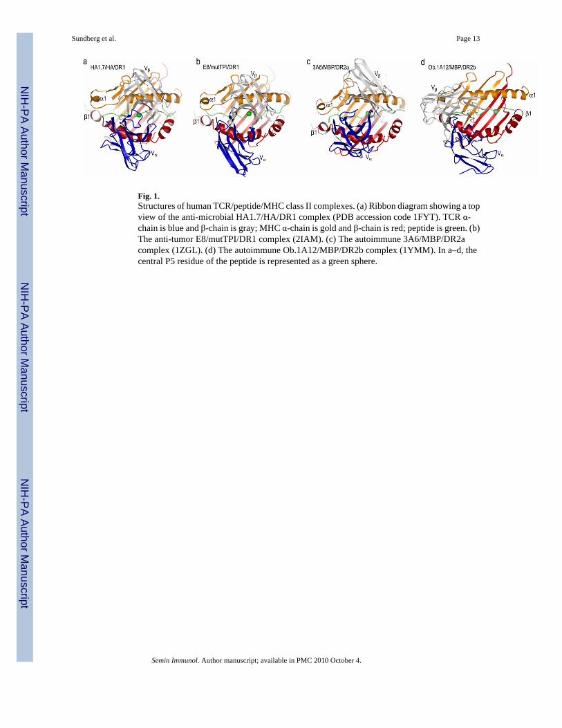
Fig. 1.Structures of human TCR/peptide/MHC class II complexes. (a) Ribbon diagram showing a topview of the anti-microbial HA1.7/HA/DR1 complex (PDB accession code 1FYT). TCR α-chain is blue and β-chain is gray; MHC α-chain is gold and β-chain is red; peptide is green. (b)The anti-tumor E8/mutTPI/DR1 complex (2IAM). (c) The autoimmune 3A6/MBP/DR2acomplex (1ZGL). (d) The autoimmune Ob.1A12/MBP/DR2b complex (1YMM). In a–d, thecentral P5 residue of the peptide is represented as a green sphere.
Sundberg et al. Page 13
Semin Immunol. Author manuscript; available in PMC 2010 October 4.
NIH
-PA Author Manuscript
NIH
-PA Author Manuscript
NIH
-PA Author Manuscript
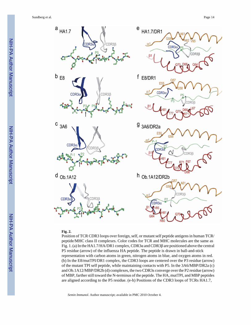
Fig. 2.Position of TCR CDR3 loops over foreign, self, or mutant self peptide antigens in human TCR/peptide/MHC class II complexes. Color codes for TCR and MHC molecules are the same asFig. 1. (a) In the HA1.7/HA/DR1 complex, CDR3α and CDR3β are positioned above the centralP5 residue (arrow) of the influenza HA peptide. The peptide is drawn in ball-and-stickrepresentation with carbon atoms in green, nitrogen atoms in blue, and oxygen atoms in red.(b) In the E8/mutTPI/DR1 complex, the CDR3 loops are centered over the P3 residue (arrow)of the mutant TPI self peptide, while maintaining contacts with P5. In the 3A6/MBP/DR2a (c)and Ob.1A12/MBP/DR2b (d) complexes, the two CDR3s converge over the P2 residue (arrow)of MBP, farther still toward the N-terminus of the peptide. The HA, mutTPI, and MBP peptidesare aligned according to the P5 residue. (e-h) Positions of the CDR3 loops of TCRs HA1.7,
Sundberg et al. Page 14
Semin Immunol. Author manuscript; available in PMC 2010 October 4.
NIH
-PA Author Manuscript
NIH
-PA Author Manuscript
NIH
-PA Author Manuscript

E8, 3A6, and Ob.1A12 on the peptide/MHC surface. Peptide residues located within the pocketformed by CDR3α and CDR3β in the four complexes are indicated by spheres. For eachcomplex, MHC residues contacted by the CDR3 loops are labeled.
Sundberg et al. Page 15
Semin Immunol. Author manuscript; available in PMC 2010 October 4.
NIH
-PA Author Manuscript
NIH
-PA Author Manuscript
NIH
-PA Author Manuscript
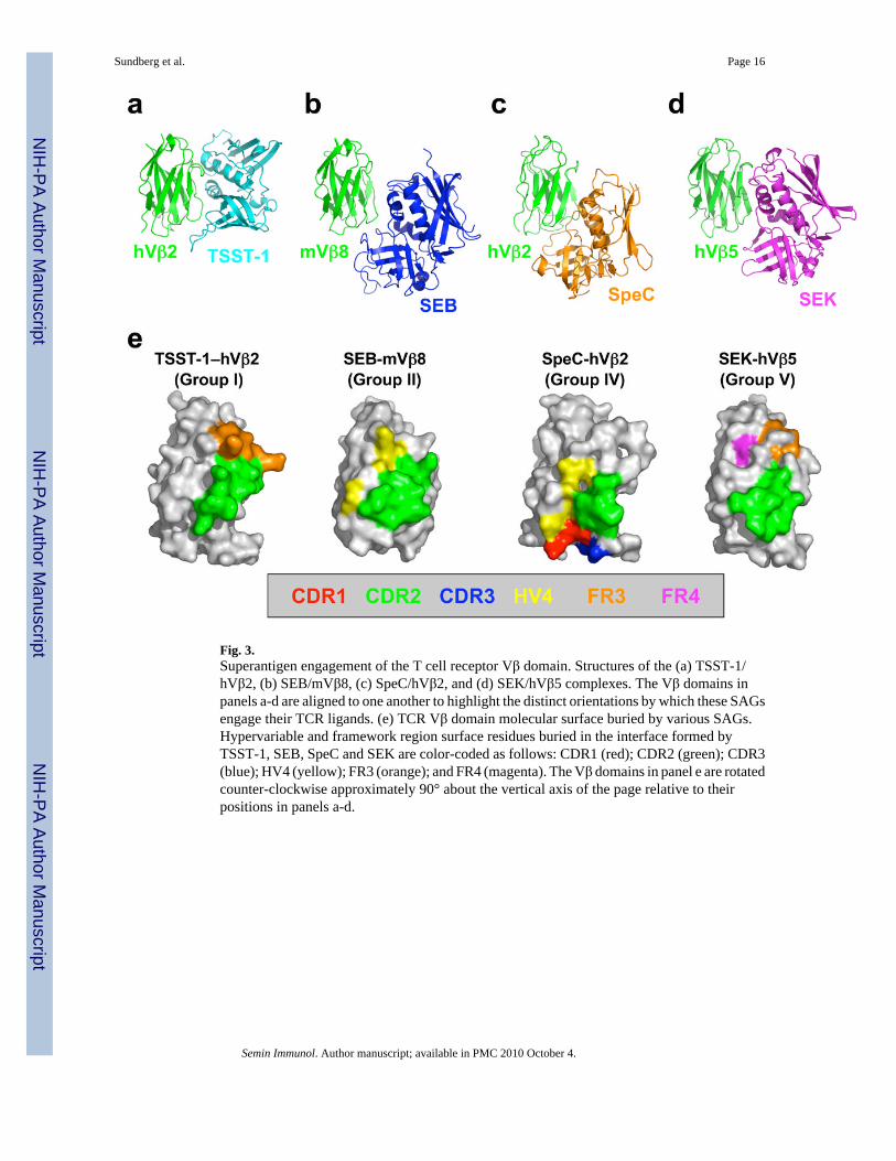
Fig. 3.Superantigen engagement of the T cell receptor Vβ domain. Structures of the (a) TSST-1/hVβ2, (b) SEB/mVβ8, (c) SpeC/hVβ2, and (d) SEK/hVβ5 complexes. The Vβ domains inpanels a-d are aligned to one another to highlight the distinct orientations by which these SAGsengage their TCR ligands. (e) TCR Vβ domain molecular surface buried by various SAGs.Hypervariable and framework region surface residues buried in the interface formed byTSST-1, SEB, SpeC and SEK are color-coded as follows: CDR1 (red); CDR2 (green); CDR3(blue); HV4 (yellow); FR3 (orange); and FR4 (magenta). The Vβ domains in panel e are rotatedcounter-clockwise approximately 90° about the vertical axis of the page relative to theirpositions in panels a-d.
Sundberg et al. Page 16
Semin Immunol. Author manuscript; available in PMC 2010 October 4.
NIH
-PA Author Manuscript
NIH
-PA Author Manuscript
NIH
-PA Author Manuscript

Fig. 4.MHC/SAG/TCR ternary signaling complexes mediated by (a) TSST-1, (b) SEB, (c) SpeC, and(d) MAM. Colors are as follows: MHC α subunit, green; MHC β subunit, blue; antigenicpeptide, gray; TCR α chain, orange; TCR β chain, red; SAGs, yellow. For clarity, the MHC/SAG/TCR complexes mediated by SpeC (panel c) and MAM (panel d) are rotatedapproximately 90° clockwise about the vertical axis of the page relative to those mediated byTSST-1 (panel a) and SEB (panel b).
Sundberg et al. Page 17
Semin Immunol. Author manuscript; available in PMC 2010 October 4.
NIH
-PA Author Manuscript
NIH
-PA Author Manuscript
NIH
-PA Author Manuscript
Related Documents


![SEMIN ,EÉTNIÉ SEMIN E, ÉTANCHÉITÉ À L’AIR · 2019. 11. 20. · SEMIN E SEMIN ,EÉTNIÉ SEMIN E, ÉTANCHÉITÉ À L’AIR] ÉTANCHÉITÉ À L’AIR Les murs extérieurs, plancher,](https://static.cupdf.com/doc/110x72/60babccc2cb2867d0b0d5ba5/semin-etni-semin-e-tanchit-la-2019-11-20-semin-e-semin-etni.jpg)