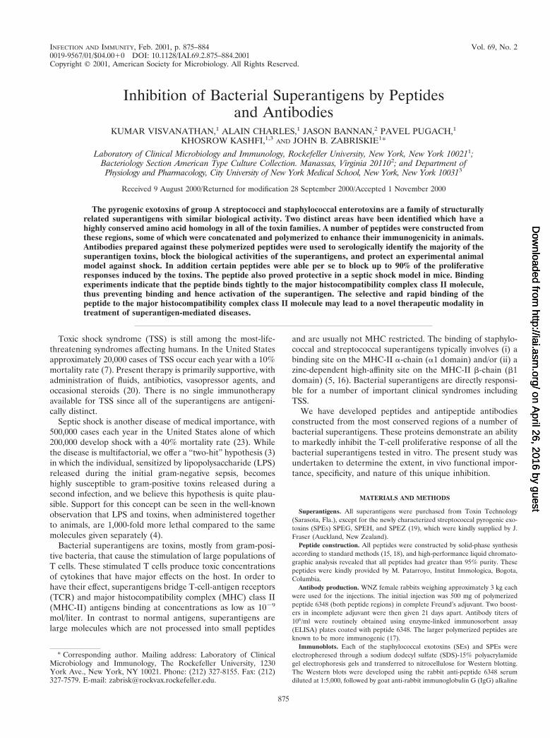
INFECTION AND IMMUNITY, 0019-9567/01/$04.0010 DOI: 10.1128/IAI.69.2.875–884.2001 Feb. 2001, p. 875–884 Vol. 69, No. 2 Copyright © 2001, American Society for Microbiology. All Rights Reserved. Inhibition of Bacterial Superantigens by Peptides and Antibodies KUMAR VISVANATHAN, 1 ALAIN CHARLES, 1 JASON BANNAN, 2 PAVEL PUGACH, 1 KHOSROW KASHFI, 1,3 AND JOHN B. ZABRISKIE 1 * Laboratory of Clinical Microbiology and Immunology, Rockefeller University, New York, New York 10021 1 ; Bacteriology Section American Type Culture Collection. Manassas, Virginia 20110 2 ; and Department of Physiology and Pharmacology, City University of New York Medical School, New York, New York 10031 3 Received 9 August 2000/Returned for modification 28 September 2000/Accepted 1 November 2000 The pyrogenic exotoxins of group A streptococci and staphylococcal enterotoxins are a family of structurally related superantigens with similar biological activity. Two distinct areas have been identified which have a highly conserved amino acid homology in all of the toxin families. A number of peptides were constructed from these regions, some of which were concatenated and polymerized to enhance their immunogenicity in animals. Antibodies prepared against these polymerized peptides were used to serologically identify the majority of the superantigen toxins, block the biological activities of the superantigens, and protect an experimental animal model against shock. In addition certain peptides were able per se to block up to 90% of the proliferative responses induced by the toxins. The peptide also proved protective in a septic shock model in mice. Binding experiments indicate that the peptide binds tightly to the major histocompatibility complex class II molecule, thus preventing binding and hence activation of the superantigen. The selective and rapid binding of the peptide to the major histocompatibility complex class II molecule may lead to a novel therapeutic modality in treatment of superantigen-mediated diseases. Toxic shock syndrome (TSS) is still among the most-life- threatening syndromes affecting humans. In the United States approximately 20,000 cases of TSS occur each year with a 10% mortality rate (7). Present therapy is primarily supportive, with administration of fluids, antibiotics, vasopressor agents, and occasional steroids (20). There is no single immunotherapy available for TSS since all of the superantigens are antigeni- cally distinct. Septic shock is another disease of medical importance, with 500,000 cases each year in the United States alone of which 200,000 develop shock with a 40% mortality rate (23). While the disease is multifactorial, we offer a “two-hit” hypothesis (3) in which the individual, sensitized by lipopolysaccharide (LPS) released during the initial gram-negative sepsis, becomes highly susceptible to gram-positive toxins released during a second infection, and we believe this hypothesis is quite plau- sible. Support for this concept can be seen in the well-known observation that LPS and toxins, when administered together to animals, are 1,000-fold more lethal compared to the same molecules given separately (4). Bacterial superantigens are toxins, mostly from gram-posi- tive bacteria, that cause the stimulation of large populations of T cells. These stimulated T cells produce toxic concentrations of cytokines that have major effects on the host. In order to have their effect, superantigens bridge T-cell-antigen receptors (TCR) and major histocompatibility complex (MHC) class II (MHC-II) antigens binding at concentrations as low as 10 29 mol/liter. In contrast to normal antigens, superantigens are large molecules which are not processed into small peptides and are usually not MHC restricted. The binding of staphylo- coccal and streptococcal superantigens typically involves (i) a binding site on the MHC-II a-chain (a1 domain) and/or (ii) a zinc-dependent high-affinity site on the MHC-II b-chain (b1 domain) (5, 16). Bacterial superantigens are directly responsi- ble for a number of important clinical syndromes including TSS. We have developed peptides and antipeptide antibodies constructed from the most conserved regions of a number of bacterial superantigens. These proteins demonstrate an ability to markedly inhibit the T-cell proliferative response of all the bacterial superantigens tested in vitro. The present study was undertaken to determine the extent, in vivo functional impor- tance, specificity, and nature of this unique inhibition. MATERIALS AND METHODS Superantigens. All superantigens were purchased from Toxin Technology (Sarasota, Fla.), except for the newly characterized streptococcal pyrogenic exo- toxins (SPEs) SPEG, SPEH, and SPEZ (19), which were kindly supplied by J. Fraser (Auckland, New Zealand). Peptide construction. All peptides were constructed by solid-phase synthesis according to standard methods (15, 18), and high-performance liquid chromato- graphic analysis revealed that all peptides had greater than 95% purity. These peptides were kindly provided by M. Patarroyo, Institut Immologica, Bogota, Columbia. Antibody production. WNZ female rabbits weighing approximately 3 kg each were used for the injections. The initial injection was 500 mg of polymerized peptide 6348 (both peptide regions) in complete Freund’s adjuvant. Two boost- ers in incomplete adjuvant were then given 21 days apart. Antibody titers of 10 6 /ml were routinely obtained using enzyme-linked immunosorbent assay (ELISA) plates coated with peptide 6348. The larger polymerized peptides are known to be more immunogenic (17). Immunoblots. Each of the staphylococcal exotoxins (SEs) and SPEs were electropheresed through a sodium dodecyl sulfate (SDS)-15% polyacrylamide gel electrophoresis gels and transferred to nitrocellulose for Western blotting. The Western blots were developed using the rabbit anti-peptide 6348 serum diluted at 1:5,000, followed by goat anti-rabbit immunoglobulin G (IgG) alkaline * Corresponding author. Mailing address: Laboratory of Clinical Microbiology and Immunology, The Rockefeller University, 1230 York Ave., New York, NY 10021. Phone: (212) 327-8155. Fax: (212) 327-7579. E-mail: [email protected]. 875 on April 26, 2016 by guest http://iai.asm.org/ Downloaded from on April 26, 2016 by guest http://iai.asm.org/ Downloaded from on April 26, 2016 by guest http://iai.asm.org/ Downloaded from

Welcome message from author
This document is posted to help you gain knowledge. Please leave a comment to let me know what you think about it! Share it to your friends and learn new things together.
Transcript
INFECTION AND IMMUNITY,0019-9567/01/$04.0010 DOI: 10.1128/IAI.69.2.875–884.2001
Feb. 2001, p. 875–884 Vol. 69, No. 2
Copyright © 2001, American Society for Microbiology. All Rights Reserved.
Inhibition of Bacterial Superantigens by Peptidesand Antibodies
KUMAR VISVANATHAN,1 ALAIN CHARLES,1 JASON BANNAN,2 PAVEL PUGACH,1
KHOSROW KASHFI,1,3 AND JOHN B. ZABRISKIE1*
Laboratory of Clinical Microbiology and Immunology, Rockefeller University, New York, New York 100211;Bacteriology Section American Type Culture Collection. Manassas, Virginia 201102; and Department of
Physiology and Pharmacology, City University of New York Medical School, New York, New York 100313
Received 9 August 2000/Returned for modification 28 September 2000/Accepted 1 November 2000
The pyrogenic exotoxins of group A streptococci and staphylococcal enterotoxins are a family of structurallyrelated superantigens with similar biological activity. Two distinct areas have been identified which have ahighly conserved amino acid homology in all of the toxin families. A number of peptides were constructed fromthese regions, some of which were concatenated and polymerized to enhance their immunogenicity in animals.Antibodies prepared against these polymerized peptides were used to serologically identify the majority of thesuperantigen toxins, block the biological activities of the superantigens, and protect an experimental animalmodel against shock. In addition certain peptides were able per se to block up to 90% of the proliferativeresponses induced by the toxins. The peptide also proved protective in a septic shock model in mice. Bindingexperiments indicate that the peptide binds tightly to the major histocompatibility complex class II molecule,thus preventing binding and hence activation of the superantigen. The selective and rapid binding of thepeptide to the major histocompatibility complex class II molecule may lead to a novel therapeutic modality intreatment of superantigen-mediated diseases.
Toxic shock syndrome (TSS) is still among the most-life-threatening syndromes affecting humans. In the United Statesapproximately 20,000 cases of TSS occur each year with a 10%mortality rate (7). Present therapy is primarily supportive, withadministration of fluids, antibiotics, vasopressor agents, andoccasional steroids (20). There is no single immunotherapyavailable for TSS since all of the superantigens are antigeni-cally distinct.
Septic shock is another disease of medical importance, with500,000 cases each year in the United States alone of which200,000 develop shock with a 40% mortality rate (23). Whilethe disease is multifactorial, we offer a “two-hit” hypothesis (3)in which the individual, sensitized by lipopolysaccharide (LPS)released during the initial gram-negative sepsis, becomeshighly susceptible to gram-positive toxins released during asecond infection, and we believe this hypothesis is quite plau-sible. Support for this concept can be seen in the well-knownobservation that LPS and toxins, when administered togetherto animals, are 1,000-fold more lethal compared to the samemolecules given separately (4).
Bacterial superantigens are toxins, mostly from gram-posi-tive bacteria, that cause the stimulation of large populations ofT cells. These stimulated T cells produce toxic concentrationsof cytokines that have major effects on the host. In order tohave their effect, superantigens bridge T-cell-antigen receptors(TCR) and major histocompatibility complex (MHC) class II(MHC-II) antigens binding at concentrations as low as 1029
mol/liter. In contrast to normal antigens, superantigens arelarge molecules which are not processed into small peptides
and are usually not MHC restricted. The binding of staphylo-coccal and streptococcal superantigens typically involves (i) abinding site on the MHC-II a-chain (a1 domain) and/or (ii) azinc-dependent high-affinity site on the MHC-II b-chain (b1domain) (5, 16). Bacterial superantigens are directly responsi-ble for a number of important clinical syndromes includingTSS.
We have developed peptides and antipeptide antibodiesconstructed from the most conserved regions of a number ofbacterial superantigens. These proteins demonstrate an abilityto markedly inhibit the T-cell proliferative response of all thebacterial superantigens tested in vitro. The present study wasundertaken to determine the extent, in vivo functional impor-tance, specificity, and nature of this unique inhibition.
MATERIALS AND METHODS
Superantigens. All superantigens were purchased from Toxin Technology(Sarasota, Fla.), except for the newly characterized streptococcal pyrogenic exo-toxins (SPEs) SPEG, SPEH, and SPEZ (19), which were kindly supplied by J.Fraser (Auckland, New Zealand).
Peptide construction. All peptides were constructed by solid-phase synthesisaccording to standard methods (15, 18), and high-performance liquid chromato-graphic analysis revealed that all peptides had greater than 95% purity. Thesepeptides were kindly provided by M. Patarroyo, Institut Immologica, Bogota,Columbia.
Antibody production. WNZ female rabbits weighing approximately 3 kg eachwere used for the injections. The initial injection was 500 mg of polymerizedpeptide 6348 (both peptide regions) in complete Freund’s adjuvant. Two boost-ers in incomplete adjuvant were then given 21 days apart. Antibody titers of106/ml were routinely obtained using enzyme-linked immunosorbent assay(ELISA) plates coated with peptide 6348. The larger polymerized peptides areknown to be more immunogenic (17).
Immunoblots. Each of the staphylococcal exotoxins (SEs) and SPEs wereelectropheresed through a sodium dodecyl sulfate (SDS)-15% polyacrylamidegel electrophoresis gels and transferred to nitrocellulose for Western blotting.The Western blots were developed using the rabbit anti-peptide 6348 serumdiluted at 1:5,000, followed by goat anti-rabbit immunoglobulin G (IgG) alkaline
* Corresponding author. Mailing address: Laboratory of ClinicalMicrobiology and Immunology, The Rockefeller University, 1230York Ave., New York, NY 10021. Phone: (212) 327-8155. Fax: (212)327-7579. E-mail: [email protected].
875
on April 26, 2016 by guest
http://iai.asm.org/
Dow
nloaded from
on April 26, 2016 by guest
http://iai.asm.org/
Dow
nloaded from
on April 26, 2016 by guest
http://iai.asm.org/
Dow
nloaded from
phosphatase conjugate (Sigma). A similar gel was stained with Coomassie bluefor molecular weight locations and proteins.
Protein A Enrichment of peptide 6348 antibody. Some of the peptide 6348antibody raised in rabbits was further enriched on a column as described below.In brief, the column of protein A-Sepharose (15 ml) (Sigma) beads was preparedat room temperature. Ten milliliters of immune rabbit serum, diluted 1:1 inphosphate-buffered saline (PBS), was run through the column. The column wasthen washed with four column volumes of PBS and eluted with 10 ml of 0.1 MNaAc (pH 2.5), and 10 1-ml fractions were collected. One hundred microliters of1 M sodium bicarbonate was added to each fraction to neutralize the acidicelution step. Protein elution and concentration were then determined via spec-trophotometer, and the protein A-enriched antibody preparation was readjustedappropriately to be equal to the anti peptide titers of the original serum. Similarexperiments were performed with the streptococcal group A carbohydrate anti-body.
Proliferation assays. Human peripheral blood mononuclear cells (PBMCs)were isolated by standard Ficoll-Hypaque techniques and adjusted to 2 3 106
cells/ml. PBMCs (2 3 105) in 200 ml of complete medium (RPMI medium1640 1 10% human type AB serum) were placed in 96-well titer plates andstimulated with various doses of superantigen or with a combination of each
toxin and various doses of peptide. The cells were incubated for 6 days, and theresults were measured via tritiated thymidine incorporation. The data presentedare averages of the results of four different experiments. All tests were done intriplicate.
Viability studies. Human PBMCs were isolated as described above, and 2 3105 cells were placed in 96-well titer plates. Phytohemagglutinin (PHA) wasadded at a concentration of 5 mg/well. All peptides were added to PHA at aconcentration of 200 mg/well. The plates were incubated at 37°C in a CO2
incubator for 72 h at which time tritated thymidine was added to the cells. Thecells were harvested 18 h later, and the counts per minute of tritated thymidinewere measured. All experiments were carried out in triplicate.
A second viability test was performed as follows: 2 3 105 PBMCs were platedin 96-well titer plates to which various concentrations of peptide 6343 wereadded. Aliquots of cells with and without peptide were stained with trypan blueeach day for 5 days to observe viability of the cells in the presence or absence ofpeptide.
Rabbit experiments. Female NZW rabbits .1 year of age were obtained fromHazelton Dutchland Labs, Inc. (Denver, Pa.). Older animals are used in theseexperiments because they are more sensitive to the effects of LPS and toxins (9).The lethal shock experiments were performed as follows: Animals were given 50
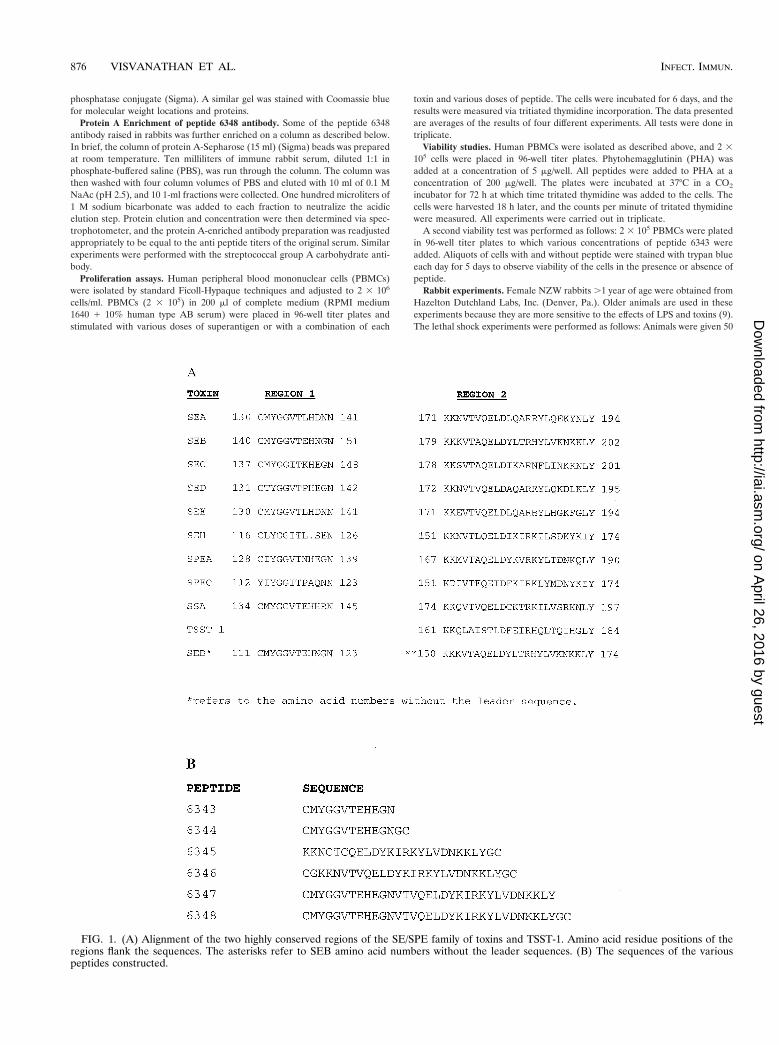
FIG. 1. (A) Alignment of the two highly conserved regions of the SE/SPE family of toxins and TSST-1. Amino acid residue positions of theregions flank the sequences. The asterisks refer to SEB amino acid numbers without the leader sequences. (B) The sequences of the variouspeptides constructed.
876 VISVANATHAN ET AL. INFECT. IMMUN.
on April 26, 2016 by guest
http://iai.asm.org/
Dow
nloaded from

mg of either SEB or SPEA toxins per kg of body weight, mixed with 0.5 ml ofeither preimmune normal rabbit serum or anti-peptide antibody. Four hourslater, all rabbits received 5 mg of LPS (List Biological Labs, Campbell, Calif.) perkg of body weight. Animals were monitored for 72 h for clinical signs of toxicity.Moribund animals were sacrificed with an overdose of nembutal. All toxins andLPS were delivered intravenously.
Murine experiments. Eight-week-old Female BALB/c mice were used for allexperiments. Animals were housed at the Rockefeller Laboratory Animal Re-search Facility, and experiments were undertaken after approval by institutionalanimal care and ethics boards. All mice were sensitized with 0.001 mg of LPS and20 mg of D-galactosamine via intraperitoneal injection (4). Eight hours later,mice were injected with various doses of superantigen that had been shown tocause 100% lethality. In protection experiments, at 6 h postinoculation saline or1.5 mg of the peptide was administered to the experimental mice by subcutane-ous injection. One hour later, the mice were injected again with either saline or1.5 mg of peptide (3.0 mg total). One hour after the second injection, all micewere challenged with the appropriate dose of toxin (via intraperitoneal injec-tion), and the mice were observed for 24 to 48 h.
Endotoxin determination. Assays were performed with a stimulus a mebocytelysate chromogenic ELISA (Associates of Cape Cod). Control standards andsolutions were provided with the kit. Control standard endotoxin was reconsti-tuted and was used to construct a standard curve. The negative control, controlstandard, test samples, and a positive control were plated on a Linbro 96-wellELISA plate (ICN Biomedicals Inc., Aurora, Ohio). Pyrochrome was placed ineach well to yield a 1:1 solution. The plate was agitated for 30 s to ensureincorporation. The plate was then incubated for 30 min in a 37°C water bath.After 30 min, the plate was read at an optical density of 405 nm. Using thestandard curve, endotoxin concentrations of the toxin solutions were determined(14).
MHC class II binding assay. The plates were coated with immunoaffinity-purified soluble human DR-1 (kindly provided by J. Strominger, Harvard Uni-versity) overnight at 4°C in 0.1 M Tris (pH 8.0) at a concentration of 1 mg perwell. A solution of 1% bovine serum albumin in PBS was used to block thecoated plates for 1 h. The primary antibody (anti-6348) was diluted in RPMI1640, added to the wells of the plate, and incubated for 1 h. Horseradishperoxidase-conjugated antibodies of appropriate affinity were used at a dilutionof 1:1,000. One hundred microliters of a 1:1 mixture of hydrogen peroxide andTMB substrate (Kirkegaard and Perry Inc.) was applied in the dark for 20 minafter which the plate was read. All incubation steps were carried out at roomtemperature. Plates were washed three times with ELISA wash buffer betweenevery incubation step. The pH of the binding medium was adjusted to ensure thatall assays were at pH 7.0. Care was taken to ensure that the ionic strength wasadjusted for in each assay. Apparent Kd, the dissociation constant at equilibrium,was calculated using the Lineweaver-Burk equation (21) as previously describedby Fridkis-Hareli and Strominger (6).
RESULTS
Peptide and antibody design. Two distinct regions of theSE/SPE toxins have been identified which share highly con-served amino acid homology, and consensus patterns havebeen identified as common to all members of this family of
toxins (3). The first consensus region has the amino acid se-quence Y-G-G-(LIV)-T-x(4)-N. This pattern has been identi-fied in all of the SEs and SPEs pyrogenic exotoxins, but not inTSS toxin 1 (TSST-1). The sequence begins immediately at theCOOH-terminal side of the cysteine loop. In selected cases wehave given the amino acid sequence without the leader se-quence for more appropriate orientation. The second consen-sus region has the following amino acid sequence: K-x(2)-(LIV)-x(4)-(LIV)-D-x(3)-R-x(2)-L-x(5)-(LIV)-Y. This pattern hadbeen identified in all of the SEs and SPEs, including TSST-1(Fig. 1A).
Considering the high degree of conservation of the SE/SPEtoxins, and the immunologic cross-reactivity previously men-tioned for these toxins, the possibility that antibodies raisedagainst the two conserved regions might block the biologicalactivities of all toxins was considered (2). A number of peptideswere constructed based on the consensus sequences describedabove. These were made and purified using high-performanceliquid chromatography according to previous protocols (17).As illustrated in Fig. 1B, peptides 6343 and 6344 were derivedfrom the first consensus sequence, peptides 6345 and 6346were constructed from the second consensus sequence, andpeptides 6347 and 6348 were combinations of both sequencesattached in the proper order. Peptides 6344, 6346, and 6348were polymerized with the addition of cysteine residues at theN and C terminals with glycine molecules used as spacers. Thisprocess was used to increase the immunogenicity of these pep-tides for vaccination (18).
Anti-peptide antibody activity in Western blotting. Peptidesconstructed of the combined consensus regions of the toxins(6348) were used to raise antibodies in rabbits. Sera from allrabbits showed similar antibody production. Western blots in-dicated that the peptide antiserum was able not only to recog-nize the conserved regions of SEB but also those of otherstreptococcal and staphylococcal toxins including SEA, SEB,SEC, SED, SPEA, and SPEC, but not TSST-1 (Fig. 2A). Fig-ure 2B is an SDS-15% gel with the same toxins stained withCoomassie blue. As seen in both the Western blot and in thestained gel, the upper bands most likely represent eitherdimers of the toxins or partially digested larger-molecular-sizefractions of the toxins. The major bands correspond to theknown molecular size of the toxins. Similar results were ob-tained using sera from three other rabbits (data not shown).
FIG. 2. (A) SDS-15% gel of various toxins transferred to and developed by Western blot. The blot demonstrates the fact that the 6348anti-peptide antibody (raised in rabbit) immunoprecipitates with SEA (lane 1), SEB (lane 2), SEC (lane 3), SED (lane 4), and SPEA (lane 5), anda faint band is seen with SPEC (lane 6). No band was seen with TSST-1 (lane 7). (B) SDS-15% gel of the same toxins stained with Coomassie blue.All lanes were loaded with 5 mg of each toxin. Upper bands most likely represent dimers or partially digested forms of the toxins. The major bandsare seen between 28 and 25 kDa, corresponding to their known molecular sizes.
VOL. 69, 2001 PEPTIDES, SUPERANTIGENS, AND TOXIC AND SEPTIC SHOCK 877
on April 26, 2016 by guest
http://iai.asm.org/
Dow
nloaded from

The effect of protein A-enriched anti-peptide antibody onblastogenesis. To further enrich our serum anti-peptide anti-bodies, we isolated the IgG fraction of our anti-peptide anti-body using a protein A column and used these antibodies inexperiments similar to that described above. The antibodiesshowed strong inhibition of blastogenesis to all of the staphy-
lococcal and streptococcal superantigens tested (Fig. 3A). Inaddition we determined that 117 ng of total IgG was enough toachieve 93 to 100% inhibition of all the superantigens. Figure3B demonstrates that a high-titer anti-group A streptococcalcarbohydrate antibody enriched in a similar manner was un-able to block the biological properties of the toxins.
FIG. 3. (A) Inhibition of toxin blastogenesis of PBMCs by protein A-enriched anti-peptide antibody. PBMCs (2 3 105) were stimulated witheither 2 mg of each toxin or a combination of 2 mg of each toxin with anti-peptide antibody. These cells were incubated for 6 days, and the resultswere measured via tritiated thymidine incorporation. cpm, counts per minute. (B) Note the lack of inhibition of blastogenesis when a high-titeredgroup A carbohydrate antibody (Ab) was used in the assay.
878 VISVANATHAN ET AL. INFECT. IMMUN.
on April 26, 2016 by guest
http://iai.asm.org/
Dow
nloaded from

Rabbit challenge experiments. To determine whether ouranti-peptide antibody could block the lethal effects of the tox-ins, animals were given either toxins plus preimmune normalrabbit serum or anti-peptide antibody. As seen in Table 1, allrabbits who received anti-peptide antibody survived while rab-bits that received toxin plus pre immune rabbit serum did not.
Direct peptide inhibition of blastogenesis. Theoretically, thepeptides we designed could act as peptidomimetics and directlyinhibit superantigen stimulation of T cells. Using various con-centrations of peptide 6343, up to 83% inhibition of blastogen-esis was seen (Fig. 4A). This inhibition was dose responsive innature and indicated probable competitive inhibition. Otherpeptides tested, including 6346 and 6348, also demonstratedsome inhibition but less than that induced by 6343 (data notshown), whereas other peptides including 6345 (Fig. 4B)showed no inhibition by the peptide. Most important was ourobservation that a single-amino-acid substitution of a lysine forcysteine in the original 6343 peptide at the N-terminal-end-labeled 6343X resulted in complete loss of the original block-ing activity (Fig. 4C). These experiments indicate the specific-ity of the 6343 peptide for the inhibition of blastogenesis.
The most striking feature about this inhibition was that itinhibited all of the streptococcal and staphylococcal toxinstested, including TSST-1, which shows no sequence homologyto the 6343 peptide (see Fig. 5).
Peptide inhibition of newly described SEs. We tested thethree new SEs recently described by Proft et al. (19). Thesewere identified and cloned from the Streptococcus pyogenes M1genomic database at the University of Oklahoma. These toxinshave been shown to have the characteristics of superantigens,including specific Vb stimulation profiles. Peptide 6343 indoses of 75 and 150 mg/well showed between 59 and 83%inhibition of blastogenesis of PBMCs (Fig. 6).
Viability studies. While the studies described above indi-cated that the peptide was quite specific in its inhibition of thetoxins, we wanted to be sure that the peptide was not interfer-ing with cell function in some other manner. We approachedthis question in two different ways.
First, 200 mg each of different lots of 6343 peptide includinga dimer form of 6343 was added to 2 3 105 cells per well incomplete medium in a 96-well microtiter plate. Controls were2 3 105 cells/well without peptide. Aliquots were removedfrom the wells each day. The cells were centrifuged at 250 3 gand diluted 1:1 with trypan blue, and the numbers of viablecells were counted each day. Daily inspection of the cells re-
vealed that the same number of cells remained viable through-out the 5 days, with or without added peptides.
The second approach was to test the PHA stimulation of cellwith and without different peptides in a 72-h blastogenesisassay. Peptides 6343, 6345, and 6343X, as described in Fig. 4,were added at concentrations of 200 mg of each peptide toPHA-stimulated PBMCs. As seen in Fig. 7, the addition ofeach peptide to PHA-stimulated cells did not inhibit PHAblastogenesis, attesting to the viability of the PHA-stimulatedcells in the presence of the peptide.
“Two-hit” animal model of septic shock. Based on a modelof the synergistic effects of LPS and the toxins when adminis-tered together (3, 4), called the “two hit”model of septic shock,we established an animal model of septic shock. After primingwith D-galactosamine (20 mg/mouse), BALB/c mice (JacksonLaboratory) were challenged intraperitoneally with LPS fol-lowed by SEB. The results demonstrated that extremely smallamounts of LPS and SEB were needed to effect lethality (4,13). The synergy between these two mediators of shock wasextremely impressive and extended for at least an 18-h period.We chose an 8-h delay between the two toxins for our model tomimic the clinical picture of septic shock. We established andoptimized doses of toxins that would lead to 100% lethality(see Table 2).
Peptide inhibition in animal model of sepsis. Our prelimi-nary experiments (Table 2) indicated that at a concentration ofLPS of 0.001 mg in D-galactosamine-primed mice, only smallamounts of toxins were needed to cause 100% lethality in themice. This LPS dosage combined with 0.2 mg of SPEA, 0.02 mgof SEB, or 0.2 mg of TSST-1 resulted in approximately 100%lethality in three control groups of six mice each.
Introduction of peptide 6343 at a total of 3 mg per mouseinjected subcutaneously at 2 and 1 h before administration ofthe above toxins completely protected most of the mice foreach toxin tested (ratio of surviving mice per experimentalgroup: SPEA 1 6343, 6/6 (100%), SEB 1 6343, 5/6 (83%);TSST-1 1 6343, 6/6 (100%). Studies in which the peptide isgiven at specified intervals after the administration of toxin arecurrently under investigation.
Direct binding of peptide to MHC-II molecules. Using anELISA technique and purified human DR-1 molecules (withand without peptides in the groove) supplied by J. Strominger(Harvard University), we were able to demonstrate strongbinding of the 6343 peptide to the DR-1 molecule in thenanomolar range. The results are shown in Fig. 8.
DISCUSSION
In this report we have clearly demonstrated that both the6343 peptide and the antibody to the 6348 peptide can inde-pendently block the proliferative effects of all of the staphylo-coccal and streptococcal superantigens and that this inhibitionis specific for the 6343 peptide. We have also demonstratedthat the anti-peptide antibody can provide passive protectionagainst toxic shock in a rabbit model. In addition the peptideitself is protective in a two-hit model of septic shock describedabove.
This is not the first time synthesized peptides have been usedto study the structure-function relationship of the various tox-ins. A number of synthetic peptides corresponding to different
TABLE 1. Passive protection of rabbits with anti-peptide antibodya
Toxin (dosage) Anti-peptide antibody Live/total
Control 2/2
SEB (50–100 mg/kg) 2 0/3SEB (50–100 mg/kg) 1 2/2
SPEA (50–100 mg/kg) 2 0/2SPEA (50–100 mg/kg) 1 2/2
a Rabbits were given 5 mg of LPS/Kg of body weight alone (control) or incombination with the indicated toxin with (1) or without (2) the anti-peptideantibody.
VOL. 69, 2001 PEPTIDES, SUPERANTIGENS, AND TOXIC AND SEPTIC SHOCK 879
on April 26, 2016 by guest
http://iai.asm.org/
Dow
nloaded from
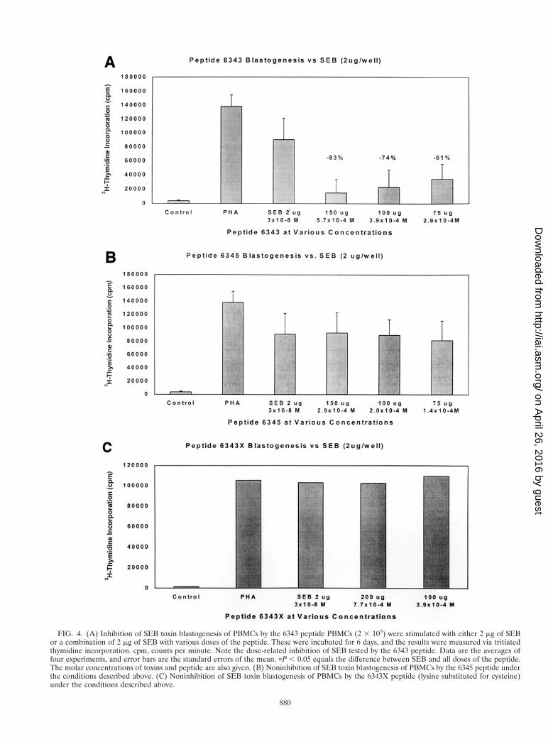
FIG. 4. (A) Inhibition of SEB toxin blastogenesis of PBMCs by the 6343 peptide PBMCs (2 3 105) were stimulated with either 2 mg of SEBor a combination of 2 mg of SEB with various doses of the peptide. These were incubated for 6 days, and the results were measured via tritiatedthymidine incorporation. cpm, counts per minute. Note the dose-related inhibition of SEB tested by the 6343 peptide. Data are the averages offour experiments, and error bars are the standard errors of the mean. pP , 0.05 equals the difference between SEB and all doses of the peptide.The molar concentrations of toxins and peptide are also given. (B) Noninhibition of SEB toxin blastogenesis of PBMCs by the 6345 peptide underthe conditions described above. (C) Noninhibition of SEB toxin blastogenesis of PBMCs by the 6343X peptide (lysine substituted for cysteine)under the conditions described above.
880
on April 26, 2016 by guest
http://iai.asm.org/
Dow
nloaded from

regions of various superantigens including SEA and TSST-1have been studied by other researchers (5, 8, 11, 12). Importantsites for cytokine production and other functions were foundincluding inhibiting peptides. However, we are the first todescribe a single peptide that inhibits all of the known bacterialsuperantigens.
The amount of peptide required to achieve the blockingeffects (i.e., 1,000-fold higher than the amount of toxin admin-istered) against the toxins is puzzling. This is especially truebecause the peptide has such high-affinity binding to MHC-IImolecules. However, the fact that unrelated peptides or thelysine-substituted peptide 6343X did not inhibit the prolifera-tive activity of the toxins (Fig. 4) strengthens our belief that theinhibition is specific for peptide 6343. Whether the peptide isdegraded by serum enzymes or needs to be in excess to coverthe MHC receptor sites for the toxins is unknown and is cur-rently under investigation.
As the various superantigen toxins bind and stimulate dif-ferent Vb regions of the TCR, we believe the more likely siteof binding of the 6343 peptide is to the MHC-II molecule. Thishypothesis is strengthened by the fact that TSST-1, which doesnot have any sequence similarity to the first consensus region(Fig. 1), is nevertheless inhibited (50%) by the peptide in theproliferation assay. While this concept is attractive, it should beemphasized that the peptide does not appear to bind to theresidues of SEB recognized to bind to the MHC molecule (10).However, it is true that a cysteine is involved in the interactionbetween the superantigens and the MHC. The observation thatsubstitution of a lysine for the N-terminal cysteine in our pep-tide abolished its biological activity suggests that the cysteine
might play an important role in the inhibition of the binding ofsuperantigens to the MHC.
Using SEA as a template, the two regions of the superanti-gens from which the peptides are made are the b5 region andpart of the a4 helix. These areas, although they are highlyconserved, are not known binding regions of the superantigento the MHC molecule. Also, neither seems to represent surfaceepitopes. However, it is clear from the evidence presented thatthe peptide (especially the one that is derived from the b5region) binds very strongly to the intact MHC-II molecule. Webelieve that the 6343 peptide itself in its 12-amino-acid con-figuration (with its lack of tertiary structure) is able to fit intothe MHC superantigen-binding area and thus compete withthe natural superantigens. This effect is very specific becauseeither a single amino acid variation of the peptide sequence oran unrelated peptide leads to poor binding and lack of func-tional activity. Experiments in which alanine has been substi-tuted for each amino acid of the 12-amino-acid peptide arecurrently underway to determine whether other amino acidsare important for the biological activity of this peptide.
The antibody to the same core regions of the superantigensdescribed above also has substantial activity against the varioussuperantigens. As demonstrated in the Western blot (Fig. 2),the antibody clearly binds to the majority of the superantigenswith the exception of TSST-1. The binding of the antibody tothe superantigens, even though the residues are not normallyexposed, indicates that either the antibody binding involvessome aspect of the tertiary structure of the antibody or buriedresidues are variably exposed. The disparity between the lackof serological binding of the antibody to TSST-1 and the inhi-
FIG. 5. Inhibition by the 6343 peptide of PBMC blastogenesis by various toxins. PBMCs (2 3 105) were stimulated with 2 mg of each of thevarious toxins or a combination of 2 mg of each toxin with 150 mg of peptide. These cells were incubated for 6 days, and the results were measuredvia tritiated thymidine incorporation. Note that the single peptide (6343) inhibited all of the superantigens tested.
VOL. 69, 2001 PEPTIDES, SUPERANTIGENS, AND TOXIC AND SEPTIC SHOCK 881
on April 26, 2016 by guest
http://iai.asm.org/
Dow
nloaded from

bition of TSST-1 proliferation by the peptide emphasizes thefact that serological recognition or lack thereof to a given toxindoes not always predict the biological activities of these toxinswith respect to the peptides.
Functionally we have shown that our peptide is able toeffectively block the clinical effects of superantigens in a two-hit mouse model of septic shock. Using this model, we haveshown that the peptide can block the effects of the toxins evenwhen the interaction between the superantigens and LPS en-hances the lethal potency of both antigens by 1,000-fold. Thismay account for the fact that endotoxin-mediated events are,in many circumstances, insufficient to explain the deleteriouseffects of septic shock in clinical practice. Hence, at least two
independent pathways of lethal shock can occur. LPS andpeptidoglycan interact with macrophages whereas superanti-gens interact with T cells. In both cases target cells are inducedto release large amounts of cytokines. Since both gram-nega-tive and gram-positive organisms frequently can be recoveredfrom patients with sepsis, we believe a two-hit hypothesis isoperative and that the interaction between LPS and the su-perantigens markedly enhances the lethal properties of bothmolecules. The interruption of the toxin pathway by peptidesmay prevent the onset of lethal shock induced by the combi-nation of the two molecules.
Repeated injections of the 6343 superantigen peptide (non-polymerized) into normal rabbits even with the addition of
FIG. 6. Inhibition of PBMC blastogenesis of three newly described SEs by peptide 6343. PBMCs (2 3 105) were stimulated with either 2 mgof each toxin or a combination of 2 mg of each toxin with various doses of peptide. These were incubated for 6 days, and the results were measuredvia tritiated thymidine incorporation. Note the strong inhibition of all three toxins by 6343 peptide. Addition of 100 mg of peptide did not inhibitthe PHA stimulation.
FIG. 7. Stimulation of human PBMCs with PHA in the presence or absence of peptides 6343, 6345, and 6343X. All peptides were added at aconcentration of 200 mg/well, and all experiments were carried out in triplicate. PHA was added at a concentration of 5 mg/well. Note that theaddition of each peptide did not decrease the PHA stimulation of the cells, attesting to the viability of the cells in the presence of the peptide.
882 VISVANATHAN ET AL. INFECT. IMMUN.
on April 26, 2016 by guest
http://iai.asm.org/
Dow
nloaded from

Freund’s adjuvant failed to produce any antibodies. This sug-gests that the smallest peptide (6343) is poorly immunogenic(data not shown) and thus can be used for repeated adminis-tration as a therapeutic agent.
All peptides and reagents were tested for endotoxin contam-ination using the Limulus amebocyte lysate assay and wereshown to have less than 0.1 ng of endotoxin per ml (9). Theseresults indicate that endotoxin itself did not play an importantrole in these experiments. Our preliminary evidence confirmsthe specific binding of the peptide directly to MHC-II mole-cules and suggests that it does not affect the normal immunefunction of this molecule.
During the drafting of this manuscript, Arad et al. (1)published findings similar to but not identical to ours. Usinga slightly different and shorter segment of region II (see Fig.1), YNKKKATVQELD, which is a variant of SEB (150-TNKKKVTAQELD-161), they were able to inhibit expressionof interleukin 2 RNA by 18- to 40-fold after stimulation withthe native toxin. Arad et al. also noted that their peptide wasnot close to the domains of SEB known to participate in bind-ing to the TCR or MHC-II molecules. They tested only alimited number of toxins while our inhibition was present in alarge number of toxins tested. Their peptide was also able torescue mice from toxic shock in a mouse model similar to ours.No binding studies were carried out by these authors. Aninteresting and as yet unexplained feature of their studies wasthat mice protected by the peptide and toxin challenge werethen resistant to a second challenge of the same toxin 2 weekslater, even though no peptide was administered.
In summary, these experiments indicate that both peptidesand antibodies thereto directly block the interaction of staph-ylococcal and streptococcal superantigens, with their bindingsite thus preventing the proliferative and massive inflammatoryresponses these antigens usually generate. Vaccination witheither the combination peptide (6348) or potentially thesmaller peptide (6344) conjugated to an appropriate carriermay be protective in humans against a large number of bacte-rial superantigen toxins. The peptide may prove to be usefuldirectly or indirectly as an antigen in a vaccine for the treat-ment of both TSS and septic shock as well as other diseasesthat are due to superantigen activation.
FIG. 8. Binding of 6343 peptide to soluble human DR-1. The plateswere coated with immunoaffinity purified soluble human DR-1 (kindlyprovided by J. Strominger, Harvard University) overnight at 4°C in 0.1M Tris (pH 8.0) at a concentration of 1 mg per well. A solution of 1%bovine serum albumin in PBS was used to block the coated plates for1 h. Peptide 6343 was added to the wells at various concentrations andallowed to incubate for 1 h. After washing in ELISA wash buffer threetimes, the rabbit anti-peptide (6348) at a dilution of 1:500 was addedand incubated for another hour. After washing, horseradish peroxi-dase-conjugated signal antibody was used at a dilution of 1:1,000. Onehundred microliters of a 1:1 mixture of H2O2 and TMB substrate wereapplied in the dark for 20 min, after which the plate was read. Allincubation steps were carried out at room temperature. Inset is theLineweaver-Burk plot of the binding data. The apparent Kd was cal-culated to be 1.1 3 1029 M.
TABLE 2. Minimum doses of LPS and/or toxins needed to induce lethality either alone or in the two-hit shock modela
Experimental groupbDosage (mg) of Response
(live mice/total mice)LPSc SEB SPEA
Group I (single inoculum)LPS alone 100 0 0 0/3
10 0 0 0/35 0 0 2/3
SEB alone 0 10 0 0/30 2.5 0 0/30 1.0 0 0/3
SPEA alone 0 0 10 0/30 0 5 0/30 0 2.5 2/3
Group II (“two-hit” inoculum)LPS 1 SEB 0.01 0.02 0 0/3
0.001 0.02 0 0/30.0005 0.02 0 3/3
LPS 1 SPEA 0.01 0 0.1 0/30.001 0 0.1 0/30.0005 0 0.1 2/3
a Note the marked synergism of up to 1,000-fold when the toxin is used in conjunction with LPS.b Three mice were used in each phase of the experiment.c Animals receiving LPS also received 20 mg of D-galactosamine each.
VOL. 69, 2001 PEPTIDES, SUPERANTIGENS, AND TOXIC AND SEPTIC SHOCK 883
on April 26, 2016 by guest
http://iai.asm.org/
Dow
nloaded from

REFERENCES
1. Arad, G., R. Levy, D. Hillman, and R. Kaempfer. 2000. Superantigen antag-onist protects against lethal shock and defines a new domain for T-cellactivation. Nat. Med. 6:414–420.
2. Bannan, J. D., F. Mingo, A. Viteri, and J. B. Zabriskie. 1997. Neutralizationof streptococcal pyrogenic exotoxins and staphylococcal enterotoxins by an-tisera to synthetic peptides representing conserved amino acid motifs. Adv.Exp. Med. Biol. 418:903–907.
3. Bannan, J., K. Visvanathan, and J. B. Zabriskie. 1999. Structure and func-tion of streptococcal and staphylococcal superantigens in septic shock. In-fect. Dis. Clin. North Am. 13(2):387–96.
4. Blank, C., A. Luz, S. Bendigs, A. Erdmann, H. Wagner, and K. Heeg. 1997.Superantigens and endotoxin synergize in the induction of lethal shock. Eur.J. Immunol. 27(4):825–833.
5. Eriksson, A., S. E. Holm, and M. Norgren. 1998. Identification of domainsinvolved in superantigenicity of streptococcal pyrogenic exotoxin F (SpeF).Microb. Pathog. 25:279–290.
6. Fridkis-Hareli, M., and J. L. Strominger. 1998. Promiscuous binding ofsynthetic copolymer 1 to purified HLA-DR molecules. J. Immunol. 160(9):4386–4397.
7. Howe, L. M. 1998. Treatment of endotoxic shock: glucocorticoids, lazaroids,nonsteroidals, others. Vet. Clin. N. Am. Small Anim. Pract. 28:249–267.
8. Hu, D. L., K. Omoe, A. Nakane, S. Sugii, K. Ono, S. Sasaki, and K. Shina-gawa. 1999. Studies on the functional site on staphylococcal enterotoxin Aresponsible for production of murine gamma interferon. FEMS Immunol.Med. Microbiol. 25:237–244.
9. Hull, D., J. McIntyre, and J. Vinter. 1993. Age-related changes in endotoxinsensitivity and the febrile response of newborn rabbits. Biol. Neonate 63:370–379.
10. Jardetzky, T., J. Brown, J. Gorga, L. Stern, R. Urban, Y. Chi, C. Stauffacher,J. Strominger, and D. Wiley. 1994. Three-dimensional structure of a humanclass II histocompatibility molecule complexed with superantigen. Nature368:711–718.
11. Jett, M., R. Neill, C. Welch, T. Boyle, E. Bernton, D. Hoover, G. Lowell, R. E.Hunt, S. Chatterjee, and P. Gemski. 1994. Identification of staphylococcal en-terotoxin B sequences important for induction of lymphocyte proliferation byusing synthetic peptide fragments of the toxin. Infect. Immun. 62:3408–3415.
12. Kum, W. W., K. B. Laupland, and A. W. Chow. 2000. Defining a noveldomain of staphylococcal toxic shock syndrome toxin-1 critical for majorhistocompatibility complex class II binding, superantigenic activity, and le-thality. Can. J. Microbiol. 46:171–179.
13. Leonard, B. A., and P. M. Schlievert. 1992. Immune cell lethality induced bystreptococcal pyrogenic exotoxin A and endotoxin. Infect. Immun. 60:3747–3755.
14. Lindsay, G. K., P. F. Roslansky, and T. J. Novitsky. 1989. Single-step,chromogenic Limulus amebocyte lysate assay for endotoxin. J. Clin. Micro-biol. 27:947–951.
15. Merrifield, B. 1986. Solid phase synthesis. Science 232:341–347.16. Olson, M. A., and L. Cuff. 1997. Molecular docking of superantigens with
class II major histocompatibility complex proteins. J. Mol. Recognit. 10:277–289.
17. Patarroyo, M. E., P. Romero, M. L. Torres, P. Clavijo, A. Moreno, A.Martinez, R. Rodriquez, F. Guzman, and E. Cabezas. 1987. Induction ofprotective immunity against experimental infection with malaria using syn-thetic peptides. Nature 328:629–632.
18. Patarroyo, M. E., R. Amador, P. Clavijo, A. Moreno, F. Guzman, P. Romero,R. Tascon, A. Franco, L. A. Murillo, G. Ponton, et al. 1988. A syntheticvaccine protects humans against challenge with asexual blood stages ofPlasmodium falciparum malaria. Nature 332:158–161.
19. Proft, T., S. L. Moffatt, C. J. Berkahn, and J. D. Fraser. 1999. Identificationand characterization of novel superantigens from Streptococcus pyogenes. J.Exp. Med. 189:89–102.
20. Schoenberg, M. H., M. Weiss, and P. Radermacher. 1998. Outcome ofpatients with sepsis and septic shock after ICU treatment. LangenbecksArch. Surg. 383(1):44–48.
21. Segel, I. H. 1975. Enzyme kinetics, p. 107–108. In Behavior and analysis ofrapid equilibrium and steady-state enzyme systems. John Wiley & Sons, Inc.,New York, NY.
22. Soos, J. M., and H. M. Johnson. 1994. Multiple binding sites on the su-perantigen, staphylococcal enterotoxin B, imparts versatility in binding toMHC class II molecules. Biochem. Biophys. Res. Commun. 201:596–602.
23. Weiss, K. A., and M. Laverdiere. 1997. Group A Streptococcus invasiveinfections: a review. Can. J. Surg. 40(1):18–25.
Editor: J. D. Clements
884 VISVANATHAN ET AL. INFECT. IMMUN.
on April 26, 2016 by guest
http://iai.asm.org/
Dow
nloaded from

INFECTION AND IMMUNITY, Oct. 2002, p. 5900 Vol. 70, No. 100019-9567/02/$04.00�0 DOI: 10.1128/IAI.70.10.5900.2002
ERRATA
Inhibition of Bacterial Superantigens by Peptides and AntibodiesKumar Visvanathan, Alain Charles, Jason Bannan, Pavel Pugach, Khosrow Kashfi,
and John B. ZabriskieLaboratory of Clinical Microbiology and Immunology, Rockefeller University, New York, New York 10021;
Bacteriology Section, American Type Culture Collection, Manassas, Virginia 20110; and Departmentof Physiology and Pharmacology, City University of New York Medical School,
New York, New York 10031
Volume 69, no. 2, p. 875–884, 2001. Page 876, Fig. 1B: For peptide 6345, the sequence should read KKNVTVQELDYKIRKYLVDNKKLYGC; for peptide 6347, the sequence should read CMYGGVTEHEGNKKNVTVQELDYKIRKYLVDNKKLY; for peptide 6348, the sequence should read CMYGGVTEHEGNKKNVTVQELDYKIRKYLVDNKKLYGC.
ADP and Other Metabolites Released from Acanthamoeba castellaniiLead to Human Monocytic Cell Death through Apoptosis and
Stimulate the Secretion of Proinflammatory CytokinesA. Mattana, V. Cappai, L. Alberti, C. Serra, P. L. Fiori, and P. Cappuccinelli
Department of Pharmaceutical Sciences and Department of Biomedical Sciences Division of Experimentaland Clinical Microbiology, University of Sassari, 07100 Sassari, Italy
Volume 70, no. 8, p. 4424–4432, 2002. Page 4426, column 2, line 2: “P2y3” should read “P2y1,4,6.”Page 4426, column 2, line 12: “P2y3” should read “P2y2.”
5900
Related Documents