Sachiko Sakon 1 , 2 , Xin Xue 1 , Mutsuhiro Takekawa 3 , 4 , Tomonari Sasazuki 1 , Tatsuma Okazaki 1 , Yuko Kojima 1 , Jian-Hu Piao 1 , Hideo Yagita 1 , Ko Okumura 1 , Takahiro Doi 5 and Hiroyasu Nakano 1 , 2 , 6 1 Department of Immunology, Juntendo University School of Medicine, 2-1-1 Hongo, Bunkyo-ku, Tokyo 113-8421, 2 Precursory Research for Embryonic Science and Technology (PRESTO), Japan Science and Technology Corporation (JST), 1-11-2 Yoyogi, Shibuya-ku, Tokyo 151-0053, 3 Division of Molecular Cell Signaling, Institute of Medical Science, The University of Tokyo, 4-6-1 Shirokanedai, Minato-ku, Tokyo 108-8639, 4 PRESTO, JST, Kawaguchi, Saitama 332-0012 and 5 Subteam for BioResponse Integration, RIKEN (Institute of Physical and Chemical Research), BioResource Center, Tsukuba, Ibaraki 305-0074, Japan 6 Corresponding author e-mail: [email protected] NF-kB downregulates tumor necrosis factor (TNF)- induced c-Jun N-terminal kinase (JNK) activation that promotes cell death, but the mechanism is not yet fully understood. By using murine embryonic fibro- blasts (MEFs) that are deficient in TNF receptor-asso- ciated factor (TRAF) 2 and TRAF5 (DKO) or p65 NF-kB subunit (p65KO), we demonstrate here that TNF stimulation leads to accumulation of reactive oxygen species (ROS), which is essential for prolonged mitogen-activated protein kinase (MAPK) activation and cell death. Interestingly, dying cells show necrotic as well as apoptotic morphological changes as assessed by electron microscopy and flow cytometry, and necrotic, but not apoptotic, cell death is substantially inhibited by antioxidant. Importantly, TNF does not induce ROS accumulation or prolonged MAPK acti- vation in wild-type MEFs, indicating that TRAF- mediated NF-kB activation normally suppresses the TNF-induced ROS accumulation that subsequently induces prolonged MAPK activation and necrotic cell death Keywords: MAPK/NF-kB/ROS/TNF/TRAF Introduction Tumor necrosis factor (TNF) exerts a variety of biological effects, including production of inflammatory cytokines, upregulation of adhesion molecules, proliferation, differ- entiation and cell death (Tracey and Cerami, 1994). While such pleiotropic effects are mediated by two cognate TNF receptors, TNF-R1 and TNF-R2, TNF-induced cell death is mainly mediated by TNF-R1. In response to TNF, TNF-R1 is trimerized and recruits an adapter molecule, TRADD. In the apoptotic signaling pathway, the recruited TRADD interacts with FADD, which then recruits and activates caspase-8 (Mak and Yeh, 2002; Wallach et al., 2002). The activated caspase-8 in turn activates effector caspases, such as caspase-3 and -7, resulting in apoptosis. On the other hand, recruited TRADD directly or indirectly interacts with RIP, TNF receptor-associated factor (TRAF) 2 and TRAF5, which are implicated in nuclear factor (NF)-kB and c-Jun N-terminal kinase (JNK) activation (Mak and Yeh, 2002; Wallach et al., 2002). TRAFs have been identified as being signaling intermedi- ates in both TNF receptor and interleukin (IL)-1 receptor/ Toll-like receptor (TLR) superfamilies (Inoue et al., 2000; Chung et al., 2002). TRAFs interact directly or indirectly with several mitogen-activated protein kinase (MAPK) kinase kinases (MAPKKKs), including apoptosis signal- regulating kinase (ASK) 1, TAK1 and MEKK1, and thereby activate MAPK cascades (Nishitoh et al., 1998; Baud et al., 1999; Ninomiya-Tsuji et al., 1999). TRAF2, TRAF5 and TRAF6 have been shown to be involved in TNFR- and IL-1R/TLR-mediated NF-kB and MAPK activation (Yeh et al., 1997; Lomaga et al., 1999; Naito et al., 1999; Tada et al., 2001). The MAPK cascades are activated by various cellular stresses and growth factors, and are involved in various biological responses such as cytokine production, differ- entiation, proliferation and cell death (Ichijo, 1999; Davis, 2000). In mammals, MAPK cascades are composed of three distinct signaling modules, JNK, p38 MAPK and extracellular signal-regulated kinase (ERK). Upon cyto- kine or growth factor stimulation, or in response to various stresses, MAPKKKs are rapidly activated by oligomeriza- tion or undefined mechanisms, and phosphorylate down- stream MAP kinases, called MAPK kinases (MAPKKs) (Ichijo, 1999; Davis, 2000). Then, activated MAPKKs finally phosphorylate and activate MAPKs, which in turn phosphorylate specific targets and activate their transcrip- tional activities. TNF- and IL-1-induced MAPK activation are usually rapid and transient with a peak at ~10 min and then declining to basal level by 60 min. On the other hand, genotoxic stresses such as UV or g-irradiation induce long- lasting or prolonged MAPK activation. Several lines of evidences suggested that transient MAPK activation is associated with gene expression, proliferation or differen- tiation, whereas prolonged MAPK activation promotes cell death in a cell type- and stimulus-dependent manner (Xia et al., 1995; Chen et al., 1996; Guo et al., 1998). NF-kB is a transcriptional factor that regulates expres- sion of various inflammatory cytokines, chemokines, and adhesion molecules (Ghosh et al., 1998). NF-kB is activated by inflammatory cytokines and cellular stresses, including TNF, IL-1, lipopolysaccharide (LPS), UV, or g-irradiation. NF-kB also plays a critical role in protection from TNF-induced cell death (Barkett and Gilmore, 1999; Karin and Lin, 2002). Cells lacking NF-kB subunit p65 (p65), IkB kinase b (IKKb) and IkB kinase g NF-kB inhibits TNF-induced accumulation of ROS that mediate prolonged MAPK activation and necrotic cell death The EMBO Journal Vol. 22 No. 15 pp. 3898–3909, 2003 3898 ª European Molecular Biology Organization

Welcome message from author
This document is posted to help you gain knowledge. Please leave a comment to let me know what you think about it! Share it to your friends and learn new things together.
Transcript

Sachiko Sakon1,2, Xin Xue1,Mutsuhiro Takekawa3,4,Tomonari Sasazuki1, Tatsuma Okazaki1,Yuko Kojima1, Jian-Hu Piao1, Hideo Yagita1,Ko Okumura1, Takahiro Doi5 andHiroyasu Nakano1,2,6
1Department of Immunology, Juntendo University School of Medicine,2-1-1 Hongo, Bunkyo-ku, Tokyo 113-8421, 2Precursory Research forEmbryonic Science and Technology (PRESTO), Japan Science andTechnology Corporation (JST), 1-11-2 Yoyogi, Shibuya-ku, Tokyo151-0053, 3Division of Molecular Cell Signaling, Institute of MedicalScience, The University of Tokyo, 4-6-1 Shirokanedai, Minato-ku,Tokyo 108-8639, 4PRESTO, JST, Kawaguchi, Saitama 332-0012 and5Subteam for BioResponse Integration, RIKEN (Institute of Physicaland Chemical Research), BioResource Center, Tsukuba, Ibaraki305-0074, Japan
6Corresponding authore-mail: [email protected]
NF-kB downregulates tumor necrosis factor (TNF)-induced c-Jun N-terminal kinase (JNK) activationthat promotes cell death, but the mechanism is not yetfully understood. By using murine embryonic ®bro-blasts (MEFs) that are de®cient in TNF receptor-asso-ciated factor (TRAF) 2 and TRAF5 (DKO) or p65NF-kB subunit (p65KO), we demonstrate here thatTNF stimulation leads to accumulation of reactiveoxygen species (ROS), which is essential for prolongedmitogen-activated protein kinase (MAPK) activationand cell death. Interestingly, dying cells show necroticas well as apoptotic morphological changes as assessedby electron microscopy and ¯ow cytometry, andnecrotic, but not apoptotic, cell death is substantiallyinhibited by antioxidant. Importantly, TNF does notinduce ROS accumulation or prolonged MAPK acti-vation in wild-type MEFs, indicating that TRAF-mediated NF-kB activation normally suppresses theTNF-induced ROS accumulation that subsequentlyinduces prolonged MAPK activation and necrotic celldeathKeywords: MAPK/NF-kB/ROS/TNF/TRAF
Introduction
Tumor necrosis factor (TNF) exerts a variety of biologicaleffects, including production of in¯ammatory cytokines,upregulation of adhesion molecules, proliferation, differ-entiation and cell death (Tracey and Cerami, 1994). Whilesuch pleiotropic effects are mediated by two cognate TNFreceptors, TNF-R1 and TNF-R2, TNF-induced cell deathis mainly mediated by TNF-R1. In response to TNF,TNF-R1 is trimerized and recruits an adapter molecule,TRADD. In the apoptotic signaling pathway, the recruitedTRADD interacts with FADD, which then recruits and
activates caspase-8 (Mak and Yeh, 2002; Wallach et al.,2002). The activated caspase-8 in turn activates effectorcaspases, such as caspase-3 and -7, resulting in apoptosis.On the other hand, recruited TRADD directly or indirectlyinteracts with RIP, TNF receptor-associated factor(TRAF) 2 and TRAF5, which are implicated in nuclearfactor (NF)-kB and c-Jun N-terminal kinase (JNK)activation (Mak and Yeh, 2002; Wallach et al., 2002).TRAFs have been identi®ed as being signaling intermedi-ates in both TNF receptor and interleukin (IL)-1 receptor/Toll-like receptor (TLR) superfamilies (Inoue et al., 2000;Chung et al., 2002). TRAFs interact directly or indirectlywith several mitogen-activated protein kinase (MAPK)kinase kinases (MAPKKKs), including apoptosis signal-regulating kinase (ASK) 1, TAK1 and MEKK1, andthereby activate MAPK cascades (Nishitoh et al., 1998;Baud et al., 1999; Ninomiya-Tsuji et al., 1999). TRAF2,TRAF5 and TRAF6 have been shown to be involved inTNFR- and IL-1R/TLR-mediated NF-kB and MAPKactivation (Yeh et al., 1997; Lomaga et al., 1999; Naitoet al., 1999; Tada et al., 2001).
The MAPK cascades are activated by various cellularstresses and growth factors, and are involved in variousbiological responses such as cytokine production, differ-entiation, proliferation and cell death (Ichijo, 1999; Davis,2000). In mammals, MAPK cascades are composed ofthree distinct signaling modules, JNK, p38 MAPK andextracellular signal-regulated kinase (ERK). Upon cyto-kine or growth factor stimulation, or in response to variousstresses, MAPKKKs are rapidly activated by oligomeriza-tion or unde®ned mechanisms, and phosphorylate down-stream MAP kinases, called MAPK kinases (MAPKKs)(Ichijo, 1999; Davis, 2000). Then, activated MAPKKs®nally phosphorylate and activate MAPKs, which in turnphosphorylate speci®c targets and activate their transcrip-tional activities. TNF- and IL-1-induced MAPK activationare usually rapid and transient with a peak at ~10 min andthen declining to basal level by 60 min. On the other hand,genotoxic stresses such as UV or g-irradiation induce long-lasting or prolonged MAPK activation. Several lines ofevidences suggested that transient MAPK activation isassociated with gene expression, proliferation or differen-tiation, whereas prolonged MAPK activation promotescell death in a cell type- and stimulus-dependent manner(Xia et al., 1995; Chen et al., 1996; Guo et al., 1998).
NF-kB is a transcriptional factor that regulates expres-sion of various in¯ammatory cytokines, chemokines, andadhesion molecules (Ghosh et al., 1998). NF-kB isactivated by in¯ammatory cytokines and cellular stresses,including TNF, IL-1, lipopolysaccharide (LPS), UV, org-irradiation. NF-kB also plays a critical role in protectionfrom TNF-induced cell death (Barkett and Gilmore, 1999;Karin and Lin, 2002). Cells lacking NF-kB subunitp65 (p65), IkB kinase b (IKKb) and IkB kinase g
NF-kB inhibits TNF-induced accumulation of ROSthat mediate prolonged MAPK activation andnecrotic cell death
The EMBO Journal Vol. 22 No. 15 pp. 3898±3909, 2003
3898 ã European Molecular Biology Organization

(IKKg)/NEMO show increased sensitivity to TNF-inducedcell death (Barkett and Gilmore, 1999; Karin and Lin,2002). One of the mechanisms by which NF-kB inhibitsTNF-induced cell death is to upregulate anti-apoptoticgenes including the Bcl-2 and XIAP families, and FLIP(Barkett and Gilmore, 1999; Karin and Lin, 2002).However, the survival signals elicited by NF-kB are notfully understood. Recently, several groups reported thatTNF, but not IL-1, induces prolonged JNK activation incells lacking p65, IKKb, or stably expressing super-repressor of NF-kB, and this prolonged JNK activationparticipates in TNF-induced cell death (De Smaele et al.,2001; Javelaud and Besancon, 2001; Tang et al., 2001).These studies also revealed that NF-kB upregulates theexpression of XIAP and GADD45b, which downregulateTNF-induced JNK activation. However, the molecularmechanism by which XIAP and GADD45b inhibit TNF-induced JNK activation remains unknown.
Reactive oxygen species (ROS), including superoxideanions, hydrogen peroxide and hydroxyl radicals, arenormally generated in the mitochondria and act assignaling intermediates (Adler et al., 1999b; Thannickaland Fanburg, 2000). Under physiological conditions,generated ROS are rapidly eliminated by antioxidant
enzymes, including superoxide dismutases (SODs), cata-lase, glutathione peroxidases (GPxs) and peroxiredoxins(PRxs) (Thannickal and Fanburg, 2000). In variouspathological conditions such as ischemia, excessivelyaccumulated ROS induce apoptosis or necrosis by acti-vating MAPK, caspase cascades, and/or disruption ofmitochondrial membrane potential (Fiers et al., 1999).However, it remains controversial whether ROS play acritical role in cytokine-induced MAPK activation underphysiological conditions.
We previously demonstrated that murine embryonic®broblasts (MEFs) derived from TRAF2 and TRAF5double knockout (DKO) mice are defective in TNF-induced NF-kB activation and rapid/transient JNK acti-vation, and are highly susceptible to TNF-induced celldeath (Tada et al., 2001). In the present study, we foundthat TNF stimulation induces delayed/prolonged MAPKactivation in DKO MEFs, as well as in p65KO MEFs, butnot in wild-type MEFs. We have further investigated themolecular mechanism by which NF-kB inhibits TNF-induced prolonged JNK activation and cell death. Wereport here that TNF induces ROS accumulation in DKOand p65KO, but not wild-type, MEFs, which is essentialfor TNF-induced prolonged MAPK activation and cell
Fig. 1. Prolonged MAPK activation by TNF, but not IL-1, in DKO and p65KO MEFs. (A, B, E and F) Wild-type, DKO and p65KO MEFs werestimulated with TNF (50 ng/ml) for the indicated time periods, and then the lysates were blotted with antibodies speci®c for the activated form of JNK(phospho-JNK), p38 (phospho-p38) or ERK (phospho-ERK). The membranes were reblotted with antibodies to total JNK, p38 or ERK. (C andD) Wild-type, DKO and p65KO MEFs were stimulated with IL-1 (50 ng/ml) for the indicated time periods and the lysates were blotted as above.
NF-kB inhibits ROS accumulation by TNF
3899

death. Electron microscopy and ¯ow cytometry analysesshow that TNF induces necrosis as well as apoptosis inDKO and p65KO MEFs, and necrosis is inhibited by anti-oxidant. Collectively, these results suggested that one ofthe pro-survival functions of NF-kB is to inhibit TNF-induced ROS accumulation that mediates prolongedMAPK activation and necrotic cell death.
Results
TNF induces delayed and prolonged MAPKactivation in TRAF-de®cient MEFsProlonged JNK activation has been implicated in in-creased sensitivity to TNF-induced cell death. We previ-ously demonstrated that MEFs derived from TRAF2 andTRAF5 DKO mice have a defect in TNF-induced NF-kBand JNK activation, and show increased sensitivity toTNF-induced cell death (Tada et al., 2001). Given thatrapid and transient MAPK activation depends on TRAFs,
it is intriguing to test whether prolonged JNK activation isinduced in TRAF-de®cient cells. Thus, we ®rst examinedthe time-course of JNK phosphorylation in DKO MEFsafter TNF stimulation. As previously reported (Tada et al.,2001), phosphorylation of JNK at 10 min was severelyimpaired in DKO MEFs compared with wild-type MEFs(Figure 1A). Unexpectedly, however, phosphorylation ofJNK gradually increased thereafter up to 120 min afterTNF stimulation (Figure 1A). Moreover, in addition toJNK, we detected prolonged phosphorylation of p38 andERK (Figure 1A). We also con®rmed that kinase activitiesof JNK and ERK were prolonged in DKO MEFs byimmune complex kinase assays using glutathione S-transferase (GST)±c-Jun and MBP as substrates, respect-ively (data not shown).
We next investigated whether prolonged activation ofp38 and ERK as well as JNK was also induced in p65KOMEFs, since previous studies showed that activation ofJNK, but not ERK or p38, was prolonged in TNF-stimulated p65KO or IKKb KO MEFs (De Smaele et al.,2001; Tang et al., 2001). In contrast to these previousreports, activation of both p38 and ERK as well as JNKwas also prolonged in TNF-stimulated p65KO MEFs(Figure 1B). On the other hand, IL-1 did not induce such aprolonged MAPK activation in DKO and p65KO MEFs(Figure 1C and D).
We also investigated duration of MAPK activation inTNF-stimulated DKO and p65KO MEFs. In both MEFs,phosphorylation of MAPKs peaked at 2 h and persisted upto 5 h, and then gradually declined (Figure 1E and F). Thelater decline re¯ected the decreased protein levels ofMAPKs due to cell death (Figure 1E and F).
Prolonged MAPK activation is mediated by ROSTo explore the molecular mechanism for prolongedMAPK activation, we pretreated the cells with a broadcaspase inhibitor, z-VAD-fmk, or an antioxidant, buty-lated hydroxyanisole (BHA), before TNF stimulation.Previous studies showed that BHA inhibits TNF-inducednecrosis in murine ®brosarcoma, L929 (Vercammen et al.,1998; Fiers et al., 1999). As shown in Figure 2A and B,BHA, but not z-VAD-fmk, almost completely inhibitedprolonged MAPK activation in DKO and p65KO MEFs.Interestingly, in contrast to the strong inhibitory effect ofBHA on prolonged MAPK activation at 30 min and later,BHA only slightly inhibited early MAPK activation at10 min in both DKO and p65KO MEFs (Figure 2A and B).Similarly, BHA only slightly inhibited activation ofMAPKs at 10 min after TNF stimulation in wild-typeMEFs (Figure 2C). These weak inhibitory effects of BHAon rapid and transient MAPK activation are partlyconsistent with previous reports demonstrating inhibitoryeffect of antioxidants on TNF-induced MAPK activation(Gotoh and Cooper, 1998; Saitoh et al., 1998).Collectively, these results demonstrate that transientactivation and prolonged activation of MAPKs are inducedby different mechanisms.
TNF induces accumulation of ROS in DKO andp65KO MEFs, but not wild-type MEFsThe fact that BHA treatment inhibited prolongedMAPK activation in DKO and p65KO MEFs promptedus to examine whether TNF stimulation induces ROS
Fig. 2. BHA, but not z-VAD-fmk, inhibits prolonged MAPK activationby TNF in DKO and p65KO MEFs. (A and B) DKO (A) and p65KO(B) MEFs were untreated or pretreated with z-VAD-fmk (10 mM) orBHA (100 mM) for 20 min, then stimulated with TNF (50 ng/ml) forthe indicated time periods. The cell lysates were analyzed as describedin Figure 1. (C) Wild-type MEFs were untreated or treated with BHA(100 mM) for 20 min, then stimulated with TNF (50 ng/ml) for 10 min.The cell lysates were analyzed as described in Figure 1.
S.Sakon et al.
3900

accumulation in these cells. It is still controversial whetherTNF induces ROS accumulation in primary ®broblasts.Wild-type, DKO and p65KO MEFs were stimulated withTNF for 1±8 h, then the cells were labeled with cell-permeable ¯uorescent dyes, 5-(and-6)-chloromethyl-2¢,7¢-dichlorodihydro¯uorescence diacetate (CM-H2DCFDA)or dihydroethidium (DHE). When these dyes are oxidizedby ROS in the cells, their ¯uorescent signals increase.While CM-H2DCFDA is mainly oxidized by hydrogenperoxides (H2O2) and hydroxyl radical, DHE is oxidizedby superoxide anion (O2
±). Fluorescent signals wereanalyzed by ¯ow cytometry. Importantly, no substantialincrease in ¯uorescent signals of either CM-H2DCFDA orDHE was observed in wild-type MEFs up to 24 h afterTNF stimulation, indicating that TNF does not induceROS accumulation in wild-type MEFs (Figure 3A and B;data not shown). In contrast, in DKO and p65KO MEFs,
¯uorescent intensity of CM-H2DCFDA, but not DHE,slightly increased at 2 h, and peaked at 4±8 h after TNFstimulation (Figure 3A). We next examined whether BHAor z-VAD-fmk inhibits ROS accumulation in DKO andp65KO MEFs. As shown in Figure 3C, BHA almostcompletely inhibited ROS accumulation in DKO andp65KO MEFs, while z-VAD-fmk did not (Figure 3C). Wealso observed inhibitory effects of other antioxidants,including glutathione (GSH) and N-acetyl cystein (NAC),although their effects were not complete in p65KO MEFscompared with BHA (Figure 3C). Consistent with thepartial inhibitory effect of NAC on TNF-induced ROSaccumulation, NAC only partially inhibited TNF-inducedMAPK activation (see Supplementary ®gure 1, availableat The EMBO Journal Online). On the other hand, IL-1stimulation did not induce ROS accumulation in wild-type, DKO or p65KO MEFs (Figure 3D). These results
Fig. 3. TNF, but not IL-1, induces accumulation of ROS in DKO and p65KO MEFs. (A and B) Wild-type, DKO and p65KO MEFs were unstimulated(thin line) or stimulated (bold line) with TNF (10 ng/ml) for the indicated time periods, then the cells were labeled with CM-H2DCFDA (1 mM) (A) orDHE (1 mM) (B) for the last 30 min, and analyzed by ¯ow cytometry. The ratios of mean ¯uorescent intensity of stimulated cells to unstimulated cellsare indicated at the right upper corner (A). (C) Wild-type, DKO and p65KO MEFs were untreated or pretreated with z-VAD-fmk (10 mM), BHA(100 mM), GSH (5 mM) or NAC (10 mM) for 20 min, then stimulated with TNF (10 ng/ml) for 4 h, labeled with CM-H2DCFDA and analyzed asdescribed in (A). (D) Wild-type, DKO and p65KO MEFs were stimulated with IL-1 (10 ng/ml) for 4 h, labeled with CM-H2DCFDA and the¯uorescent signals analyzed as described in (A). (E) Wild-type MEFs were stimulated with H2O2 (1 mM) for the indicated time periods, and thelysates were analyzed as described in Figure 1.
NF-kB inhibits ROS accumulation by TNF
3901

demonstrate that accumulation of ROS perfectly coincideswith prolonged MAPK activation.
Previous studies showed that ROS including H2O2
activates JNK, p38 and ERK depending on the cell-type(Adler et al., 1999b; Thannickal and Fanburg, 2000).Thus, we tested whether exogenously added H2O2 couldactivate JNK, p38 and ERK in MEFs. As shown inFigure 3E, H2O2 potently activated JNK, p38 and ERK inwild-type MEFs. Together, these results substantiatethe fact that accumulated ROS mediate prolongedMAPK activation in DKO and p65KO MEFs upon TNFstimulation.
Cellular GSH and NADPH levels are decreased inDKO and p65KO, but not wild-type, MEFs afterTNF stimulationUnder normal conditions, ROS generated in mitochondriaare rapidly eliminated by cellular antioxidants. In general,O2
± is dismutated to H2O2 by SODs. H2O2 is subsequentlyeliminated by catalase, GPxs and PRxs (Figure 4A).Optimum cellular GSH level is essential for maintainingthe activities of GPxs. NADPH is also required forconverting oxidized glutathione disul®de form (GSSG) toreduced form GSH. Given that TNF induced accumulation
of ROS in DKO and p65KO MEFs, we examined thecellular levels of GSH and NADPH before and after TNFstimulation. While either GSH or NADPH was notsigni®cantly altered in wild-type MEFs before and afterTNF stimulation, both GSH and NADPH levels weresigni®cantly reduced in DKO and p65KO MEFs afterstimulation (Figure 4B and C). These results suggest thatthe machinery to maintain the cellular GSH and NADPHlevels is compromised in DKO and p65KO MEFs.Consistent with this notion, exogenously added GSHinhibited TNF-induced ROS accumulation in DKO andp65KO MEFs (Figure 3C).
Ectopic expression of TRAFs or p65, but notGADD45b or XIAP, inhibits TNF-induced ROSaccumulation, prolonged MAPK activation, andreduction of GSH and NADPH levels in DKO andp65KO MEFsTo rule out the possibility that some other defects in theTNF-induced signaling pathways are present in DKO andp65KO MEFs, we tested whether TNF-induced ROSaccumulation, prolonged MAPK activation, and reductionof GSH and NADPH levels could be suppressed by ectopicexpression of TRAF2 and TRAF5 or p65. To do this, we
Fig. 4. TNF induces reduction of GSH and NADPH levels in DKO and p65KO MEFs. (A) Pathways of ROS elimination by cellular antioxidants.O2
± is converted into H2O2 by SODs. Then, H2O2 is eliminated by catalase, GPxs and PRxs. During elimination of H2O2, reduced glutathione (GSH)is converted to disul®de form (GSSG) by GPxs, and then GSSG is recycled to GSH by glutathione reductase (GR). On the other hand, PRxs also cata-lyze conversion of H2O2 into H2O by reduced thioredoxin (TRx). Oxidized TRx is recylcled back to redTRx by thioredoxin reductase (TR). NADPHis essential for both recycling reactions. (B) Wild-type, DKO and p65KO MEFs were unstimulated (open columns) or stimulated (closed columns)with TNF (10 ng/ml) for 4 h, and then the cellular levels of GSH were measured as described in Materials and methods. The levels of GSH werenormalized by protein contents. GSH levels are presented as percentage reduction in stimulated cells compared with unstimulated cells. Results arepresented as mean 6 SEs of triplicate samples and represent two independent experiments with similar results. *P < 0.05 compared with unstimulatedcells. (C) Wild-type, DKO and p65KO MEFs were unstimulated (open columns) or stimulated (closed columns) with TNF (10 ng/ml) for 4 h, andthen the cellular levels of NADPH were measured as described in Materials and methods. NADPH levels are presented as percentage reduction instimulated cells compared with unstimulated cells. Results are presented as mean 6 SEs of triplicate samples and represent two independentexperiments with similar results. *P < 0.05 compared with unstimulated cells.
S.Sakon et al.
3902

stably transfected DKO MEFs with TRAF2 and TRAF5,and p65KO MEFs with p65. Expression of the transfectedgenes was veri®ed by western blotting with anti-Flag (forTRAF2 and TRAF5) or anti-HA (for p65) antibody(Figure 5A). Reconstitution of TRAF2 and TRAF5 inDKO MEFs or p65 in p65KO MEFs almost completelyinhibited TNF-induced ROS accumulation, prolongedJNK activation, and reduction of GSH and NADPH levels(Figure 5B and C; Supplementary ®gure 2). These resultsverify that the accumulation of ROS, prolonged MAPKactivation, reduction of GSH and NADPH levels in DKOand p65KO MEFs are due to the absence of TRAF2 andTRAF5 or p65.
Previous studies showed that GADD45b and XIAPwere induced by TNF in an NF-kB-dependent manner andinhibited JNK activation in p65KO MEFs (De Smaeleet al., 2001; Tang et al., 2001). Thus, we next examinedwhether ectopic expression of GADD45b or XIAP mightinhibit ROS accumulation and prolonged JNK activation.
Expression of the transfected genes was veri®ed bywestern blotting with anti-Flag antibody (Figure 5A). Incontrast to the previous data, transfection of eitherGADD45b or XIAP did not inhibit TNF-induced ROSaccumulation or prolonged MAPK activation in p65KOMEFs (Figure 5D and E). However, expression of XIAP,but not GADD45b, substantially inhibited TNF-inducedcell death (Figure 5F).
Contribution of ROS and MAPKs to TNF-inducedcell death in DKO and p65KO MEFsWe next examined whether accumulated ROS per se orprolonged MAPK activation participates in TNF-inducedcell death. We stimulated DKO and p65KO MEFs withTNF in the presence or absence of inhibitors for caspases,ROS or MAP kinases. As shown in Figure 6A, BHA orzVAD-fmk alone substantially increased cell viability ofDKO and p65KO MEFs. Moreover, a combined treatmentwith BHA and z-VAD-fmk further increased cell viability,
Fig. 5. Introduction of TRAF2 and TRAF5 or p65, but not GADD45b or XIAP, inhibits TNF-induced ROS accumulation, prolonged JNK activation,and reduction of GSH and NADPH levels in DKO and p65KO MEFs. (A) Total cell lysates were blotted with anti-Flag (for TRAF2, TRAF5,GADD45b and XIAP) or anti-HA antibody (for p65). (B and D) Mock- or TRAF2- and TRAF5 (T2/T5)-transfected DKO MEFs, and mock-, p65-,GADD45b- or XIAP-transfected p65KO MEFs were stimulated with TNF (10 ng/ml) for 4 h and analyzed as described in Figure 3. (C andE) Transfectants were stimulated with TNF (50 ng/ml) for the indicated time periods, and the lysates were analyzed as described in Figure 1.(F) Mock-, GADD45b-, XIAP- or p65-transfected p65KO MEFs were stimulated with the indicated doses of TNF for 16 h. Cell viability wasdetermined by WST assay. Results are presented as mean 6 SEs of triplicate samples and represent four independent experiments with similar results.*P < 0.05 compared with mock transfectants.
NF-kB inhibits ROS accumulation by TNF
3903
suggesting that ROS and caspases induce cell deathindependently, at least in part. SB203580 (speci®cinhibitor for p38) and PD98059 (speci®c inhibitor forERK) did not increase cell viability, but rather enhancedTNF-induced death of DKO and p65KO MEFs. Incontrast, SP600125 (speci®c inhibitor for JNK) signi®-cantly increased viability of DKO, but not p65KO, MEFs.We veri®ed that these inhibitors actually inhibited MAPkinase activities by using antibodies speci®c for phos-phorylated form of JNK, ERK, or speci®c substrate of p38(MAPKAP2) (Supplementary ®gure 3). On the other hand,these MAPK inhibitors did not affect TNF-induced ROSaccumulation (data not shown), indicating that prolongedMAPK activation is a downstream event of ROSaccumulation.
To further evaluate the contribution of MAPK cascadesto TNF-induced cell death, we inhibited each MAPKcascade by using dominant-negative mutants of upstreamactivators, including MKK1-KM (for ERK), MKK7-KM(for JNK) and MKK6-KM (for p38). We transientlytransfected DKO and p65KO MEFs with these dominant-negative kinases along with an expression vector for green¯uorescence protein (GFP). We also transfected expres-sion vectors for TRAF2 or p65 as positive control.Expression of transfected genes was veri®ed by westernblotting (data not shown). Then, cells were treated with
TNF, and the morphology of GFP-positive cells wasexamined. As expected, transient expression of TRAF2 orp65 into DKO or p65KO MEFs, respectively, substantiallyreduced dead cells (Figure 6B). In contrast, onlyMKK7-KM partially decreased dead cells in DKO, butnot p65KO, MEFs. Collectively, these results indicate thatROS are involved in enhanced TNF-induced cell death ofDKO and p65KO MEFs, but contribution of prolongedJNK activation to TNF-induced cell death is relatively celldependent.
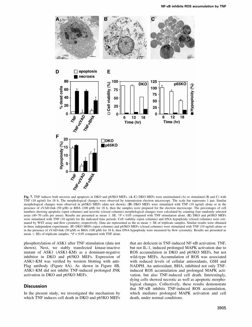
TNF induces both necrosis and apoptosis in DKOand p65KO MEFs, and necrosis is mainly mediatedby ROSSince ROS have been reported to induce both apoptosisand necrosis depending on the cellular context (Fiers et al.,1999), we examined the morphology of dying cells bytransmission electron microscopy. Notably, comparedwith the unstimulated DKO MEFs (Figure 7A), somedying cells showed a necrotic morphological change,which was characterized by loss of membrane integritywithout apparent damage of nuclei (Figure 7B), whereasother cells showed typical apoptotic changes such aschromatin condensation, cell shrinking and apoptoticbodies (Figure 7C). Furthermore, number of necroticcells was substantially reduced in the presence of BHA,but not zVAD-fmk (Figure 7D). Similar morphologicalchanges and inhibitory effects were also observed inp65KO MEFs (data not shown).
To assess the contribution of apoptosis and necrosis toTNF-induced cell death more quantitatively, we per-formed time-course analyses of TNF-induced DNAhypoploidy as an indication of apoptosis using ¯owcytometry. In parallel with these experiments, we alsoexamined the cell viability by WST assay. Viability ofDKO and p65KO MEFs gradually decreased to 50% at18 h after TNF stimulation (Figure 7E). Although the cellswith DNA hypoploidy increased to 15% at 18 h, thispopulation was substantially less than the dead cellpopulation (Figure 7E). These results, in combinationwith the electron microscopy data, indicate that asubstantial part of DKO and p65KO MEFs died ofnecrosis after TNF stimulation (Figure 7E). We alsotested the effects of BHA and z-VAD-fmk on TNF-induced apoptosis. As expected, the TNF-induced apop-tosis was markedly inhibited by z-VAD-fmk. In contrast,BHA treatment substantially increased cell viability(Figure 6A), BHA only slightly decreased population ofDNA hypoploidy (Figure 7F).
A kinase-inactive mutant of ASK1 does not inhibitprolonged JNK activation in DKO and p65KO MEFsPrevious studies have shown that ASK1 is activated byH2O2 and involved in prolonged JNK activation in somecells (Gotoh and Cooper, 1998; Saitoh et al., 1998). Thus,we next examined the contribution of ASK1 to prolongedJNK activation in DKO and p65KO MEFs after TNFstimulation. We ®rst tried an in vitro kinase assay usingimmunoprecipitates with anti-ASK1 antibody or westernblotting with antibody speci®c for phosphorylated ASK1antibody. However, since the expression level of ASK1 inMEFs was low (H.Ichijo, unpublished result), we couldnot detect substantial levels of ASK1 kinase activities or
Fig. 6. Inhibition of TNF-induced cell death in DKO and p65KO MEFsby BHA, z-VAD-fmk and MAPK inhibitors. (A) DKO and p65KOMEFs were stimulated with TNF (10 ng/ml) in the presence ofz-VAD-fmk (10 or 50 mM), BHA (100 mM), BHA (100 mM) +z-VAD-fmk (10 or 50 mM), SP600125 (10 or 30 mM), SB203580(10 or 30 mM) or PD98059 (10 or 30 mM) for 16 h. Cell viability wasdetermined by WST assay as described in Figure 5. (B) DKO andp65KO MEFs were co-transfected with the indicated expression vectors(0.75 mg) along with pEGFP (0.25 mg). After 24 h, the cells weretreated with TNF (10 ng/ml) for 16 h, and then counted by ¯uorescentmicroscopy. The percentage of dead cells among GFP-positive cellswas calculated by counting >100 cells in randomly selected areas.Results are presented as means 6 SEs of triplicate samples andrepresent two individual experiments. *P < 0.05 compared with mocktransfectants.
S.Sakon et al.
3904
phosphorylation of ASK1 after TNF stimulation (data notshown). Next, we stably transfected kinase-inactivemutant of ASK1 (ASK1-KM) as a dominant-negativeinhibitor in DKO and p65KO MEFs. Expression ofASK1-KM was veri®ed by western blotting with anti-Flag antibody (Figure 8A). As shown in Figure 8B,ASK1-KM did not inhibit TNF-induced prolonged JNKactivation in DKO and p65KO MEFs.
Discussion
In the present study, we investigated the mechanism bywhich TNF induces cell death in DKO and p65KO MEFs
that are de®cient in TNF-induced NF-kB activation. TNF,but not IL-1, induced prolonged MAPK activation due toROS accumulation in DKO and p65KO MEFs, but notwild-type MEFs. Accumulation of ROS was associatedwith reduced levels of cellular antioxidants, GSH andNADPH. An antioxidant, BHA, inhibited not only TNF-induced ROS accumulation and prolonged MAPK acti-vation, but also TNF-induced cell death. Interestingly,dying cells showed necrotic as well as apoptotic morpho-logical changes. Collectively, these results demonstratethat NF-kB inhibits TNF-induced ROS accumulation,which mediates prolonged MAPK activation and celldeath, under normal conditions.
Fig. 7. TNF induces both necrosis and apoptosis in DKO and p65KO MEFs. (A±C) DKO MEFs were unstimulated (A) or stimulated (B and C) withTNF (10 ng/ml) for 18 h. The morphological changes were observed by transmission electron microscopy. The scale bar represents 1 mm. Similarmorphological changes were observed in p65KO MEFs (data not shown). (D) DKO MEFs were stimulated with TNF (10 ng/ml) alone or in thepresence of zVAD-fmk (50 mM) or BHA (100 mM) for 18 h, then the samples were prepared for the electron microscope. The percentages of cellnumbers showing apoptotic (open columns) and necrotic (closed columns) morphological changes were calculated by counting four randomly selectedareas (40±70 cells per areas). Results are presented as mean 6 SE. *P < 0.05 compared with TNF stimulation alone. (E) DKO and p65KO MEFswere stimulated with TNF (10 ng/ml) for the indicated time periods. Cell viability (open columns) and DNA hypoploidy (closed columns) were esti-mated by WST assay and ¯ow cytometry, respectively. Data are represented as the as mean 6 SE of triplicate samples. Similar results were obtainedin three independent experiments. (F) DKO MEFs (open columns) and p65KO MEFs (closed columns) were stimulated with TNF (10 ng/ml) alone orin the presence of zVAD-fmk (50 mM) or BHA (100 mM) for 18 h, then DNA hypoploidy were measured by ¯ow cytometry. Results are presented asmean 6 SEs of triplicate samples. *P < 0.05 compared with TNF alone.
NF-kB inhibits ROS accumulation by TNF
3905

It has been reported that NF-kB-inducible genes such asXIAP and GADD45b play an important role in suppress-ing prolonged JNK activation (De Smaele et al., 2001;Tang et al., 2001). However, the molecular mechanism bywhich XIAP and GADD45b inhibit JNK activationremains obscure. We previously identi®ed GADD45b asa molecule interacting with MTK1 and showed thatoverexpression of GADD45b potently activates JNK andp38 pathways (Takekawa and Saito, 1998). Consistentwith these results, stable expression of GADD45b inp65KO MEFs did not substantially inhibit TNF-inducedROS accumulation or prolonged MAPK activation, andcell death (Figure 5D±F). Similarly, XIAP did not inhibitTNF-induced ROS accumulation or prolonged MAPKactivation, although XIAP substantially increased cellviability of TNF-stimulated cells (Figure 5D±F). Thesediscrepancies might result from cell-type differences togenerate GADD45b transfectants (T cell clone versusMEF) and/or differences in experimental systems (transi-ent versus stable transfection of XIAP).
Previous studies suggested an important role of ROS inTNF-induced MAPK activation (Gotoh and Cooper, 1998;Saitoh et al., 1998). However, at least under our experi-mental conditions, we could not detect ROS accumulationin TNF-stimulated wild-type MEFs (Figure 3A). While
BHA almost completely inhibited both ROS accumulationand prolonged MAPK activation in DKO and p65KOMEFs, BHA only slightly inhibited TNF-induced rapid/transient MAPK activation (Figures 2 and 3). Together,ROS appear not to play an essential role in TNF-inducedtransient MAPK activation in MEFs. However, we cannotexclude the possibility that ROS might play a critical rolein other types of cells. In contrast to previous studies (DeSmaele et al., 2001; Javelaud and Besancon, 2001; Tanget al., 2001), activation of not only JNK, but also p38 andERK, was prolonged after TNF stimulation in DKO andp65KO MEFs (Figure 1A and B), which were allabrogated by BHA (Figure 2). Thus, accumulated ROSappear to activate all three MAPK cascades. Consistentwith this notion, exogenously added ROS actually acti-vated three MAPK cascades in MEFs (Figure 3E).Concerning the molecular mechanism by which ROSmediate prolonged JNK activation, ASK1 might partici-pate in this process (Tobiume et al., 2001). However,under our experimental conditions, expression ofASK1-KM did not inhibit TNF-induced prolonged JNKactivation in DKO and p65KO MEFs (Figure 8). Theseresults suggested that a kinase other than ASK1, such asMEKK1, might be also involved in the ROS-induced JNKactivation. It has been also reported that under non-stressed conditions, JNK binds cellular JNK inhibitor,GST, and this interaction is disrupted by ROS, resulting inactivation of JNK (Adler et al., 1999a). Although thesescenarios could explain the mechanism for prolonged JNKactivation, they cannot explain the mechanism for pro-longed ERK activation. In this respect, it is noteworthythat ROS- and UV-induced ERK activation has beenreported to be mediated by c-src- and EGF receptor-dependent pathways (Chen et al., 2001; Kitagawa et al.,2002). We are currently pursuing these possibilities.
While TNF or IL-1 stimulation generally induces rapidand transient MAPK activation, genotoxic stresses such asUV and g-irradiation induce prolonged MAPK activationthat promotes cell death (Xia et al., 1995; Chen et al.,1996; Guo et al., 1998). Consistent with these studies,prolonged JNK activation partly contributed to TNF-induced cell death in DKO MEFs, as demonstrated by theinhibitory effect of a JNK inhibitor, SP600125 (Figure 6A).However, SP600125 did not inhibit TNF-induced celldeath of p65KO MEFs (Figure 6A). Consistent with thedata using MAPK inhibitors, expression of MKK7-KMonly partially inhibited TNF-induced cell death of DKO,but not p65KO, MEFs (Figure 6B). The fact that inhibitionof JNK cascade did not affect TNF-induced cell death ofp65KO MEFs may be explained by the fact that TRAF-mediated transient JNK activation was intact in p65KOMEFs but not DKO MEFs (Figure 1A and B). Given thattransient JNK activation transmits survival signals (Davis,2000), SP600125 or MKK7-KM might cancel bothsurvival and apoptotic signals in p65KO MEFs, resultingin no apparent inhibitory effect on TNF-induced celldeath.
Our present study also demonstrates that TNF-inducedROS accumulation can be induced independently ofTRAF2 and TRAF5. In addition, IL-1 signaling that ismediated by TRAF6 could not induce ROS accumulation(Figure 3D). Together, TRAF family proteins appear notto be essential for ROS accumulation. Given that
Fig. 8. Introduction of ASK1-KM does not inhibit TNF-induced pro-longed JNK activation in DKO and p65KO MEFs. (A) Total celllysates were blotted with anti-Flag antibody. (B) Mock- or ASK1-KM-transfected DKO and p65KO MEFs were stimulated with TNF(50 ng/ml) for the indicated time periods, and the lysates were analyzedas described in Figure 1.
S.Sakon et al.
3906
z-VAD-fmk did not inhibit TNF-induced ROS accumula-tion (Figure 3C), TRADD, FADD or RIP, but notcaspase-8, might play a critical role in this process. Inthis respect, RIP has been reported to be responsible forFas-, TNF- or TRAIL-induced necrosis (Holler et al.,2000), but the contribution of ROS to RIP-mediated celldeath has not been determined. Further study is required toaddress whether RIP is essential for TNF-induced ROSaccumulation.
Here we show that reduced cellular levels of GSH andNADPH are correlated with accumulation of ROS in TNF-stimulated DKO and p65KO MEFs. ROS are generated inmitochondria and rapidly eliminated by antioxidants undernormal conditions. Thus, the function of some antioxidantenzyme, the expression level or activity of which isregulated by NF-kB, might be impaired in DKO andp65KO MEFs. Previous studies showed that manganese-dependent SOD (MnSOD) is upregulated by TNF in anNF-kB-dependent manner, and overexpression of MnSODinhibits ROS-induced apoptosis (Tanaka et al., 2002).While basal expression levels of MnSOD mRNA were notdifferent between wild-type and DKO or p65KO MEFs,induction of MnSOD mRNA by TNF was substantiallyimpaired in DKO and p65KO MEFs (data not shown).However, stable transfection of MnSOD into DKO orp65KO MEFs did not signi®cantly inhibit TNF-inducedROS accumulation or cell death (data not shown). Takenthat the primary target of MnSOD is O2
± and the mainaccumulated ROS in DKO and p65KO MEFs were H2O2
or hydroxyl radical, the basal expression level of MnSODmight be suf®cient for elimination of O2
±. Regardingscavengers to detoxify H2O2, there are many antioxidantenzymes, including catalase, GPxs and PRxs (Thannickaland Fanburg, 2000). So far, we have observed nosigni®cant inhibitory effect of such antioxidant enzymes,including GPx1, GPx2, PRx1 and PRx2 on TNF-inducedROS accumulation in DKO or p65KO MEFs after stabletransfection (data not shown). Further study is nowunderway to identify an antioxidant enzyme responsiblefor the ROS accumulation.
Another interesting point from this study is that asubstantial part of DKO and p65KO MEFs died by necrosisafter TNF stimulation, as assessed by electron microscopyand ¯ow cytometry (Figure 7A±E). Furthermore, takenthat BHA only marginally inhibited the emergence ofapoptotic cells (Figure 7F), but substantially inhibitednecrotic morphological changes (Figure 7D), ROS mainly,but not exclusively, contribute to necrotic cell death. Theseresults are reminiscent of murine ®brosarcoma cell line,L929, in which TNF induces ROS-dependent necrosis(Vercammen et al., 1998). On the other hand, previousstudies using p65KO mice implied an important role ofNF-kB in protection from TNF-induced apoptosis ofhepatocytes (Beg and Baltimore, 1996; Doi et al., 1999).It will be interesting to determine whether necroticmorphological changes are also observed in hepatocytesin p65KO mice.
In summary, our present study demonstrates a criticalrole of NF-kB to inhibit TNF-induced ROS accumulationunder normal conditions. Elucidation of the mechanism bywhich NF-kB eliminates ROS would be helpful inmanipulating various pathological conditions, such as
ischemia or aging, in which ROS are deeply implicated inthe pathogenesis.
Materials and methods
Reagents and cell cultureRecombinant murine TNF and murine IL-1b were purchased from BDPharMingen. NAC and reduced form of GSH were purchased fromSigma. SP600125, SB203580 and PD98059 were purchased from Biomoland Calbiochem. Antibodies speci®c for phospho-JNK, phospho-p38,phospho-ERK, phospho-MAPKAP2, JNK, p38, ERK and MAPKAP2were purchased from Cell Signaling. Anti-Flag and anti-HA antibodieswere purchased from Sigma and Roche Diagnostics Corp., respectively.BHA, z-VAD-fmk, CM-H2DCFDA and DHE were purchased from WakoPure Chemicals, Peptide Institute and Molecular Probes. Wild-type, DKOand p65KO MEFs, and Phoenix-Eco cells were cultured in high-glucoseDMEM containing 10% fetal calf serum (FCS).
Western blot analysisMEFs (1 3 106 cells) were lysed in a buffer containing 50 mM Tris±HClpH 8.0, 150 mM NaCl, 1% NP-40, 0.5% deoxycholate, 0.1% SDS, 1 mMphenylmethylsulfonyl ¯uoride and 1 mg/ml aprotinin. After centrifuga-tion, cell lysates were subjected to 10% SDS±PAGE and transferred ontopolyvinylidene di¯uoride membranes (Millipore). The membranes wereblotted with antibodies to phospho-JNK, phospho-p38, phospho-ERK orphospho-MAPKAP2, and reblotted with antibodies to total JNK, totalp38, total ERK or total MAPKAP2, respectively. To detect transfectedgene products, the membranes were blotted with anti-Flag or anti-HAantibody. The membranes were developed with EnhancedChemiluminescence (ECL) Western Blotting Detection System Plus(Amersham Pharmacia Biotech.) according to the manufacturer'sinstruction.
Measurement of ROS accumulationMEFs (2 3 105 cells) were plated in six-well plates and stimulated withTNF or IL-1 in phenol red-free medium for the indicated time periods.After stimulation, the cells were incubated with CM-H2DCFDA or DHEin the dark for 30 min at 37°C. Then, the cells were harvested andanalyzed on a ¯ow cytometer (FACSCalibur; BD Biosciences). Datawere processed by using the CellQuest program (BD Biosciences).
Generation of stable transfectantsGeneration of stable transfectants by using retroviral vectors wasperformed as described previously (Sasazuki et al., 2002). Retroviralexpression vectors for TRAF2, TRAF5, p65, GADD45b, XIAP or ASK1-KM were constructed by inserting each cDNA into pMX-puro. Phoenix-Eco cells (1.5 3 106 cells) were transiently transfected with 3 mg of theindicated vectors using LipofectAMINE according to the manufacturer'sinstruction (Invitrogen) to generate viral supernatants. After infection,DKO and p65KO MEFs were selected with 2.5 mg/ml puromycin (Sigma)to isolate stable transfectants. Puromycin-resistant pools were used for theexperiments.
Measurement of GSHIntracellular GSH levels were measured by using a colorimetric assay kit(OxisResearchÔ) according to the manufacturer's instruction. Brie¯y,MEFs (2 3 106 cells) were unstimulated or stimulated with TNF for 4 hand harvested. The cells were resuspended in 500 ml of 5% meta-phosphoric acid solution and sonicated using an ultrasonic sonicator(AstrasonÔ). After centrifugation, the supernatants were sequentiallyincubated with 4-chloro-1-methyl-7-tri¯uromethyl-quindinium methyl-sulfate and NaOH, and then the absorbance at 400 nm was measured on aspectrophotometer. The pellets were lysed in the lysis buffer and theprotein concentration was measured by the Bradford method. GSHcontents were normalized by the protein concentration of each sample andare presented as GSH level per microgram of cell lysate.
Measurement of NADPHNADPH levels were measured by the method of Gibon and Larher(1997). Brie¯y, MEFs (2.5 3 106 cells) were unstimulated or stimulatedwith TNF for 4 h. The cells were harvested and resuspended in 250 ml of0.1 N NaOH, followed by brief homogenization. The homogenates wereheated at 95°C for 5 min. After centrifugation, the supernatants wereneutralized with 250 ml of 0.1 N HCl. Then, the supernatantswere incubated with a buffer containing 400 mM NaCl, 100 mM
NF-kB inhibits ROS accumulation by TNF
3907

Tris±HCl pH. 8.0, 4 mM EDTA, 0.42 mM 3-(4,5-dimethylthiazolyl-2)-2,5-diphenyltetrazolium bromide (MTT), 1.66 mM phenzine ethosulfate(PES), 2.5 mM NADP+ and 14 U of glucose 6-phosphate dehydrogenase(G6PDH) at 37°C for 40 min. The reaction was stopped by addition of666 ml of 5 M NaCl, then centrifuged at 17 400 g at 4°C for 5 min. Thepellets were lysed in 1 ml of 98% ethanol and the absorbance at 570 nmwas measured on a spectrophotometer. The cellular NADPH contentswere determined by using an NADPH standard curve.
WST assayMEFs (5 3 103 cells) were plated in 96-well plates and cultured for 12 hin DMEM containing 10% FCS. The cells were then stimulated with TNFin the presence or absence of various inhibitors for 16 h. Cell viability wasdetermined by WST assay using a Cell Counting kit (Dojindo).
Cell death assayMEFs (1 3 105 cells) were cotransfected with the indicated expressionvectors along with an expression vector for GFP (pEGFP) (Clontech)using LipofectAMINE. Expression vectors for MKK1-KM, MKK7-KMand MKK6-KM were constructed by inserting each cDNA intopcDNA3.1 (Invitrogen). After 24 h, cells were treated with TNF for16 h, then GFP-positive cells were counted by ¯uorescent microscopy.Cells showing ¯at or round morphology were considered to be live ordead, respectively. Expression of transfected gene products was veri®edby western blotting with anti-Myc (for MKK1-KM and MKK7-KM) oranti-HA (for MKK6, TRAF2 and p65) antibody (data not shown).
Electron microscopyMEFs (4 3 106 cells) were unstimulated or stimulated with TNF in thepresence or absence of various inhibitors for 18 h, and then serially ®xedwith 2% glutaraldehyde in phosphate-buffered saline for 2 h and then with2% OsO4 for 2 h before embedding in Epon 812. Thin sections wereprepared using a MT-5000 ultramicrotome (Dupont Pharmaceuticals),stained with uranyl acetate followed by lead citrate, and then observed ona JEM1230 electron transmission microscope (JEOL). To evaluate thepercentages of cell numbers showing apoptotic and necrotic morpho-logical changes, sections were stained with toluidine blue and used forcell counting.
Statistical analysisStatistical analysis was performed by Student's t-test. A P value <0.05was considered to be signi®cant.
Supplementary dataSupplementary data are available at The EMBO Journal Online.
Acknowledgements
We thank H.Sakurai, R.Takahashi, T.Kitamura, E.Nishida, C.Marshall,G.P.Nolan, W.-C.Yeh and T.W.Mak for providing reagents and Traf2±/±
mice. We also thank H.Ichijo and H.Nishina for helpful discussion, andA.Matsuzawa and H.Nishitoh for technical advice. This work wassupported in part by Grants-in-Aid for Scienti®c Research on PriorityAreas (C) from the Ministry of Education, Culture, Sports, Science andTechnology, Grant-in-Aid for Scienti®c Research (B) from Japan Societyfor the Promotion of Science, Japan, and by a Grant from Human FrontierScience Program (HFSP).
References
Adler,V. et al. (1999a) Regulation of JNK signaling by GSTp. EMBO J.,18, 1321±1334.
Adler,V., Yin,Z., Tew,K.D. and Ronai,Z. (1999b) Role of redoxpotential and reactive oxygen species in stress signaling. Oncogene,18, 6104±6111.
Barkett,M. and Gilmore,T.D. (1999) Control of apoptosis by Rel/NF-kBtranscription factors. Oncogene, 18, 6910±6924.
Baud,V., Liu,Z.G., Bennett,B., Suzuki,N., Xia,Y. and Karin,M. (1999)Signaling by proin¯ammatory cytokines: oligomerization of TRAF2and TRAF6 is suf®cient for JNK and IKK activation and target geneinduction via an amino-terminal effector domain. Genes Dev., 13,1297±1308.
Beg,A.A. and Baltimore,D. (1996) An essential role for NF-kB inpreventing TNF-a-induced cell death. Science, 274, 782±784.
Chen,K., Vita,J.A., Berk,B.C. and Keaney,J.F.,Jr (2001) c-Jun
N-terminal kinase activation by hydrogen peroxide in endothelialcells involves SRC-dependent epidermal growth factor receptortransactivation. J. Biol. Chem., 276, 16045±16050.
Chen,Y.R., Wang,X., Templeton,D., Davis,R.J. and Tan,T.H. (1996) Therole of c-Jun N-terminal kinase (JNK) in apoptosis induced byultraviolet C and gamma radiation. Duration of JNK activation maydetermine cell death and proliferation. J. Biol. Chem., 271, 31929±31936.
Chung,J.Y., Park,Y.C., Ye,H. and Wu,H. (2002) All TRAFs are notcreated equal: common and distinct molecular mechanisms of TRAF-mediated signal transduction. J. Cell Sci., 115, 679±688.
Davis,R.J. (2000) Signal transduction by the JNK group of MAP kinases.Cell, 103, 239±252.
De Smaele,E., Zazzeroni,F., Papa,S., Nguyen,D.U., Jin,R., Jones,J.,Cong,R. and Franzoso,G. (2001) Induction of gadd45b by NF-kBdownregulates pro-apoptotic JNK signalling. Nature, 414, 308±313.
Doi,T.S., Marino,M.W., Takahashi,T., Yoshida,T., Sakakura,T., Old,L.J.and Obata,Y. (1999) Absence of tumor necrosis factor rescues RelA-de®cient mice from embryonic lethality. Proc. Natl Acad. Sci. USA,96, 2994±2999.
Fiers,W., Beyaert,R., Declercq,W. and Vandenabeele,P. (1999) Morethan one way to die: apoptosis, necrosis and reactive oxygen damage.Oncogene, 18, 7719±7730.
Ghosh,S., May,M.J. and Kopp,E.B. (1998) NF-kB and Rel proteins:evolutionarily conserved mediators of immune responses. Annu. Rev.Immunol., 16, 225±260.
Gibon,Y. and Larher,F. (1997) Cycling assay for nicotinamide adeninedinucleotides: NaCl precipitation and ethanol solubilization of thereduced tetrazolium. Anal. Biochem., 251, 153±157.
Gotoh,Y. and Cooper,J.A. (1998) Reactive oxygen species- anddimerization-induced activation of apoptosis signal-regulating kinase1 in tumor necrosis factor-alpha signal transduction. J. Biol. Chem.,273, 17477±17482.
Guo,Y.L., Baysal,K., Kang,B., Yang,L.J. and Williamson,J.R. (1998)Correlation between sustained c-Jun N-terminal protein kinaseactivation and apoptosis induced by tumor necrosis factor-alpha inrat mesangial cells. J. Biol. Chem., 273, 4027±4034.
Holler,N. et al. (2000) Fas triggers an alternative, caspase-8-independentcell death pathway using the kinase RIP as effector molecule. Nat.Immunol., 1, 489±495.
Ichijo,H. (1999) From receptors to stress-activated MAP kinases.Oncogene, 18, 6087±6093.
Inoue,J., Ishida,T., Tsukamoto,N., Kobayashi,N., Naito,A., Azuma,S.and Yamamoto,T. (2000) Tumor necrosis factor receptor-associatedfactor (TRAF) family: adapter proteins that mediate cytokinesignaling. Exp. Cell Res., 254, 14±24.
Javelaud,D. and Besancon,F. (2001) NF-kB activation results in rapidinactivation of JNK in TNFa-treated Ewing sarcoma cells: amechanism for the anti-apoptotic effect of NF-kappa B. Oncogene,20, 4365±4372.
Karin,M. and Lin,A. (2002) NF-kB at the crossroads of life and death.Nat. Immunol., 3, 221±227.
Kitagawa,D. et al. (2002) Activation of extracellular signal-regulatedkinase by ultraviolet is mediated through Src-dependent epidermalgrowth factor receptor phosphorylation. Its implication in an anti-apoptotic function. J. Biol. Chem., 277, 366±371.
Lomaga,M.A. et al. (1999) TRAF6 de®ciency results in osteopetrosisand defective interleukin-1, CD40 and LPS signaling. Genes Dev., 13,1015±1024.
Mak,T.W. and Yeh,W.C. (2002) Signaling for survival and apoptosis inthe immune system. Arthritis Res., 4, S243±S252.
Naito,A. et al. (1999) Severe osteopetrosis, defective interleukin-1signalling and lymph node organogenesis in TRAF6-de®cient mice.Genes Cells, 4, 353±362.
Ninomiya-Tsuji,J., Kishimoto,K., Hiyama,A., Inoue,J., Cao,Z. andMatsumoto,K. (1999) The kinase TAK1 can activate the NIK-IkBas well as the MAP kinase cascade in the IL-1 signalling pathway.Nature, 398, 252±256.
Nishitoh,H., Saitoh,M., Mochida,Y., Takeda,K., Nakano,H., Rothe,M.,Miyazono,K. and Ichijo,H. (1998) ASK1 is essential for JNK/SAPKactivation by TRAF2. Mol. Cell, 2, 389±395.
Saitoh,M., Nishitoh,H., Fujii,M., Takeda,K., Tobiume,K., Sawada,Y.,Kawabata,M., Miyazono,K. and Ichijo,H. (1998) Mammalianthioredoxin is a direct inhibitor of apoptosis signal-regulating kinase(ASK) 1. EMBO J., 17, 2596±2606.
Sasazuki,T. et al. (2002) Identi®cation of a novel transcriptional
S.Sakon et al.
3908

activator, BSAC, by a functional cloning to inhibit tumor necrosisfactor-induced cell death. J. Biol. Chem., 277, 28853±28860.
Tada,K. et al. (2001) Critical roles of TRAF2 and TRAF5 in tumornecrosis factor-induced NF-kB activation and protection from celldeath. J. Biol. Chem., 276, 36530±36534.
Takekawa,M. and Saito,H. (1998) A family of stress-inducibleGADD45-like proteins mediate activation of the stress-responsiveMTK1/MEKK4 MAPKKK. Cell, 95, 521±530.
Tanaka,H., Matsumura,I., Ezoe,S., Satoh,Y., Sakamaki,T., Albanese,C.,Machii,T., Pestell,R.G. and Kanakura,Y. (2002) E2F1 and c-Mycpotentiate apoptosis through inhibition of NF-kB activity thatfacilitates MnSOD-mediated ROS elimination. Mol. Cell, 9, 1017±1029.
Tang,G., Minemoto,Y., Dibling,B., Purcell,N.H., Li,Z., Karin,M. andLin,A. (2001) Inhibition of JNK activation through NF-kB targetgenes. Nature, 414, 313±317.
Thannickal,V.J. and Fanburg,B.L. (2000) Reactive oxygen species in cellsignaling. Am. J. Physiol. Lung Cell. Mol. Physiol., 279, L1005±L1028.
Tobiume,K. et al. (2001) ASK1 is required for sustained activations ofJNK/p38 MAP kinases and apoptosis. EMBO Rep., 2, 222±228.
Tracey,K.J. and Cerami,A. (1994) TUMOR NECROSIS FACTOR: Apleiotropic cytokine and therapeutic target. Annu. Rev. Immunol., 45,491±503.
Vercammen,D., Beyaert,R., Denecker,G., Goossens,V., Van Loo,G.,Declercq,W., Grooten,J., Fiers,W. and Vandenabeele,P. (1998)Inhibition of caspases increases the sensitivity of L929 cells tonecrosis mediated by tumor necrosis factor. J. Exp. Med., 187, 1477±1485.
Wallach,D. et al. (2002) How are the regulators regulated? The searchfor mechanisms that impose speci®city on induction of cell death andNF-kB activation by members of the TNF/NGF receptor family.Arthritis Res., 4, S189±S196.
Xia,Z., Dickens,M., Raingeaud,J., Davis,R.J. and Greenberg,M.E. (1995)Opposing effects of ERK and JNK-p38 MAP kinases on apoptosis.Science, 270, 1326±1331.
Yeh,W.-C. et al. (1997) Early lethality, functional NF-kB activation andincreased sensitivity to TNF-induced cell death in TRAF2-de®cientmice. Immunity, 7, 715±725.
Received January 16, 2003; revised May 22, 2003;accepted June 6, 2003
NF-kB inhibits ROS accumulation by TNF
3909
Related Documents