International Journal of Scientific and Research Publications, Volume 5, Issue 3, March 2015 1 ISSN 2250-3153 www.ijsrp.org New Approach for Improving Production of Naja haje Snake Antivenom Heba M. Karam*, Esmat A. Shaaban*, Aly F. Mohamed**, Hala F. Zaki, and Sanaa A. Kenawy Department of Pharmacology and Toxicology − Faculty of Pharmacy − Cairo University, Egypt * Department of Drug Radiation Research−National Center for Radiation Research and Technology −Atomic Energy Authority, Egypt ** The Holding Company for Biological Products-Vaccines and Drugs (VACSERA), Egypt Abstract- Snake-bite is considered a neglected tropical disease that affects thousands of people worldwide. Administration of antivenom is the corner stone in the therapy of snake bite. The study aimed to improve the production of antivenom using calcium phosphate nanoparticles (CPN) as adjuvant and gamma irradiation to detoxify venom. This was carried out by studying the toxicological and immunological properties of the Naja haje venom before and after exposure to 2 KGy gamma radiation. Furthermore, the cardiotoxic and hepatotoxic biomarkers of the envenomed rats were examined to compare the effect of native and gamma irradiated venoms. Moreover, in order to achieve the goal of the present study the immune response of immunized rabbits was evaluated through determination of antibody titer using ELISA technique and comparing the neutralizing capacity for lethality and enzyme activities of the serum obtained from rabbits inoculated with Naja haje venom in its native and γ irradiated form in presence of CPN as adjuvant or complete Freund's adjuvant. Data revealed that the toxicity of γ irradiated Naja haje venom was reduced 6 times as compared to the native venom. There was no change in the antigenic reactivity between both native and γ irradiated Naja haje venoms. Furthermore, injection of γ irradiated Naja haje venom did not significantly change activities of serum LDH, CPK, CK-MB, ALT and AST as compared to the normal group. In addition, serum titer produced with γ irradiated venom loaded on CPN showed highest titer as compared to other sera. Serum produced from irradiated Naja haje showed higher neutralizing capacity than that from native venom. All prepared antivenoms were able to neutralize the cardiotoxic and hepatotoxic biomarkers. Index Terms- Antivenom production, ELISA, gamma irradiation, Naja haje venom, nanoparticles. I. INTRODUCTION enomous snakes are some of the most dangerous poisoning animals in the world. Their bites may be serious depending on the amount of venom injected, the location of the bite, the size of the victim, the species of the snake and the amount of time between the bite and the injection of the right antivenin. Poisoning by snake bite is a real clinical problem, especially in tropical areas, and efficacious treatment should be available. (Chippaux, 1991). There are many varieties of snakes in Egypt, some of them cause severe damage to snake bite victims. Cobra is one of the major causes of snake bites death in Egypt. (Shaaban 2005). Cobra venoms cause death by the action of their neurotoxic and cardiotoxic components (Mebs, 2002). Serotherapy is the treatment of choice in snake-bite accidents. Clinical investigations have established that generally antivenoms are highly effective in the neutralization of toxins responsible for systemic effects such as hemorrhage, coagulopathy, hemodynamic disturbances and neurotoxicity (Warrell, 2003). The production of therapeutic antivenoms against venoms from Elapidae family has proven to be very difficult where, the low molecular size of the neurotoxins confers low immunogenicity, resulting in the production of antibodies of relatively low potency (Ownby and Colberg 1988). To improve antisera production and extend the useful life of immunized horse much effort has been devoted to decrease chronic venom toxicity. Several techniques have been used to detoxify venom, for preparing effective toxoid, such as mixing the venom with adjuvant which adsorbs the venom, as aluminum hydroxide gel (Christensen, 1955), using mixture of the venom with carboxymethyl cellulose (Moroz et al., 1963), adding chemical agent as formaldehyde (Costa et al., 1985), controlled iodination of the venom (Daniel et al., 1987) and encapsulation of purified toxins in liposomes (Freitas and Frezard, 1997). Towards more effective and safer antivenins, one method that has been shown to be effective for attenuating venom toxicity and maintaining venom immunogenicity is gamma irradiation (Nascimento et al., 1996; Shaaban et al., 1996; Clissa et al., 1999; Souza et al., 2002; Oussedik-Oumehdi & Laraba-Djebari, 2011). Adjuvants are substances injected along with an antigen that are intended to enhance the immune response to the antigen. The most widely used is Freund’s adjuvant but it poses a great problem in commercial antivenom production since they induce inflammation and lesions at the inoculum site leading to shortening the longevity of serum-producing animals (Ferreira et al. 2010). Many adverse effects were noted in horses used for the production of antivenin, mainly in the form of tissue reaction at the site of injection such as of edema, abscesses, myonecrosis and fibrosis. Micro and nanocarriers such as microspheres, liposomes and nanoparticles have many advantages concerning drug delivery and targeting. These advantages include high drug loading, lack of chemical interaction with drug, which is necessary for encapsulation and considerable protection of the drug molecules (Crommelin et al., 2001). In this respect, this study aimed to enhance the production of snake antivenoms through the use of gamma irradiation of Naja haje venom as detoxifying tool and calcium phosphate nanoparticles as an adjuvant to minimize the adverse reactions during the hyper-immunization process and reduce manufacturing costs. V

Welcome message from author
This document is posted to help you gain knowledge. Please leave a comment to let me know what you think about it! Share it to your friends and learn new things together.
Transcript
International Journal of Scientific and Research Publications, Volume 5, Issue 3, March 2015 1 ISSN 2250-3153
www.ijsrp.org
New Approach for Improving Production of Naja haje
Snake Antivenom
Heba M. Karam*, Esmat A. Shaaban*, Aly F. Mohamed**, Hala F. Zaki, and Sanaa A. Kenawy
Department of Pharmacology and Toxicology − Faculty of Pharmacy − Cairo University, Egypt *Department of Drug Radiation Research−National Center for Radiation Research and Technology −Atomic Energy Authority, Egypt
**The Holding Company for Biological Products-Vaccines and Drugs (VACSERA), Egypt
Abstract- Snake-bite is considered a neglected tropical disease
that affects thousands of people worldwide. Administration of
antivenom is the corner stone in the therapy of snake bite. The
study aimed to improve the production of antivenom using
calcium phosphate nanoparticles (CPN) as adjuvant and gamma
irradiation to detoxify venom. This was carried out by studying
the toxicological and immunological properties of the Naja haje
venom before and after exposure to 2 KGy gamma radiation.
Furthermore, the cardiotoxic and hepatotoxic biomarkers of the
envenomed rats were examined to compare the effect of native
and gamma irradiated venoms. Moreover, in order to achieve the
goal of the present study the immune response of immunized
rabbits was evaluated through determination of antibody titer
using ELISA technique and comparing the neutralizing capacity
for lethality and enzyme activities of the serum obtained from
rabbits inoculated with Naja haje venom in its native and γ
irradiated form in presence of CPN as adjuvant or complete
Freund's adjuvant. Data revealed that the toxicity of γ irradiated
Naja haje venom was reduced 6 times as compared to the native
venom. There was no change in the antigenic reactivity between
both native and γ irradiated Naja haje venoms. Furthermore,
injection of γ irradiated Naja haje venom did not significantly
change activities of serum LDH, CPK, CK-MB, ALT and AST
as compared to the normal group. In addition, serum titer
produced with γ irradiated venom loaded on CPN showed
highest titer as compared to other sera. Serum produced from
irradiated Naja haje showed higher neutralizing capacity than
that from native venom. All prepared antivenoms were able to
neutralize the cardiotoxic and hepatotoxic biomarkers.
Index Terms- Antivenom production, ELISA, gamma
irradiation, Naja haje venom, nanoparticles.
I. INTRODUCTION
enomous snakes are some of the most dangerous poisoning
animals in the world. Their bites may be serious depending
on the amount of venom injected, the location of the bite, the size
of the victim, the species of the snake and the amount of time
between the bite and the injection of the right antivenin.
Poisoning by snake bite is a real clinical problem, especially in
tropical areas, and efficacious treatment should be available.
(Chippaux, 1991). There are many varieties of snakes in Egypt,
some of them cause severe damage to snake bite victims. Cobra
is one of the major causes of snake bites death in Egypt.
(Shaaban 2005). Cobra venoms cause death by the action of
their neurotoxic and cardiotoxic components (Mebs, 2002).
Serotherapy is the treatment of choice in snake-bite accidents.
Clinical investigations have established that generally
antivenoms are highly effective in the neutralization of toxins
responsible for systemic effects such as hemorrhage,
coagulopathy, hemodynamic disturbances and neurotoxicity
(Warrell, 2003). The production of therapeutic antivenoms
against venoms from Elapidae family has proven to be very
difficult where, the low molecular size of the neurotoxins confers
low immunogenicity, resulting in the production of antibodies of
relatively low potency (Ownby and Colberg 1988). To improve
antisera production and extend the useful life of immunized
horse much effort has been devoted to decrease chronic venom
toxicity. Several techniques have been used to detoxify venom,
for preparing effective toxoid, such as mixing the venom with
adjuvant which adsorbs the venom, as aluminum hydroxide gel
(Christensen, 1955), using mixture of the venom with
carboxymethyl cellulose (Moroz et al., 1963), adding chemical
agent as formaldehyde (Costa et al., 1985), controlled iodination
of the venom (Daniel et al., 1987) and encapsulation of purified
toxins in liposomes (Freitas and Frezard, 1997). Towards more
effective and safer antivenins, one method that has been shown
to be effective for attenuating venom toxicity and maintaining
venom immunogenicity is gamma irradiation (Nascimento et al.,
1996; Shaaban et al., 1996; Clissa et al., 1999; Souza et al.,
2002; Oussedik-Oumehdi & Laraba-Djebari, 2011). Adjuvants are substances injected along with an antigen that
are intended to enhance the immune response to the antigen. The
most widely used is Freund’s adjuvant but it poses a great
problem in commercial antivenom production since they induce
inflammation and lesions at the inoculum site leading to
shortening the longevity of serum-producing animals (Ferreira
et al. 2010). Many adverse effects were noted in horses used for
the production of antivenin, mainly in the form of tissue reaction
at the site of injection such as of edema, abscesses, myonecrosis
and fibrosis. Micro and nanocarriers such as microspheres,
liposomes and nanoparticles have many advantages concerning
drug delivery and targeting. These advantages include high drug
loading, lack of chemical interaction with drug, which is
necessary for encapsulation and considerable protection of the
drug molecules (Crommelin et al., 2001). In this respect, this study aimed to enhance the production of
snake antivenoms through the use of gamma irradiation of Naja
haje venom as detoxifying tool and calcium phosphate
nanoparticles as an adjuvant to minimize the adverse reactions
during the hyper-immunization process and reduce
manufacturing costs.
V
International Journal of Scientific and Research Publications, Volume 5, Issue 3, March 2015 2
ISSN 2250-3153
www.ijsrp.org
II. MATERIALS AND METHODS
Animals used in the present study included New Zealand male
rabbits (2.5-3 kg), Swiss albino male mice (20-25 g) and Wistar
albino male rats (180-200 g). Animals were purchased from the
National Research Center (Giza, Egypt). The study was
conducted in accordance with the regulations approved by the
Ethics Committee at Faculty of Pharmacy, Cairo University.
Venom: Lyophilized crude venom of Naja haje (Cobra) snake
venom was kindly supplied from the laboratory animal unit of
Medical Research Center, Faculty of Medicine, Ain Shams
University.
Irradiation of venom: In this study, Naja haje venom was
dissolved in saline solution (1mg/ml). Samples were subjected to
radiation dose level of 2 KGy at the National Center for
Radiation Research and Technology (NCRRT) using cobalt-60
Indian gamma cell (GE 4000A). The radiation dose rate was 1.26
Gy/sec at the time of experiment. This dose was selected as it
gets rid of venom toxicity while maintaining immunogenicity
(Clissa et al. 1999; Karam et al., 2010).
Determination of lethal dose fifty (LD50) of native and γ
irradiated venoms. LD50 of native and γ irradiated Naja haje
venoms were determined according to Spearman-karber method
by Finney (1964).
Evaluation of the immunological properties of native and γ
irradiated venoms. Effect of irradiation on the immunological
properties of Naja haje venom was evaluated using double
immunodiffusion technique as described by Ouchterlony (1948).
In immunodiffusion plates saline, native and γ irradiated Naja
haje venoms solution (20 µl) were placed in peripheral wells
(venom concentration were 20 mg/ml), whereas the central well
was filled with 20 µl of antivenom. After developing of the
precipitation bands (72 h), slides were washed and dried then
stained and photographed.
Evaluation of the biochemical activities of native and γ
irradiated venoms. Toxic effects of native and γ irradiated Naja
haje venoms were evaluated through determination of the
cardiotoxic and hepatotoxic biomarkers in rats. Since, the LD50 of
native venom was measured in mice; the equivalent rat dose was
calculated according to Paget and Barnes (1964). Rats were
classified into three groups, each consisting of seven rats that
were treated as follows:
Group І: received 0.1 ml saline i.p. and served as normal control.
Group ІІ: received native Naja haje venom (0.163 mg/kg; i.p.).
Group ІІІ: received γ irradiated Naja haje venom (0.163 mg/kg;
i.p.).
After 4 h of envenomation (Mohamed et al., 1981), rats were
anesthetized by i.p. injection of urethane (1.2 g/kg) (Flecknell,
1987). Blood samples were withdrawn via the retro-orbital vein
using heparinized capillary tubes (Cocchetto and Bjornsoon,
1983) for serum separation.
Lactate dehydrogenase (LDH) activity was measured using a
test reagent kit according to the method of Stentz (2010), creatine
phosphokinase (CPK) activity was measured using a test reagent
kit according to the method of Szasz et al. (1976), and creatine
kinase isoenzyme (CK-MB) activity was measured using a test
reagent kit according to the method of Lott and Stang (1980).
Moreover, serum aspartate aminotransferase (AST) and alanine
aminotransferase (ALT) activities were determined using a test
reagent kit according to the method of Retiman and Frankel
(1957).
Preparation and characterization of calcium phosphate
nanoparticles (CPN) were prepared to be used as adjuvant for
Naja haje venom in antivenoms preparation. This was performed
according to the method of He et al. (2000). Particle size and
morphological feature of prepared CPN was observed using
transmission electron microscope TEM (JEOL JEM-1230, Japan)
(Vacsera) according to the method described by Van der et al.
(2003). The structure features of venom and venom loaded
nanoparticles were estimated by Fourier transform infrared
(FTIR) (3600 JASCO, Colchester United Kingdom) (Vacsera) at
room temperature. For comparison, venom solution was
measured by the same process.
Preparation of antivenoms
Rabbits were used as antivenom producing animals. They
were classified into four groups each included 3 rabbits that were
treated as follows:
Group 1: was injected s.c. with native venom emulsified in 0.5
ml complete Freund's adjuvant (CFA).
Group 2: was injected s.c. with 2KGy irradiated venom
emulsified in 0.5 ml CFA.
Group 3: was injected s.c. with native venom loaded on calcium
phosphate nanoparticles (CPN).
Group 4: was injected s.c. with 2KGy irradiated venom loaded
on CPN.
Immunization was carried out as described by WHO (2010).
Ten days after the final dose, rabbits were injected with a booster
dose without adjuvants 500 µg/ml of native venom for groups 1
and 3 meanwhile 500 µg/ml of irradiated venom were used for
groups 2 and 4. Blood samples were collected ten days thereafter.
Serum was distributed in small tubes and kept at -20 °C until the
moment of use for evaluation.
Evaluation of immune response post immunization using
enzyme-linked immunosorbent assay (ELISA) Blood samples
were withdrawn post immunization. The levels of specific
antibodies in serum samples were compared using ELISA
according to the method of Nascimento (1996).
Neutralization of lethality
1- In-vitro neutralization (Pre-incubation type assay)
This was done according to the method of Gutiearrez et al.
(1990). Mixtures containing a constant amount of venom (10
LD50) and varying dilutions of antivenoms were incubated at
37ºC for 1h. Aliquots of the mixtures (0.5 ml) were i.p. injected
into groups of six mice. Control included venom alone. Deaths
were recorded after 24 h. Median effective dose (ED50) was
calculated by spearman-karber analysis and defined as the ratio
of antivenom (µl): venom (mg) at which 50% of mice were
protected
International Journal of Scientific and Research Publications, Volume 5, Issue 3, March 2015 3
ISSN 2250-3153
www.ijsrp.org
2- In-vivo neutralization (independent type assay)
This assay is considered as in vivo neutralization where
independent injection of venom and antivenom attempted to
simulate the natural route of envenomation. In these assays, a
challenge dose of venom (2 LD50) was i.p. injected first and then,
at various time intervals, antivenom is administered i.v.
according to the method of Leon et al. (2001).
Neutralization of biochemical activities
It was assessed as described by Ghazal, et al. (1975). Venom
was incubated with each of the prepared antivenom for 1 h at
37°C in a ratio of 1: 4 (1 mg venom to 4 ml serum). Then,
aliquots of the mixtures containing a challenge dose equivalent to
1 LD50 of venom were i.p. injected to rats that were classified
into five groups, each consisting of seven rats that were treated as
follows:
Group І: received native Naja haje venom (0.163 mg/kg; i.p.) and
served as control.
Group ІІ: received a mixture of venom and the antivenom raised
against native venom emulsified in complete Freund's adjuvant
(CFA) (0.815 ml/kg; i.p.).
Group ІІІ: received a mixture of venom and the antivenom
raised against 2 KGy irradiated venom emulsified in CFA (0.815
ml/kg; i.p.).
Group ІV: received a mixture of venom and the antivenom
raised against native venom loaded on calcium phosphate
nanoparticles (CPN) (0.815 ml/kg; i.p.).
Group V: received a mixture of venom and the antivenom raised
against 2 KGy irradiated venom loaded on CPN (0.815 ml/kg; i.p.).
After 4 h of envenomation, rats were anesthetized by i.p.
injection of urethane (1.2 g/kg). Blood was collected and used for
estimation of LDH, CPK, CK-MB, ALT and AST activities.
Statistical analysis
Values were calculated as mean ± standard error (S.E) of the
mean. Comparisons between different groups were carried out by
one way analysis of variance (ANOVA) followed by Tukey-
Kramer multiple comparison test. The p value was set at ≤ 0.05.
III. RESULTS AND DISCUSSION
Lethal dose fifty (LD50) of native and 2 KGy irradiated
venoms LD50 for native Naja haje venom was 0.233 mg/kg (i.e.
4.66 µg/20 g mice) with 95% confidence limits of 0.198 to 0.273
mg/kg. Meanwhile, LD50 for γ irradiated Naja haje venom was
1.39 mg/kg (i.e. 27.8 µg/20 g mice) with 95% confidence limits
of 1.276 to 1.513 mg/kg.
Lethality is the most important activity in the study of the
toxicity of snake venoms (WHO, 1981). In the present study, the
toxicity of Naja haje venom was reduced 6 times following
exposure to 2 KGy gamma radiation compared to its native
venom. These results were in accordance with results of
Shaaban, (2003) who reported that irradiated Naja haje venom
at dose level of 15 KGy was at least 28.1 % less toxic than non-
irradiated one. Furthermore, Clissa et al., (1999) showed that the
2 KGy dose showed to be the best radiation dose to promote
venom detoxification with maintenance of its immunogenicity.
Moreover, Bennacef-Heffar and Laraba-Djebari (2003) showed
that when, Vipera lebtina venom was irradiated with 1 KGy and
2 KGy, there was significant decrease in the toxicity four and
nine times, respectively. Furthermore, the study of Caproni et
al., (2009) added that, gamma irradiation of bothrops toxin
protein leads to significant structural modifications. There is
qualitative difference in the protein composition of snake venom
as a result of gamma irradiation as, both chromatographic and
electrophoretic profiles of the gamma irradiated venom were
drastically changed as compared with that of the native venom
(Shaaban et al., 2010). Effects of gamma irradiation on venom
solution could be attributed to its known effects on protein
molecules, as venoms are mainly protein in nature, as well as,
ionizing radiation can change the molecular structure and the
biological properties of protein molecules (Boni-Mitake et al.,
2001). This can occur by two forms: direct process by which
ionizing radiation interacts directly on target molecules and an
indirect process by which the product generated by water
radiolysis, like e-, O2-, H° and OH˚ interact with target molecules
and can modify the biological activity of protein and peptides by
reacting with certain sites or groups in the molecule (Garrison,
1987; Casare et al., 2006). These radicals act by removing
hydrogen, breaking disulfide bonds, promoting deamination as
well as inducing the formation of intramolecular and
intermolecular covalent bonds (Alexander & Hamilton 1962;
Halliwell & Gutteridge, 1989). These structural changes result in
a decrease or loss of the enzymatic and biological activities of
the proteins (Gallacci et al., 2000).
Immunological properties of native and γ irradiated venoms
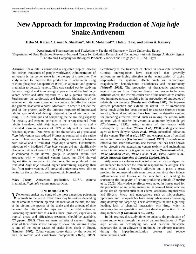
Results of the double immunodiffusion test showed that, there
was no change in the antigenic reactivity of native and 2 KGy
Naja haje venom. The visible lines obtained were identical,
continuous and joined smoothly at the corner, indicating that
there was no change in antigenic determinants i.e. the antivenom
cannot distinguish between the native and γ irradiated Naja haje
venoms as they are immunologically identical (Figure 1).
This finding is in harmony with that of Rogero &
Nascimento, (1995) who reported that, the part of protein
responsible for toxicity of the venom was associated with the
radio-labile group while, the immunogenic part of the venom was
located in a confined portion, which was either resistant to
gamma radiation or was structurally shielded from it. Radiation
is able to induce changes in the structural and antigenic
properties of egg albumin and bovine serum albumin. This
finding is attributed to that, the main part of conformation
dependent antigenic structures (conformational epitopes) is easily
lost by radiation, but some antigenicity, which is mostly due to
the amino acid sequence-dependent antigenic structures
(sequential epitopes) remain, even at high doses (Kume &
Matsuda, 1995).
Figure (1): Immunodiffusion reaction of commercial
polyvalent antivenom with native and γ irradiated Naja haje
venoms.
S = Saline
A = Antivenom
N = Native Naja haje venom.
I = Irradiated Naja haje venom
International Journal of Scientific and Research Publications, Volume 5, Issue 3, March 2015 4
ISSN 2250-3153
www.ijsrp.org
Biochemical activities of native and γ irradiated venoms
Activities of serum LDH, CPK and CK-MB of normal group
were 626.40 ± 44.01 U/l, 410.80 ± 37.17 U/l and 278.76 ± 25.08
U/l, respectively. Injection of native Naja haje venom
significantly elevated activities of serum LDH, CPK and CK-MB
by 49.82%, 267.87% and 80.15%, respectively as compared to
the normal group. In addition, activities of serum ALT and AST
of normal group were 70.60 ± 1.51 U/l and 132.40 ± 1.69 U/l,
respectively. Injection of native Naja haje venom significantly
elevated activities of serum ALT and AST by 208.16% and
99.98%, respectively as compared to the normal group. On the
other hand, injection of irradiated Naja haje venom did not
significantly change activities of serum LDH, CPK, CK-MB,
ALT and AST as compared to the normal group (Table 1).
Thus, the results of this study clearly demonstrated that a
single injection of native Naja haje venom at a dose equal its
LD50 caused a significant elevation in activities of serum LDH,
CPK, CK-MB, ALT and AST as compared to the normal group.
The present increase in enzymes activities due to Naja haje
envenomations was in accordance with the results of the studies
carried out by Fernando et al., (1989) who reported that B. asper
venom caused serum AST, LDH and CPK to increase
significantly, the highest peak being observed at 3h in the cases
of AST and CPK, and at 6 h in the case of LDH. Furthermore, in
a study by Aguiyi et al., (2001), the effect of lethal Echis
carinatus venom on serum enzyme levels and blood plasma
coagulation parameters in rats subjected to (i.p.) venom injection
was investigated. Measurements of the enzyme and coagulation
parameter levels 4 h after venom administration showed an
increase in the level of enzymes; creatinine phosphokinase
(CPK), lactate dehydrogenase (LDH) and glutamic pyruvic
transaminase (ALT) as well as a change in the level of
coagulation parameters due to envenomation. Mebs et al., (1983)
suggested that, the increase in enzymatic activities of CPK and
CPK-MB in the serum could be explained by an increase of the
permeability of the cell membrane. Elevation of creatine
phosphokinase is an indication of damage to muscle, therefore
indicative of injury, myocardial infarction, muscular dystrophy
and myocarditis (Wallimann & Hemmer, 1994).
In addition, Shaaban & Hafez (2003) reported that, tissue
destruction occurs in most of the organs secondary to venom
injections. The increase in enzymatic activity of the serum
attributed to the release of enzymes from liver, kidney and heart.
Organ damage is followed by an increase in levels of ALT, AST
and ALP.
However, the 2KGy gamma irradiated Naje haje venom,
showed non-significant changes in the rats' serum LDH, CPK,
CK-MB, ALT and AST compared to the normal rats. These
findings were attributed to loss of the myotoxic activity of snake
venoms as a secondary event following the exposure to gamma
radiation. Previous studies have emphasized that irradiation of
protein has been shown to cause several chemical changes and
alterations of the physico-chemical properties and of the
secondary and tertiary structure of the proteins, all these changes
are closely connected with the loss of enzymatic, hormonal and
toxic activity of venom after irradiation (Skalka & Antoni, 1970;
Souza-Filho et al., 1992). This is attributed to the
disorganization of the molecular structure of venom after
exposure to gamma radiation, resulting in a change in its
biological activity (Shaaban et al., 1996; Hayes, 2001). In this
respect, radiation is able to detoxify snake venoms and decrease
its harmful effects. In this context, gamma radiation has showed
to be a promising tool for snake venom detoxification without
affecting their immunogenic properties.
Table (1): Effect of native and irradiated Naja haje snake
venoms on serum lactate dehydrogenase (LDH), creatine
phosphokinase (CPK), creatine kinase isoenzyme (CK-MB),
alanine aminotransferase (ALT) and aspartate
aminotransferase (AST) activities in rats
Groups
Parameters
Normal
(Saline)
(0.1 ml; i.p.)
Native Naja haje
(0.163 mg/kg; i.p.)
Irradiated Naja haje
(0.163 mg/kg; i.p.)
LDH (U/l) 626.40 ± 44.01 938.50* ± 33.57 726.22# ± 24.27
CPK (U/l) 410.80 ± 37.17 1510.46* ± 78.82 709.57# ± 50.64
CK-MB (U/l) 278.76 ± 25.08 502.20* ± 9.58 330.21# ± 29.88
ALT (U/l) 70.60 ± 1.51 217.56* ±1.32 83.71# ± 2.96
AST (U/l) 132.40 ± 1.69 264.77* ± 1.14 149.72# ± 2.55
Native and irradiated Naja haje were injected as single doses (a dose equivalent
to native LD50). Blood samples were collected 4 h thereafter. Each value represents the mean ± S.E (n=7).
Statistical analysis was carried out by one-way ANOVA followed by Tukey-
Kramer multiple comparison test. *Significantly different from the normal group at p ≤ 0.05. #Significantly different from native Naja haje group at p ≤ 0.05.
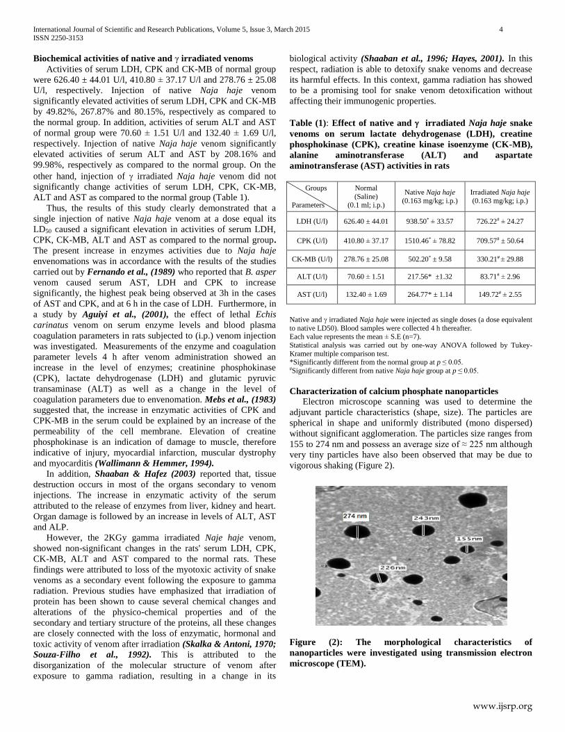
Characterization of calcium phosphate nanoparticles
Electron microscope scanning was used to determine the
adjuvant particle characteristics (shape, size). The particles are
spherical in shape and uniformly distributed (mono dispersed)
without significant agglomeration. The particles size ranges from
155 to 274 nm and possess an average size of ≈ 225 nm although
very tiny particles have also been observed that may be due to
vigorous shaking (Figure 2).
Figure (2): The morphological characteristics of
nanoparticles were investigated using transmission electron
microscope (TEM).
International Journal of Scientific and Research Publications, Volume 5, Issue 3, March 2015 5
ISSN 2250-3153
www.ijsrp.org
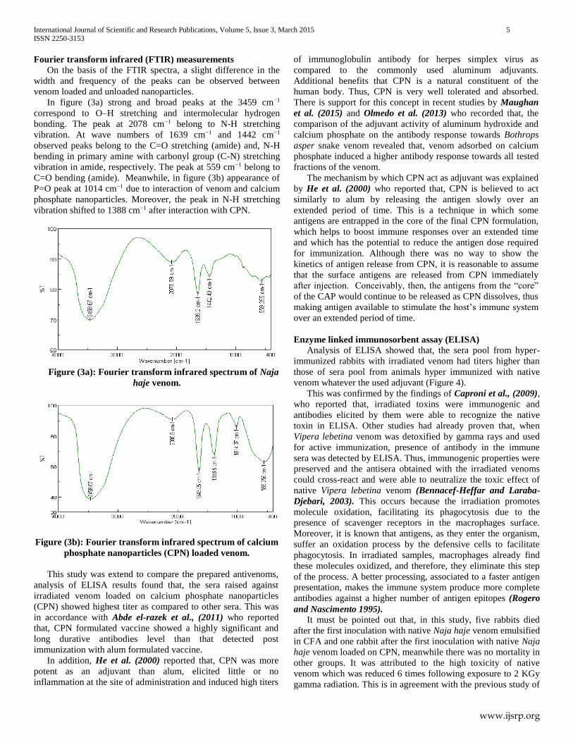
Fourier transform infrared (FTIR) measurements
On the basis of the FTIR spectra, a slight difference in the
width and frequency of the peaks can be observed between
venom loaded and unloaded nanoparticles.
In figure (3a) strong and broad peaks at the 3459 cm−1
correspond to O–H stretching and intermolecular hydrogen
bonding. The peak at 2078 cm−1 belong to N-H stretching
vibration. At wave numbers of 1639 cm−1 and 1442 cm−1
observed peaks belong to the C=O stretching (amide) and, N-H
bending in primary amine with carbonyl group (C-N) stretching
vibration in amide, respectively. The peak at 559 cm−1 belong to
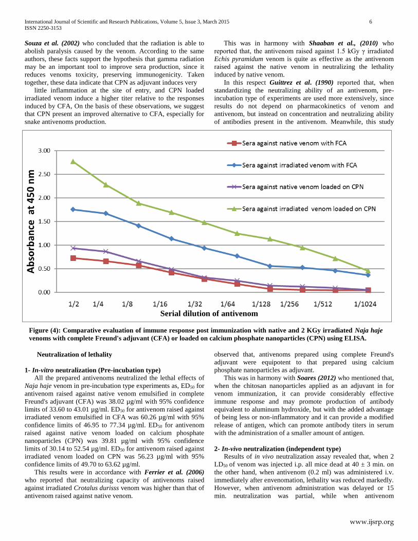
C=O bending (amide). Meanwhile, in figure (3b) appearance of
P=O peak at 1014 cm−1 due to interaction of venom and calcium
phosphate nanoparticles. Moreover, the peak in N-H stretching
vibration shifted to 1388 cm−1 after interaction with CPN.
Figure (3a): Fourier transform infrared spectrum of Naja
haje venom.
Figure (3b): Fourier transform infrared spectrum of calcium
phosphate nanoparticles (CPN) loaded venom.
This study was extend to compare the prepared antivenoms,
analysis of ELISA results found that, the sera raised against
irradiated venom loaded on calcium phosphate nanoparticles
(CPN) showed highest titer as compared to other sera. This was
in accordance with Abde el-razek et al., (2011) who reported
that, CPN formulated vaccine showed a highly significant and
long durative antibodies level than that detected post
immunization with alum formulated vaccine.
In addition, He et al. (2000) reported that, CPN was more
potent as an adjuvant than alum, elicited little or no
inflammation at the site of administration and induced high titers
of immunoglobulin antibody for herpes simplex virus as
compared to the commonly used aluminum adjuvants.
Additional benefits that CPN is a natural constituent of the
human body. Thus, CPN is very well tolerated and absorbed.
There is support for this concept in recent studies by Maughan
et al. (2015) and Olmedo et al. (2013) who recorded that, the
comparison of the adjuvant activity of aluminum hydroxide and
calcium phosphate on the antibody response towards Bothrops
asper snake venom revealed that, venom adsorbed on calcium
phosphate induced a higher antibody response towards all tested
fractions of the venom.
The mechanism by which CPN act as adjuvant was explained
by He et al. (2000) who reported that, CPN is believed to act
similarly to alum by releasing the antigen slowly over an
extended period of time. This is a technique in which some
antigens are entrapped in the core of the final CPN formulation,
which helps to boost immune responses over an extended time
and which has the potential to reduce the antigen dose required
for immunization. Although there was no way to show the
kinetics of antigen release from CPN, it is reasonable to assume
that the surface antigens are released from CPN immediately
after injection. Conceivably, then, the antigens from the “core”
of the CAP would continue to be released as CPN dissolves, thus
making antigen available to stimulate the host’s immune system
over an extended period of time.
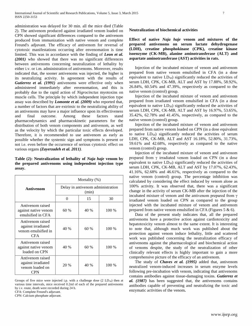
Enzyme linked immunosorbent assay (ELISA) Analysis of ELISA showed that, the sera pool from hyper-
immunized rabbits with irradiated venom had titers higher than
those of sera pool from animals hyper immunized with native
venom whatever the used adjuvant (Figure 4).
This was confirmed by the findings of Caproni et al., (2009),
who reported that, irradiated toxins were immunogenic and
antibodies elicited by them were able to recognize the native
toxin in ELISA. Other studies had already proven that, when
Vipera lebetina venom was detoxified by gamma rays and used
for active immunization, presence of antibody in the immune
sera was detected by ELISA. Thus, immunogenic properties were
preserved and the antisera obtained with the irradiated venoms
could cross-react and were able to neutralize the toxic effect of
native Vipera lebetina venom (Bennacef-Heffar and Laraba-
Djebari, 2003). This occurs because the irradiation promotes
molecule oxidation, facilitating its phagocytosis due to the
presence of scavenger receptors in the macrophages surface.
Moreover, it is known that antigens, as they enter the organism,
suffer an oxidation process by the defensive cells to facilitate
phagocytosis. In irradiated samples, macrophages already find
these molecules oxidized, and therefore, they eliminate this step
of the process. A better processing, associated to a faster antigen
presentation, makes the immune system produce more complete
antibodies against a higher number of antigen epitopes (Rogero
and Nascimento 1995). It must be pointed out that, in this study, five rabbits died
after the first inoculation with native Naja haje venom emulsified
in CFA and one rabbit after the first inoculation with native Naja
haje venom loaded on CPN, meanwhile there was no mortality in
other groups. It was attributed to the high toxicity of native
venom which was reduced 6 times following exposure to 2 KGy
gamma radiation. This is in agreement with the previous study of
International Journal of Scientific and Research Publications, Volume 5, Issue 3, March 2015 6
ISSN 2250-3153
www.ijsrp.org
Souza et al. (2002) who concluded that the radiation is able to
abolish paralysis caused by the venom. According to the same
authors, these facts support the hypothesis that gamma radiation
may be an important tool to improve sera production, since it
reduces venoms toxicity, preserving immunogenicity. Taken
together, these data indicate that CPN as adjuvant induces very
little inflammation at the site of entry, and CPN loaded
irradiated venom induce a higher titer relative to the responses
induced by CFA, On the basis of these observations, we suggest
that CPN present an improved alternative to CFA, especially for
snake antivenoms production.
Neutralization of lethality
1- In-vitro neutralization (Pre-incubation type)
All the prepared antivenoms neutralized the lethal effects of
Naja haje venom in pre-incubation type experiments as, ED50 for
antivenom raised against native venom emulsified in complete
Freund's adjuvant (CFA) was 38.02 µg/ml with 95% confidence
limits of 33.60 to 43.01 µg/ml. ED50 for antivenom raised against
irradiated venom emulsified in CFA was 60.26 µg/ml with 95%
confidence limits of 46.95 to 77.34 µg/ml. ED50 for antivenom
raised against native venom loaded on calcium phosphate
nanoparticles (CPN) was 39.81 µg/ml with 95% confidence
limits of 30.14 to 52.54 µg/ml. ED50 for antivenom raised against
irradiated venom loaded on CPN was 56.23 µg/ml with 95%
confidence limits of 49.70 to 63.62 µg/ml.
This results were in accordance with Ferrier et al. (2006)
who reported that neutralizing capacity of antivenoms raised
against irradiated Crotalus durisss venom was higher than that of
antivenom raised against native venom.
This was in harmony with Shaaban et al., (2010) who
reported that, the antivenom raised against 1.5 kGy γ irradiated
Echis pyramidum venom is quite as effective as the antivenom
raised against the native venom in neutralizing the lethality
induced by native venom.
In this respect Guittrez et al. (1990) reported that, when
standardizing the neutralizing ability of an antivenom, pre-
incubation type of experiments are used more extensively, since
results do not depend on pharmacokinetics of venom and
antivenom, but instead on concentration and neutralizing ability
of antibodies present in the antivenom. Meanwhile, this study
observed that, antivenoms prepared using complete Freund's
adjuvant were equipotent to that prepared using calcium
phosphate nanoparticles as adjuvant.
This was in harmony with Soares (2012) who mentioned that,
when the chitosan nanoparticles applied as an adjuvant in for
venom immunization, it can provide considerably effective
immune response and may promote production of antibody
equivalent to aluminum hydroxide, but with the added advantage
of being less or non-inflammatory and it can provide a modified
release of antigen, which can promote antibody titers in serum
with the administration of a smaller amount of antigen.
2- In-vivo neutralization (independent type)
Results of in vivo neutralization assay revealed that, when 2
LD50 of venom was injected i.p. all mice dead at 40 ± 3 min. on
the other hand, when antivenom (0.2 ml) was administered i.v.
immediately after envenomation, lethality was reduced markedly.
However, when antivenom administration was delayed or 15
min. neutralization was partial, while when antivenom
Figure (4): Comparative evaluation of immune response post immunization with native and 2 KGy irradiated Naja haje
venoms with complete Freund's adjuvant (CFA) or loaded on calcium phosphate nanoparticles (CPN) using ELISA.
Serial dilution of antivenom
International Journal of Scientific and Research Publications, Volume 5, Issue 3, March 2015 7
ISSN 2250-3153
www.ijsrp.org
administration was delayed for 30 min. all the mice died (Table
2). The antivenom produced against irradiated venom loaded on
CPN showed significant differences compared to the antivenom
produced from immunization with native venom and complete
Freund's adjuvant. The efficacy of antivenom for reversal of
cytotoxic manifestations occurring after envenomation is time
limited. This was in accordance with the finding of Leon et al.
(2001) who showed that there was no significant differences
between antivenoms concerning neutralization of lethality by
either i.v. or i.m. administration of antivenoms. Moreover, results
indicated that, the sooner antivenoms was injected, the higher is
its neutralizing activity. In agreement with the results of
Gutierrez et al. (1991) antivenoms were effective only when
administered immediately after envenomation, and this is
probably due to the rapid action of Nigrocinctus myotoxins on
muscle cells. The principle by which independent injection type
assay was described by Lomonte et al. (2009) who reported that,
a number of factors that are extrinsic to the neutralizing ability of
an antivenoms may have a major influence upon its effectiveness
and final outcome. Among these factors stand
pharmacodynamics and pharmacokinetic parameters for the
distribution of both venom components and antivenom, as well
as the velocity by which the particular toxic effects developed.
Therefore, it is recommended to use antivenom as early as
possible whether the systemic sign and symptoms is present or
not i.e. even before the occurrence of serious cytotoxic effect on
various organs (Zayerzadeh et al. 2011).
Table (2): Neutralization of lethality of Naja haje venom by
the prepared antivenoms using independent injection type
assay.
Antivenom
Mortality (%)
Delay in antivenom administration
(min)
0 15 30
Antivenom raised
against native venom
emulsified in CFA
60 % 40 % 100 %
Antivenom raised
against irradiated
venom emulsified in
CFA
40 % 60 % 100 %
Antivenom raised
against native venom
loaded on CPN
40 % 60 % 100 %
Antivenom raised
against irradiated
venom loaded on
CPN
20 % 40 % 100 %
Groups of five mice were injected i.p. with a challenge dose (2 LD50) then at
various time intervals, mice received 0.2ml of each of the prepared antivenoms by i.v. route, death were recorded during 24 h.
CFA: Complete Freund's adjuvant.
CPN: Calcium phosphate adjuvant.
Neutralization of biochemical activities
Effect of native Naja haje venom and mixtures of the
prepared antivenoms on serum lactate dehydrogenase
(LDH), creatine phosphokinase (CPK), creatine kinase
isoenzyme (CK-MB), alanine aminotransferase (ALT) and
aspartate aminotransferase (AST) activities in rats.
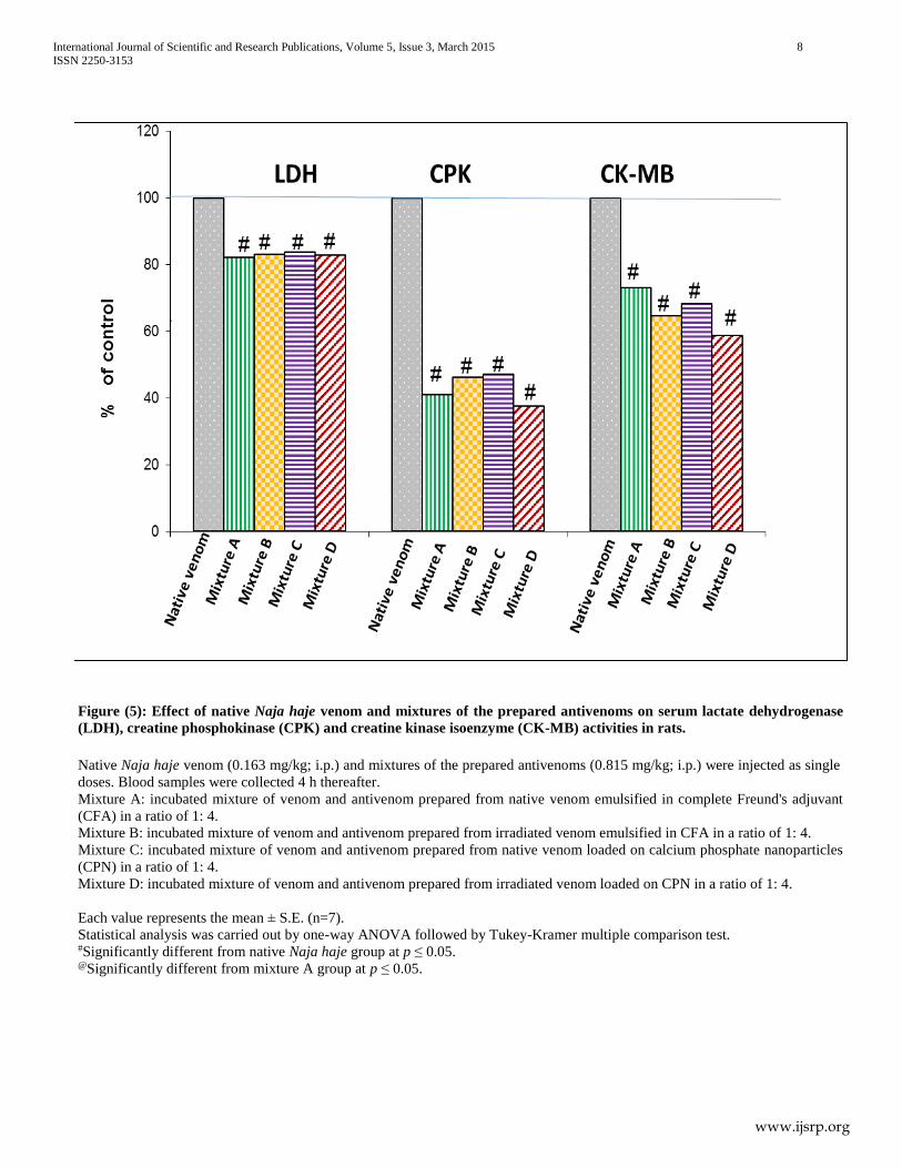
Injection of the incubated mixture of venom and antivenom
prepared from native venom emulsified in CFA (in a dose
equivalent to native LD50) significantly reduced the activities of
serum LDH, CPK, CK-MB, ALT and AST by 17.88%, 58.92%,
26.84%, 60.54% and 47.39%, respectively as compared to the
native venom (control) group.
Injection of the incubated mixture of venom and antivenom
prepared from irradiated venom emulsified in CFA (in a dose
equivalent to native LD50) significantly reduced the activities of
serum LDH, CPK, CK-MB, ALT and AST by 16.97%, 53.65%,
35.42%, 62.78% and 41.45%, respectively, as compared to the
native venom (control) group.
Injection of the incubated mixture of venom and antivenom
prepared from native venom loaded on CPN (in a dose equivalent
to native LD50) significantly reduced the activities of serum
LDH, CPK, CK-MB, ALT and AST 16.18%, 52.80%, 31.73%,
59.61% and 42.68%, respectively as compared to the native
venom (control) group.
Injection of the incubated mixture of venom and antivenom
prepared from γ irradiated venom loaded on CPN (in a dose
equivalent to native LD50) significantly reduced the activities of
serum LDH, CPK, CK-MB, ALT and AST by 17.07%, 62.43%,
41.16%, 62.68% and 46.61%, respectively as compared to the
native venom (control) group. The percentage inhibition was
calculated by considering the effect induced by venom alone as
100% activity. It was observed that, there was a significant
change in the activity of serum CK-MB after the injection of the
incubated mixture of venom and the antivenom prepared from γ
irradiated venom loaded on CPN as compared to the group
injected with the incubated mixture of venom and antivenom
prepared from native venom emulsified in CFA (Figures 5 & 6).
Data of the present study indicates that, all the prepared
antivenoms have a protective action against cardiotoxicity and
hepatotoxicity venom almost to the same extent. It is interesting
to note that, although much work was published about the
protection against venom induce lethality, little and scattered
work was published concerning the neutralization efficacy of
antivenoms against the pharmacological and biochemical action
of venoms despite, the study of the neutralization of other
clinically relevant effects is highly important to gain a more
comprehensive picture of the efficacy of an antivenom.
The study of Chaves et al. (1995) added that, antivenom
neutralized venom-induced increases in serum enzyme levels
following pre-incubation with venom, indicating that antivenoms
contains antibodies against tissue-damaging toxins. Gutierrez et
al. (1987) has been suggested that, the antivenoms contains
antibodies capable of preventing and neutralizing the toxic and
enzymatic activities of the venom.
International Journal of Scientific and Research Publications, Volume 5, Issue 3, March 2015 8
ISSN 2250-3153
www.ijsrp.org
Figure (5): Effect of native Naja haje venom and mixtures of the prepared antivenoms on serum lactate dehydrogenase
(LDH), creatine phosphokinase (CPK) and creatine kinase isoenzyme (CK-MB) activities in rats.
Native Naja haje venom (0.163 mg/kg; i.p.) and mixtures of the prepared antivenoms (0.815 mg/kg; i.p.) were injected as single
doses. Blood samples were collected 4 h thereafter.
Mixture A: incubated mixture of venom and antivenom prepared from native venom emulsified in complete Freund's adjuvant
(CFA) in a ratio of 1: 4.
Mixture B: incubated mixture of venom and antivenom prepared from irradiated venom emulsified in CFA in a ratio of 1: 4.
Mixture C: incubated mixture of venom and antivenom prepared from native venom loaded on calcium phosphate nanoparticles
(CPN) in a ratio of 1: 4.
Mixture D: incubated mixture of venom and antivenom prepared from irradiated venom loaded on CPN in a ratio of 1: 4.
Each value represents the mean ± S.E. (n=7).
Statistical analysis was carried out by one-way ANOVA followed by Tukey-Kramer multiple comparison test. #Significantly different from native Naja haje group at p ≤ 0.05. @Significantly different from mixture A group at p ≤ 0.05.
International Journal of Scientific and Research Publications, Volume 5, Issue 3, March 2015 9
ISSN 2250-3153
www.ijsrp.org
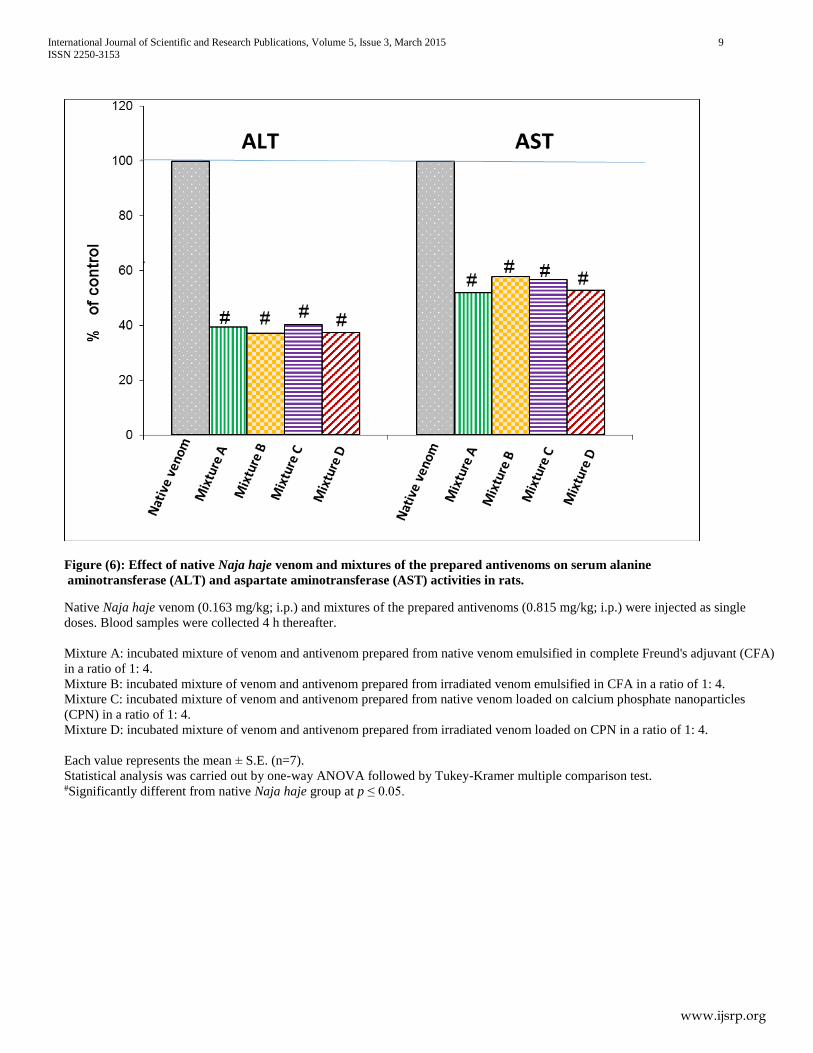
Figure (6): Effect of native Naja haje venom and mixtures of the prepared antivenoms on serum alanine
aminotransferase (ALT) and aspartate aminotransferase (AST) activities in rats.
Native Naja haje venom (0.163 mg/kg; i.p.) and mixtures of the prepared antivenoms (0.815 mg/kg; i.p.) were injected as single
doses. Blood samples were collected 4 h thereafter.
Mixture A: incubated mixture of venom and antivenom prepared from native venom emulsified in complete Freund's adjuvant (CFA)
in a ratio of 1: 4.
Mixture B: incubated mixture of venom and antivenom prepared from irradiated venom emulsified in CFA in a ratio of 1: 4.
Mixture C: incubated mixture of venom and antivenom prepared from native venom loaded on calcium phosphate nanoparticles
(CPN) in a ratio of 1: 4.
Mixture D: incubated mixture of venom and antivenom prepared from irradiated venom loaded on CPN in a ratio of 1: 4.
Each value represents the mean ± S.E. (n=7).
Statistical analysis was carried out by one-way ANOVA followed by Tukey-Kramer multiple comparison test. #Significantly different from native Naja haje group at p ≤ 0.05.
International Journal of Scientific and Research Publications, Volume 5, Issue 3, March 2015 10
ISSN 2250-3153
www.ijsrp.org
IV. CONCLUSION
In conclusion, based on the experimental findings,
irradiation was found to be a reliable tool in detoxification of
venoms, minimizing the toxic effect while maintaining the
immunogenicity. In addition, calcium phosphate nanoparticles
when applied as adjuvant, provide enhancement of immune
response with the adjuvant of being less or non-inflammatory
and it can provide a modified release of antigen, which can
promote obtaining antibody titers in serum with the
administration of a smaller amount of antigen. Taken together,
this study showed an immunization adjuvant system for Naja
haje snake venom that should be tested with venom of other
snakes. Thus, this approach allows achieving new
biotechnological antivenoms to be used in the future.
REFERENCES
[1] Aguiyi, J.C., Guerranti, R., Pagani, R. & Marinello, E. (2001). Blood
chemistry of rats pretreated with Mucuna pruriens seed aqueous extract MP101UJ after Echis carinatus venom challenge. Phytother Res, 15: 712-714.
[2] ALexander, P. & Hamilton, L.D. (1962). Infulence of oxygen and absorbed water on changes produced in bovine serum albumin. Radiat. Res., 15: 193-201.
[3] Bennacef-Heffar, N. & Laraba-Djebari, F. (2003). Evaluation of the effect of gamma rays on the venom of Vipera lebetina by biochemical study. Can J Physiol Pharmacol, 81: 1110-1117.
[4] Boni-Mitake, M., Costa, H., Spencer, P.J., Vassilieff, V.S. & Rogero,
J.R. (2001). Effects of (60)Co gamma radiation on crotamine. Braz J Med Biol Res, 34: 1531-1538.
[5] Caproni, P., Baptista, J., Almeida, T.d., Passos, L. & Nascimento, N.
(2009). Study of irradiated bothropstoxin-1 with 60Co gamma rays: immune system behavior. J. Venom. Anim. Toxins incl. Trop. Dis, 15: 216-225.
[6] Casare, M.S., Spencer, P., Campos, L.A. & Nascimento, N. (2006).
Study of gamma-radiation effects on crotamine and crotoxin. Journal of Radioanalytical and Nuclear Chemistry, 269: 571-577.
[7] Chaves, F., Barboza, M., & Gutiérrez, J. (1995). Pharmacological study of edema induced by venom of the snake Bothrops asper (terciopelo) in mice. Toxicon, 33(1), 31-39.
[8] Chippaux, J.P., Williams, V. & White, J. (1991). Snake venom variability: methods of study, results and interpretation. Toxicon, 29: 1279-1303.
[9] Christensen, P.A. (1955). South African snake venoms and antivenoms. South African Institute for Medical Research, Johannesburg, p147.
[10] Clissa, P.B., do Nascimento, N. & Rogero, J.R. (1999). Toxicity and immunogenicity of Crotalus durissus terrificus venom treated with different doses of gamma rays. Toxicon, 37: 1131-1141.
[11] Cocchetto, D.M. & Bjornsson, T.D. (1983). Methods for vascular access and collection of body fluids from the laboratory rat. J Pharm Sci, 72: 465-492.
[12] Costa, L.M., Takeda, A.K., Barbosa, S.F., Berra, J.A., Adelina, M.G.,
Soerensen, B., Pinto, J.R. & Vancetto, M.D. (1985). Study of immune response in horse immunized with Crotalus durissus terrificus, in natura, submitted to formaldehyde treatment and thermic action. Vacinas Soros, 1: 24-29.
[13] Crommelin, D.J., Hennink, W.E. & Storm, G., (2001). Drug Targeting Systems: Fundamentals and Applications to Parenteral Drug Delivery. In: M.H. Anya, A.W. Lloyd and J. Swarbrick. (Eds.), Drug Delivery and Targeting for Pharmacists and Pharmaceutical Scientists. Taylor and Fracncis, London, pp: 118-144.
[14] Daniel, J.P., Heneine, L.G., Tavares, C.A., Nascimento, M.C. &
Heneine, I.F. (1987). Generation of protective immune sera by Crotalus durissus terrificus venom detoxified by controlled iodination. Braz J Med Biol Res, 20: 713-720.
[15] Fernando, C., Jose, M.G., Bruno, L. & Luis, C. (1989). Histopathological and biochemical alterations induced by intramuscular injection of Bothrops Asper venom in mice. . Toxicon 27 1085-1093.
[16] Finney, D.J. (1964). Statistical method in biological assay. Hafner Pub. Co., New York, p668.
[17] Flecknell, P.A. (1987). Laboratory animal anaesthesia : an introduction for research workers and technicians. Academic Press, London ; San Diego, p125.
[18] Ferreira Junior, R. S., Nascimento, N., Couto, R., Alves, J. B., Meira,
D. A., & Barraviera, B. (2006). Laboratory evaluation of young ovines inoculated with natural or 60Co-irradiated Crotalus durissus terrificus venom during hyperimmunization process. Journal of Venomous Animals and Toxins including Tropical Diseases, 12(4), 620-631.
[19] Ferreira Junior, R. S., Nascimento, N., Martinez J. C., Alves J. B.,
Meira D. A. and Barraviera B. (2010). Immune response and neutralization capacity of antibodies produced in young sheep immunized with Crotalus durissus terrificus native or Cobalt-60 irradiated venom. J.Venom. Anim. Toxins incl. Trop. Dis. 11: (4) 447-464.
[20] Freitas, T.V. & Frezard, F. (1997). Encapsulation of native crotoxin in liposomes: a safe approach for the production of antivenom and vaccination against Crotalus durissus terrificus venom. Toxicon, 35: 91-100.
[21] Gallacci, M., Nascimento, N., Rogero, J.R. & Vassilieff, V.S. (2000).
Influence of temperature upon effects of crotoxin and gamma-irradiated crotoxin at rat neuromuscular transmission. Toxicol Lett, 114: 77-80.
[22] Garrison, W. M. (1987). Reaction mechanisms in the radiolysis of peptides, polypeptides, and proteins. Chemical Reviews, 87(2), 381-398.
[23] Ghazal, A., Ismail, M., Abdel-Rahman, A. A., & El-Asmar, M. F.
(1975). Pharmacological studies of scorpion (Androctonus amoreuxi Aud. & Sav.) venom. Toxicon, 13(4), 253-254.
[24] Gutiérrez, J., Rojas, G., & Cerdas, L. (1987). Ability of a polyvalent antivenom to neutralize the venom of Lachesis muta melanocephala, a new Costa Rican subspecies of the bushmaster. Toxicon, 25(7), 713-720.
[25] Gutierrez, J.M., Chaves, F., Rojas, E., Elizondo, J., Avila, C. & Cerdas,
L. (1988). Production of monovalent anti-Bothrops asper antivenom: development of immune response in horses and neutralizing ability. Rev Biol Trop, 36: 511-517.
[26] Gutiérrez, J. M., Dos Santos, M. C., de Fatima Furtado, M., & Rojas,
G. (1991). Biochemical and pharmacological similarities between the venoms of newborn Crotalus durissus durissus and adult Crotalus durissus terrificus rattlesnakes. Toxicon, 29(10), 1273-1277.
[27] Gutierrez, J.M., Romero, M., Nunez, J., Chaves, F., Borkow, G. &
Ovadia, M. (1997). Skeletal muscle necrosis and regeneration after injection of BaH1, a hemorrhagic metalloproteinase isolated from the venom of the snake Bothrops asper (Terciopelo). Exp Mol Pathol, 62: 28-41.
[28] Halliwell, B. & Gutteridge, J.M.C. (1989). the chemistry of oxygen radicals and other oxygen derived species. Free radicals in biology and medicine. Clarendon Press, New York, p543.
[29] Hayes, A.W., Ed. (2001). Principles and Methods of Toxicology, Taylor and Francis, Philadelphia, p715.
[30] He, Q., Mitchell, A. R., Johnson, S. L., Wagner-Bartak, C., Morcol, T.,
& Bell, S. J. (2000). Calcium phosphate nanoparticle adjuvant. Clinical and diagnostic laboratory immunology, 7(6), 899-903.
[31] Karam H. M., Shaaban E. A. and Kenawy S. A. (2010). Effect of Gamma Irradiation on Toxicity, Immunological properties and Oxidative Damages of Cerastes Cerastes and Echis Pyramidum Snake Venoms
[32] Kume, T. & Matsuda, T. (1995). Changes in structural and antigenic properties of proteins by radiation. . Radiat. Phys. Chem. , 46: 225-231.
[33] León, G., Monge, M., Rojas, E., Lomonte, B., & Gutiérrez, J. M. (2001). Comparison between IgG and F (ab′) 2 polyvalent antivenoms: neutralization of systemic effects induced by Bothrops asper venom in mice, extravasation to muscle tissue, and potential for induction of adverse reactions. Toxicon, 39(6), 793-801.
[34] Lomonte, B., León, G., Angulo, Y., Rucavado, A., & Núñez, V. (2009).
Neutralization of Bothrops asper venom by antibodies, natural products and synthetic drugs: Contributions to understanding snakebite envenomings and their treatment. Toxicon, 54(7), 1012-1028.
[35] Lott, J. A., & Stang, J. M. (1980). Serum enzymes and isoenzymes in the diagnosis and differential diagnosis of myocardial ischemia and necrosis. Clinical chemistry, 26(9), 1241-1250.
International Journal of Scientific and Research Publications, Volume 5, Issue 3, March 2015 11
ISSN 2250-3153
www.ijsrp.org
[36] Maughan, C. N., Preston, S. G., & Williams, G. R. (2015). Particulate inorganic adjuvants: recent developments and future outlook. Journal of Pharmacy and Pharmacology.
[37] Mebs, D. (2002). Scorpions and snakes, such as cobras, mambas and vipers made the African continent famous for venomous animals. Bull Soc Pathol Exot, 95: 131.
[38] Mebs, D., Ehrenfeld, M. & Samejima, Y. (1983). Local necrotizing effect of snake venoms on skin and muscle: relationship to serum creatine kinase. Toxicon, 21: 393-404.
[39] Mohamed, A.H., Fouad, S., El-Aasar, S., Salem, A.M., Abdel-Aal, A.,
Hassan, A.A., Zahran, F. & Abbas, N. (1981). Effects of several snake venoms on serum and tissue transaminases, alkaline phosphatase and lactate dehydrogenase. Toxicon, 19: 605-609.
[40] Moroz, C., Goldblum, N. & Devries, A. (1963). Preparation of Vipera Palestinae Antineurotoxin Using Carboxymethyl-Cellulose-Bound Neurotoxin as Antigen. Nature, 200: 697-698.
[41] Nascimento, N., Seebart, C.S., Francis, B., Rogero, J.R. & Kaiser, II
(1996). Influence of ionizing radiation on crotoxin: biochemical and immunological aspects. Toxicon, 34: 123-131.
[42] Olmedo, H., Herrera, M., Rojas, L., Villalta, M., Vargas, M., Leiguez,
E., ... & Montero, M. L. (2013). Comparison of the adjuvant activity of aluminum hydroxide and calcium phosphate on the antibody response towards Bothrops asper snake venom. Journal of immunotoxicology, 11(1), 44-49.
[43] Ouchterlony, O. (1948). In vitro method for testing the toxin production capacity of diphtheria bacteria Acta Path. Microbiol. Scand, 25: 186-189.
[44] Oussedik-Oumehdi, H. & Laraba-Djebari, F. (2008). Irradiated Cerastes cerastes venom as a novel tool for immunotherapy. Immunopharmacol Immunotoxicol, 30: 37-52.
[45] Ownby, C.L. & Colberg, T.R. (1988). Classification of myonecrosis induced by snake venoms: venoms from the prairie rattlesnake (Crotalus viridis viridis), western diamondback rattlesnake (Crotalus atrox) and the Indian cobra (Naja naja naja). Toxicon, 26: 459-474.
[46] Paget, G.E. & Barnes, J.M. (1964). Toxicity tests. In Evaluation of drug activation; Pharmacometrics., New york, p135.
[47] Reitman, S. & Frankel, S. (1957). A colorimetric method for the determination of serum glutamic oxalacetic and glutamic pyruvic transaminases. Am J Clin Pathol, 28: 56-63.
[48] Rogero, J.R. & Nascimento, N. (1995). - Detoxification of snake venom using ionizing radiation. . J. Venom. Anim. Toxins 1: 7-10.
[49] Shaaban, E.A. (2005). Active immunization of rabbit with gamma irradiated cobra (Naja haje) venom toxoid. The Egyptian journal of hospital medicine 13: 99-111.
[50] Shaaban, E.A., Ahmed, A.A. & Ayobe, M.H. (1996). Gamma irradiation of Egyptian Cobra (Naja haje) Venom. J Egypt Public Health Assoc, 71: 257-267, 269-271.
[51] Shaaban, E.A. & Hafez, M.N. (2003). Ability of gamma-irradiated polyvalent antivenin to neutralize the toxicity of the Egyptian Cobra (Naja haje) venom. The Egyptian Journal of Hospital Medicine 13: 135-152.
[52] Shaaban, E., El-missiry, A., Abd-Elbaset, A., Abdallah, N. & Moustafa,
M. (2010). infuluence of ionizing radiation on Echis Pyramidum snake venom: biochemical and immunological aspect. the Egyptian journal of hospital medicine, 40: 314-334.
[53] Skalka, M. & Antoni, F. (1970). Effect of radiation on the biological properties of proteins. In: Radiation Sensitivity of Toxins and Animal Poisons, 1969. Proceedings of a Panel Bangkok. 19-22 May, 1969. International Atomic Energy Agency, Vienna City. p. 1-11.
[54] Soares, K. S. R., Fonseca, J. L. C., Bitencourt, M. A. O., Santos, K. S.,
Silva-Júnior, A. A., & Fernandes-Pedrosa, M. F. (2012). Serum production against Tityus serrulatus scorpion venom using cross-linked chitosan nanoparticles as immunoadjuvant. Toxicon, 60(8), 1349-1354.
[55] Souza-Filho, J.N., Guarnieri-Cruz, M.C., Murata, Y. & Rogero, J.R.
(1992). Detoxification of the crotoxin complex by gamma radiation. Braz J Med Biol Res, 25: 103-113.
[56] Souza, F.A., Spencer, P.J., Rogero, J.R., Nascimento, N., Dal Pai-Silva,
M. & Gallacci, M. (2002). 60Co gamma irradiation prevents Bothrops jararacussu venom neurotoxicity and myotoxicity in isolated mouse neuromuscular junction. Toxicon, 40: 1101-1106.
[57] Stentz, R., Bongaerts, R. J., Gunning, A. P., Gasson, M., & Shearman,
C. (2010). Controlled release of protein from viable Lactococcus lactis cells. Applied and environmental microbiology, 76(9), 3026-3031.
[58] Szasz, G., Gruber, W. & Bernt, E. (1976). Creatine kinase in serum: 1. Determination of optimum reaction conditions. Clin Chem, 22: 650-656.
[59] Van der Lubben, I. M., Kersten, G., Fretz, M. M., Beuvery, C.,
Verhoef, J. C., & Junginger, H. E. (2003). Chitosan microparticles for mucosal vaccination against diphtheria: oral and nasal efficacy studies in mice. Vaccine, 21(13), 1400-1408.
[60] Wallimann, T. & Hemmer, W. (1994). Creatine kinase in non-muscle tissues and cells. Mol Cell Biochem, 133-134: 193-220.
[61] Warrell, D.A. (2003). Injuries, envenoming, poisoning and allergic reactions caused by animal. Oxford textbook of medicine. Oxford University Press, New York p.298.
[62] World Health Organization. (1981). Progress in the characterization of venoms and standardization of antivenoms.
[63] World Health Organization (2010). Guidelines for the Production, Control and Regulation of Antivenom Immunoglobulins. World Health Organization, Geneva, Switzerland. WHO Offset Publication No.83.
[64] Zayerzadeh, E., Koohi, M. K., Mirakabadi, A. Z., Fardipoor, A.,
Kassaian, S. E., Rabbani, S., & Anvari, M. S. (2012). Amelioration of cardio-respiratory perturbations following Mesobuthus eupeus envenomation in anesthetized rabbits with commercial polyvalent F (ab′) 2 antivenom. Toxicon, 59(2), 249-256.
AUTHORS
Heba M. Karam- Assistant Lecturer in Drug Radiation Research
Department − National Center for Radiation Research and
Technology − Atomic Energy Authority, Egypt.
Esmat A. Shaaban- Professor of Pharmacology & Toxicology −
National Center for Radiation Research and Technology − Atomic
Energy Authority, Egypt. [email protected]
Aly F. Mohamed −Head of Applied Research Sector −
Holding Company for Biological Products, Vaccines and Drug
(VACSERA), Egypt. [email protected]
Hala F. Zaki − Professor of Pharmacology & Toxicology
Faculty of Pharmacy − Cairo University. [email protected]
Sanaa A. Kenawy − Professor of Pharmacology & Toxicology
Faculty of Pharmacy − Cairo University.
Correspondence Author – Heba M. Karam,
[email protected], or [email protected],
+201114786345
Related Documents