neuropathic pain in burn scars 1 NEUROPATHIC PAIN IN POST-BURN HYPERTROPHIC SCARS: A PSYCHOPHYSICAL AND NEUROPHYSIOLOGICAL STUDY Gianluca Isoardo, MD, a,b Maurizio Stella, MD, c Dario Cocito, MD, d Daniela Risso, MD, c Giuseppe Migliaretti, PhD, e Franco Cauda, PhD, f Angela Palmitessa, PhD, a,b Giuliano Faccani, MD, b Palma Ciaramitaro, MD. a,b a Unit of Neurophysiology, Hospital CTO-Maria Adelaide, Torino, Italy b Department of Neurosurgery, Hospital CTO-Maria Adelaide, Torino, Italy c Department of Plastic Surgery, Burn Center, Hospital CTO-Maria Adelaide, Torino, Italy d Department of Neurosciences, Univesity of Torino, Torino, Italy. e Department of Public Health and Microbiology, University of Torino, Italy f CCS fMRI, Hospital Koelliker and Department of Psychology, University of Torino, Italy. Corresponding author: Dr Gianluca Isoardo, MD Unit of Neurophysiology Department of Neurosurgery Hospital CTO-Maria Adelaide Via Zuretti 29 10126 Torino, Italy Tel + 390116933882 e-mail: [email protected] Running title: neuropathic pain in burn scars Page 3 of 29 John Wiley & Sons, Inc. Muscle & Nerve

Welcome message from author
This document is posted to help you gain knowledge. Please leave a comment to let me know what you think about it! Share it to your friends and learn new things together.
Transcript
neuropathic pain in burn scars
1
NEUROPATHIC PAIN IN POST-BURN HYPERTROPHIC SCARS: A
PSYCHOPHYSICAL AND NEUROPHYSIOLOGICAL STUDY
Gianluca Isoardo, MD,a,b
Maurizio Stella, MD,c Dario Cocito, MD,
d Daniela Risso, MD,
c Giuseppe
Migliaretti, PhD,e Franco Cauda, PhD,
f Angela Palmitessa, PhD,
a,b Giuliano Faccani, MD,
b Palma
Ciaramitaro, MD.a,b
aUnit of Neurophysiology, Hospital CTO-Maria Adelaide, Torino, Italy
bDepartment of Neurosurgery, Hospital CTO-Maria Adelaide, Torino, Italy
cDepartment of Plastic Surgery, Burn Center, Hospital CTO-Maria Adelaide, Torino, Italy
dDepartment of Neurosciences, Univesity of Torino, Torino, Italy.
eDepartment of Public Health and Microbiology, University of Torino, Italy
fCCS fMRI, Hospital Koelliker and Department of Psychology, University of Torino, Italy.
Corresponding author:
Dr Gianluca Isoardo, MD
Unit of Neurophysiology
Department of Neurosurgery
Hospital CTO-Maria Adelaide
Via Zuretti 29
10126 Torino, Italy
Tel + 390116933882
e-mail: [email protected]
Running title: neuropathic pain in burn scars
Page 3 of 29
John Wiley & Sons, Inc.
Muscle & Nerve
neuropathic pain in burn scars
2
NEUROPATHIC PAIN IN POST-BURN HYPERTROPHIC SCARS: A
PSYCHOPHYSICAL AND NEUROPHYSIOLOGICAL STUDY
ABSTRACT
Introduction. Pain complicates hypertrophic post-burn pathologic scars (PPS) Methods. To
investigate the possible neuropathic origin of pain, 13 patients with painful-PPS involving at least
one hand underwent clinical examination including the Douleur Neuropathique en 4 questions
(DN4) questionnaire, median, ulnar and radial nerve conduction studies (NCS), cold (CDT) and
heat-induced pain thresholds evaluation by quantitative sensory testing, and cutaneous silent period
(CSP) of the abductor pollicis brevis. Controls were 9 patients with non painful-PPS, 52 healthy
subjects, and 28 patients with carpal tunnel syndrome (CTS). Results. All patients with painful-PPS
had possible neuropathic pain (DN4 score ≥ 4). NCS signs of CTS were similarly present in PPS
subjects with or without pain. Hands with painful-PPS had lower CDT and CSP duration, more
frequent cold and heat pain hypesthesia and thermal allodynia than controls. Discussion. In PPS,
possible neuropathic pain is associated with psychophysical and neurophysiological abnormalities
suggestive of small fiber damage.
Key words: neuropathic pain, burns, post-burn scars, quantitative sensory testing, cutaneous silent
period
Page 4 of 29
John Wiley & Sons, Inc.
Muscle & Nerve
neuropathic pain in burn scars
3
INTRODUCTION
Post-burn pathologic scars (PPS) may commonly complicate the healing of burn wounds in up to
77% of patients. PPS encompass different type of scars, i.e. hypertrophic, contracture and atrophic,
but hypertrophy is the most frequent presentation.1 Pain is a frequent and often severe
accompanying symptom of hypertrophic PPS, and it has been hypothesized that pain may be of
neuropathic origin.2
The grading of certainty for the diagnosis of neuropathic pain labels it as definite if clinical and
laboratory evaluations demonstrate the presence of both a neuroanatomically plausible distribution
of pain itself, and the evidence of a lesion of the somatosensory system.3 In patients with burns,
entrapment neuropathies are frequent.4 In hypertrophic PPS there is a moderate-to-severe reduction
of skin nerve fiber density.5,6
Therefore, patients with PPS are at increased risk of both small- and
large-fiber sensory nerve damage, which may be the basis for the development of neuropathic pain.
Standardized screening tools may allow for identification of patients with possible neuropathic pain.
Therefore, they are recommended as standardized case identification tools in research studies.7 The
Douleur Neuropathique en 4 questions (DN4) questionnaire is a validated clinician-administered
10-item pain questionnaire that indicates the presence of neuropathic pain for scores ≥ to 4.7-9
This
questionnaire consists of a patient interview (seven items related to symtoms) and a standardized
clinical examination (consisting of search for pinprick and touch hypesthesia and allodynia to
brush).8 The DN4 in Italian translation has been adopted to identify patients with neuropathic pain
associated with carpal tunnel syndrome (CTS).9
In this study, patients with PPS involving at least one hand were screened by the DN4 questionnaire
for the presence/absence of possible neuropathic pain in the PPS on the hands. Then, they
underwent psychophysical and neurophysiological evaluation to assess for damage to either large or
small peripheral sensory nerve fibers in the PPS site. Large fibers were evaluated by routine nerve
conduction studies (NCS). Quantitative sensory testing (QST) and the cutaneous silent period (CSP)
were performed to assess the function of small fibers. In fact, both QST and CSP are non-invasive
Page 5 of 29
John Wiley & Sons, Inc.
Muscle & Nerve
neuropathic pain in burn scars
4
tools that are frequently abnormal in patients with small fiber neuropathy.10-13
QST measures the
function of small fibers by assessing temperature thresholds.10,11
CSP is a transient suppression of
EMG activity that occurs during sustained muscle contraction and after electrical stimulation
delivered to the fingers.12-18
CSP is mediated mainly by Aδ afferents.13-15,17
The aim of this study was to investigate the possible neuropathic origin of the pain in PPS by
examining clinical features and by investigating the function of both small and large fibers to search
for a lesion of the somatosensory system. The latter provides at the same time a pathophysiological
basis for pain.
Page 6 of 29
John Wiley & Sons, Inc.
Muscle & Nerve
neuropathic pain in burn scars
5
METHODS
Patients.
One hundred-twenty patients with PPS followed by the Burn Center of our hospital were screened
for inclusion in the study. Diagnosis of hypertrophic PPS was made, as previously described,1
on a
clinical basis by two expert plastic surgeons (MS and DR), taking into account color, thickness,
width, pigmentation, contour, degree of vascular congestion, vascularity, shape, height, texture,
consistency and extensibility of the scar. Ancillary laboratory evaluation included ultrasound and
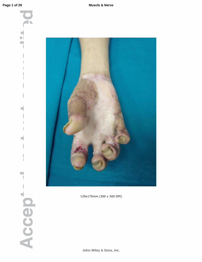
videocapillaroscopy studies. An example of a hypertrophic PPS involving the hand is provided in
Figure 1.
The inclusion criteria for this study were: 1) a PPS that involved both the dorsum and the palmar
surface of at least one hand; 2) complaint of pain at the site of PPS. Patients were screened for the
presence of possible neuropathic pain by the DN4 questionnaire, which defines the occurrence of
possible neuropathc pain for scores ≥ 4.7,8
Patients who had PPS involving at least one hand but did
not suffer from pain were also included in the study as a control group, to better elucidate the
clinical and neurophysiological features of PPS that were associated with pain.
All patients underwent a standardized clinical assessment19
that included Medical Research Council
scale score of six muscles of each arm and four of each leg, and pinprick, touch, vibration and
position sense assessment in the upper and lower limbs. Vibration sense impairment was quantified
using a graduated 128 Hz Rydel-Seifer tuning fork.19
Allodynia to brush was also evaluated at the
PPS site as part of the DN4 questionnaire.8 Itch intensity was measured by an 11-point Likert scale
(0=no itch, 10=worst possible itch).7 Severe itch was defined for scores that were ≥ 6. The Visual
Analog Scale (VAS) to evaluate the intensity of pain, Beck Depression Index and Mini Mental State
Examination were also performed. Exclusion criteria were: age lower than 14 years and higher than
80 years; severe depression defined as a Beck Depression Index score of more than 30; Mini Mental
State examination score below 26; inability to complete the QST examination with sufficient
accuracy (see below); concurrent treatment with neuroleptics, antiepileptics, benzodiazepines or
Page 7 of 29
John Wiley & Sons, Inc.
Muscle & Nerve
neuropathic pain in burn scars
6
antidepressants; history of alcohol and/or illicit drug abuse; family history of inherited neurological
disease; past history or clinical or laboratory evidence of cervical radiculopathy, myelopathy,
polyneuropathy, or other neurological diseases; history of diabetes and/or other known causes of
autoimmune, metabolic, or toxic peripheral neuropathies. Normative data for QST and CSP were
obtained from 52 (22 men, 30 women; age 41.1 + 13.4 years) and 22 (7 men, 15 women, age: 41.2
+ 17.2 years) healthy subjects, respectively for a total of 104 and 44 hands. As burn patients have an
increased susceptibility to focal mononeuropathies,4 the results of the NCS, CSP and QST
evaluations were compared to those obtained in 28 consecutive patients with CTS who were
evaluated routinely in our EMG laboratory (7 men, 21 women, age 49.9 + 14.4 years). The
diagnosis of CTS was based on the clinical and neurophysiological criteria set by the American
Academy of Neurology and the American Association of Electrodiagnostic Medicine.20-21
CTS was
bilateral in 16 patients and unilateral in 12, for a total of 44 hands considered in the analysis.
The study was approved by the local ethics committee, and both patients and controls gave their
informed consent to the performance of laboratory evaluations.
Neurophysiological assessment.
Patients underwent bilateral motor NCS of median and ulnar nerves and antidromic sensory NCS of
median, radial and ulnar nerves according to standard techniques.19
Needle EMG examination was
performed, when necessary, to evaluate the degree of denervation or to rule out cervical
radiculopathy. NCS were performed with a commercially available electrodiagnostic machine
(Viking Quest, Carefusion, Madison,Wisconsin). Comparison of both antidromic median and ulnar
sensory latency to the fourth digit was performed bilaterally in patients with PPS and in subjects
with clinical suspicion of CTS who had normal motor and sensory conduction of the median
nerve.21,22
A median sensory latency at least 0.5 ms greater than the ulnar latency at the fourth digit
was considered suggestive of CTS.22
Severity of NCS abnormalities suggestive of CTS were
graded as minimal, mild, moderate, severe and extreme.23
Page 8 of 29
John Wiley & Sons, Inc.
Muscle & Nerve
neuropathic pain in burn scars
7
The CSP of the abductor pollicis brevis muscle was recorded with surface electrodes in a bipolar
belly-tendon montage after electrical stimulation of the index finger.18
The patients and controls
performed an isometric contraction at maximum force against resistance, and they were provided
with audio feedback to maintain constant contraction. Stimulation was delivered through ring
electrodes with the cathode placed at the proximal interphalangeal joint of the second digit. The
CSP was obtained after stimulation at an intensity 8 times the perception threshold for electric
shock.14
This threshold was evaluated separately in each hand by slowly increasing the intensity of
stimulation delivered at 1 Hz, until the subjects perceived a sensation of non-painful electric shock.
EMG activity was rectified and averaged over 8 trials in each hand. The onset and offset of the CSP
were defined by visual inspection as the beginning of an abrupt decrease and recovery of EMG
activity, as previously described.18
Quantitative sensory testing.
QST was performed to evaluate the thresholds for both cold- and heat-induced pain sensation.
The evaluation of cold- and heat-induced pain sensation, the sites of QST evaluation, and the
algorithms were chosen in order to estimate the function of small fibers (C and Aδ)24
with sufficient
accuracy and to keep the time needed sufficiently short to avoid subject fatigue. In patients with
PPS and healthy controls, the QST evaluation was performed on the dorsal radial surface of the
hand and on the palmar surface of the index finger. In patients with CTS the evaluation was
performed on the palmar surface of the index finger. QST was performed with a commercially
available thermal stimulation device (Medoc TSA II, Durham, North Carolina).
Heat-induced pain threshold (HPT) was evaluated by the method of limits.24
Stimulation started at
32° C and increased by a rate of 1° C per second until the subject perceived a change from heat
sensation to pain, or the temperature of the probe reached 50°C. Five trials on each site were
averaged to evaluate the HPT. Cold detection threshold (CDT) was evaluated by a staircase method
with null stimulations.24,25
Briefly, three ranges of steps of cooling are presented, beginning with a
Page 9 of 29
John Wiley & Sons, Inc.
Muscle & Nerve
neuropathic pain in burn scars
8
gross 3° C decrease of temperature. Stimulation started at 32° C. In this reaction-time-independent
evaluation, the subject was asked to define whether he/she had perceived the cooling step.
Threshold was evaluated by a computerized algorithm. QST was considered insufficiently accurate
if subjects failed to identify at least two of five null stimuli during CDT evaluation.
Hypesthesia for cold and heat-induced pain was defined if CDT was lower and HPT higher than the
minimal and maximal cut-off values for the site, respectively. Thermal allodynia was defined if
HPT was lower than the minimal cut off value for the site. A similar definition for loss or gain of
function was previously reported.10,26
Statistical analysis.
The normality of the quantitative parameters distribution was analyzed using the Kolmogorov-
Smirnov test. The parameters that were non-normally distributed were log-transformed in order to
be analyzed using the parametric methods of inferential analysis. Cut-off values of the non-
parametrically distributed variables were calculated as mean ± 2 SD of the log transformed data,
and the results were retransformed into the original units. A similar approach to define the reference
cut-off of the QST has been previously reported.26, 27
Analyzing neurophysiological data from patients with bilateral CTS may overstate a statistical
significance if the comparison was made only by hand.28
Since both PPS and CTS were frequently
bilateral in our series, in order to avoid this bias, we performed the statistical analysis both by hands
and by patients according to the suggestions of Padua et al.28
The differences among the groups of
hands/patients (with painful-PPS, with non painful-PPS, healthy and with CTS) were analyzed
using the one-way ANOVA with the Bonferroni post hoc test. A multivariate ANOVA test was
used to adjust the results for sex and age. Correlations were analyzed by estimating the parametric r-
Pearson correlation coefficient separately for each group of hands. Continuous data were expressed
as mean ± SD. Categoric data were compared using the Chi-square test or the Fisher exact test when
appropriate. Statistical analysis was carried out using the Statistical Package for the Social Sciences
Page 10 of 29
John Wiley & Sons, Inc.
Muscle & Nerve

neuropathic pain in burn scars
9
software version 9.0 (SPSS Inc., Chicago, IL, USA). In all the analyses, P-values<0.05 were
considered to be statistically significant.
Page 11 of 29
John Wiley & Sons, Inc.
Muscle & Nerve
neuropathic pain in burn scars
10
RESULTS
Clinical features
Thirteen patients (10 men, 3 women; age 48.6 ± 8.8 years) satisfied both criteria, and, therefore, in
all of them, the pain at the site of PPS had a possible neuropathic origin. An additional 9 patients
had only PPS in the hands but did not complain of pain in any part of their bodies (5 men, 4 women,
age 41.7 ± 16.7 years). All patients with PPS, both painful and non-painful, had suffered from deep-
partial or full-thickness burns involving 16-45% of body surface area. Painful-PPS were bilateral in
4 patients and unilateral in 9. Eight patients with unilateral painful-PPS had non painful-PPS
affecting the contralateral hand. In patients with only non-painful-PPS, these were bilateral in 6 and
unilateral in 3 patients. A total of 39 hands with PPS were evaluated, 17 with painful PPS and 22
with non-painful PPS.
In patients with PPS and pain, the DN4 score was 7 ± 1.9 (range 4-9), and the VAS score was 5.6 ±
1.8 (range 3.5-9). The pain was located on the dorsum of the hand in all patients and also on the
palmar surface of hands and fingers in 7 patients. Pain was continuous in 12 patients, paroxysmal in
the remaining one. Severe itch was present in 6 patients (11-point Likert score range 6 to 9). Tactile
and pinprick hypesthesia were detected at the site of painful-PPS, but not in the remaining part of
the body surface in all patients. Allodynia to brush was present at the site of painful-PPS in 7 hands.
Clinical and NCS signs of severe CTS were present only in one patient with PPS who underwent
surgical decompression of the median nerve at the wrist with partial resolution but not complete
disappearance of pain. The neurological examination was otherwise unremarkable in all patients.
There were not any significant differences among the groups for age, and between patients with
PPS and healthy controls for gender distribution. Women were more frequent in the CTS group than
in the other groups (P<0.01).
Page 12 of 29
John Wiley & Sons, Inc.
Muscle & Nerve
neuropathic pain in burn scars
11
Neurophysiological assessment
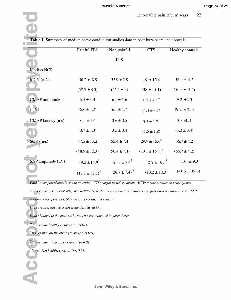
The results of median motor and sensory NCS are summarized in Table 1. The NCS of both the
ulnar and radial nerve were normal in all groups and did not differ among them. NCS of the median
nerve showed abnormalities suggestive of CTS in 4 hands with painful-PPS and 3 with non painful-
PPS (P=NS). These abnormalities involved only limbs with PPS, and were bilateral in 2 patients.
The severity of NCS abnormalities suggestive of CTS was graded as minimal in one hand (14.2),
mild in 2 (28.5%), moderate in 3 (42.8%), and extreme in 1 (14.2%). In the control group of hands
with CTS, median NCS abnormalities were graded as minimal in 4 (9 %), mild in 5 (11.4 %),
moderate in 27 (61.3 %), severe in 5 (11.4 %), and extreme in 3 (6.8 %). The amplitude of the
sensory nerve action potential was lower in hands with both painful and non painful-PPS than in
healthy controls.
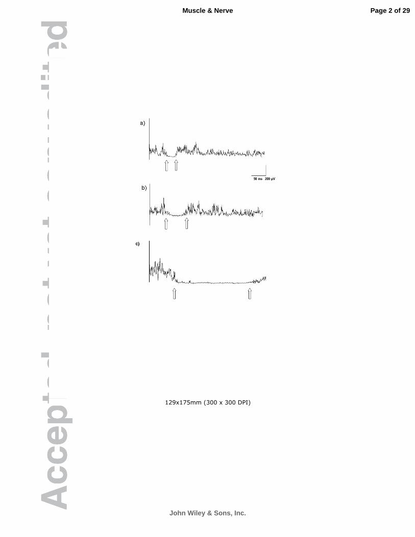
Examples of CSP obtained in hands with painful-PPS, CTS and healthy controls are provided in
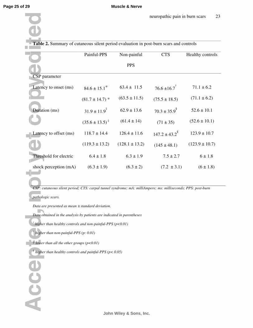
Figure 2. Results of CSP data evaluation are summarized in Table 2. There were no interside
differences for the CSP parameters considered in the analysis in healthy controls. The most salient
findings of the CSP evaluation were: 1) significantly shorter duration in hands with painful-PPS
than the other groups; 2) significantly longer duration and latency to offset in hands with CTS than
healthy controls and painful-PPS. The analysis made by patients confirmed the significant reduction
of CSP duration in patients with painful-PPS than in other groups (Table 2). In patients with
painful-PPS, the duration of CSP in the painful hands was significantly shorter than in the non-
painful-hands (32.6 + 11.8 ms vs 51.3 + 8.2 ms, P=0.001). No effect of age or gender was evident
in the comparison of median NCS and CSP parameters among the groups of hands. Taking a
duration of CSP lower than 32.4 ms as the minimal cut-off, the CSP was abnormally shortened in 7
of 17 hands with painful-PPS, but in none in the other groups (P<0.01, in all comparisons)
Quantitative sensory testing.
Page 13 of 29
John Wiley & Sons, Inc.
Muscle & Nerve
neuropathic pain in burn scars
12
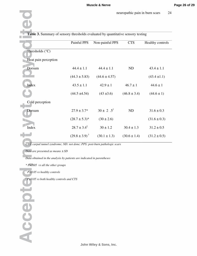
Analysis of parameter distribution showed a non-normal distribution of QST results, and therefore
they were log-transformed before ANOVA. Results of QST evaluation are summarized in Tables 3
and 4. In healthy controls there was no significant difference between right and left hands for all
QST parameters. The CDT on the dorsum was lower in hands with painful-PPS than in both healthy
controls and non-painful PPS. The CDT in the index finger was lower in hands with painful-PPS
than in healthy controls and CTS. The HPT was not different among groups. The analysis made by
patients confirmed the significant reduction of CDT at both sites identified in the analysis by hands
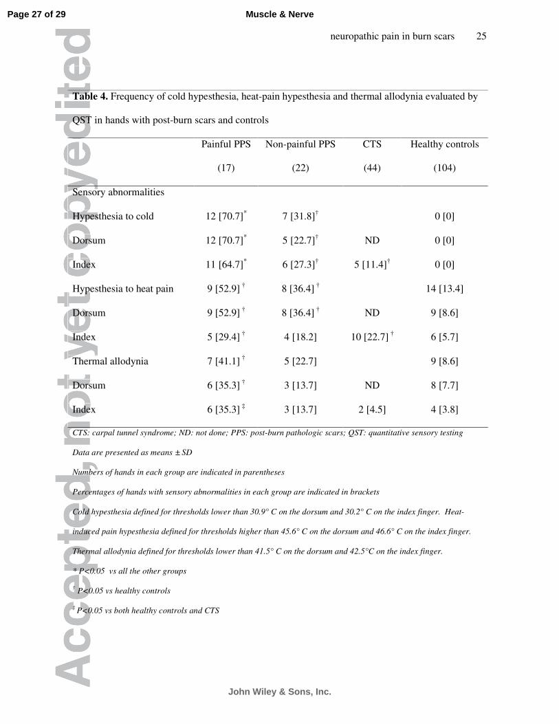
(Table 3). Hypesthesia to cold, to heat-pain and thermal allodynia was more frequent in painful-PPS
than in healthy controls at both sites. Reduced cold sensation was also more frequent in painful-PPS
than both in non-painful-PPS and CTS groups at all sites evaluated. Warm and heat-pain anesthesia
to 50°C was detected in only 4 of 17 hands with painful-PPS, but not in the other groups (P<0.05 in
all comparisons). In summary, at least one abnormal QST finding in at least one site was detected in
all hands with painful-PPS, 12 of 22 with non painful-PPS, and 22 of 104 of healthy controls
(p<0.0001) No effect of age or gender was evident in the comparison of QST parameters among the
groups.
Correlation analysis
The VAS score did not correlate with any neurophysiological or QST parameter in patients with
painful-PPS. In hands with painful-PPS, the CSP duration correlated significantly only with CDT
on the dorsum of the hand (r: 0.53, P<0.05). On the contrary, in the other groups, the CSP duration
correlated significantly with latency to offset (non painful-PPS, r: 0.59,p<0.05; healthy controls, r:
0.79, p<0.0001; CTS, r: 0.92, p<0.0001). Latency to offset correlated with sensory conduction
velocity in painful-PPS (r: -0.57, p<0.05), non-painful-PPS (r: -0.77, p<0.0001) and healthy
controls (r: -0.60, p<0.05), but not in CTS. In painful-PPS, the CDT on the dorsum correlated
significantly with CDT on the index finger (r: 0.67, P<0.05), and it was lower when CSP duration
was reduced than when it was normal (25.4 ± 3.5 ° C vs 29.5 ± 2.8 ° C, P= 0.02).
Page 14 of 29
John Wiley & Sons, Inc.
Muscle & Nerve
neuropathic pain in burn scars
13
DISCUSSION
Pain is a frequent and often severe accompanying symptom of PPS. Previous reports put forward
the hypothesis that it may be of neuropathic origin.2 However no previous studies have
systematically investigated the occurrence of possible neuropathic pain in patients with PPS using
standardized screening tools, such as the DN4 questionnaire. Moreover, no previous studies have
attempted to correlate the occurrence of possible neuropathic pain with the presence of large as well
small sensory fiber damage in PPS.
The most striking findings in our series were: 1) all patients complaining of pain at the PPS site had
a DN4 questionnaire score ≥ 4, suggesting a possible neuropathic origin of pain itself; 2) in
hands/patients with painful-PPS, both QST and CSP findings differed significantly from those
obtained in the other groups. In fact, in the presence of painful-PPS, there were lower CDT, shorter
CSP duration, more frequent hypesthesia to cold than in healthy controls and in non-painful-PPS,
and more frequent thermal allodynia and hypesthesia to heat pain than in healthy controls. The
absence of a significant difference for HPT among the groups may be due to the significant
presence of both abnormally lower and higher HPT in the hands with painful-PPS, reflecting
thermal allodynia and hypesthesia to heat pain, respectively. Therefore, it is reasonable to presume
that, in this setting, the mean HPT is less informative than is the number of hands with abnormally
reduced or increased HPT values. Even if warm perception was not evaluated, it is noteworthy that
about 25% of hands with painful-PPS, but none in the other groups lacked warm or heat pain
perception at 50°C. Assessment of CDT is considered a suitable method to evaluate the function of
Aδ fibers,13,24,29
and CSP duration is influenced primarily by Aδ afferents.13-15
In patients with
painful-PPS, our study showed a correlation between the duration of CSP and CDT; moreover, an
abnormally reduced CSP duration seems to be associated with a more severe degree of CDT
reduction in these patents. Similar results have been described in Fabry disease13
and suggest that
CSP duration is reduced only when damage to Aδ fibers is moderate-to-severe.
Page 15 of 29
John Wiley & Sons, Inc.
Muscle & Nerve
neuropathic pain in burn scars
14
Taken together, these observations suggest that there may be a substantial impairment of Aδ and C
fibers at the PPS sites.13,24,29
Previous series reported a similar pattern of sensory impairment in
chronic burn lesions.30,31
Nedelec et al31
reported significantly reduced CDT, anesthesia to both
warm and heat-pain in 27% of cases, but no significant abnormality of mean HPT in grafted skin
after burns. The pattern of QST abnormalities in their study was very similar to that observed in our
series. Histological evaluations disclosed a severe reduction of both dermal and epidermal nerve
fibers in skin grafts after burns.31
Similar histological findings have been reported in other
evaluations of patients with PPS.6 Therefore, skin lesions after burns seem to be characterized by a
moderate-to severe loss of small fibers. However, none of the previous studies of burn lesions
correlated with the presence and extent of small fiber damage to the occurrence of possible
neuropathic pain.
The prominent role of small fiber damage in the genesis of neuropathic pain has been increasingly
recognized.9,10,32,33
In our patients, pain at the PPS site seems to be associated with moderate-to
severe abnormalities of QST and CSP; this observation is in line with the hypothesis that the degree
of small sensory fiber loss is related to the probability of development of neuropathic pain as well
as to its severity.33
We are aware of some possible criticisms of our results. The first point is the role of median
mononeuropathy at the wrist in the development of neuropathic pain, and, secondarily, in the
genesis of QST as well as CSP abnormalities, in patients with PPS. Some observations suggest that
this role seems to be irrelevant: 1) previous studies emphasized the lack of association between Aß
fiber damage and the development of neuropathic pain at least in CTS and peripheral
neuropathies;9,32
2) the frequency of NCS abnormalities suggestive of CTS did not differ between
hands with painful and non painful-PPS; 3) only one patient with painful-PPS had clear signs and
symptoms suggestive of CTS; 4) the CSP duration is significantly prolonged in hands with CTS
than in hands with painful-PPS or in healthy controls, which is in line with other reports.18
Taken
Page 16 of 29
John Wiley & Sons, Inc.
Muscle & Nerve
neuropathic pain in burn scars
15
as a whole, these observations suggest that the occurrence of possible neuropathic pain does not
seem to be correlated with the presence of a median mononeuropathy at wrist.
A second possible criticism is the possibility that abnormalities of CDT and the CSP may be due to
modulation of spinal circuitry induced by the pain itself. Furthermore, previous studies have put
into question the role of both QST and CSP for the definition of the neuropathic ethiology of
pain.7,34
Previous reports have suggested that both nociceptive and neuropathic pain may modulate
spinal cord circuitry through the activation of diffuse noxious inhibitory control, which acts
through inhibition of wide dynamic range neurons in the dorsal horn.16,35
This mechanism of spinal
cord modulation by pain was investigated by the use of painful heterotopic stimulation which, when
applied unilaterally, reduces the duration of the CSP16
and CDT35
bilaterally. However, our study
showed no correlation with this mechanism, because in patients with painful-PPS, the QST and
CSP abnormalities were confined only to the painful hands, even if there was a non painful-PPS in
the other hand. We agree with previous reports that the best neurophysiological tool to confirm the
neuropathic nature of pain is laser evoked potentials.7,34
However, no safety studies are available for
the use of these potentials in patients with burn lesions. Therefore our choice fell to non-invasive
and previously tested evaluations such as QST in this particular setting.30,31
When all these observations are taken into consideration, some thought should be given to the level
of certainty of neuropathic pain in PPS. Our study documents that in PPS, a condition characterized
by a moderate-to-severe loss of epidermal and dermal nerve fibers,5,6,31
possible neuropathic pain is
associated with laboratory evidence of small fiber damage. Moreover, pain is confined only to areas
involved by PPS. Therefore, pain in PPS seems to be associated with a definite lesion of the
somatosensory system (i.e. the small sensory fiber loss that occurs in PPS) and has a
neuroanatomically plausible distribution (i.e. is confined to areas involved by the PPS with
laboratory evidence of more severe small fiber damage). Therefore, in a very conservative way, the
grading of certainty for pain in PPS may be, at least, probable neuropathic pain.3
Page 17 of 29
John Wiley & Sons, Inc.
Muscle & Nerve
neuropathic pain in burn scars
16
Further studies are required to obtain a more precise definition of the nature and degree of small
fiber damage necessary to induce neuropathic pain in patients with PPS. This may be of great
importance for the therapy of this pain for which treatment has not been satisfactory.2
ACKNOWLEDGMENT
The authors thanks Mrs Barbara Wade for her linguistic device.
Page 18 of 29
John Wiley & Sons, Inc.
Muscle & Nerve

neuropathic pain in burn scars
17
LIST OF ABBREVATIONS
cMAP: compound muscle action potential; CDT: cold detection threshold; CSP: cutaneous silent
period; CTS: carpal tunnel syndrome; DML: distal motor latency; DN-4: douleur Neuropathique en
4 questions; HPT: heat-induced pain threshold; mA: milliAmpere; MCV: motor conduction
velocity; ms: milliseconds; mV: milliVolts; µV: microVolts; NCS: nerve conduction studies; ND:
not done; PPS: post burn pathologic scars; QST: quantitative sensory testing; SAP: sensory action
potential; SCV: sensory conduction velocity. SD: standard deviation; VAS: visual analog scale
Page 19 of 29
John Wiley & Sons, Inc.
Muscle & Nerve
neuropathic pain in burn scars
18
REFERENCES.
1. Gangemi EN, Gregori D, Berchialla P, Zingarelli E, Cairo M, Bollero D, et al.
Epidemiology and risk factors for Pathologic scarring after burn wounds. Arch Facial Plast Surg
2008; 10: 93-102
2. Schneider JC, Harris NL, El Shami A, Sheridan RL, Schulz JT, Bilodeau ML, et al. A descriptive
review of neuropathic-like pain after burn injury. Burn Care and Res 2006; 27: 524-528
3. Treede RD, Jensen TS, Campbell JN, Cruccu G, Dostrovsky JO, Griffin JW, et al. Neuropathic
pain. Redefinition and a grading system for clinical and research purposes. Neurology 2008; 70:
1630-163
4. Marquez S, Turley JJE, Peters WJ. Neuropathy in burn patients. Brain 1993; 116: 471-483. 1.
5.Altun V, Hakvoort TE, van Zyijlen PPM, van der Kwaast H, Prens EP. Nerve outgrowth and
neuropeptide expression during the remodelling of human burn wound scars. A 7-month follow-up
study of 22 patients. Burns 2001; 27: 717-722
6. Stella M, Calcagni M, Teich-Alasia S, Ramieri G, Cellino G, Panzica G. Sensory endings in skin
grafts and scars after extensive burns. Burns. 1994; 20: 491-5.
7. Haampaa M, Attal N, Backonja M, Baron R, Bennett M, Bouhassira D, et al. New guidelines on
neuropathic pain assessment. Pain 2011; 152(1): 14-27.
8. Bouhassira D, Attal N, Alchaar H, Boureau F, Brochet B, Bruxelle J, et al. Comparison of pain
syndromes associated with nervous or somatic lesions and development of a new neuropathic pain
diagnostic questionnaire (DN4). Pain 2005; 114: 29-36
9. Truini A, Padua L, Biasiotta A, Caliandro P, Pazzaglia C, Galeotti F, et al. Differential
involvement of A-delta and A-beta fibers in neuropathic pain related to carpal tunnel syndrome.
Pain. 2009; 145: 105-9
10. Devigili G, Tugnoli V, Penza P, Camozzi F, Lombardi R, Melli G, et al. The diagnostic criteria
for small fiber neuropathy. From symptmos to neuropathology. Brain 2008; 131: 1912-1925.
Page 20 of 29
John Wiley & Sons, Inc.
Muscle & Nerve
neuropathic pain in burn scars
19
11. Lacomis D. Small fiber neuropathy. Muscle Nerve 2002; 26: 173-188.
12. Onal MR, Ulas UH, Oz O, Bek VS, Yucel M, Taslipinar A, et al. Cutaneous silent period
changes in type 2 diabetes mellitus patients with small fiber neuropathy. Clin Neurophysiol 2010;
121: 714-718.
13. Syed NA, Sandbrink F, Luciano CA, Altarescu G, Weibel T, Schiffmann R, et al. Cutaneous
silent period in patients with Fabry disease. Muscle Nerve 2000; 23: 1179-1186.
14. Floeter MK. Cutaneous silent periods. Muscle Nerve. 2003;28:391-401.
15. Inghilleri M, Cruccu G, Argenta M, Polidori L, Manfredi M. Silent period in upper limb
muscles after noxious cutaneous stimulation in man. Electroencephalogr Clin Neurophysiol. 1997;
105: 109-15.
16. Rossi P, Pierelli F, Parisi L, Perrotta A, Bartolo M, Amabile G, et al. Effect of painful
heterotopic stimulation on the cutaneous silent period in the upper limbs. Clin Neurophysiol 2003;
114: 1-6
17. Serrao M, Parisi L, Pierelli F, Rossi P. Cutaneous afferents mediating the cutaneous silent
period in the upper limbs: evidence for a role of low-threshold sensory fibers. Clin Neurophysiol
2001; 112: 2007-2014.
18. Svilpauskatie J, Truffert A, Vaiciene N, Magistris MR. Cutaneous silent period in carpal tunnel
syndrome. Muscle Nerve 2006; 33: 487-493.
19. Isoardo G, Migliaretti G, Ciaramitaro P, Rota E, Poglio F, Tavella A, et al. Differential
diagnosis of chronic dysimmune demyelinating polyneuropathies with and without anti-MAG
antibodies. Muscle Nerve. 2005; 31: 52-8.
20. American Academy of Neurology. Practice parameters for carpal tunnel syndrome. Neurology
1993; 43: 2406-2409
Page 21 of 29
John Wiley & Sons, Inc.
Muscle & Nerve
neuropathic pain in burn scars
20
21. Jablecki CK, Andary MT, Floeter MK, Miller RG, Quartly CA, Vennix MJ, et al. Practice
parameter: electrodiagnostic studies in carpal tunnel syndrome. Report of the American Academy
of electrodiagnostic Medicine, America Academy of Neurology and the American Academy of
Physical Medicine and Rehabilitation. Neurology 2002; 58: 1598-1593.
22. Preston DC. Distal median neuropathies. In Logigian EL, editor. Neurologic Clinics.
Entrapment and other focal neuropathies. Philadelphia: WB Saunders, 1999: p 407-424
23. Padua L, Lo Monaco M, Gregori B, Valente EM, Padua R, Tonali P. Neurophysiological
classification and sensitivity in 500 carpal tunnel syndrome hands. Acta Neurol Scand 1997; 96:
211-217
24. Chong PST, Cros D. Technology literature review: quantitative sensory testing. Muscle Nerve
2004; 23:734-747
25. Fowler CJ, Carroll MB, Burns D, Howe N, Robinson K. A portable system for measuring
cutaneous thresholds for warming and cooling. J Neurol Neurosurg Psychiatry 1987; 50: 1211-
1215.
26. Maier C, Baron R, Tolle TR, Binder A, Birbaumer N, Birklein F, et al. Quantitative sensory
testing in the German Research Network on Neuropathic Pain (DFNS): somatosensory
abnormalities in 1236 patients with different neuropathic pain syndromes. Pain 2010; 150: 439-450
27. Rolke R, Baron R, Maier C, Tölle TR, Treede RD, Beyer A, et al. Quantitative sensory testing
in the German Research Network on Neuropathic Pain (DFNS): standardised protocol and reference
values. Pain 2006; 123: 231-243.
28. Padua L, Pasqualetti P, Rosenbaum R. One patient, two carpaal tunnels: statistical and clinical
analysis- by hand or by patient? Clin Neurophysiol 2005; 16: 241-243
29. Dyck PJ, O Brien P, Johnson DM, Klein CJ, Dyck PJB. Quantitative sensory testing In: Dyck
PJ, Thomas PK editors. Peripheral neuropathy. Philadelphia: Elsever Saunders, 2005: p 1063-1093
Page 22 of 29
John Wiley & Sons, Inc.
Muscle & Nerve
neuropathic pain in burn scars
21
30. Malenfant A, Forget R, Ansel R, Papilon J, Frigon JY, Choiniere M. Tactile, thermal and pain
sensibility in burned patients with and without chronic pain and paresthesia problems. Pain 1998:
77: 241-251.
31. Nedelec B, Hou Q, Sohbi I, Choiniere M, Beauregard G, Dykes RW. Sensory perception and
neuroanatomical structures in normal and grafted skin of burned survivors. Burns 2005; 31:817-830
32. Truini A, Biasiotta A, La Cesa S, Di Stefano G, Galeotti F, Petrucci MT, et al. Mechanism of
pain in distal simmetrici polyneuropathy: A combined clinical and neurophysiological study. Pain
2010; 150: 516-521.
33. Sommer C, Lauria G. Skin biopsy in the management of peripheral neuropathy. Lancet Neurol
2007; 6: 632-642
34. Truini A, Galeotti F, Biasiotta A, Gabriele M, Inghilleri M, Petrucci MT,et al.. Dissociation
between cutaneous silent period and laser evoked potentials in assessing neuropathic pain. Muscle
Nerve 2009; 39: 369-373
35. Leffler AS, Kosek E, Hansson P. The influence of pain intensity on somatosensory perception
in patients suffering from subacute/chronic lateral epicondylalgia. Eur J Pain 2000; 4: 57-71
Page 23 of 29
John Wiley & Sons, Inc.
Muscle & Nerve
neuropathic pain in burn scars
22
Table 1. Summary of median nerve conduction studies data in post-burn scars and controls
Painful-PPS Non-painful
PPS
CTS Healthy controls
Median NCS
MCV (m/s) 50.2 ± 8.9
(52.7 ± 8.3)
55.9 ± 2.9
(56.1 ± 3)
48 ± 15.4
(48 ± 15.1)
56.9 ± 4.5
(56.9 ± 4.5)
CMAP amplitude
(mV)
6.5 ± 3.3
(6.6 ± 3.2)
6.1 ± 1.6
(6.1 ± 1.7)
5 3 ± 3.1*
(5.4 ± 3.1)
9.2 ±2.5
(9.2 ± 2.5)
CMAP latency (ms) 3.7 ± 1.6
(3.7 ± 1.3)
3.6 ± 0.5
(3.5 ± 0.4)
5.5 ± 1.7†
(5.5 ± 1.8)
3.3 ±0.4
(3.3 ± 0.4)
SCV (m/s) 47.5 ± 13.2
(48.9 ± 12.3)
55.4 ± 7.4
(56.4 ± 7.4)
29.9 ± 15.6‡
(30.1 ± 15.4) ‡
56.7 ± 4.2
(56.7 ± 4.2)
SAP amplitude (µV) 19.2 ± 14.9§
(18.7 ± 13.2) §
26.8 ± 7.4§
(26.7 ± 7.4) §
12.9 ± 10.5§
(13.2 ± 10.3)
41.6 ±19.3
(41.6 ± 19.3)
CMAP: compound muscle action potential; CTS: carpal tunnel syndrome; MCV: motor conduction velocity; ms:
milliseconds; µV: microVolts; mV: milliVolts; NCS: nerve conduction studies; PPS: post-burn pathologic scars; SAP:
sensory action potential; SCV: sensory conduction velocity.
Data are presented as mean ± standard deviation.
Data obtained in the analysis by patients are indicated in parentheses
* lower than healthy controls (p: 0.001)
† higher than all the other groups (p<0.0001)
‡ lower than all the other groups (p<0.01)
§ lower than healthy controls (p< 0.01).
Page 24 of 29
John Wiley & Sons, Inc.
Muscle & Nerve
neuropathic pain in burn scars
23
Table 2. Summary of cutaneous silent period evaluation in post-burn scars and controls
Painful-PPS Non-painful
PPS
CTS Healthy controls
CSP parameter
Latency to onset (ms) 84.6 ± 15.1*
(81.7 ± 14.7) *
63.4 ± 11.5
(63.5 ± 11.5)
76.6 ±16.7†
(75.5 ± 18.5)
71.1 ± 6.2
(71.1 ± 6.2)
Duration (ms) 31.9 ± 11.9‡
(35.6 ± 13.5) ‡
62.9 ± 13.6
(61.4 ± 14)
70.3 ± 35.9§
(71 ± 35)
52.6 ± 10.1
(52.6 ± 10.1)
Latency to offset (ms) 118.7 ± 14.4
(119.3 ± 13.2)
126.4 ± 11.6
(128.1 ± 13.2)
147.2 ± 43.2§
(145 ± 48.1)
123.9 ± 10.7
(123.9 ± 10.7)
Threshold for electric
shock perception (mA)
6.4 ± 1.8
(6.3 ± 1.9)
6.3 ± 1.9
(6.3 ± 2)
7.5 ± 2.7
(7.2 ± 3.1)
6 ± 1.8
(6 ± 1.8)
CSP: cutaneous silent period; CTS: carpal tunnel syndrome; mA: milliAmpere; ms: milliseconds; PPS: post-burn
pathologic scars.
Data are presented as mean ± standard deviation.
Data obtained in the analysis by patients are indicated in parentheses
* higher than healthy controls and non-painful-PPS (p<0.01)
† higher than non-painful-PPS (p: 0.01)
‡ lower than all the other groups (p<0.01)
§ higher than healthy controls and painful-PPS (p< 0.05)
Page 25 of 29
John Wiley & Sons, Inc.
Muscle & Nerve
neuropathic pain in burn scars
24
Table 3. Summary of sensory thresholds evaluated by quantitative sensory testing
Painful PPS Non-painful PPS CTS Healthy controls
Thresholds (°C)
Heat pain perception
Dorsum 44.4 ± 1.1
(44.3 ± 5.83)
44.4 ± 1.1
(44.6 ± 4.57)
ND 43.4 ± 1.1
(43.4 ±1.1)
Index
43.5 ± 1.1
(44.5 ±4.54)
42.9 ± 1
(43 ±3.6)
46.7 ± 1
(46.8 ± 3.4)
44.6 ± 1
(44.6 ± 1)
Cold perception
Dorsum 27.9 ± 3.7*
(28.7 ± 5.3)*
30 ± 2 .5†
(30 ± 2.6)
ND 31.6 ± 0.3
(31.6 ± 0.3)
Index 28.7 ± 3.4‡
(29.8 ± 3.9) †
30 ± 1.2
(30.1 ± 1.3)
30.4 ± 1.3
(30.6 ± 1.4)
31.2 ± 0.5
(31.2 ± 0.5)
CTS: carpal tunnel syndrome; ND: not done; PPS: post-burn pathologic scars
Data are presented as means ± SD
Data obtained in the analysis by patients are indicated in parentheses
* P<0.05 vs all the other groups
† P<0.05 vs healthy controls
‡ P<0.05 vs both healthy controls and CTS
Page 26 of 29
John Wiley & Sons, Inc.
Muscle & Nerve
neuropathic pain in burn scars
25
Table 4. Frequency of cold hypesthesia, heat-pain hypesthesia and thermal allodynia evaluated by
QST in hands with post-burn scars and controls
Painful PPS
(17)
Non-painful PPS
(22)
CTS
(44)
Healthy controls
(104)
Sensory abnormalities
Hypesthesia to cold 12 [70.7]*
7 [31.8]† 0 [0]
Dorsum 12 [70.7]* 5 [22.7]
† ND 0 [0]
Index 11 [64.7]* 6 [27.3]
† 5 [11.4]
† 0 [0]
Hypesthesia to heat pain 9 [52.9] †
8 [36.4] †
14 [13.4]
Dorsum 9 [52.9] †
8 [36.4] †
ND 9 [8.6]
Index 5 [29.4] †
4 [18.2] 10 [22.7] †
6 [5.7]
Thermal allodynia 7 [41.1] †
5 [22.7] 9 [8.6]
Dorsum 6 [35.3] †
3 [13.7] ND 8 [7.7]
Index 6 [35.3] ‡
3 [13.7] 2 [4.5] 4 [3.8]
CTS: carpal tunnel syndrome; ND: not done; PPS: post-burn pathologic scars; QST: quantitative sensory testing
Data are presented as means ± SD
Numbers of hands in each group are indicated in parentheses
Percentages of hands with sensory abnormalities in each group are indicated in brackets
Cold hypesthesia defined for thresholds lower than 30.9° C on the dorsum and 30.2° C on the index finger. Heat-
induced pain hypesthesia defined for thresholds higher than 45.6° C on the dorsum and 46.6° C on the index finger.
Thermal allodynia defined for thresholds lower than 41.5° C on the dorsum and 42.5°C on the index finger.
* P<0.05 vs all the other groups
† P<0.05 vs healthy controls
‡ P<0.05 vs both healthy controls and CTS
Page 27 of 29
John Wiley & Sons, Inc.
Muscle & Nerve
neuropathic pain in burn scars
26
LEGEND TO FIGURES
Figure 1. An example of hyperthrophic post-burn pathologic scar, involving the hand.
Figure 2. Examples of cutaneous silent period in painful post-burn pathologic scars (a), healthy
control (b) and carpal tunnel syndrome (c). Cutaneous silent period was recorded from the abductor
pollicis brevis during an isometric contraction at maximum force against resistance. Electrical
stimulation was delivered to the second digit at an intensity 8 times the sensory threshold for an
electric shock. Latency to onset and latency to offset are indicated by arrows.
The duration of the CSP is reduced in post-burn pathologic scar (28 ms, a) and increased in carpal
tunnel syndrome (254 ms, c) compared to a healthy control (43 ms, b). The latency to onset is
increased in carpal tunnel syndrome (98 ms), compared to post-burn pathologic scars (70 ms) and in
the healthy control (72 ms)
Page 28 of 29
John Wiley & Sons, Inc.
Muscle & Nerve
129x175mm (300 x 300 DPI)
Page 1 of 29
John Wiley & Sons, Inc.
Muscle & Nerve
129x175mm (300 x 300 DPI)
Page 2 of 29
John Wiley & Sons, Inc.
Muscle & Nerve
Related Documents