Citation: Soni, K.K.; Jeong, H.-S.; Jang, S. Neurons for Ejaculation and Factors Affecting Ejaculation. Biology 2022, 11, 686. https://doi.org/ 10.3390/biology11050686 Academic Editor: Paul S. Cooke Received: 25 March 2022 Accepted: 28 April 2022 Published: 29 April 2022 Publisher’s Note: MDPI stays neutral with regard to jurisdictional claims in published maps and institutional affil- iations. Copyright: © 2022 by the authors. Licensee MDPI, Basel, Switzerland. This article is an open access article distributed under the terms and conditions of the Creative Commons Attribution (CC BY) license (https:// creativecommons.org/licenses/by/ 4.0/). biology Review Neurons for Ejaculation and Factors Affecting Ejaculation Kiran Kumar Soni * ,† , Han-Seong Jeong † and Sujeong Jang * Department of Physiology, Chonnam National University Medical School, Hwasun 58128, Korea; [email protected] * Correspondence: [email protected] (K.K.S.); [email protected] (S.J.) † These authors contributed equally to this work. Simple Summary: Sexual dysfunctions are rarely discussed in our current society. Males experience different sexual dysfunctions, including erectile, infertility, and ejaculatory dysfunctions. In this review only the ejaculatory dysfunction will be discussed. Ejaculation is defined as the ejection of contents collectively from the vas deferens, seminal vesicle, prostate and Cowper’s glands. It is completely controlled by a population of neurons present in the lumbar spinal cord. The presence of lesion in these neurons ceases the ejaculatory behavior in males. This population of neurons was first identified in rats; however, recently it was confirmed that these neurons are present in human males as well. The issues are known as ejaculatory dysfunction. The following are the different types of ejaculatory dysfunctions: early ejaculation, ejaculation into the urinary bladder, late ejaculation and no ejaculation. Abstract: Ejaculation is a reflex and the last stage of intercourse in male mammals. It consists of two coordinated phases, emission and expulsion. The emission phase consists of secretions from the vas deferens, seminal vesicle, prostate, and Cowper’s gland. Once these contents reach the posterior urethra, movement of the contents becomes inevitable, followed by the expulsion phase. The urogenital organs are synchronized during this complete event. The L3–L4 (lumbar) segment, the spinal cord region responsible for ejaculation, nerve cell bodies, also called lumbar spinothalamic (LSt) cells, which are denoted as spinal ejaculation generators or lumbar spinothalamic cells [Lst]. Lst cells activation causes ejaculation. These Lst cells coordinate with [autonomic] parasympathetic and sympathetic assistance in ejaculation. The presence of a spinal ejaculatory generator has recently been confirmed in humans. Different types of ejaculatory dysfunction in humans include premature ejaculation (PE), retrograde ejaculation (RE), delayed ejaculation (DE), and anejaculation (AE). The most common form of ejaculatory dysfunction studied is premature ejaculation. The least common forms of ejaculation studied are delayed ejaculation and anejaculation. Despite the confirmation of Lst in humans, there is insufficient research on animals mimicking human ejaculatory dysfunction. Keywords: ejaculation; spinal ejaculatory generator; lumbar spinothalamic cells; ejaculatory dysfunctions 1. Introduction Ejaculation is a spinal reflex. It is the forceful ejection of seminal fluid by the males at the end of coitus [1,2]. During ejaculation, the prostatic urethra has pressure of more than 5 m water pressure [3,4]. Specifically, the parasympathetic nervous system maintains the heart rate at relaxed levels; however, the sympathetic nervous system increases the rate of heart rate through the secretion of epinephrine. At the time of ejaculation, the heart rate may increase by 100% in males owing to sympathetic effects [5–7]. In rats, ejaculation is associated with reward [8]. It should not be confused with climax or orgasm, which provides pleasurable feelings. Orgasm and ejaculation can be considered a single incident, however, both are different biological processes [9]. Orgasm can be absent in some men with ejection of seminal contents during ejaculation [10], whereas in some men, ejaculation is absent, and they experience intense orgasm [11,12]. Biology 2022, 11, 686. https://doi.org/10.3390/biology11050686 https://www.mdpi.com/journal/biology

Welcome message from author
This document is posted to help you gain knowledge. Please leave a comment to let me know what you think about it! Share it to your friends and learn new things together.
Transcript

Citation: Soni, K.K.; Jeong, H.-S.;
Jang, S. Neurons for Ejaculation and
Factors Affecting Ejaculation. Biology
2022, 11, 686. https://doi.org/
10.3390/biology11050686
Academic Editor: Paul S. Cooke
Received: 25 March 2022
Accepted: 28 April 2022
Published: 29 April 2022
Publisher’s Note: MDPI stays neutral
with regard to jurisdictional claims in
published maps and institutional affil-
iations.
Copyright: © 2022 by the authors.
Licensee MDPI, Basel, Switzerland.
This article is an open access article
distributed under the terms and
conditions of the Creative Commons
Attribution (CC BY) license (https://
creativecommons.org/licenses/by/
4.0/).
biology
Review
Neurons for Ejaculation and Factors Affecting EjaculationKiran Kumar Soni *,† , Han-Seong Jeong † and Sujeong Jang *
Department of Physiology, Chonnam National University Medical School, Hwasun 58128, Korea;[email protected]* Correspondence: [email protected] (K.K.S.); [email protected] (S.J.)† These authors contributed equally to this work.
Simple Summary: Sexual dysfunctions are rarely discussed in our current society. Males experiencedifferent sexual dysfunctions, including erectile, infertility, and ejaculatory dysfunctions. In thisreview only the ejaculatory dysfunction will be discussed. Ejaculation is defined as the ejection ofcontents collectively from the vas deferens, seminal vesicle, prostate and Cowper’s glands. It iscompletely controlled by a population of neurons present in the lumbar spinal cord. The presence oflesion in these neurons ceases the ejaculatory behavior in males. This population of neurons was firstidentified in rats; however, recently it was confirmed that these neurons are present in human malesas well. The issues are known as ejaculatory dysfunction. The following are the different types ofejaculatory dysfunctions: early ejaculation, ejaculation into the urinary bladder, late ejaculation andno ejaculation.
Abstract: Ejaculation is a reflex and the last stage of intercourse in male mammals. It consists oftwo coordinated phases, emission and expulsion. The emission phase consists of secretions fromthe vas deferens, seminal vesicle, prostate, and Cowper’s gland. Once these contents reach theposterior urethra, movement of the contents becomes inevitable, followed by the expulsion phase.The urogenital organs are synchronized during this complete event. The L3–L4 (lumbar) segment,the spinal cord region responsible for ejaculation, nerve cell bodies, also called lumbar spinothalamic(LSt) cells, which are denoted as spinal ejaculation generators or lumbar spinothalamic cells [Lst].Lst cells activation causes ejaculation. These Lst cells coordinate with [autonomic] parasympatheticand sympathetic assistance in ejaculation. The presence of a spinal ejaculatory generator has recentlybeen confirmed in humans. Different types of ejaculatory dysfunction in humans include prematureejaculation (PE), retrograde ejaculation (RE), delayed ejaculation (DE), and anejaculation (AE). Themost common form of ejaculatory dysfunction studied is premature ejaculation. The least commonforms of ejaculation studied are delayed ejaculation and anejaculation. Despite the confirmation ofLst in humans, there is insufficient research on animals mimicking human ejaculatory dysfunction.
Keywords: ejaculation; spinal ejaculatory generator; lumbar spinothalamic cells; ejaculatory dysfunctions
1. Introduction
Ejaculation is a spinal reflex. It is the forceful ejection of seminal fluid by the malesat the end of coitus [1,2]. During ejaculation, the prostatic urethra has pressure of morethan 5 m water pressure [3,4]. Specifically, the parasympathetic nervous system maintainsthe heart rate at relaxed levels; however, the sympathetic nervous system increases therate of heart rate through the secretion of epinephrine. At the time of ejaculation, the heartrate may increase by 100% in males owing to sympathetic effects [5–7]. In rats, ejaculationis associated with reward [8]. It should not be confused with climax or orgasm, whichprovides pleasurable feelings. Orgasm and ejaculation can be considered a single incident,however, both are different biological processes [9]. Orgasm can be absent in some menwith ejection of seminal contents during ejaculation [10], whereas in some men, ejaculationis absent, and they experience intense orgasm [11,12].
Biology 2022, 11, 686. https://doi.org/10.3390/biology11050686 https://www.mdpi.com/journal/biology
Biology 2022, 11, 686 2 of 16
Ejaculation delivers sperm to the female genital tract for offspring generation. The dis-tal epididymis, vas deferens, seminal vesicle, prostate, prostatic urethra, and bladder neckare the male reproductive organs involved in ejaculation [2,13]. The duration and degreeof ejaculation vary remarkably among men and individuals in different situations [14]. Inrats, ejaculation is distinguished by a prolonged, intense force (750–2000 ms), and muchslower dismount [15]. This review aims to explain the neural regulation of ejaculation, itsabnormalities, and its etiology. Moreover, this review discusses ejaculation physiology andabnormalities in animals and their correlation with humans, as well as ways to improvestudies that allow mimicking this pathology in humans.
2. Neurophysiology of Ejaculation
Ejaculation remains in the control of the parasympathetic (sacral) and sympathetic(thoracic) autonomic nervous systems and spinal centers [16]. It remains intact in animalswith complete spinal cord transection, which provides evidence that ejaculation is con-trolled at the spinal level, despite the loss in connection from supraspinal regions [17].The integration between spinal centers and the autonomic nervous system is organizedby interneurons that shape the spinal ejaculation generator [SEG] [18]. Based on a de-tailed study of the spinal gray matter and its cellular composition under a microscope,the gray matter of the spinal cord was found to be split into 10 laminae [I–X] [19]. Theparasympathetic nucleus (sacral) innervates the prostate and seminal vesicles located inthe S2–S4 segments of lamina VII [16]. The sympathetic nucleus (thoracic) innervates thesmooth involuntary muscles of the seminal tract and bladder neck located in the T12-L2segments of lamina VII [20,21]. Motor neurons that govern the pelvic-perineal striatedmuscles remain in the Onuf’s nucleus of the ventral horn of the segments [22]. The spinalejaculation generator or lumbar spinothalamic cells are located in lamina X and the medialpart of lamina VII of the gray matter in the lumbar L3–L4 spinal cord of rats [23,24]. Theyare called lumbar spinothalamic cells as they have connections in the lumbar spinal cordand thalamus [22]. Recently, it was confirmed that the spinal ejaculation generator in malehumans is in the L3–L5 segment [16].
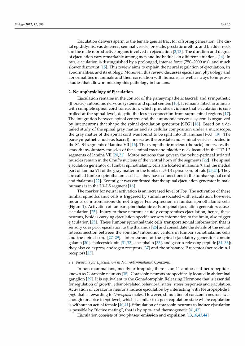
The marker for neural activation is an increased level of Fos. The activation of theselumbar spinothalamic cells is triggered by stimuli associated with ejaculation; however,mounts or intromissions do not trigger Fos expression in lumbar spinothalamic cells(Figure 1). Activation of lumbar spinothalamic cells or spinal ejaculation generators causesejaculation [25]. Injury to these neurons acutely compromises ejaculation; hence, theseneurons, besides carrying ejaculation-specific sensory information to the brain, also triggerejaculation [25]. These lumbar spinothalamic cells transport sexual information that issensory cues prior ejaculation to the thalamus [26] and consolidate the details of the neuralinterconnection between the somatic/autonomic centers in lumbar spinothalamic cellsand the spinal cord [27–29]. Interneurons of the spinal ejaculatory generator containgalanin [30], cholecystokinin [31,32], encephalin [33], and gastrin-releasing peptide [34–36];they also co-express androgen receptors [37] and the substance P receptor (neurokinin-1receptor) [23].
2.1. Neurons for Ejaculation in Non-Mammalians: Corazonin
In non-mammalians, mostly arthropods, there is an 11 amino acid neuropeptidesknown as Corazonin neurons [38]. Corazonin neurons are specifically located in abdominalganglion [39]. It is equivalent to the Gonadotrophin Releasing Hormone that is essentialfor regulation of growth, ethanol-related behavioral states, stress responses and ejaculation.Activation of corazonin neurons induce ejaculation by interacting with Neuropeptide F(npf) that is rewarding to Drosophila males. However, stimulation of corazonin neurons wasenough for a rise in npf level, which is similar to a post-copulation state where copulationis without an actual female [40,41]. Stimulation of corazonin neurons to induce ejaculationis possible by “fictive mating”, that is by opto- and thermogenetic [41,42].
Ejaculation consists of two phases: emission and expulsion [13,16,43,44].
Biology 2022, 11, 686 3 of 16
Figure 1. Schematic diagram of level of spinal cord and neurons regulating ejaculation. Activatedspinal ejaculation generator directs and synchronizes the activity of: (a) parasympathetic output thatinnervate prostate and seminal vesicles secreting seminal fluid. (b) Sympathetic output that innervatesmooth muscle cells of the seminal tract and the bladder neck. Contraction of the seminal tractaccumulate spermatozoa that is mixed with the seminal fluid to the prostatic urethra. The neck ofbladder remains closed to prevent retrograde ejaculation. (c) Somatic output that innervate the pelvicstriated muscles (not shown in this figure). The external urethral sphincter relaxes and rhythmiccontractions of the bulbospongiosus and ischiocavernosus muscles are responsible for rhythmicforceful expulsion of sperm at the urethral meatus. (a) and (b) = emission; (c) = expulsion.
2.2. Emission
The beginning of this phase involves the shutdown of the bladder neck to check forretrograde ejaculation [45]. It is followed by a mixture of seminal vesicle, prostatic, vasdeferens, and Cowper’s gland secretions into the prostatic urethra [46]. The initiation ofthe emission phase of ejaculation is not under individual will or controlled cerebrally andcan be evoked through visual erotic stimulation or physical stimulation [47,48]. The organsinvolved in this phase obtain deep autonomic innervations formed by the sympathetic andparasympathetic nerves from the pelvic plexus [48]. The basic neurotransmitter required inthe stimulation of the sympathetic nervous system is norepinephrine, and this is balancedby acetylcholine, the parasympathetic neurotransmitter. Sympathetic nerves at levelsT10–L2 that leave the spinal cord commence peristaltic contraction of the prostate smoothmuscle, seminal vesicles, vas deferens, and epididymis [49]. Once the semen reaches theposterior urethra, ejection of the semen and its content becomes unavoidable [48].
2.3. Expulsion [Anterograde Ejaculation]
Expulsion follows emission, where semen is pushed out as the consequence of therhythmic contractions of the striated muscles of the pelvis and the ischiocavernosus, bul-bospongiosus, and perineal muscles [43]. Electromyographic [EMG] studies of the bul-bocavernosus or bulbospongiosus have reported evidence of ejaculation or expulsion in

Biology 2022, 11, 686 4 of 16
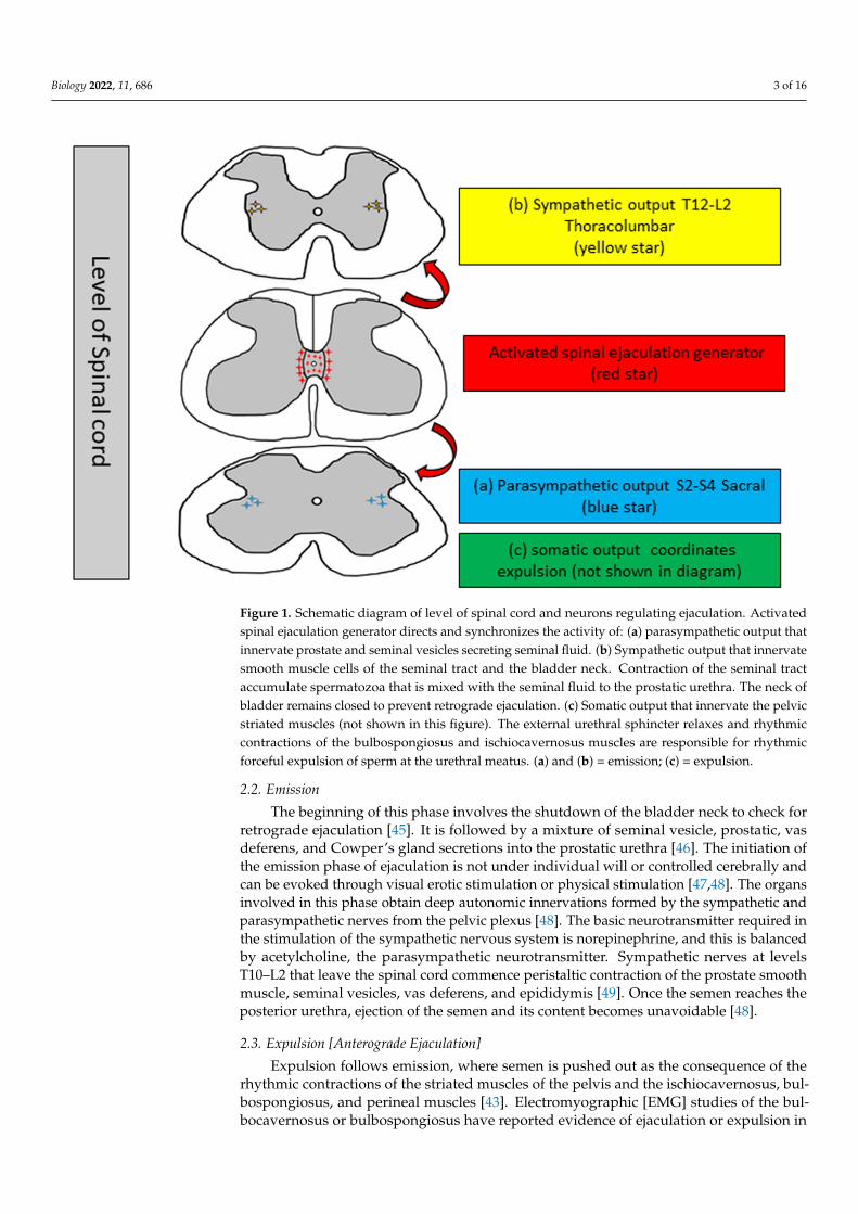
animals following electrical or mechanical stimulation of genital structures or of the dorsalnerve of the penis [32,50], which is controlled by lumbar spinothalamic cells. This inducesejaculation by consolidating the sensory information conveyed by the dorsal nerve ofthe penis, which is the sensory branch of the pudendal nerve [34]. The pudendal nerve,which begins at the S2–S4 level of the sacral spinal cord, causes rhythmic involuntarycontractions (Figure 2) [49]. The ejaculation response in men is generally approximately10–15 contractions [51]. The intra-seminal vesicle pressure during expulsion in rats isapproximately 61.4 mmHg [52]. Men cannot undergo a series of ejaculations rapidly; menenter a refractory period immediately after ejaculation; it is a regaining time in which noejaculation is possible. The refractory period time varies among individuals, ranging froma few minutes to hours [53].
Figure 2. Schematic diagram of organs involved in ejaculation. Anterograde ejaculation (green arrow)and retrograde ejaculation (black arrow) shown in same figure, retrograde ejaculation is due to theinability of the neck of bladder to close completely during expulsion phase. Some amount of semencan be seen in the urinary bladder.
3. Ejaculatory Dysfunction
Ejaculatory dysfunction can be classified into four types: premature ejaculation, retro-grade ejaculation, delayed ejaculation, and anejaculation [54]. Here, delayed ejaculationand anejaculation have been discussed together because the most severe form of delayedejaculation is anejaculation. Almost all factors for anejaculation are extreme conditions ofdelayed ejaculation (Figure 3) [20,55,56]. Premature ejaculation and retrograde ejaculationhave been widely studied [57]. Unfortunately, delayed ejaculation and anejaculation are theminimally studied ejaculatory dysfunctions [58,59].

Biology 2022, 11, 686 5 of 16
Figure 3. Table showing the normal and abnormal types of ejaculation. Their etiologies are describedin brief.
4. Premature Ejaculation
Premature ejaculation [PE] is the most common type of ejaculation dysfunction inmen, with a prevalence of up to 75% [48,60]. Premature ejaculation [PE] is a male sexualdysfunction identified by ejaculation that always or nearly always occurs prior to or within1 min of vaginal penetration [61]. Female partners of men with premature ejaculation [PE]were reported to have worse relationships compared to men without premature ejaculation[PE] [62].
Some attempts have been made in rats to create a premature ejaculation model fordetailed study. Based on the number of ejaculations in a 30 min period, a premature anddelayed ejaculatory animal model has been proposed [63]. A pharmacological model forpremature ejaculation has been developed by administering selective 5-HT1A receptoragonists, including flesinoxan, FG-5893, and 8- OH-DPAT [63–65]. These drugs reduceejaculation time, intromission, and mount frequency; however, the actual mechanism ofaction of these drugs is not clear.
Waldinger suggested four subtypes of PE: [1] lifelong, [2] acquired, [3] natural variable,and [4] premature-like ejaculatory dysfunction [66]. Lifelong PE, which affects men whohave never attained ejaculatory control, but have no erectile or desire difficulties; acquiredPE, which affects elderly men and is associated with erectile difficulties; natural variablePE is a normal variation in sexual functioning in which a man will have normal and PEat different periods of time; premature-like ejaculatory dysfunction describes men whopresent with ejaculatory functioning within the normal range, but their PE occurs due tomisunderstandings or partner factors [67].
ETIOPATHOGENESIS OF PREMATURE EJACULATION: Psychological and bio-logical factors have conventionally been responsible for this [68].
4.1. Psychological
Factors include anxiety, depression, guilt, stress, history of sexual suppression, lackof poor body image, sexual abuse, problems in understanding among partners, and earlysexual experience [43,48,69–72]. Anxiety is considered the primary cause of rapid ejacula-tion. The sympathetic nervous system increases anxiety levels and is responsible for rapidejaculation, whereas low anxiety delays ejaculation [48]. Depression is significantly corre-

Biology 2022, 11, 686 6 of 16
lated with decreased orgasm in patients suffering from myasthenia gravis [73]. Cortisol is ahormone associated with mental stress related to high alertness in stressful situations. Arecent study showed a relationship between cortisol, stress, and premature ejaculation [74].
4.2. Biological
Factors include endocrine, genetic, urological, and others [48,69].The thyroid gland is also an endocrine factor considered in sexual disorders. It has
been reported that most animals [75] and patients with thyroid hormone disorders ex-perience sexual dysfunction, such as PE, which can be changed by normalizing thyroidhormone levels [76]. Premature ejaculation is observed in patients with thyroid disorders.However, it decreased from 50% to 15% after 2–4 months of treatment [76]. The hyperthy-roid rat ejaculation time was shorter than that of the control and returned to normal aftertreatment [75]. Excessive thyroid hormone levels cause premature ejaculation, as these areclinically related. A high level of thyroid hormones should be regarded as a reversible andnovel causative risk factor for premature ejaculation [77,78].
Thyroid hormone levels and ejaculatory time duration have shown an inverse relation-ship in different reports. Hyperthyroidism is associated with shorter ejaculatory time or PE,whereas hypothyroidism is strongly accompanied by longer ejaculatory time or delayedejaculation [76,79]. Previous patient data showed the widespread presence of PE in patientswith hyperthyroidism, which was approximately 42.4%. In the aforementioned study, itwas observed that thyroid-stimulating hormone [TSH] levels altered ejaculation latency,independent of age and testosterone level. Furthermore, the findings showed that hikes inTSH levels also caused increases in intravaginal ejaculatory delay time [IELT] levels [79]. Astudy involving 94 healthy men and 107 men with PE concluded that free T4 levels werenotably higher in the PE group than in healthy controls [80]. In another case–control studyinvolving 39 control men and 63 men with PE, TSH levels were notably lower in men withPE, however, no significant changes were found in their free T3 or free T4 levels [81]. Basedon this evidence, it has been suggested that the connection between hyperthyroidism andPE may be secondary to higher sympathetic activity [76].
Testosterone is considered the principal hormone involved in male gonad forma-tion and ejaculation control. However, there are some contradictory data regarding thecorrelation between testosterone levels and PE [69]. In a study between men with andwithout PE, there were no notable differences in the levels of gonadal hormones (luteinizinghormone and free and total testosterone) [82]. In contrast, young patients with PE havebeen reported to have higher total and free serum testosterone levels. It has been proposedthat testosterone plays an excitatory role in ejaculatory control [83]. Another study showedthat follicle-stimulating hormones and free testosterone in the serum were elevated inpatients who had earlier PE compared with control men [84]. Based on a comprehensivereview, there are contradictory results regarding the correlation between PE and testos-terone. Therefore, more and larger studies are needed to better understand the relationshipbetween PE and testosterone.
Diabetes mellitus (DM) is another common disease. Diabetes mellitus [DM] is adisorder in which there is a markedly higher level of blood glucose due to insufficientinsulin, β-cell dysfunction, insulin resistance, or both [85]. Many animal experiments andhuman records have shown ejaculatory disorders in diabetes subjects. Due to the differ-ences between normal ejaculation, PE, and anejaculation, it is difficult to explain ejaculatorydysfunction in animals. Many factors affect ejaculation in animals, such as animal type,models formed by different techniques, lab experimental plans, and the number of animalsused. Some results show variations in the results of DM model animal experiments. Someexperiments showed that there was no change in sexual performance in the control and DManimal models [86,87]. A long ejaculation time or delayed ejaculation has been shown insome animal models [88–90]. No ejaculation or anejaculation has been observed in some an-imal models [91,92]. Some animal models have shown reduced PE ejaculation times [93,94].These findings suggest that there are various types of ejaculatory dysfunction in animal

Biology 2022, 11, 686 7 of 16
models. Early insulin replacement has been shown to control seminal emission, suggestingthat insulin can play a role in preventing ejaculatory dysfunction. Long-term exposure toglucose may cause permanent ejaculatory dysfunction, which shows that once the dysfunc-tion or the issue begins, late insulin therapy cannot recover normal ejaculation function [95].Animal studies have shown that hyperglycemia is effective in amending the contractilityof the epididymis, vas deferens, seminal vesicles, prostate, bladder neck, and urethra bymodulating neurotransmitter release [96,97]. The pathology of ejaculatory dysfunctioncaused by diabetes can be understood by some experiments that show the effects of exper-imental diabetes on the emission of semen. Chronically, streptozotocin-diabetic animalsshowed decreased reaction to stimulation of the sympathetic supply of the vas deferens,which may be due to degenerative changes in the autonomic nervous system [96,98,99].Moreover, reactive oxygen species (ROS) may be the reason for the reduced sympatheticneurotransmission and unusual function of diabetic vas deferens in streptozotocin-induceddiabetic animals [99,100]. Changes in serotonin receptors 5-HT also impair serotonergictransmission to the rat brain in animals with long-term hyperglycemia [101,102]. PE isassociated with reduced serotonin neurotransmission [103]. However, the mechanisms bywhich diabetes causes ejaculatory dysfunction remain unclear. Ejaculatory dysfunctionoccurs in 40% of men with diabetes [104]. PE was higher in men with diabetes than inthose without [78.8% vs. 47.5%, p = 0.001], which indicates the prevalence of PE in indi-viduals with diabetes [105]. ED [erectile dysfunction] is the primary cause of PE in type2 DM. With this in mind, one study showed that ED was reported in 95% of patients withtype 2 diabetes. Furthermore the study also reported that males with a diabetes history of≥10 years have a 2.7 fold greater likelihood of PE than those with a history of <5 years [106].
Obesity and metabolic syndrome are characterized by a lack of outdoor activitiesand aging [107,108]. Patients with metabolic syndrome showed a higher prevalence ofPE and higher waist circumference, (35.2%) and (51%), respectively, than the controlgroups (7.6%) and (24%) [109,110]. The mechanism underlying the relationship betweenmetabolic syndrome and PE is not yet fully understood. Some reports have shown that thecause may be depression, since it is known that depression can cause PE and metabolicsyndrome [111,112].
Vitamin D is a steroid hormone produced in the skin. It is produced by exposureto sunlight [113]. Administration of 2.5 mg of vitamin D3 completely prevented malerat ejaculation [114]. Vitamin D causes anxiety, which may be a probable cause of PE invitamin D deficiency patients [115,116]. Vitamin D supplementation after 6 months showedimprovement in anxiety symptoms [117].
Genetic factor studies indicate that 5-HT1a receptor gene polymorphisms, the 5-HTtransporter gene-linked polymorphic region (5-HTTLPR), and 5-HT2c receptor gene poly-morphisms may be involved in the progression of PE [118,119]. Lifelong PE has beengenetically determined in some men [120].
Urological factors are part of the urological organs involved in ejaculation (Figure 2).It can be understood that inflammation or disease in these organs may be a cause ofejaculatory dysfunction. Patients with chronic prostatitis may experience PE [121]. Theexact pathology connecting prostatitis and PE has not yet been elucidated. However, someresearchers have proposed that inflammation of the prostate may lead to changes in theregulation and sensation of the ejaculation reflex via a neurophysiologic pathway [122].
Other factors include low seminal plasma magnesium levels and significantly de-creased levels of magnesium in the seminal plasma in PE patients [123]. Lower seminalplasma magnesium levels can cause an increase in thromboxane A2 levels, which in turncauses an increase in endothelial intracellular calcium and a decrease in nitric oxide lev-els. A decline in nitric oxide levels can cause contraction of the penile muscle, leading toPE [124,125].
Varicocele engorges the testicular veins in men. It is also considered one of the causesof male infertility [126]. Additionally, it is associated with PE in patients [127,128]. Theexact mechanism of varicocele and PE is difficult to explain; however, it is speculated that
Biology 2022, 11, 686 8 of 16
varicocele causes intrapelvic congestion, which causes prostatitis or prostate inflammation.Further protection may lead to changes in the regulation and sensation of the reflex ofejaculation via a neurophysiological pathway [122,129].
5. Retrograde Ejaculation
We have already previously described that ejaculation is a reflex. Retrograde ejacula-tion causes ejaculatory dysfunction, wherein partial or complete passage of semen entersthe urinary bladder rather than passing out through the urethra, which occurs due to com-plete contraction inhibition of the bladder neck [2]. This effect is due to nerve pathology inthe sympathetic nervous system or due to biological injury [130,131]. Approximately 0.3%to 2% of infertility in men is due to RE [132]. It occurs when little or no semen is ejaculated,however, the sensation of orgasm and contraction can be felt at the base of the penis in theischeo- and bulbo-cavernosus muscles. Patients complain of dry ejaculation if no semen isejaculated, followed by “white urine” when emptying the urinary bladder. Detection ofsperm in the urine is a confirmed diagnostic test for RE [130].
ETIOPATHOGENESIS OF RETROGRADE EJACULATION: These factors are clas-sified as pharmacological,neurogenic and other causes of retrograde ejaculation [133].
5.1. Pharmacologic Factors
RE can be induced in one individual undergoing treatment for lower urinary tractsymptoms using an alpha-receptor antagonist. Other drugs used for the treatment of hyper-tension, antidepressants, and antipsychotics have similar effects. Since it is understood thatthe sympathetic nerves control the contraction of the bladder neck, any drug that obstructsthe shutdown of the bladder neck can lead to partial contraction of the bladder neck andcause RE [14,133,134].
5.2. Neurogenic Factors
Include nerve injury or damage. Spinal cord injury is considered a major neurologicalfactor for RE [133]. In patients with spinal cord injury [SCI], sexual function is one ofthe top priorities for a good quality of life [135,136]. Other neurogenic factors includemultiple sclerosis, myelodysplasia, and the most common type of uncontrolled diabetesthat causes neuropathy [137]. Diabetic RE patients have lower intraureteral pressure thanhealthy individuals with diabetes who have somatic innervation of the outer ureteralsphincter [138]. RE is more regularly linked to the composition of the internal urethralsphincter at the time of prostatectomy or after retroperitoneal lymph node dissection[RPLND] [133].
5.3. Other Factors
BPH (benign prostatic hyperplasia) is the enlargement of prostate gland in elderlymale population causing lower urinary tract symptoms (LUTS). There are many evidencesshowing BPH surgery, one of the causative factor for retrograde ejaculation [139].
6. Delayed Ejaculation and Anejaculation
These are unusual forms of male ejaculatory dysfunction, indicated by a noticeabledelay in ejaculation or an inability to achieve ejaculation [55,140]. These are the least com-monly studied forms of male ejaculatory dysfunction, with an approximate prevalence of1–4% in males [141]. Difficulties in defining DE are associated with the fact that orgasm andejaculations generally occur simultaneously, despite being two different phenomena [140].An animal model for DE/AE has been developed, as previously described in the PE sec-tion [63]. An important clinical feature seen in DE is that men are incapable of ejectingsemen during intercourse with a partner, despite reaching orgasm and ejaculating duringsolo masturbation [141].
ETIOPATHOGENESIS OF DELAYED EJACULATION AND ANEJACULATION:These factors are psychological and biological.

Biology 2022, 11, 686 9 of 16
6.1. Psychological Factors Include Religious Factors, Insufficient Arousal, Masturbation, andHomosexuality
Insufficient arousal: Actual subjective arousal is absent in males, whereas sufficienterection is present; however, orgasm is absent. This type of insufficient arousal causesDE/AE [142]. Drugs that treat erectile dysfunction also cause insufficient arousal andabsence of orgasm [140].
Masturbation: This is also a cause of DE/AE. Men who masturbate to achieve sexualsatisfaction have individual durations, pressures, speeds, and strengths required to producean orgasm, and these can differ from those required with a partner. As a result, they considerit impossible or difficult to attain orgasm with their partners [143].
Homosexuality: Men in homosexual situations are said to have a highly elevatedprevalence of DE compared to heterosexual men, however this finding is not so precise dueto very little work done on it [144,145].
6.2. Biological Factors Include Age, Race, Genetic, Congenital, Endocrine, Neurogenic,Infection/Inflammation, and Pharmacological Factors
Age: This may be linked to a decrease in the sensitivity of the penis, which is due toloss of penile receptors and sensory axons [146,147].
Congenital: The female reproductive organs, such as the oviduct and uterus, areformed by the Müllerian duct during the gestational period. The Wolffian duct gives riseto the kidneys and male reproductive organs [148]. Any inborn deformity of the Wolffianduct or partial remnant of the Müllerian ducts may cause DE/AE [149].
Genetic: Patients taking serotonergic antidepressants have been reported to havesexual dysfunction. There is higher risk of DE/AE in patients who take selective serotoninreuptake inhibitors (SSRI) [150]. Genomic methods can be used to identify genes andpossible genetic risk factors for SSRI-induced DE [151].
Neurogenic: Men with multiple sclerosis report DE/AE [152]. In addition, the poten-tial to ejaculate is compromised by SCI. The ejaculation rate was higher in patients withlower motor neuron lesions (15%) than in those with upper motor neuron lesions (5%).The feeling of orgasm or pleasure may be absent or completely lost in patients who areable to successfully ejaculate, which can also cause DE [47]. Retroperitoneal lymph nodedissection (RPLND) and bladder neck surgery are also causes of DE [153].
Urinary tract infection, pelvic inflammation, and chronic prostatitis are also causes ofDE/AE [154].
Endocrine: A reverse connection has been revealed between thyroid hormone levelsand ejaculatory duration. Hypothyroidism is robustly linked to a longer ejaculatory dura-tion in DE/AE [79]. Older men with reduced total testosterone and free testosterone levelsshowed a higher incidence of DE [83].
Pharmacological: Antidepressant drugs composed of serotonin also cause DE/AE. Aseven-fold risk of DE/AE can be observed in selective serotonin reuptake inhibitor (SSRI)users [150].
Prostate cancer: It is the third most common type of cancer diagnosed [155]. Radiationtherapy and prostatectomy is the method of treatment for prostate cancer. More than 89%patients reported lack of ejaculation after radiation therapy [156]. Anejaculation is antici-pated following prostatectomy because prostate and seminal vesicles are removed [157].
7. Future Directions and Limitations
Male ejaculation and ejaculatory dysfunction remains mostly unexplored topic inthe field of scientific study. Spinal ejaculatory generator (SEG) or lumbar spinothalamicneurons offer a highly compliant tool for studying different types of ejaculatory dysfunction.Ejaculation in lab animals can be studied in three different ways.
First is manually allowing the opposite gender animals to mate. Ejaculation in malesshows activation of Lst neurons, which can be traced by increased Fos level immediatelyafter ejaculation [25]. Fos level can be examined by immunohistochemistry. Second is

Biology 2022, 11, 686 10 of 16
mechanical or electrical stimulation of genital structures or dorsal penile nerve, respectively.Stimulation causes contraction of bulbocavernosus or bulbospongiosus muscles. This canbe traced by electromyographic study [32,50]. Third is optogenetics, where light is usedto stimulate the nerve cells [158]. Optogenetics stimulating ejaculation neurons are alsosuggested in non-mammalians species [41,42].
Previous attempts explained the use of male rats to create premature and delayedejaculation models by number of ejaculations in a 30 min period, usage of drugs in pharma-cological models of premature and delayed ejaculation [63–65]. There is a lack of sufficientfindings with these animal models in relation to spinal ejaculatory generator. Pharma-cological and biological factors affecting retrograde ejaculation are summarized in thisreview. Spinal cord injury can be considered as one of the ideal models in animal researchbecause spinal cord injury (SCI) is a devastating neurological disorder, which affects around250,000 to 500,000 individuals each year [159]. Only 9% of men with SCI can ejaculate bymasturbation [160], whereas penile vibratory stimulation or electroejaculation is requiredby a large population of men with SCI [161,162]. Spinal cord injury model in rats havebeen used to collect partial ejaculation recovery after infusion of dopamine agonist [163].However, it is not clear whether it is anterograde ejaculation or retrograde ejaculation.
Limitations of this studies are Fos tracing in spinal cord is a terminal experiment, sowe will not be able to get further findings for a second attempt. Frequent stimulation ofdorsal penile nerve may not respond to stimulus because after ejaculation, the animal hasrefractory period during which the response of stimulus cannot be seen [25]. Anotherlimitation is within the animal model, diabetes causes premature ejaculation and alsoretrograde ejaculation in humans, therefore it may be difficult in finding among animals ifthey show premature or retrograde ejaculation. Similar is the case with SCI model, whereanimals can show retrograde or anejaculation in laboratory findings.
8. Conclusions
The identification of a potential spinal ejaculation generator is an important break-through in the field of sexual function. Several studies have been conducted to understandthe regulation of ejaculation after the identification of a spinal ejaculation generator inrats [23]. Furthermore, recent findings have confirmed the presence of a spinal ejaculationgenerator in humans [16]. Ejaculation is a complicated process that includes differentanatomical and neural compositions as well as large-scale neurochemical and hormonalregulation. As evidenced in this review, sexual problems are one of the major issues inrelationships between men and women in our society. It has not been addressed properly,which may be due to the lack of interconnection between human ejaculatory dysfunctionand the spinal ejaculatory generator. A detailed study on the different ejaculatory dysfunc-tion types should be conducted in animal models, with the spinal ejaculation generator orlumbar spinothalamic cells at the center of the experiment. The spinal ejaculation generatoror lumbar spinothalamic cells present in the L3–L4 region and their activation and inhi-bition at different time intervals may be the best mechanisms to study in animal models.Ejaculation in animal models can also be predicted by contraction of the bulbocavernosusmuscle. Thus, knowledge about spinal ejaculation generators might be useful in the produc-tion of different animal models for the different types of ejaculatory dysfunctions discussedin this review.
Author Contributions: Each author has substantially contributed to the study conception, designand first draft of this manuscript. K.K.S., H.-S.J. and S.J. critical reading, commenting. All authorshave read and agreed to the published version of the manuscript.
Funding: This research was supported by grants from the National Research Foundation of Korea(grant numbers NRF-2021R1I1A3060435, and NRF-2020R1F1A1076616); a grant from the ChonnamNational University Hospital Biomedical Research Institute (BCRI22051); Chonnam National Uni-versity (Grant number: 2022-0036) and a grant from the Jeollanam-do Science and Technology R&DProject (Development of Stem Cell-Derived New Drug), funded by the Jeollanam-do, Korea.

Biology 2022, 11, 686 11 of 16
Institutional Review Board Statement: Not applicable.
Informed Consent Statement: Not applicable.
Data Availability Statement: Not applicable.
Conflicts of Interest: The authors declare no conflict of interest related to this article.
References1. Clement, P.; Giuliano, F. Physiology and pharmacology of ejaculation. Basic Clin. Pharmacol. Toxicol. 2016, 119 (Suppl. 3), 18–25.
[CrossRef] [PubMed]2. Revenig, L.; Leung, A.; Hsiao, W. Ejaculatory physiology and pathophysiology: Assessment and treatment in male infertility.
Transl. Androl. Urol. 2014, 3, 41–49. [CrossRef] [PubMed]3. Everaert, K.; de Waard, W.I.; Van Hoof, T.; Kiekens, C.; Mulliez, T.; D’Herde, C. Neuroanatomy and neurophysiology related to
sexual dysfunction in male neurogenic patients with lesions to the spinal cord or peripheral nerves. Spinal Cord 2010, 48, 182–191.[CrossRef] [PubMed]
4. Shafik, A. Response of the urethral and intracorporeal pressures to cavernosus muscle stimulation: Role of the muscles in erectionand ejaculation. Urology 1995, 46, 85–88. [CrossRef]
5. Nemec, E.D.; Mansfield, L.; Kennedy, J.W. Heart rate and blood pressure responses during sexual activity in normal males. Am.Heart J. 1976, 92, 274–277. [CrossRef]
6. Kruger, T.; Exton, M.S.; Pawlak, C.; von zur Muhlen, A.; Hartmann, U.; Schedlowski, M. Neuroendocrine and cardiovascularresponse to sexual arousal and orgasm in men. Psychoneuroendocrinology 1998, 23, 401–411. [CrossRef]
7. Rowland, D.L. Genital and heart rate response to erotic stimulation in men with and without premature ejaculation. Int. J. Impot.Res. 2010, 22, 318–324. [CrossRef]
8. Coolen, L.M.; Allard, J.; Truitt, W.A.; McKenna, K.E. Central regulation of ejaculation. Physiol. Behav. 2004, 83, 203–215. [CrossRef]9. Wibowo, E.; Wassersug, R.J. Multiple orgasms in men-what we know so far. Sex Med. Rev. 2016, 4, 136–148. [CrossRef]10. Arafa, M.M.; Zohdy, W.A.; Shamloul, R. Prostatic massage: A simple method of semen retrieval in men with spinal cord injury.
Int. J. Androl. 2007, 30, 170–173. [CrossRef]11. Sipski, M.; Alexander, C.J.; Gomez-Marin, O. Effects of level and degree of spinal cord injury on male orgasm. Spinal Cord 2006,
44, 798–804. [CrossRef] [PubMed]12. Lu, S.; Cui, Y.; Li, X.; Zhang, H.; Hu, J.; Liu, J.; Chen, Z.J. Sperm retrieval in anejaculatory diabetic men who failed in drug
treatment and penile vibratory stimulation during blood sugar under control. Andrologia 2014, 46, 370–373. [CrossRef] [PubMed]13. Alwaal, A.; Breyer, B.N.; Lue, T.F. Normal male sexual function: Emphasis on orgasm and ejaculation. Fertil. Steril. 2015, 104,
1051–1060. [CrossRef] [PubMed]14. Barazani, Y.; Stahl, P.J.; Nagler, H.M.; Stember, D.S. Management of ejaculatory disorders in infertile men. Asian J. Androl. 2012,
14, 525–529. [CrossRef]15. Beyer, C.; Contreras, J.L.; Morali, G.; Larsson, K. Effects of castration and sex steroid treatment on the motor copulatory pattern of
the rat. Physiol. Behav. 1981, 27, 727–730. [CrossRef]16. Chehensse, C.; Facchinetti, P.; Bahrami, S.; Andrey, P.; Soler, J.M.; Chretien, F.; Bernabe, J.; Clement, P.; Denys, P.; Giuliano, F.
Human spinal ejaculation generator. Ann. Neurol. 2017, 81, 35–45. [CrossRef]17. Pescatori, E.S.; Calabro, A.; Artibani, W.; Pagano, F.; Triban, C.; Italiano, G. Electrical stimulation of the dorsal nerve of the penis
evokes reflex tonic erections of the penile body and reflex ejaculatory responses in the spinal rat. J. Urol. 1993, 149, 627–632.[CrossRef]
18. Veening, J.G.; Coolen, L.M. Neural mechanisms of sexual behavior in the male rat: Emphasis on ejaculation-related circuits.Pharmacol. Biochem. Behav. 2014, 121, 170–183. [CrossRef]
19. Sengul, G.; Puchalski, R.B.; Watson, C. Cytoarchitecture of the spinal cord of the postnatal (P4) mouse. Anat. Rec. 2012, 295,837–845. [CrossRef]
20. Mostafa, T.; Abdel-Hamid, I.A. Ejaculatory dysfunction in men with diabetes mellitus. World J. Diabetes 2021, 12, 954–974.[CrossRef]
21. Morgan, C.; de Groat, W.C.; Nadelhaft, I. The spinal distribution of sympathetic preganglionic and visceral primary afferentneurons that send axons into the hypogastric nerves of the cat. J. Comp. Neurol. 1986, 243, 23–40. [CrossRef] [PubMed]
22. Sakamoto, H. Sexually dimorphic nuclei in the spinal cord control male sexual functions. Front. Neurosci. 2014, 8, 184. [CrossRef][PubMed]
23. Truitt, W.A.; Coolen, L.M. Identification of a potential ejaculation generator in the spinal cord. Science 2002, 297, 1566–1569.[CrossRef] [PubMed]
24. Oti, T.; Ueda, R.; Kumagai, R.; Nagafuchi, J.; Ito, T.; Sakamoto, T.; Kondo, Y.; Sakamoto, H. Sexual experience induces theexpression of gastrin-releasing peptide and oxytocin receptors in the spinal ejaculation generator in rats. Int. J. Mol. Sci. 2021, 22,10362. [CrossRef]
25. Truitt, W.A.; Shipley, M.T.; Veening, J.G.; Coolen, L.M. Activation of a subset of lumbar spinothalamic neurons after copulatorybehavior in male but not female rats. J. Neurosci. 2003, 23, 325–331. [CrossRef] [PubMed]

Biology 2022, 11, 686 12 of 16
26. Ju, G.; Melander, T.; Ceccatelli, S.; Hokfelt, T.; Frey, P. Immunohistochemical evidence for a spinothalamic pathway co-containingcholecystokinin- and galanin-like immunoreactivities in the rat. Neuroscience 1987, 20, 439–456. [CrossRef]
27. Xu, C.; Yaici, E.D.; Conrath, M.; Blanchard, P.; Leclerc, P.; Benoit, G.; Verge, D.; Giuliano, F. Galanin and neurokinin-1 receptorimmunoreactive [corrected] spinal neurons controlling the prostate and the bulbospongiosus muscle identified by transsynapticlabeling in the rat. Neuroscience 2005, 134, 1325–1341. [CrossRef]
28. Xu, C.; Giuliano, F.; Yaici, E.D.; Conrath, M.; Trassard, O.; Benoit, G.; Verge, D. Identification of lumbar spinal neurons controllingsimultaneously the prostate and the bulbospongiosus muscles in the rat. Neuroscience 2006, 138, 561–573. [CrossRef]
29. Sun, X.Q.; Xu, C.; Leclerc, P.; Benoit, G.; Giuliano, F.; Droupy, S. Spinal neurons involved in the control of the seminal vesicles:A transsynaptic labeling study using pseudorabies virus in rats. Neuroscience 2009, 158, 786–797. [CrossRef]
30. Newton, B.W. A sexually dimorphic population of galanin-like neurons in the rat lumbar spinal cord: Functional implications.Neurosci. Lett. 1992, 137, 119–122. [CrossRef]
31. Phan, D.C.; Newton, B.W. Cholecystokinin-8-like immunoreactivity is sexually dimorphic in a midline population of rat lumbarneurons. Neurosci. Lett. 1999, 276, 165–168. [CrossRef]
32. Kozyrev, N.; Coolen, L.M. Activation of galanin and cholecystokinin receptors in the lumbosacral spinal cord is required forejaculation in male rats. Eur. J. Neurosci. 2017, 45, 846–858. [CrossRef] [PubMed]
33. Nicholas, A.P.; Zhang, X.; Hokfelt, T. An immunohistochemical investigation of the opioid cell column in lamina X of the male ratlumbosacral spinal cord. Neurosci. Lett. 1999, 270, 9–12. [CrossRef]
34. Wiggins, J.W.; Sledd, J.E.; Coolen, L.M. Spinal cord injury causes reduction of galanin and gastrin releasing peptide mRNAexpression in the spinal ejaculation generator of male rats. Front. Neurol. 2021, 12, 670536. [CrossRef]
35. Sakamoto, H.; Matsuda, K.; Zuloaga, D.G.; Hongu, H.; Wada, E.; Wada, K.; Jordan, C.L.; Breedlove, S.M.; Kawata, M. Sexuallydimorphic gastrin releasing peptide system in the spinal cord controls male reproductive functions. Nat. Neurosci. 2008, 11,634–636. [CrossRef]
36. Kozyrev, N.; Lehman, M.N.; Coolen, L.M. Activation of gastrin-releasing peptide receptors in the lumbosacral spinal cord isrequired for ejaculation in male rats. J. Sex Med. 2012, 9, 1303–1318. [CrossRef]
37. Facchinetti, P.; Giuliano, F.; Laurin, M.; Bernabe, J.; Clement, P. Direct brain projections onto the spinal generator of ejaculation inthe rat. Neuroscience 2014, 272, 207–216. [CrossRef]
38. Veenstra, J.A. Isolation and structure of corazonin, a cardioactive peptide from the American cockroach. FEBS Lett. 1989, 250,231–234. [CrossRef]
39. Pan, Y.; Robinett, C.C.; Baker, B.S. Turning males on: Activation of male courtship behavior in Drosophila melanogaster. PLoSONE 2011, 6, e21144. [CrossRef]
40. Khan, Z.; Tondravi, M.; Oliver, R.; Vonhoff, F.J. Drosophila corazonin neurons as a hub for regulating growth, stress responses,ethanol-related behaviors, copulation persistence and sexually dimorphic reward pathways. J. Dev. Biol. 2021, 9, 26. [CrossRef]
41. Zer-Krispil, S.; Zak, H.; Shao, L.; Ben-Shaanan, S.; Tordjman, L.; Bentzur, A.; Shmueli, A.; Shohat-Ophir, G. Ejaculation induced bythe activation of crz neurons is rewarding to Drosophila males. Curr. Biol. 2018, 28, 1445–1452.e1443. [CrossRef] [PubMed]
42. Tayler, T.D.; Pacheco, D.A.; Hergarden, A.C.; Murthy, M.; Anderson, D.J. A neuropeptide circuit that coordinates sperm transferand copulation duration in Drosophila. Proc. Natl. Acad. Sci. USA 2012, 109, 20697–20702. [CrossRef] [PubMed]
43. Porst, H.; Burri, A. Fortacin spray for the treatment of premature ejaculation. Urologia 2017, 84, 1–10. [CrossRef] [PubMed]44. Federighi, G.; Asteriti, S.; Cangiano, L. Lumbar spinal cord neurons putatively involved in ejaculation are sexually dimorphic in
early postnatal mice. J. Comp. Neurol. 2020, 528, 624–636. [CrossRef]45. Master, V.A.; Turek, P.J. Ejaculatory physiology and dysfunction. Urol. Clin. N. Am. 2001, 28, 363–375. [CrossRef]46. Deeh Defo, P.B.; Asongu, E.; Wankeu, M.N.; Ngadjui, E.; Bonsou Fazin, G.R.; Kemka, F.X.; Carro-Juarez, M.; Kamanyi, A.;
Kamtchouing, P.; Watcho, P. Guibourtia tessmannii-induced fictive ejaculation in spinal male rat: Involvement of D1, D2-likereceptors. Pharm. Biol. 2017, 55, 1138–1143. [CrossRef]
47. Comarr, A.E. Sexual function among patients with spinal cord injury. Urol. Int. 1970, 25, 134–168. [CrossRef]48. El-Hamd, M.A.; Saleh, R.; Majzoub, A. Premature ejaculation: An update on definition and pathophysiology. Asian J..Androl.
2019, 21, 425–432. [CrossRef]49. Bettocchi, C.; Verze, P.; Palumbo, F.; Arcaniolo, D.; Mirone, V. Ejaculatory disorders: Pathophysiology and management. Nat. Clin.
Pract. Urol. 2008, 5, 93–103. [CrossRef]50. Carro-Juarez, M.; Rodriguez-Manzo, G.; de Lourdes Rodriguez Pena, M.; Franco, M.A. Rhythmic motor patterns accompanying
ejaculation in spinal cord-transected male rats. Int. J. Impot. Res. 2014, 26, 191–195. [CrossRef]51. Gerstenberg, T.C.; Levin, R.J.; Wagner, G. Erection and ejaculation in man. Assessment of the electromyographic activity of the
bulbocavernosus and ischiocavernosus muscles. Br. J. Urol. 1990, 65, 395–402. [CrossRef]52. Sung, H.H.; Kim, J.J.; Han, D.H.; Kang, S.J.; Chae, M.R.; Kim, C.Y.; Park, J.K.; Lee, S.W. New methodology for investigating
ejaculation dysfunction: Measuring intraluminal seminal vesicle pressure in rats with a telemetric device. J. Sex Med. 2015, 12,2134–2140. [CrossRef]
53. Puppo, V.; Puppo, G. Comprehensive review of the anatomy and physiology of male ejaculation: Premature ejaculation is not adisease. Clin. Anat. 2016, 29, 111–119. [CrossRef]
54. Masugi-Tokita, M.; Tomita, K.; Kobayashi, K.; Yoshida, T.; Kageyama, S.; Sakamoto, H.; Kawauchi, A. Metabotropic glutamatereceptor subtype 7 is essential for ejaculation. Mol. Neurobiol. 2020, 57, 5208–5218. [CrossRef]

Biology 2022, 11, 686 13 of 16
55. Abdel-Hamid, I.A.; Ali, O.I. Delayed ejaculation: Pathophysiology, diagnosis, and treatment. World J. Mens Health 2018, 36, 22–40.[CrossRef]
56. Gray, M.; Zillioux, J.; Khourdaji, I.; Smith, R.P. Contemporary management of ejaculatory dysfunction. Transl. Androl. Urol. 2018,7, 686–702. [CrossRef]
57. Hu, J.; Nagao, K.; Tai, T.; Kobayashi, H.; Nakajima, K. Randomized crossover trial of amoxapine versus vitamin B12 for retrogradeejaculation. Int. Braz. J. Urol. 2017, 43, 496–504. [CrossRef]
58. Jannini, E.A.; Lenzi, A. Ejaculatory disorders: Epidemiology and current approaches to definition, classification and subtyping.World J. Urol. 2005, 23, 68–75. [CrossRef]
59. Wolters, J.P.; Hellstrom, W.J. Current concepts in ejaculatory dysfunction. Rev. Urol. 2006, 8 (Suppl. 4), S18–S25.60. Lee, S.W.; Lee, J.H.; Sung, H.H.; Park, H.J.; Park, J.K.; Choi, S.K.; Kam, S.C. The prevalence of premature ejaculation and its
clinical characteristics in Korean men according to different definitions. Int. J. Impot. Res. 2013, 25, 12–17. [CrossRef]61. Tiefer, L. Medicine, sexual norms, and the role of the DSM. Virtual Mentor 2014, 16, 923–927. [CrossRef]62. Althof, S.E.; McMahon, C.G.; Waldinger, M.D.; Serefoglu, E.C.; Shindel, A.W.; Adaikan, P.G.; Becher, E.; Dean, J.; Giuliano, F.;
Hellstrom, W.J.; et al. An update of the international society of sexual medicine’s guidelines for the giagnosis and treatment ofpremature ejaculation (PE). Sex Med. 2014, 2, 60–90. [CrossRef]
63. Waldinger, M.D.; Olivier, B. Animal models of premature and retarded ejaculation. World J. Urol. 2005, 23, 115–118. [CrossRef]64. Andersson, G.; Larsson, K. Effects of FG 5893, a new compound with 5-HT1A receptor agonistic and 5-HT2 receptor antagonistic
properties, on male rat sexual behavior. Eur. J. Pharmacol. 1994, 255, 131–137. [CrossRef]65. Haensel, S.M.; Slob, A.K. Flesinoxan: A prosexual drug for male rats. Eur. J. Pharmacol. 1997, 330, 1–9. [CrossRef]66. Waldinger, M.D. Premature ejaculation: Definition and drug treatment. Drugs 2007, 67, 547–568. [CrossRef]67. Wincze, J.P. Psychosocial aspects of ejaculatory dysfunction and male reproduction. Fertil. Steril. 2015, 104, 1089–1094. [CrossRef]68. Waldinger, M.D. Recent advances in the classification, neurobiology and treatment of premature ejaculation. Adv. Psychosom.
Med. 2008, 29, 50–69. [CrossRef]69. Coskuner, E.R.; Ozkan, B. Premature ejaculation and endocrine disorders: A literature review. World J. Mens Health 2022, 40,
38–51. [CrossRef]70. Xia, Y.; Li, J.; Shan, G.; Qian, H.; Wang, T.; Wu, W.; Chen, J.; Liu, L. Relationship between premature ejaculation and depression:
A PRISMA-compliant systematic review and meta-analysis. Medicine 2016, 95, e4620. [CrossRef]71. Mourikis, I.; Antoniou, M.; Matsouka, E.; Vousoura, E.; Tzavara, C.; Ekizoglou, C.; Papadimitriou, G.N.; Vaidakis, N.; Zervas, I.M.
Anxiety and depression among Greek men with primary erectile dysfunction and premature ejaculation. Ann. Gen. Psychiatry2015, 14, 34. [CrossRef]
72. Son, H.; Song, S.H.; Lee, J.Y.; Paick, J.S. Relationship between premature ejaculation and depression in Korean males. J. Sex Med.2011, 8, 2062–2070. [CrossRef]
73. Wang, J.; Yan, C.; Zhao, Z.; Chen, H.; Shi, Z.; Du, Q.; Zhang, Y.; Qiu, Y.; Lang, Y.; Kong, L.; et al. Sexual dysfunction in patientswith myasthenia gravis. J. Neuroimmunol. 2021, 358, 577669. [CrossRef]
74. Fiala, L.; Lenz, J.; Konecna, P.; Zajicova, M.; Cerna, J.; Sajdlova, R. Premature ejaculation and stress. Andrologia 2021, 53, e14093.[CrossRef]
75. Cihan, A.; Murat, N.; Demir, O.; Aslan, G.; Demir, T.; Gidener, S.; Esen, A.A. An experimental approach to the interrelationshipbetween hyperthyroidism and ejaculation latency time in male rats. J. Urol. 2009, 181, 907–912. [CrossRef]
76. Carani, C.; Isidori, A.M.; Granata, A.; Carosa, E.; Maggi, M.; Lenzi, A.; Jannini, E.A. Multicenter study on the prevalence of sexualsymptoms in male hypo- and hyperthyroid patients. J. Clin. Endocrinol. Metab. 2005, 90, 6472–6479. [CrossRef]
77. Cihan, A.; Demir, O.; Demir, T.; Aslan, G.; Comlekci, A.; Esen, A. The relationship between premature ejaculation and hyperthy-roidism. J. Urol. 2009, 181, 1273–1280. [CrossRef]
78. Cihan, A.; Esen, A.A. Systematic review and meta-analysis for the value of thyroid disorder screening in men with ejaculatorydysfunction. Int. J. Clin. Pract. 2021, 75, e14419. [CrossRef]
79. Corona, G.; Jannini, E.A.; Lotti, F.; Boddi, V.; De Vita, G.; Forti, G.; Lenzi, A.; Mannucci, E.; Maggi, M. Premature and delayedejaculation: Two ends of a single continuum influenced by hormonal milieu. Int. J. Androl. 2011, 34, 41–48. [CrossRef]
80. Ozturk, M.I.; Koca, O.; Tuken, M.; Keles, M.O.; Ilktac, A.; Karaman, M.I. Hormonal evaluation in premature ejaculation. Urol. Int.2012, 88, 454–458. [CrossRef]
81. Canat, L.; Erbin, A.; Canat, M.; Dinek, M.; Caskurlu, T. Assessment of hormonal activity in patients with premature ejaculation.Int. Braz. J. Urol. 2017, 43, 311–316. [CrossRef] [PubMed]
82. Pirke, K.M.; Kockott, G.; Aldenhoff, J.; Besinger, U.; Feil, W. Pituitary gonadal system function in patients with erectile impotenceand premature ejaculation. Arch. Sex Behav. 1979, 8, 41–48. [CrossRef]
83. Corona, G.; Jannini, E.A.; Mannucci, E.; Fisher, A.D.; Lotti, F.; Petrone, L.; Balercia, G.; Bandini, E.; Chiarini, V.; Forti, G.; et al.Different testosterone levels are associated with ejaculatory dysfunction. J. Sex Med. 2008, 5, 1991–1998. [CrossRef] [PubMed]
84. Mohseni, M.G.; Hosseini, S.R.; Alizadeh, F.; Rangzan, N. Serum testosterone and gonadotropins levels in patients with prematureejaculation: A comparison with normal men. Adv. Biomed Res. 2014, 3, 6. [CrossRef] [PubMed]
85. Olokoba, A.B.; Obateru, O.A.; Olokoba, L.B. Type 2 diabetes mellitus: A review of current trends. Oman Med. J. 2012, 27, 269–273.[CrossRef] [PubMed]

Biology 2022, 11, 686 14 of 16
86. Clark, J.T. Sexual function in altered physiological states: Comparison of effects of hypertension, diabetes, hyperprolactinemia,and others to “normal” aging in male rats. Neurosci. Biobehav. Rev. 1995, 19, 279–302. [CrossRef]
87. Scarano, W.R.; Messias, A.G.; Oliva, S.U.; Klinefelter, G.R.; Kempinas, W.G. Sexual behaviour, sperm quantity and quality aftershort-term streptozotocin-induced hyperglycaemia in rats. Int. J. Androl. 2006, 29, 482–488. [CrossRef]
88. De, A.; Singh, M.F.; Singh, V.; Ram, V.; Bisht, S. Treatment effect of l-norvaline on the sexual performance of male rats withstreptozotocin induced diabetes. Eur. J. Pharmacol. 2016, 771, 247–254. [CrossRef]
89. Shi, G.J.; Zheng, J.; Wu, J.; Qiao, H.Q.; Chang, Q.; Niu, Y.; Sun, T.; Li, Y.X.; Yu, J.Q. Protective effects of Lycium barbarumpolysaccharide on male sexual dysfunction and fertility impairments by activating hypothalamic pituitary gonadal axis instreptozotocin-induced type-1 diabetic male mice. Endocr. J. 2017, 64, 907–922. [CrossRef]
90. Li, Z.M.; Liu, N.; Jiang, Y.P.; Yang, J.M.; Zheng, J.; Sun, M.; Li, Y.X.; Sun, T.; Wu, J.; Yu, J.Q. Vitexin alleviates streptozotocin-inducedsexual dysfunction and fertility impairments in male mice via modulating the hypothalamus-pituitary-gonadal axis. Chem. Biol.Interact. 2019, 297, 119–129. [CrossRef]
91. Pontes, D.A.; Fernandes, G.S.; Piffer, R.C.; Gerardin, D.C.; Pereira, O.C.; Kempinas, W.G. Ejaculatory dysfunction in streptozotocin-induced diabetic rats: The role of testosterone. Pharmacol. Rep. 2011, 63, 130–138. [CrossRef]
92. Lert-Amornpat, T.; Maketon, C.; Fungfuang, W. Effect of Kaempferia parviflora on sexual performance in streptozotocin-induceddiabetic male rats. Andrologia 2017, 49, e12770. [CrossRef]
93. Ghaheri, M.; Miraghaee, S.; Babaei, A.; Mohammadi, B.; Kahrizi, D.; Saivosh Haghighi, Z.M.; Bahrami, G. Effect of Steviarebaudiana Bertoni extract on sexual dysfunction in streptozotocin-induced diabetic male rats. Cell Mol. Biol. 2018, 64, 6–10.[CrossRef] [PubMed]
94. Minaz, N.; Razdan, R.; Hammock, B.D.; Mujwar, S.; Goswami, S.K. Impact of diabetes on male sexual function in streptozotocin-induced diabetic rats: Protective role of soluble epoxide hydrolase inhibitor. Biomed. Pharm. 2019, 115, 108897. [CrossRef][PubMed]
95. Yonezawa, A.; Ebiko, M.; Yoshizumi, M.; Ise, S.N.; Watanabe, C.; Mizoguchi, H.; Iwasaki, M.; Kimura, Y.; Sakurada, S. Effects ofinsulin replacement on ejaculatory dysfunction in streptozotocin-induced diabetic rats. Int. J. Urol. 2009, 16, 208–211. [CrossRef][PubMed]
96. Longhurst, P.A.; Brotcke, T.P.; Burrell, C.L.; Belis, J.A. Comparison of the effects of castration and streptozotocin-induced diabetesmellitus on contractile responses of the rat vas deferens. Pharmacology 1989, 38, 253–262. [CrossRef]
97. Kaschube, M.; Moller-Hartmann, H.; Zetler, G. The field-stimulated vas deferens of the streptozotocin-diabetic mouse: Effects ofprazosin, alpha, beta-methylene ATP, and variation of stimulation parameters. J. Neural Transm. 1989, 77, 171–180. [CrossRef]
98. Kamata, K.; Kirisawa, H. Changes in electrophysiological properties and noradrenaline response in vas deferens of diabetic rats.Eur. J. Pharmacol. 1998, 350, 237–241. [CrossRef]
99. Gunes, A.; Ceylan, A.; Sarioglu, Y.; Stefek, M.; Bauer, V.; Karasu, C. Antioxidants in diabetes-induced complications study group.Reactive oxygen species mediate abnormal contractile response to sympathetic nerve stimulation and noradrenaline in the vasdeferens of chronically diabetic rats: Effects of in vivo treatment with antioxidants. Fundam. Clin. Pharmacol. 2005, 19, 73–79.[CrossRef]
100. Tsounapi, P.; Honda, M.; Dimitriadis, F.; Shimizu, S.; Shiomi, T.; Hikita, K.; Saito, M.; Tomita, S.; Sofikitis, N.; Takenaka, A.Antioxidant treatment ameliorates diabetes-induced dysfunction of the vas deferens in a rat model. Andrologia 2018, 50, e12795.[CrossRef]
101. Sandrini, M.; Vitale, G.; Vergoni, A.V.; Ottani, A.; Bertolini, A. Streptozotocin-induced diabetes provokes changes in serotoninconcentration and on 5-HT1A and 5-HT2 receptors in the rat brain. Life Sci. 1997, 60, 1393–1397. [CrossRef]
102. Abraham, P.M.; Anju, T.R.; Jayanarayanan, S.; Paulose, C.S. Serotonergic receptor upregulation in cerebral cortex and downregulation in brainstem of streptozotocin induced diabetic rats: Antagonism by pyridoxine and insulin. Neurosci. Lett. 2010, 483,23–27. [CrossRef] [PubMed]
103. Martin-Tuite, P.; Shindel, A.W. Management options for premature ejaculation and delayed ejaculation in men. Sex Med. Rev.2020, 8, 473–485. [CrossRef]
104. Dunsmuir, W.D.; Holmes, S.A. The aetiology and management of erectile, ejaculatory, and fertility problems in men with diabetesmellitus. Diabet. Med. 1996, 13, 700–708. [CrossRef]
105. Majzoub, A.; Arafa, M.; Al-Said, S.; Dabbous, Z.; Aboulsoud, S.; Khalafalla, K.; Elbardisi, H. Premature ejaculation in type IIdiabetes mellitus patients: Association with glycemic control. Transl. Androl. Urol. 2016, 5, 248–254. [CrossRef]
106. El-Sakka, A.I. Premature ejaculation in non-insulin-dependent diabetic patients. Int. J. Androl. 2003, 26, 329–334. [CrossRef]107. Hammarsten, J.; Peeker, R. Urological aspects of the metabolic syndrome. Nat. Rev. Urol. 2011, 8, 483–494. [CrossRef]108. Lee, R.K.; Chung, D.; Chughtai, B.; Te, A.E.; Kaplan, S.A. Central obesity as measured by waist circumference is predictive of
severity of lower urinary tract symptoms. BJU Int. 2012, 110, 540–545. [CrossRef]109. Salama, N.; Eid, A.; Swedan, A.; Hatem, A. Increased prevalence of premature ejaculation in men with metabolic syndrome.
Aging Male 2017, 20, 89–95. [CrossRef]110. Bolat, D.; Kocabas, G.U.; Gunlusoy, B.; Aydogdu, O.; Aydin, M.E. The relationship between acquired premature ejaculation and
metabolic syndrome: A prospective, comparative study. Int. J. Impot. Res. 2017, 29, 105–109. [CrossRef]

Biology 2022, 11, 686 15 of 16
111. Pan, A.; Keum, N.; Okereke, O.I.; Sun, Q.; Kivimaki, M.; Rubin, R.R.; Hu, F.B. Bidirectional association between depressionand metabolic syndrome: A systematic review and meta-analysis of epidemiological studies. Diabetes Care 2012, 35, 1171–1180.[CrossRef] [PubMed]
112. Skilton, M.R.; Moulin, P.; Terra, J.L.; Bonnet, F. Associations between anxiety, depression, and the metabolic syndrome. Biol.Psychiatry 2007, 62, 1251–1257. [CrossRef] [PubMed]
113. Holick, M.F. Resurrection of vitamin D deficiency and rickets. J. Clin. Investig. 2006, 116, 2062–2072. [CrossRef] [PubMed]114. Mirzahosseini, S.; Karabelyos, C.; Dobozy, O.; Csaba, G. Changes in sexual behavior of adult male and female rats neonatally
treated with vitamin D3. Hum. Exp. Toxicol. 1996, 15, 573–576. [CrossRef]115. Hartmann, U.; Schedlowski, M.; Kruger, T.H. Cognitive and partner-related factors in rapid ejaculation: Differences between
dysfunctional and functional men. World J. Urol. 2005, 23, 93–101. [CrossRef] [PubMed]116. Groves, N.J.; Kesby, J.P.; Eyles, D.W.; McGrath, J.J.; Mackay-Sim, A.; Burne, T.H. Adult vitamin D deficiency leads to behavioural
and brain neurochemical alterations in C57BL/6J and BALB/c mice. Behav. Brain Res. 2013, 241, 120–131. [CrossRef] [PubMed]117. Zhu, C.; Zhang, Y.; Wang, T.; Lin, Y.; Yu, J.; Xia, Q.; Zhu, P.; Zhu, D.M. Vitamin D supplementation improves anxiety but not
depression symptoms in patients with vitamin D deficiency. Brain Behav. 2020, 10, e01760. [CrossRef]118. Yang, D.W.; Sun, J. Correlation of lifelong premature ejaculation with 5-HT system gene polymorphism. Zhonghua Nan Ke Xue
2021, 27, 748–752.119. Waldinger, M.D. The neurobiological approach to premature ejaculation. J. Urol. 2002, 168, 2359–2367. [CrossRef]120. Waldinger, M.D.; Rietschel, M.; Nothen, M.M.; Hengeveld, M.W.; Olivier, B. Familial occurrence of primary premature ejaculation.
Psychiatr. Genet. 1998, 8, 37–40. [CrossRef]121. Liang, C.Z.; Hao, Z.Y.; Li, H.J.; Wang, Z.P.; Xing, J.P.; Hu, W.L.; Zhang, T.F.; Ge, W.W.; Zhang, X.S.; Zhou, J.; et al. Prevalence of
premature ejaculation and its correlation with chronic prostatitis in Chinese men. Urology 2010, 76, 962–966. [CrossRef] [PubMed]122. Jannini, E.A.; Simonelli, C.; Lenzi, A. Disorders of ejaculation. J. Endocrinol. Investig. 2002, 25, 1006–1019. [CrossRef] [PubMed]123. Nikoobakht, M.; Aloosh, M.; Hasani, M. Seminal plasma magnesium and premature ejaculation: A case-control study. Urol. J.
2005, 2, 102–105. [CrossRef]124. Omu, A.E.; Al-Bader, A.A.; Dashti, H.; Oriowo, M.A. Magnesium in human semen: Possible role in premature ejaculation. Arch.
Androl. 2001, 46, 59–66. [CrossRef]125. Aloosh, M.; Hassani, M.; Nikoobakht, M. Seminal plasma magnesium and premature ejaculation: A case-control study. BJU Int.
2006, 98, 402–404. [CrossRef]126. Soni, K.K.; Zhang, L.T.; Choi, B.R.; Karna, K.K.; You, J.H.; Shin, Y.S.; Lee, S.W.; Kim, C.Y.; Zhao, C.; Chae, H.J.; et al. Protective
effect of MOTILIPERM in varicocele-induced oxidative injury in rat testis by activating phosphorylated inositol requiring kinase1alpha (p-IRE1alpha) and phosphorylated c-Jun N-terminal kinase (p-JNK) pathways. Pharm. Biol. 2018, 56, 94–103. [CrossRef]
127. Lotti, F.; Corona, G.; Mancini, M.; Biagini, C.; Colpi, G.M.; Innocenti, S.D.; Filimberti, E.; Gacci, M.; Krausz, C.; Sforza, A.; et al.The association between varicocele, premature ejaculation and prostatitis symptoms: Possible mechanisms. J. Sex Med. 2009, 6,2878–2887. [CrossRef]
128. Ahmed, A.F.; Abdel-Aziz, A.S.; Maarouf, A.M.; Ali, M.; Emara, A.A.; Gomaa, A. Impact of varicocelectomy on prematureejaculation in varicocele patients. Andrologia 2015, 47, 276–281. [CrossRef]
129. Sakamoto, H.; Ogawa, Y. Is varicocele associated with underlying venous abnormalities? varicocele and the prostatic venousplexus. J. Urol. 2008, 180, 1427–1431. [CrossRef]
130. Gupta, S.; Sharma, R.; Agarwal, A.; Parekh, N.; Finelli, R.; Shah, R.; Kandil, H.; Saleh, R.; Arafa, M.; Ko, E.; et al. A comprehensiveguide to sperm recovery in infertile men with retrograde ejaculation. World J. Mens Health 2021, 40, 208–216. [CrossRef]
131. Yavetz, H.; Yogev, L.; Hauser, R.; Lessing, J.B.; Paz, G.; Homonnai, Z.T. Retrograde ejaculation. Hum. Reprod. 1994, 9, 381–386.[CrossRef] [PubMed]
132. Jefferys, A.; Siassakos, D.; Wardle, P. The management of retrograde ejaculation: A systematic review and update. Fertil. Steril.2012, 97, 306–312. [CrossRef] [PubMed]
133. Parnham, A.; Serefoglu, E.C. Retrograde ejaculation, painful ejaculation and hematospermia. Transl. Androl. Urol. 2016, 5, 592–601.[CrossRef] [PubMed]
134. Hisasue, S.; Furuya, R.; Itoh, N.; Kobayashi, K.; Furuya, S.; Tsukamoto, T. Ejaculatory disorder caused by alpha-1 adrenoceptorantagonists is not retrograde ejaculation but a loss of seminal emission. Int. J. Urol. 2006, 13, 1311–1316. [CrossRef] [PubMed]
135. Anderson, K.D. Targeting recovery: Priorities of the spinal cord-injured population. J. Neurotrauma 2004, 21, 1371–1383. [CrossRef][PubMed]
136. Steadman, C.J.; Vangoor, S.S.; Hubscher, C.H. Telemetric monitoring of penile pressure during mating in rats after chronic spinalcord injury. Am. J. Physiol. Regul. Integr. Comp. Physiol. 2019, 317, R673–R683. [CrossRef]
137. Mehta, A.; Sigman, M. Management of the dry ejaculate: A systematic review of aspermia and retrograde ejaculation. Fertil. Steril.2015, 104, 1074–1081. [CrossRef]
138. Ibragimov, A.Z.; Aliev, T.A.; Abdullaev, K.I.; Mirza-Zade, V.A. The function of the closure apparatus of the bladder in retrogradeejaculation in diabetics. Urol. Nefrol. 1990, 3, 65–68.
139. Couteau, N.; Duquesne, I.; Frederic, P.; Thiounn, N.; Timsit, M.O.; Mejean, A.; Pinar, U.; Audenet, F. Ejaculations and benignprostatic hyperplasia: An impossible compromise? a comprehensive review. J. Clin. Med. 2021, 10, 5788. [CrossRef]
140. Chen, J. The pathophysiology of delayed ejaculation. Transl. Androl. Urol. 2016, 5, 549–562. [CrossRef]

Biology 2022, 11, 686 16 of 16
141. Perelman, M.A. Patient highlights. Delayed ejaculation. J. Sex Med. 2013, 10, 1189–1190. [CrossRef]142. Rowland, D.L.; Keeney, C.; Slob, A.K. Sexual response in men with inhibited or retarded ejaculation. Int. J. Impot. Res. 2004, 16,
270–274. [CrossRef] [PubMed]143. Perelman, M.A.; Rowland, D.L. Retarded ejaculation. World J. Urol. 2006, 24, 645–652. [CrossRef] [PubMed]144. Jern, P.; Santtila, P.; Johansson, A.; Alanko, K.; Salo, B.; Sandnabba, N.K. Is there an association between same-sex sexual
experience and ejaculatory dysfunction? J. Sex Marital. Ther. 2010, 36, 303–312. [CrossRef] [PubMed]145. Bancroft, J.; Carnes, L.; Janssen, E.; Goodrich, D.; Long, J.S. Erectile and ejaculatory problems in gay and heterosexual men. Arch.
Sex Behav. 2005, 34, 285–297. [CrossRef] [PubMed]146. Edwards, A.E.; Husted, J.R. Penile sensitivity, age, and sexual behavior. J. Clin. Psychol. 1976, 32, 697–700. [CrossRef]147. Rowland, D.L. Penile sensitivity in men: A composite of recent findings. Urology 1998, 52, 1101–1105. [CrossRef]148. Chiga, M.; Ohmori, T.; Ohba, T.; Katabuchi, H.; Nishinakamura, R. Preformed wolffian duct regulates mullerian duct elongation
independently of canonical wnt signaling or lhx1 expression. Int. J. Dev. Biol. 2014, 58, 663–668. [CrossRef]149. Phillips, E.; Carpenter, C.; Oates, R.D. Ejaculatory dysfunction. Urol. Clin. North Am. 2014, 41, 115–128. [CrossRef]150. Corona, G.; Ricca, V.; Bandini, E.; Mannucci, E.; Lotti, F.; Boddi, V.; Rastrelli, G.; Sforza, A.; Faravelli, C.; Forti, G.; et al. Selective
serotonin reuptake inhibitor-induced sexual dysfunction. J. Sex Med. 2009, 6, 1259–1269. [CrossRef]151. Bishop, J.R.; Moline, J.; Ellingrod, V.L.; Schultz, S.K.; Clayton, A.H. Serotonin 2A-1438 G/A and G-protein Beta3 subunit C825T
polymorphisms in patients with depression and SSRI-associated sexual side-effects. Neuropsychopharmacology 2006, 31, 2281–2288.[CrossRef] [PubMed]
152. Redelman, M.J. Sexual difficulties for persons with multiple sclerosis in New South Wales, Australia. Int. J. Rehabil. Res. 2009, 32,337–347. [CrossRef] [PubMed]
153. Kamischke, A.; Nieschlag, E. Update on medical treatment of ejaculatory disorders. Int. J. Androl. 2002, 25, 333–344. [CrossRef][PubMed]
154. Tuncel, A.; Akbulut, Z.; Atan, A.; Basar, M.M. Common symptoms in men with prostatic inflammation. Int. Urol. Nephrol. 2006,38, 583–586. [CrossRef] [PubMed]
155. Giona, S. The epidemiology of prostate cancer. In Prostate Cancer; Bott, S.R.J., Ng, K.L., Eds.; Exon Publications: Brisbane, QLD,Australia, 2021; pp. 1–15.
156. Green, T.P.; Saavedra-Belaunde, J.; Wang, R. Ejaculatory and orgasmic dysfunction following prostate cancer therapy: Clinicalmanagement. Med. Sci. 2019, 7, 109. [CrossRef]
157. Benson, C.R.; Serefoglu, E.C.; Hellstrom, W.J. Sexual dysfunction following radical prostatectomy. J. Androl. 2012, 33, 1143–1154.[CrossRef]
158. Uta, D.; Oti, T.; Sakamoto, T.; Sakamoto, H. In vivo electrophysiology of peptidergic neurons in deep layers of the lumbar spinalcord after optogenetic stimulation of hypothalamic paraventricular oxytocin neurons in rats. Int. J. Mol. Sci. 2021, 22, 3400.[CrossRef]
159. Quadri, S.A.; Farooqui, M.; Ikram, A.; Zafar, A.; Khan, M.A.; Suriya, S.S.; Claus, C.F.; Fiani, B.; Rahman, M.; Ramachandran, A.;et al. Recent update on basic mechanisms of spinal cord injury. Neurosurg. Rev. 2020, 43, 425–441. [CrossRef]
160. Kathiresan, A.S.; Ibrahim, E.; Modh, R.; Aballa, T.C.; Lynne, C.M.; Brackett, N.L. Semen quality in ejaculates produced bymasturbation in men with spinal cord injury. Spinal Cord 2012, 50, 891–894. [CrossRef]
161. Brackett, N.L.; Ibrahim, E.; Iremashvili, V.; Aballa, T.C.; Lynne, C.M. Treatment for ejaculatory dysfunction in men with spinalcord injury: An 18-year single center experience. J. Urol. 2010, 183, 2304–2308. [CrossRef]
162. Chehensse, C.; Bahrami, S.; Denys, P.; Clement, P.; Bernabe, J.; Giuliano, F. The spinal control of ejaculation revisited: A systematicreview and meta-analysis of anejaculation in spinal cord injured patients. Hum. Reprod. Update 2013, 19, 507–526. [CrossRef][PubMed]
163. Kozyrev, N.; Staudt, M.D.; Brown, A.; Coolen, L.M. Chronic contusion spinal cord injury impairs ejaculatory reflexes in male rats:Partial recovery by systemic infusions of dopamine D3 receptor agonist 7OHDPAT. J. Neurotrauma 2016, 33, 943–953. [CrossRef][PubMed]
Related Documents