Neuron-Specific Phosphorylation of Alzheimer’s b-Amyloid Precursor Protein by Cyclin-Dependent Kinase 5 *Ko-ichi Iijima, *Kanae Ando, *²‡Shizu Takeda, *Yasushi Satoh, §Tatsunori Seki, ²Shigeyoshi Itohara, \Paul Greengard, *Yutaka Kirino, \Angus C. Nairn, and *Toshiharu Suzuki *Laboratory of Neurobiophysics, School of Pharmaceutical Sciences, The University of Tokyo, Tokyo; ² The Institute of Physical and Chemical Research (RIKEN) Brain Science Institute, Saitama; ‡Bio-oriented Technology Research Advancement Institution, Tokyo; and §Department of Anatomy, Juntendo University School of Medicine, Tokyo, Japan; and \Laboratory of Molecular and Cellular Neuroscience, The Rockefeller University, New York, New York, U.S.A. Abstract: The mature form of Alzheimer’s b-amyloid pre- cursor protein (APP) is phosphorylated specifically at Thr 668 in neurons. In mature neurons, phosphorylated APP is detected in neurites, with dephosphorylated APP being found mostly in the cell body. In vitro, active cyclin- dependent kinase 5 (Cdk5) phosphorylated the cytoplas- mic domain of APP at Thr 668 . Treatment of mature neu- rons with an antisense oligonucleotide to Cdk5 sup- pressed Cdk5 expression and significantly diminished the level of phosphorylated APP. The expression of APP was unaffected in antisense-treated neurons. These re- sults indicate that in neurons APP is phosphorylated by Cdk5, and that this may play a role in its localization. Key Words: Alzheimer’s b-amyloid precursor protein—Alz- heimer’s disease —Cyclin-dependent kinase 5—Protein phosphorylation. J. Neurochem. 75, 1085–1091 (2000). The extracellular cerebral deposition of b-amyloid is a characteristic hallmark of patients with Alzheimer’s dis- ease (AD). b-Amyloid is derived from b-amyloid pre- cursor protein (APP) (Price et al., 1998), an integral membrane protein (Kang et al., 1987) that is phosphor- ylated in the cytoplasmic (Suzuki et al., 1994; Oishi et al., 1997; Iijima et al., 1998) and extracellular (Hung and Selkoe, 1994; Walter et al., 1997) domains. Imma- ture APP (imAPP; N-glycosylated form) is localized to the endoplasmic reticulum and cis-Golgi, whereas ma- ture APP (mAPP; N- and O-glycosylated form) is local- ized to compartments following trans-Golgi and on the plasma membrane. We previously reported that in cul- tured cell lines imAPP is preferentially phosphorylated, compared with mAPP, at Thr 668 (all numbers refer to those of the APP 695 isoform) within the cytoplasmic domain by Cdc2 kinase during the G2/M phase of the cell cycle (Suzuki et al., 1994; Oishi et al., 1997). Fur- thermore, our recent studies using a phosphorylation state-specific antibody raised against phospho-Thr 668 of APP (pAbThr668) demonstrated that APP is phosphor- ylated in rat brain (Oishi et al., 1997) and in differenti- ated PC12 cells (Ando et al., 1999). These results suggest that APP can be phosphorylated at Thr 668 in neuronal cells by a mechanism distinct from that found in cultured cell lines. However, the protein kinase responsible for the phosphorylation of APP in neuronal cells is un- known. In the present study, we have characterized the phosphorylation of APP at Thr 668 (P i APP) in neurons of the CNS. We have identified the protein kinase that phosphorylates Thr 668 as cyclin-dependent kinase 5 (Cdk5), a neuronal homologue of Cdc2 kinase. MATERIALS AND METHODS Antibodies Polyclonal anti-APP (AbAPP, UT-421) and phosphorylation state-specific (pAbThr668, UT-33) antibodies were described previously (Tomita et al., 1998; Ando et al., 1999). Anti-APP extracellular domain LN-27 and anti-a-tubulin TU-01 mono- clonal antibodies were purchased from Zymed Laboratories Inc. (South San Francisco, CA, U.S.A.). Anti-Cdk5 polyclonal antibody, AbCdk5 G-516, was raised against the Cdk5 carbox- yl-terminal sequence Cdk5 277–291 [Cys]. AbCdk5 C-8, which recognizes the last eight carboxyl-terminal amino acid residues of Cdk5, was purchased from Santa Cruz Biotechnology (Santa Cruz, CA, U.S.A.). These antigenic sequences are not con- served among other Cdc2-related protein kinases (Meyerson et al., 1992). Noncommercial antibodies were affinity-purified (Oishi et al., 1997). Anti-Cdk5 antibodies used in this study, Received January 10, 2000; revised manuscript received April 10, 2000; accepted April 18, 2000. Address correspondence and reprint requests to Dr. T. Suzuki at Laboratory of Neurobiophysics, School of Pharmaceutical Sciences, The University of Tokyo, Hongo 7-3-1, Bunkyo-ku, Tokyo, 113-0033 Japan. E-mail: [email protected] Abbreviations used: AD, Alzheimer’s disease; APLP2, amyloid pre- cursor-like protein 2; APP, Alzheimer’s b-amyloid precursor protein; Cdk5, cyclin-dependent kinase 5; FITC, fluorescein isothiocyanate; GSK3b, glycogen synthase kinase-3b; GST, glutathione S-transferase; imAPP, immature APP; mAPP, mature APP; PAGE, polyacrylamide gel electrophoresis; P i APP, phosphorylated APP; SDS, sodium dodecyl sulfate; TRITC, tetramethylrhodamine B isothiocyanate. 1085 Journal of Neurochemistry Lippincott Williams & Wilkins, Inc., Philadelphia © 2000 International Society for Neurochemistry

Welcome message from author
This document is posted to help you gain knowledge. Please leave a comment to let me know what you think about it! Share it to your friends and learn new things together.
Transcript
Neuron-Specific Phosphorylation of Alzheimer’sb-AmyloidPrecursor Protein by Cyclin-Dependent Kinase 5
*Ko-ichi Iijima, *Kanae Ando, *†‡Shizu Takeda, *Yasushi Satoh, §Tatsunori Seki,†Shigeyoshi Itohara,\Paul Greengard, *Yutaka Kirino,\Angus C. Nairn, and *Toshiharu Suzuki
*Laboratory of Neurobiophysics, School of Pharmaceutical Sciences, The University of Tokyo, Tokyo;†The Institute of Physicaland Chemical Research (RIKEN) Brain Science Institute, Saitama;‡Bio-oriented Technology Research Advancement Institution,Tokyo; and§Department of Anatomy, Juntendo University School of Medicine, Tokyo, Japan; and\Laboratory of Molecular and
Cellular Neuroscience, The Rockefeller University, New York, New York, U.S.A.
Abstract: The mature form of Alzheimer’s b-amyloid pre-cursor protein (APP) is phosphorylated specifically atThr668 in neurons. In mature neurons, phosphorylatedAPP is detected in neurites, with dephosphorylated APPbeing found mostly in the cell body. In vitro, active cyclin-dependent kinase 5 (Cdk5) phosphorylated the cytoplas-mic domain of APP at Thr668. Treatment of mature neu-rons with an antisense oligonucleotide to Cdk5 sup-pressed Cdk5 expression and significantly diminishedthe level of phosphorylated APP. The expression of APPwas unaffected in antisense-treated neurons. These re-sults indicate that in neurons APP is phosphorylated byCdk5, and that this may play a role in its localization. KeyWords: Alzheimer’s b-amyloid precursor protein—Alz-heimer’s disease—Cyclin-dependent kinase 5—Proteinphosphorylation.J. Neurochem. 75, 1085–1091 (2000).
The extracellular cerebral deposition ofb-amyloid is acharacteristic hallmark of patients with Alzheimer’s dis-ease (AD).b-Amyloid is derived fromb-amyloid pre-cursor protein (APP) (Price et al., 1998), an integralmembrane protein (Kang et al., 1987) that is phosphor-ylated in the cytoplasmic (Suzuki et al., 1994; Oishiet al., 1997; Iijima et al., 1998) and extracellular (Hungand Selkoe, 1994; Walter et al., 1997) domains. Imma-ture APP (imAPP;N-glycosylated form) is localized tothe endoplasmic reticulum andcis-Golgi, whereas ma-ture APP (mAPP;N- andO-glycosylated form) is local-ized to compartments followingtrans-Golgi and on theplasma membrane. We previously reported that in cul-tured cell lines imAPP is preferentially phosphorylated,compared with mAPP, at Thr668 (all numbers refer tothose of the APP695 isoform) within the cytoplasmicdomain by Cdc2 kinase during the G2/M phase of thecell cycle (Suzuki et al., 1994; Oishi et al., 1997). Fur-thermore, our recent studies using a phosphorylationstate-specific antibody raised against phospho-Thr668 ofAPP (pAbThr668) demonstrated that APP is phosphor-
ylated in rat brain (Oishi et al., 1997) and in differenti-ated PC12 cells (Ando et al., 1999). These results suggestthat APP can be phosphorylated at Thr668 in neuronalcells by a mechanism distinct from that found in culturedcell lines. However, the protein kinase responsible forthe phosphorylation of APP in neuronal cells is un-known. In the present study, we have characterized thephosphorylation of APP at Thr668 (PiAPP) in neurons ofthe CNS. We have identified the protein kinase thatphosphorylates Thr668 as cyclin-dependent kinase 5(Cdk5), a neuronal homologue of Cdc2 kinase.
MATERIALS AND METHODS
AntibodiesPolyclonal anti-APP (AbAPP, UT-421) and phosphorylation
state-specific (pAbThr668, UT-33) antibodies were describedpreviously (Tomita et al., 1998; Ando et al., 1999). Anti-APPextracellular domain LN-27 and anti-a-tubulin TU-01 mono-clonal antibodies were purchased from Zymed LaboratoriesInc. (South San Francisco, CA, U.S.A.). Anti-Cdk5 polyclonalantibody, AbCdk5 G-516, was raised against the Cdk5 carbox-yl-terminal sequence Cdk5277–291[Cys]. AbCdk5 C-8, whichrecognizes the last eight carboxyl-terminal amino acid residuesof Cdk5, was purchased from Santa Cruz Biotechnology (SantaCruz, CA, U.S.A.). These antigenic sequences are not con-served among other Cdc2-related protein kinases (Meyersonet al., 1992). Noncommercial antibodies were affinity-purified(Oishi et al., 1997). Anti-Cdk5 antibodies used in this study,
Received January 10, 2000; revised manuscript received April 10,2000; accepted April 18, 2000.
Address correspondence and reprint requests to Dr. T. Suzuki atLaboratory of Neurobiophysics, School of Pharmaceutical Sciences,The University of Tokyo, Hongo 7-3-1, Bunkyo-ku, Tokyo, 113-0033Japan. E-mail: [email protected]
Abbreviations used:AD, Alzheimer’s disease; APLP2, amyloid pre-cursor-like protein 2; APP, Alzheimer’sb-amyloid precursor protein;Cdk5, cyclin-dependent kinase 5; FITC, fluorescein isothiocyanate;GSK3b, glycogen synthase kinase-3b; GST, glutathioneS-transferase;imAPP, immature APP; mAPP, mature APP; PAGE, polyacrylamidegel electrophoresis; PiAPP, phosphorylated APP; SDS, sodium dodecylsulfate; TRITC, tetramethylrhodamine B isothiocyanate.
1085
Journal of NeurochemistryLippincott Williams & Wilkins, Inc., Philadelphia© 2000 International Society for Neurochemistry

AbCdk5 G-516 and C-8, detected a single polypeptide of 33kDa when rat brain lysate was analyzed by immunoblot (datanot shown). An anti-glycogen synthase kinase-3b (anti-GSK3b) polyclonal antibody was kindly provided by Dr. JamesWoodgett (Woodgett, 1990).
Immunoblot analysis of APP in various tissuesTissues from adult Wistar rats (7–10 weeks) or brain sam-
ples from adult C57BL/6J mice (8 weeks) were homogenizedin RIPA buffer [50 mM Tris-HCl (pH 8.0), 150 mM NaCl, 1%NP-40, 0.5% deoxycholate, and 0.1% sodium dodecyl sulfate(SDS)] containing 1mM microcystin LR, 25mg/ml pepstatin,25 mg/ml leupeptin, and 25mg/ml chymostatin. Samples werecentrifuged (10,000g, 10 min), and APP in the supernatant wasimmunoprecipitated using UT-421, separated by SDS–polyac-rylamide gel electrophoresis (PAGE) and transferred to nitro-cellulose membranes. Membranes were probed with appropri-ate antibodies and125I-protein A (Amersham Pharmacia Bio-tech). Radioactivity was quantified using a Fuji BAS 2000Imaging Analyzer. UT-33 was highly specific toward PiAPPand did not recognize dephospho-APP (Ando et al., 1999). Inaddition, UT-33 did not recognize the amyloid precursor-likeprotein 2 (APLP2) phosphorylated at Thr736 (APLP2763 iso-form numbering), the site homologous to Thr668 of APP (Su-zuki et al., 1997) (data not shown).
Immunoblot analysis of PiAPP in glial cellsGlial cells were prepared from cortex of embryonic day 18
rats. Dissociated cells were cultured for 1 week in the presenceof fetal calf serum (10%, vol/vol), and the resulting cells, whichwere highly enriched in glia, were collected and lysed asdescribed previously (Oishi et al., 1997). The enrichment ofglial cells was indicated by the presence of glial fibrillary acidicprotein, and the absence of the microtubule-associated protein,MAP-2, in the cultures (data not shown). The supernatant wascollected, APP was immunoprecipitated with UT-421, andsamples were immunoblotted as described above.
Immunocytochemical studiesRat hippocampal neuronal cultures were prepared from em-
bryonic day 18 rats (Bartlett and Banker, 1984). Dissociatedcells (104–105 cells) were plated on glass coverslips coatedwith poly-L-lysine (1 mg/ml) in culture dishes (12-mm diam-eter) supplied with Neurobasal medium (Life Technologies)containing B-27 supplement (Life Technologies), 0.5 mM L-glutamine, and 0.01 mg/ml pyruvate, and cultured at 37°C.After culture for the indicated time, cells were fixed for 10 minat room temperature with 4% (wt/vol) paraformaldehyde inphosphate-buffered saline (pH 7.4) containing 4% (wt/vol)sucrose, and permeabilized for 5 min at room temperature with0.2% (vol/vol) Triton X-100 in phosphate-buffered saline.Cells were further incubated with primary antibody (as indi-cated) for 12 h at 4°C, washed well, and then incubated for 1 hat room temperature with tetramethylrhodamine B isothiocya-nate (TRITC)- or fluorescein isothiocyanate (FITC)-conjugatedsecondary antibodies (Zymed). The cells were viewed using aconfocal microscope, Bio-Rad MRC 600.
In vitro phosphorylation of the cytoplasmic domainof APP by Cdk5
The cytoplasmic domain of human APP695 (APPCOOH) wassubcloned into pGEX-4T-1 (Amersham Pharmacia Biotech)(Tomita et al., 1999). Affinity-purified glutathioneS-transferase(GST)–APPCOOH fusion protein was used as a substrate forCdk5. Active Cdk5 was recovered from the lysate of embryonicday 18 rat brain by immunoprecipitation with AbCdk5 C-8
[known to precipitate active Cdk5 (Lee et al., 1997) moreefficiently than AbCdk5 G-516 (data not shown)]. Brain tissuewas homogenized in an extraction buffer (Lee et al., 1997), andthe supernatant (;5 mg of protein) of the homogenate wasincubated with 5mg of C-8 antibody. The Cdk5/antibodycomplex was recovered by addition of protein A–Sepharosebeads. The beads were washed twice with the extraction bufferand twice with Cdk5 reaction buffer containing 20 mM MOPS(pH 7.4) and 30 mM MgCl2. The beads were incubated with 5mg of GST–APPCOOHin the reaction buffer containing 100mMATP for 1 h at30°C. After removal of the resin by centrifu-gation, GST–APPCOOH was recovered by immunoprecipitationwith UT-421, subjected to SDS-PAGE [12.5% (wt/vol) poly-acrylamide], transferred to a membrane, and incubated withUT-33 or UT-421, and125I-protein A. The resulting membranewas analyzed by autoradiography using a Fuji BAS 2000Imaging Analyzer.
Treatment of mature neurons with antisense/senseoligonucleotide
Antisense oligonucleotide, 59-TTCTCGTATTTCTGC-39,and sense oligonucleotide, 59-GCAGAAATACGAGAA-39,corresponding to rat Cdk5149–163 were designed as described(Pigino et al., 1997). Antisense oligonucleotide, 59-CCCCGA-CATGATGGCTCTTC-39, and sense oligonucleotide, 59-GAAGAGCCATCATGTCGGGG-39, corresponding to ratGSK3b211–19 were designed as described (Woodgett, 1990).The oligonucleotides were synthesized using phosphorothioatechemistry and purified by HPLC (Nissinbo Co., Tokyo, Japan).The 59-terminal region of each oligonucleotide was attached toTRITC. Mature neurons, which had been allowed to grow for.96 h in culture after plating, were washed once with culturemedium and each oligonucleotide was administered in 200mlof culture medium at 10mM. The medium was supplementedagain with 10mM oligonucleotide after 12 and 24 h of culture(Ferreria et al., 1993). Twelve hours after the last addition ofoligonucleotide, cells were fixed and permeabilized as de-scribed above for immunocytochemical studies.
RESULTS
Neuron-specific phosphorylation of APPAPP was immunoprecipitated with UT-421 from ap-
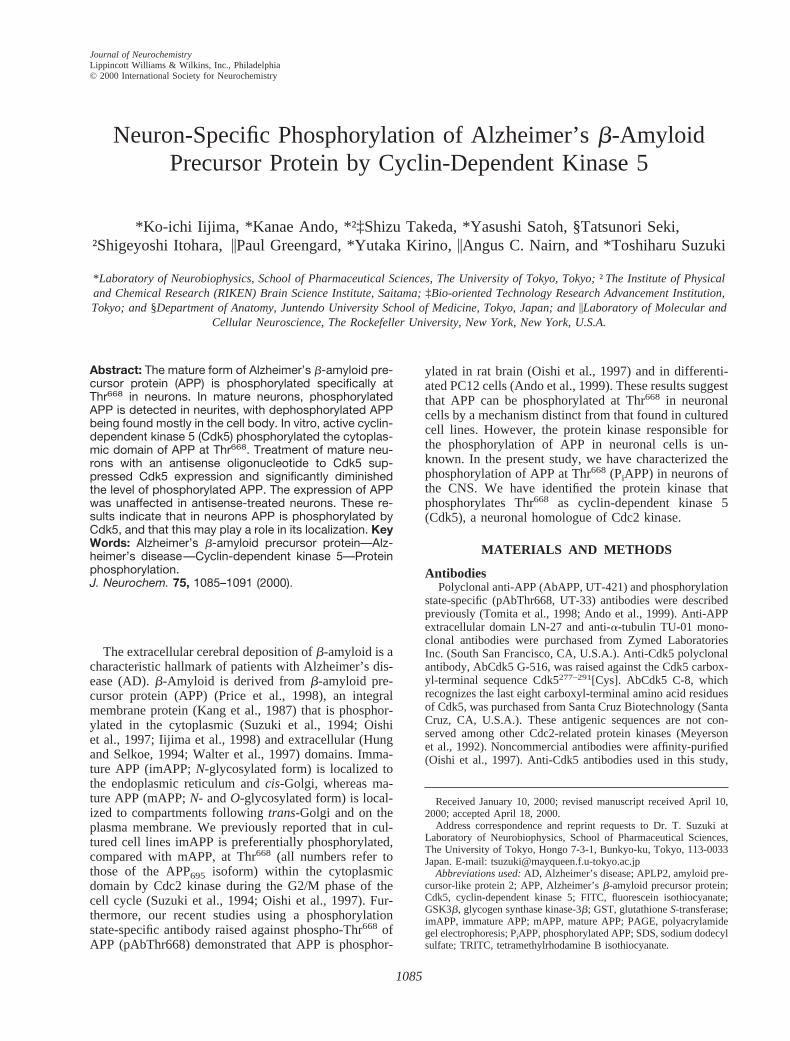
propriate amounts of various adult rat tissues and ana-lyzed by immunoblotting with UT-421 (Fig. 1A, left) orwith UT-33 (Fig. 1A, right). Similar amounts of APPdistributed between different isoforms were recoveredfrom the different tissues. PiAPP was detected only insamples from cerebrum and cerebellum. Mouse brainwas also dissected and fractionated. APP from the indi-cated brain tissues was analyzed with UT-421 (Fig. 1B,left) or with UT-33 (Fig. 1B, right). In all brain regionsexamined, PiAPP was detected. These results clearlyindicate that basal phosphorylation of APP at Thr668 isspecific to nervous tissue.
APP from brain exhibits three major isoforms (Fig.1B, left), and we determined that the two higher molec-ular weight isoforms are mAPP695 variants differing inO-glycosylation. When APP recovered from brain wastreated with a combination of neuraminidase plusO-Glycanase, a process that releasesO-glycan from mAPP(Tomita et al., 1998), the two APP isoforms of highestmolecular weight migrated at the position of imAPP695,
J. Neurochem., Vol. 75, No. 3, 2000
1086 K. IIJIMA ET AL.
the lowest isoform (data not shown). As only the twohighest molecular weight forms were phosphorylated(Fig. 1B, right), this indicates that only mAPP695, but notimAPP695, is phosphorylated at Thr668 in brain tissues.
To investigate if phosphorylation of APP occurs inneurons but not in glial cells, APP was immunoprecipi-tated with UT-421 from cerebrum and from a glial cellpreparation, and analyzed by immunoblotting with UT-421 (Fig. 1C, left) or with UT-33 (Fig. 1C, right). Twomature and immature APP isoforms, probably APP770and APP751, were recovered from glial lysates; however,PiAPP was not detected. These results suggest that phos-
phorylation of APP at Thr668 is neuron-specific, and thiswas investigated in more detail using neuronal culturesas described below.
Localization of APP and PiAPP in cultured rathippocampal neurons
The localization of APP and PiAPP was analyzed inmature rat hippocampal neurons by immunocytochemis-try. Neurons were dissected from embryonic day 18 ratand cultured for 120 h. Mature neurons were double-stained with UT-33 (Fig. 2A), UT-421 (Fig. 2C), oranti-a-tubulin monoclonal antibody TU-01 (Fig. 2B andD). PiAPP was localized mainly to the somatic plasmamembrane and to neurites rather than being presentwithin the cell body. In contrast, APP was restrictedmostly to the cell body. These observations suggest thatthe majority of plasma membrane-associated APP isphosphorylated.
Phosphorylation of the cytoplasmic domain of APPby Cdk5 in vitro
We next examined which protein kinase phosphory-lates APP at Thr668 in neurons. The most likely candidateis Cdk5 because (a) Cdk5 is a neuronal Cdc2-relatedprotein kinase, (b) Cdk5 recognizes an amino acid se-quence similar to that of Cdc2 kinase substrates (Mey-erson et al., 1992; Lew and Wang, 1994), (c) Cdk5 isactive in the brain during late embryonic and adult life(Tsai et al., 1994; Lee et al., 1997), and (d) a recombinantCdk5/p25 activator complex phosphorylates the APPcytoplasmic peptide, APP695
645–694, in vitro (J. Bibb andA. C. Nairn, unpublished observation). Active Cdk5 re-covered from a lysate of embryonic rat brain was incu-bated with GST–APPCOOH (Fig. 3).GST–APPCOOH wasphosphorylated by immunoprecipitated Cdk5. Additionof the peptide Cdk5277–292 (which includes the aminoacids used as antigen) during immunoprecipitation abol-ished the signal observed for phosphorylation of GST–APPCOOH. These results demonstrated that in vitro Cdk5directly phosphorylates APP at Thr668.
Phosphorylation of APP by Cdk5 in neuronsTo investigate whether Cdk5 is the protein kinase
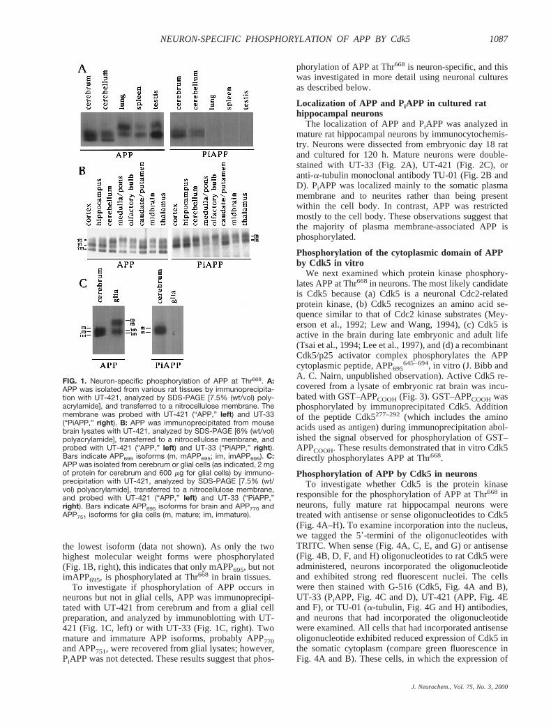
responsible for the phosphorylation of APP at Thr668 inneurons, fully mature rat hippocampal neurons weretreated with antisense or sense oligonucleotides to Cdk5(Fig. 4A–H). To examine incorporation into the nucleus,we tagged the 59-termini of the oligonucleotides withTRITC. When sense (Fig. 4A, C, E, and G) or antisense(Fig. 4B, D, F, and H) oligonucleotides to rat Cdk5 wereadministered, neurons incorporated the oligonucleotideand exhibited strong red fluorescent nuclei. The cellswere then stained with G-516 (Cdk5, Fig. 4A and B),UT-33 (PiAPP, Fig. 4C and D), UT-421 (APP, Fig. 4Eand F), or TU-01 (a-tubulin, Fig. 4G and H) antibodies,and neurons that had incorporated the oligonucleotidewere examined. All cells that had incorporated antisenseoligonucleotide exhibited reduced expression of Cdk5 inthe somatic cytoplasm (compare green fluorescence inFig. 4A and B). These cells, in which the expression of
FIG. 1. Neuron-specific phosphorylation of APP at Thr668. A:APP was isolated from various rat tissues by immunoprecipita-tion with UT-421, analyzed by SDS-PAGE [7.5% (wt/vol) poly-acrylamide], and transferred to a nitrocellulose membrane. Themembrane was probed with UT-421 (“APP,” left) and UT-33(“PiAPP,” right). B: APP was immunoprecipitated from mousebrain lysates with UT-421, analyzed by SDS-PAGE [6% (wt/vol)polyacrylamide], transferred to a nitrocellulose membrane, andprobed with UT-421 (“APP,” left) and UT-33 (“PiAPP,” right).Bars indicate APP695 isoforms (m, mAPP695; im, imAPP695). C:APP was isolated from cerebrum or glial cells (as indicated, 2 mgof protein for cerebrum and 600 mg for glial cells) by immuno-precipitation with UT-421, analyzed by SDS-PAGE [7.5% (wt/vol) polyacrylamide], transferred to a nitrocellulose membrane,and probed with UT-421 (“APP,” left) and UT-33 (“PiAPP,”right). Bars indicate APP695 isoforms for brain and APP770 andAPP751 isoforms for glia cells (m, mature; im, immature).
J. Neurochem., Vol. 75, No. 3, 2000
1087NEURON-SPECIFIC PHOSPHORYLATION OF APP BY Cdk5

Cdk5 was suppressed, displayed a corresponding de-crease in the PiAPP signal (compare green fluorescencein Fig. 4C and D). Neither APP (compare green fluores-cence in Fig. 4E and F) nora-tubulin (compare greenfluorescence in Fig. 4G and H) expression was affectedin cells that had incorporated antisense oligonucleotides(Fig. 4F and H). Cells that had incorporated sense oli-gonucleotide showed normal levels of Cdk5 and PiAPP(Fig. 4A and C). These results suggest strongly that inneurons Cdk5 phosphorylates APP at Thr668.
GSK3b does not phosphorylate APPat Thr 668 in neurons
In vitro, GSK3b phosphorylates APPCOOH at Thr668
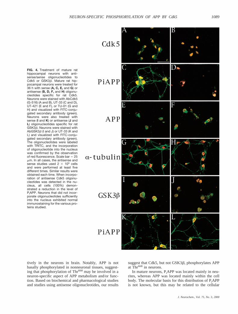
(Aplin et al., 1996). Therefore, we examined whetherantisense oligonucleotide specific for GSK3b might de-crease PiAPP in neurons (Fig. 4I–L). Mature rat hip-pocampal neurons were treated with antisense (Fig. 4Jand L) or sense (Fig. 4I and K) oligonucleotides to ratGSK3b that had been tagged with TRITC at their 59-termini. Neurons that had incorporated antisense oligo-nucleotide exhibited decreased GSK3b expression (com-pare Fig. 4I and J); however, the level of PiAPP inneurites was not affected (compare Fig. 4K and L).Treatment with sense oligonucleotide had no effect onthe expression of GSK3b and PiAPP (Fig. 4I and K).Longer incubation of mature neurons with antisense oli-gonucleotide to GSK3b resulted in cell death, but evenas cell lysis began to occur, PiAPP was detected (data notshown). Moreover, treatment of neurons with lithiumchloride (10 mM), which inhibits GSK3b activity spe-cifically (Stambolic et al., 1996), had no effect on thePiAPP signal (Fig. 5).These observations indicate thatGSK3b does not make a major contribution to the phos-phorylation of APP at Thr668 in neurons.
DISCUSSION
We have identified that the mature form of APP,mAPP695, but not imAPP695, is phosphorylated constitu-
FIG. 2. Localization of APP and PiAPP in cultured rat hippocampal neurons. Embryonic day 18 hippocampal neurons were cultured for120 h. Neurons were double-stained with (A) pAbThr668 (UT-33) and (B) anti-a-tubulin antibody (TU-01). Other neurons weredouble-stained with (C) AbAPP (UT-421) and (D) TU-01. Scale bar 5 25 mm.
FIG. 3. Phosphorylation of the cytoplasmic domain of APP atThr668 by Cdk5. Cdk5 was immunoprecipitated from rat embry-onic brain with (antibody “1”) or without (antibody “2”) AbCdk5(C-8) in the presence (peptide “1”) or absence (peptide “2”) ofa peptide that includes the amino acids used as antigen. Theresulting Cdk5/C-8 complex coupled to resin was incubatedwith GST–APPCOOH fusion protein (arrow). GST–APPCOOH wasrecovered by immunoprecipitation with UT-421, and sampleswere analyzed by immunoblot using UT-33 (left) or UT-421(right).
J. Neurochem., Vol. 75, No. 3, 2000
1088 K. IIJIMA ET AL.
tively in the neurons in brain. Notably, APP is notbasally phosphorylated in nonneuronal tissues, suggest-ing that phosphorylation of Thr668 may be involved in aneuron-specific aspect of APP metabolism and/or func-tion. Based on biochemical and pharmacological studiesand studies using antisense oligonucleotides, our results
suggest that Cdk5, but not GSK3b, phosphorylates APPat Thr668 in neurons.
In mature neurons, PiAPP was located mainly in neu-rites, whereas APP was located mainly within the cellbody. The molecular basis for this distribution of PiAPPis not known, but this may be related to the cellular
FIG. 4. Treatment of mature rathippocampal neurons with anti-sense/sense oligonucleotides toCdk5 or GSK3b. Mature rat hip-pocampal neurons were treated for36 h with sense (A, C, E, and G) orantisense (B, D, F, and H) oligonu-cleotides specific for rat Cdk5.Neurons were stained with AbCdk5(G-516) (A and B), UT-33 (C and D),UT-421 (E and F), or TU-01 (G andH) and visualized with FITC-conju-gated secondary antibody (green).Neurons were also treated withsense (I and K) or antisense (J andL) oligonucleotides specific for ratGSK3b. Neurons were stained withAbGSK3b (I and J) or UT-33 (K andL) and visualized with FITC-conju-gated secondary antibody (green).The oligonucleotides were labeledwith TRITC, and the incorporationof oligonucleotide into the nucleuswas confirmed by the observationof red fluorescence. Scale bar 5 25mm. In all cases, the antisense andsense studies used 2 3 105 cellsand were performed at least fivedifferent times. Similar results wereobtained each time. When incorpo-ration of antisense Cdk5 oligonu-cleotides was detected in the nu-cleus, all cells (100%) demon-strated a reduction in the level ofPiAPP. Neurons that did not incor-porate oligonucleotides sufficientlyinto the nucleus exhibited normalimmunostaining for the various pro-teins studied.
J. Neurochem., Vol. 75, No. 3, 2000
1089NEURON-SPECIFIC PHOSPHORYLATION OF APP BY Cdk5
distribution of Cdk5 and/or its activator protein p35, orof the protein phosphatase(s) that dephosphorylateThr668. Together with our recent studies using PC12 cells(Ando et al., 1999), the present results suggest thatphosphorylation of APP at Thr668 may play a specificrole in regulation of APP function in neurites. Possibly,a decrease in PiAPP level and/or unregulated phosphor-ylation of APP may induce retraction of neurites andmay result in damage of neuronal circuits in the adultbrain. In fact, we have observed that prolonged incuba-tion of neurons with antisense oligonucleotide to Cdk5brings about the retraction of neurites (data not shown).The disappearance of PiAPP in neurons treated withantisense oligonucleotide to Cdk5 occurred before theretraction of neurites.
Cdk5 is activated by the regulatory subunit, p35 (Ish-iguro et al., 1994; Lew et al., 1994; Tsai et al., 1994). Arecent study has indicated that a truncated form of p35,termed p25, accumulates in postmortem brain samplesobtained from patients with AD (Patrick et al., 1999;Ahlijanian et al., 2000). The interaction of p25 withCdk5 results in the constitutive activation of the kinaseand increased phosphorylation of tau, a microtubule-binding protein that has been implicated in paired-helicalfilament formation in AD (Patrick et al., 1999). As ourpresent studies indicate that APP is phosphorylated atThr668 in a neuron-specific manner by Cdk5, increasedphosphorylation of APP in brain of AD patients might beexpected. However, in preliminary studies, we have ob-served a slight decrease in the level of phosphorylationof APP in brain tissue from AD patients (Suzuki et al.,unpublished observation). Active Cdk5 may possibly be
localized to a specific subcellular location within neu-rons, or may be specifically localized together with tau inthe brain of AD patients. In any case, our results and theresults from Patrick et al. (1999) indicate that APP andtau, two proteins implicated in AD, are physiologicalsubstrates for Cdk5. It is hoped that further studies of thephosphorylation of APP by Cdk5 will contribute to ourunderstanding of the role of APP in neuronal differenti-ation and possibly of the role of APP in the pathogenesisof AD.
Acknowledgment: We thank Dr. H. Gomi (RIKEN, Japan)for technical advice and helpful discussions. This work wassupported by a Grant-in-Aid for Scientific Research from theJapanese Ministry of Education, Science, Sports and Culture(T.S.), by a grant from the Program for Promotion of BasicResearch Activities for Innovative Biosciences Japan (T.S.),and USPHS grant AG09464 (A.C.N. and P.G.). K. Ando is arecipient of a fellowship from Japan Society for the Promotionof Science Research Fellowships for Young Scientists.
REFERENCES
Ahlijanian M. K., Barrezueta N. X., Williams R. D., Jakowski A.,Kowsz K. P., McCarthy S., Coskran T., Carlo A., Seymour P. A.,Burkhardt J. E., Nelson R. B., and McNeish J. D. (2000) Hyper-phosphorylated tau and neurofilament and cytoskeletal disruptionsin mice overexpressing human p25, an activator of cdk5.Proc.Natl. Acad. Sci. USA97, 2910–2915.
Ando K., Oishi M., Takeda S., Iijima K., Isohara T., Nairn A. C.,Kirino Y., Greengard P., and Suzuki T. (1999) Role of phosphor-ylation of Alzheimer’s amyloid precursor protein during neuronaldifferentiation.J. Neurosci.19, 4421–4427.
Aplin A. E., Gibb G. M., Jacobsen J. S., Gallo J.-M., and AndertonB. H. (1996) In vitro phosphorylation of the cytoplasmic domain
FIG. 5. Treatment of neurons with lithium chloride has no effect on APP phosphorylation. Mature rat hippocampal neurons were treatedfor 2.5 h with 10 mM lithium chloride (LiCl) or sodium chloride (control). Neurons were double-stained with UT-33 (PiAPP) and TU-01(a-tubulin) and visualized with FITC- and TRITC-conjugated secondary antibody. Scale bar 5 25 mm.
J. Neurochem., Vol. 75, No. 3, 2000
1090 K. IIJIMA ET AL.
of the amyloid precursor protein by glycogen synthase kinase-3b.J. Neurochem.67, 699–707.
Bartlett W. and Banker G. (1984) An electron microscopic study of thedevelopment of axons and dendrites by hippocampal neurons inculture. I. Cells which develop without intercellular contacts.J. Neurosci.4, 1944–1953.
Bradke F. and Dotti C. G. (1997) Neuronal polarity: vectorial cyto-plasmic flow precedes axon formation.Neuron19, 1175–1186.
Ferreria A., Caceres A., and Kosik K. S. (1993) Intraneutral compart-ments of the amyloid precursor protein.J. Neurosci.13, 3112–3123.
Hung A. J. and Selkoe D. J. (1994) Selective ectodomain phosphory-lation and regulated cleavage ofb-amyloid precursor protein.EMBO J.13, 534–542.
Iijima K., Lee D.-S., Okutsu J., Tomita S., Hirashima N., Kirino Y., andSuzuki T. (1998) cDNA isolation of Alzheimer’s amyloid precur-sor protein from cholinergic nerve terminals of the electric organof the electric ray.Biochem. J.330,29–33.
Ishiguro K., Kobayashi S., Omori A., Takamatsu M., Yonekura S.,Anzai K., Imahori K., and Uchida T. (1994) Identification of the23 kDa subunit of tau protein kinase II as a putative activator ofCdk5 in bovine brain.FEBS Lett.342,203–208.
Kang J., Lemaire H. G., Unterbeck A. J., Salbaum J. M., Masters C. L.,Grzeschik K. H., Multhaup G., Beyreuther K., and Mu¨ller-Hill B.(1987) The precursor of Alzheimer’s disease amyloid A4 proteinresembles a cell-surface receptor.Nature325,733–736.
Lee K.-Y., Helbing C. C., Choi K.-S., Johnston R. N., and Wang J. H.(1997) Neuronal cdc2-like kinase (Nclk) binds and phosphorylatesthe retinoblastoma protein.J. Biol. Chem.272,5622–5626.
Lew J. and Wang J. H. (1994) Neuronal cdc2-like kinase.TrendsBiochem. Sci.20, 33–37.
Lew J., Huang Q. Q., Qi Z., Winkfein R. J., Aebersold R., Hunt T., andWang J. H. (1994) A brain-specific activator of cyclin-dependentkinase 5.Nature371,423–426.
Meyerson M., Enders G. H., Wu C.-L., Su L.-K., Gorka C., Nelson C.,Harlow E., and Tsai L.-H. (1992) A family of human cdc2-relatedprotein kinases.EMBO J.11, 2909–2917.
Oishi M., Nairn A. C., Czernik A. J., Lim G. S., Isohara T., GandyS. E., Greengard P., and Suzuki T. (1997) The cytoplasmic do-main of Alzheimer’s amyloid precursor protein is phosphorylated
at Thr654, Ser655, and Thr668 in adult rat brain and cultured cells.Mol. Med.3, 111–123.
Patrick G. N., Zukerberg L., Nikolic M., de la Monte S., Dikkes P., andTsai L.-H. (1999) Conversion of p35 to p25 deregulates Cdk5activity and promotes neurodegeneration.Nature402,615–622.
Pigino G., Paglini G., Ulloa L., Avila J., and Caceres A. (1997)Analysis of the expression, distribution and function of cyclindependent kinase 5 (cdk5) in developing cerebellar macroneurons.J. Cell Sci.110,257–270.
Price D. L., Sisodia S. S., and Borchelt D. R. (1998) Genetic neuro-degenerative disease: the human illness and transgenic models.Science282,1079–1083.
Stambolic V., Ruel L., and Woodgett J. R. (1996) Lithium inhibitsglycogen synthase kinase-3 activity and mimics wingless signal-ling in intact cells.Curr. Biol. 6, 1664–1668.
Suzuki T., Oishi M., Marshak D. M., Czernik A. J., Nairn A. C., andGreengard P. (1994) Cell cycle-dependent regulation of the phos-phorylation and metabolism of the Alzheimer amyloid precursorprotein.EMBO J.13, 1114–1122.
Suzuki T., Ando K., Isohara T., Oishi M., Lim G. S., Satoh Y., WascoW., Tanzi R. E., Nairn A. C., Greengard P., Gandy S. E., andKirino Y. (1997) Phosphorylation of Alzheimerb-amyloid pre-cursor-like proteins.Biochemistry36, 4643–4649.
Tomita S., Kirino Y., and Suzuki T. (1998) Cleavage of Alzheimer’samyloid precursor protein by secretases occurs afterO-glycosyl-ation of APP in the protein secretory pathway.J. Biol. Chem.273,6277–6284.
Tomita S., Ozaki T., Taru H., Oguchi S., Takeda S., Yagi Y., SakiyamaS., Kirino Y., and Suzuki T. (1999) Interaction of a neuron-specific protein containing PDZ domains with Alzheimer’s amy-loid precursor protein.J. Biol. Chem.274,2243–2254.
Tsai L. H., Delalle I., Caviness V. S. Jr., and Harlow E. (1994) p35 isa neural-specific regulatory subunit of cyclin-dependent kinase 5.Nature371,419–423.
Walter J., Capell A., Hung A. Y., Langen H., Schno¨lzer M., ThinakaranG., Sisodia S. S., Selkoe D. J., and Haass C. (1997) Ectodomainphosphorylation ofb-amyloid precursor protein at two distinctcellular locations.J. Biol. Chem.272,1896–1903.
Woodgett J. R. (1990) Molecular cloning and expression of glycogensynthase kinase-3.EMBO J.9, 2431–2438.
J. Neurochem., Vol. 75, No. 3, 2000
1091NEURON-SPECIFIC PHOSPHORYLATION OF APP BY Cdk5
Related Documents


