Neuroglia Drawing by Ramon y Cajal Lecture by Dr. Jennifer Ziegenfuss Columbia University Medical School

Welcome message from author
This document is posted to help you gain knowledge. Please leave a comment to let me know what you think about it! Share it to your friends and learn new things together.
Transcript
Neuroglia
Drawing by Ramon y Cajal
Lecture by Dr. Jennifer Ziegenfuss
Columbia University Medical School

441 pages long…..
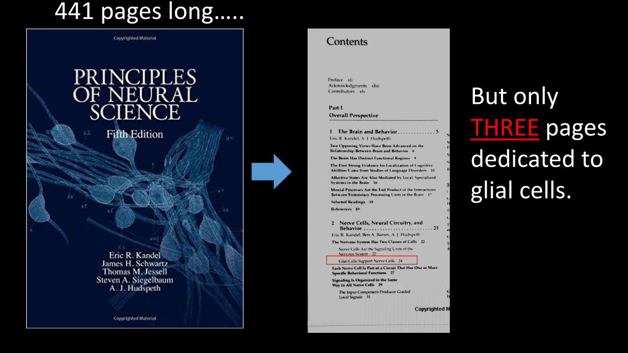
441 pages long…..
But onlyTHREE pagesdedicated to glial cells.

441 pages long….. 599 pages long…..

Why so long?599 pages long…..
- There are roughly equal number of neurons and glia in the CNS: - 98 billion neurons to 96
billion glia (+ or – 9 billion cells)
- This guy on the right had an abnormally higher amount of glia than typical.
- There are SIX different glial subtypes.- 4 in CNS- 2 in PNS
- They do way more than just act as “glue”.

I think it’s about time to become
“glia-evangelized”
599 pages long…..
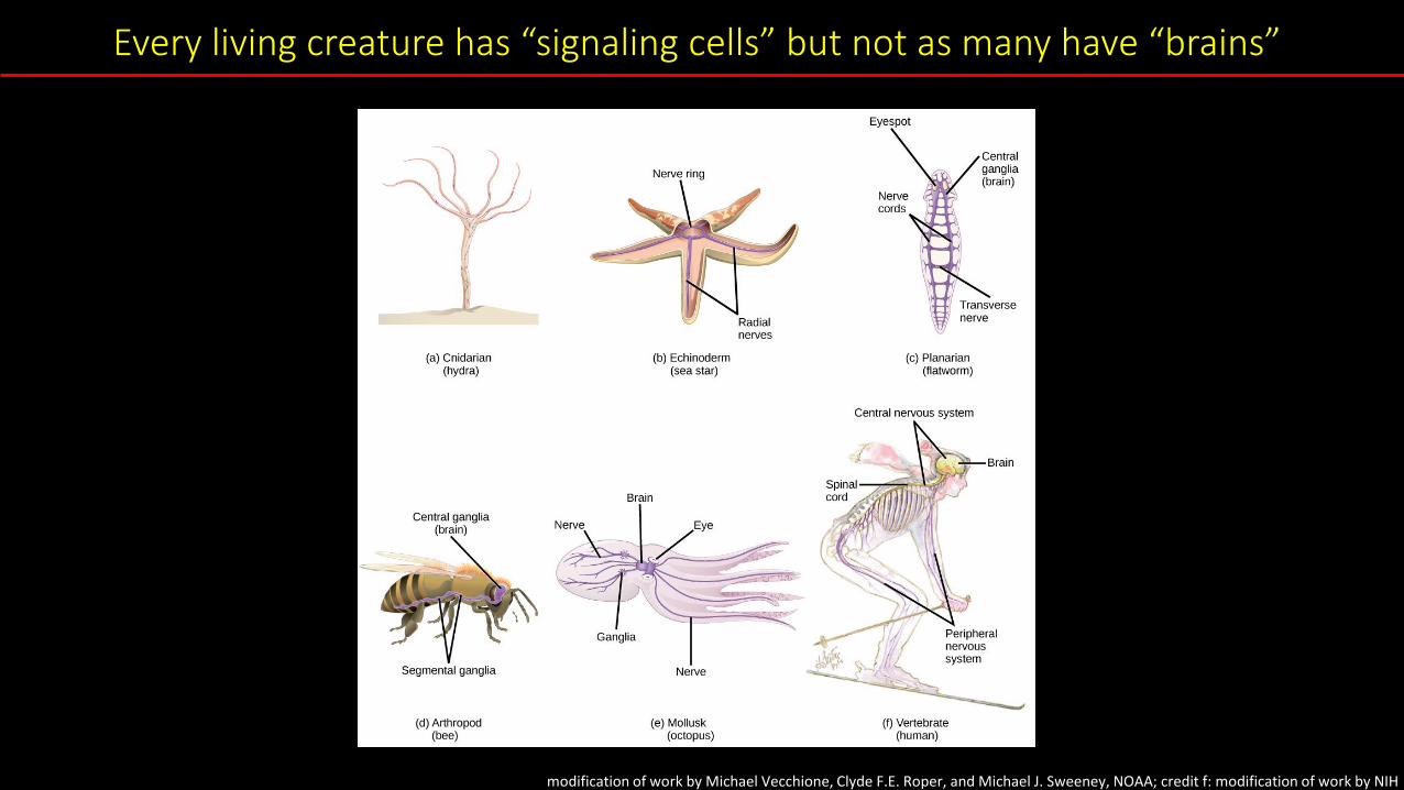
Every living creature has “signaling cells” but not as many have “brains”
modification of work by Michael Vecchione, Clyde F.E. Roper, and Michael J. Sweeney, NOAA; credit f: modification of work by NIH

Neuroglia roles in nervous system function
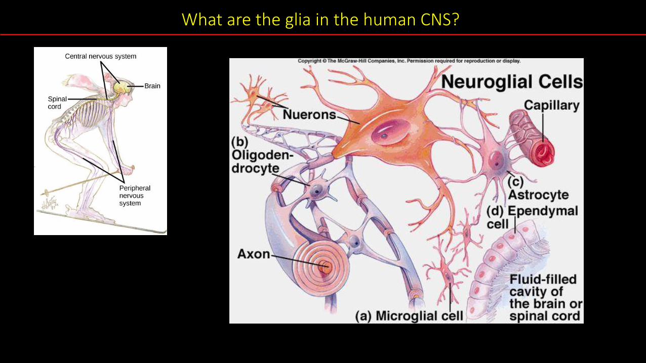
What are the glia in the human CNS?
~100 billion neurons
(50% all brain cells are glia)
24,000 genes
• Fruit flies share 60% of its
DNA with humans.
• 75% of all human
disease-causing genes in
humans are also found in
fruit flies.
• Fruit flies have roughly
same glia subtypes as
mammals/humans
Using fruit flies to study glia?
100,000 neurons
(30% all brain cells are glia)
14,000 genes
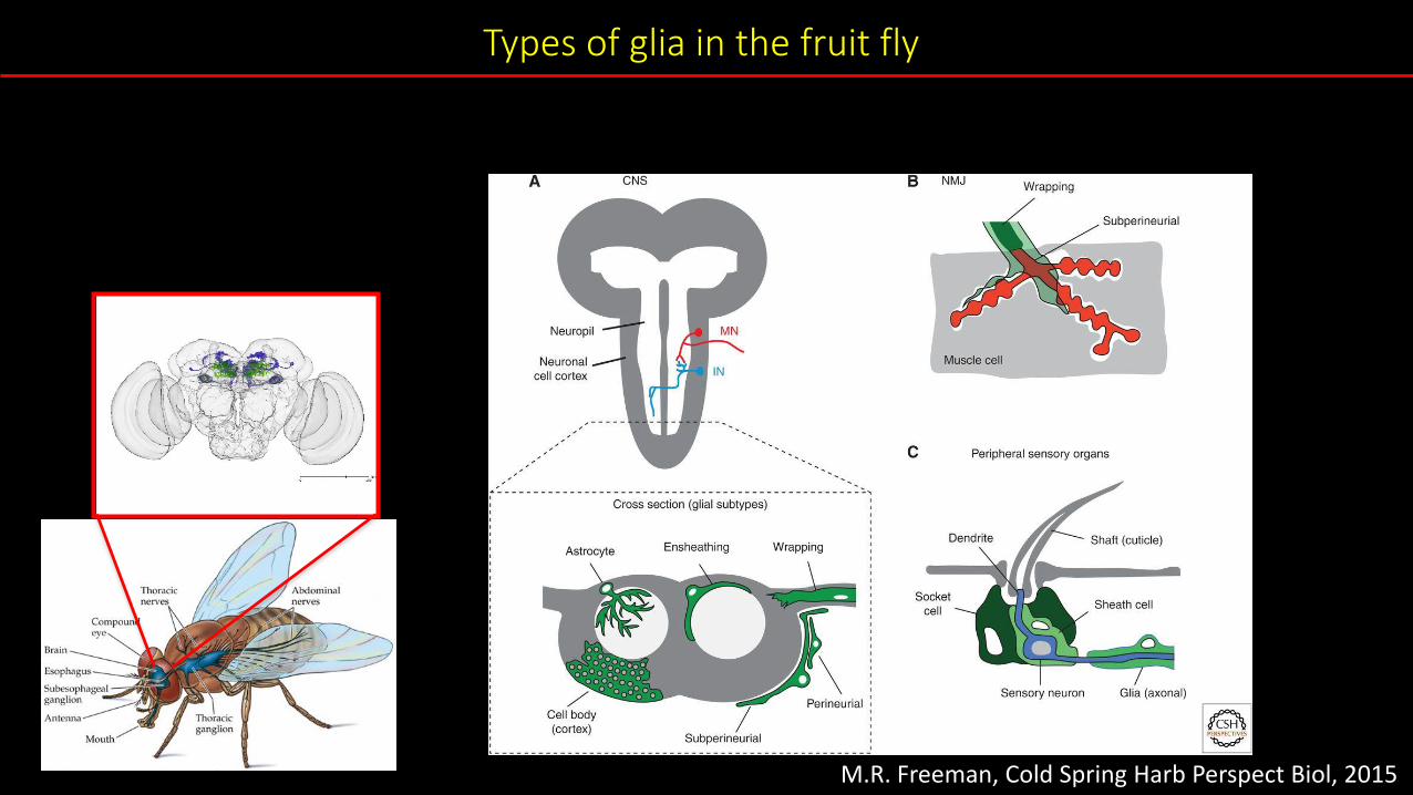
Types of glia in the fruit fly
M.R. Freeman, Cold Spring Harb Perspect Biol, 2015
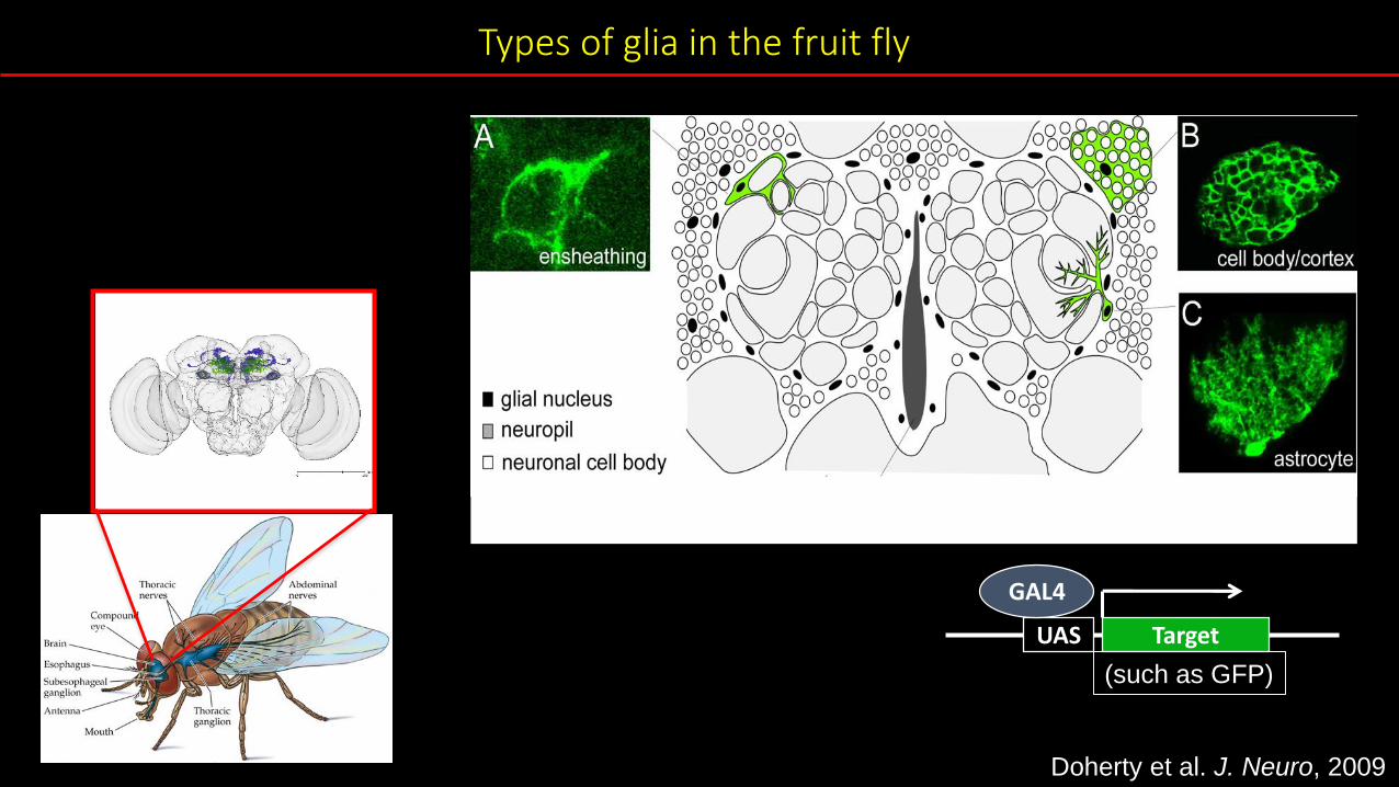
UAS Target
GAL4
(such as GFP)
Doherty et al. J. Neuro, 2009
Types of glia in the fruit fly
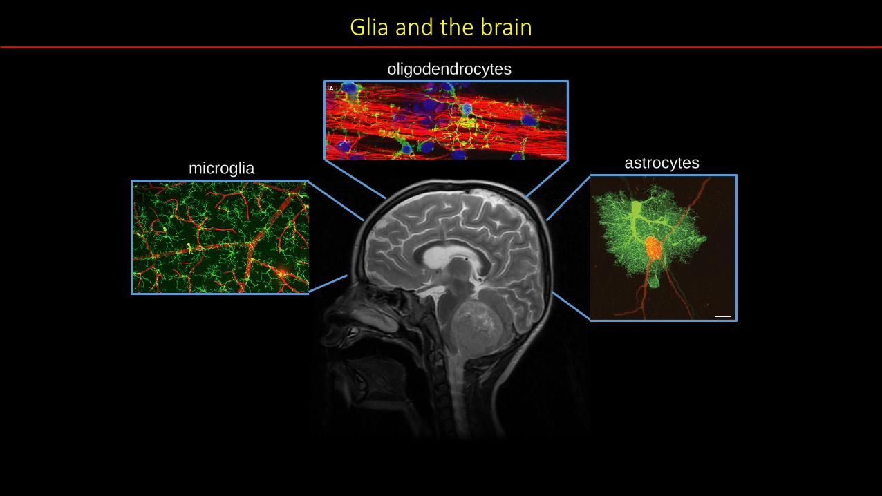
Glia and the brain
oligodendrocytes
microglia astrocytes

Oligodendrocytes
oligodendrocytes
• Oligodendrocytes, which are continuously remodeling
the myelin, and thus finely tuning axonal conduction
velocity, in response to neuronal activity.

Oligodendrocytes (this is also the same for peripheral Schwann cells)
Oligodendrocyte - mouse
• Oligodendrocytes, which are continuously remodeling
the myelin, and thus finely tuning axonal conduction
velocity, in response to neuronal activity.

Astrocytes (I)
astrocyte - mouse
• Astrocytes – help provide energy to neurons
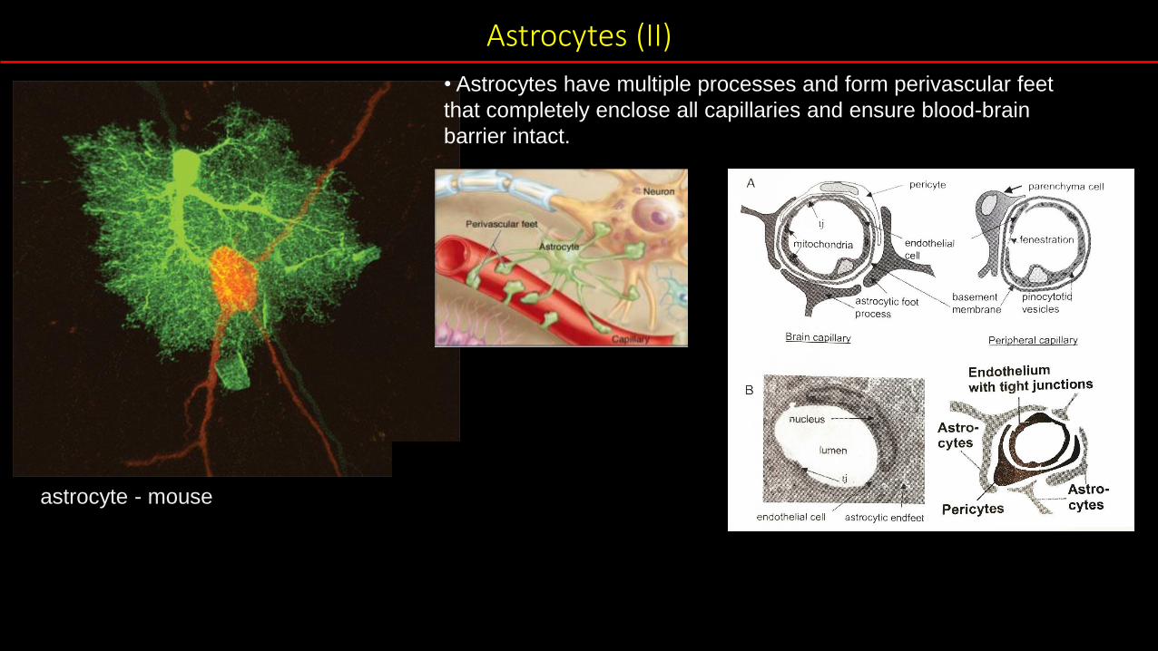
Astrocytes (II)
astrocyte - mouse
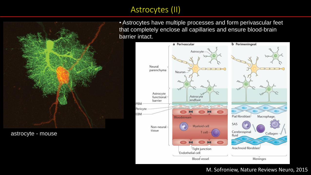
• Astrocytes have multiple processes and form perivascular feet
that completely enclose all capillaries and ensure blood-brain
barrier intact.
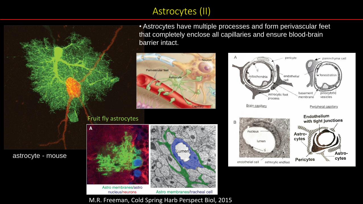
Astrocytes (II)
astrocyte - mouse
• Astrocytes have multiple processes and form perivascular feet
that completely enclose all capillaries and ensure blood-brain
barrier intact.
M.R. Freeman, Cold Spring Harb Perspect Biol, 2015
Fruit fly astrocytes
Astrocytes (II)
astrocyte - mouse
• Astrocytes have multiple processes and form perivascular feet
that completely enclose all capillaries and ensure blood-brain
barrier intact.
M. Sofroniew, Nature Reviews Neuro, 2015
Astrocytes (III)
astrocyte - mouse
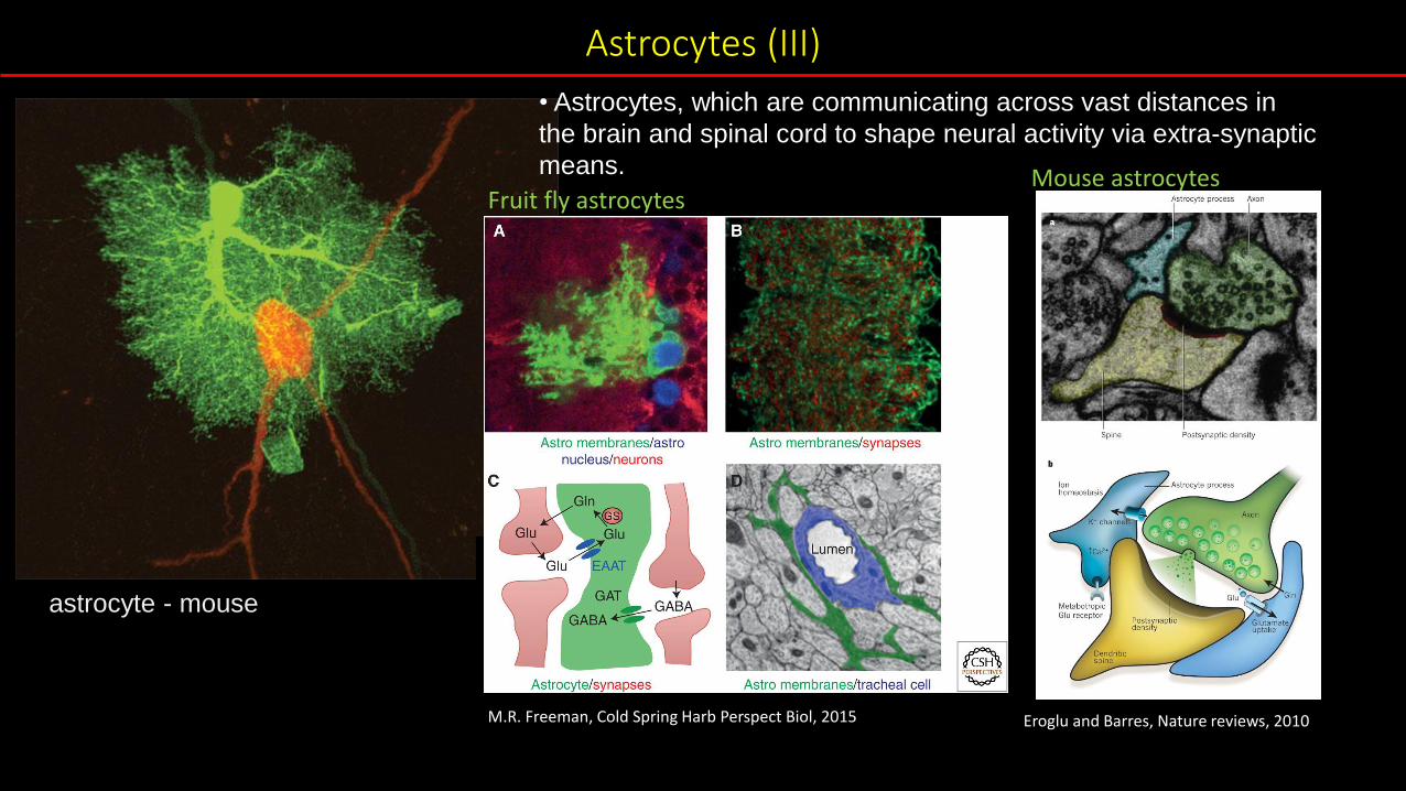
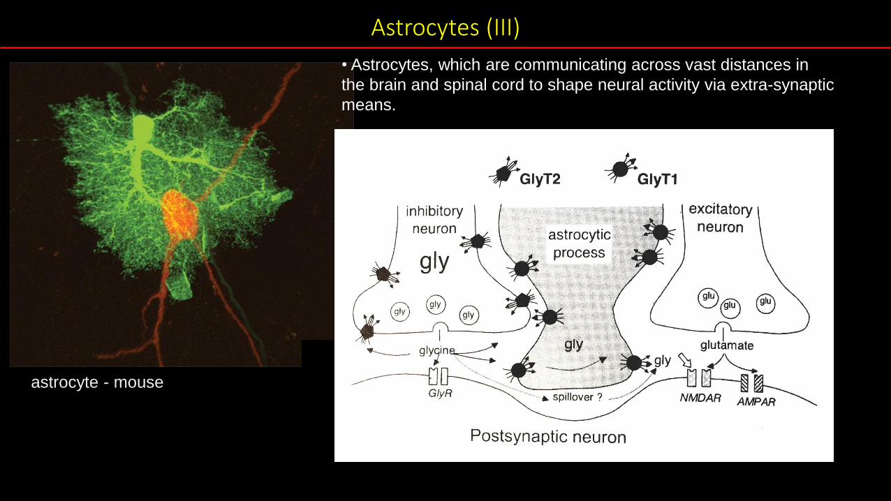
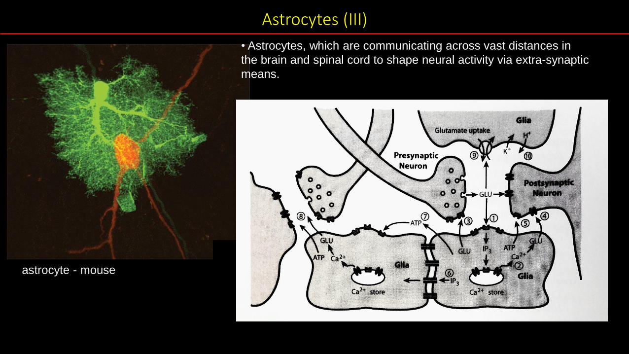
• Astrocytes, which are communicating across vast distances in
the brain and spinal cord to shape neural activity via extra-synaptic
means.
M.R. Freeman, Cold Spring Harb Perspect Biol, 2015 Eroglu and Barres, Nature reviews, 2010
Fruit fly astrocytesMouse astrocytes
Astrocytes (III)
astrocyte - mouse
• Astrocytes, which are communicating across vast distances in
the brain and spinal cord to shape neural activity via extra-synaptic
means.
Astrocytes (III)
astrocyte - mouse
• Astrocytes, which are communicating across vast distances in
the brain and spinal cord to shape neural activity via extra-synaptic
means.
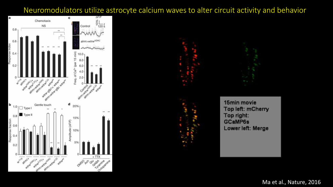
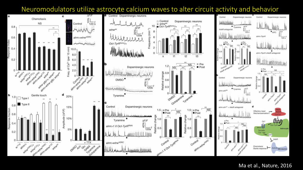
Neuromodulators utilize astrocyte calcium waves to alter circuit activity and behavior
Ma et al., Nature, 2016
Neuromodulators utilize astrocyte calcium waves to alter circuit activity and behavior
Ma et al., Nature, 2016

Astrocytes (III)
astrocyte - mouse
• Astrocytes, which are communicating across vast distances in
the brain and spinal cord to shape neural activity via extra-synaptic
means.Fruit fly astrocytes
M.R. Freeman, Cold Spring Harb Perspect Biol, 2015
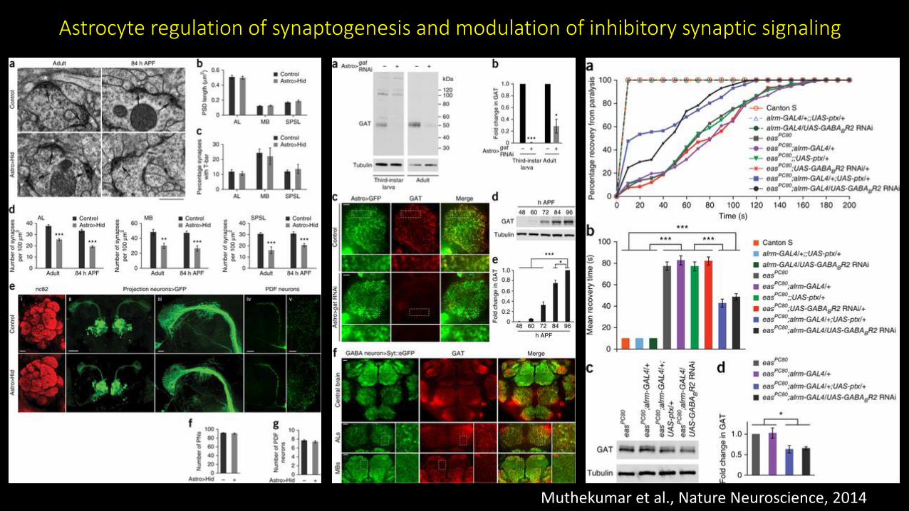
Muthekumar et al., Nature Neuroscience, 2014
Astrocyte regulation of synaptogenesis and modulation of inhibitory synaptic signaling
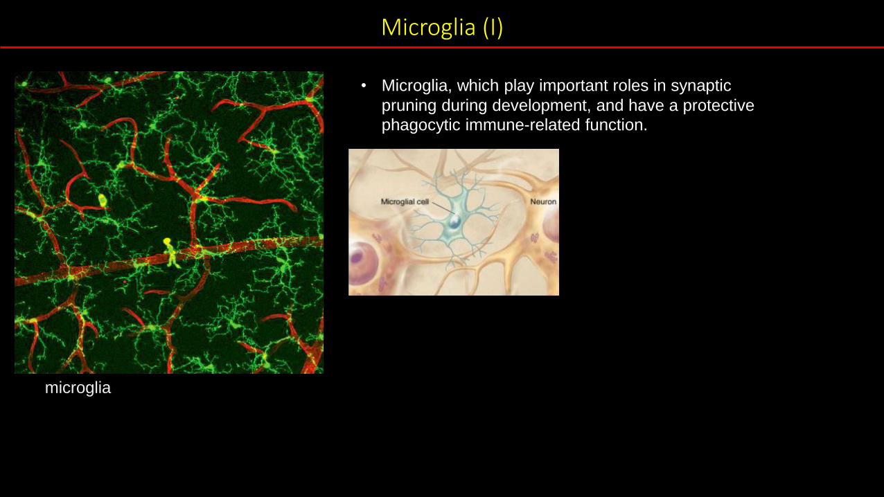
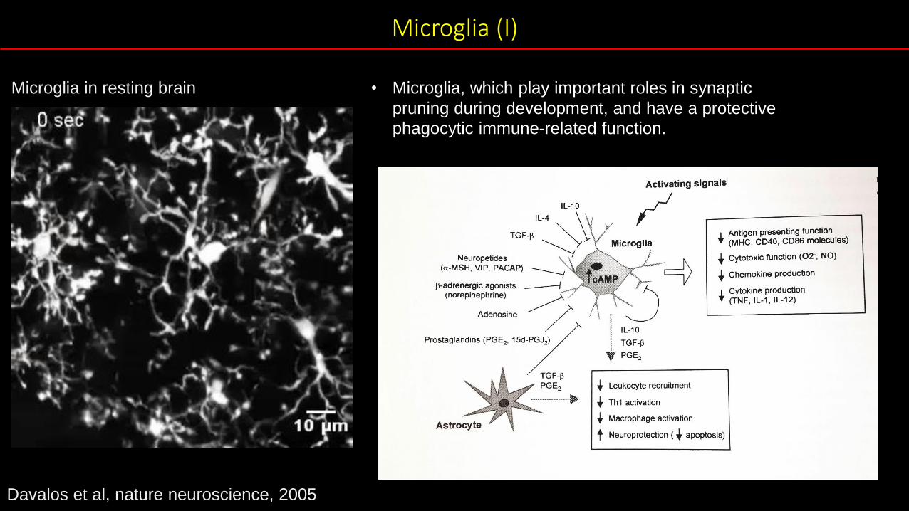
Microglia (I)
microglia
• Microglia, which play important roles in synaptic
pruning during development, and have a protective
phagocytic immune-related function.
Microglia (I)
Microglia in resting brain • Microglia, which play important roles in synaptic
pruning during development, and have a protective
phagocytic immune-related function.
Davalos et al, nature neuroscience, 2005
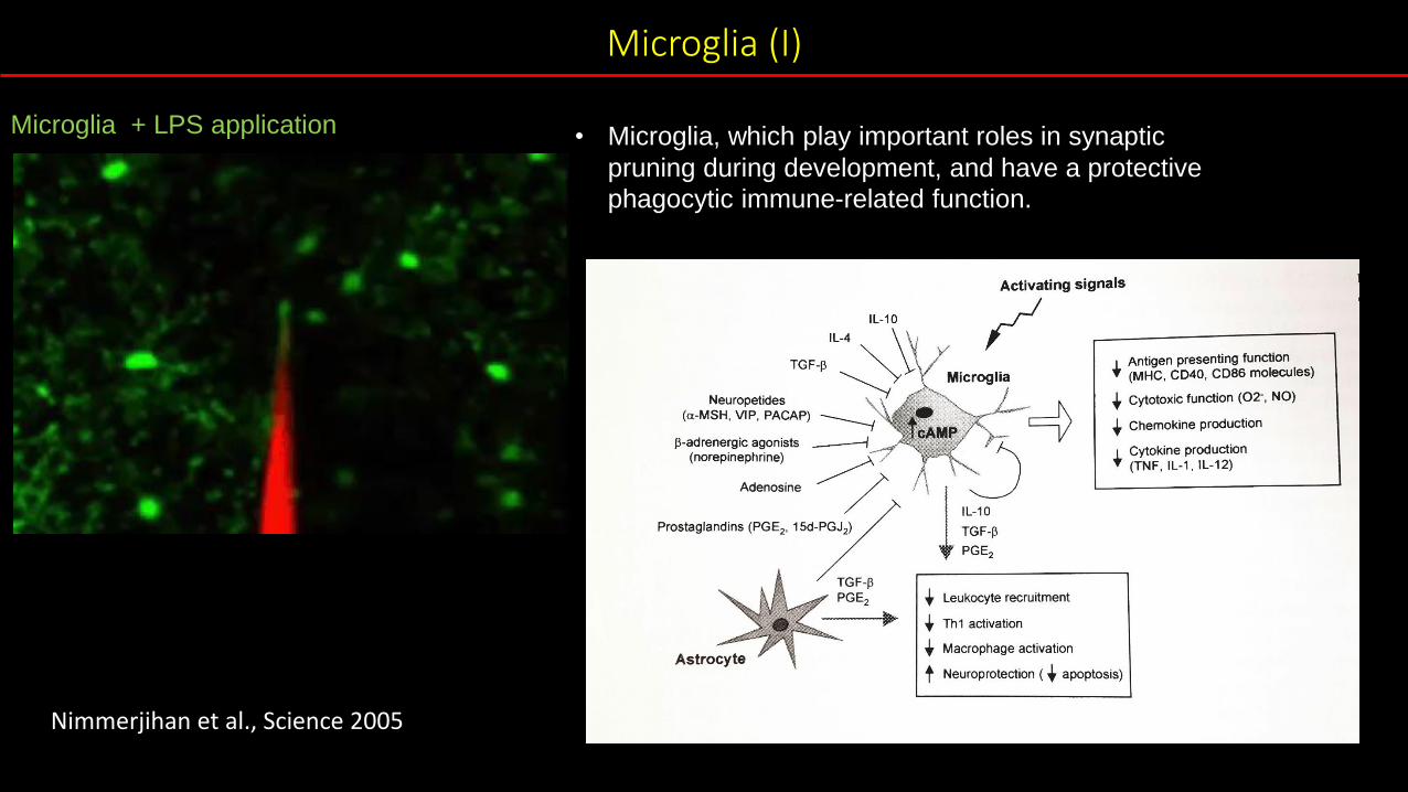
Microglia (I)
Microglia + LPS application • Microglia, which play important roles in synaptic
pruning during development, and have a protective
phagocytic immune-related function.
Nimmerjihan et al., Science 2005

Microglia (I)
• Microglia, which play important roles in synaptic
pruning during development, and have a protective
phagocytic immune-related function.
Glial response and
axon debris removal.
Draper
EG
F re
pe
ats
Draper 7-AAD Repo
(cell corpse)
Freeman et al, Neuron 2003

Microglia (II)
microglia
• Microglia, which play important roles in synaptic
pruning during development, and have a protective
phagocytic immune-related function.
Schafer et al, Glia 2013

Microglia (II)
microglia
• Microglia, which play important roles in synaptic
pruning during development, and have a protective
phagocytic immune-related function.
Schafer et al, Glia 2013
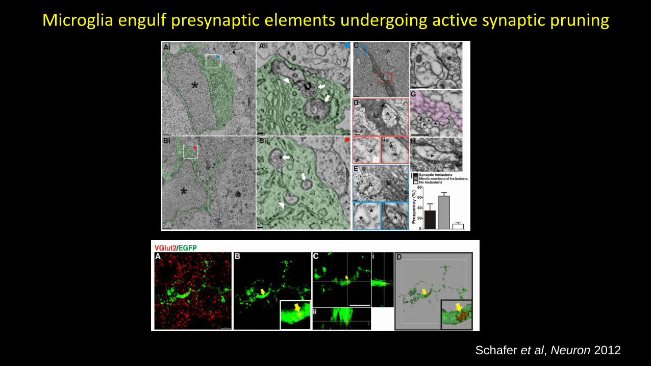
Microglia engulf presynaptic elements undergoing active synaptic pruning
Schafer et al, Neuron 2012

CR3/C3-dependent signaling regulates engulfment of synaptic inputs by microglia)
Schafer et al, Neuron 2012

Glia and the brain
oligodendrocytes
microglia astrocytes
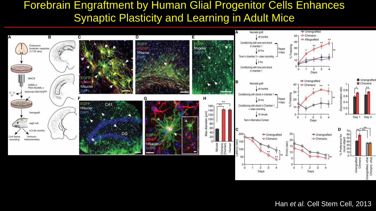
Forebrain Engraftment by Human Glial Progenitor Cells Enhances Synaptic Plasticity and Learning in Adult Mice
Han et al. Cell Stem Cell, 2013

Break

Neuroglia roles in nervous system, disease,
and degeneration
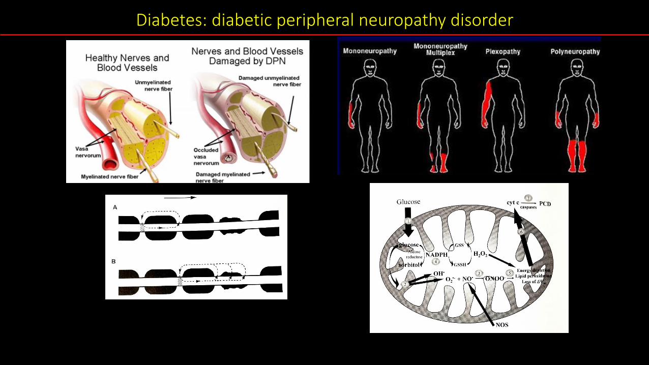
Diabetes: diabetic peripheral neuropathy disorder
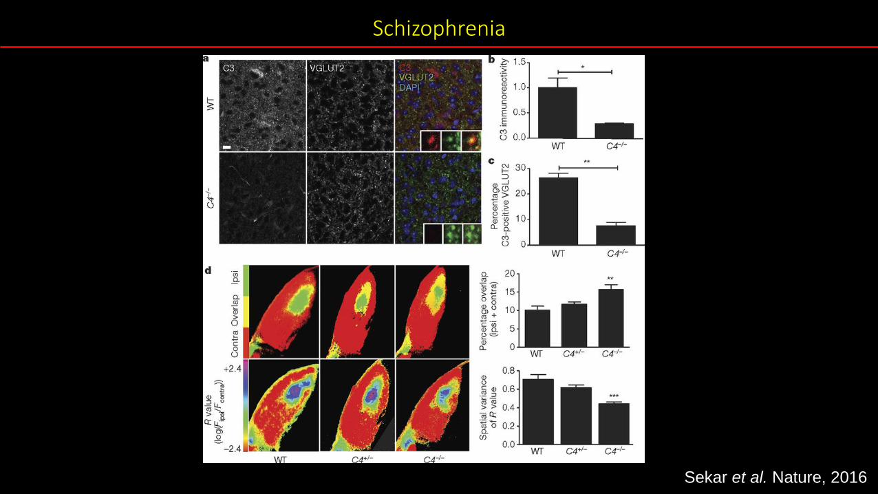
Schizophrenia
Sekar et al. Nature, 2016
Petrelli et al. Front. Cell. Neurosci., 2016
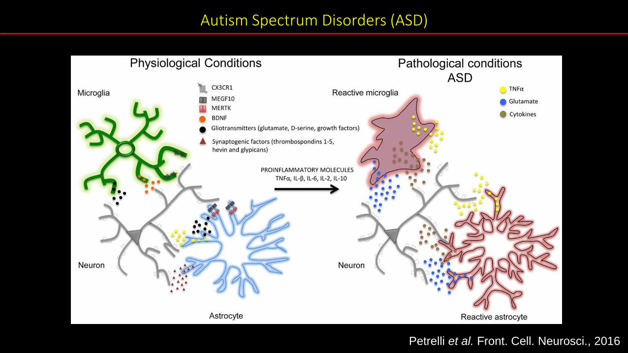
Autism Spectrum Disorders (ASD)

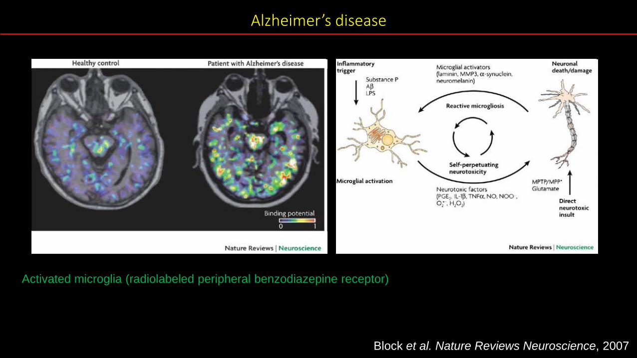
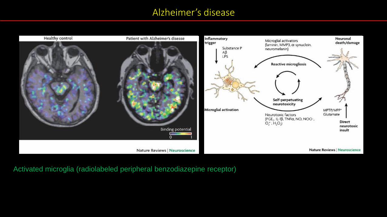
Alzheimer’s disease
Block et al. Nature Reviews Neuroscience, 2007
Shown are PET scans that track tau (top row) and beta-amyloid from two normal older people and one patient with Alzheimer’s disease (AD). The normal older adult on the left has no brain amyloid deposition and minimal tau in the medial temporal lobe. In the normal older adult in the middle, amyloid deposition is present throughout the brain, and tau has spread out into the temporal cortex. In the AD patient, both amyloid and tau are spread through the brain. (Image by Michael Schöll)
Block et al. Nature Reviews Neuroscience, 2007
Alzheimer’s disease
Activated microglia (radiolabeled peripheral benzodiazepine receptor)
Block et al. Nature Reviews Neuroscience, 2007

Alzheimer’s disease
Block et al. Nature Reviews Neuroscience, 2007
Activated microglia (radiolabeled peripheral benzodiazepine receptor)
Block et al. Nature Reviews Neuroscience, 2007
Alzheimer’s disease
Activated microglia (radiolabeled peripheral benzodiazepine receptor)

Block et al. Nature Reviews Neuroscience, 2007
Alzheimer’s disease
Activated microglia (radiolabeled peripheral benzodiazepine receptor)

ALS- Amyotrophic Lateral Sclerosis
Nagai et al. Nature Neuroscience, 2007
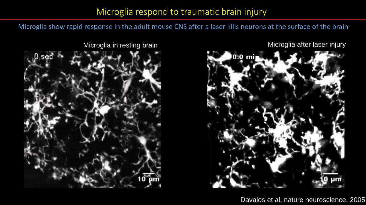
Microglia respond to traumatic brain injury
Davalos et al., Nat Neurosci 2005Microglia (Cx3cr1: fractalkine receptor)
Microglia show rapid response in the adult mouse CNS after a laser kills neurons at the surface of the brain
Microglia in resting brain
Davalos et al, nature neuroscience, 2005
Microglia after laser injury
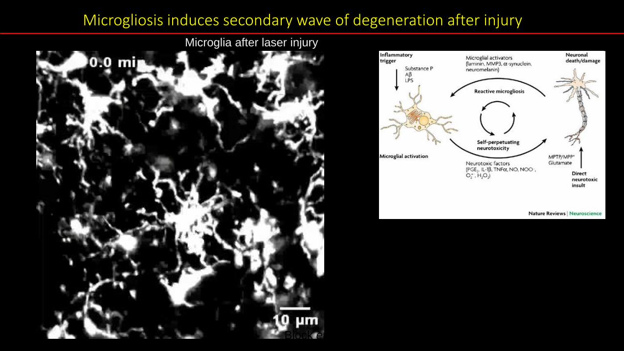
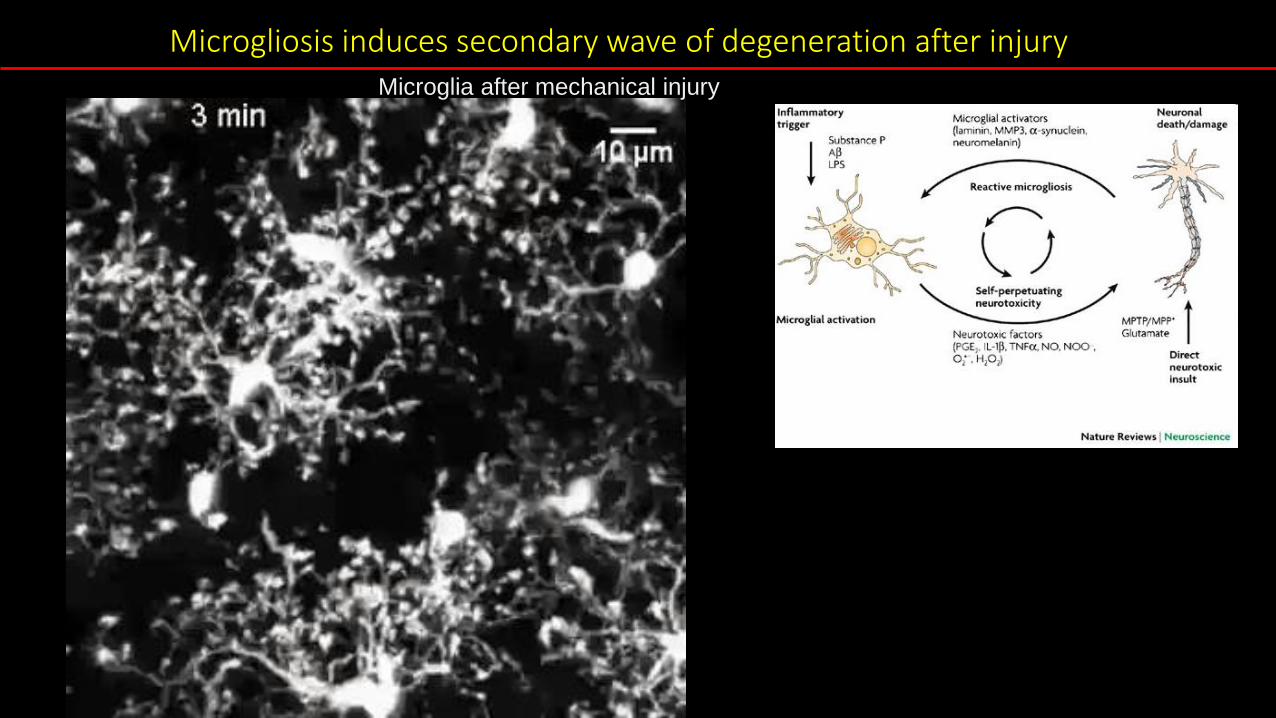
Microgliosis induces secondary wave of degeneration after injury
Block et al. Nature Reviews Neuroscience, 2007
Microglia after laser injury

Block et al. Nature Reviews Neuroscience, 2007
Microgliosis induces secondary wave of degeneration after injuryMicroglia after laser injury
Block et al. Nature Reviews Neuroscience, 2007
Microgliosis induces secondary wave of degeneration after injuryMicroglia after mechanical injury
Block et al. Nature Reviews Neuroscience, 2007
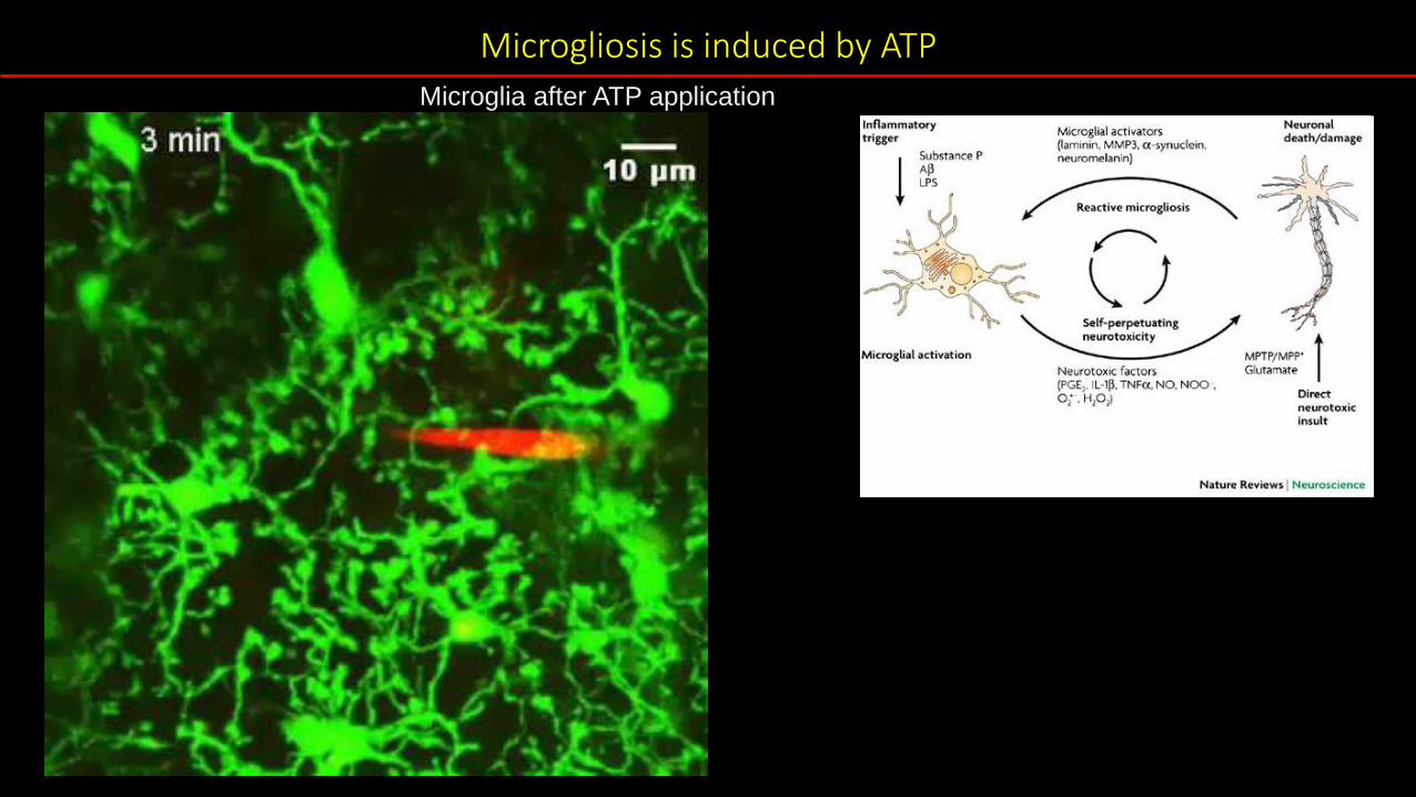
Microgliosis is induced by ATPMicroglia after ATP application

Block et al. Nature Reviews Neuroscience, 2007
Microglia after ATP application
Microgliosis is induced by ATP

• Where is the balance between beneficial and detrimental glial activation?

• Where is the balance between beneficial and detrimental glial activation?
• What is the basic biology behind glial activation?
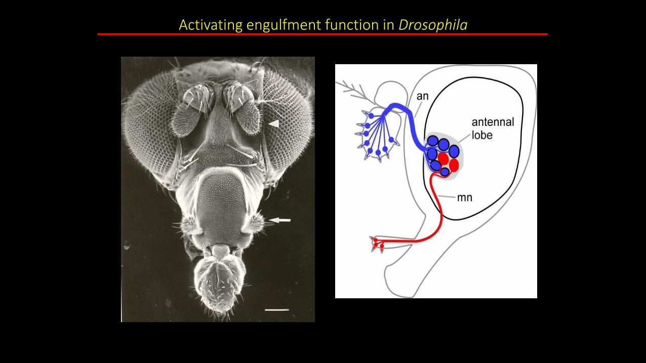
Activating engulfment function in Drosophila
antenna
maxillary palp
MacDonald et al., Neuron 2006
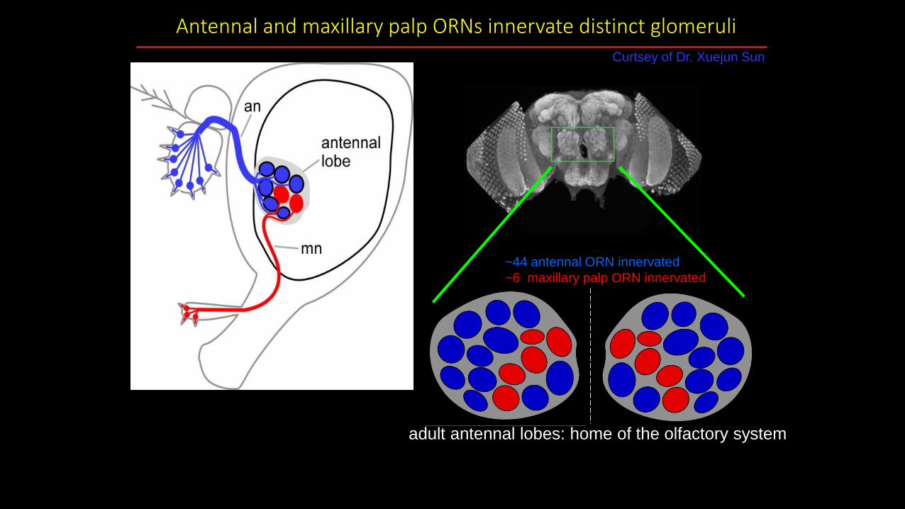
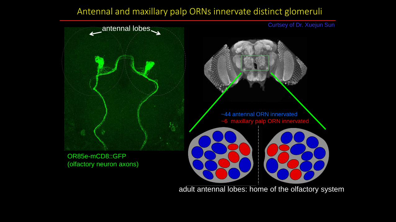
Antennal and maxillary palp ORNs innervate distinct glomeruli
adult fly brain
Curtsey of Dr. Xuejun Sun
antenna
maxillary palp
adult antennal lobes: home of the olfactory system
50 glomeruli total:
~44 antennal ORN innervated
~6 maxillary palp ORN innervated
Antennal and maxillary palp ORNs innervate distinct glomeruli
adult fly brain
adult antennal lobes: home of the olfactory system
OR85e-mCD8::GFP
(olfactory neuron axons)
antennal lobesCurtsey of Dr. Xuejun Sun
50 glomeruli total:
~44 antennal ORN innervated
~6 maxillary palp ORN innervated
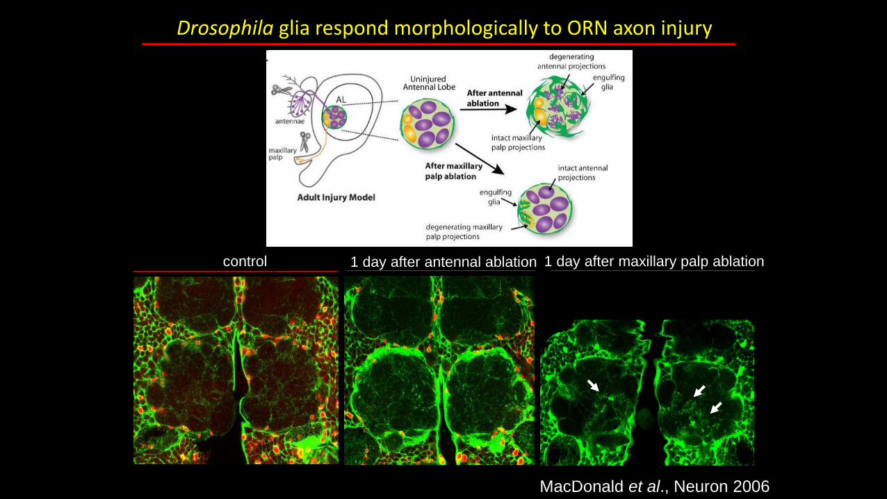
Drosophila glia respond morphologically to ORN axon injury
1 day after antennal ablationcontrol 1 day after maxillary palp ablation
MacDonald et al., Neuron 2006Logan and Freeman, 2007
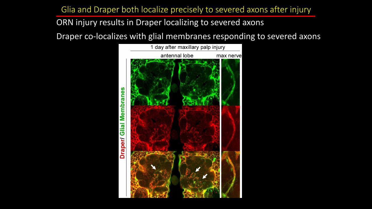
Glia and Draper both localize precisely to severed axons after injury
ORN injury results in Draper localizing to severed axons
Draper co-localizes with glial membranes responding to severed axons
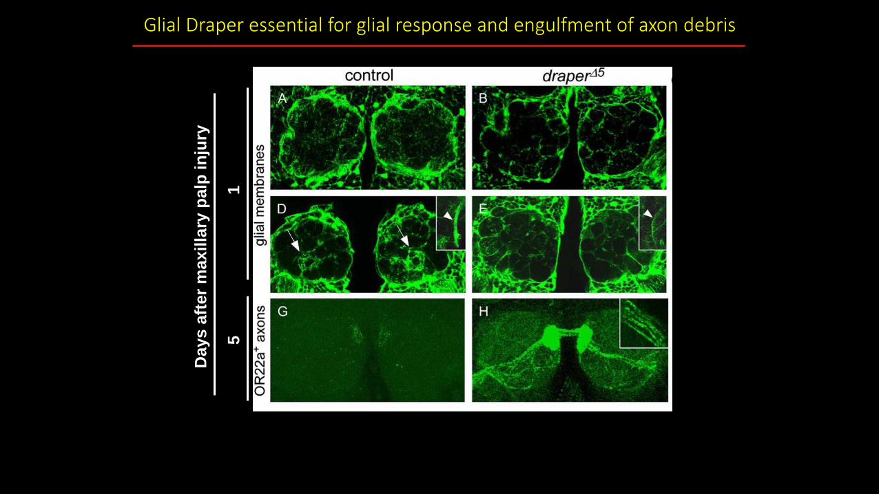
Glial Draper essential for glial response and engulfment of axon debris
MacDonald et al., Neuron 2006
Days a
fter
max
illa
ry p
alp
in
jury
51

Severed ORN axons are cleared from the CNS within 5 days
MacDonald et al., Neuron 2006
Axons (22a-Gal4/UAS-GFP)
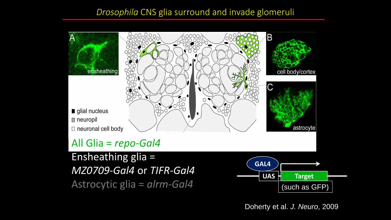
All Glia = repo-Gal4Ensheathing glia =MZ0709-Gal4 or TIFR-Gal4Astrocytic glia = alrm-Gal4
Drosophila CNS glia surround and invade glomeruli
UAS Target
GAL4
(such as GFP)
Doherty et al. J. Neuro, 2009
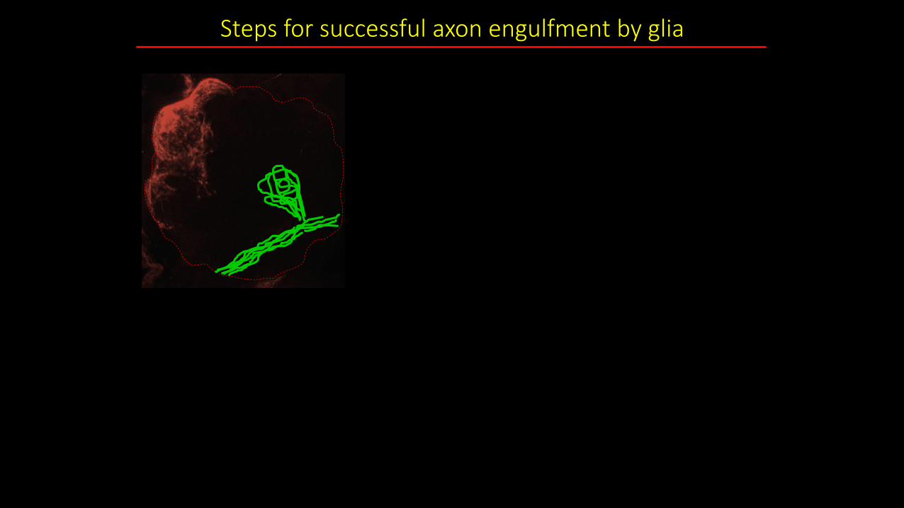
Steps for successful axon engulfment by glia

Engulfment Steps: Glial Activation
Step 1:
Glial Activation
•Recognition
~ 1 hour
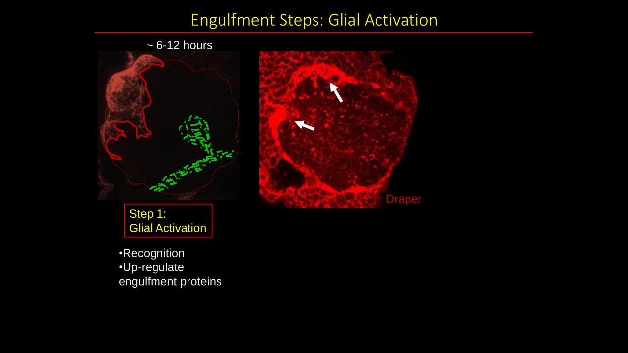
Engulfment Steps: Glial Activation
Step 1:
Glial Activation
•Recognition
•Up-regulate
engulfment proteins
Draper
~ 6-12 hours
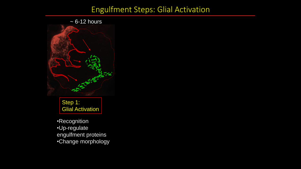
Engulfment Steps: Glial Activation
Step 1:
Glial Activation
•Recognition
•Up-regulate
engulfment proteins
•Change morphology
~ 6-12 hours

Engulfment Steps: Glial Activation
Step 1:
Glial Activation Glia membranes
Draper•Recognition
•Up-regulate
engulfment proteins
•Change morphology
Glia membranes
1 day after injury
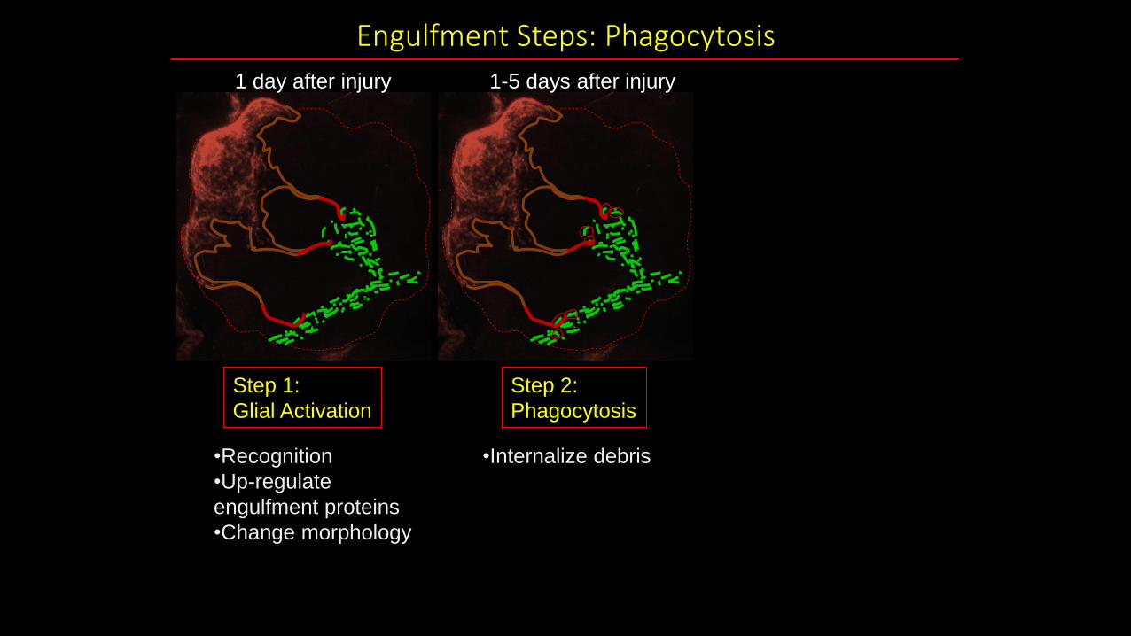
Engulfment Steps: Phagocytosis
Step 1:
Glial Activation
Step 2:
Phagocytosis
•Internalize debris•Recognition
•Up-regulate
engulfment proteins
•Change morphology
1 day after injury 1-5 days after injury
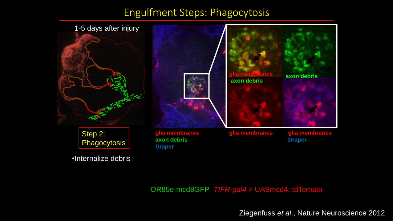
axon debrisglia membranes
axon debris
glia membranes
Draper
glia membranes
axon debris
Draper
OR85e-mcd8GFP; TIFR-gal4 > UASmcd4::tdTomato
glia membranesStep 2:
Phagocytosis
•Internalize debris
1-5 days after injury
Engulfment Steps: Phagocytosis
4um
Ziegenfuss et al., Nature Neuroscience 2012
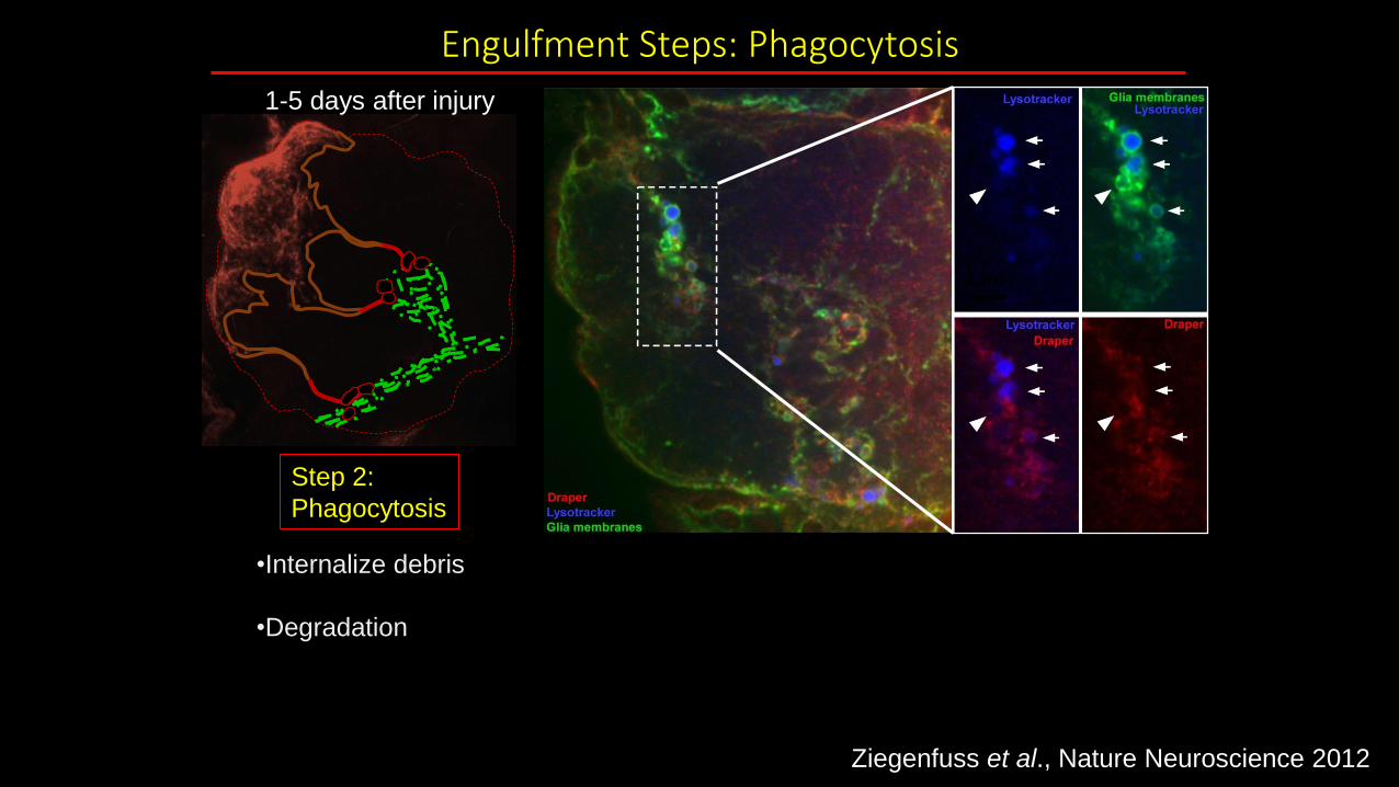
Engulfment Steps: Phagocytosis
Step 2:
Phagocytosis
•Internalize debris
•Degradation
1-5 days after injury
4um
Ziegenfuss et al., Nature Neuroscience 2012

Engulfment Steps: Phagocytosis and Termination of response
Step 1:
Glial Activation
Step 2:
Phagocytosis
Step 3:
Termination
•Down-regulate
engulfment proteins
•Return to resting
morphology
•Internalize debris
•Degradation
•Recognition
•Up-regulate
engulfment proteins
•Change morphology
1 day after injury 1-5 days after injury 5-10 days after injury

C
Ziegenfuss et al. Nature, 2008
Injury induced changes in glial morphology and Draper require Shark
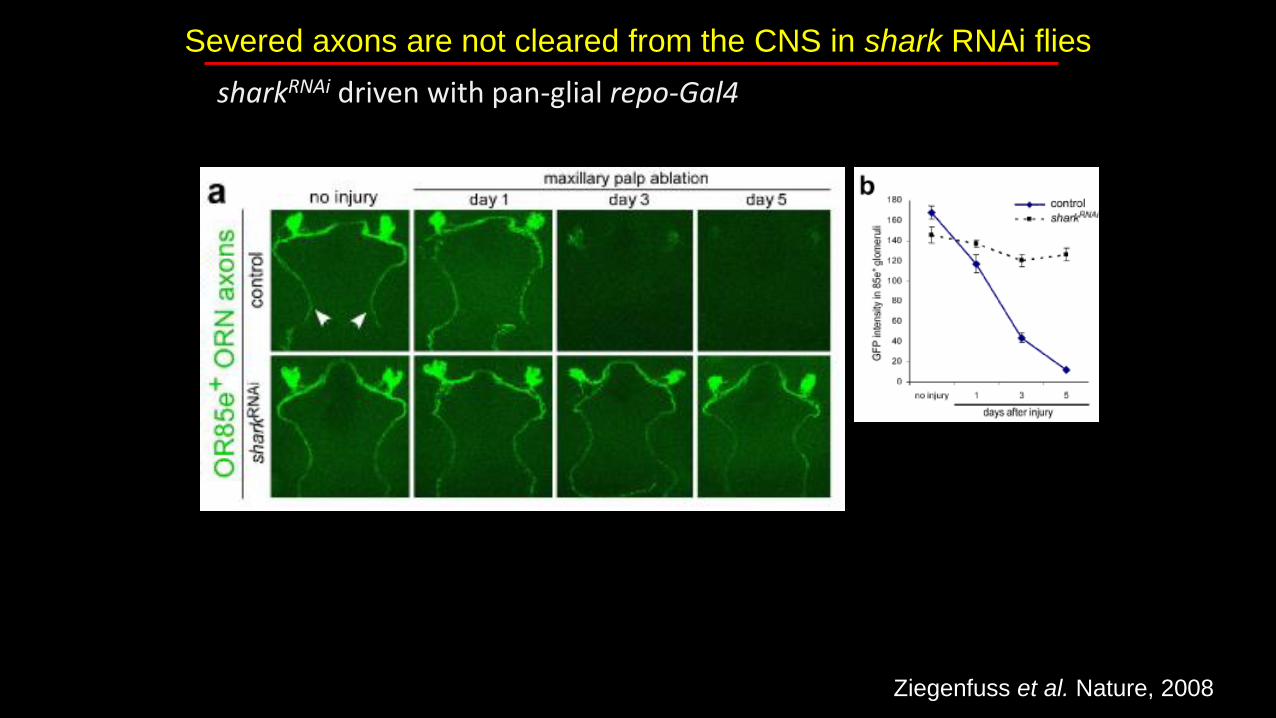
Severed axons are not cleared from the CNS in shark RNAi flies
sharkRNAi driven with pan-glial repo-Gal4
Ziegenfuss et al. Nature, 2008

What happens to severed axons when activation is impaired?
glial activation
Draper
Step 1:
Glial Activation
•Recognition
•Up-regulate
engulfment proteins
•Change morphology
SharkITAM
p
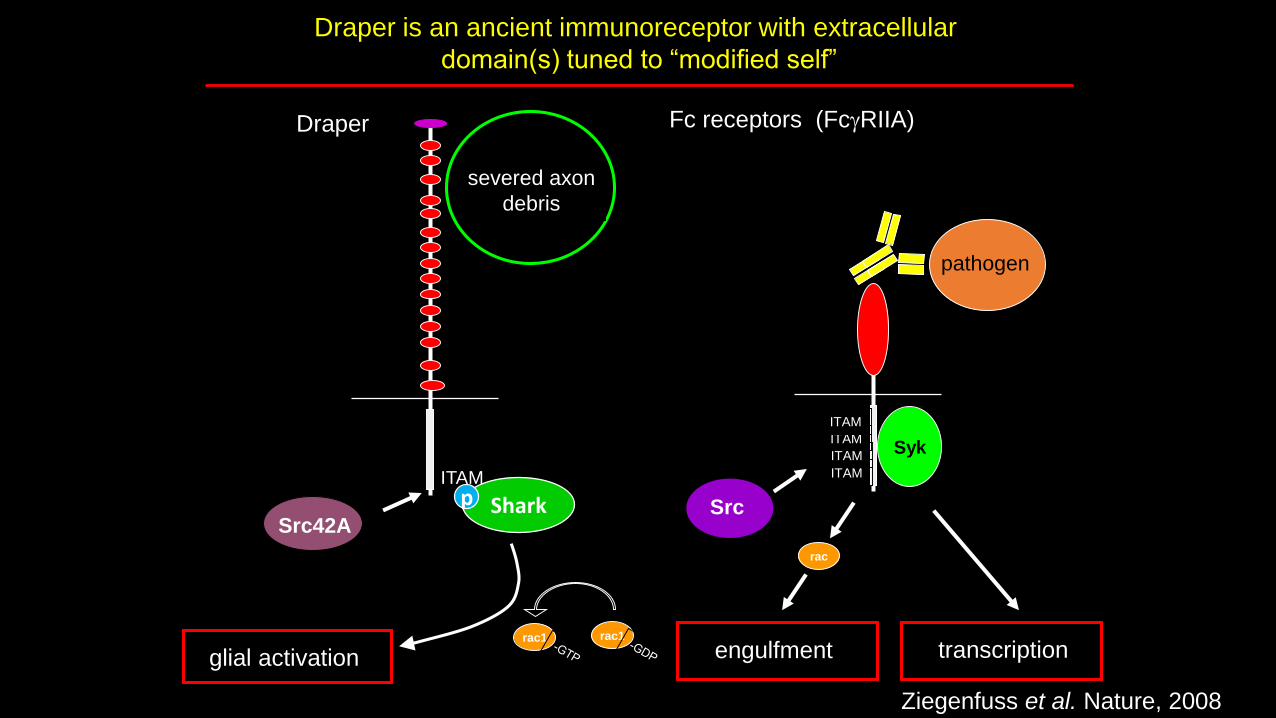
Draper is an ancient immunoreceptor with extracellular
domain(s) tuned to “modified self”
Src
Fc receptors (FcgRIIA)
ITAM
ITAM
ITAM
ITAM
pathogen
engulfment transcription
severed axon
debris
Syk
Src42A
Draper
SharkITAM
p
glial activationrac1 rac1
rac
Ziegenfuss et al. Nature, 2008
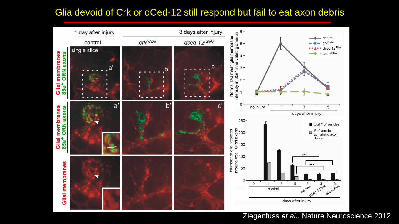
Glia devoid of Crk or dCed-12 still respond but fail to eat axon debris
Ziegenfuss et al., Nature Neuroscience 2012

Lysotracker: labels acidified mature phagosomes = ones that are digesting axon debris
Ziegenfuss et al., Nature Neuro 2012
Glia devoid of Crk or dCed-12 fail to degrade axon debris
Ziegenfuss et al., Nature Neuroscience 2012
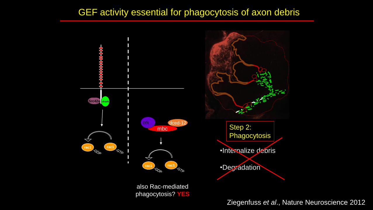
GEF activity essential for phagocytosis of axon debris
Step 2:
Phagocytosis
•Internalize debris
•Degradation
Drosophila
Src42a shark
activation of glia to
severed axons
mbcdced-12crk
also Rac-mediated
phagocytosis? YES
rac1 rac1
rac1 rac1
Ziegenfuss et al., Nature Neuroscience 2012
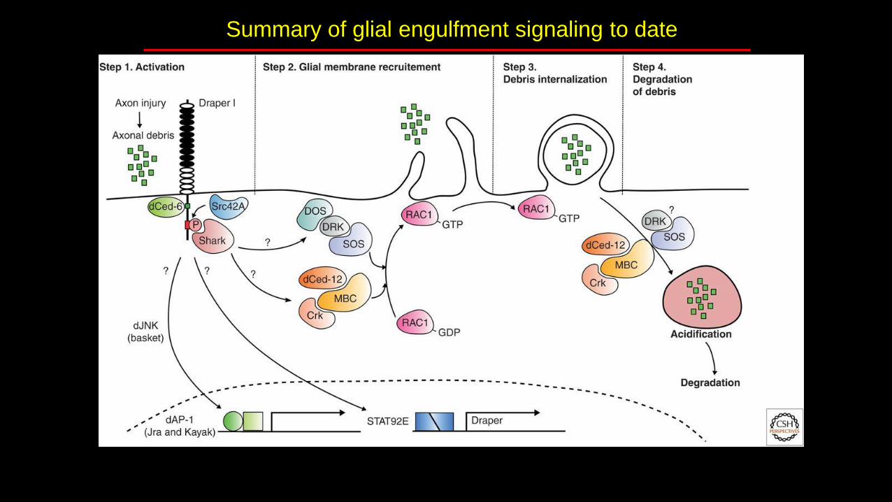
Summary of glial engulfment signaling to date

Thank you!I hope you are more
“glia-evangelized”to the coolness of glia.
Related Documents

![clase 2 neuroglia-C[1]](https://static.cupdf.com/doc/110x72/5517b05349795947228b4ab9/clase-2-neuroglia-c1.jpg)