Neuropsychologia 43 (2005) 12–19 Neural mechanisms involved in the detection of our first name: a combined ERPs and PET study Fabien Perrin a,b,∗ , Pierre Maquet a , Philippe Peigneux a , Perrine Ruby a , Christian Degueldre a , Evelyne Balteau a , Guy Del Fiore a , Gustave Moonen c , Andr´ e Luxen a , Steven Laureys a a Centre de Recherches du Cyclotron, Universit´ e de Li` ege, Belgique b Laboratoire de Neurosciences and Syst` emes Sensoriels, Universit´ e Claude Bernard Lyon 1, CNRS-UMR 5020, 50 Av T. Garnier, 69366 Lyon Cedex 07, France c Service de Neurologie, Universit´ e de Li` ege, Belgique Received 3 October 2003; received in revised form 29 April 2004; accepted 2 July 2004 Abstract In everyday social interactions, hearing our own first name captures our attention and gives rise to a sense of self-awareness, since it is one of the most socially self related stimulus. In the present study, we combined ERPs and PET scan methods to explore the cerebral mechanisms underlying the detection of our own name. While categorical analyses of PET data failed to reveal significant results, we found that the amplitude of the P3 component, elicited when hearing one’s own name, correlates with regional cerebral blood changes in right superior temporal sulcus, precuneus and medial prefrontal cortex. Additionally, the latter was more correlated to the P3 obtained for the subject’s name compared to that obtained for other first names. These results suggest that the medial prefrontal cortex plays the most prominent role in self-processing. © 2004 Elsevier Ltd. All rights reserved. Keywords: Medial prefrontal cortex; P3; Self-processing 1. Introduction Our own first name is intrinsically meaningful for each of us because of its personal significance, its emotional content and repetition along life. Beyond our day-to-day experience, the extreme salience of one’s own first name was highlighted in various experimental and clinical studies. Moray’s (1959) early study of the ‘cocktail party’ phenomenon has shown that approximately one-third of the subjects report hearing their own name in an unattended auditory channel, while none of them could recall the details of the unattended speech (see also Wood & Cowan, 1995). This suggests that the subject’s own name (SON) is so potent that it could “capture atten- tion and subsequently bring that stimulus into awareness” ∗ Corresponding author. Tel.: +33 4 37 28 74 98; fax: +33 4 37 28 76 01. E-mail address: [email protected] (F. Perrin). (Mack, Pappas, Silverman, & Gay, 2002). The powerful de- tection of the SON in comparison with other stimuli was also shown in situations of reduced consciousness. For example, it has been shown that the presentation of the SON during sleep could evoke behavioural responses (Oswald, Taylor, & Treisman, 1960), as well as a differential brain electri- cal response associated to its discrimination (Perrin, Garc´ ıa- Larrea, Maugui` ere, & Bastuji, 1999; Perrin, Bastuji, Maugui` ere, & Garc´ ıa-Larrea, 2000; Pratt, Berlad, & Lavie, 1999). Behavioural responses were demonstrated in 4–5 months old babies, who are able to recognise the sound patterns of their own names (Mandel, Jusczyk, & Pisoni, 1995). Robust responses were also found in demented pa- tients whose perception of their own name deteriorated well after perception of time, place and recognition (Fishback, 1977). At last, after general anaesthesia, reactivity to the SON occurs first, before reactivity to pain or to a noise (Kurtz et al., 1977), and its presentation in comatose patients increases the 0028-3932/$ – see front matter © 2004 Elsevier Ltd. All rights reserved. doi:10.1016/j.neuropsychologia.2004.07.002

Welcome message from author
This document is posted to help you gain knowledge. Please leave a comment to let me know what you think about it! Share it to your friends and learn new things together.
Transcript
Neuropsychologia 43 (2005) 12–19
Neural mechanisms involved in the detection of our first name:a combined ERPs and PET study
Fabien Perrina,b,∗, Pierre Maqueta, Philippe Peigneuxa, Perrine Rubya, Christian Degueldrea,Evelyne Balteaua, Guy Del Fiorea, Gustave Moonenc, Andre Luxena, Steven Laureysa
a Centre de Recherches du Cyclotron, Universit´e de Liege, Belgiqueb Laboratoire de Neurosciences and Syst`emes Sensoriels, Universit´e Claude Bernard Lyon 1,
CNRS-UMR 5020, 50 Av T. Garnier, 69366 Lyon Cedex 07, Francec Service de Neurologie, Universit´e de Liege, Belgique
Received 3 October 2003; received in revised form 29 April 2004; accepted 2 July 2004
Abstract
ince it is oneo mechanismsu d that thea ht superiort e subject’sn ominent rolei©
K
1
uatieaotaot
-lso
mple,uring,ri-
,,4–5
oundi,d pa-well,ON
s the
0d
In everyday social interactions, hearing our own first name captures our attention and gives rise to a sense of self-awareness, sf the most socially self related stimulus. In the present study, we combined ERPs and PET scan methods to explore the cerebralnderlying the detection of our own name. While categorical analyses of PET data failed to reveal significant results, we founmplitude of the P3 component, elicited when hearing one’s own name, correlates with regional cerebral blood changes in rig
emporal sulcus, precuneus and medial prefrontal cortex. Additionally, the latter was more correlated to the P3 obtained for thame compared to that obtained for other first names. These results suggest that the medial prefrontal cortex plays the most pr
n self-processing.2004 Elsevier Ltd. All rights reserved.
eywords:Medial prefrontal cortex; P3; Self-processing
. Introduction
Our own first name is intrinsically meaningful for each ofs because of its personal significance, its emotional contentnd repetition along life. Beyond our day-to-day experience,
he extreme salience of one’s own first name was highlightedn various experimental and clinical studies.Moray’s (1959)arly study of the ‘cocktail party’ phenomenon has shown thatpproximately one-third of the subjects report hearing theirwn name in an unattended auditory channel, while none ofhem could recall the details of the unattended speech (seelsoWood & Cowan, 1995). This suggests that the subject’swn name (SON) is so potent that it could “capture atten-ion and subsequently bring that stimulus into awareness”
∗ Corresponding author. Tel.: +33 4 37 28 74 98; fax: +33 4 37 28 76 01.E-mail address:[email protected] (F. Perrin).
(Mack, Pappas, Silverman, & Gay, 2002). The powerful detection of the SON in comparison with other stimuli was ashown in situations of reduced consciousness. For exait has been shown that the presentation of the SON dsleep could evoke behavioural responses (Oswald, Taylor& Treisman, 1960), as well as a differential brain electcal response associated to its discrimination (Perrin, Garcıa-Larrea, Mauguiere, & Bastuji, 1999; Perrin, BastujiMauguiere, & Garcıa-Larrea, 2000; Pratt, Berlad, & Lavie1999). Behavioural responses were demonstrated inmonths old babies, who are able to recognise the spatterns of their own names (Mandel, Jusczyk, & Pison1995). Robust responses were also found in dementetients whose perception of their own name deterioratedafter perception of time, place and recognition (Fishback1977). At last, after general anaesthesia, reactivity to the Soccurs first, before reactivity to pain or to a noise (Kurtz et al.,1977), and its presentation in comatose patients increase
028-3932/$ – see front matter © 2004 Elsevier Ltd. All rights reserved.oi:10.1016/j.neuropsychologia.2004.07.002
F. Perrin et al. / Neuropsychologia 43 (2005) 12–19 13
chances of obtaining differential brain responses associatedto information processing (Signorino, D’Acunto, Angeleri,& Pietropaoli, 1995).
The neural correlates of the discrimination of the SONhave not been yet properly investigated. However, someanatomical hypotheses may be put forward, since hearingour first name elicits a self-experience, i.e. a “consciousnessof oneself as an immediate subject of experience, unextendedin time” (Gallagher, 2000; for a review seeKircher & David,2003). This stimulus is of particular interest in the study ofself-processing because its detection is associated with an en-hancement of self-awareness. Since it is used by the others todirectly refer to ourself, the SON plays an important part inour everyday social interactions. For this reason, it can be con-sidered as a very ecological and highly socially self-relatedstimulus. Prior neuroimaging studies identified several corti-cal structures involved in various self-processing: the medialprefrontal cortex (MPFC) and/or the anterior cingulate cor-tex, the right temporo-parietal junction and the precuneus.For instance, MPFC could be involved in tasks where sub-jects have to make a self-referential judgement (Gusnard,Akbudak, Shulman, & Raichle, 2001; Lane, Fink, Chau, &Dolan, 1997) and in tasks requiring (self-)reflection on theirpersonal abilities, traits or attitudes (Johnson et al., 2002;Kelley et al., 2000, 2002; Kircher et al., 2001; Kjaer, Nowak,& de-s nar-r or asA e ac-t ;R aidt if ther urallyi t it is“ ,R
bralm firstn 300)p en-h sucha ction( ,1 incei N isp ulusp re-s f theS i-m ET)i nalc itudet du-l SONc
2. Methods
2.1. Subjects
Fifteen right-handed (Edinburgh Inventory,Oldfield,1971) healthy volunteers (five women, 22.7± 3 years)without hearing deficit and without medical, neurological,nor psychiatric history participated to the study. The ex-periment was conducted in agreement with the guidelinesof the ‘Declaration of Helsinki’ and was approved by theEthics Committee of the Faculty of Medicine of the Uni-versity of Liege. Written informed consent was obtainedfrom all subjects. All subjects had a disyllabic first name,which was non-commonly used in French language (i.e. em-ployed less than 30,000 times in France during the 20th cen-tury: seehttp://www.prenoms.com/, Kilos Mutimedia Sarl,IDDN.FR.010.0077897.000.R.P.2000.028.40000).
2.2. Stimuli
Three types of auditory stimuli were used: the subject’sown name (SON), uncommon first names (UFN), and com-mon first names (CFN, i.e. employed more than 300,000times during the 20th century).
The experimental material was constructed using a 2× 2f FN)a ver-s t in-c ucea mea-s uli,c
( our;
( lled
( ”];( lled
uliw 90-sp
nts.T pre-s re thes twoc es.M em-p frome
wasr eries,fi was2 amen at
Lou, 2002). Activation of all three regions have beencribed in studies where subjects were implicated in aative history, i.e. when they had to take a first-personelf-perspective (Vogeley et al., 2001; Vogeley & Fink, 2003).t last, both MPFC and precuneus were reported to b
ivated in the conscious resting state (Mazoyer et al., 2001aichle et al., 2001), i.e. in a condition where subjects p
heir attention towards various internal processes. Evenesting state is considered by some authors as a behavioll-defined heterogeneous condition, other suggested thathe ultimate state of inspection of the self” (Wicker, Rubyoyet, & Fonlupt, 2003).The aim of the present study was to identify the cere
echanisms underlying the capacity to discriminate ourame in the external world. We investigated the P3 (or Potential, for which the parietal component (the ‘P3b’) isanced when subjects detect an implicit target stimuluss the SON, even in the absence of any explicit instruBerlad & Pratt, 1995; Folmer & Yingling, 1997; Perrin et al.999). We focused our attention on the parietal P3 wave s
ts amplitude is sensitive to both task relevance (the SOarticularly relevant and inherently attended) and stimrobability, thus may reflect the information-processingources allocated to the categorisation/discrimination oON (for a review, seePicton, 1992). Furthermore, we sultaneously acquired positron emission tomography (P
mages in the aim to identify brain regions in which regioerebral blood flow changes correlated with the P3 amplo the SON, i.e. brain areas which were specifically moated when the resources allocated to detection of thehanged.
actorial design, where the type of stimuli (SON versus Und the probability of occurrence of the first name (20%us 50%) were independently manipulated. (We did nolude a high SON occurrence condition since it would indhabituation effect and preclude P300 and related rCBFurements.) We elaborated four conditions, of 48–60 stimontaining:
1) SON with a probability of occurrence of 20% and fCFN with a probability of 20% each [labelled “S20”]
2) SON at 50% and four CFN at 12.5% each [labe“S50”];
3) UFN at 20% and four CFN at 20% each [labelled “U204) UFN at 50% and four CFN at 12.5% each [labe
“U50”].
The probability of occurrence of the three types of stimas the same within the entire sequence and within theeriod of PET scanning.
The UFN were the first names of the other participawelve first names were used as CFN and randomlyented across conditions, series and subjects; they weame for all participants. Thus, the group comparison ofonditions eliminated the acoustical effect of all first namoreover, prior to the experiment a list of the five mostloyed first names, of relatives and friends, was obtainedach subject and eliminated from the lists.
Each condition was repeated three times andandomly presented across subjects. Within each srst names were pseudo-randomly presented. SOA000 ms. All first names were recorded by the seutral male voice, digitised and replayed binaurally
14 F. Perrin et al. / Neuropsychologia 43 (2005) 12–19
about 80 dB SPL maximal intensity with Cogent softwareenvironment (Wellcome Department of Imaging Neuro-science, London, UK,http://www.vislab.ucl.ac.uk/Cogent/).All were disyllabic and had a maximum length of600 ms.
Subjects were instructed to listen passively and to remainattentive to the stimuli all along the recording session. Wedid not use an active attention task in the aim to further applythis protocol in subjects with altered states of consciousness(such as coma, general anaesthesia, sleep, dementia, mini-mally conscious state, etc.).
2.3. ERPs, PET and MRI acquisitions
Electroencephalographic (EEG) signals from nine AgClelectrodes (Fz, Cz, Pz, F3, C3, P3, F4, C4, P4), placed ac-cording to the International 10–20 system (Jasper, 1958;Klem, Luders, Jasper, & Elger, 1999) and referenced to thenose, electrooculogram (EOG) from two electrodes diago-nally above and below the right eye, and electrocardiogram(ECG) from two electrodes on pectoral muscles, were ampli-fied (150,000×) and sampled at 1000 Hz by a Synamps acqui-sition system connected to a Neuroscan (NeuroSoft, Sterling,VA, USA) equipment, with an analog bandpass of 0.1–70 Hz.A ground electrode was placed near Fpz and impedance wask
wasi s sta-b wasi onesw dataw ree-d ebralb mesi twof Thes ret tedf Dataw ncy0 ounda
anA mm× s,T
2
pre-s duale 00 mse aver-a 50),t lec-t ining
eye movement or muscular artefact with amplitude exceed-ing ±50�V were excluded from the average. Then, ERPswere averaged across subjects and digitally filtered between0.1 and 30 Hz (roll off: 24 dB/oct.) to create grand-averagedERPs (used for illustrative purposes). Statistical computa-tions were performed on averaged traces from each individ-ual: amplitudes (from baseline) and latencies of the N1, P2,N2 and P3 components were calculated for each individualaverage. These values were tested with a three-way analysisof variance (ANOVA) with repeated measures on the type offirst names (SON or UFN versus CFN), the condition (S20versus S50 versus U20 versus U50) and the electrode po-sition (Fz versus Cz versus Pz). Post-hoc Bonferroni testswere performed when significant interactions emerged onANOVA.
The difference in P3 amplitude between the SON or theUFN and the CFN was calculated for each individual averageat its maximum, i.e. at Pz, and these values were used ascovariate of interest in the subsequent SPM analyses.
Separate ANOVAs were performed to test the existenceof laterality effects on the P300 under our experimentalconditions. P300 amplitudes were submitted to similar re-peated measurements ANOVAs than described above, butusing lateral temporal electrodes (T3 and T4) as levels forthe ‘electrode position’ factor. For all analyses, no sig-n lec-t e re-s theP them
2
ara-m entou ,S gnedt ernelo
ari-a cord-i wasc ac-c antc ditione
pre-s nce[ 0)],t thet 50)+ hen,w me( 20,S
ept below 5 k�.After the installation of the electrodes, the subject
nstalled on the (PET) scanner couch. Her/his head wailised by a thermoplastic facemask. A venous catheter
nserted in a left anterobrachial vein and two earphere inserted into the external acoustic canals. PETere acquired on a CTI 951 R 16/31 scanner in thimensional mode (Siemens, Erlangen, Germany). Cerlood flow was estimated while subjects listened to na
n one of the four conditions. Each scan consisted oframes: a 30-s background frame and a 90-s frame.low intravenous water (H215O) infusion begun just befohe second frame. Six millicurie (222 MBq) were injecor each scan. The infusion was totally automated.ere reconstructed using a Hanning filter (cut-off freque.5 cycle/pixel) and corrected for attenuation and backgrctivity.
Structural T1-weighted MRI scans were obtained byllegra 3 T MR(Siemens, Erlangen, Germany) at a [0.90.9 mm× 0.9 mm] voxel size (MPRAGE, TR = 1960 m
E = 4.43 ms, FA = 8◦).
.4. ERPs data analyses
The EEG analysis was conducted on the stimuliented during PET scanning (i.e. the 90-s frame). Indivivent-related potentials (ERPs) were analysed over a 17poch, including a prestimulus baseline of 200 ms, andged according to the condition (S20, S50, U20, and U
he type of stimulus (SON or UFN and CFN) and the erode position. Prior to averaging, single epochs conta
ificant effect nor interactions were noted for the erode side factor, and therefore further analyses wertricted to midline electrodes in order to maximise300 effect, which is known to predominate overidline.
.5. PET data analyses
PET data were analysed using the statistical petric mapping software SPM99 (Wellcome Departmf Cognitive Neurology, London, UK,http://www.fil.ion.cl.ac.uk/spm/) implemented in MATLAB (Mathworksherborn, MA). For each subject, all scans were reali
ogether, normalised and smoothed using a Gaussian kf 16-mm full width at half maximum.
The scan condition, covariate (P3), condition by covte interaction and subject effects were estimated ac
ng to the general linear model (only one design matrixonstructed). Changes in global CBF were taken intoount by a subjects specific ANCOVA. Areas of significhanges were determined using linear contrasts of constimates.
We first assessed the main effects of first nameentation independently of the probability of occurre(S20–U20) + (S50–U50) and (U20–S20) + (U50–S5hose of the probability of occurrence independently ofype of first name [(S50–S20) + (U50–U20) and (S20–S
(U20–U50)] and the interactions between factors. Te looked for the simple effects of the type of first na
SON or UFN) at each probability of occurrence (S20–U50–U50, U20–S20 and U50–S50).
F. Perrin et al. / Neuropsychologia 43 (2005) 12–19 15
Furthermore, we looked for the brain areas that covariedwith the P3 amplitude obtained when subjects heard theirown first name (SON) or an uncommon first name (UFN) atboth probabilities of occurrence (S20 + S50 and U20 + U50).We also looked for the brain areas that were more correlatedto the P3 amplitude obtained for the SON than that obtainedfor the UFN [(S20 + S50)− (U20 + U50)].
The resulting set of voxel values for each contrast con-stituted a map of the t statistic, SPM (T), thresholded atP(uncorrected)≤ 0.001. Results were considered signifi-cant atP < 0.05 after a small volume correction of 10-mmradius-sphere centred on the predetermined regions of inter-est. The latter were the medial prefrontal cortex (stereotaxiccoordinatesx = ±6, y = 54, z = 11, averaged coordinatestaken fromGusnard et al., 2001; Johnson et al., 2002; Kelleyet al., 2002; Lane et al., 1997; Mazoyer et al., 2001; Raichleet al., 2001; Vogeley et al., 2001), the right temporo-parietaljunction (x = 58, y = −56, z = 28, Kjaer et al., 2002;Vogeley et al., 2001) and the precuneus (x = ±6, y = −52,z= 45,Kelley et al., 2002; Kircher et al., 2000, 2002; Kjaeret al., 2002; Johnson et al., 2002; Mazoyer et al., 2001;Raichle et al., 2001; Vogeley et al., 2001). Functional re-sults were positioned on the averaged structural MRI of oursubjects.
FaCa
3. Results
3.1. ERPs data
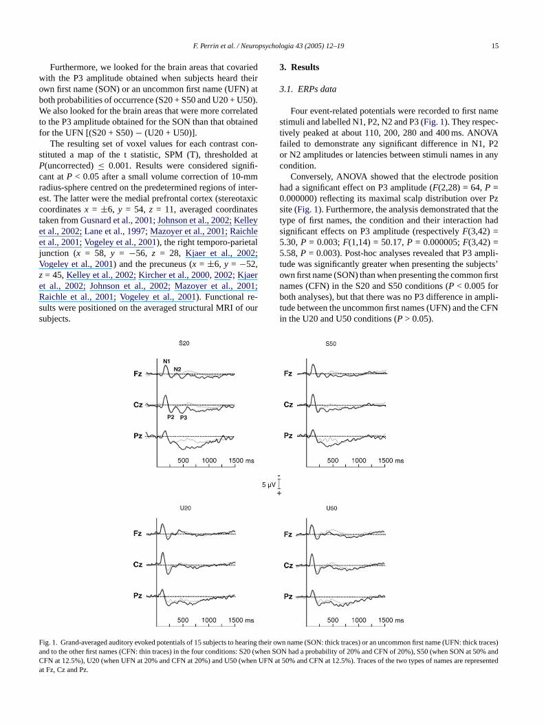
Four event-related potentials were recorded to first namestimuli and labelled N1, P2, N2 and P3 (Fig. 1). They respec-tively peaked at about 110, 200, 280 and 400 ms. ANOVAfailed to demonstrate any significant difference in N1, P2or N2 amplitudes or latencies between stimuli names in anycondition.
Conversely, ANOVA showed that the electrode positionhad a significant effect on P3 amplitude (F(2,28) = 64,P =0.000000) reflecting its maximal scalp distribution over Pzsite (Fig. 1). Furthermore, the analysis demonstrated that thetype of first names, the condition and their interaction hadsignificant effects on P3 amplitude (respectivelyF(3,42) =5.30,P = 0.003;F(1,14) = 50.17,P = 0.000005;F(3,42) =5.58,P = 0.003). Post-hoc analyses revealed that P3 ampli-tude was significantly greater when presenting the subjects’own first name (SON) than when presenting the common firstnames (CFN) in the S20 and S50 conditions (P < 0.005 forboth analyses), but that there was no P3 difference in ampli-tude between the uncommon first names (UFN) and the CFNin the U20 and U50 conditions (P > 0.05).
ig. 1. Grand-averaged auditory evoked potentials of 15 subjects to hearing tnd to the other first names (CFN: thin traces) in the four conditions: S20 (whFN at 12.5%), U20 (when UFN at 20% and CFN at 20%) and U50 (when Ut Fz, Cz and Pz.
heir own name (SON: thick traces) or an uncommon first name (UFN: thick traces)en SON had a probability of 20% and CFN of 20%), S50 (when SON at 50% andFN at 50% and CFN at 12.5%). Traces of the two types of names are represented
16 F. Perrin et al. / Neuropsychologia 43 (2005) 12–19
Table 1Brain areas where rCBF was linearly correlated to the P3 response evoked by hearing one’s own first name
Region Brodmann area x (mm) y (mm) z (mm) Z value Small volume correctedP-value
Right medial prefrontal cortex 10 8 64 12 3.66 0.006Left precuneus 7 −6 −66 48 3.17 0.023Right superior temporal sulcus 39 64 −58 28 3.16 0.023
Right medial frontal cortex 10 8 64 16 3.88 0.003
The last line corresponds to the brain area which is more correlated to the P3 response obtained for the subject’s own name than that obtained for another firstname (x, y andzare coordinates in the standardTalairach and Tournoux (1988)stereotactic space).
3.2. PET data
3.2.1. Effects of the type of name and of the probabilityof occurrence
Categorical comparisons based on the stimulus type (SONversus UFN) or its probability of occurrence (20% versus50%) failed to reveal significant effects in regions of interest(even after a small volume correction centred on the pre-determined regions of interest). These analyses did not in-clude the electrophysiological measures. Thus, it could besuggested that PET data alone are less adapted to assess thebrain responses to sparse events, as in the passive detectionof the SON (in particular with our protocol in which similarconditions, except for one stimulus, were contrasted).
3.2.2. Areas varying with the P3 amplitudeThe SPM analysis looking for brain areas that showed a
linear correlation between rCBF and the amplitude of the P3
Fc
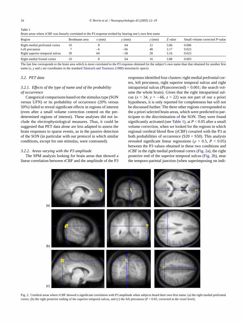
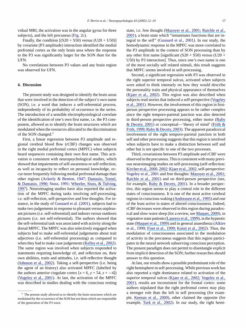
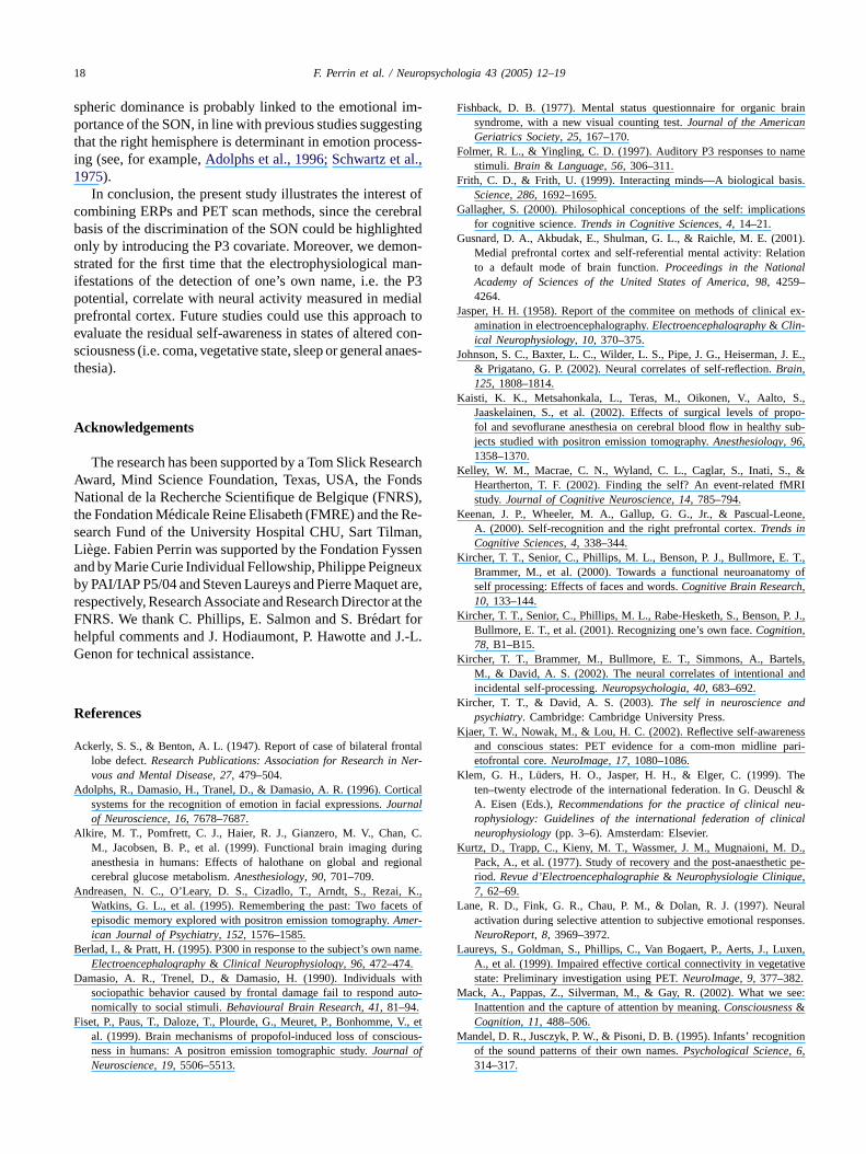
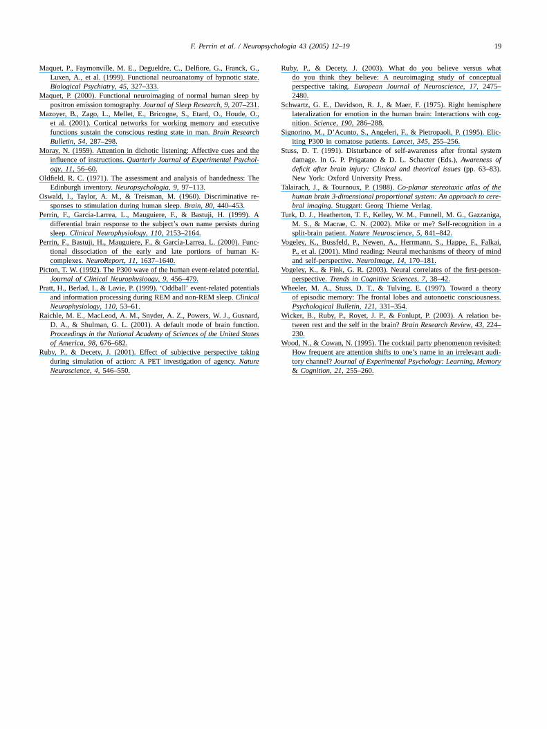
responses identified four clusters: right medial prefrontal cor-tex, left precuneus, right superior temporal sulcus and rightintraparietal sulcus (P(uncorrected) < 0.001; the search vol-ume the whole brain). Given that the right intraparietal sul-cus (x = 34, y = −66, z = 22) was not part of our a priorihypotheses, it is only reported for completeness but will notbe discussed further. The three other regions corresponded tothe a priori selected brain areas, which were predicted to par-ticipate to the discrimination of the SON. They were foundsignificantly activated (seeTable 1), atP< 0.05 after a smallvolume correction, when we looked for the regions in whichregional cerebral blood flow (rCBF) covaried with the P3 atboth probabilities of occurrence (S20 + S50). This analysisrevealed significant linear regressions (ρ > 0.5, P < 0.05)between the P3 values obtained in these two conditions andrCBF in the right medial prefrontal cortex (Fig. 2a), the rightposterior end of the superior temporal sulcus (Fig. 2b), nearthe temporo-parietal junction (when superimposing on indi-
ig. 2. Cerebral areas where rCBF showed a significant correlation with P3 aortex, (b) the right posterior ending of the superior temporal sulcus, and (c)
mplitude when subjects heard their own first name: (a) the right medial prefrontalthe left precuneus (P < 0.05, corrected at the voxel level).
F. Perrin et al. / Neuropsychologia 43 (2005) 12–19 17
vidual MRI, the activation was in the angular gyrus for threesubjects), and the left precuneus (Fig. 2c).
Finally, the condition [(S20 + S50) versus (U20 + U50)]by covariate (P3 amplitude) interaction identified the medialprefrontal cortex as the only brain area where the responseto the P3 was significantly larger for the SONthan for theUFN.
No correlations between P3 values and any brain regionwas observed for UFN.
4. Discussion
The present study was designed to identify the brain areasthat were involved in the detection of the subject’s own name(SON), i.e. a word that induces a self-referential process,independently of its probability of occurrence in the series.The introduction of a sensible electrophysiological correlateof the identification of one’s own first name, i.e. the P3 com-ponent, allowed us to identify the brain structures which aremodulated when the resources allocated to the discriminationof the SON changed.1
First, a linear regression between P3 amplitude and re-gional cerebral blood flow (rCBF) changes was observedin the right medial prefrontal cortex (MPFC) when subjectsh acti-v hichs tion,a , oc-c hano l,& ,1 tiva-t ing,i r in-s om pleasa oorsp thatt g thed hens t traita d towT ed tos theiro ght( ngt byt( Cw sting
h arem nsibleo
state, i.e. free thought (Mazoyer et al., 2001; Raichle et al.,2001), a brain state which “instantiates functions that are in-tegral to the self” (Gusnard et al., 2001). In our study, thehemodynamic response in the MPFC was more correlated tothe P3 amplitude in the context of SON processing than byany other first name [significant (S20 + S50) versus (U20 +U50) by P3 interaction]. Thus, since one’s own name is oneof the most socially self related stimuli, this result suggeststhat MPFC seems involved in self-processing.
Second, a significant regression with P3 was observed inthe right superior temporal sulcus, activated when subjectswere asked to think intensely on how they would describethe personality traits and physical appearance of themselves(Kjaer et al., 2002). This region was also described whensubjects read stories that induced a self-perspective (Vogeleyet al., 2001). However, the involvement of this region in first-person perspective processing appears to be rather complexsince the right temporo-parietal junction was also detectedin third-person perspective processing, either motor (Ruby& Decety, 2001) or conceptual—’theory of mind’ (Frith &Frith, 1999; Ruby & Decety, 2003). The apparent paradoxicalinvolvement of the right temporo-parietal junction in bothself and other processing suggests that this region is involvedwhen subjects have to make a distinction between self andother but is not specific to one of the two processes.
ereo revi-o tion:K :V ;R ee,f c-t rents rebralro deed,r dox-ivsem lationo rtici-p ption.T plicitf oulda
f ther hasa f thes l.,2 mea playa am-p ore i-
eard sequences containing their own first name. Thisation is consistent with neuropsychological studies, whowed that impairments of self-awareness or self-reflecs well as incapacity to reflect on personal knowledgeur more frequently following medial prefrontal damage tther regions (Ackerly & Benton, 1947; Damasio, TreneDamasio, 1990; Stuss, 1991; Wheeler, Stuss, & Tulving
997). Neuroimaging studies have also reported the acion of the MPFC during tasks involving self-process.e. self-reflection, self-perspective and free thoughts. Fotance, in the study ofGusnard et al. (2001), subjects had take two judgements in response to pleasant versus unnt pictures (i.e. self-referential) and indoors versus outdictures (i.e. not self-referential). The authors showed
he self-referential task was associated with activity alonorsal MPFC. The MPFC was also selectively engaged wubjects had to make self-referential judgements aboudjectives (i.e. self-referential processing) as comparehen they had to make case judgements (Kelley et al., 2002).he same region was involved when subjects respondtatements requiring knowledge of, and reflection on,wn abilities, traits and attitudes, i.e. self-reflective thouJohnson et al., 2002). Taking a self-perspective (i.e. beihe agent of an history) also activated MPFC (labelledhe authors anterior cingulate cortex [x = 6, y = 54,z= −4])Vogeley et al., 2001). At last, the activation of the MPFas described in studies dealing with the conscious re
1 The present study allowed us to identify the brain structures whicodulated by the occurrence of the SON but not those which are respof the generation of the P3 wave.
-
Third, covariations between P3 amplitude and rCBF wbserved in the precuneus. This is consistent with many pus neuroimaging studies on self-processing (self-reflecircher et al., 2000, 2002; Kjaer et al., 2002, self-perspectiveogeley et al., 2001and free thoughts:Mazoyer et al., 2001aichle et al., 2001) and on third-person perspective (s
or example,Ruby & Decety, 2001). In a broader perspeive, this region seems to play a central role in the diffetates of consciousness. It is one of the most active ceegions in conscious waking (Andreasen et al., 1995) and onef the least active in states of altered consciousness. InCBF decreases were observed in this region during paracal and slow-wave sleep (for a review, seeMaquet, 2000), inegetative state patients (Laureys et al., 1999), in the hypnotictate (Maquet et al., 1999) and in general anaesthesia (Alkiret al., 1999; Fiset et al., 1999; Kaisti et al., 2002). Thus, theodulation of consciousness associated to the moduf activity in the precuneus suggests that this region paates in the neural network subserving conscious percehe present paradigm does not permit to disentangle ex
rom implicit detection of the SON; further researches shnswer to this question.
At last, our results show a possible predominant role oight hemisphere in self-processing. While previous worklso reported a right dominance related to activation ouperior temporal sulcus (Kjaer et al., 2002; Vogeley et a001), results are inconsistent for the frontal cortex: southors stipulated that the right prefrontal cortex maystronger role than the left in self processing (for ex
le, Keenan et al., 2000), other claimed the opposite (fxample,Turk et al., 2002). In our study, the right hem
18 F. Perrin et al. / Neuropsychologia 43 (2005) 12–19
spheric dominance is probably linked to the emotional im-portance of the SON, in line with previous studies suggestingthat the right hemisphere is determinant in emotion process-ing (see, for example,Adolphs et al., 1996; Schwartz et al.,1975).
In conclusion, the present study illustrates the interest ofcombining ERPs and PET scan methods, since the cerebralbasis of the discrimination of the SON could be highlightedonly by introducing the P3 covariate. Moreover, we demon-strated for the first time that the electrophysiological man-ifestations of the detection of one’s own name, i.e. the P3potential, correlate with neural activity measured in medialprefrontal cortex. Future studies could use this approach toevaluate the residual self-awareness in states of altered con-sciousness (i.e. coma, vegetative state, sleep or general anaes-thesia).
Acknowledgements
The research has been supported by a Tom Slick ResearchAward, Mind Science Foundation, Texas, USA, the FondsNational de la Recherche Scientifique de Belgique (FNRS),the Fondation Medicale Reine Elisabeth (FMRE) and the Re-search Fund of the University Hospital CHU, Sart Tilman,L yssena euxb t are,r at theFh J.-L.G
R
A ntalNer-
A tical
A , C.ring
gional
A K.,ts of
B ame.
D ithauto-
F V., etious-
Fishback, D. B. (1977). Mental status questionnaire for organic brainsyndrome, with a new visual counting test.Journal of the AmericanGeriatrics Society, 25, 167–170.
Folmer, R. L., & Yingling, C. D. (1997). Auditory P3 responses to namestimuli. Brain & Language, 56, 306–311.
Frith, C. D., & Frith, U. (1999). Interacting minds—A biological basis.Science, 286, 1692–1695.
Gallagher, S. (2000). Philosophical conceptions of the self: implicationsfor cognitive science.Trends in Cognitive Sciences, 4, 14–21.
Gusnard, D. A., Akbudak, E., Shulman, G. L., & Raichle, M. E. (2001).Medial prefrontal cortex and self-referential mental activity: Relationto a default mode of brain function.Proceedings in the NationalAcademy of Sciences of the United States of America, 98, 4259–4264.
Jasper, H. H. (1958). Report of the commitee on methods of clinical ex-amination in electroencephalography.Electroencephalography& Clin-ical Neurophysiology, 10, 370–375.
Johnson, S. C., Baxter, L. C., Wilder, L. S., Pipe, J. G., Heiserman, J. E.,& Prigatano, G. P. (2002). Neural correlates of self-reflection.Brain,125, 1808–1814.
Kaisti, K. K., Metsahonkala, L., Teras, M., Oikonen, V., Aalto, S.,Jaaskelainen, S., et al. (2002). Effects of surgical levels of propo-fol and sevoflurane anesthesia on cerebral blood flow in healthy sub-jects studied with positron emission tomography.Anesthesiology, 96,1358–1370.
Kelley, W. M., Macrae, C. N., Wyland, C. L., Caglar, S., Inati, S., &Heartherton, T. F. (2002). Finding the self? An event-related fMRIstudy.Journal of Cognitive Neuroscience, 14, 785–794.
Keenan, J. P., Wheeler, M. A., Gallup, G. G., Jr., & Pascual-Leone,A. (2000). Self-recognition and the right prefrontal cortex.Trends in
K . T.,y ofh
K P. J.,
K els,and
K nd
K esspari-
K hehl &eu-ical
K D.,ic pe-
L uralnses.
L uxen,tive
.M see:
M ition
i ege. Fabien Perrin was supported by the Fondation Fnd by Marie Curie Individual Fellowship, Philippe Peigny PAI/IAP P5/04 and Steven Laureys and Pierre Maqueespectively, Research Associate and Research DirectorNRS. We thank C. Phillips, E. Salmon and S. Bredart forelpful comments and J. Hodiaumont, P. Hawotte andenon for technical assistance.
eferences
ckerly, S. S., & Benton, A. L. (1947). Report of case of bilateral frolobe defect.Research Publications: Association for Research invous and Mental Disease, 27, 479–504.
dolphs, R., Damasio, H., Tranel, D., & Damasio, A. R. (1996). Corsystems for the recognition of emotion in facial expressions.Journalof Neuroscience, 16, 7678–7687.
lkire, M. T., Pomfrett, C. J., Haier, R. J., Gianzero, M. V., ChanM., Jacobsen, B. P., et al. (1999). Functional brain imaging duanesthesia in humans: Effects of halothane on global and recerebral glucose metabolism.Anesthesiology, 90, 701–709.
ndreasen, N. C., O’Leary, D. S., Cizadlo, T., Arndt, S., Rezai,Watkins, G. L., et al. (1995). Remembering the past: Two faceepisodic memory explored with positron emission tomography.Amer-ican Journal of Psychiatry, 152, 1576–1585.
erlad, I., & Pratt, H. (1995). P300 in response to the subject’s own nElectroencephalography& Clinical Neurophysiology, 96, 472–474.
amasio, A. R., Trenel, D., & Damasio, H. (1990). Individuals wsociopathic behavior caused by frontal damage fail to respondnomically to social stimuli.Behavioural Brain Research, 41, 81–94.
iset, P., Paus, T., Daloze, T., Plourde, G., Meuret, P., Bonhomme,al. (1999). Brain mechanisms of propofol-induced loss of conscness in humans: A positron emission tomographic study.Journal ofNeuroscience, 19, 5506–5513.
Cognitive Sciences, 4, 338–344.ircher, T. T., Senior, C., Phillips, M. L., Benson, P. J., Bullmore, E
Brammer, M., et al. (2000). Towards a functional neuroanatomself processing: Effects of faces and words.Cognitive Brain Researc,10, 133–144.
ircher, T. T., Senior, C., Phillips, M. L., Rabe-Hesketh, S., Benson,Bullmore, E. T., et al. (2001). Recognizing one’s own face.Cognition,78, B1–B15.
ircher, T. T., Brammer, M., Bullmore, E. T., Simmons, A., BartM., & David, A. S. (2002). The neural correlates of intentionalincidental self-processing.Neuropsychologia, 40, 683–692.
ircher, T. T., & David, A. S. (2003).The self in neuroscience apsychiatry. Cambridge: Cambridge University Press.
jaer, T. W., Nowak, M., & Lou, H. C. (2002). Reflective self-awarenand conscious states: PET evidence for a com-mon midlineetofrontal core.NeuroImage, 17, 1080–1086.
lem, G. H., Luders, H. O., Jasper, H. H., & Elger, C. (1999). Tten–twenty electrode of the international federation. In G. DeuscA. Eisen (Eds.),Recommendations for the practice of clinical nrophysiology: Guidelines of the international federation of clinneurophysiology(pp. 3–6). Amsterdam: Elsevier.
urtz, D., Trapp, C., Kieny, M. T., Wassmer, J. M., Mugnaioni, M.Pack, A., et al. (1977). Study of recovery and the post-anaesthetriod. Revue d’Electroencephalographie& Neurophysiologie Clinique,7, 62–69.
ane, R. D., Fink, G. R., Chau, P. M., & Dolan, R. J. (1997). Neactivation during selective attention to subjective emotional respoNeuroReport, 8, 3969–3972.
aureys, S., Goldman, S., Phillips, C., Van Bogaert, P., Aerts, J., LA., et al. (1999). Impaired effective cortical connectivity in vegetastate: Preliminary investigation using PET.NeuroImage, 9, 377–382
ack, A., Pappas, Z., Silverman, M., & Gay, R. (2002). What weInattention and the capture of attention by meaning.Consciousness&Cognition, 11, 488–506.
andel, D. R., Jusczyk, P. W., & Pisoni, D. B. (1995). Infants’ recognof the sound patterns of their own names.Psychological Science, 6,314–317.
F. Perrin et al. / Neuropsychologia 43 (2005) 12–19 19
Maquet, P., Faymonville, M. E., Degueldre, C., Delfiore, G., Franck, G.,Luxen, A., et al. (1999). Functional neuroanatomy of hypnotic state.Biological Psychiatry, 45, 327–333.
Maquet, P. (2000). Functional neuroimaging of normal human sleep bypositron emission tomography.Journal of Sleep Research, 9, 207–231.
Mazoyer, B., Zago, L., Mellet, E., Bricogne, S., Etard, O., Houde, O.,et al. (2001). Cortical networks for working memory and executivefunctions sustain the conscious resting state in man.Brain ResearchBulletin, 54, 287–298.
Moray, N. (1959). Attention in dichotic listening: Affective cues and theinfluence of instructions.Quarterly Journal of Experimental Psychol-ogy, 11, 56–60.
Oldfield, R. C. (1971). The assessment and analysis of handedness: TheEdinburgh inventory.Neuropsychologia, 9, 97–113.
Oswald, I., Taylor, A. M., & Treisman, M. (1960). Discriminative re-sponses to stimulation during human sleep.Brain, 80, 440–453.
Perrin, F., Garcıa-Larrea, L., Mauguiere, F., & Bastuji, H. (1999). Adifferential brain response to the subject’s own name persists duringsleep.Clinical Neurophysiology, 110, 2153–2164.
Perrin, F., Bastuji, H., Mauguiere, F., & Garcıa-Larrea, L. (2000). Func-tional dissociation of the early and late portions of human K-complexes.NeuroReport, 11, 1637–1640.
Picton, T. W. (1992). The P300 wave of the human event-related potential.Journal of Clinical Neurophysioogy, 9, 456–479.
Pratt, H., Berlad, I., & Lavie, P. (1999). ‘Oddball’ event-related potentialsand information processing during REM and non-REM sleep.ClinicalNeurophysiology, 110, 53–61.
Raichle, M. E., MacLeod, A. M., Snyder, A. Z., Powers, W. J., Gusnard,D. A., & Shulman, G. L. (2001). A default mode of brain function.Proceedings in the National Academy of Sciences of the United States
R king
Ruby, P., & Decety, J. (2003). What do you believe versus whatdo you think they believe: A neuroimaging study of conceptualperspective taking.European Journal of Neuroscience, 17, 2475–2480.
Schwartz, G. E., Davidson, R. J., & Maer, F. (1975). Right hemispherelateralization for emotion in the human brain: Interactions with cog-nition. Science, 190, 286–288.
Signorino, M., D’Acunto, S., Angeleri, F., & Pietropaoli, P. (1995). Elic-iting P300 in comatose patients.Lancet, 345, 255–256.
Stuss, D. T. (1991). Disturbance of self-awareness after frontal systemdamage. In G. P. Prigatano & D. L. Schacter (Eds.),Awareness ofdeficit after brain injury: Clinical and theorical issues(pp. 63–83).New York: Oxford University Press.
Talairach, J., & Tournoux, P. (1988).Co-planar stereotaxic atlas of thehuman brain 3-dimensional proportional system: An approach to cere-bral imaging. Stuggart: Georg Thieme Verlag.
Turk, D. J., Heatherton, T. F., Kelley, W. M., Funnell, M. G., Gazzaniga,M. S., & Macrae, C. N. (2002). Mike or me? Self-recognition in asplit-brain patient.Nature Neuroscience, 5, 841–842.
Vogeley, K., Bussfeld, P., Newen, A., Herrmann, S., Happe, F., Falkai,P., et al. (2001). Mind reading: Neural mechanisms of theory of mindand self-perspective.NeuroImage, 14, 170–181.
Vogeley, K., & Fink, G. R. (2003). Neural correlates of the first-person-perspective.Trends in Cognitive Sciences, 7, 38–42.
Wheeler, M. A., Stuss, D. T., & Tulving, E. (1997). Toward a theoryof episodic memory: The frontal lobes and autonoetic consciousness.Psychological Bulletin, 121, 331–354.
Wicker, B., Ruby, P., Royet, J. P., & Fonlupt, P. (2003). A relation be-tween rest and the self in the brain?Brain Research Review, 43, 224–230.
W ited:audi-ory
of America, 98, 676–682.uby, P., & Decety, J. (2001). Effect of subjective perspective ta
during simulation of action: A PET investigation of agency.NatureNeuroscience, 4, 546–550.
ood, N., & Cowan, N. (1995). The cocktail party phenomenon revisHow frequent are attention shifts to one’s name in an irrelevanttory channel?Journal of Experimental Psychology: Learning, Mem& Cognition, 21, 255–260.
Related Documents