Neoplasia-I Dr.Mohamid Afroz Khan

Welcome message from author
This document is posted to help you gain knowledge. Please leave a comment to let me know what you think about it! Share it to your friends and learn new things together.
Transcript
Neoplasia-I
Dr.Mohamid Afroz Khan

Neoplasia
• Cancer is one of the leading causes of death worldwide.
• Emotional and physical suffering by the patient.
• Different mortality rate – Some are curable – Others are fatal

Characteristics of cancers:
• Genetic disorder caused by DNA mutations; by environmental insults.
• Epigenetic changes:DNA methylation and alterations in histone modifications.
• These genetic and epigenetic changes alter the expression genes that regulate fundamental cellular processes, such as growth, survival, and senescence.

• These genetic alterations are heritable, being passed to daughter cells upon cell division .
• Cells harboring these alterations are subject to darwinian selection
cells bearing mutations that provide them with growth or survival advantages outcompeting their neighbours and thus coming to dominate the population
Conferred on a single cell that gives rise
to the tumor, all tumors are clonal (i.e., the progeny of one cell). .
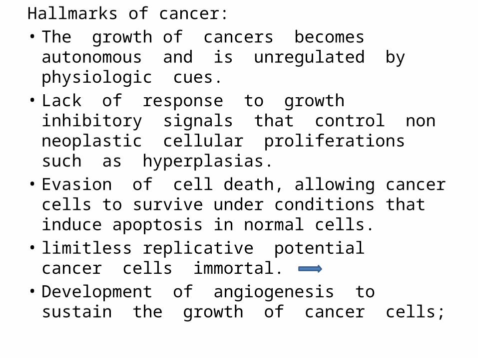
Hallmarks of cancer:• The growth of cancers becomes autonomous
and is unregulated by physiologic cues.• Lack of response to growth inhibitory
signals that control non neoplastic cellular proliferations such as hyperplasias.
• Evasion of cell death, allowing cancer cells to survive under conditions that induce apoptosis in normal cells.
• limitless replicative potential cancer cells immortal.
• Development of angiogenesis to sustain the growth of cancer cells;
• Ability to invade local tissues and spread to distant sites.
• Reprogramming of metabolic pathways—specifically, a switch to aerobic glycolysis even when there is abundant oxygen.
• Ability to evade the immune system.

Definition of Neoplasia
• “A neoplasm is an abnormal mass of tissue, the growth of which exceeds and is uncoordinated with that of the normal tissues and persists in the same excessive manner after cessation of the stimuli which evoked the change”
• Genetic changes• Autonomous • Clonal
Nomenclature
Benign tumors :– Will remain localized– Cannot spread to distant sites– Generally can be locally excised– Patient generally survives
- can produce more than localized lumps, they are responsible for serious disease.

Nomenclature
Malignant neoplasms:– Can invade and destroy adjacent structure– Can spread to distant sites(metastasize)– Cause death (if not treated )
• All tumors have two basic components:– Parechyma: made up of neoplastic cells– Stroma: made up of connective tissue,
blood vessels, and macrophages and lymphocytes.
• The neoplastic cells largely determine a tumor's behaviour & pathologic consequences.
• Growth and evolution is dependent on their stroma.
• Adequate stromal blood supply is requisite for the tumor cells to live and divide.
• The stromal connective tissue provides the structural framework essential for the growing cells.

• In some tumors, the stromal support is scant and so the neoplasm is soft & fleshy.
• In other cases the parenchymal cells stimulate the formation of an abundant collagenous stroma, referred to as desmoplasia.
• Demoplastic tumors—for example, some cancers of the female breast—are stony hard or scirrhous.
Nomenclature – Benign Tumors
• -oma = benign neoplasm• Examples: -Benign tumor arising in fibrous tissue:
Fibro + oma = Fibroma-Benign tumor arising in fatty tissue: Lipo + oma = lipoma
• Epithelial benign tumors are classified on the basis of :– The cell of origin – Microscopic pattern– Macroscopic pattern
.Benign epithelial tumors
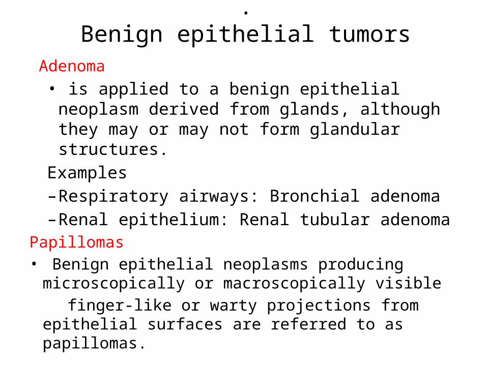
Adenoma • is applied to a benign epithelial neoplasm
derived from glands, although they may or may not form glandular structures.
Examples– Respiratory airways: Bronchial adenoma– Renal epithelium: Renal tubular adenoma
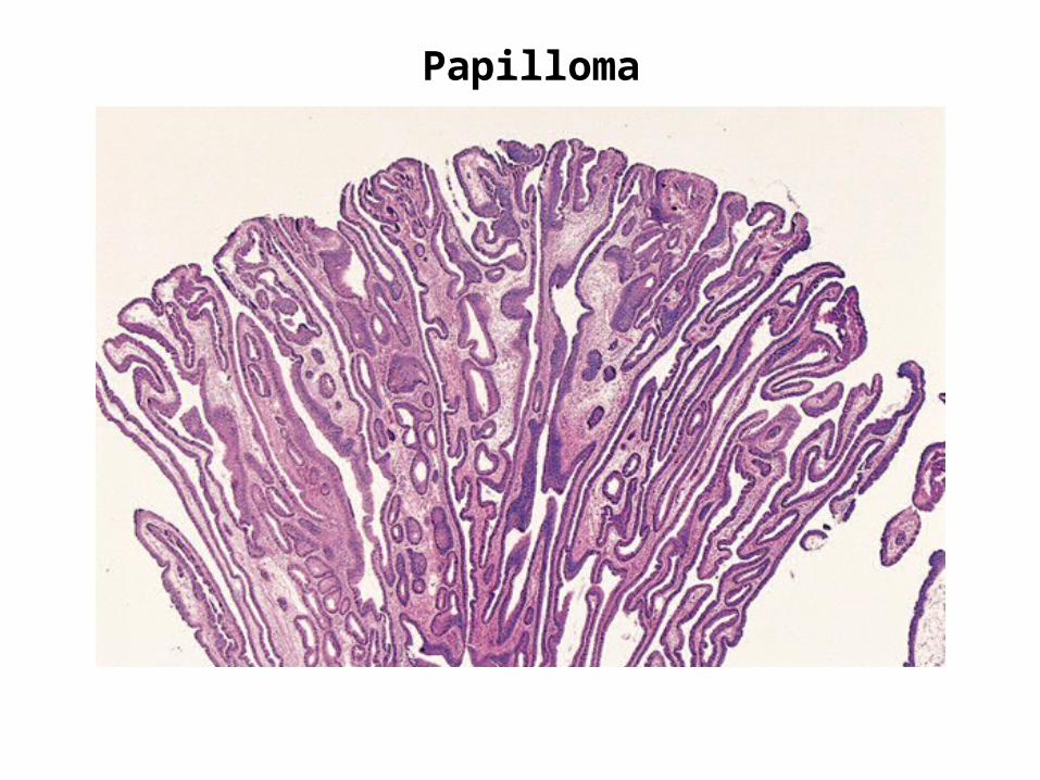
Papillomas• Benign epithelial neoplasms producing
microscopically or macroscopically visible finger-like or warty projections from epithelial
surfaces are referred to as papillomas.
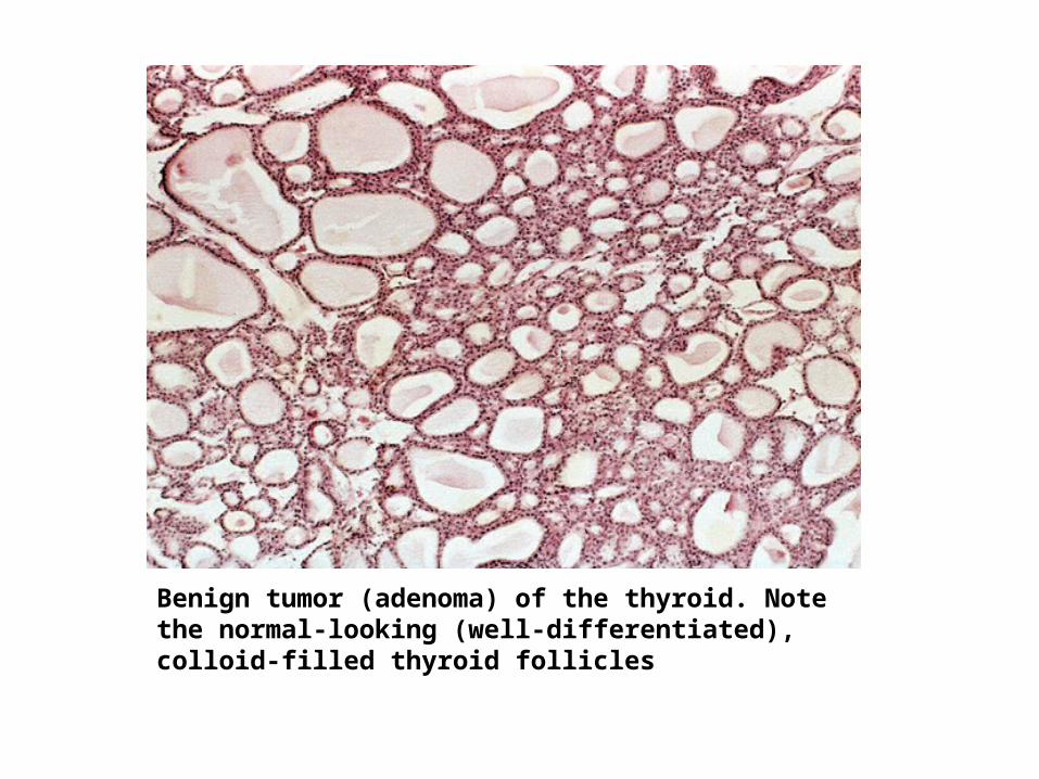
Benign tumor (adenoma) of the thyroid. Note the normal-looking (well-differentiated), colloid-filled thyroid follicles
.Benign epithelial tumors
Papillomas• Benign epithelial neoplasms producing
microscopically or macroscopically visible finger-like or warty projections from epithelial surfaces are referred to as papillomas.
Examples• Squamous epithelium: squamous papilloma • Large cystic masses, as in the ovary, are referred to
as cystadenomas. • Some tumors produce papillary patterns that
protrude into cystic spaces and are called papillary cystadenomas.
Papilloma
Papilloma
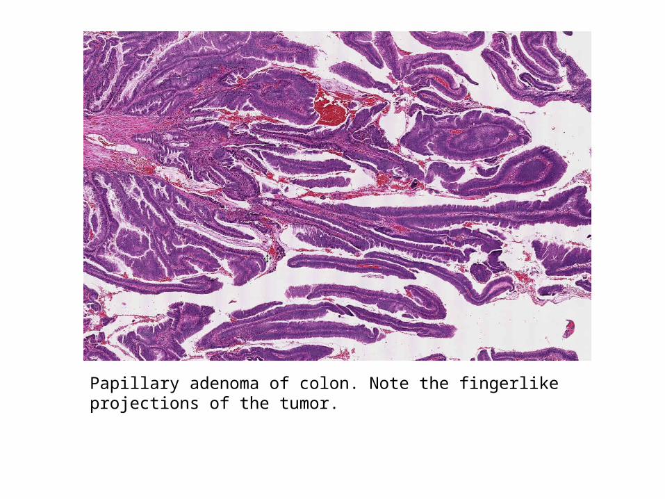
Papillary adenoma of colon. Note the fingerlike projections of the tumor.
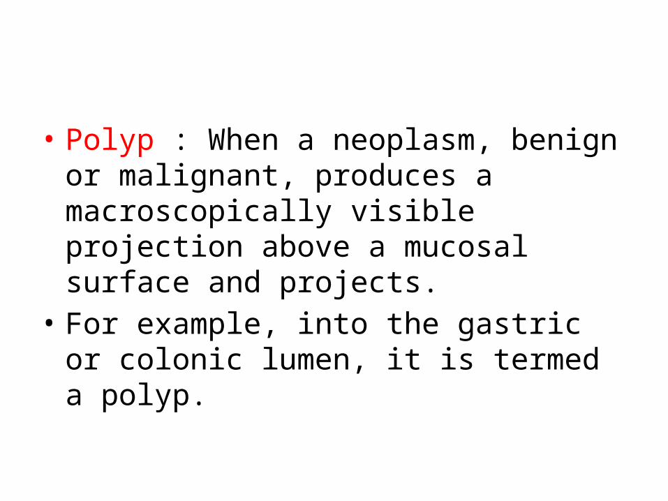
• Polyp : When a neoplasm, benign or malignant, produces a macroscopically visible projection above a mucosal surface and projects.
• For example, into the gastric or colonic lumen, it is termed a polyp.
Polyp
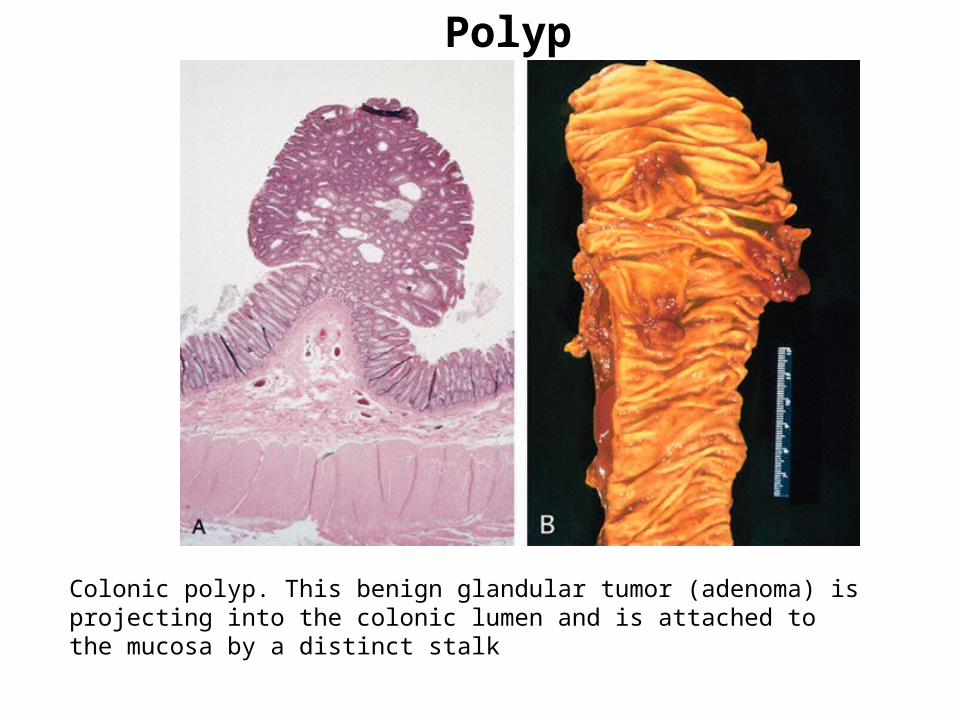
Colonic polyp. This benign glandular tumor (adenoma) is projecting into the colonic lumen and is attached to the mucosa by a distinct stalk
Nomenclature – Malignant Tumors
• Malignant neoplasms arising in “solid” mesenchymal tissues or its derivatives are called sarcomas.
• Cancer of fibrous tissue origin is a fibrosarcoma. • Malignant neoplasm composed of chondrocytes is
a chondrosarcoma
• Those arising from the mesenchymal cells of the blood are called leukemias or lymphomas.

• Malignant tumors arising from epithelial origin :derived from all three germ layers, are called carcinomas.
• Renal tubular epithelium (mesoderm) is a carcinoma.
• Arising in the skin (ectoderm) and • Lining epithelium of the gut (endoderm)

Carcinoma subdivided:• Carcinomas that grow in a glandular
pattern are called adenocarcinomas. tissue or organ of origin renal cell
adenocarcinoma.• That produce squamous cells are called
squamous cell carcinomas.• The tumor shows little or no
differentiation called poorly differentiated or undifferentiated carcinoma.

This view shows the transition from normal squamous epithelium into invasive carcinoma.
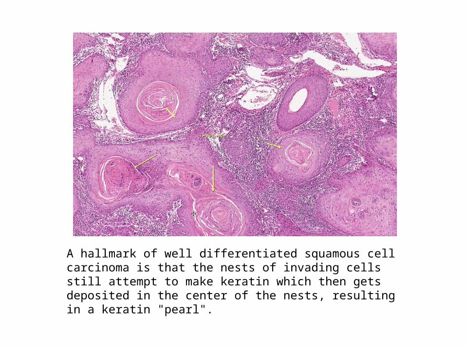
A hallmark of well differentiated squamous cell carcinoma is that the nests of invading cells still attempt to make keratin which then gets deposited in the center of the nests, resulting in a keratin "pearl".

Another characteristic of a well differentiated squamous cell carcinoma is that it still makes visible intercellular bridges.
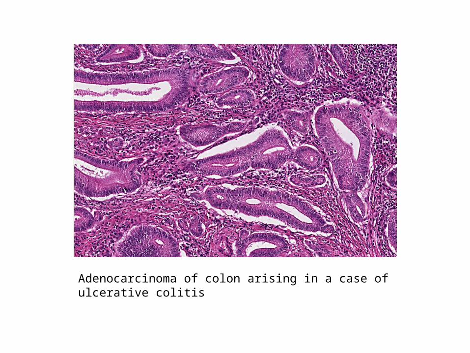
Adenocarcinoma of colon arising in a case of ulcerative colitis
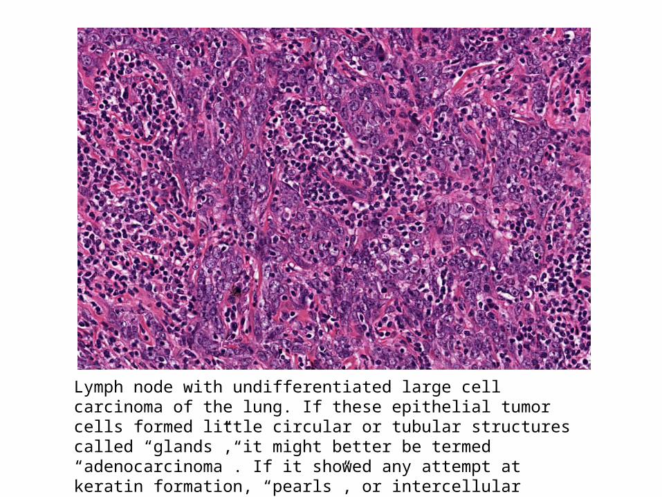
Lymph node with undifferentiated large cell carcinoma of the lung. If these epithelial tumor cells formed little circular or tubular structures called “glands”, it might better be termed “adenocarcinoma”. If it showed any attempt at keratin formation, “pearls”, or intercellular bridges between tumor cells, it might best be termed “squamous cell” carcinoma.
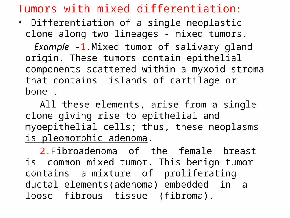
Tumors with mixed differentiation:• Differentiation of a single neoplastic clone along two
lineages - mixed tumors. Example -1.Mixed tumor of salivary gland origin.
These tumors contain epithelial components scattered within a myxoid stroma that contains islands of cartilage or bone .
All these elements, arise from a single clone giving rise to epithelial and myoepithelial cells; thus, these neoplasms is pleomorphic adenoma.
2.Fibroadenoma of the female breast is common mixed tumor. This benign tumor contains a mixture of proliferating ductal elements(adenoma) embedded in a loose fibrous tissue (fibroma).
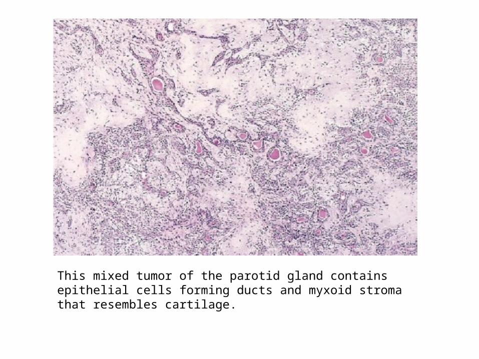
This mixed tumor of the parotid gland contains epithelial cells forming ducts and myxoid stroma that resembles cartilage.
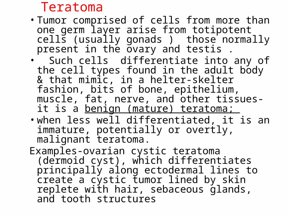
Teratoma• Tumor comprised of cells from more than one
germ layer arise from totipotent cells (usually gonads ) those normally present in the ovary and testis .
• Such cells differentiate into any of the cell types found in the adult body & that mimic, in a helter-skelter fashion, bits of bone, epithelium, muscle, fat, nerve, and other tissues- it is a benign (mature) teratoma;
• when less well differentiated, it is an immature, potentially or overtly, malignant teratoma.
Examples-ovarian cystic teratoma (dermoid cyst), which differentiates principally along ectodermal lines to create a cystic tumor lined by skin replete with hair, sebaceous glands, and tooth structures
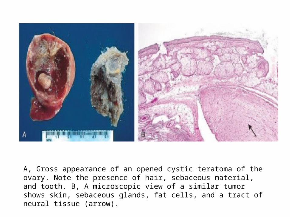
A, Gross appearance of an opened cystic teratoma of the ovary. Note the presence of hair, sebaceous material, and tooth. B, A microscopic view of a similar tumor shows skin, sebaceous glands, fat cells, and a tract of neural tissue (arrow).

• Aberrant differentiation (not true neoplasms):
Hamartomas :present as disorganized but benign-appearing masses composed of cells indigenous to the particular site.
Example : pulmonary chondroid harmatoma contains islands of disorganized, but histologically normal cartilage, bronchi, and vessels.
Choriostoma:congenital anomaly ectopic focus of normal tissue (heterotopia). Example, a small nodule of well-developed pancreatic substance found in the submucosa of the stomach, duodenum, or small intestine.

Tissue of Origin Benign MalignantCOMPOSED OF ONE PARENCHYMAL CELL TYPETumors of Mesenchymal OriginConnective tissue and derivatives Fibroma Fibrosarcoma
Chondroma Chondrosarcoma
Endothelial and Related TissuesBlood vessels Hemangioma AngiosarcomaLymph vessels Lymphangioma LymphangiosarcomaSynovium Synovial sarcomaMesothelium MesotheliomaBrain coverings Meningioma Invasive meningiomaBlood Cells and Related CellsHematopoietic cells LeukemiasLymphoid tissue LymphomasMuscleSmooth Leiomyoma LeiomyosarcomaStriated RhabdomyomaRhabdom
yosarcoma
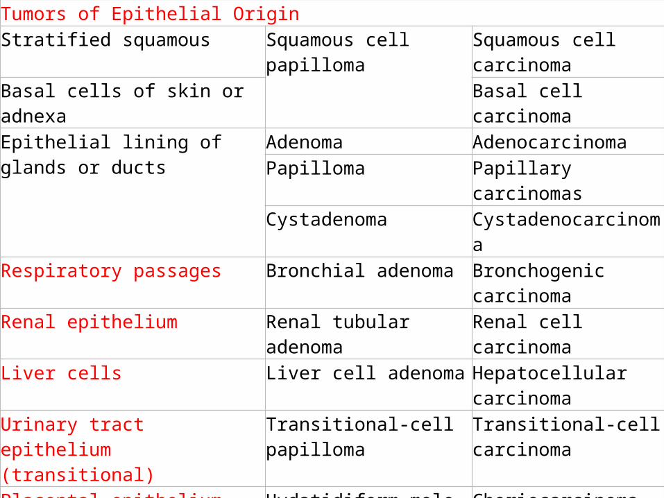
Tumors of Epithelial OriginStratified squamous Squamous cell papilloma Squamous cell carcinomaBasal cells of skin or adnexa Basal cell carcinomaEpithelial lining of glands or ducts Adenoma Adenocarcinoma
Papilloma Papillary carcinomasCystadenoma Cystadenocarcinoma
Respiratory passages Bronchial adenoma Bronchogenic carcinomaRenal epithelium Renal tubular adenoma Renal cell carcinomaLiver cells Liver cell adenoma Hepatocellular carcinomaUrinary tract epithelium (transitional)
Transitional-cell papilloma Transitional-cell carcinoma
Placental epithelium Hydatidiform mole ChoriocarcinomaTesticular epithelium (germ cells) SeminomaTumors of Melanocytes Nevus Malignant melanomaMORE THAN ONE NEOPLASTIC CELL TYPESalivary glands Pleomorphic adenoma (mixed
tumor of salivary origin)Malignant mixed tumor of salivary gland origin
MORE THAN ONE NEOPLASTIC CELL TYPE DERIVED FROM MORE THAN ONE GERM CELL Totipotential cells in gonads or in embryonic rests
Mature teratoma, dermoid cyst
Immature teratoma, teratocarcinoma
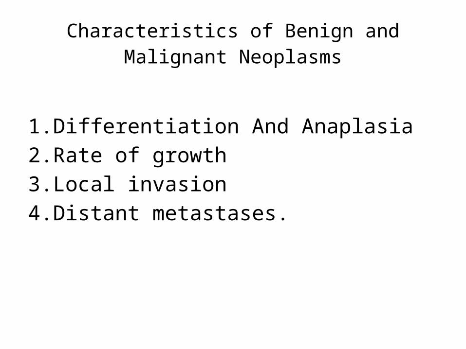
Characteristics of Benign and Malignant Neoplasms
1.Differentiation And Anaplasia2.Rate of growth 3.Local invasion4.Distant metastases.

Differentiation
• Refers to extent to which neoplastic parenchymal cells resemble the corresponding normal parenchymal cells, both morphologically and functionally.
1.Well differentiated neoplasm– Resembles mature cells of tissue of origin
2.Poorly diffentiated neoplasm– Composed of primitive cells with little diffrerentiation
3.Undifferentiated or “anaplastic” tumor

• Benign neoplasms are composed of well differentiated cells that closely resemble their normal counterparts.
• Mitoses are usually rare• Ex. A lipoma is made up of mature fat cells
laden with cytoplasmic lipid vacuoles. A chondroma is made up of mature
cartilage cells that synthesize their usual cartilaginous matrix—evidence of morphologic and functional differentiation.

Leiomyoma of the uterus. This benign, well-differentiated tumor contains interlacing bundles of neoplastic smooth muscle cells that are virtually identical in appearance to normal smooth muscle cells in the myometrium.
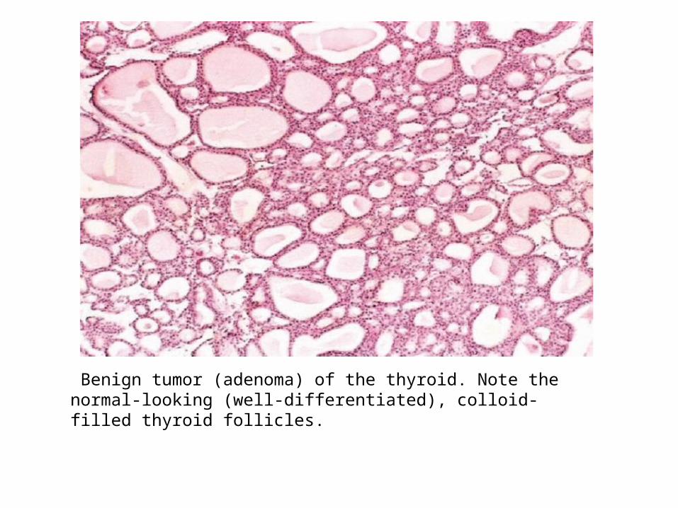
Benign tumor (adenoma) of the thyroid. Note the normal-looking (well-differentiated), colloid-filled thyroid follicles.

• Malignant neoplasms are characterized by a wide range of parenchymal cell differentiation, from surprisingly well differentiated to completely undifferentiated.
• The amount of stromal connective tissue determine the consistency of a neoplasm.
• Certain cancers induce a dense, abundant fibrous stroma (desmoplasia)- hard, so called scirrhous tumors.
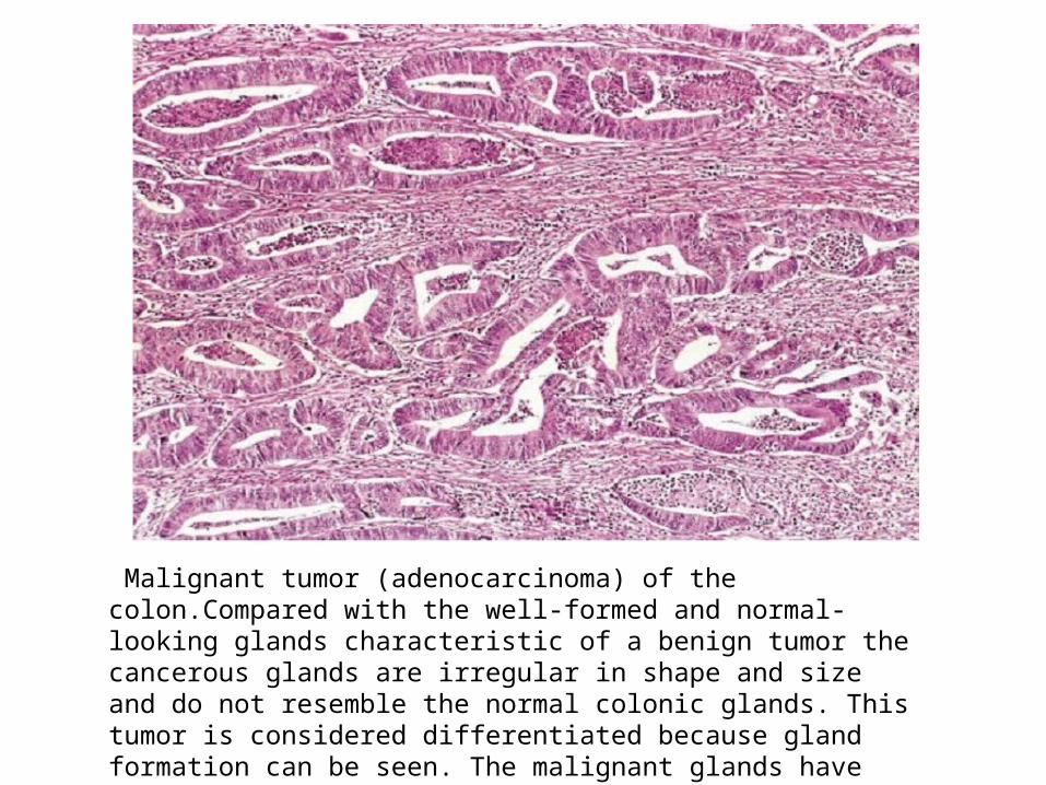
Malignant tumor (adenocarcinoma) of the colon.Compared with the well-formed and normal-looking glands characteristic of a benign tumor the cancerous glands are irregular in shape and size and do not resemble the normal colonic glands. This tumor is considered differentiated because gland formation can be seen. The malignant glands have invaded the muscular layer of the colon.
• Malignant neoplasms that are composed of undifferentiated cells are said to be anaplastic.
• The term anaplasia literally means “back ward formation”implying dedifferentiation, or loss of the structural and functional differentiation of normal cells.
• However, that most cancers do not represent “reverse differentiation” of mature normal cells but, in fact, arise from less mature cells with “stem-cell-like” properties, such as tissue stem cells .

Anaplasia
• Pleomorphism– Size– shape
• Abnormal nuclear morphology– Hyperchromasia– High nuclear cytoplasmic ratio– Coarsely Chromatin ,clumping– Prominent nucleoli
• Mitoses– Mitotic rate– Atypical bizzare mitotic figure –tripolar ,multipolar ,qudripolar spindle
• Loss of polarity -anaplastic cell orientation disturbed

Anaplastic tumor of the skeletal muscle (rhabdomyosarcoma). Note the marked cellular and nuclear pleomorphism, hyperchromatic nuclei, and tumor giant cells.

Anaplastic tumor showing cellular and nuclear variation in size and shape. The prominent cell in the center field has an abnormal tripolar spindle.

Metaplasia
• Is defined as the replacement of one type of cell with another type.
• Is nearly always found in association with tissue damage, repair, and regeneration.
• Often the replacing cell type is more suited to a change in environment.
• For e.g., gastroesophageal reflux damages the squamous epithelium of the esophagus, leading to its replacement by glandular (gastric or intestinal) epithelium,& suited to the acidic environment.
Dysplasia• Is a term that means disordered growth.• Dysplasia often occurs in metaplastic
epithelium, but not all metaplastic epithelium is dysplastic.
• Dysplasia is occure principally in epithelia & • Is characterized by changes that include a loss
in the uniformity of the individual cells as well as a loss in their architectural orientation.
Dysplasia
• Cells exhibit pleomorphism & contain large hyperchromatic nuclei with a high nuclear to-cytoplasmic ratio.
• E.g. in squamous epithelium the usual progressive maturation of tall cells in the basal layer to flattened squamous on the surface may be lost and replaced by a scrambling of dark basal-appearing cells throughout the epithelium.
• Mitotic figures - more abundant than usual.
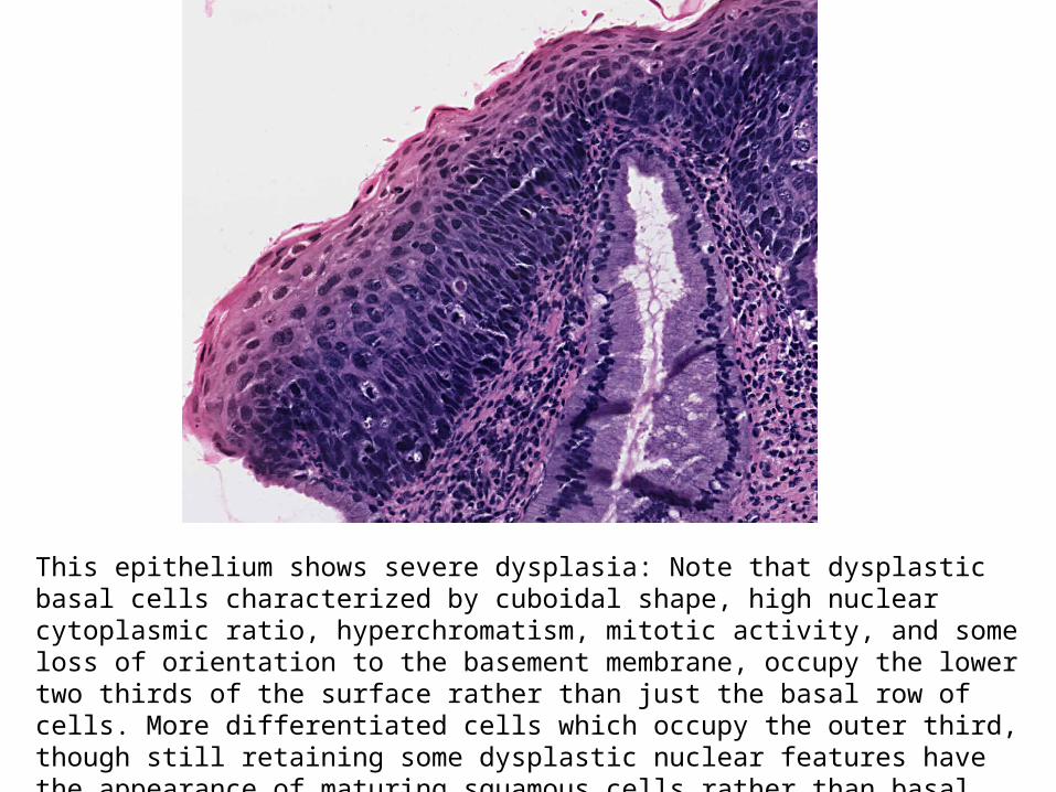
This epithelium shows severe dysplasia: Note that dysplastic basal cells characterized by cuboidal shape, high nuclear cytoplasmic ratio, hyperchromatism, mitotic activity, and some loss of orientation to the basement membrane, occupy the lower two thirds of the surface rather than just the basal row of cells. More differentiated cells which occupy the outer third, though still retaining some dysplastic nuclear features have the appearance of maturing squamous cells rather than basal cells, and eventually become flattened on the surface

Dysplasia• When dysplastic changes are marked & involve the entire
thickness of the epithelium but the lesion remains confined by the basement membrane,
• It is considered a preinvasive neoplasm and is referred to as carcinoma in situ
• Once the tumor cells breach the basement membrane, the tumor is said to be invasive.
• Dysplastic changes are found adjacent to foci of invasive carcinoma, & in some situations, such as in long-term cigarette smokers and persons with Barrett esophagus, severe epithelial dysplasia frequently antedates the appearance of cancer.
• Does not necessarily progress to cancer. Mild to moderate changes that do not involve the entire thickness of epithelium may be reversible.
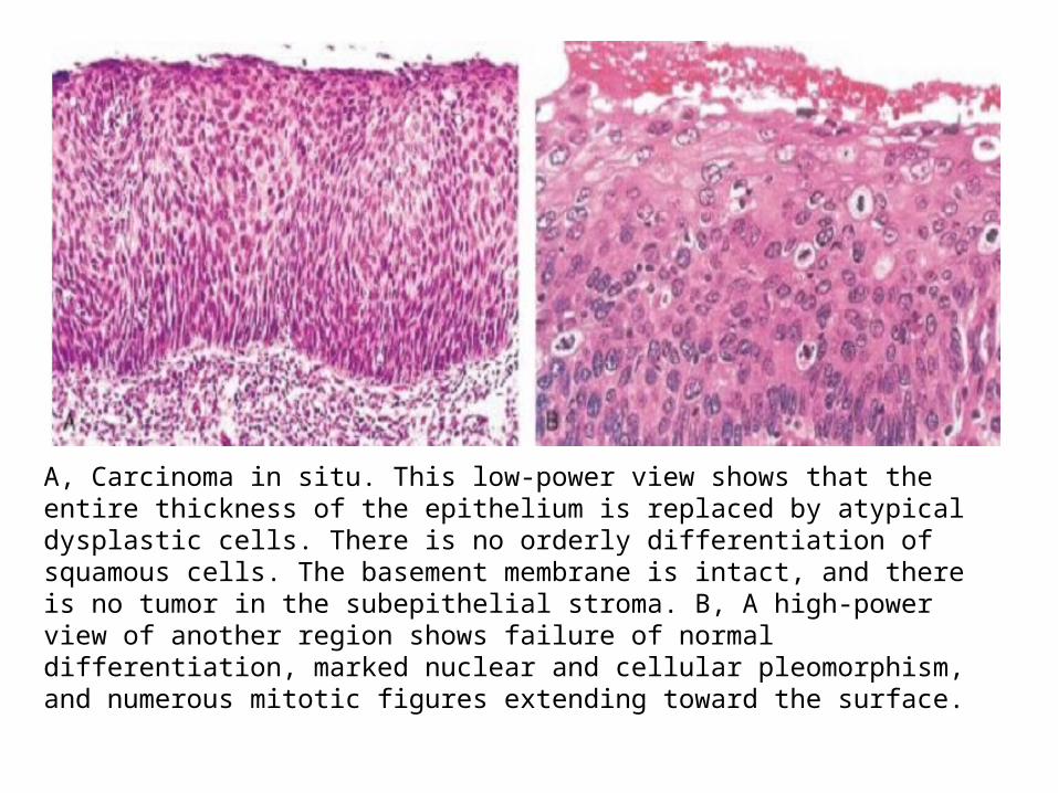
A, Carcinoma in situ. This low-power view shows that the entire thickness of the epithelium is replaced by atypical dysplastic cells. There is no orderly differentiation ofsquamous cells. The basement membrane is intact, and there is no tumor in the subepithelial stroma. B, A high-power view of another region shows failure of normal differentiation, marked nuclear and cellular pleomorphism, and numerous mitotic figures extending toward the surface.
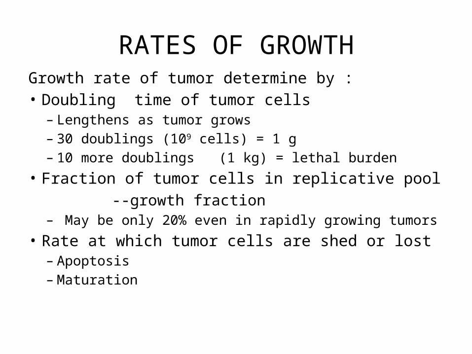
RATES OF GROWTHGrowth rate of tumor determine by :• Doubling time of tumor cells
– Lengthens as tumor grows– 30 doublings (109 cells) = 1 g – 10 more doublings (1 kg) = lethal burden
• Fraction of tumor cells in replicative pool --growth fraction
– May be only 20% even in rapidly growing tumors• Rate at which tumor cells are shed or lost
– Apoptosis– Maturation

Schematic representation of tumor growth. As the cell population expands, a progressively higher percentage of tumor cells leaves the replicative pool by reversion to G0, differentiation, and death.
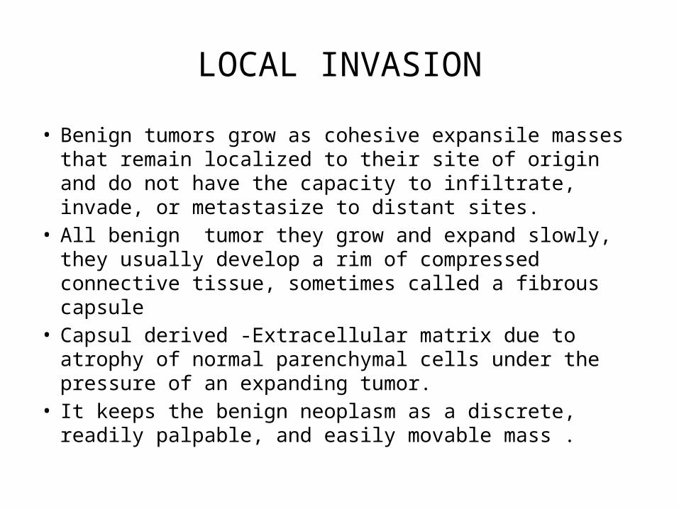
LOCAL INVASION
• Benign tumors grow as cohesive expansile masses that remain localized to their site of origin and do not have the capacity to infiltrate, invade, or metastasize to distant sites.
• All benign tumor they grow and expand slowly, they usually develop a rim of compressed connective tissue, sometimes called a fibrous capsule
• Capsul derived -Extracellular matrix due to atrophy of normal parenchymal cells under the pressure of an expanding tumor.
• It keeps the benign neoplasm as a discrete, readily palpable, and easily movable mass .
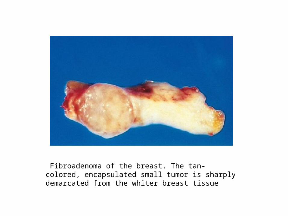
Fibroadenoma of the breast. The tan-colored, encapsulated small tumor is sharply demarcated from the whiter breast tissue
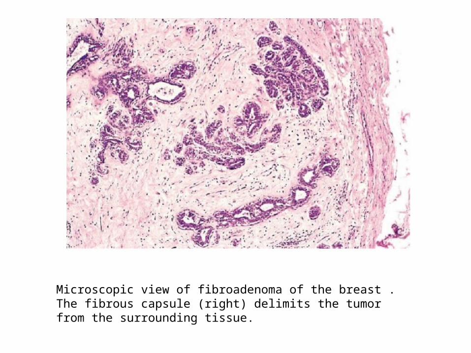
Microscopic view of fibroadenoma of the breast . The fibrous capsule (right) delimits the tumor from the surrounding tissue.
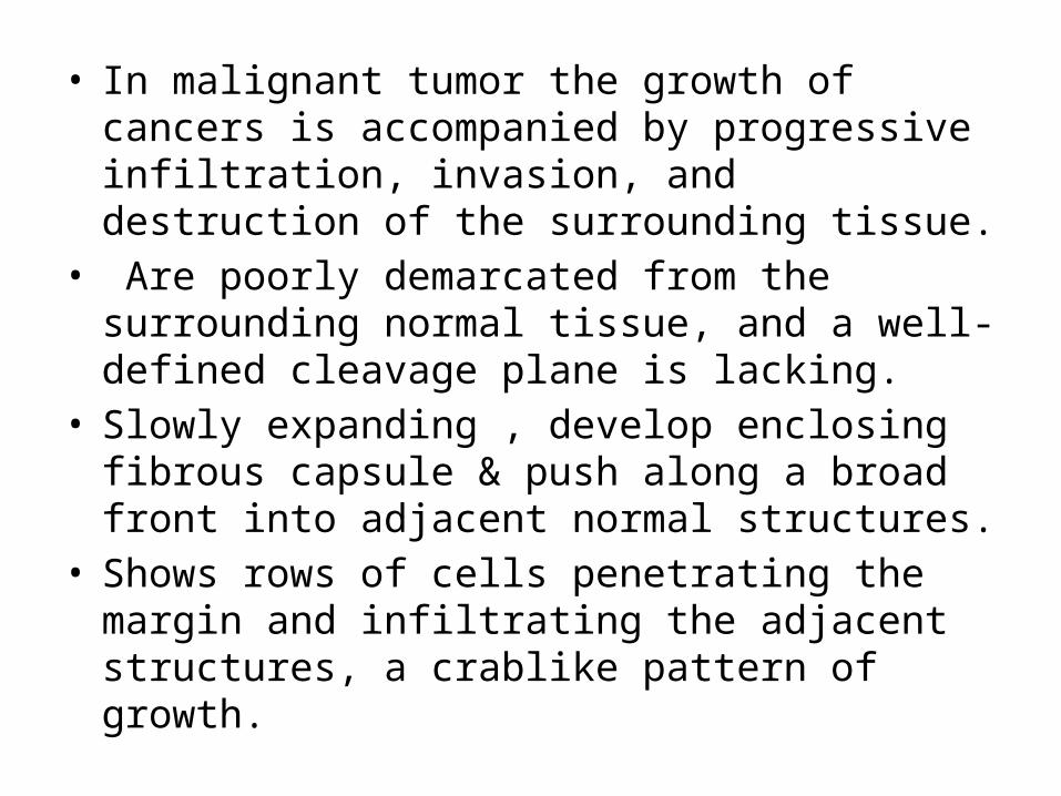
• In malignant tumor the growth of cancers is accompanied by progressive infiltration, invasion, and destruction of the surrounding tissue.
• Are poorly demarcated from the surrounding normal tissue, and a well-defined cleavage plane is lacking.
• Slowly expanding , develop enclosing fibrous capsule & push along a broad front into adjacent normal structures.
• Shows rows of cells penetrating the margin and infiltrating the adjacent structures, a crablike pattern of growth.

Cut section of an invasive ductal carcinoma of the breast. The lesion is retracted, infiltrating the surrounding breast substance, and would be stony hard on palpation.
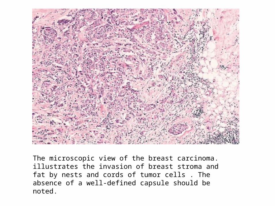
The microscopic view of the breast carcinoma. illustrates the invasion of breast stroma and fat by nests and cords of tumor cells . The absence of a well-defined capsule should be noted.
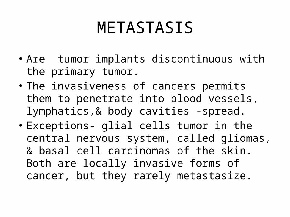
METASTASIS
• Are tumor implants discontinuous with the primary tumor.
• The invasiveness of cancers permits them to penetrate into blood vessels, lymphatics,& body cavities -spread.
• Exceptions- glial cells tumor in the central nervous system, called gliomas, & basal cell carcinomas of the skin. Both are locally invasive forms of cancer, but they rarely metastasize.
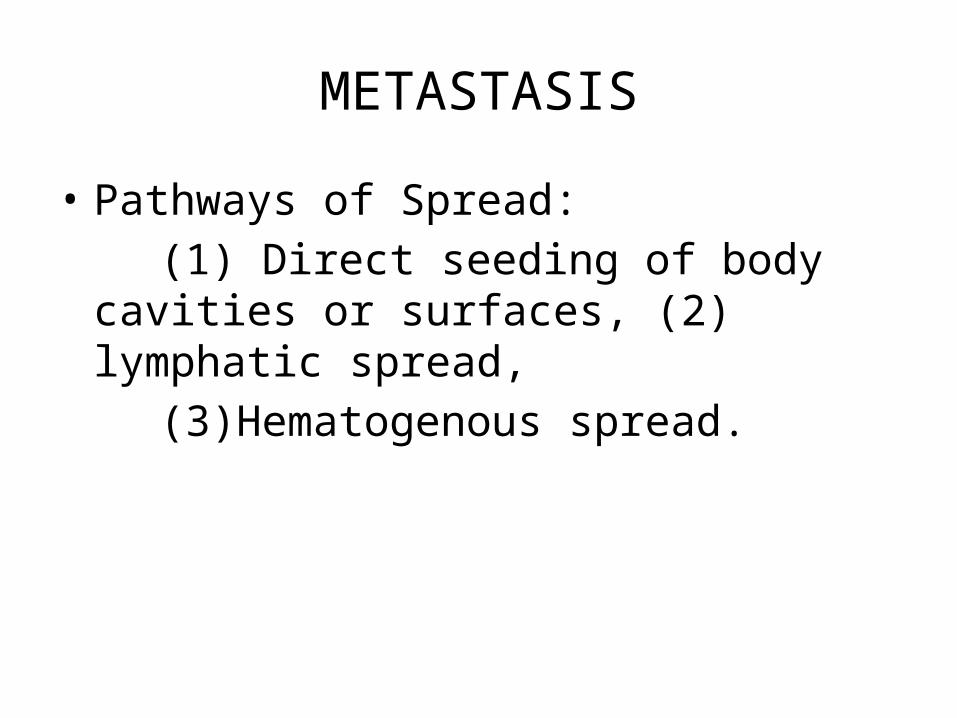
METASTASIS
• Pathways of Spread: (1) Direct seeding of body cavities or surfaces,
(2) lymphatic spread, (3)Hematogenous spread.

Direct seeding of body cavities or surfaces:• Most often involved is the peritoneal cavity but any
other cavity—pleural, pericardial, subarachnoid, and joint space—may be affected
• Such seeding is particularly characteristic of carcinomas arising in the ovaries, all peritoneal surfaces become coated with a heavy layer of cancerous glaze.
• Mucus-secreting appendiceal carcinomas fill the peritoneal cavity with a gelatinous neoplastic mass called as pseudomyxoma peritonei.

Colon carcinoma invading pericolonic adipose tissue.
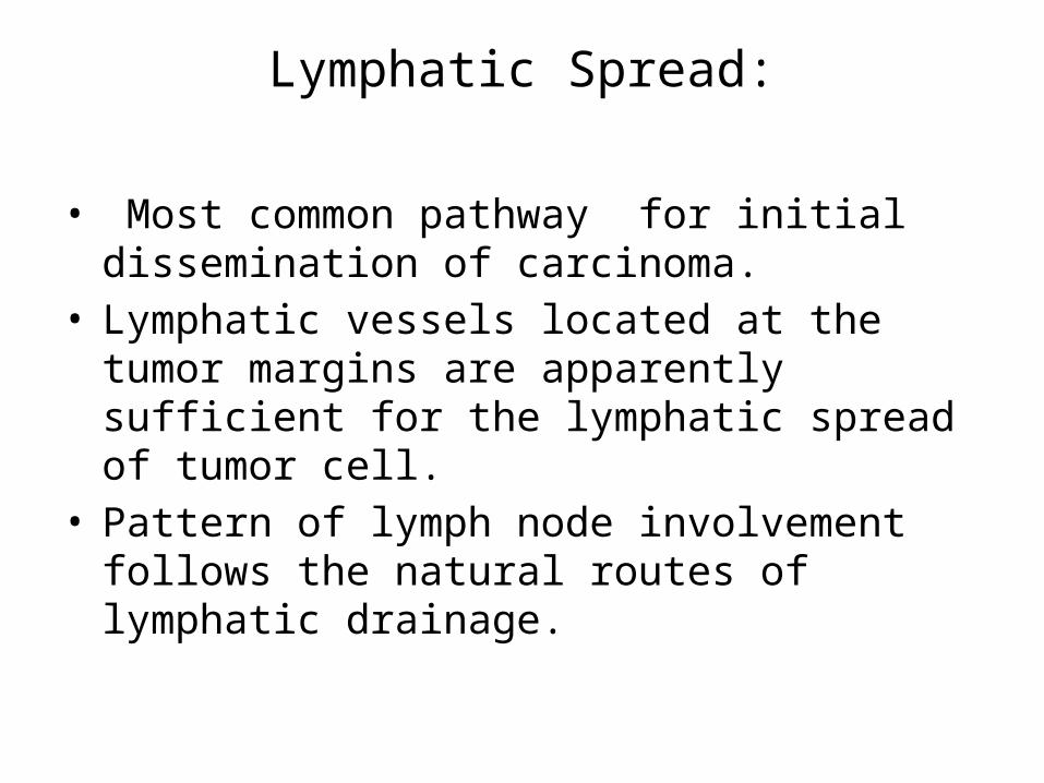
Lymphatic Spread:
• Most common pathway for initial dissemination of carcinoma.
• Lymphatic vessels located at the tumor margins are apparently sufficient for the lymphatic spread of tumor cell.
• Pattern of lymph node involvement follows the natural routes of lymphatic drainage.

Axillary lymph node with metastatic breast carcinoma. The subcapsular sinus (top) is distended with tumor cells. Nests of tumor cells have also invaded the subcapsular cortex.
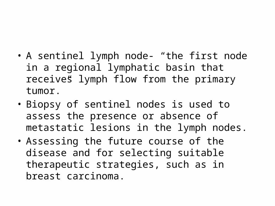
• A sentinel lymph node- “the first node in a regional lymphatic basin that receives lymph flow from the primary tumor.”
• Biopsy of sentinel nodes is used to assess the presence or absence of metastatic lesions in the lymph nodes.
• Assessing the future course of the disease and for selecting suitable therapeutic strategies, such as in breast carcinoma.
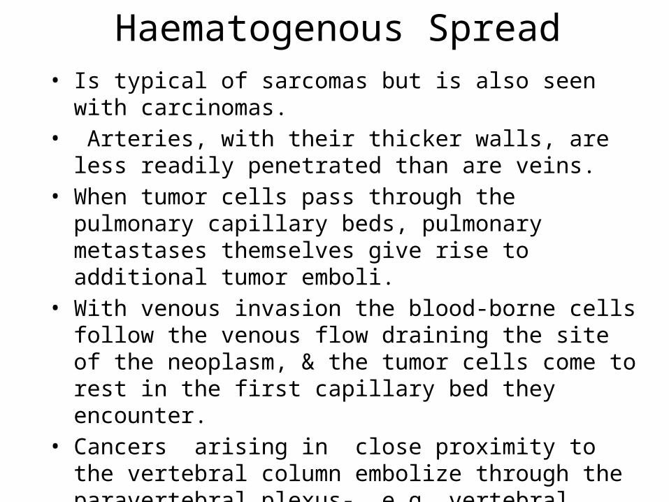
Haematogenous Spread• Is typical of sarcomas but is also seen with carcinomas.• Arteries, with their thicker walls, are less readily penetrated
than are veins.• When tumor cells pass through the pulmonary capillary beds,
pulmonary metastases themselves give rise to additional tumor emboli.
• With venous invasion the blood-borne cells follow the venous flow draining the site of the neoplasm, & the tumor cells come to rest in the first capillary bed they encounter.
• Cancers arising in close proximity to the vertebral column embolize through the paravertebral plexus- e.g. vertebral metastases of carcinomas of the thyroid and prostate.
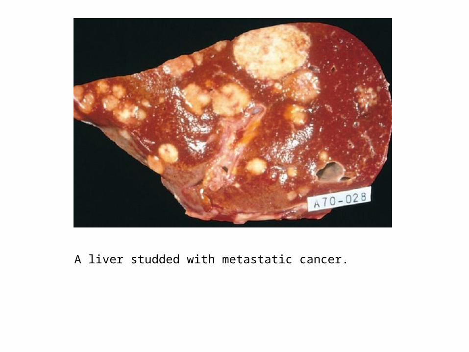
A liver studded with metastatic cancer.

Microscopic view of liver metastasis. A pancreatic adenocarcinoma has formed a metastatic nodule in the liver.

Benign vs Malignant FeaturesFeature Benign Malignant
Rate of growth Progressive but slow. Mitoses few and normal
Variable. Mitoses more frequent and may be abnormal
Differentiation Well differentiated
Some degree of anaplasia
Local invasion Cohesive growth. Capsule & BM not breached
Poorly cohesive and infiltrative.
Metastasis Absent May occur
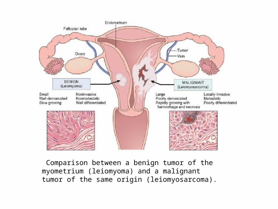
Comparison between a benign tumor of the myometrium (leiomyoma) and a malignant tumor of the same origin (leiomyosarcoma).
Epidemiology
• Cancer is a disorder of cell growth and behavior, its ultimate cause at the cellular and subcellular levels
• Study of cancer patterns - origins of cancer. Epidemiologic studies have established the causative link-
- Between smoking and lung cancer, -Comparison of diet and cancer rates in the
Western world -Africa has implicated high dietary fat and low fiber
in the development of colon cancer.

GEOGRAPHIC AND ENVIRONMENTAL FACTORS
• Sun exposure– Melanomas 6x incidence New Zealand vs Iceland– Blacks have low incidence of melanoma
• Smoking and alcohol abuse• Body mass
– Overweight = 50% increase in cancer
• Environmental vs racial factors– Japanese immigrants to USA
• Viral exposure– Human papilloma virus (HPV) and cervical cancer– Hepatitis B virus (HBV) and liver cancer (Africa)– Epstein-Barr Virus (EBV) and lymphoma

Predisposing Factors for Cancer• Age
– Most cancers occur in persons ≥ 55 years– Childhood cancers
• Leukemias & CNS neoplasms• Bone tumors
• Genetic predispostion– Familial cancer syndromes
• Early age at onset• Two or more primary relatives with the cancer• Multiple or bilateral tumors
– Polymorphisms that metabolize procarcinogens, e.g., nitrites
• Nonhereditary predisposing conditions– Chronic inflammation– Precancerous conditions
• Chronic ulcerative colitis• Atrophic gastritis of pernicious anemia• Leukoplakia of mucous membranes

THANK YOU.....


Related Documents