Neglected Time: Impaired Temporal Perception of Multisecond Intervals in Unilateral Neglect James Danckert 1 , Susanne Ferber 2 , Carson Pun 2 , Carol Broderick 1 , Christopher Striemer 1 , Sherry Rock 3 , and Dwight Stewart 1,4 Abstract & Recent neuroimaging and neuropsychological studies have suggested that the right hemisphere, particularly frontal re- gions, is important for the perception of the passage of time. We examined the ability to estimate durations of up to 60 sec in a group of eight patients with unilateral neglect. When es- timating multisecond intervals, neglect patients grossly un- derestimated all durations. On average, healthy controls (HC) demonstrated reasonably accurate estimates of all durations tested. Although the right hemisphere lesioned control pa- tients without neglect also tended to underestimate durations, these underestimations were significantly better than the per- formance of the neglect group. These findings suggest a piv- otal role for a right hemisphere fronto-parietal network in the accurate perception of multisecond durations. Furthermore, these findings add to a growing body of literature suggesting that neglect cannot be understood simply in terms of a bias in orienting attention to one side of space. Additional deficits of the kind demonstrated here are likely to be crucial in deter- mining the nature and extent of the loss of conscious aware- ness for contralesional events. & INTRODUCTION Lesions of the right posterior parietal or superior tem- poral cortex often lead to the disorder of unilateral neglect in which patients fail to attend to or respond to stimuli in the contralesional—in this case left—side of space (Danckert & Ferber, 2006; Mort et al., 2003; Karnath, Ferber, & Himmelbach, 2001). Patients may fail to eat food from the left half of a plate, fail to groom the left side of their body or face, and will bump into objects on the left side of space (see Danckert & Ferber, 2006 for review). Neglect is not typically considered a dis- order of primary perceptual or motor capacities, but is instead thought to be due to deficient orienting of at- tention toward contralesional space (Husain & Rorden, 2003; Driver & Mattingley, 1998). More recent findings have suggested that, in addition to impaired attentional orienting—which may manifest itself as a severe bias toward processing ipsilesional stimuli—neglect patients suffer from impairments to spatial working memory and saccadic or spatial remapping (Ferber & Danckert, 2006; Malhotra et al., 2005; Malhotra, Mannan, Driver, & Husain, 2004; Pisella, Berbevoric, & Mattingley, 2004; Pisella & Mattingley, 2004; Wojciulik, Rorden, Clarke, Husain, & Driver, 2004; Husain et al., 2001). This strongly suggests that a simple attentional bias toward ipsilesional space is unlikely to be the only factor needed to produce the cardinal symptom of neglect—a loss of awareness for contralesional events. Furthermore, several authors have demonstrated im- pairments in temporal processing in neglect. One exam- ple of a spatio-temporal deficit in neglect comes from the so-called temporal order judgment (TOJ) task in which subjects must determine which of two lateralized stimuli was presented first (Berberovic, Pisella, Morris, & Mattingley, 2004; Robertson, Mattingley, Rorden, & Driver, 1998; Rorden, Mattingley, Karnath, & Driver, 1997; note: Rorden et al., 1997 investigated patients with extinction). In this task, the left stimulus can precede the right (or vice versa) by varying amounts of time, and the two targets can also be presented simultaneously. For healthy individuals, the subjective point of simulta- neity, in which they respond ‘‘left first’’ (or ‘‘right first’’) on around 50% of trials, coincides nicely with the objective point of simultaneity (Stelmach & Herdman, 1991). In contrast, for neglect patients, a bias is seen in TOJs such that the left stimulus must precede the right by more than 250 msec before the patient accurately reports that it was presented first (Berberovic et al., 2004; Robertson et al., 1998). However, the delayed processing of the left stimulus may be due to various sources of error, including a difficulty in disengaging attention from the ipsilesional side in a timely manner. Demonstrations of nonspatial impairments in neglect patients—impairments that could be taken to reflect to some extent impaired temporal processing—have 1 University of Waterloo, Canada, 2 University of Toronto, Canada, 3 Freeport Rehabilitation Hospital, Kitchener, ON, Canada, 4 Grand River Hospital, Kitchener, ON, Canada D 2007 Massachusetts Institute of Technology Journal of Cognitive Neuroscience 19:10, pp. 1706–1720

Welcome message from author
This document is posted to help you gain knowledge. Please leave a comment to let me know what you think about it! Share it to your friends and learn new things together.
Transcript
Neglected Time: Impaired Temporal Perceptionof Multisecond Intervals in Unilateral Neglect
James Danckert1, Susanne Ferber2, Carson Pun2, Carol Broderick1,Christopher Striemer1, Sherry Rock3, and Dwight Stewart1,4
Abstract
& Recent neuroimaging and neuropsychological studies havesuggested that the right hemisphere, particularly frontal re-gions, is important for the perception of the passage of time.We examined the ability to estimate durations of up to 60 secin a group of eight patients with unilateral neglect. When es-timating multisecond intervals, neglect patients grossly un-derestimated all durations. On average, healthy controls (HC)demonstrated reasonably accurate estimates of all durationstested. Although the right hemisphere lesioned control pa-tients without neglect also tended to underestimate durations,
these underestimations were significantly better than the per-formance of the neglect group. These findings suggest a piv-otal role for a right hemisphere fronto-parietal network in theaccurate perception of multisecond durations. Furthermore,these findings add to a growing body of literature suggestingthat neglect cannot be understood simply in terms of a bias inorienting attention to one side of space. Additional deficits ofthe kind demonstrated here are likely to be crucial in deter-mining the nature and extent of the loss of conscious aware-ness for contralesional events. &
INTRODUCTION
Lesions of the right posterior parietal or superior tem-poral cortex often lead to the disorder of unilateralneglect in which patients fail to attend to or respondto stimuli in the contralesional—in this case left—side ofspace (Danckert & Ferber, 2006; Mort et al., 2003;Karnath, Ferber, & Himmelbach, 2001). Patients may failto eat food from the left half of a plate, fail to groom theleft side of their body or face, and will bump into objectson the left side of space (see Danckert & Ferber, 2006for review). Neglect is not typically considered a dis-order of primary perceptual or motor capacities, but isinstead thought to be due to deficient orienting of at-tention toward contralesional space (Husain & Rorden,2003; Driver & Mattingley, 1998). More recent findingshave suggested that, in addition to impaired attentionalorienting—which may manifest itself as a severe biastoward processing ipsilesional stimuli—neglect patientssuffer from impairments to spatial working memoryand saccadic or spatial remapping (Ferber & Danckert,2006; Malhotra et al., 2005; Malhotra, Mannan, Driver,& Husain, 2004; Pisella, Berbevoric, & Mattingley,2004; Pisella & Mattingley, 2004; Wojciulik, Rorden,Clarke, Husain, & Driver, 2004; Husain et al., 2001). Thisstrongly suggests that a simple attentional bias toward
ipsilesional space is unlikely to be the only factor neededto produce the cardinal symptom of neglect—a loss ofawareness for contralesional events.
Furthermore, several authors have demonstrated im-pairments in temporal processing in neglect. One exam-ple of a spatio-temporal deficit in neglect comes fromthe so-called temporal order judgment (TOJ) task inwhich subjects must determine which of two lateralizedstimuli was presented first (Berberovic, Pisella, Morris,& Mattingley, 2004; Robertson, Mattingley, Rorden, &Driver, 1998; Rorden, Mattingley, Karnath, & Driver, 1997;note: Rorden et al., 1997 investigated patients withextinction). In this task, the left stimulus can precedethe right (or vice versa) by varying amounts of time, andthe two targets can also be presented simultaneously.For healthy individuals, the subjective point of simulta-neity, in which they respond ‘‘left first’’ (or ‘‘right first’’)on around 50% of trials, coincides nicely with theobjective point of simultaneity (Stelmach & Herdman,1991). In contrast, for neglect patients, a bias is seen inTOJs such that the left stimulus must precede the rightby more than 250 msec before the patient accuratelyreports that it was presented first (Berberovic et al.,2004; Robertson et al., 1998). However, the delayedprocessing of the left stimulus may be due to varioussources of error, including a difficulty in disengagingattention from the ipsilesional side in a timely manner.
Demonstrations of nonspatial impairments in neglectpatients—impairments that could be taken to reflectto some extent impaired temporal processing—have
1University of Waterloo, Canada, 2University of Toronto, Canada,3Freeport Rehabilitation Hospital, Kitchener, ON, Canada, 4GrandRiver Hospital, Kitchener, ON, Canada
D 2007 Massachusetts Institute of Technology Journal of Cognitive Neuroscience 19:10, pp. 1706–1720
come from studies examining auditory attention (e.g.,sustained auditory attention, frequency and pitch discrim-ination; Cusack, Carlyon, & Robertson, 2000; Robertsonet al., 1997) or the allocation of attention in the tem-poral domain (e.g., using the attentional blink task;Hillstrom, Husain, Shapiro, & Rorden, 2004; Snyder &Chatterjee, 2004; Shapiro, Hillstrom, & Husain, 2002;Husain, Shapiro, Martin, & Kennard, 1997; Chun &Potter, 1995; Raymond, Shapiro, & Arnell, 1992; seeSchneider, 1999 for review; see also Rizzo, Akutsu, &Dawson, 2001 for a demonstration of an altered atten-tional blink in patients with discrete cortical lesionswithout neglect). It is important to emphasize here thatthere is a difference both in behavioral and neural termsbetween attention to time, the allocation of attentionover time, and the perception of time per se (Buhusi &Meck, 2005; Coull, 2004). Although the first two capac-ities will undoubtedly rely on and interact with mecha-nisms of attentional selection and temporal perception,the perception of the passage of time itself is also likelyto depend on distinct behavioral and neural propertiesfrom those networks and mechanisms involved in at-tention (see Buhusi & Meck, 2005, for a review). For ex-ample, although an increased attentional blink may notentail any specific spatial biases, it may, nevertheless, beindicative of a more fundamental problem of disen-gaging attention evident in neglect patients, rather thanreflecting any impairment of the perception of the pas-sage of time itself. Finally, in a recent review, Husain andRorden (2003) suggested that deficits in the temporalallocation of attention (evident in such tasks as theattentional blink) merely exacerbated the spatial deficitsevident in neglect. This point has also been made byRobertson et al. (1997) who demonstrated a strong cor-relation between nonlateralized deficits of sustained au-ditory attention and spatial biases, as demonstrated onclinical tests of neglect. Another related possibility isthat the temporal deficits in attention are related toreduced levels of tonic arousal in neglect patients. Thishypothesis gains some support from the finding thata phasic alerting cue (a loud, spatially noninformativetone) can improve TOJs in neglect patients (Robertsonet al., 1998).
As mentioned above, exploration of the temporal dy-namics of the allocation of attention in and of itself doesnot explicitly address the capacity to perceive the pas-sage of time. Although we appreciate that the percep-tion of the passage of time is necessarily distinct fromperceiving basic physical properties of events in ourenvironment (i.e., given that we do not perceive timeitself per se), there are, nevertheless, commonly usedtasks to assess the perception of the duration of phys-ically perceivable events. More specifically, one task in-volves the discrimination of various stimulus durationswith reference to a standard duration (see Buhusi &Meck, 2005; Wearden, 1999, for a review). For example,the subject may be presented with a pair of tones sep-
arated by a standard temporal duration (usually in themillisecond range) followed some time later by a secondpair of tones that may be separated by a longer, shorter,or identical temporal duration. The task then is to deter-mine whether the duration between the two tones inthe second pair was longer or shorter than the standardduration. Typically, poorer discrimination performanceis seen for durations that are longer than the standardwhen contrasted with durations that are shorter thanthe standard (see Wearden, 1999, for a review). Using aprocedure similar to this, Harrington, Haaland, andKnight (1998) found that patients with right hemispherebrain damage were more impaired than patients withleft hemisphere damage. The nature of the impairmentwas such that difference thresholds were elevated in thepatients, indicating that durations longer than the stan-dard were more often confused with it than durationsthat were shorter than the standard. In other words, theright hemisphere patients demonstrated an exaggera-tion of the normal tendency observed in this kind of in-terval timing task. Lesion overlay analysis demonstratedthat the prefrontal and premotor cortices were com-monly involved in a group with anterior right hemi-sphere lesions, whereas the inferior parietal cortex wasalways involved in the group with posterior right hemi-sphere lesions (Harrington et al., 1998). These findingsare in accordance with single neuron recordings in non-human primates using similar duration discriminationtasks (Oshio, Chiba, & Inase, 2006; Leon & Shadlen,2003). That is, neurons on the lateral intraparietal regiondemonstrate an increase in firing rate during encodingof the standard duration and throughout the delay pe-riod prior to making a comparison with the test duration(Leon & Shadlen, 2003). Prefrontal neurons show mod-ulation of firing rates depending on the presentationorder of short and long durations, indicating a role forthis region in the implementation of a strategic setrelated to temporal processing (Oshio et al., 2006).Interestingly, of the human patients with right hemi-sphere lesions studied by Harrington et al. (1998), only 2of the 18 demonstrated neglect. The authors concludedthat a prefrontal–parietal network in the right hemi-sphere is critical for temporal perception and wentfurther to suggest that the inferior parietal involvementmay be explicitly linked to covert shifts of attentionwithin the temporal domain (Harrington et al., 1998).This latter hypothesis is somewhat akin to the possibilitywe raised above that impaired TOJs and a prolongedattentional blink in neglect patients may be related to amore general deficit in disengaging attention.
Only one single case study to our knowledge has ex-plicitly examined temporal perception in neglect (Basso,Nichelli, Frassinetti, & di Pellegrino, 1996). In one ex-periment, the patient was asked to discriminate betweenshort (300 msec) and long (700 msec) stimulus dura-tions presented at various locations within the right vi-sual field (the patient had already demonstrated neglect
Danckert et al. 1707
for the leftmost stimuli presented within the right visualfield). Stimuli in the leftmost positions were judgedby the patient to be longer than stimuli in the right-most positions (Basso et al., 1996). That is, the patientmade substantially more discrimination errors whenpresented with a short duration in leftmost positionsand, conversely, made more discrimination errors forlong durations presented in the rightmost positions. In asubsequent experiment, the patient was asked to esti-mate 1-sec durations by pressing a space bar to extin-guish a visual stimulus presented on a computer screen.After training with stimuli presented directly abovefixation, the patient’s performance was similar to thatof controls. However, when stimuli were now presentedthroughout the right visual field (as in the first experi-ment), the patient again tended to overestimate the1-sec duration for the leftmost stimulus positions (Bassoet al., 1996).
Both the single case study (Basso et al., 1996) and theinvestigation of a larger group of right hemispherelesioned patients discussed above (Harrington et al.,1998) examined temporal perception at the subsecondlevel (or at most a single second level; Basso et al., 1996)and found a general tendency toward overestimation ofdurations as a consequence of right hemisphere dam-age. In a recent study using repetitive pulse transcranialmagnetic stimulation (rTMS), results showed that reac-tion times (RTs) to make subsecond temporal discrim-inations were slowed only after rTMS was applied to theright inferior parietal cortex and not the left (Alexander,Cowey, & Walsh, 2005). The effects were not only site-specific but also task-specific, given that pitch discrim-inations were unaffected by rTMS over either the rightor left parietal cortex. Taken together, these resultsmake a strong case for a role for the right parietal cor-tex in temporal perception at the subsecond level. Oneinterpretation of this role would suggest that such fine-grained temporal distinctions are crucial for the controlof goal-directed actions (Alexander et al., 2005; Buhusi& Meck, 2005; Walsh, 2003a). The inferior parietal cortexis ideally poised to integrate processing from the dorsal‘‘action’’ stream—from V1 to more superior regions ofthe posterior parietal cortex—with processing in theventral ‘‘perception’’ stream—from V1 to the inferotem-poral cortex (Goodale & Milner, 1992). Recent researchhas also suggested that the right parietal cortex is in-volved in comparing forward models of intended ac-tions with actual sensory outcomes in order to modifygoal-directed actions on-line (e.g., Danckert et al., 2002;Sirigu et al., 1996). For these and other motor functions,subsecond timing will obviously be critical. One finalsuggestion based on neurophysiological studies of non-human primates and functional neuroimaging in hu-mans would be that there are two distinct systems forprocessing subsecond and multisecond intervals (thelatter is often referred to simply as ‘‘interval timing’’;Buhusi & Meck, 2005; Lewis & Miall, 2003a, 2003b). This
model would suggest that millisecond timing dependsmost heavily on the cerebellum and is more automaticin nature, whereas flexible control of multisecond tem-poral perception is more likely to depend on the basalganglia and its connections with parietal and prefrontalcortices (Buhusi & Meck, 2005; Lewis & Miall, 2003a).
We wanted to investigate temporal perception inneglect patients at the multisecond level to determinewhether neglect leads to a more fundamental and per-vasive impairment in the ability to perceive the passageof time. In other words, would neglect patients showimpaired temporal perception for durations longer thanthose thought to be involved in accurate motor con-trol (e.g., Alexander et al., 2005)? A second goal of ourstudy was to develop a test for the perception of thepassage of time that does not involve comparisons to astandard interval. We felt that this was important giventhat neglect patients seem to have difficulty disengag-ing attention in a timely manner even from nonspatialstimuli and that their spatial working memory capacityhas been demonstrated to be severely compromised(Ferber & Danckert, 2006; Malhotra et al., 2004, 2005).To do this, we had a group of patients with neglect (NP)perform a temporal estimation task in which an illusorymotion stimulus was presented centrally for various mul-tisecond durations after which the patient gave a verbalestimate of the duration in seconds (Figure 1). A groupof eight healthy older individuals (HC) and six patientswith right brain damage (RBD) without neglect actedas controls.
METHODS
Participants
Clinical details for the patients with neglect (NP) are pre-sented in Table 1.
Patients were defined as having neglect on the basis ofline bisection, cancellation, and figure copying perform-ance. A significant rightward bias on line bisection wasdefined as anything greater than 5% of the total linelength, whereas impaired cancellation performance wasdefined as anything greater than 10% omissions of tar-gets on the left side. Neglect was scored as present orabsent on figure copying by visual inspection of thepatient’s performance. Patients were considered to haveneglect if they met at least two of these three criteria(deviation in line bisection, more than 10% omissions ofleftward targets in cancellation tasks, omissions in figurecopying). Neglect patients were further characterized ashaving mild, moderate, or severe levels of impairmentbased on laterality scores for cancellation, line bisection,and figure copying (see Schindler, Clavagnier, Karnath,Derex, & Perenin, 2006). For line bisection, rightwarddeviations were converted to a percentage score basedon total line length, whereas for cancellation perform-ance the laterality score was derived from the raw
1708 Journal of Cognitive Neuroscience Volume 19, Number 10
number of omissions to the left side minus the right-sided omissions expressed as a percentage of the totalnumber of omissions. For each task within each individ-ual patient, laterality scores were assigned a level of se-verity in the following manner: 0–33% = mild, 34–66% =moderate, and 67–100% = severe. An overall severityrating for each individual patient was then obtained bytaking the median severity score across all tasks for thatparticular patient (Schindler et al., 2006). Six patientswith RBD (all men) also acted as controls. None of theRBD patients presented with neglect on any of the testsused. Mean age of the RBD group was 66 (±14.4 SD)years. On average, the patients were tested more than24 months post onset of their stroke (patient RBD3 wasthe only patient tested within 1 month of stroke onset).
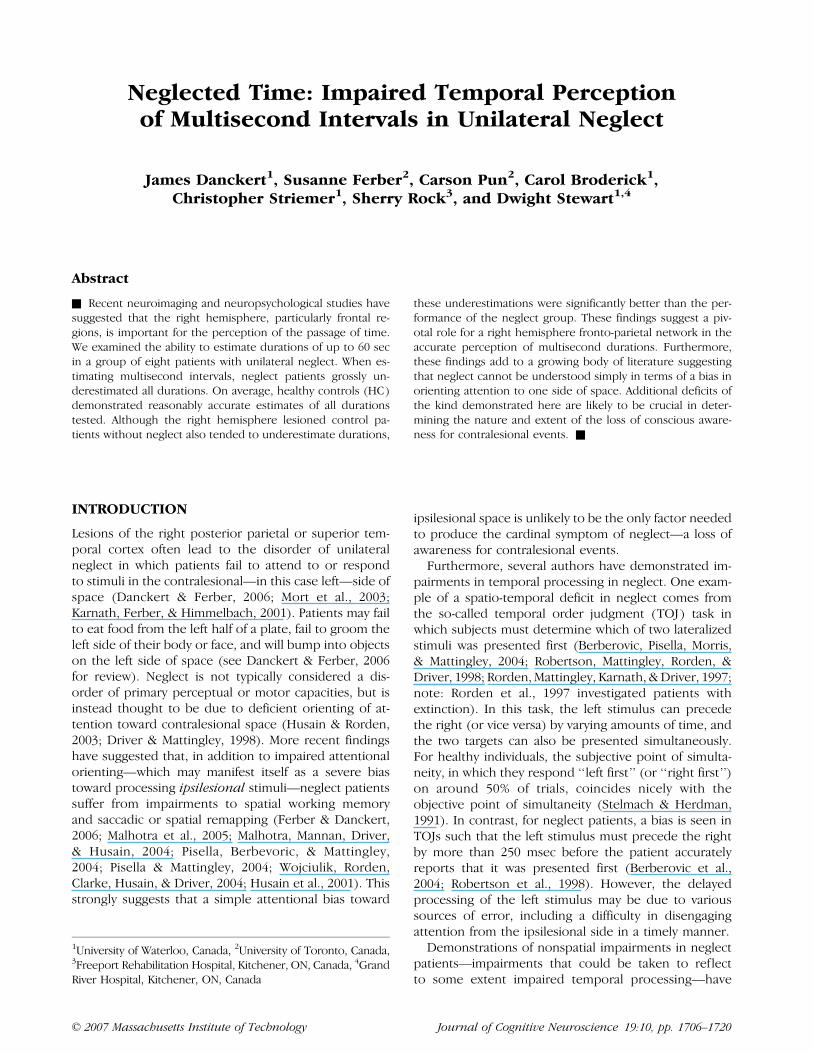
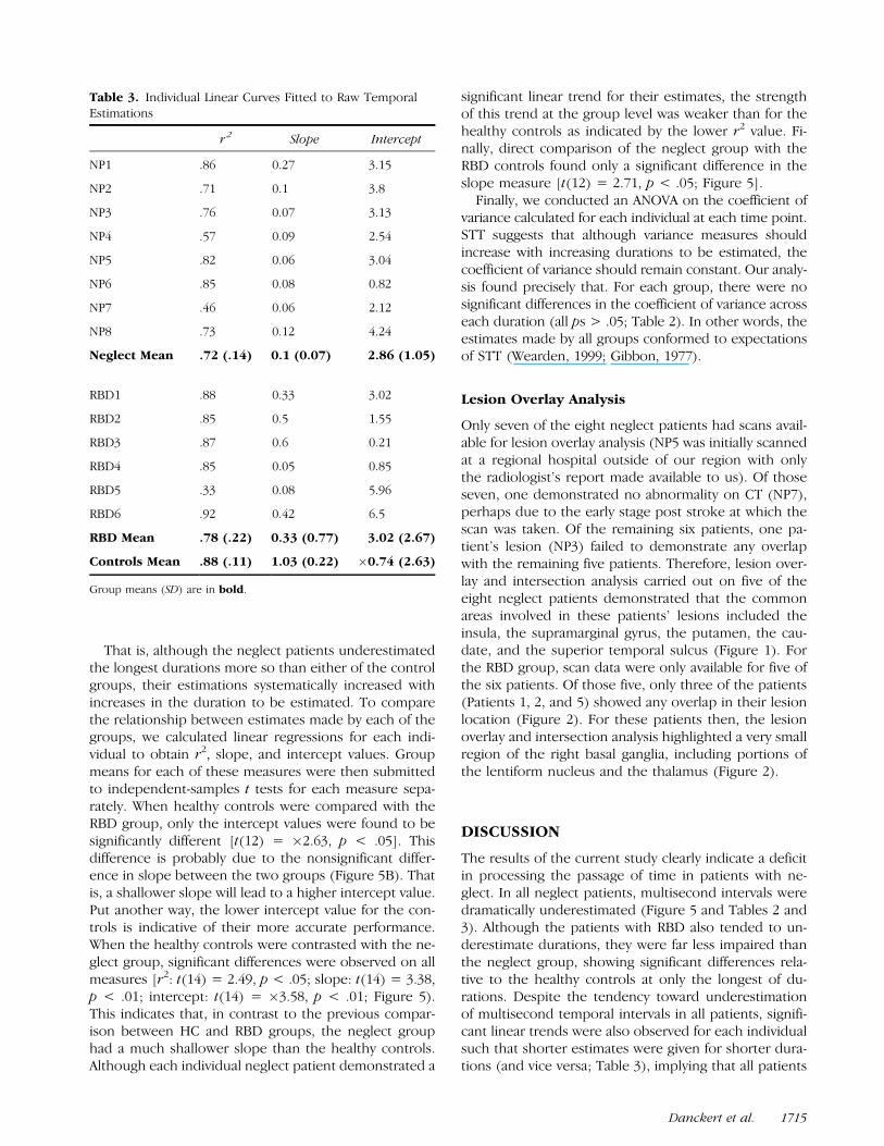
Individual lesions for the neglect patients are presentedin Figure 1, whereas the RBD patients’ lesions are pre-sented in Figure 2.
Lesion overlay analysis was conducted for each patientgroup to control for possible effects of lesion sites onour behavioral data. For the neglect group, one patient’sscans (NP7) showed no abnormality. This may havebeen related to the time at which the scan was takenas computed tomography (CT) images collected in thevery earliest stages poststroke often fail to show ab-normalities. Unfortunately, this patient does not residelocally, making it unfeasible to conduct a repeat scan.For a second patient (NP5), although we were able toobtain a radiologist’s report for her CT scans, we couldnot obtain the scan data itself due to the fact that she
Figure 1. (A) Individual lesion
maps for the six neglect
patients with scan data
available. (B) Lesion overlayanalysis for five of the six
patients (NP3 failed to show
any overlap and so was notincluded in this analysis).
Each patient’s lesion is given a
distinct color with the region
of overlap for all five patientsindicated in red. (C) Lesion
intersection analysis. The
common areas lesioned in five
of the six patients are indicatedin red (essentially, this is the
same region highlighted in
red in B presented here inisolation for clarity). We found
overlap mainly in the insula,
supramarginal gyrus, caudate,
putamen, and the superiortemporal gyrus.
Danckert et al. 1709
had been seen initially at a regional hospital outside ofour area. Lesion data were analyzed using the protocoloutlined by Ferber and Danckert (2006). All lesions (de-fined as the hypointense or hypodense stroke com-pared to its surrounding parenchyma) were traced onCT scans on a slice-by-slice basis using ANALYZE 7.0 AVWSoftware (Biomedical Imaging Resource, Mayo Founda-tion, Rochester, MN; Robb, 2001; Robb & Barillot, 1989;Robb et al., 1989). All scans were then transferred to theICBM152 template from the Montreal Neurological In-stitute (www.bic.mni.mcgill.ca/cgi/icbm_view), based onthe average of 152 normal MRI scans approximatelymatched to Talairach space. This transformation was atwo-step process using Automatic Image Registration ver-sion 5.2.5 software (AIR; http://bishopw.loni.ucla.edu/AIR5): the first step was a spatial normalization protocolincluding a linear 12-parameter affine transformation (in-cluding aligning scans to AC–PC aligned Talairach space).The second step was a nonlinear fourth-order parameterwarping model to make scans fit best to the template.The resulting images had a voxel size of 1 mm � 1 mm �1 mm. Using the transformed lesion maps, we esti-mated the proportion of each Brodmann’s area oranatomical region involved in each patient’s lesion,using the ‘‘broadmann’’ and ‘‘AAL’’ templates in MRIcro(http://people.cas.sc.edu/rorden/). To then superimpose
the individual brain lesion maps, the template ‘‘ch2’’ inMRIcro was used (Rorden & Brett, 2000). We shouldpoint out here, however, that lesion overlay analysis onsuch a small sample size is problematic. Certainly, withsuch a small sample size, we are not able to addressquestions related to the critical lesion site for neglect or,for that matter, the critical lesion location for any ob-served temporal estimation deficits (see Karnath et al.,2001).
All participants gave informed consent prior to partic-ipating in the experiment and the experimental protocolreceived ethics approval from the Office of Research atthe Universities of Waterloo and Toronto and the Tri-Hospital Research Ethics Board of Kitchener-Waterloo inaccordance with the Helsinki declaration. Eight neurolog-ically healthy older controls with normal or corrected-to-normal vision (4 men; mean age = 73 years; range = 60–74 years) also completed the temporal estimation taskafter giving written consent. Healthy controls were ex-cluded prior to participation if they reported any historyof neurological or psychiatric illness.
Apparatus and Procedure
Patients and controls were seated approximately 50 cmin front of a computer screen with a refresh rate of 75 Hz.
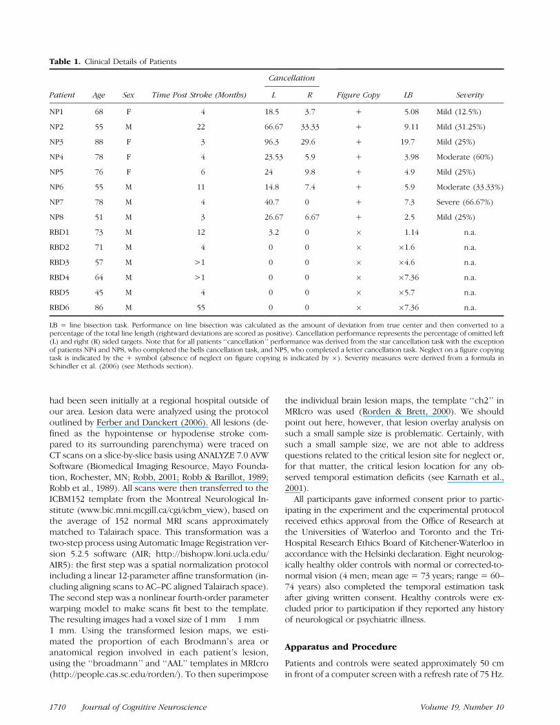
Table 1. Clinical Details of Patients
Cancellation
Patient Age Sex Time Post Stroke (Months) L R Figure Copy LB Severity
NP1 68 F 4 18.5 3.7 + 5.08 Mild (12.5%)
NP2 55 M 22 66.67 33.33 + 9.11 Mild (31.25%)
NP3 88 F 3 96.3 29.6 + 19.7 Mild (25%)
NP4 78 F 4 23.53 5.9 + 3.98 Moderate (60%)
NP5 76 F 6 24 9.8 + 4.9 Mild (25%)
NP6 55 M 11 14.8 7.4 + 5.9 Moderate (33.33%)
NP7 78 M 4 40.7 0 + 7.3 Severe (66.67%)
NP8 51 M 3 26.67 6.67 + 2.5 Mild (25%)
RBD1 73 M 12 3.2 0 � 1.14 n.a.
RBD2 71 M 4 0 0 � �1.6 n.a.
RBD3 57 M >1 0 0 � �4.6 n.a.
RBD4 64 M >1 0 0 � �7.36 n.a.
RBD5 45 M 4 0 0 � �5.7 n.a.
RBD6 86 M 55 0 0 � �7.36 n.a.
LB = line bisection task. Performance on line bisection was calculated as the amount of deviation from true center and then converted to apercentage of the total line length (rightward deviations are scored as positive). Cancellation performance represents the percentage of omitted left(L) and right (R) sided targets. Note that for all patients ‘‘cancellation’’ performance was derived from the star cancellation task with the exceptionof patients NP4 and NP8, who completed the bells cancellation task, and NP5, who completed a letter cancellation task. Neglect on a figure copyingtask is indicated by the + symbol (absence of neglect on figure copying is indicated by �). Severity measures were derived from a formula inSchindler et al. (2006) (see Methods section).
1710 Journal of Cognitive Neuroscience Volume 19, Number 10
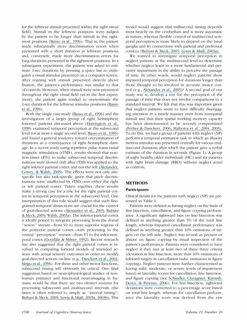
Participants were free to move their head and eyes butwere encouraged to maintain a stable posture through-out the experiment. E-Prime software (Psychology Soft-ware Tools) was used to present stimuli and record theparticipant’s responses, which were made via a standardkeyboard. An illusory motion stimulus was created asthe stimulus event that required temporal estimation.The stimulus array consisted of eight open circles (eachcircle subtends 3.58 of visual angle) arranged in a largercircle around a central point on the screen. The radiusof the larger circle around which the smaller circles werearranged subtended approximately 88 of visual angle.Each circle was filled-in in sequence in a clockwisedirection (Figure 3). No fixation point was used; how-ever, a secondary requirement to read aloud numbers
(subtending 18 of visual angle horizontally and 1.58 ver-tically) presented in the center of the illusory motionstimulus (described in more detail below) effectivelymeant that participants fixated the center of the display.
Illusory motion was used as the stimulus duration tobe estimated to ensure there was a salient event tocapture and maintain the patient’s attention throughouta trial. In previous work on temporal estimation in hu-mans, the stimulus event to be estimated is often sim-ply the presence of a geometrical shape (see Wearden,1999, for a review). We felt that it was important toemploy a more engaging stimulus to ensure full atten-tion throughout a trial. In initial development, we alsofelt that modulating the speed of the illusory motionstimulus may prove to be a useful means of examining
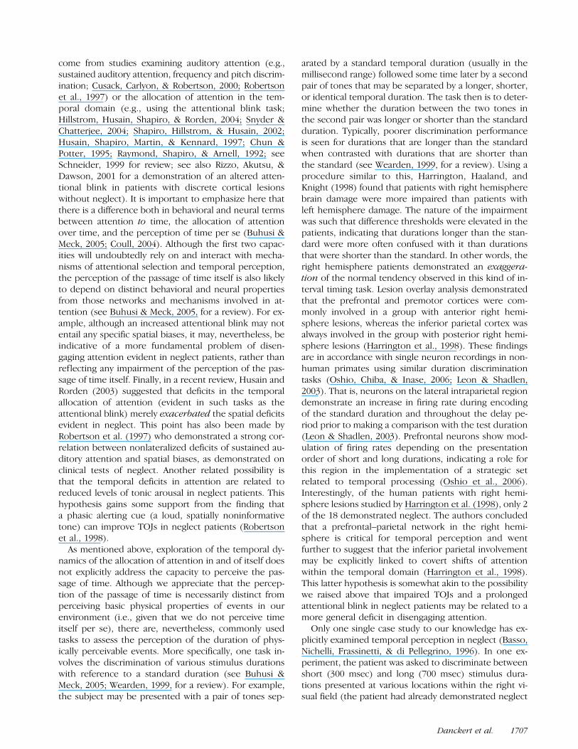
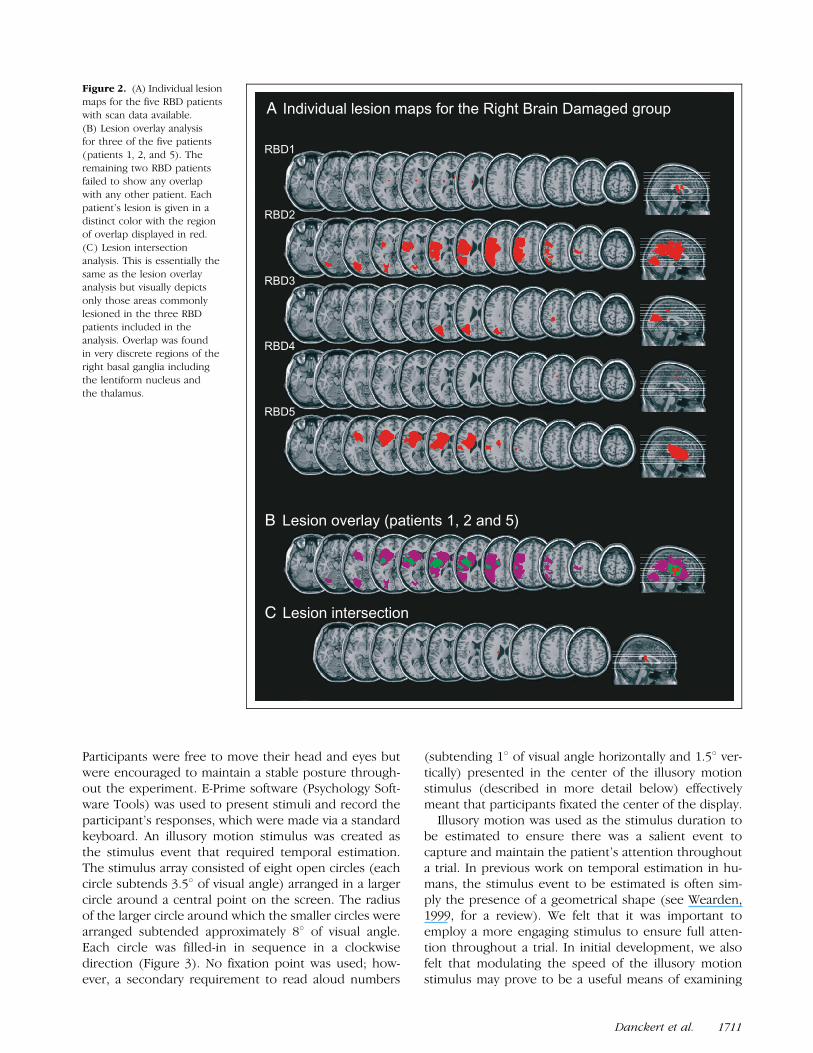
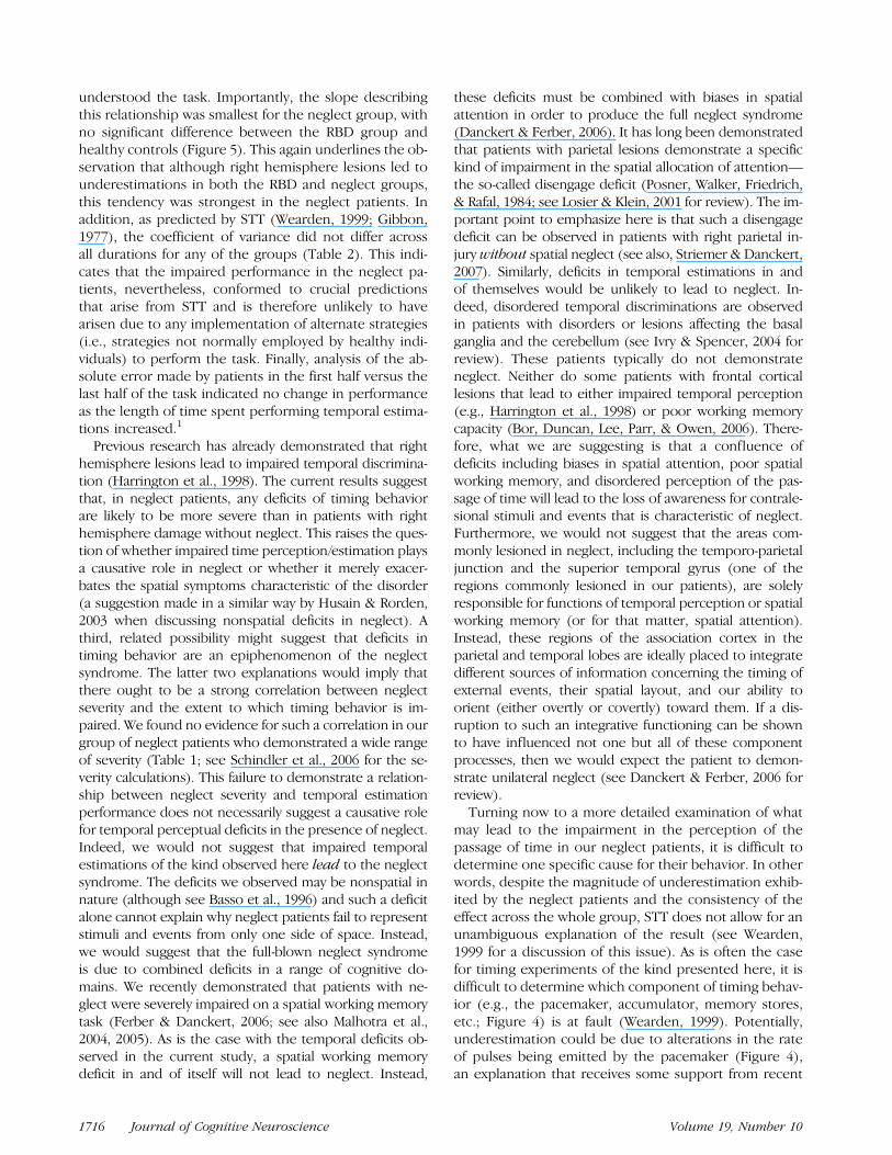
Figure 2. (A) Individual lesion
maps for the five RBD patients
with scan data available.
(B) Lesion overlay analysisfor three of the five patients
(patients 1, 2, and 5). The
remaining two RBD patientsfailed to show any overlap
with any other patient. Each
patient’s lesion is given in a
distinct color with the regionof overlap displayed in red.
(C) Lesion intersection
analysis. This is essentially the
same as the lesion overlayanalysis but visually depicts
only those areas commonly
lesioned in the three RBDpatients included in the
analysis. Overlap was found
in very discrete regions of the
right basal ganglia includingthe lentiform nucleus and
the thalamus.
Danckert et al. 1711
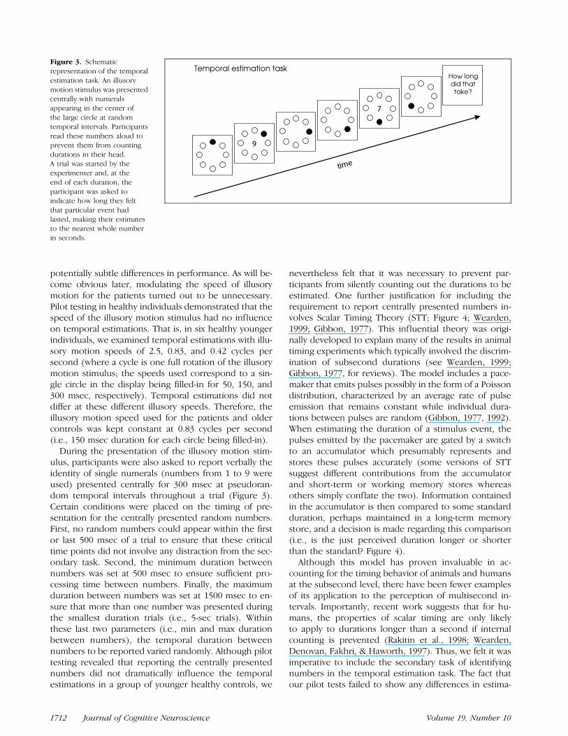
potentially subtle differences in performance. As will be-come obvious later, modulating the speed of illusorymotion for the patients turned out to be unnecessary.Pilot testing in healthy individuals demonstrated that thespeed of the illusory motion stimulus had no influenceon temporal estimations. That is, in six healthy youngerindividuals, we examined temporal estimations with illu-sory motion speeds of 2.5, 0.83, and 0.42 cycles persecond (where a cycle is one full rotation of the illusorymotion stimulus; the speeds used correspond to a sin-gle circle in the display being filled-in for 50, 150, and300 msec, respectively). Temporal estimations did notdiffer at these different illusory speeds. Therefore, theillusory motion speed used for the patients and oldercontrols was kept constant at 0.83 cycles per second(i.e., 150 msec duration for each circle being filled-in).
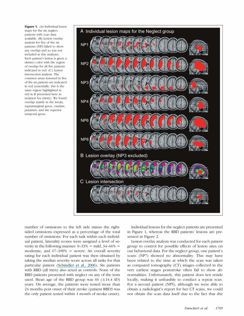
During the presentation of the illusory motion stim-ulus, participants were also asked to report verbally theidentity of single numerals (numbers from 1 to 9 wereused) presented centrally for 300 msec at pseudoran-dom temporal intervals throughout a trial (Figure 3).Certain conditions were placed on the timing of pre-sentation for the centrally presented random numbers.First, no random numbers could appear within the firstor last 500 msec of a trial to ensure that these criticaltime points did not involve any distraction from the sec-ondary task. Second, the minimum duration betweennumbers was set at 500 msec to ensure sufficient pro-cessing time between numbers. Finally, the maximumduration between numbers was set at 1500 msec to en-sure that more than one number was presented duringthe smallest duration trials (i.e., 5-sec trials). Withinthese last two parameters (i.e., min and max durationbetween numbers), the temporal duration betweennumbers to be reported varied randomly. Although pilottesting revealed that reporting the centrally presentednumbers did not dramatically influence the temporalestimations in a group of younger healthy controls, we
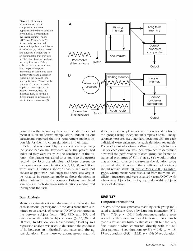
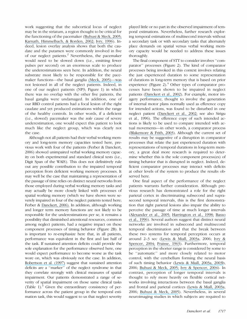
nevertheless felt that it was necessary to prevent par-ticipants from silently counting out the durations to beestimated. One further justification for including therequirement to report centrally presented numbers in-volves Scalar Timing Theory (STT; Figure 4; Wearden,1999; Gibbon, 1977). This influential theory was origi-nally developed to explain many of the results in animaltiming experiments which typically involved the discrim-ination of subsecond durations (see Wearden, 1999;Gibbon, 1977, for reviews). The model includes a pace-maker that emits pulses possibly in the form of a Poissondistribution, characterized by an average rate of pulseemission that remains constant while individual dura-tions between pulses are random (Gibbon, 1977, 1992).When estimating the duration of a stimulus event, thepulses emitted by the pacemaker are gated by a switchto an accumulator which presumably represents andstores these pulses accurately (some versions of STTsuggest different contributions from the accumulatorand short-term or working memory stores whereasothers simply conflate the two). Information containedin the accumulator is then compared to some standardduration, perhaps maintained in a long-term memorystore, and a decision is made regarding this comparison(i.e., is the just perceived duration longer or shorterthan the standard? Figure 4).
Although this model has proven invaluable in ac-counting for the timing behavior of animals and humansat the subsecond level, there have been fewer examplesof its application to the perception of multisecond in-tervals. Importantly, recent work suggests that for hu-mans, the properties of scalar timing are only likelyto apply to durations longer than a second if internalcounting is prevented (Rakitin et al., 1998; Wearden,Denovan, Fakhri, & Haworth, 1997). Thus, we felt it wasimperative to include the secondary task of identifyingnumbers in the temporal estimation task. The fact thatour pilot tests failed to show any differences in estima-
Figure 3. Schematic
representation of the temporal
estimation task. An illusory
motion stimulus was presentedcentrally with numerals
appearing in the center of
the large circle at randomtemporal intervals. Participants
read these numbers aloud to
prevent them from counting
durations in their head.A trial was started by the
experimenter and, at the
end of each duration, the
participant was asked toindicate how long they felt
that particular event had
lasted, making their estimatesto the nearest whole number
in seconds.
1712 Journal of Cognitive Neuroscience Volume 19, Number 10
tions when the secondary task was included does notmean it is an ineffective manipulation. Indeed, all ourparticipants reported that this requirement made it im-possible for them to count durations in their head.
Each trial was started by the experimenter pressingthe space bar on the keyboard once the patient hadindicated they were ready. At the conclusion of the du-ration, the patient was asked to estimate to the nearestsecond how long the stimulus had been present onthe computer screen. Durations of 5, 15, 30, and 60 secwere used. Durations shorter than 5 sec were notchosen as pilot work had suggested there was very lit-tle variance in responses made at these durations ineither patients or healthy controls. Patients completedfour trials at each duration with durations randomizedthroughout the task.
Data Analysis
Mean raw estimates at each duration were calculated foreach individual participant. These data were then sub-mitted to an analysis of variance (ANOVA) with group asthe between-subject factor (HC, RBD, and NP) andduration as the within-subjects factor (5, 15, 30, and60 msec). In addition, for each individual, a least squaresregression analysis was used to determine the goodnessof fit between an individual’s estimates and the ac-tual durations. From these equations, group mean r2,
slope, and intercept values were contrasted betweenthe groups using independent-samples t tests. Finally,variance measures (i.e., standard deviation, SD) for eachindividual were calculated at each duration separately.The coefficient of variance (SD/mean) for each individ-ual, for each duration, was then examined to determinehow well the performance of each group conformed toexpected properties of STT. That is, STT would predictthat although variance increases as the duration to beestimated also increases, the coefficient of varianceshould remain stable (Buhusi & Meck, 2005; Wearden,1999). Group means were calculated from individual co-efficient measures and were assessed via an ANOVA witha between-subjects factor of group and a within-subjectsfactor of duration.
RESULTS
Temporal Estimations
ANOVA of the raw estimates made by each group indi-cated a significant Group by Duration interaction [F(6,57) = 7.93; p < .001]. Independent-samples t testsat each of the durations tested indicated that controlsmade substantially higher estimates at all but the veryfirst duration when contrasted directly with the ne-glect patients [5-sec duration: t(9.67) = 1.62, p = .13;15-sec duration: t(8.3) = 3.231, p < .01; 30-sec duration:
Figure 4. Schematic
representation of the
component processes
hypothesized to be responsiblefor temporal perception in
the Scalar Timing Theory
(STT; see Wearden, 1999).A pacemaker or internal
clock emits pulses in a Poisson
distribution (A). These pulses
are gated by a switch (B) toan accumulator that may also
involve short-term or working
memory functions. Pulses
collected in the accumulatorare compared to prior
experience in some long-term
memory store and a decisionregarding the current time
interval is made. Theoretically,
attentional resources can be
applied at any stage of themodel, however, they are
indicated here as having a
direct impact on processing
within the accumulator (B).
Danckert et al. 1713
t(7.12) = 2.82, p < .05; 60-sec duration: t(7.17) = 3.39,p < .01]. Note that Levene’s test for equality of variancewas significant at all four durations, indicating that vari-
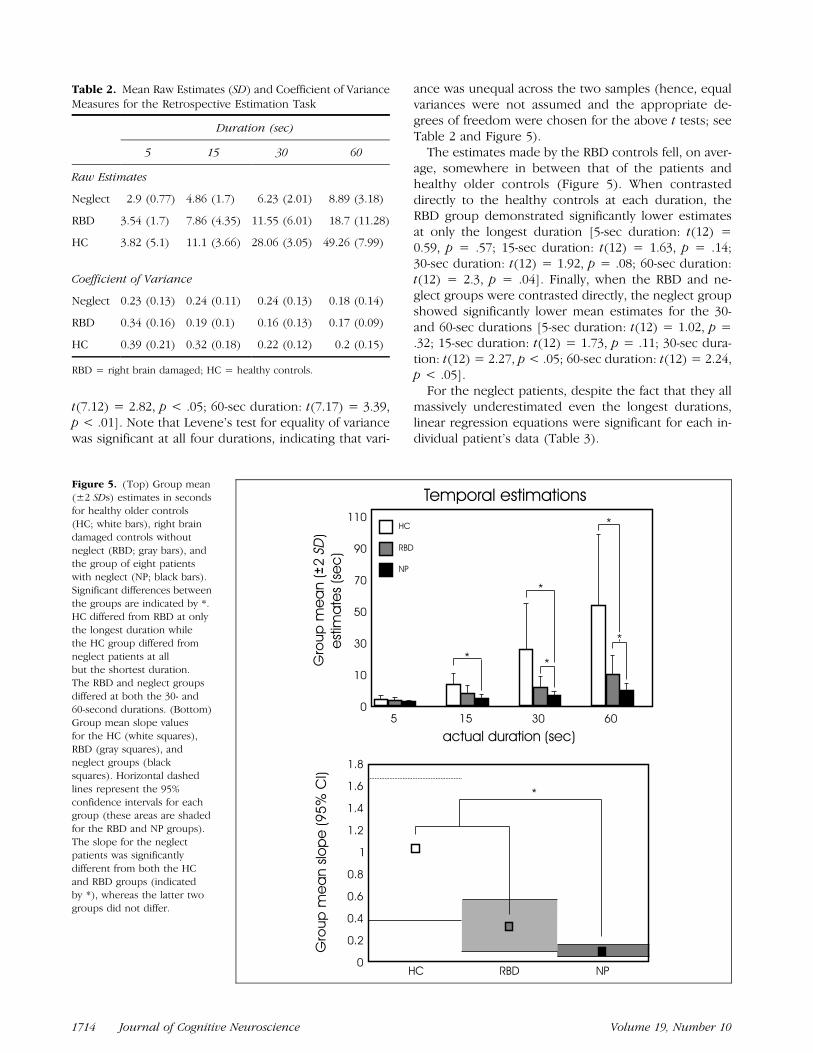
ance was unequal across the two samples (hence, equalvariances were not assumed and the appropriate de-grees of freedom were chosen for the above t tests; seeTable 2 and Figure 5).
The estimates made by the RBD controls fell, on aver-age, somewhere in between that of the patients andhealthy older controls (Figure 5). When contrasteddirectly to the healthy controls at each duration, theRBD group demonstrated significantly lower estimatesat only the longest duration [5-sec duration: t(12) =0.59, p = .57; 15-sec duration: t(12) = 1.63, p = .14;30-sec duration: t(12) = 1.92, p = .08; 60-sec duration:t(12) = 2.3, p = .04]. Finally, when the RBD and ne-glect groups were contrasted directly, the neglect groupshowed significantly lower mean estimates for the 30-and 60-sec durations [5-sec duration: t(12) = 1.02, p =.32; 15-sec duration: t(12) = 1.73, p = .11; 30-sec dura-tion: t(12) = 2.27, p < .05; 60-sec duration: t(12) = 2.24,p < .05].
For the neglect patients, despite the fact that they allmassively underestimated even the longest durations,linear regression equations were significant for each in-dividual patient’s data (Table 3).
Table 2. Mean Raw Estimates (SD) and Coefficient of VarianceMeasures for the Retrospective Estimation Task
Duration (sec)
5 15 30 60
Raw Estimates
Neglect 2.9 (0.77) 4.86 (1.7) 6.23 (2.01) 8.89 (3.18)
RBD 3.54 (1.7) 7.86 (4.35) 11.55 (6.01) 18.7 (11.28)
HC 3.82 (5.1) 11.1 (3.66) 28.06 (3.05) 49.26 (7.99)
Coefficient of Variance
Neglect 0.23 (0.13) 0.24 (0.11) 0.24 (0.13) 0.18 (0.14)
RBD 0.34 (0.16) 0.19 (0.1) 0.16 (0.13) 0.17 (0.09)
HC 0.39 (0.21) 0.32 (0.18) 0.22 (0.12) 0.2 (0.15)
RBD = right brain damaged; HC = healthy controls.
Figure 5. (Top) Group mean
(±2 SDs) estimates in seconds
for healthy older controls(HC; white bars), right brain
damaged controls without
neglect (RBD; gray bars), andthe group of eight patients
with neglect (NP; black bars).
Significant differences between
the groups are indicated by *.HC differed from RBD at only
the longest duration while
the HC group differed from
neglect patients at allbut the shortest duration.
The RBD and neglect groups
differed at both the 30- and60-second durations. (Bottom)
Group mean slope values
for the HC (white squares),
RBD (gray squares), andneglect groups (black
squares). Horizontal dashed
lines represent the 95%
confidence intervals for eachgroup (these areas are shaded
for the RBD and NP groups).
The slope for the neglectpatients was significantly
different from both the HC
and RBD groups (indicated
by *), whereas the latter twogroups did not differ.
1714 Journal of Cognitive Neuroscience Volume 19, Number 10
That is, although the neglect patients underestimatedthe longest durations more so than either of the controlgroups, their estimations systematically increased withincreases in the duration to be estimated. To comparethe relationship between estimates made by each of thegroups, we calculated linear regressions for each indi-vidual to obtain r2, slope, and intercept values. Groupmeans for each of these measures were then submittedto independent-samples t tests for each measure sepa-rately. When healthy controls were compared with theRBD group, only the intercept values were found to besignificantly different [t(12) = �2.63, p < .05]. Thisdifference is probably due to the nonsignificant differ-ence in slope between the two groups (Figure 5B). Thatis, a shallower slope will lead to a higher intercept value.Put another way, the lower intercept value for the con-trols is indicative of their more accurate performance.When the healthy controls were contrasted with the ne-glect group, significant differences were observed on allmeasures [r2: t(14) = 2.49, p < .05; slope: t(14) = 3.38,p < .01; intercept: t(14) = �3.58, p < .01; Figure 5).This indicates that, in contrast to the previous compar-ison between HC and RBD groups, the neglect grouphad a much shallower slope than the healthy controls.Although each individual neglect patient demonstrated a
significant linear trend for their estimates, the strengthof this trend at the group level was weaker than for thehealthy controls as indicated by the lower r2 value. Fi-nally, direct comparison of the neglect group with theRBD controls found only a significant difference in theslope measure [t(12) = 2.71, p < .05; Figure 5].
Finally, we conducted an ANOVA on the coefficient ofvariance calculated for each individual at each time point.STT suggests that although variance measures shouldincrease with increasing durations to be estimated, thecoefficient of variance should remain constant. Our analy-sis found precisely that. For each group, there were nosignificant differences in the coefficient of variance acrosseach duration (all ps > .05; Table 2). In other words, theestimates made by all groups conformed to expectationsof STT (Wearden, 1999; Gibbon, 1977).
Lesion Overlay Analysis
Only seven of the eight neglect patients had scans avail-able for lesion overlay analysis (NP5 was initially scannedat a regional hospital outside of our region with onlythe radiologist’s report made available to us). Of thoseseven, one demonstrated no abnormality on CT (NP7),perhaps due to the early stage post stroke at which thescan was taken. Of the remaining six patients, one pa-tient’s lesion (NP3) failed to demonstrate any overlapwith the remaining five patients. Therefore, lesion over-lay and intersection analysis carried out on five of theeight neglect patients demonstrated that the commonareas involved in these patients’ lesions included theinsula, the supramarginal gyrus, the putamen, the cau-date, and the superior temporal sulcus (Figure 1). Forthe RBD group, scan data were only available for five ofthe six patients. Of those five, only three of the patients(Patients 1, 2, and 5) showed any overlap in their lesionlocation (Figure 2). For these patients then, the lesionoverlay and intersection analysis highlighted a very smallregion of the right basal ganglia, including portions ofthe lentiform nucleus and the thalamus (Figure 2).
DISCUSSION
The results of the current study clearly indicate a deficitin processing the passage of time in patients with ne-glect. In all neglect patients, multisecond intervals weredramatically underestimated (Figure 5 and Tables 2 and3). Although the patients with RBD also tended to un-derestimate durations, they were far less impaired thanthe neglect group, showing significant differences rela-tive to the healthy controls at only the longest of du-rations. Despite the tendency toward underestimationof multisecond temporal intervals in all patients, signifi-cant linear trends were also observed for each individualsuch that shorter estimates were given for shorter dura-tions (and vice versa; Table 3), implying that all patients
Table 3. Individual Linear Curves Fitted to Raw TemporalEstimations
r 2 Slope Intercept
NP1 .86 0.27 3.15
NP2 .71 0.1 3.8
NP3 .76 0.07 3.13
NP4 .57 0.09 2.54
NP5 .82 0.06 3.04
NP6 .85 0.08 0.82
NP7 .46 0.06 2.12
NP8 .73 0.12 4.24
Neglect Mean .72 (.14) 0.1 (0.07) 2.86 (1.05)
RBD1 .88 0.33 3.02
RBD2 .85 0.5 1.55
RBD3 .87 0.6 0.21
RBD4 .85 0.05 0.85
RBD5 .33 0.08 5.96
RBD6 .92 0.42 6.5
RBD Mean .78 (.22) 0.33 (0.77) 3.02 (2.67)
Controls Mean .88 (.11) 1.03 (0.22) �0.74 (2.63)
Group means (SD) are in bold.
Danckert et al. 1715
understood the task. Importantly, the slope describingthis relationship was smallest for the neglect group, withno significant difference between the RBD group andhealthy controls (Figure 5). This again underlines the ob-servation that although right hemisphere lesions led tounderestimations in both the RBD and neglect groups,this tendency was strongest in the neglect patients. Inaddition, as predicted by STT (Wearden, 1999; Gibbon,1977), the coefficient of variance did not differ acrossall durations for any of the groups (Table 2). This indi-cates that the impaired performance in the neglect pa-tients, nevertheless, conformed to crucial predictionsthat arise from STT and is therefore unlikely to havearisen due to any implementation of alternate strategies(i.e., strategies not normally employed by healthy indi-viduals) to perform the task. Finally, analysis of the ab-solute error made by patients in the first half versus thelast half of the task indicated no change in performanceas the length of time spent performing temporal estima-tions increased.1
Previous research has already demonstrated that righthemisphere lesions lead to impaired temporal discrimina-tion (Harrington et al., 1998). The current results suggestthat, in neglect patients, any deficits of timing behaviorare likely to be more severe than in patients with righthemisphere damage without neglect. This raises the ques-tion of whether impaired time perception/estimation playsa causative role in neglect or whether it merely exacer-bates the spatial symptoms characteristic of the disorder(a suggestion made in a similar way by Husain & Rorden,2003 when discussing nonspatial deficits in neglect). Athird, related possibility might suggest that deficits intiming behavior are an epiphenomenon of the neglectsyndrome. The latter two explanations would imply thatthere ought to be a strong correlation between neglectseverity and the extent to which timing behavior is im-paired. We found no evidence for such a correlation in ourgroup of neglect patients who demonstrated a wide rangeof severity (Table 1; see Schindler et al., 2006 for the se-verity calculations). This failure to demonstrate a relation-ship between neglect severity and temporal estimationperformance does not necessarily suggest a causative rolefor temporal perceptual deficits in the presence of neglect.Indeed, we would not suggest that impaired temporalestimations of the kind observed here lead to the neglectsyndrome. The deficits we observed may be nonspatial innature (although see Basso et al., 1996) and such a deficitalone cannot explain why neglect patients fail to representstimuli and events from only one side of space. Instead,we would suggest that the full-blown neglect syndromeis due to combined deficits in a range of cognitive do-mains. We recently demonstrated that patients with ne-glect were severely impaired on a spatial working memorytask (Ferber & Danckert, 2006; see also Malhotra et al.,2004, 2005). As is the case with the temporal deficits ob-served in the current study, a spatial working memorydeficit in and of itself will not lead to neglect. Instead,
these deficits must be combined with biases in spatialattention in order to produce the full neglect syndrome(Danckert & Ferber, 2006). It has long been demonstratedthat patients with parietal lesions demonstrate a specifickind of impairment in the spatial allocation of attention—the so-called disengage deficit (Posner, Walker, Friedrich,& Rafal, 1984; see Losier & Klein, 2001 for review). The im-portant point to emphasize here is that such a disengagedeficit can be observed in patients with right parietal in-jury without spatial neglect (see also, Striemer & Danckert,2007). Similarly, deficits in temporal estimations in andof themselves would be unlikely to lead to neglect. In-deed, disordered temporal discriminations are observedin patients with disorders or lesions affecting the basalganglia and the cerebellum (see Ivry & Spencer, 2004 forreview). These patients typically do not demonstrateneglect. Neither do some patients with frontal corticallesions that lead to either impaired temporal perception(e.g., Harrington et al., 1998) or poor working memorycapacity (Bor, Duncan, Lee, Parr, & Owen, 2006). There-fore, what we are suggesting is that a confluence ofdeficits including biases in spatial attention, poor spatialworking memory, and disordered perception of the pas-sage of time will lead to the loss of awareness for contrale-sional stimuli and events that is characteristic of neglect.Furthermore, we would not suggest that the areas com-monly lesioned in neglect, including the temporo-parietaljunction and the superior temporal gyrus (one of theregions commonly lesioned in our patients), are solelyresponsible for functions of temporal perception or spatialworking memory (or for that matter, spatial attention).Instead, these regions of the association cortex in theparietal and temporal lobes are ideally placed to integratedifferent sources of information concerning the timing ofexternal events, their spatial layout, and our ability toorient (either overtly or covertly) toward them. If a dis-ruption to such an integrative functioning can be shownto have influenced not one but all of these componentprocesses, then we would expect the patient to demon-strate unilateral neglect (see Danckert & Ferber, 2006 forreview).
Turning now to a more detailed examination of whatmay lead to the impairment in the perception of thepassage of time in our neglect patients, it is difficult todetermine one specific cause for their behavior. In otherwords, despite the magnitude of underestimation exhib-ited by the neglect patients and the consistency of theeffect across the whole group, STT does not allow for anunambiguous explanation of the result (see Wearden,1999 for a discussion of this issue). As is often the casefor timing experiments of the kind presented here, it isdifficult to determine which component of timing behav-ior (e.g., the pacemaker, accumulator, memory stores,etc.; Figure 4) is at fault (Wearden, 1999). Potentially,underestimation could be due to alterations in the rateof pulses being emitted by the pacemaker (Figure 4),an explanation that receives some support from recent
1716 Journal of Cognitive Neuroscience Volume 19, Number 10
work suggesting that the subcortical locus of neglectmay be in the striatum, a region thought to be critical forthe functioning of the pacemaker (Buhusi & Meck, 2005;Karnath, Himmelbach, & Rorden, 2002; Ivry, 1996). In-deed, lesion overlay analysis shows that both the cau-date and the putamen were commonly involved in fiveof our neglect patients.2 Nevertheless, the pacemakerwould need to be slowed down (i.e., emitting fewerpulses per second) on an enormous scale to producethe underestimations seen here. In addition, the neuralsubstrate most likely to be responsible for the pace-maker functions—the basal ganglia (Meck, 2005)—wasnot lesioned in all of the neglect patients. Indeed, inone of our neglect patients (NP3; Figure 1) in whichthere was no overlap with the other five patients, thebasal ganglia were undamaged. In addition, one ofour RBD control patients had a focal lesion of the rightcaudate and yet produced estimations within the rangeof the healthy controls. In other words, if a deficient(i.e., slowed) pacemaker was the sole cause of severeunderestimation, one would expect this patient to lookmuch like the neglect group, which was clearly notthe case.
Although not all patients had their verbal working mem-ory and long-term memory capacities tested here, pre-vious work with four of the patients (Ferber & Danckert,2006) showed unimpaired verbal working memory capac-ity on both experimental and standard clinical tests (i.e.,Digit Span of the WAIS). This does not definitively ruleout any possible contribution to the impaired temporalperception from deficient working memory processes. Itmay well be the case that maintaining a representation ofthe passage of time relies on distinct neural networks fromthose employed during verbal working memory tasks andmay actually be more closely linked with processes ofspatial working memory (which we have shown were se-verely impaired in four of the neglect patients tested here;Ferber & Danckert, 2006). In addition, although workingand longer term memory functions are not likely to beresponsible for the underestimations per se, it remains apossibility that diminished attentional resources, commonamong neglect patients, had a negative impact on thesecomponent processes of timing behavior (Figure 2B). Itis important to re-emphasize here that, in all patients,performance was equivalent in the first and last half ofthe task. If sustained attention deficits could provide thesole explanation for the performance observed here, onewould expect performance to become worse as the taskwore on, which was obviously not the case. In addition,Robertson et al. (1997) suggest that sustained attentiondeficits are a ‘‘marker’’ of the neglect syndrome in thatthey correlate strongly with clinical measures of spatialimpairment. Our patients demonstrated a range of se-verity of spatial impairment on those same clinical tasks(Table 1).3 Given the extraordinary consistency of per-formance across the patient group in the temporal esti-mation task, this would suggest to us that neglect severity
played little or no part in the observed impairment of tem-poral estimations. Nevertheless, further research explor-ing temporal estimations of multisecond intervals withouta secondary task or with secondary tasks that alternatelyplace demands on spatial versus verbal working mem-ory capacity would be needed to address these issuesthoroughly.
The final component of STT to consider involves ‘‘com-parator’’ processes (Figure 2). The kind of comparatorprocesses being invoked in this context involves relatingthe just experienced duration to some representationof durations in long-term memory that is based on priorexperience (Figure 2).4 Other types of comparator pro-cesses have been shown to be impaired in neglectpatients (Danckert et al., 2002). For example, motor im-agery performance, thought to involve the generationof internal motor plans normally used as efference copyfor intended actions, was found to be disturbed in oneneglect patient (Danckert et al., 2002; see also Siriguet al., 1996). The efference copy of such intended ac-tions is likely to be used to compare intended with ac-tual movements—in other words, a comparator process(Blakemore & Frith, 2003). Although the current set ofresults may be suggestive of a disruption in comparatorprocesses that relate the just experienced duration withrepresentations of temporal durations in long-term mem-ory, a great deal more research is required to deter-mine whether this is the sole component process(es) oftiming behavior that is disrupted in neglect. Indeed, de-ficient comparator processes may interact with deficitsat other levels of the system to produce the results ob-served here.
One final aspect of the performance of the neglectpatients warrants further consideration. Although pre-vious research has demonstrated a role for the rightparietal cortex in discriminating subsecond and supra-second temporal intervals, this is the first demonstra-tion that right parietal lesions also impair the ability toperceive the passage of time at much longer intervals(Alexander et al., 2005; Harrington et al., 1998; Bassoet al., 1996). Several authors suggest that distinct neuralnetworks are involved in subsecond and multisecondtemporal discrimination and that the break betweenthese two systems for temporal perception occurs ataround 2–3 sec (Lewis & Miall, 2003a, 2006; Ivry &Spencer, 2004; Fraisse, 1963). Furthermore, temporalperception in the shorter range is considered by some tobe ‘‘automatic’’ and more closely related to motorcontrol, with the cerebellum forming the neural basisof such timing behavior (Lewis & Miall, 2003a, 2003b,2006; Buhusi & Meck, 2005; Ivry & Spencer, 2004). Incontrast, perception of longer temporal intervals isthought to rely more heavily on flexible cortical net-works involving interactions between the basal gangliaand frontal and parietal cortices (Lewis & Miall, 2003a,2006; Buhusi & Meck, 2005). Nevertheless, in severalneuroimaging studies in which subjects are required to
Danckert et al. 1717
produce responses aligned with various temporal inter-vals spanning subsecond, suprasecond, and multisecondranges, virtually identical networks of activation arecommonly observed (Macar et al., 2002; Rao, Mayer, &Harrington, 2001; Rubia et al., 1998). In addition, al-though cortical activations during temporal discrimina-tion tasks can be observed bilaterally, one study foundthat only the right hemisphere activity in the parietal,caudate, and parahippocampal regions was significantlycorrelated with behavioral measures (Harrington et al.,2004). In an excellent review of neuroimaging studiesof temporal perception, Lewis and Miall (2003a) demon-strate two distinct clusters of activations across studiesdepending on the nature of the task used, the involve-ment of motor responses to define temporal intervals,and the intervals tested. Their analysis clearly indicatedthat right hemisphere involvement including prefrontaland parietal cortices was most strongly evident in tasksin which motor responses were not necessary for de-fining temporal intervals and the intervals tested weresupra- or multisecond durations (Lewis & Miall, 2003a).
One further possibility is that the involvement of theparietal cortex in a broad range of timing tasks is in-dicative of a role in ‘‘magnitude’’ judgments (Walsh,2003b). Interestingly, this hypothesis has the potentialto explain one apparent discrepancy between the currentresults and previous work. That is, previous work sug-gests that subsecond and suprasecond intervals tend tobe overestimated by patients with parietal lesions, where-as the current results clearly demonstrate underestima-tion (Harrington et al., 1998; Basso et al., 1996). Thisapparent discrepancy can be explained with recourse topsychophysical functions applied to magnitude or inten-sity judgments of various kinds (e.g., brightness, weight,force, and spatial extent; Mennemeier et al., 2005; Ricci& Chatterjee, 2001; see also Hollingworth, 1909 for theseminal work on this ‘‘magnitude’’ function). That is,healthy individuals tend to overestimate the magnitudeor intensity of smaller stimuli and underestimate the mag-nitude or intensity of larger stimuli across a range of dif-ferent tasks and perceptual properties. Indeed, the classic‘‘crossover’’ effect in line bisection, in which neglect pa-tients bisect larger lines to the right of true center,whereas much shorter lines are bisected to the left oftrue center, may well be due to this same kind of psy-chophysical relationship applied to magnitude judgmentsof line length (Mennemeier et al., 2005). One suggestionin the context of this magnitude explanation of variousneglect phenomena is that neglect patients demonstratean exaggeration of the normal tendency to overestimateat the smaller end of a range of stimulus intensities whileunderestimating the longer range (Mennemeier et al.,2005; Ricci & Chatterjee, 2001). Further studies in whichthe tests used span both subsecond and multisecondduration discrimination within the same group of patientswould provide insights into this potential explanation forthe underestimation observed here.
It is becoming increasingly clear that models of neglectthat simply refer to impaired mechanisms of attention-al orienting are insufficient to explain the full range ofsymptoms and impairments that characterize the disorder(Danckert & Ferber, 2006; Pisella & Mattingley, 2004).Deficits of saccadic or spatial remapping, spatial work-ing memory, and now temporal perception, will all needto be considered when constructing neurocognitive mod-els of the disorder (Ferber & Danckert, 2006; Pisella& Mattingley, 2004; Wojciulik et al., 2004; Husain et al.,2001; Wojciulik, Husain, Clarke, & Driver, 2001; Heide,Blankenburg, Zimmermann, & Kompf, 1995). As sug-gested above, neglect is likely to result from the conflu-ence of these component deficits and concomitantly, theregion of the parietal and temporal cortex commonly le-sioned in neglect patients is likely to be crucial for inte-grating information processing related to these distinctfunctions. A loss of that integrative function, combinedwith the classic demonstration of biases in spatial atten-tion, will lead to an extraordinary difficulty in consciouslyrepresenting stimuli and events on the left side of space(Danckert & Ferber, 2006). In relation to time perceptionper se, the experience of the passage of time is intimatelylinked with the experience of change. Therefore, in or-der to create and maintain an accurate representation ofan ever-changing environment, it would be necessary toaccurately ‘‘time-stamp’’ behaviorally relevant or salientevents in that environment. Inaccurate temporal percep-tion of the kind demonstrated here would presumablymake that process of ‘‘time-stamping’’ salient changes instimuli or events difficult to perform. As already stated,this difficulty, in and of itself, would presumably not leadto a loss of awareness for contralesional events only. In-stead, the temporal perception deficit would need to becoupled with a bias in orienting attention to one side ofspace. If one considers that all events in the environ-ment compete for conscious representation (Desimone& Duncan, 1995), then such a combination of deficitswould make it extremely difficult for contralesional events(or changes in the environment occurring in contrale-sional space) to compete with ipsilesional events for ac-cess to memory systems and, eventually, consciousness.
Acknowledgments
This work was supported by the Natural Sciences and Engineer-ing Research Council (NSERC) of Canada Discovery Grants toJD and SF, an NSERC Canada Research Chair (Tier II) awardto JD, NSERC graduate scholarships to CS and CB, Heart andStroke Foundation (HSF) of Ontario Grants to JD, an HSFMasters Scholarship to CS, a Canadian Institutes of Health Re-search grant to SF, and the Leverhulme Trust. We thank Drs.F. Gao and S. Black for help with the lesion analysis.
Reprint requests should be sent to Dr. James Danckert, CanadaResearch Chair (Tier II) in Cognitive Neuroscience, Depart-ment of Psychology, University of Waterloo, 200 University Ave-nue West, Waterloo, Ontario, Canada N2L 3G1, or via e-mail:[email protected].
1718 Journal of Cognitive Neuroscience Volume 19, Number 10
Notes
1. Absolute error was determined via a difference score calcu-lated for each individual trial. That is, the patient’s responsewas subtracted from the actual duration to be estimated witha positive score indicative of underestimation (there were nooverestimations in any patient negating the need to deal withthe sign of the difference score). t Tests comparing first halfand last half performance for the group as a whole and for eachpatient individually were all nonsignificant.2. The small number of both NP and RBD patients that wewere able to include in our lesion overlay analyses makes anyconclusions concerning the role of commonly lesioned areas inour data necessarily speculative. This is especially true for theRBD group in which only three patients were included in theanalysis, which showed overlap in the thalamus and lentiformnucleus of the right hemisphere (Figure 2).3. If severity was to be based solely on cancellation perform-ance, we would then classify as ‘‘mild’’ neglect patients 1 and6, with patients 4, 5, and 8 classified as ‘‘moderate’’ and pa-tients 2, 3, and 7 classified as ‘‘severe’’. The point remains un-changed, as despite the range of severity in our patient group,their performance on the temporal estimation task was in-credibly consistent across all patients.4. In many cases, a ‘‘standard’’ duration is actually given tothe subject to which all test durations must be compared. Inour case, no such standard was used and patients were re-quired to compare the test duration to some other storedrepresentation, perhaps of how long a second or a minute‘‘should’’ take to elapse.
REFERENCES
Alexander, I., Cowey, A., & Walsh, V. (2005). The rightparietal cortex and time perception: Back to Critchley andthe Zeitraffer phenomenon. Cognitive Neuropsychology,22, 306–315.
Basso, G., Nichelli, P., Frassinetti, F., & di Pellegrino, G.(1996). Time perception in a neglected space. NeuroReport,7, 2111–2114.
Berberovic, N., Pisella, L., Morris, A. P., & Mattingley, J. B.(2004). Prismatic adaptation reduces biased temporalorder judgements in spatial neglect. NeuroReport, 15,1199–1204.
Blakemore, S.-J., & Frith, C. D. (2003). Self-awareness andaction. Current Opinion in Neurobiology, 13, 219–224.
Bor, D., Duncan, J., Lee, A. C. H., Parr, L., & Owen, A. M.(2006). Frontal lobe involvement in spatial span:Converging studies of normal and impaired function.Neuropsychologia, 44, 229–237.
Buhusi, C. V., & Meck, W. H. (2005). What makes us tick?Functional and neural mechanisms of interval timing.Nature Reviews Neuroscience, 6, 755–765.
Chun, M. M., & Potter, M. C. (1995). A two-stage model formultiple target detection in rapid serial visual presentation.Journal of Experimental Psychology: Human Perceptionand Performance, 21, 109–127.
Coull, J. T. (2004). fMRI studies of temporal attention:Allocating attention within, or towards time. CognitiveBrain Research, 21, 216–226.
Cusack, R., Carlyon, R. P., & Robertson, I. H. (2000).Neglect between but not within auditory objects. Journalof Cognitive Neuroscience, 12, 1056–1065.
Danckert, J., & Ferber, S. (2006). Revisiting unilateralneglect. Neuropsychologia, 44, 987–1006.
Danckert, J., Ferber, S., Doherty, T., Steinmetz, H., Nicolle, D.,& Goodale, M. A. (2002). Selective, non-lateralised
impairment of motor imagery following right parietaldamage. Neurocase, 8, 194–204.
Desimone, R., & Duncan, J. (1995). Neural mechanisms ofselective visual attention. Annual Review of Neuroscience,18, 193–222.
Driver, J., & Mattingley, J. B. (1998). Parietal neglect andvisual awareness. Nature Neuroscience, 1, 17–22.
Ferber, S., & Danckert, J. (2006). Lost in space—The fateof memory representations for non-neglected stimuli.Neuropsychologia, 44, 320–325.
Fraisse, P. (1963). The psychology of time. New York: Harper& Row.
Gibbon, J. (1977). Scalar expectancy theory and Weber’slaw in animal timing. Psychological Review, 84, 279–325.
Gibbon, J. (1992). Ubiquity of scalar timing with a Poissonclock. Journal of Mathematical Psychology, 36, 283–293.
Goodale, M. A., & Milner, A. D. (1992). Separate visualpathways for perception and action. Trends inNeurosciences, 15, 20–25.
Harrington, D. L., Boyd, L. A., Mayer, A. R., Sheltraw, D. M.,Lee, R. R., Huang, M., et al. (2004). Neural representationof interval encoding and decision making. CognitiveBrain Research, 21, 193–205.
Harrington, D. L., Haaland, K. Y., & Knight, R. T. (1998).Cortical networks underlying mechanisms of timeperception. Journal of Neuroscience, 18, 1085–1095.
Heide, W., Blankenburg, M., Zimmermann, E., & Kompf, D.(1995). Cortical control of double-step saccades:Implications for spatial orientation. Annals of Neurology,38, 739–748.
Hillstrom, A. P., Husain, M., Shapiro, K. L., & Rorden, C.(2004). Spatiotemporal dynamics of attention in visualneglect: A case study. Cortex, 40, 433–440.
Hollingworth, H. L. (1909). The indifference point. In H. L.Hollingworth (Ed.), The inaccuracy of movement. NewYork: The Science Press.
Husain, M., Mannan, S., Hodgson, T., Wojciulik, E., Driver, J.,& Kennard, C. (2001). Impaired spatial working memoryacross saccades contributes to abnormal search in parietalneglect. Brain, 124, 941–952.
Husain, M., & Rorden, C. (2003). Non-spatially lateralisedmechanisms in neglect. Nature Neuroscience Reviews, 4,26–36.
Husain, M., Shapiro, K., Martin, J., & Kennard, C. (1997).Abnormal temporal dynamics of visual attention inspatial neglect patients. Nature, 385, 154–156.
Ivry, R. B. (1996). The representation of temporal informationin perception and motor control. Current Opinion inNeurobiology, 6, 851–857.
Ivry, R. B., & Spencer, R. M. C. (2004). The neuralrepresentation of time. Current Opinion in Neurobiology,14, 225–232.
Karnath, H.-O., Ferber, S., & Himmelbach, M. (2001). Spatialawareness is a function of the temporal not the posteriorparietal lobe. Nature, 411, 950–953.
Karnath, H.-O., Himmelbach, M., & Rorden, C. (2002). Thesubcortical anatomy of human spatial neglect: Putamen,caudate nucleus and pulvinar. Brain, 125, 350–360.
Leon, M. I., & Shadlen, M. N. (2003). Representation oftime by neurons in the posterior parietal cortex of themacaque. Neuron, 38, 317–327.
Lewis, P. A., & Miall, R. C. (2003a). Distinct systems forautomatic and cognitively controlled time measurement:Evidence from neuroimaging. Current Opinion inNeurobiology, 13, 250–255.
Lewis, P. A., & Miall, R. C. (2003b). Brain activation patternsduring measurement of sub- and supra-second intervals.Neuropsychologia, 41, 1583–1592.
Danckert et al. 1719
Lewis, P. A., & Miall, R. C. (2006). Remembering the time:A continuous clock. Trends in Cognitive Sciences, 10,401–406.
Losier, B. J., & Klein, R. M. (2001). A review of the evidencefor a disengage deficit following parietal lobe damage.Neuroscience and Biobehavioral Reviews, 25, 1–13.
Macar, F., Lejeune, H., Bonnet, M., Ferrara, A., Poutha, V.,Vidal, F., et al. (2002). Activation of the supplementarymotor area and of attention networks during temporalprocessing. Experimental Brain Research, 142, 475–485.
Malhotra, P., Jager, H. R., Parton, A., Greenwood, R.,Playford, E. D., Brown, M. M., et al. (2005). Spatialworking memory capacity in unilateral neglect. Brain,128, 424–435.
Malhotra, P., Mannan, S., Driver, J., & Husain, M. (2004).Impaired spatial working memory: One component ofthe visual neglect syndrome? Cortex, 40, 667–676.
Meck, W. H. (2005). Neuropsychology of timing and timeperception. Brain & Cognition, 58, 1–8.
Mennemeier, M., Pierce, C. A., Chatterjee, A., Anderson, B.,Jewell, G., Dowler, R., et al. (2005). Biases in attentionalorientation and magnitude estimation explain crossover:Neglect is a disorder of both. Journal of CognitiveNeuroscience, 17, 1194–1211.
Mort, D. J., Malhotra, P., Mannan, S. K., Rorden, C.,Pambakian, A., Kennard, C., et al. (2003). The anatomyof visual neglect. Brain, 126, 1986–1997.
Oshio, K., Chiba, A., & Inase, M. (2006). Delay period activity ofmonkey prefrontal neurons during duration-discriminationtask. European Journal of Neuroscience, 23, 2779–2790.
Pisella, L., Berberovic, N., & Mattingley, J. B. (2004).Impaired working memory for location but not for colouror shape in visual neglect: A comparison of parietal andnon-parietal lesions. Cortex, 40, 379–390.
Pisella, L., & Mattingley, J. B. (2004). The contribution ofspatial remapping to unilateral visual neglect. Neuroscienceand Biobehavioral Reviews, 28, 181–200.
Posner, M. I., Walker, J. A., Friedrich, F. J., & Rafal, R. D.(1984). Effects of parietal injury on covert orienting ofattention. Journal of Neuroscience, 4, 1863–1874.
Rakitin, B. C., Gibbon, J., Penney, T. B., Malapani, C., Hinton,S. C., & Meck, W. H. (1998). Scalar expectancy theory andpeak-interval timing in humans. Journal of ExperimentalPsychology: Animal Behavior Processes, 24, 15–33.
Rao, S. M., Mayer, A. R., & Harrington, D. L. (2001). Theevolution of brain activation during temporal processing.Nature Neuroscience, 4, 317–323.
Raymond, J. E., Shapiro, K. L., & Arnell, K. M. (1992).Temporary suppression of visual processing in an RSVPtask: An attentional blink? Journal of ExperimentalPsychology, 18, 849–860.
Ricci, R., & Chatterjee, A. (2001). Context and crossoverin unilateral neglect. Neuropsychologia, 39, 1138–1143.
Rizzo, M., Akutsu, H., & Dawson, J. (2001). Increasedattentional blink after focal cerebral lesions. Neurology,57, 795–800.
Robb, R. A. (2001). The Biomedical Imaging Resource atMayo Clinic. Guest editorial. IEEE Transactions onMedical Imaging, 20, 854–867.
Robb, R. A., & Barillot, C. (1989). Interactive display andanalysis of 3-D medical images. IEEE Transactions onMedical Imaging, 8, 217–226.
Robb, R. A., Hanson, D. P., Karwoski, R. A., Larson, A. G.,Workman, E. L., & Stacy, M. C. (1989). ANALYZE: Acomprehensive, operator-interactive software package
for multidimensional medical image display and analysis.Computerized Medical Imaging and Graphics, 13,433–454.
Robertson, I. H., Manly, T., Beschin, N., Daini, R.,Haeske-Dewick, H., Homberg, V., et al. (1997). Auditorysustained attention is a marker of unilateral neglect.Neuropsychologia, 35, 1527–1532.
Robertson, I. H., Mattingley, J. B., Rorden, C., & Driver, J.(1998). Phasic alerting of neglect patients overcomes theirspatial deficit in visual awareness. Nature, 395, 169–172.
Rorden, C., & Brett, M. (2000). Stereotaxic display of brainlesions. Behavioural Neurology, 12, 191–200.
Rorden, C., Mattingley, J. B., Karnath, H.-O., & Driver, J.(1997). Visual extinction and prior entry: Impairedperception of temporal order with intact motionperception after unilateral parietal damage.Neuropsychologia, 35, 421–433.
Rubia, K., Overmeyer, S., Taylor, E., Brammer, M.,Williams, S., Simmons, A., et al. (1998). Prefrontalinvolvement in ‘‘temporal bridging’’ and timingmovement. Neuropsychologia, 36, 1283–1293.
Schindler, I., Clavagnier, S., Karnath, H.-O., Derex, L.,& Perenin, M.-T. (2006). A common basis for visualand tactile exploration deficits in spatial neglect?Neuropsychologia, 44, 1444–1451.
Schneider, W. (1999). Visual–spatial working memory,attention, and scene representation: A neuro-cognitivetheory. Psychological Research, 62, 220–236.
Shapiro, K., Hillstrom, A. P., & Husain, M. (2002). Controlof visuotemporal attention by inferior parietal andsuperior temporal cortex. Current Biology, 12, 1320–1325.
Sirigu, A., Duhamel, J.-R., Cohen, L., Pillon, B., Dubois, B.,& Agid, Y. (1996). The mental representation of handmovements after parietal cortex damage. Science, 273,1564–1568.
Snyder, J. J., & Chatterjee, A. (2004). Spatial–temporalanisometries following right parietal damage.Neuropsychologia, 42, 1703–1708.
Stelmach, L. B., & Herdman, C. M. (1991). Directedattention and perception of temporal order. Journalof Experimental Psychology: Human Perception andPerformance, 17, 539–550.
Striemer, C., & Danckert, J. (2007). Prism adaptation reducesthe disengage deficit in patients with right brain damage.NeuroReport, 18, 99–103.
Walsh, V. (2003a). Time: The back-door of perception.Trends in Cognitive Sciences, 7, 335–338.
Walsh, V. (2003b). A theory of magnitude: Common corticalmetrics of time, space and quantity. Trends in CognitiveSciences, 7, 483–488.
Wearden, J. H. (1999). ‘‘Beyond the fields we know. . .’’:Exploring and developing scalar timing theory.Behavioral Processes, 45, 3–21.
Wearden, J. H., Denovan, L., Fakhri, M., & Haworth, R. (1997).Scalar timing in temporal generalization in humans withlonger stimulus durations. Journal of ExperimentalPsychology: Animal Behavior Processes, 23, 502–511.
Wojciulik, E., Husain, M., Clarke, K., & Driver, J. (2001).Spatial working memory deficit in unilateral neglect.Neuropsychologia, 39, 390–396.
Wojciulik, E., Rorden, C., Clarke, K., Husain, M., &Driver, J. (2004). Group study of an ‘‘undercover’’ testfor visuospatial neglect: Invisible cancellation can revealmore neglect than standard cancellation. Journal ofNeurology, Neurosurgery, and Psychiatry, 75, 1356–1358.
1720 Journal of Cognitive Neuroscience Volume 19, Number 10
Related Documents


