Case Reports in Clinical Medicine, 2014, 3, 639-643 Published Online December 2014 in SciRes. http://www.scirp.org/journal/crcm http://dx.doi.org/10.4236/crcm.2014.312136 How to cite this paper: Korekawaka, A., Nakajima, K., Nakano, H. and Sawamura, D. (2014) Necrobiosis Lipoidica in the Absence of Diabetes Mellitus: A Case Report and an Analysis of 116 Japanese Cases. Case Reports in Clinical Medicine, 3, 639-643. http://dx.doi.org/10.4236/crcm.2014.312136 Necrobiosis Lipoidica in the Absence of Diabetes Mellitus: A Case Report and an Analysis of 116 Japanese Cases Ayumi Korekawaka, Koji Nakajima, Hajime Nakano, Daisuke Sawamura * Department of Dermatology, Hirosaki University Graduate School of Medicine, Hirosaki, Japan Email: * [email protected] Received 23 October 2014; revised 22 November 2014; accepted 3 December 2014 Copyright © 2014 by authors and Scientific Research Publishing Inc. This work is licensed under the Creative Commons Attribution International License (CC BY). http://creativecommons.org/licenses/by/4.0/ Abstract A 60-year-old Japanese woman was referred to our hospital for yellow-brown plaques accompa- nied by ulceration on her left lower leg. Her medical history included neither diabetes mellitus nor minor trauma. A histopathological examination of the plaques showed necrobiotic changes within the dermal collagen surrounded by granulomas that comprised lymphocytes, histiocytes, and giant cells. The lower dermis revealed fibrotic changes that extended into the subcutaneous tissue. The patient’s blood glucose and glycated hemoglobin levels were within the normal ranges. We considered a diagnosis of necrobiosis lipoidica (NL) in the absence of diabetes mellitus. We re- viewed 116 cases of NL that were reported in the Japanese medical literature between 1986 and 2014 and found that NL onset was most common in individuals of both sexes ≥ 60 years of age. Previous reports that reviewed NL cases in the Japanese medical literature that were published during the1900s indicated that NL occurs in people ≥ 40 years of age. We suggest that the aging population and increasingly longer life spans have increased the average age of NL onset in Japan. With regard to treatments, there were no effective treatments, but skin grafts were curative. NL treatment is very difficult, especially when ulcers are present; hence, we suggest that further re- search is needed to determine effective NL treatments. Keywords Peripheral Circulatory Disturbance, Ulceration, Trauma, Lower Leg 1. Introduction Necrobiosis lipoidica (NL) is a rare and benign granulomatous disease that is associated with collagen degenera- tion and typically affects the lower legs of women. It is characterized by well-defined ulcerated plaques with * Corresponding author.

Welcome message from author
This document is posted to help you gain knowledge. Please leave a comment to let me know what you think about it! Share it to your friends and learn new things together.
Transcript

Case Reports in Clinical Medicine, 2014, 3, 639-643 Published Online December 2014 in SciRes. http://www.scirp.org/journal/crcm http://dx.doi.org/10.4236/crcm.2014.312136
How to cite this paper: Korekawaka, A., Nakajima, K., Nakano, H. and Sawamura, D. (2014) Necrobiosis Lipoidica in the Absence of Diabetes Mellitus: A Case Report and an Analysis of 116 Japanese Cases. Case Reports in Clinical Medicine, 3, 639-643. http://dx.doi.org/10.4236/crcm.2014.312136
Necrobiosis Lipoidica in the Absence of Diabetes Mellitus: A Case Report and an Analysis of 116 Japanese Cases Ayumi Korekawaka, Koji Nakajima, Hajime Nakano, Daisuke Sawamura* Department of Dermatology, Hirosaki University Graduate School of Medicine, Hirosaki, Japan Email: *[email protected] Received 23 October 2014; revised 22 November 2014; accepted 3 December 2014
Copyright © 2014 by authors and Scientific Research Publishing Inc. This work is licensed under the Creative Commons Attribution International License (CC BY). http://creativecommons.org/licenses/by/4.0/
Abstract A 60-year-old Japanese woman was referred to our hospital for yellow-brown plaques accompa-nied by ulceration on her left lower leg. Her medical history included neither diabetes mellitus nor minor trauma. A histopathological examination of the plaques showed necrobiotic changes within the dermal collagen surrounded by granulomas that comprised lymphocytes, histiocytes, and giant cells. The lower dermis revealed fibrotic changes that extended into the subcutaneous tissue. The patient’s blood glucose and glycated hemoglobin levels were within the normal ranges. We considered a diagnosis of necrobiosis lipoidica (NL) in the absence of diabetes mellitus. We re-viewed 116 cases of NL that were reported in the Japanese medical literature between 1986 and 2014 and found that NL onset was most common in individuals of both sexes ≥ 60 years of age. Previous reports that reviewed NL cases in the Japanese medical literature that were published during the1900s indicated that NL occurs in people ≥ 40 years of age. We suggest that the aging population and increasingly longer life spans have increased the average age of NL onset in Japan. With regard to treatments, there were no effective treatments, but skin grafts were curative. NL treatment is very difficult, especially when ulcers are present; hence, we suggest that further re-search is needed to determine effective NL treatments.
Keywords Peripheral Circulatory Disturbance, Ulceration, Trauma, Lower Leg
1. Introduction Necrobiosis lipoidica (NL) is a rare and benign granulomatous disease that is associated with collagen degenera-tion and typically affects the lower legs of women. It is characterized by well-defined ulcerated plaques with
*Corresponding author.
A. Korekawaka et al.
640
indurate borders and atrophic centers that often impair patient quality of life when form ulcerations in skin le-sions, especially in lower legs.
NL is associated with diabetes mellitus in 80% - 90% of cases. Although its pathogenesis has been discussed in the context of diabetes mellitus, its co-occurrence with peripheral circulatory disturbances and autoimmune disorders has yet to be established. There are no clear specific treatments for NL. Here we describe the case of a Japanese woman who was diagnosed with NL in the absence of diabetes mellitus and report on an analysis of 116 Japanese NL cases.
2. Case Report A 60-year-old Japanese woman was referred to our hospital in June 2014 with a 2-year history of yellow-brown plaques on her lower left leg. The plaques had gradually worsened over time. The lesions were irregularly shaped, well-circumscribed, atrophic yellow-brown plaques, and the largest was accompanied by a small ulcer (Figure 1(a), Figure 1(b)). There was no leg edema, associated pain, or itching as well as no varices. Her medi-cal history did not include diabetes mellitus, metabolic disorders, or minor trauma. She had stitched clothing for many years and stood for long periods of time as she worked. The patient’s lesions were clinically diagnosed as NL, and we performed a skin biopsy.
A histopathological examination showed necrobiotic changes within the dermal collagen that were sur-rounded by granulomas that comprised lymphocytes, histiocytes, and giant cells (Figures 1(c)-(e)). The lower dermis revealed fibrotic changes that extended into the subcutaneous tissue. The blood vessels in that region did not show any particularly abnormal changes. The epidermis was mildly atrophic. Laboratory test results, in-cluding blood glucose and glycated hemoglobin levels, were within the normal ranges. A 75-goral glucose to-lerance test produced normal results.
We treated the patient with topical corticosteroids but saw no noticeable improvement. Given the diagnosis of NL in the absence of diabetes mellitus, we administered topical therapy that comprised alprostadil alfadex oint-ment (0.003%). By 2 months after the treatment initiation, the lesions had decreased in size.
3. Discussion NL is an idiopathic chronic granulomatous inflammatory disorder characterized by collagen degeneration, gra-nuloma formation, fat deposition, and thickening of the blood vessel walls. NL is often associated with diabetes mellitus, thyroid dysfunction, sarcoidosis, rheumatoid arthritis, inflammatory bowel disease, and peripheral cir-culatory disturbances, and most cases of NL are idiopathic [1]. The most commonly affected regions are the pre-tibial areas.
We reviewed 116 NL cases that were reported in the Japanese medical literature between 1986 and 2014 in-cluding the present case. Thirteen menaged 21 - 75 years and 103 women aged 31 - 86 years were evaluated with regard to their epidemiological data, the presence of ulcers, lesion, laboratory test results, treatment pro-vided, and treatmenteffects. NL was considerably more frequent in women (89%) (Figure 2(a)). NL onset was most common when people of both sexes were aged ≥ 60 years, and the average age of NL onset was 55.5 years in women and 56.4 years in men (Figure 2(b), Figure 2(c)). Reports from countries other than Japan indicated that NL occurs during the third or fourth decades of life in both sexes [2]-[4] Previous reports that reviewed NL cases in the Japanese medical literature published during the 1900s indicated that the average age range at NL onset was the 40s in both sexes [5]. However, reports that reviewed NL cases in the Japanese medical li-terature published during the 2000s indicated that the average age at NL onset was ≥50 years in both sexes [6] [7]. We suggest that the aging population and extended lifes pans have increased the average age of NL onset in Japan.
The incidence of diabetes mellitus among people with NL is reportedly 77% for men and 30% for women. In 48% of men, NL lesions were found in areas other than the pretibial regions, including the forearms, chest, ab-domen, thighs, fingers, scrotum, and penis. In contrast, only 8% of women had lesions in atypical locations, in-cluding the face, back, groin, abdomen, and chest (Figure 2(d)). We found that the presence of diabetes mellitus with NL and atypical lesion locations occurred predominantly in men.
Of the patients whose cases were reviewed, ulceration of the NL lesions occurred in 14% and trauma pre-ceded NL in 23% of men and in 19% of women. Although 9.4% of all of cases reviewed had neither metabolic disorders nor trauma, they did have histories that were indicative of circulatory disturbances, which included

A. Korekawaka et al.
641
Figure 1. Clinical manifestations and histopathological findings. (a) (b) Irregularly shaped, well-circumscribed, atrophic yellow-brown necrobiosis lipoidica plaques, one of which is accompanied by a small ulcer on the patient’s lower left leg; (c)-(e) Necrobiotic changes within the dermal collagen that are surrounded by granulomasthatcomprise lymphocytes, histio- cytes, and giant cells. Fibrotic changes that are apparent in the lower dermis extend into the subcutaneous tissue (hema- toxylin and eosin, Figure 1(c) 40×, Figure 1(b) 100×, and Figure 1(e) 400×).
Figure 2. (a)-(c) Epidemiological data and lesion locations. Necrobiosis lipoidica (NL) is considerably more frequent in women and the onset is most common when people of both sexes are ≥60 yearsof age; (d) NL lesions are found in areas other than the pretibial regions, including the forearms, chest, abdomen, thighs, fingers, scrotum, and penis in 48% of men. standing for long periods of time while working, lymph edema of the affected parts, postoperative effects, and varices. Although there was no history of metabolic disorders, minor trauma, edema, or varices and there were no histopathological findings associated with the blood vessels in our case, the patient had worked for many years stitching clothing and had stood for long periods of time while she worked. We propose that her work had
A. Korekawaka et al.
642
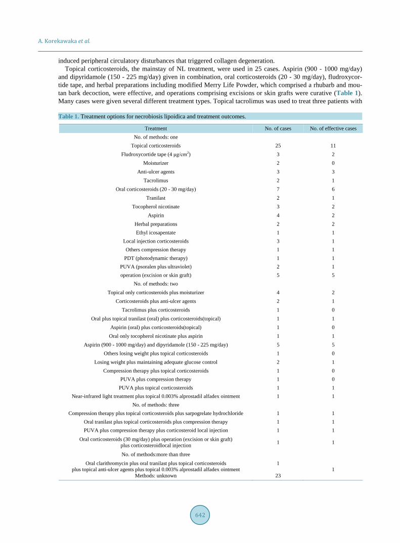
induced peripheral circulatory disturbances that triggered collagen degeneration. Topical corticosteroids, the mainstay of NL treatment, were used in 25 cases. Aspirin (900 - 1000 mg/day)
and dipyridamole (150 - 225 mg/day) given in combination, oral corticosteroids (20 - 30 mg/day), fludroxycor-tide tape, and herbal preparations including modified Merry Life Powder, which comprised a rhubarb and mou-tan bark decoction, were effective, and operations comprising excisions or skin grafts were curative (Table 1). Many cases were given several different treatment types. Topical tacrolimus was used to treat three patients with Table 1. Treatment options for necrobiosis lipoidica and treatment outcomes.
Treatment No. of cases No. of effective cases No. of methods: one
Topical corticosteroids 25 11 Fludroxycortide tape (4 μg/cm2) 3 2
Moisturizer 2 0 Anti-ulcer agents 3 3
Tacrolimus 2 1 Oral corticosteroids (20 - 30 mg/day) 7 6
Tranilast 2 1 Tocopherol nicotinate 3 2
Aspirin 4 2 Herbal preparations 2 2 Ethyl icosapentate 1 1
Local injection corticosteroids 3 1 Others compression therapy 1 1
PDT (photodynamic therapy) 1 1 PUVA (psoralen plus ultraviolet) 2 1 operation (excision or skin graft) 5 5
No. of methods: two Topical only corticosteroids plus moisturizer 4 2
Corticosteroids plus anti-ulcer agents 2 1 Tacrolimus plus corticosteroids 1 0
Oral plus topical tranilast (oral) plus corticosteroids(topical) 1 1 Aspirin (oral) plus corticosteroids(topical) 1 0
Oral only tocopherol nicotinate plus aspirin 1 1 Aspirin (900 - 1000 mg/day) and dipyridamole (150 - 225 mg/day) 5 5
Others losing weight plus topical corticosteroids 1 0 Losing weight plus maintaining adequate glucose control 2 1
Compression therapy plus topical corticosteroids 1 0 PUVA plus compression therapy 1 0
PUVA plus topical corticosteroids 1 1 Near-infrared light treatment plus topical 0.003% alprostadil alfadex ointment 1 1
No. of methods: three Compression therapy plus topical corticosteroids plus sarpogrelate hydrochloride 1 1
Oral tranilast plus topical corticosteroids plus compression therapy 1 1 PUVA plus compression therapy plus corticosteroid local injection 1 1
Oral corticosteroids (30 mg/day) plus operation (excision or skin graft) plus corticosteroidlocal injection 1 1
No. of methods:more than three Oral clarithromycin plus oral tranilast plus topical corticosteroids
plus topical anti-ulcer agents plus topical 0.003% alprostadil alfadex ointment Methods: unknown
1
23 1
A. Korekawaka et al.
643
NL, but no notable improvements were observed. While operative procedures seem to heal NL completely, there is a risk of relapse because of vascular damage and koebnerization; hence, we should be careful when making treatment decisions.
4. Conclusion Treating NL is very difficult, especially cases of ulcerated NL, and squamous cell carcinomais often associated with ulcerated NL [8]. We suggest the need for further research to determine effective treatments for NL.
Conflict of Interest Statement The authors declare no conflicts of interest.
References [1] Chung, C.G., Rosengrant, A., Helm, K.F. and Shupp, D.L. (2014) Necrobiosis Lipoidica Occurring in a Patient with
Rheumatoid Arthritis on Concurrent Tumor Necrosis Factor-α Inhibitor Therapy. International Journal of Dermatolo-gy. http://dx.doi.org/10.1111/ijd.12309
[2] Reid, S.D., Ladizinski, B., Lee, K., Baibergenova, A. and Alavi, A. (2013) Update on Necrobiosis Lipoidica: A Re-view of Etiology, Diagnosis, and Treatment Options. Journal of the American Academy of Dermatology, 69, 783-791. http://dx.doi.org/10.1016/j.jaad.2013.05.034
[3] Erfurt-Berge, C., Seitz, A.T., Rehse, C., Wollina, U., Schwede, K. and Renner, R. (2012) Update on Clinical and La-boratory Features in Necrobiosis Lipoidica: A Retrospective Multicentre Study of 52 Patients. European Journal of Dermatology, 22, 770-775.
[4] Pătraşcu, V., Giurcă, C., Ciurea, R.N., Georgescu, C.C. and Ciurea, M.E. (2014) Ulcerated Necrobiosis Lipoidica to a Teenager with Diabetes Mellitus and Obesity. Romanian Journal of Morphology and Embryology, 55, 171-176.
[5] Masatoshi, A., Kazunori, O., Osamu, I., Yoshiki, M., Toshiya, I. and Kyohei, S. (1999) Twocases of Necrobiosis Li-poidica—Its Relationships with Diabetes Mellitus and Preceding Trauma in the Japanese Patients. Rinsho Derma (To- kyo), 53, 529-532.
[6] Mariko, Y., Yasuhiko, M., Takekuni, N., Shinichiro, Y., Takashi, H. and Kenichiro, I. (2006) A Case of Necrobiosis Lipoidica without Diabetes Mellitus. Rinsho Derma (Tokyo), 60, 1020-1022.
[7] Misaki, O., Yoshihiro, S., Yuichi, T. and Seiichi, I. (2011) A Case of Necrobiosis Lipoidica Associated with Venous Insufficiency of Both Legs. Rinsho Derma (Tokyo), 65, 497-500.
[8] Uva, L., Freitas, J., Soares de Almeida, L., Vasques, H., Moura, C. and Miguel, D. (2013) Squamous Cell Carcinoma Arising in Ulcerated Necrobiosis Lipoidica Diabeticorum. International Wound Journal. http://dx.doi.org/10.1111/iwj.12206

Related Documents