Near Syncope and 1 Running head: NEAR SYNCOPE AND FATIGUE; WARNING SIGNS FOR AORTIC Near Syncope and Fatigue; Warning Signs for Aortic Stenosis Kendra L. Newland Otterbein College

Welcome message from author
This document is posted to help you gain knowledge. Please leave a comment to let me know what you think about it! Share it to your friends and learn new things together.
Transcript

Near Syncope and 1
Running head: NEAR SYNCOPE AND FATIGUE; WARNING SIGNS FOR AORTIC
Near Syncope and Fatigue; Warning Signs for Aortic Stenosis
Kendra L. Newland
Otterbein College
Near Syncope and 2
Near Syncope and Fatigue; Warning Signs for Aortic Stenosis
Chief Complaint: “I have been too weak to do my work.”
History of Present Illness: Eli Miller is a 65-year-old, married, white man. He has been
experiencing constant weakness and paroxysmal lightheadedness for the past three to four days.
Onset was sudden. He reports five or six episodes of lightheadedness in the past four days. He
denies any previous episodes. His lightheadedness worsens with exertion; however it also occurs
without any precipitating factors. He describes this lightheadedness as a “feeling that everything
is turning black” and states that he must either sit or lay down for this to subside, which it does
after a “few minutes”. He denies losing consciousness or sensations of the room spinning. He
denies headaches or recent trauma. His weakness he describes as a general lack of energy. He
states that he “feels tired all of the time”. He denies specific muscle pain or muscle weakness. He
is having trouble carrying out his normal daily activities and he is losing productivity at work.
Current medications: Mr. Miller takes the following herbals and other supplements for heart
health: Lilly of the valley 1 tsp. daily, alfalfa 2 tabs three times daily, CoQ10 twice daily,
activated charcoal four times daily.
Allergies: NKDA
Past Medical History: Positive for hypertension. He is unsure of what, if any, childhood
illnesses he had. He does not recall being sick as a child. He has not received any immunizations.
Surgical History: No past surgeries.
Social History: Mr. Miller is an Amish gentleman who is married with 10 children. He does not
smoke, drink alcohol or use illicit drugs. He drinks one cup of coffee per day. He eats three
meals per day. He completed the 8th
grade. He works in a harness shop. He gets regular exercise
with farm chores daily. He sleeps eight hours per night.
Near Syncope and 3
Family History: Father died at age 89 in an accident; no known medical problems. Mother died
in an accident at age 48; no known medical problems. He had a brother who died at the age of 41
of probable heart disease. He has a sister, in her 40’s, alive with valve disease. He has 10
children, all living with no known health problems, ages ranging from 42 years to 10 years old.
His grandparents died while he was young and he does not recall their causes of death.
Decision Point #1: Focused history, physical exam, laboratory tests
Focused History-
The possible etiologies of Mr. Miller’s vague symptoms are extensive. Based on the
history of present illness, it can be determined that Mr. Miller is experiencing near syncope.
Near syncope can be defined as, “the sense of imminent loss of consciousness without frank
syncope” (Magaziner & Walker, 2007, p. 1540). Further questioning aims to identify potential
origins of Mr. Miller’s near syncope and fatigue. This is best achieved by attaining a focused
history guided by plausible causes.
Near syncope and fatigue could arise from a neurologic pathology. A detailed history that
would give suspicion to a neurologic etiology is performed, supplementing the data that was
gathered in the history of present illness. Mr. Miller denies experiencing the feeling of losing his
balance. He denies any hearing or vision changes or trouble with speech or memory. Mr. Miller
denies numbness or tingling in his extremities or headaches. He denies loss of consciousness,
disorientation, tremors, or involuntary movements.
Infection and malignancy can present with the symptoms that Mr. Miller presents with.
He denies having a fever or chills. Mr. Miller denies having a cough, choryza, nasal congestion,
ear pain, sore throat. He denies nausea, vomiting, diarrhea or bloody stools. He denies burning or

Near Syncope and 4
pain with urination or flank pain. Mr. Miller denies any changes in his appetite or recent weight
loss.
Metabolic disorders such as hypothyroidism, hyperthyroidism, hypoglycemia and
hyperglycemia can cause a patient to have fatigue and near syncope. Mr. Miller denies being
increased thirst or hunger. He denies frequent urination. Mr. Miller denies dry skin or
constipation. He denies intolerance to heat or cold.
Cardiac etiologies are important to consider when obtaining a history from a patient with
near syncope and fatigue. Mr. Miller denies heart palpitations, chest pain, shortness of breath or
edema. He does not recall ever having been told that he has a heart murmur. He does not know if
anyone in his family was ever diagnosed with cardiomyopathy or an arrhythmia.
The practitioner must establish if the symptoms are psychogenic in nature. Mr. Miller
denies feeling nervous or anxious. He denies a significant change in his mood or dysthymia. Mr.
Miller has not had any significant changes in his life.
The detailed history does not give suspicion to any specific cause. Since these symptoms
are vague and could indicate any number of disease processes, a thorough physical examination
is performed.
Physical Examination-
Vital Signs:
Height is 5’5”; weight is 150 lbs, BMI 24.5
BP: 160/80 (right arm, lying); 154/72 (standing) HR 72 (regular) RR 18 (regular) Temp 98.6 F
General: Mr. Miller is a 65-year-old, normal weight man, well-groomed, and in good spirits. He
has no obvious physical deformities. He has a steady gait, good posture, is able to get up and
Near Syncope and 5
down from exam table without difficulty. His speech is clear. He is able to hear normal
conversational tone without difficulty. Body and breath without pungent odor.
Skin: Color pink. Skin warm and moist. No rash, petechiae, or ecchymoses. Hair with average
texture, normal distribution. Turgor good.
HEENT: Head-The head is normocephalic/atraumatic. Face is symmetric with appropriate
expression. Eyes- Symmetric, no erythema or exudate. Sclera white, conjunctiva pink.
PERRLA. Red reflex noted. Visual acuity 20/20 on Snellen chart without correction. Ears:
Acuity good to normal conversational tone at 3 feet. External ears symmetric without erythema,
masses or edema. TMs with good cone of light. Nose: External nose symmetric, without
erythema, edema. Nasal mucosa pink. No sinus tenderness. Mouth and throat: Oral mucosa pink,
dentition good, pharynx without exudates. Gums pink. Tongue midline and pink. Soft palate and
uvula mobile. Uvula midline. Neck: Trachea midline. Neck symmetric, supple, thyroid isthmus
palpable, lobes not felt. No visible lumps or pulsations. No bruits. Full range of motion, strong
muscle strength.
Lymph nodes: Tonsillar, submandibular, submental, anterior and posterior cervical, preauricular,
posterior auricular, occipital, inguinal, axillary, and inguinal nodes without noted
lymphadenopathy or tenderness.
Chest: Thorax is symmetric with good expansion. Respirations even and unlabored. Clear breath
sounds bilaterally anteriorly and posteriorly.
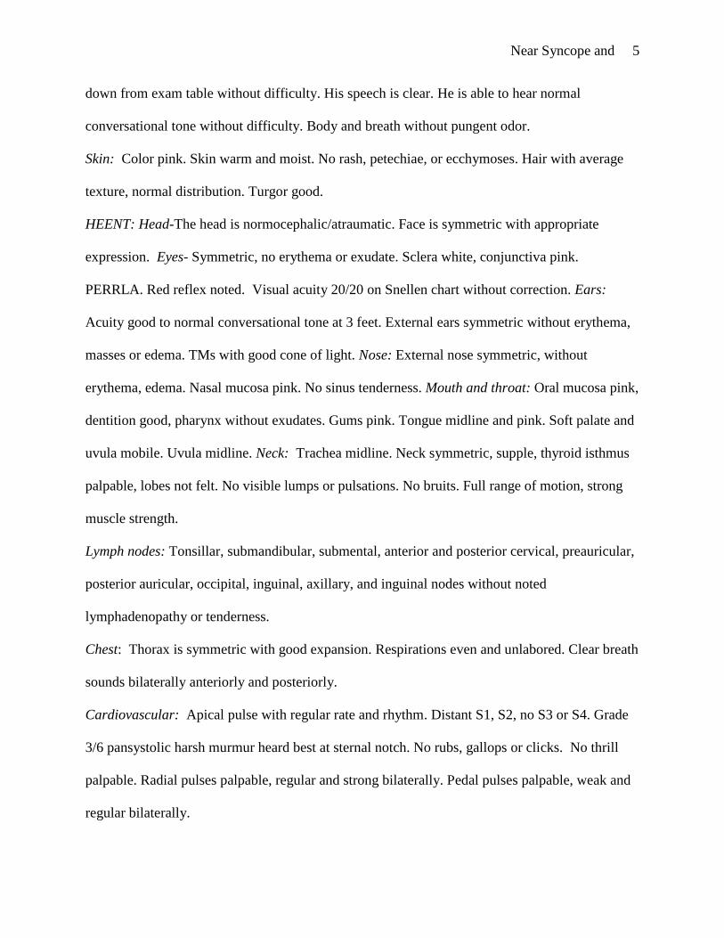
Cardiovascular: Apical pulse with regular rate and rhythm. Distant S1, S2, no S3 or S4. Grade
3/6 pansystolic harsh murmur heard best at sternal notch. No rubs, gallops or clicks. No thrill
palpable. Radial pulses palpable, regular and strong bilaterally. Pedal pulses palpable, weak and
regular bilaterally.
Near Syncope and 6
Peripheral Vascular System: Extremities are warm and without edema. No varicosities or stasis
changes.
Gastrointestinal: Abdomen is flat, soft, non-distended and non-tender. Active bowel sounds in
all 4 quadrants. No masses or hepatosplenomegaly. No costovertebral angle tenderness. No
inguinal lymphadenopathy or tenderness.
Musculoskeletal: No joint deformities. Good range of motion and strong muscle strength in
hands, wrists, elbows, shoulders, spine, hips, knees, ankles.
Neurologic: Mental Status: Alert and cooperative. Thought coherent. Oriented to person, place
and time. Cranial Nerves: CN II-XII intact. No nystagmus. Motor: Good muscle bulk and tone.
Gait stable, fluid. Negative Dix-Hallpike maneuver. Negative Brudzinski’s sign. Rapid
alternating movements, finger-to-nose and heel-to-shin intact. Romberg – maintains balance with
eyes closed. No pronator drift. Deep tendon reflexes 2+ bilaterally with plantar reflexes
downgoing.
Preliminary Labs and Testing:
With syncope or near syncope, there is no one test identified as a gold standard for
diagnosis. The practitioner must order diagnostic testing based on sound clinical judgment. From
the findings of the physical exam, explicitly the murmur that was noted, an echocardiogram is
warranted. Echocardiography is the preferred method to identify the cause and severity of heart
murmurs (McCannon, 2004). An electrocardiogram is also indicated to identify a cardiac
etiology of Mr. Miller’s near syncope. It is important to note that Mr. Miller does not have
medical insurance. This fact, along with cultural beliefs and practices, greatly influence the
degree of testing that is performed initially. The Amish are cautious and conservative in their use
of modern medical technology (Armer & Radina, 2006). Although it was highly recommended,
Near Syncope and 7
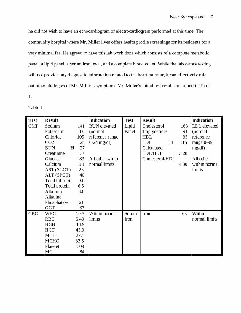
he did not wish to have an echocardiogram or electrocardiogram performed at this time. The
community hospital where Mr. Miller lives offers health profile screenings for its residents for a
very minimal fee. He agreed to have this lab work done which consists of a complete metabolic
panel, a lipid panel, a serum iron level, and a complete blood count. While the laboratory testing
will not provide any diagnostic information related to the heart murmur, it can effectively rule
out other etiologies of Mr. Miller’s symptoms. Mr. Miller’s initial test results are found in Table
1.
Table 1
Test Result Indication Test Result Indication
CMP Sodium 141
Potassium 4.6
Chloride 105
CO2 28
BUN H 27
Creatinine 1.0
Glucose 83
Calcium 9.1
AST (SGOT) 23
ALT (SPGT) 40
Total bilirubin 0.6
Total protein 6.5
Albumin 3.6
Alkaline
Phosphatase 121
GGT 37
BUN elevated
(normal
reference range
6-24 mg/dl)
All other within
normal limits
Lipid
Panel
Cholesterol 168
Triglycerides 91
HDL 35
LDL H 115
Calculated
LDL/HDL 3.28
Cholesterol/HDL
4.80
LDL elevated
(normal
reference
range 0-99
mg/dl)
All other
within normal
limits
CBC WBC 10.5
RBC 5.49
HGB 14.9
HCT 45.9
MCH 27.1
MCHC 32.5
Platelet 309
MC 84
Within normal
limits
Serum
Iron
Iron 63 Within
normal limits
Near Syncope and 8
Decision Point #2: Differential diagnoses
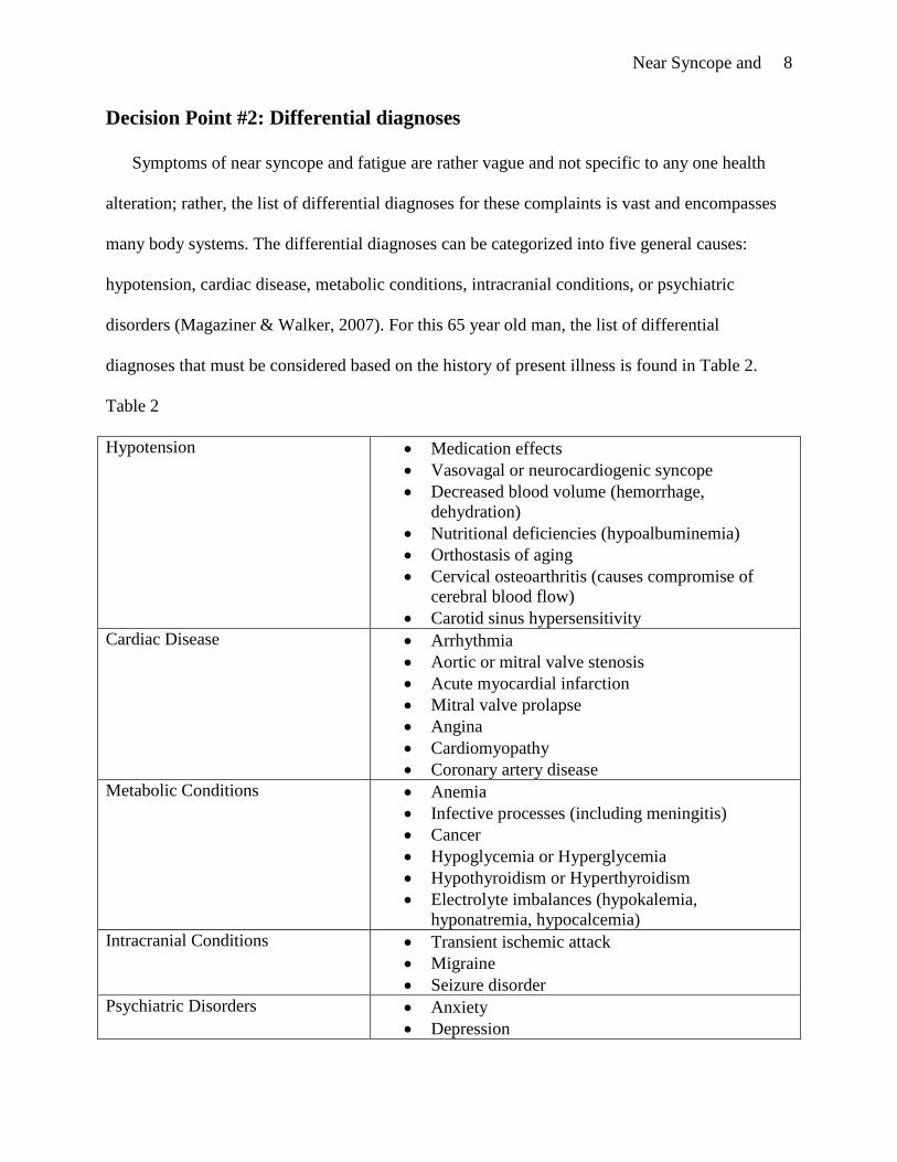
Symptoms of near syncope and fatigue are rather vague and not specific to any one health
alteration; rather, the list of differential diagnoses for these complaints is vast and encompasses
many body systems. The differential diagnoses can be categorized into five general causes:
hypotension, cardiac disease, metabolic conditions, intracranial conditions, or psychiatric
disorders (Magaziner & Walker, 2007). For this 65 year old man, the list of differential
diagnoses that must be considered based on the history of present illness is found in Table 2.
Table 2
Hypotension Medication effects
Vasovagal or neurocardiogenic syncope
Decreased blood volume (hemorrhage,
dehydration)
Nutritional deficiencies (hypoalbuminemia)
Orthostasis of aging
Cervical osteoarthritis (causes compromise of
cerebral blood flow)
Carotid sinus hypersensitivity
Cardiac Disease Arrhythmia
Aortic or mitral valve stenosis
Acute myocardial infarction
Mitral valve prolapse
Angina
Cardiomyopathy
Coronary artery disease
Metabolic Conditions Anemia
Infective processes (including meningitis)
Cancer
Hypoglycemia or Hyperglycemia
Hypothyroidism or Hyperthyroidism
Electrolyte imbalances (hypokalemia,
hyponatremia, hypocalcemia)
Intracranial Conditions Transient ischemic attack
Migraine
Seizure disorder
Psychiatric Disorders Anxiety
Depression
Near Syncope and 9
A thorough history and review of symptoms is crucial to narrowing the list of
differentials and assist the practitioner in focusing the physical exam. The physical exam will
help guide the practitioner to further limit the probable diagnoses and to determine what testing
is indicated. It is vital that the practitioner distinguishes between benign and life-threatening
causes of syncope.
In the case of Mr. Miller, the detailed history effectively ruled out a number of possible
diagnoses. Mr. Miller denied any symptoms of an acute infectious process. He denied any
symptoms of anxiety or depression. He denied a headache, effectively ruling out migraines. A
diagnosis of seizure is five times more likely if the patient is disoriented after the event
(Magaziner & Walker, 2007). Mr. Miller denied feeling disoriented after episodes of
lightheadedness, in effect ruling out seizures. He denies any chest pain therefore ruling out
angina. He denies symptoms of palpitations or heat intolerance which guides the practitioner
away from hyperthyroidism. He also denies symptoms of cold intolerance and constipation
which direct the practitioner away from hypothyroidism. Similarly, he denies symptoms of
hyper- and hypoglycemia (hunger, palpitations). The laboratory testing excluded anemias
(normal hemoglobin, hematocrit, red blood cell count), electrolyte imbalances (normal sodium,
potassium, calcium carbon dioxide levels), dehydration (normal creatinine, BUN, electrolytes),
and nutritional deficiencies (normal albumin, electrolytes). The blood work did reveal a slight
elevation in his LDL level. The physical exam ruled out orthostatic hypotension, which is
defined as a decrease in systolic blood pressure of 20 mmHg or more when patient changes from
a supine to standing position. Mr. Miller’s orthostatic blood pressure measurements did not
indicate orthostatic hypotension. Meningitis was ruled out with a negative Brudzinski’s sign.
Near Syncope and 10
The physical exam did reveal a grade 3/6 harsh pansystolic murmur which indicates a
possible cardiac etiology for Mr. Miller’s symptoms. Organic heart disease may be a life-
threatening cause of near syncope (Magaziner & Walker, 2007). Other possible diagnoses
include neurocardiogenic syncope, medication effects, transient ischemic attack, corornary artery
disease, arrhythmia, and aortic stenosis (among other diseases that inhibit cardiac outflow).
Medication effects
Alternative medicine, including the use of herbal supplementation, is routinely practiced
in the Amish culture (Armer & Radina, 2006). Many patients will not include herbals and other
supplements in their list of medications, so the practitioner must explicitly question the patient
about the use of alternative therapies. People consider herbs to be safe because they are
“natural”. Unlike prescription drugs, the Food and Drug Administration does not regulate herbs
and supplements, therefore potency and purity is not guaranteed. Many herbs have not been
thoroughly tested and safety and effectiveness are not proven. Furthermore, herbal supplements
do occasionally have serious side effects and may interact with prescription drugs. Mr. Miller
takes activated charcoal, alfalfa, Co-Q 10, and Lily of the Valley on a daily basis. Activated
charcoal is said to help lower cholesterol. Activated charcoal absorbs many materials, including
prescription drugs. Alfalfa, thought to lower cholesterol and glucose, may cause hypoglycemia
which can lead to syncope (U.S. National Library of Medicine, 2008). Coenzyme Q 10 is
produced by the body and is necessary for the basic functioning of cells. Levels are thought to be
low in some chronic conditions such as heart conditions, cancer, diabetes and Parkinson’s
disease. Coenzyme Q10 may cause hypoglycemia and dizziness, and it may lower blood
pressure, among other side effects (U.S. National Library of Medicine, 2008). Lily of the Valley
is another herb thought to help the heart. Some concerning side effects reported are heart failure,

Near Syncope and 11
coma and death (Thomson Reuters, n.d.). The side effects of herbs as a causative factor of Mr.
Miller’s symptoms cannot be ruled out without stopping the supplements to determine if the
symptoms cease. In light of the cardiac murmur that was identified with the physical exam,
medication effect is not explored as a causative factor at this time.
Neurocardiogenic syncope
Neurocardiogenic syncope, or vasovagal syncope, is the most common cause of syncope in
adults and children, accounting for 50-66% of inexplicable syncope (Chen-Scarabelli &
Scarabelli, 2004). A benign condition, it is caused by an abnormal autonomic response to stimuli
and results in self-limited bradycardia and hypotension. This bradycardia and systemic
hypotension leads to syncope; consciousness is regained promptly after the patient lies down.
Often, there is a prodromal period in which the patient may experience lightheadedness, pallor,
flushing, nausea, palpitations and throat tightness (Magaziner & Walker, 2007). Syncope can be
avoided if the patient lies down during this prodromal period. After a syncopal episode, the
patient often complains of tiredness which resolves. In more than 50% of patients with
neurocardiogenic syncope, the history and physical are non-diagnostic (Chen-Scarabelli &
Scarabelli), therefore, this is often a diagnosis of exclusion. Tilt-table testing can be done to
confirm the diagnosis.
Mr. Miller has near syncopal episodes that completely resolve with lying or sitting down.
He describes general fatigue, not associated specifically with the near syncopal episodes. In the
case of Mr. Miller, the discovery of the heart murmur during the physical exam leads the
practitioner to consider a cardiac cause rather than the benign diagnosis of neurocardiogenic
syncope. If further workup indicates that the near syncope that Mr. Miller is experiencing is not
related to a cardiac etiology, this diagnosis may be reconsidered.

Near Syncope and 12
Transient ischemic attack (TIA)
Mr. Miller is at increased risk for cerebrovascular disease due to hypertension and
hyperlipidemia. The identification of a heart murmur in his physical exam also adds to the
suspicion of a TIA. It is known that 15% to 30% of all ischemic strokes are attributed to an
embolus originating in the heart which is highly associated with cardiac arrhythmias, valvular
disease, recent myocardial infarction and dilated cardiomyopathy (Llinas & Johnson, 2007).
Hypertension and hyperlipidemia also place Mr. Miller at risk for decreased cerebral blood flow
due to diseased carotid arteries. TIA is defined as a “transient episode of focal cerebral
dysfunction, rapid in onset (from none to maximum symptoms in less than five minutes), that
usually lasts from two to fifteen minutes but always resolves completely within 24 hours (Llinas
& Johnson, 2007, p. 1573). Mr. Miller’s near syncope completely resolved within a few
minutes, as would be expected with a TIA. However, Henry and Johnston (2004) report that if
syncope (or, in this case, near syncope) occurs with a TIA, more typical TIA symptoms generally
accompany (such as slurred speech or one-sided weakness). Also, it is not likely that a transient
episode with altered consciousness is vascular in nature (Llinas & Johnson, 2007). Taking this
into account, TIA is not considered a likely diagnosis; however, it should not be ignored due to
the potential mortality and morbidity. Additionally, the clinical evidence of heart disease,
specifically the cardiac murmur, would direct the practitioner to obtain an echocardiogram
(which would identify a cardiac source of emboli) and electrocardiogram (which would identify
an arrhythmia).
Coronary Artery Disease, Arrhythmias, and Myocardial Infarction
Coronary artery disease (CAD) caused by atherosclerosis affects over 13,000,000
Americans (Chandra-Strobos & Hirsch, 2007). Mr. Miller is at increased risk for coronary artery
Near Syncope and 13
disease from hypertension and hyperlipidemia (based on recent laboratory findings) as well as a
family history of heart trouble. Patients with CAD are at risk for having a myocardial infarction
(MI). An acute MI may present without the classical symptoms, particularly in an older adult. In
fact, the sole presenting manifestation of an MI may be syncope (or near syncope) (Kyrillos,
Carissa, & Pineda, 2005). For Mr. Miller, coronary artery disease and myocardial infarction are
possible diagnoses due to his risk factors. While he does not have the classic symptoms of an
acute MI, the practitioner should not rule this out without first checking a 12-lead
electrocardiogram. An electrocardiogram may be normal in CAD, but would be abnormal in a
patient with a recent or past history of an MI.
Cardiac arrhythmias can be symptoms of disease or they can cause disease. They can be
relatively benign or they can be deadly. Arrhythmias should always be considered with near-
syncope, particularly in the older adult (Magaziner & Walker, 2007). Mr. Miller’s symptom of
lightheadedness might be caused by decreased cardiac output as a result of an arrhythmia. Mr.
Miller has a history of hypertension and has a cardiac murmur, two underlying diseases that may
be associated with arrhythmias (Gottlieb, Marine, & Calkins, 2007). Mr. Miller denies
palpitations; however, the presence or absence of symptoms does not confirm or eradicate the
possibility of an arrhythmia. Upon physical assessment, his heart rate was within normal limits
with a regular rhythm. Arrhythmias may be paroxysmal in nature, so they may not be identified
during an outpatient office visit. A single electrocardiogram tracing may not identify a
paroxysmal arrhythmia. If arrhythmia is still suspected after a normal 12-lead electrocardiogram,
a 24 hour ambulatory electrocardiogram is indicated. In Mr. Miller’s case, arrhythmia is of high
concern and cannot be ruled out at this time.
Aortic Stenosis
Near Syncope and 14
Aortic stenosis (AS) is an abnormal narrowing of the aortic valve. It is a significant cause
of morbity and mortality among the elderly population (Mallavarapu, Mankad, & Nanda, 2007).
Aortic stenosis has been documented in 2% to 4% of patients over 65 years of age (Lauck,
Mackay, Galte, & Wilson, 2008). Its precursor, aortic sclerosis, is present in an estimated 25% of
the population over 65 years of age (Keong Yeo & Low, 2007).In the older adult, the most
common cause is calcification of the aortic valve. Evidence indicates that the same factors that
influence the development of CAD, like smoking, hypercholesterolemia, hypertension and
diabetes, also influence the progression of AS (Lauck et al., 2008). Historically, AS has been
considered to be a degenerative disease. However, new evidence suggests that AS may be an
active disease similar to CAD involving lipoprotein deposition, chronic inflammation and active
calcification of the leaflets (Lauck et al., 2008). As calcification progresses, the valve leaflets
become stiff and narrow, causing impaired valve opening. The outflow of blood from the left
ventricle is obstructed; the pressure in the left ventricle increases, leading to concentric
hypertrophy of the left ventricle. This hypertrophy enables the heart to compensate and maintain
adequate cardiac output for a period of time during which the patient is clinically asymptomatic.
However, this hypertrophy also decreases coronary blood flow and leads to diastolic and systolic
left ventricular dysfunction. When the compensatory mechanisms fail, the patient starts
exhibiting classic symptoms of AS: heart failure, angina or syncope. Initially, many patients
experience decreased exercise tolerance and other vague signs and symptoms (Lauck et al.,
2008).Once symptoms develop, the average survival is two to three years. It is important to note
that the severity of AS is poorly associated with signs and symptoms. Patients with aortic
stenosis generally have a grade 1-4/6 harsh, crescendo-decrescendo, systolic murmur heard best
at the second right intercostal space or at the apex. The murmur may radiate to the carotid

Near Syncope and 15
arteries. The murmur often softens with standing and increases when the patient leans forward.
S1 and S2 may be diminished. The patient with AS may complain of fatigue, dyspnea on
exertion or angina. Mr. Miller’s cardiac exam revealed distant S1, S2, grade 2/6 pansystolic
harsh murmur heard best at the sternal notch. He complains of fatigue, but denies angina or
dyspnea. He does have exertional near syncope, which is a classic symptom of AS. Elderly
patients with severe aortic stenosis may present with subtle symptoms (Grimard & Larson,
2008). Exertional dyspnea is the most common initial complaint (Talano & Melek, 2007). In
light of the harsh systolic murmur and clinical presentation, as well as the prevalence of the
disease among the older population, aortic stenosis is a viable diagnosis. It is important to note
that in the case of Mr. Miller rheumatic valve disease should be considered since he is unable to
recall his history of childhood illnesses. Also, the Amish culture often relies on alternative
therapies to treat disease, so he may have had an untreated streptococcal infection at some point.
Decision Point #3: Further work-up
At this point, further work-up includes an echocardiogram to visualize the heart valves
and an electrocardiogram to identify any cardiac arrhythmias and rule out myocardial infarction.
The patient called the office two weeks after his original office visit and informed the
practitioner that he was experiencing dyspnea with exertion. He agreed to have an
echocardiogram and electrocardiogram performed.
Decision Point #4: Management
The presumptive diagnosis prior to the echocardiogram and electrocardiogram is aortic
stenosis based on the auscultation of a harsh systolic murmur and the presence of the classic
symptom of exertional near-syncope. A cardiology referral is made. He is given a prescription
for lisinopril 5 mg daily for his hypertension. It is estimated that 40% of patients with AS also

Near Syncope and 16
have hypertension (Grimard & Larson, 2008). Hypertension increases systemic vascular
resistance and further creates an increase in left ventricular afterload. Angiotensin-converting
enzyme inhibitors are well tolerated and will improve exercise tolerance in the symptomatic
patient (Grimard & Larson). Mr. Miller is also prescribed lovastatin 20 mg daily for
hyperlipidemia. There is conflicting evidence on the use of statins to slow the progression of AS.
Larger studies are expected to conclude in 2009 (Grimard & Larson). Mr. Miller is encouraged
to take an 81 mg aspirin daily due to his risk of cardiovascular disease. The use of herbal
medications and their possible risks is discussed. He is instructed to seek immediate medical
attention if his symptoms suddenly worsen. In 2008, the American Heart Association stated that
antibiotic prophylaxis was no longer indicated for prevention of infective endocarditis in patients
with aortic stenosis (Nishimura et al., 2008).
Watchful waiting is recommended in most asymptomatic patients, with survival rates
comparable to those in patients without AS (Grimard & Larson, 2008). Doppler echocardiology
should be performed every year for the asymptomatic patient with severe AS, every one to two
years with moderate AS and every three to five years for mild AS (Grimard & Larson, 2008).
Symptomatic patients with severe aortic stenosis have poor prognosis without valve replacement;
52% of these patients who did not receive surgery were dead at 5 years according to the National
Institutes of Health (Aronow, 2007).The only definitive treatment for symptomatic moderate to
severe AS in the elderly is aortic valve replacement (Aronow, 2007). In fact, the 10-year-
survival rate after aortic valve replacement in patients over 65 years of age is almost the same as
age- and sex-matched patients without AS (Grimard & Larson, 2008). The degree of severity
corresponds to the peak gradient across the aortic valve and the aortic valve area (as measured by

Near Syncope and 17
echocardiography) and is classified as mild, moderate or severe (see Table 3 for more
information). Mr. Miller is symptomatic with near-syncope and dyspnea.
Table 3 (Grimard & Larson, 2008, p.720)
Severity Aortic valve area
(cm2)
Aortic jet velocity
(m per second)
Mean Gradient
(mmHg)
Normal 3 to 4 < 2.5 --
Mild 1.5 to 2 2.5 to 2.9 <25
Moderate 1 to 1.5 3 to 4 25 to 40
Severe <1 >4 >40
Decision Point #5: Final diagnosis
The electrocardiogram was abnormal, showing sinus rhythm at 91 beats per minute with
a marked left ventricular hypertrophy with strain pattern. The echocardiogram showed severe
calcification of the aortic valve with severe stenosis (valve area 1 cm2, peak gradient 68 mmHg),
concentric left ventricular hypertrophy, and mild left ventricular systolic dysfunction (ejection
fraction 50%). The electrocardiogram showed no evidence of a myocardial infarction, so this can
be ruled out. Based on the results of the echocardiogram and electrocardiogram, the diagnosis of
aortic stenosis is verified and left ventricular hypertrophy is diagnosed. When the
echocardiogram shows severe aortic stenosis in a symptomatic patient, symptoms must be
presumed to be caused by the AS, even if other potential causes are present (Grimard & Larson,
2008).
The patient did follow up with the cardiologist who recommended a heart catheterization
and angiogram to assess the aortic valve and to examine the coronary arteries. The probability of
a patient requiring an aortic valve replacement was discussed. The patient wanted to consider it

Near Syncope and 18
and has not followed up with the cardiologist. On his return visit to the family practitioner, he
reported that he did not wish to have surgery. He stated that he has not filled the prescriptions
given to him and he is continuing to use the herbal supplements on a daily basis. The practitioner
discussed with Mr. Miller the seriousness of this condition and the poor prognosis without
surgical intervention. The risk of congestive heart failure, heart attack or sudden cardiac death
was discussed at length. He was instructed to follow a low salt diet so as not to develop fluid
volume overload. He was, again, strongly encouraged to take the lisinopril, lovastatin, and baby
aspirin. Mr. Miller understood all that was discussed and wished to go home and think about it.
Case Study Summary
When a patient presents with near syncope and fatigue, the practitioner must rely on a
thorough history and expert physical assessment skills to guide clinical decision making.
Whenever these symptoms are present with a cardiac murmur, a cardiac etiology must be
considered. In the case of Mr. Miller, the finding of a harsh, crescendo-decrescendo systolic
murmur indicated aortic stenosis therefore, an echocardiogram was ordered. Aortic stenosis is a
common cause of morbidity and mortality among the older population. While valvular heart
disease may be the cause these symptoms in a patient, it is not always the case, and they could
arise from another source. For this reason, other diagnosis should remain on the differential list
until proven otherwise. In Mr. Miller’s case, neurocardiogenic syncope, arrhythmia, coronary
artery disease, myocardial infarction, effects of herbal medications, and transient ischemic attack
remained in the back of the practitioner’s mind until further testing was performed. The
echocardiogram showed severe aortic stenosis, which leads the practitioner to presume that the
symptoms are related to the valvular disease versus another cause. In the case of Mr. Miller, the
practitioner prescribes an ACE inhibitor for hypertension and a statin for hyperlipidemia. He is

Near Syncope and 19
encouraged to take a baby aspirin daily and to discontinue the use of herbal medications. The
patient is referred to a cardiologist for further work-up and treatment. Cultural beliefs and
practices play a role in the management of Mr. Miller, and the practitioner must be sensitive to
this.
Near Syncope and 20
References
Armer, J. M., & Radina, M. E. (2006). Definition of health and health promotion behaviors
among Midwestern old order Amish families. The Journal of Multicultural Nursing &
Health, 12(3), 44-53.
Aronow, W. S. (2007). Recognition and management of aortic stenosis in the elderly. Geriatrics,
62(12), 23-32.
Chandra-Strobos, N., & Hirsch, G. A. (2007). Coronary artery disease. In N. H. Fiebach, D. E.
Kern, P. A. Thomas, R. C. Ziegelstein, L. R. Barker, & P. D. Zieve (Eds.), Barker,
Burton and Zieve’s principles of ambulatory medicine (pp. 949-970). Philadelphia:
Lippincott, Williams & Wilkins.
Chen-Scarabelli, C., & Scarabelli, T. M. (2004). Neurocardiogenic syncope. BMJ: British
Medical Journal, 329, 336-341.
Gottlieb, S. H., Marine, J. E., & Calkins, H. (2007). Cardiac Arrhythmias. In N. H. Fiebach, D.
E. Kern, P. A. Thomas, R. C. Ziegelstein, L. R. Barker, & P. D. Zieve (Eds.), Barker,
Burton and Zieve’s principles of ambulatory medicine (pp. 992-1028). Philadelphia:
Lippincott Williams & Wilkiins.
Grimard, B. H., & Larson, J. M. (2008). Aortic stenosis: Diagnosis and treatment. American
Family Physician, 78(6), 717-724, 725.
Henry, G., & Johnston, S. (2004). Acute neurologic symptoms -- TIA, migraine, or something
else?. Patient Care for the Nurse Practitioner. Retrieved February 22, 2009 from
CINAHL Plus with Full Text database.
Keong Yeo, K., & Low, R. I. (2007). Aortic stenosis: Assessment of the patient at risk. Journal
of Interventional Cardiology, 20(6), 509-516.
Near Syncope and 21
Kyrillos, J., Carissa, M., & Pineda, C. (2005). Syncope in the elderly patient; common and
uncommon causes. Patient Care for the Nurse Practitioner, Retrieved February 22, 2009
from CINAHL Plus with Full Text database.
Lauck, S., Mackay, M., Galte, C., & Wilson, M. (2008). A new option for the treatment of aortic
stenosis: Percutaneous aortic valve replacement. Critical Care Nurse, 28(3), 40-51.
Llinas, R. H., & Johnson, C. J. (2007). Cerebrovascular disease. In N. H. Fiebach, D. E. Kern, P.
A. Thomas, R. C. Ziegelstein, L. R. Barker, & P. D. Zieve (Eds.), Barker, Burton and
Zieve’s Principles of ambulatory medicine (pp. 1569-1580). Philadelphia: Lippincott
Williams & Wilkins.
Magaziner, J. L., & Walker, M. F. (2007). Dizziness, Vertigo, Motion Sickness, Syncope and
Near Syncope, and Disequilibrium. In N. H. Fiebach, D. E. Kern, P. A. Thomas, R. C.
Ziegelstein, L. R. Barker, & P. D. Zieve (Eds.), Barker, Burton and Zieve’s Princples of
ambulatory medicine (pp. 1531-1553). Philadelphia: Lippincott Williams & Wilkins.
Mallavarapu, R. K., Mankad, S., & Nanda, N. C. (2007). Echocardiographic assessment of aortic
stenosis in the elderly. The American Journal of Geriatric Cardiology, 16(6), 343-348.
McCannon, M. (2004). Evaluation of heart murmurs in the elderly. The American Journal for
Nurse Practitioners, 8(11), 11-26.
Nishimura, R., Carabello, B., Faxon, D., Freed, M., Lytle, B., & O’Gara, P. et al. (2008).
ACC/AHA 2008 guideline update on valvular heart disease: Focused update on infective
endocarditis. Retrieved from
http://circ.ahajournals.org/cgi/reprint/CIRCULATIONAHA.108.190377
Talano, J. V., & Melek, B. H. (2007). Aortic stenosis. Retrieved February 18, 2009, from
http://emedicine.medscape.com/article/150638-overview

Near Syncope and 22
Thomson Reuters (n.d.). Lily of the Valley herbal remedies, supplements PDR health. Retrieved
February 22, 2009, from http://www.pdrhealth.com/drugs/altmed/altmed-
mono.aspx?contentFileName=ame0251.xml&contentName=Lily+of+the+Valley&conten
tId=411#
U.S. National Library of Medicine. (2008). MedLine plus herbs and supplements: Alfalfa
(Medicago sativa L.) [Brochure]. Retrieved from
http://www.nlm.nih.gov/medlineplus/druginfo/natural/patient-alfalfa.html
U.S. National Library of Medicine. (2008). MedLine plus herbs and supplements: Coenzyme
Q10 [Brochure]. Retrieved from
http://www.nlm.nih.gov/medlineplus/druginfo/natural/patient-coenzymeq10.html
Related Documents


![Syncope AHD[1]](https://static.cupdf.com/doc/110x72/577d36611a28ab3a6b92ec10/syncope-ahd1.jpg)