HEMATOLOGY Dr. I. Quirt Adriana Cipolletti, Jeremy Gilbert and Susy Hota, chapter editors Leora Horn, associate editor APPROACH TO THE BLOOD FILM . . . . . . . . . . 2 ANEMIA . . . . . . . . . . . . . . . . . . . . . . . . . . . . . . . . . . 3 Clinical Approach to Anemia IRON METABOLISM . . . . . . . . . . . . . . . . . . . . . . . 3 Iron Intake (Dietary) Physiologic Causes of Increased Fe Requirements Iron Absorption Iron Transport Iron Storage Iron Indices Laboratory Features IRON DEFICIENCY . . . . . . . . . . . . . . . . . . . . . . . . . 5 Physiologic Causes Pathological Causes Clinical Presentation Diagnosis Treatment Recovery Time Anemia Refractory to Treatment with Oral Iron THE ANEMIA OF CHRONIC DISEASE . . . . . . 6 LEAD POISONING . . . . . . . . . . . . . . . . . . . . . . . . . 6 SIDEROBLASTIC ANEMIA . . . . . . . . . . . . . . . . . 7 HEMOGLOBIN AND . . . . . . . . . . . . . . . . . . . . . . . 7 HEMOGLOBINOPATHIES Thalassemia I. Heterozygous: ß-Thalassemia Minor II. Homozygous: ß-Thalassemia Major III. Alpha Thalassemias Sickle Cell Anemia Megaloblastic Anemia B12 Deficiency Pernicious Anemia Folate Deficiency Hemolytic Anemias I. Hereditary Hemolytic Anemias Structural Abnormalities in Cytoskeleton Enzymatic Abnormalities in RBC II. Acquired Hemolytic Anemias Autoimmune Hemolytic Anemia RBC Fragmentation Syndromes Thrombotic Thrombocytopenic Purpura and Hemolytic Uremic Syndrome APLASTIC ANEMIA . . . . . . . . . . . . . . . . . . . . . . . .15 HEMOSTASIS . . . . . . . . . . . . . . . . . . . . . . . . . . . . .16 Three Phases of Hemostasis Tests of Hemostasis Thrombocytopenia & Other Disorders of 1º Hemostasis Idiopathic (Autoimmune) Thrombocytopenic Purpura (ITP) Chronic (Adult-type) ITP Disorders of Secondary Hemostasis Hereditary Acquired Thrombosis Heparin-Induced Thrombocytopenia (HIT) MCCQE 2002 Review Notes Hematology – H1 HEMATOLOGIC MALIGNANCIES . . . . . . . . . . .24 Overview MYELOID MALIGNANCIES . . . . . . . . . . . . . . . . .24 Acute Myeloid Leukemia (AML) CHRONIC MYELOPROLIFERATIVE . . . . . . . . . .26 DISORDERS Common Features Polycythemia Rubra Vera (PRV) Chronic Granulocytic (Myelogenous) Leukemia (CML) Idiopathic Myelofibrosis Essential Thrombocythemia (ET) MYELODYSPLASTIC SYNDROMES . . . . . . . . . .29 LYMPHOID MALIGNANCIES . . . . . . . . . . . . . . . .30 ACUTE LYMPHOBLASTIC LEUKEMIA (ALL) .30 LYMPHOMAS . . . . . . . . . . . . . . . . . . . . . . . . . . . . .30 Hodgkin's Disease and Non-Hodgkin's Lymphoma Staging Hodgkin's Disease Non-Hodgkin's Lymphoma MALIGNANT CLONAL . . . . . . . . . . . . . . . . . . . . .33 PROLIFERATIONS OF B CELLS Chronic Lymphocytic Leukemia (CLL) Plasma Cell Myeloma (Multiple Myeloma) Light Chain Disease Monoclonal Gammopathy of Unknown Significance (MGUS) Macroglobulinemia of Waldenstrom Macroglobulinemia-Hyperviscosity Syndrome Bone Marrow Transplantation TUMOUR LYSIS SYNDROME . . . . . . . . . . . . . . .36 WBC DISORDERS . . . . . . . . . . . . . . . . . . . . . . . . . .36 Neutrophilia Leukemoid Reactions Neutropenia Agranulocytosis APPROACH TO SPLENOMEGALY . . . . . . . . . . .37 BLOOD PRODUCTS AND TRANSFUSIONS . .38 Blood Groups Red Cells Platelets Coagulation Factors Group and Reserve Serum Acute Complications of Blood Transfusions Delayed Complications in Transfusions MEDICATIONS COMMONLY USED IN . . . . . . .41 HEMATOLOGY REFERENCES . . . . . . . . . . . . . . . . . . . . . . . . . . . . .42

Welcome message from author
This document is posted to help you gain knowledge. Please leave a comment to let me know what you think about it! Share it to your friends and learn new things together.
Transcript

HEMATOLOGYDr. I. Quirt
Adriana Cipolletti, Jeremy Gilbert and Susy Hota, chapter editorsLeora Horn, associate editor
APPROACH TO THE BLOOD FILM . . . . . . . . . . 2
ANEMIA . . . . . . . . . . . . . . . . . . . . . . . . . . . . . . . . . . 3Clinical Approach to Anemia
IRON METABOLISM . . . . . . . . . . . . . . . . . . . . . . . 3Iron Intake (Dietary)Physiologic Causes of Increased Fe RequirementsIron AbsorptionIron TransportIron StorageIron IndicesLaboratory Features
IRON DEFICIENCY . . . . . . . . . . . . . . . . . . . . . . . . . 5Physiologic CausesPathological CausesClinical PresentationDiagnosisTreatmentRecovery TimeAnemia Refractory to Treatment with Oral Iron
THE ANEMIA OF CHRONIC DISEASE . . . . . . 6
LEAD POISONING . . . . . . . . . . . . . . . . . . . . . . . . . 6
SIDEROBLASTIC ANEMIA . . . . . . . . . . . . . . . . . 7
HEMOGLOBIN AND . . . . . . . . . . . . . . . . . . . . . . . 7HEMOGLOBINOPATHIESThalassemia
I. Heterozygous: ß-Thalassemia MinorII. Homozygous: ß-Thalassemia MajorIII. Alpha Thalassemias
Sickle Cell AnemiaMegaloblastic AnemiaB12 DeficiencyPernicious AnemiaFolate DeficiencyHemolytic Anemias
I. Hereditary Hemolytic AnemiasStructural Abnormalities in CytoskeletonEnzymatic Abnormalities in RBC
II. Acquired Hemolytic AnemiasAutoimmune Hemolytic AnemiaRBC Fragmentation SyndromesThrombotic Thrombocytopenic Purpura and Hemolytic Uremic Syndrome
APLASTIC ANEMIA . . . . . . . . . . . . . . . . . . . . . . . .15
HEMOSTASIS . . . . . . . . . . . . . . . . . . . . . . . . . . . . .16Three Phases of HemostasisTests of HemostasisThrombocytopenia & Other Disorders of 1º Hemostasis
Idiopathic (Autoimmune) Thrombocytopenic Purpura (ITP)
Chronic (Adult-type) ITPDisorders of Secondary Hemostasis
HereditaryAcquiredThrombosis
Heparin-Induced Thrombocytopenia (HIT)
MCCQE 2002 Review Notes Hematology – H1
HEMATOLOGIC MALIGNANCIES . . . . . . . . . . .24Overview
MYELOID MALIGNANCIES . . . . . . . . . . . . . . . . .24Acute Myeloid Leukemia (AML)
CHRONIC MYELOPROLIFERATIVE . . . . . . . . . .26DISORDERSCommon FeaturesPolycythemia Rubra Vera (PRV)Chronic Granulocytic (Myelogenous) Leukemia (CML)Idiopathic MyelofibrosisEssential Thrombocythemia (ET)
MYELODYSPLASTIC SYNDROMES . . . . . . . . . .29
LYMPHOID MALIGNANCIES . . . . . . . . . . . . . . . .30
ACUTE LYMPHOBLASTIC LEUKEMIA (ALL) .30
LYMPHOMAS . . . . . . . . . . . . . . . . . . . . . . . . . . . . .30Hodgkin's Disease and Non-Hodgkin's Lymphoma
StagingHodgkin's Disease Non-Hodgkin's Lymphoma
MALIGNANT CLONAL . . . . . . . . . . . . . . . . . . . . .33PROLIFERATIONS OF B CELLSChronic Lymphocytic Leukemia (CLL)Plasma Cell Myeloma (Multiple Myeloma)Light Chain DiseaseMonoclonal Gammopathy of
Unknown Significance (MGUS) Macroglobulinemia of WaldenstromMacroglobulinemia-Hyperviscosity SyndromeBone Marrow Transplantation
TUMOUR LYSIS SYNDROME . . . . . . . . . . . . . . .36
WBC DISORDERS . . . . . . . . . . . . . . . . . . . . . . . . . .36NeutrophiliaLeukemoid Reactions NeutropeniaAgranulocytosis
APPROACH TO SPLENOMEGALY . . . . . . . . . . .37
BLOOD PRODUCTS AND TRANSFUSIONS . .38Blood GroupsRed CellsPlateletsCoagulation FactorsGroup and Reserve SerumAcute Complications of Blood TransfusionsDelayed Complications in Transfusions
MEDICATIONS COMMONLY USED IN . . . . . . .41HEMATOLOGY
REFERENCES . . . . . . . . . . . . . . . . . . . . . . . . . . . . .42
H2 – Hematology MCCQE 2002 Review Notes
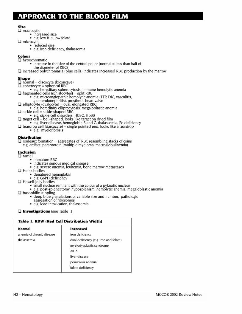
APPROACH TO THE BLOOD FILM Size❏ macrocytic
• increased size• e.g. low B12, low folate
❏ microcytic• reduced size• e.g. iron deficiency, thalassemia
Colour❏ hypochromatic
• increase in the size of the central pallor (normal = less than half ofthe diameter of RBC)
❏ increased polychromasia (blue cells) indicates increased RBC production by the marrow
Shape❏ normal = discocyte (biconcave)❏ spherocyte = spherical RBC
• e.g. hereditary spherocytosis, immune hemolytic anemia❏ fragmented cells (schistocytes) = split RBC
• e.g. microangiopathic hemolytic anemia (TTP, DIC, vasculitis,glomerulonephritis), prosthetic heart valve
❏ elliptocyte (ovalocyte) = oval, elongated RBC• e.g. hereditary elliptocytosis, megaloblastic anemia
❏ sickle cell = sickle-shaped RBC• e.g. sickle cell disorders, HbSC, HbSS
❏ target cell = bell-shaped, looks like target on dried film• e.g. liver disease, hemoglobin S and C, thalassemia, Fe deficiency
❏ teardrop cell (darcocyte) = single pointed end, looks like a teardrop• e.g. myelofibrosis
Distribution❏ rouleaux formation = aggregates of RBC resembling stacks of coins
e.g. artifact, paraprotein (multiple myeloma, macroglobulinemia)
Inclusion❏ nuclei
• immature RBC• indicates serious medical disease • e.g. severe anemia, leukemia, bone marrow metastases
❏ Heinz bodies• denatured hemoglobin• e.g. G6PD deficiency
❏ Howell-Jolly bodies• small nuclear remnant with the colour of a pyknotic nucleus• e.g. post-splenectomy, hyposplenism, hemolytic anemia, megaloblastic anemia
❏ basophilic stippling• deep blue granulations of variable size and number, pathologic
aggregation of ribosomes• e.g. lead intoxication, thalassemia
❏ Investigations (see Table 1)
Table 1. RDW (Red Cell Distribution Width)
Normal Increased
anemia of chronic disease iron deficiency
thalassemia dual deficiency (e.g. iron and folate)
myelodysplastic syndrome
AIHA
liver disease
pernicious anemia
folate deficiency

MCCQE 2002 Review Notes Hematology – H3
ANEMIACLINICAL APPROACH TO ANEMIA❏ acute vs chronic❏ decreased production vs increased destruction❏ anemia vs pancytopenia❏ based on MCV ❏ rule out dilutional anemia (low Hb due to increased effective circulating volume)
Table 2. Differential Diagnosis of Anemia Based on MCV
Hypochromic microcytic Normochromic normocytic Macrocytic(MCV<80) (80<MCV<100) (MCV>100)
• Fe deficiency• Thalassemia• Lead Poisoning• Sideroblastic• Chronic disease
(some cases)
Hematological History❏ ID: background: Mediterranean, Asian, black (thalassemia), black (sickle cell)❏ presenting symptom & HPI: depend on how rapidly the anemia develops
• fatigue, malaise, weakness, palpitations, syncope, dyspnea,headache, vertigo, tinnitus
❏ PMH: past anemias, therapies, past blood loss (GI/GU), blood donationhistory, menstrual history, signs/symptoms of renal, liver, endocrine disturbances, AIDS and other chronic diseases, malignancies
❏ family Hx: important in hereditary anemia; ask about anemia, jaundice,gallbladder disease, splenectomy
❏ medications: drugs may cause aplasia, macrocytic/megaloblastic states, hemolysis, blood loss❏ diet: iron, folic acid, vitamin B12 supplementation: amount, frequency, duration, reason❏ alcohol consumption: quantify amount and duration (toxic effect on bone
marrow or anemia due to liver disease)
Physical Exam❏ HEENT: pallor: mucous membranes, conjunctivae (Hb < 90 g/L) , icterus,
cervical lymphadenopathy, ocular bruits (Hb < 55 g/L ), glossitis❏ CVS: tachycardia, postural changes, systolic flow murmur, wide pulse pressure, CHF ❏ GI: hepatomegaly, splenomegaly, rectal (occult blood)❏ skin: pallor, jaundice, skin creases (Hb < 75 g/L ), telangiectasia as in hemolytic
anemia, koilonychia (spoon-shaped nails) as in iron deficiency anemia
IRON METABOLISM
IRON INTAKE (Dietary)❏ “average” Canadian adult diet = 10-20 mg Fe/day❏ absorption = 5-10% (0.5-2 mg/day)❏ males have a positive Fe balance❏ menstruating females have a negative Fe balance
PHYSIOLOGIC CAUSES OF INCREASED FE REQUIREMENTS❏ infancy-growth spurt 2x basal need❏ puberty-growth spurt, menarche 3x basal need❏ pregnancy-maternal RBC, fetus 4x basal need❏ blood donation 4x basal need
• 500 mL blood = 250 mg Fe • 4 donations/year = 1 g
Low Reticultocytes:• Myelodysplasia• Infiltration (leukemia,
myeloma, mets, infection)• Myelofibrosis• Aplasia• Chronic Disease
(some cases)• Liver Disease• Uremia• Endocrine
(hyper/hypothyroid,Addison’s)
High Reticulocytes:• Hemolytic anemia• Post-hemorrhagic anemia• Treated nutritional deficiency
• Megaloblastic• B12
• Folate• Drugs
• Myelodysplasia• Liver Disease• Alcohol• Reticulocytosis

H4 – Hematology MCCQE 2002 Review Notes
IRON METABOLISM . . . CONT.
IRON ABSORPTION❏ in duodenum iron combines with apoferritin to form ferritin that is absorbed through villi
Table 3. Intraluminal Factors in Absorption of Non-Heme Iron
Promoters Inhibitors
Gastric HCl AchlorhydriaAntacids
Reducing agents Oxidants• ascorbic acid
In Fe2+ form In Fe3+ form
Inorganic form Organic form
Soluble chelators Non-absorbable chelators• amino acids • phosphate (milk) • oxalate (spinach)• sugars • phytates (cereals) • tannin (tea)• alcohol
IRON TRANSPORT❏ majority of non-heme Fe in plasma is bound to transferrin❏ transferrin
• beta-globulin• carries Fe from mucosal cell to RBC precursors in marrow• carries Fe from storage pool in hepatocytes and macrophages to RBC precursors in marrow
IRON STORAGE❏ Fe is stored in two forms: ferritin and hemosiderin❏ ferritin
• ferric Fe complexed to a protein called apoferritin• hepatocytes are main site of ferritin storage• minute quantities are present in plasma in equilibrium with intracellular ferritin
❏ hemosiderin• aggregates or crystals of ferritin with the apoferritin partially removed• macrophage-monocyte system is main source of hemosiderin storage
IRON INDICES❏ bone marrow aspirate is the gold standard test for iron stores❏ serum ferritin
• single most important blood test for iron stores• falsely elevated in inflammatory disease, liver disease (from necrotic
hepatocytes), neoplasm and hyperthyroidism❏ serum iron
• varies significantly daily• a measure of all non-heme Fe present in blood• virtually all serum iron is bound to transferrin• only a trace of serum Fe is free or complexed in ferritin
❏ total iron binding capacity (TIBC)• high specificity for decreased iron, low sensitivity • measure of total amount of transferrin present in blood• normally, one third of the TIBC is saturated with Fe, remainder is unsaturated
❏ saturation• serum Fe divided by TIBC, expressed as a proportion or a %
INTERPRETING IRON INDICES
Table 4. Interpreting Iron Indices
Ferritin Serum Iron TIBC RDW Saturation
Iron Deficiency 99 9 8 8 99Chronic Disease 8/N 9/N 9/N N N
Sideroblastic Anemia 8 8 N No (dimophic picture) —
Iron Overload 8 8 N — 8
MCCQE 2002 Review Notes Hematology – H5
IRON METABOLISM . . . CONT.
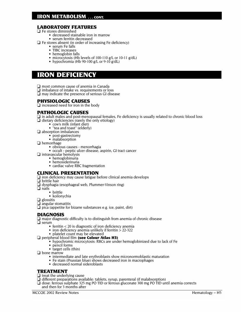
LABORATORY FEATURES❏ Fe stores diminished
• decreased stainable iron in marrow• serum ferritin decreased
❏ Fe stores absent (in order of increasing Fe deficiency)• serum Fe falls• TIBC increases• hemoglobin falls• microcytosis (Hb levels of 100-110 g/L or 10-11 g/dL)• hypochromia (Hb 90-100 g/L or 9-10 g/dL)
IRON DEFICIENCY
❏ most common cause of anemia in Canada❏ imbalance of intake vs. requirements or loss❏ may indicate the presence of serious GI disease
PHYSIOLOGIC CAUSES❏ increased need for iron in the body
PATHOLOGIC CAUSES❏ in adult males and post-menopausal females, Fe deficiency is usually related to chronic blood loss❏ dietary deficiencies (rarely the only etiology)
• cow’s milk (infant diet)• ”tea and toast” (elderly)
❏ absorption imbalances• post-gastrectomy • malabsorption
❏ hemorrhage• obvious causes - menorrhagia• occult - peptic ulcer disease, aspirin, GI tract cancer
❏ intravascular hemolysis• hemoglobinuria • hemosiderinuria• cardiac valve RBC fragmentation
CLINICAL PRESENTATION❏ iron deficiency may cause fatigue before clinical anemia develops❏ brittle hair❏ dysphagia (esophageal web, Plummer-Vinson ring)❏ nails
• brittle• koilonychia
❏ glossitis❏ angular stomatitis❏ pica (appetite for bizarre substances e.g. ice, paint, dirt)
DIAGNOSIS❏ major diagnostic difficulty is to distinguish from anemia of chronic disease❏ serum
• ferritin < 20 is diagnostic of iron deficiency anemia• iron deficiency anemia unlikely if ferritin > 22-322• platelet count may be elevated
❏ peripheral blood film (see Colour Atlas H3)• hypochromic microcytosis: RBCs are under hemoglobinized due to lack of Fe • pencil forms• target cells (thin)
❏ bone marrow• intermediate and late erythroblasts show micronormoblastic maturation• Fe stain (Prussian blue) shows decreased iron in macrophages • decreased normal sideroblasts
TREATMENT❏ treat the underlying cause❏ different preparations available: tablets, syrup, parenteral (if malabsorption)❏ dose: ferrous sulphate 325 mg PO TID or ferrous gluconate 300 mg PO TID until anemia corrects
and then for 3 months after
H6 – Hematology MCCQE 2002 Review Notes
IRON DEFICIENCY . . . CONT.
RECOVERY TIME ❏ reticulocytes begin to increase after one week❏ Hb normalizes by 10 grams per week ❏ if serum ferritin is normal then discontinue iron therapy
ANEMIA REFRACTORY TO TREATMENT WITH ORAL IRON ❏ medication
• poor preparation (e.g. expired)• drug interactions
❏ patient• poor compliance• continued bleeding • malabsorption (rare)
❏ physician• misdiagnosis
THE ANEMIA OF CHRONIC DISEASE
Etiology❏ infections❏ cancer❏ inflammatory and rheumatologic disease❏ renal disease❏ endocrine disorders (e.g. thyroid)
Pathophysiology❏ a mild hemolytic component is often present❏ red blood cell survival modestly decreased❏ erythropoietin levels are normal or slightly elevated but are inappropriately low for the degree of anemia❏ iron cannot be removed from its storage pool in hepatocytes and reticuloendothelial cells
Diagnosis❏ a diagnosis of exclusion, biochemically rule out Fe deficiency❏ serum
• serum iron, TIBC, and % saturation all normal or slightly reduced• serum ferritin is normal or increased
❏ peripheral blood• usually normocytic and normochromic if the anemia is mild • may be microcytic and normochromic if the anemia is moderate• may be microcytic and hypochromic if the anemia is severe but rarely < 90 g/L)
❏ bone marrow• normal or increased iron stores• decreased “normal” sideroblasts
Management❏ resolves if underlying disease is treated❏ erythropoietin may normalize the hemoglobin value❏ dose of erythropoietin required higher than for patients with renal disease❏ only treat patients who can benefit from a higher hemoglobin level
LEAD POISONING
L: Lead Lines on gingivae and epiphyses of long bones on X-rayE: Encephalopathy and Erythrocyte basophilic stippling A: Abdominal colic and microcytic AnemiaD: Drops: wrist and foot drop. Dimercaprol and EDTA as first line of treatment

MCCQE 2002 Review Notes Hematology – H7
SIDEROBLASTIC ANEMIA❏ group of disorders with various defects in the porphyrin biosynthetic pathway leading to a reduction in
heme synthesis resulting in an increase in cellular iron uptake ❏ characterized by presence of abnormal erythroid precursors in marrow
Types of Sideroblasts ❏ “normal” sideroblasts
• aggregates of ferritin, diffusely spread throughout the red blood cell cytoplasm• small• found in normal individuals
❏ “ring” sideroblasts• iron deposited in the mitochondria forms a ring around the red blood cell nucleus• large• abnormal finding
Etiology❏ hereditary
• rare• X-linked (defective D-aminolevulinic acid synthetase – rate-limiting enzyme in heme synthesis)• median survival is 10 years
❏ acquired• primary
• may be a preleukemic phenomenon (10%)• secondary
• toxins• drugs (isoniazid), ethanol • neoplasms and consequent chemotherapy (alkylating agents)• collagen vascular disease
Diagnosis❏ serum
• iron overload: increased serum iron, normal TIBC, increased ferritin❏ peripheral blood
• dimorphic picture (normal and hypochromic population)❏ bone marrow
• required for diagnosis• bizarre megaloblastic changes• ring sideroblasts• increased iron stores
Management❏ treatment of underlying cause❏ oral pyridoxine (vitamin B6)
• hereditary and secondary acquired forms usually responsive• myelodysplastic sideroblastic anemia not responsive
HEMOGLOBIN AND HEMOGLOBINOPATHIES
Hemoglobin Structure and Production❏ 4 α genes are located on chromosome 16❏ 2 ß genes are located on chromosome 11❏ heme group in centre with iron ❏ fetal hemoglobin, HbF (δ 2) switches to adult forms HbA (ß2) and HbA2 (δ 2) at 3-6 months of life❏ HbA constitutes 97% of adult hemoglobin❏ HbA2 constitutes 3% of adult hemoglobin❏ beware of the possibility of mixed defects e.g. ß-thalessemia minor and sickle cell trait
THALASSEMIA❏ defects in production of Hb ß that leads to microcytosis
I. HETEROZYGOUS: ß-Thalassemia Minor❏ common among people of Mediterranean and Asian descent
Clinical Presentation❏ depends on extent of disease❏ mild or no anemia❏ possible palpable spleen ❏ may be masked by Fe deficiency

H8 – Hematology MCCQE 2002 Review Notes
HEMOGLOBIN AND HEMOGLOBINOPATHIES . . . CONT.
Diagnosis❏ serum
• Hb 90-140 g/L, MCV < 70 ❏ peripheral blood
• microcytosis +/– hypochromia• target cells and increased poikilocytosis (“fish RBC”) may be present• basophilic stippling usually present
❏ Hb electrophoresis • specific: Hb A2 increased to 0.025-0.05 (2.5-5%) (normal 1.5-3.5%)• non-specific: 50% have slight increase in HbF
Management❏ not necessary to treat❏ patient and family should receive genetic counselling
II. HOMOZYGOUS: ß-Thalassemia Major
Pathophysiology❏ autosomal recessive❏ ineffective chain synthesis leading to ineffective erythropoiesis and hemolysis of RBC ❏ increase in HbF
Clinical Presentation❏ initial presentation at 3-6 months due to replacement of HbF by HbA❏ severe anemia develops in the first year of life❏ jaundice❏ stunted growth and development (hypogonadal dwarf)❏ gross hepatosplenomegaly (extramedullary hematopoiesis)❏ changes (expanded marrow cavity)
• skull x-ray has “hair-on-end” appearance• pathological fractures common
❏ evidence of increased Hb catabolism (e.g. gallstones)❏ death from
• untreated anemia (transfuse)• infection (treat early)• hemochromatosis (late, secondary to transfusions), usually 20-30 years old
Diagnosis❏ CBC
• hemoglobin 40-60 g/L ❏ peripheral blood
• hypochromic microcytosis• increased reticulocytes• basophilic stippling, target cells • postsplenectomy blood film shows Howell Jolly bodies, erythroblasts, and thrombocytosis
❏ Hb electrophoresis• Hb A: 0-0.10 (0-10%) , (normal > 95%)• Hb F: 0.90-1.00 (90-100%)
Management❏ transfusion❏ Fe chelation to prevent iron overload (e.g. desferal)❏ bone marrow transplant
III. ALPHA THALASSEMIA❏ similar distribution to thalassemia but a higher frequency among Asians
Pathophysiology❏ autosomal recessive❏ deficit of α chains❏ 4 grades of severity depending on the number of defective alpha genes
• 1 - silent • 2 - trait • 3 - HbH Disease (presents in adults due to excess chain production)• 4 - Hb Bart’s (hydrops fetalis, not compatible with life)
Diagnosis❏ peripheral blood film
• microcytes, hypochromia, occasional target cells • screen for HbH inclusion bodies
❏ Hb electrophoresis not diagnostic❏ DNA analysis using alpha gene probe

MCCQE 2002 Review Notes Hematology – H9
HEMOGLOBIN AND HEMOGLOBINOPATHIES . . . CONT.
Management❏ same as ß thalassemia
SICKLE CELL ANEMIA❏ autosomal recessive❏ amino acid substitution of valine for glutamate in position 6 of beta globin chain
Mechanisms of Sickling (see Figure 1)❏ at low pO2, deoxy Hb S polymerizes, leading to rigid crystal-like rods that distort membranes = SICKLES ❏ the pO2 level at which sickling occurs is related to the precentage of Hb S present
• in heterozygotes (Hb AS) sickling occurs at a pO2 of 40 mmHg• in homozygous (Hb SS), sickling occurs at a pO2 of 80 mmHg
❏ sickling is aggravated by• increased H+
• increased CO2
• increased 2,3-DPG• increased temperature and osmolality
Figure 1. Pathophysiology of Sickling
Heterozygous: Hb S Trait❏ clinical presentation
• patient will appear normal except at times of extreme hypoxia and infection❏ diagnosis
• serum: Hb normal• peripheral blood: normal except for possibly a few target cells• Hb electrophoresis (confirmatory test): Hb A fraction of 0.65 (65%);• Hb S fraction of 0.35 (35%)
Homozygous: Hb S Disease❏ clinical presentation
• chronic hemolytic anemia • jaundice in the first year of life• vaso-occlusive crises (infarction) leading to pain, fever and leukocytosis
e.g. acute chest syndrome (pulmonary infarct) associated with infection, such as parvovirus, leading to aplastic anemia, acidosis, dehydration, and hypoxia
• susceptibility to infections by encapsulated organisms due to hyposplenism• retarded growth and development +/– skeletal changes• spleen enlarged in child and atrophic in adult
❏ diagnosis• peripheral blood: sickled cells (see Colour Atlas H6)• screening test: sickle cell prep• Hb electrophoresis (confirmatory test): Hb S fraction > 0.80
Management❏ prevention of crises is the key
• establish diagnosis• avoid conditions that favor sickling (hypoxia, acidosis, dehydration, fever)• vaccination in childhood e.g. pneumococcus, meningococcus• consider prophylaxis - penicillin V 250 mg PO bid• good hygiene and nutrition
❏ genetic counselling❏ folic acid to avoid folate deficiency
impactioninfarction
distorted RBC sickle cells
bloodviscosity
deoxy Hb S
pO2
blood flowslows
H+
CO2
Hb S polymers
H10 – Hematology MCCQE 2002 Review Notes
HEMOGLOBIN AND HEMOGLOBINOPATHIES . . . CONT.
❏ hydroxyurea to enhance production of HbF• causes depression of the gene for HbF or by initiating differentiation of stem cells in which this gene
is active; presence of HbF in the SS cells decreases polymerization and precipitation of HbS • Note: hydroxyurea is cytotoxic and may cause bone marrow suppression
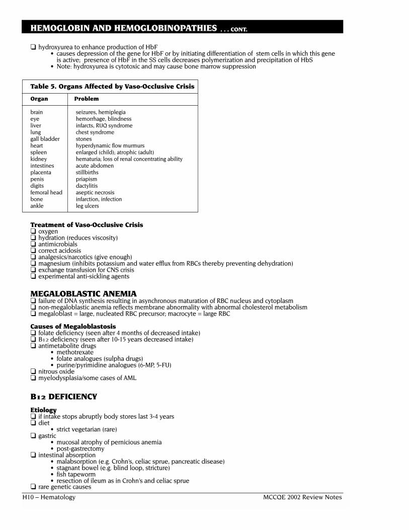
Table 5. Organs Affected by Vaso-Occlusive Crisis
Organ Problem
brain seizures, hemiplegiaeye hemorrhage, blindnessliver infarcts, RUQ syndromelung chest syndromegall bladder stonesheart hyperdynamic flow murmursspleen enlarged (child); atrophic (adult)kidney hematuria; loss of renal concentrating abilityintestines acute abdomenplacenta stillbirthspenis priapismdigits dactylitisfemoral head aseptic necrosisbone infarction, infectionankle leg ulcers
Treatment of Vaso-Occlusive Crisis❏ oxygen❏ hydration (reduces viscosity)❏ antimicrobials❏ correct acidosis❏ analgesics/narcotics (give enough)❏ magnesium (inhibits potassium and water efflux from RBCs thereby preventing dehydration)❏ exchange transfusion for CNS crisis❏ experimental anti-sickling agents
MEGALOBLASTIC ANEMIA❏ failure of DNA synthesis resulting in asynchronous maturation of RBC nucleus and cytoplasm❏ non-megaloblastic anemia reflects membrane abnormality with abnormal cholesterol metabolism❏ megaloblast = large, nucleated RBC precursor; macrocyte = large RBC
Causes of Megaloblastosis❏ folate deficiency (seen after 4 months of decreased intake)❏ B12 deficiency (seen after 10-15 years decreased intake)❏ antimetabolite drugs
• methotrexate• folate analogues (sulpha drugs)• purine/pyrimidine analogues (6-MP, 5-FU)
❏ nitrous oxide❏ myelodysplasia/some cases of AML
B12 DEFICIENCY
Etiology❏ if intake stops abruptly body stores last 3-4 years❏ diet
• strict vegetarian (rare)❏ gastric
• mucosal atrophy of pernicious anemia• post-gastrectomy
❏ intestinal absorption• malabsorption (e.g. Crohn’s, celiac sprue, pancreatic disease)• stagnant bowel (e.g. blind loop, stricture)• fish tapeworm• resection of ileum as in Crohn’s and celiac sprue
❏ rare genetic causes

MCCQE 2002 Review Notes Hematology – H11
HEMOGLOBIN AND HEMOGLOBINOPATHIES . . . CONT.
Pernicious Anemia❏ auto-antibodies produced against gastric parietal cells leading to
achlorhydria and no intrinsic factor secretion• intrinsic factor is required to stabilize B12 as it passes through the bowel• decreased intrinsic factor leads to decreased ileal absorption of B12
❏ female:male = 1.6:1❏ may be associated with other autoimmune disorders e.g. thyroid and adrenal deficiency ❏ often > 60 years old
Neurological Lesions in B12 Deficiency❏ cerebral (common; reversible with B12 therapy)
• confusion• delirium• dementia
❏ cranial nerves• optic atrophy (rare)
❏ cord (irreversible damage)• subacute combined degeneration• posterior columns - paresthesias, disturbed vibration, decreased proprioception• pyramidal tracts - spastic weakness, hyperactive reflexes
❏ peripheral neuropathy (variable reversibility)• usually symmetrical• affecting lower limbs more than upper limbs
Diagnosis❏ serum
• anemia often severe +/– neutropenia +/– thrombocytopenia• MCV > 120 • low reticulocyte count relative to the degree of anemia
❏ serum B12 and RBC folate• caution: low serum B12 leads to low RBC folate because of failure of
folate polyglutamate synthesis in the absence of B12
❏ blood film • oval macrocytes (see Colour Atlas H2A)• hypersegmented neutrophils (see Colour Atlas H2B)
❏ bone marrow• differentiates between megaloblastic and myelodysplastic anemias• hypercellularity • failure of nuclear maturation• elevated unconjugated bilirubin and LDH due to marrow cell breakdown
❏ Schilling test to distinguish pernicious anemia from other causes• Schilling test: part 1
• tracer dose (1g µg) of labelled B12 (cobalamin (Co*)), PO• flushing dose (1mg) of cold B12, IM to saturate tissue binders
of B12 thus allowing radioactive B12 to be excreted in urine• 24 hour urine Co* measured • normal ––> 5% excretion
• Schilling test: part 2• tracer dose B12 (Co*) plus intrinsic factor, PO• flushing dose of cold B12, injected IM• 24 hour urine Co* measured • normal test result (> 5% excretion) = pernicious anemia • abnormal test result (< 5% excretion) = intestinal causes (malabsorption)
Management❏ B12 100 µg IM monthly for life or oral B12
❏ watch for hypokalemia (due to return of potassium to intracellular sites) and thrombocythemia
FOLATE DEFICIENCY❏ more common than B12 deficiency because folate stores are depleted in 3-6 months ❏ folate complexes with gastric R binder❏ R binder is replaced by intrinsic factor in the duodenum❏ this complex is absorbed in the jejunum
Etiology❏ diet (folate is present in leafy green vegetables)
• most common cause• e.g. infancy, poverty, alcoholism
❏ intestinal• malabsorption

H12 – Hematology MCCQE 2002 Review Notes
HEMOGLOBIN AND HEMOGLOBINOPATHIES . . . CONT.
❏ drugs/chemicals• alcohol• anticonvulsants• antifolates (MTX)• birth control pills
❏ increased demand• pregnancy• prematurity• hemolysis• hemodialysis• psoriasis, exfoliative dermatitis
Clinical Presentation❏ mildly jaundiced due to hemolysis of RBC secondary to ineffective hemoglobin synthesis❏ glossitis and angular stomatitis❏ rare
• melanin pigmentation• purpura secondary to thrombocytopenia
❏ folate deficiency at time of conception and early pregnancy has been linked to neural tube defects
Management❏ never give folate alone to individual with megaloblastic anemia because it
will mask B12 deficiency and neurological degeneration will continue❏ folic acid 15 mg PO/day x 3 months; then 5 mg PO/day maintenance if cause not reversible❏ folic acid supplementation 1 mg PO/day will protect against elevated homocysteine levels
(risk factor for CAD)
HEMOLYTIC ANEMIAS (HA) (see Colour Atlas H4)
Classification❏ hereditary causes (intrinsic)
• abnormal membrane (spherocytosis, elliptocytosis)• abnormal enzymes (pyruvate kinase deficiency, G6PD deficiency)• abnormal hemoglobin synthesis (thalassemias, hemoglobinopathies)
❏ acquired causes (extrinsic)• immune
• hemolytic transfusion reaction• idiopathic immune HA• drugs• cold agglutinins• secondary autoimmune HA
• non-immune • RBC fragmentation syndromes• paroxysmal nocturnal hemoglobinuria• liver disease• hypersplenism• march hemoglobinuria
Clinical Presentation❏ jaundice❏ cholelithiasis❏ splenomegaly❏ skeletal abnormalities❏ leg ulcers❏ regenerative crisis❏ folic acid deficiency❏ iron overload with extravascular hemolysis❏ iron deficiency with intravascular hemolysis
Diagnosis❏ indirect - not specific to hemolytic anemias
• increased reticulocyte count • reduced haptoglobin• increased unconjugated bilirubin• increased urine bilinogen• increased LDH
❏ tests exclusive for intravascular hemolysis • serum free hemoglobin present• methemalbuminemia (heme + albumin)• hemoglobinuria (immediate)• hemosiderinuria (delayed)
MCCQE 2002 Review Notes Hematology – H13
HEMOGLOBIN AND HEMOGLOBINOPATHIES . . . CONT.
Antiglobulin Tests (Coombs’ Tests)❏ direct Coombs’ test (direct antiglobulin test)
• purpose: detect antibodies or complement on the surface of RBC • by adding anti-antibodies to the RBC; the RBC agglutinate in a positive test• indications
• hemolytic disease of newborn• hemolytic anemia• AIHA• hemolytic transfusion reaction
❏ indirect Coombs’ test (indirect antiglobulin test)• purpose: detect antibodies in serum that can recognize antigens on RBC• by mixing serum with donor RBC and then anti-antibodies; RBCs
agglutinate in a positive test• indications
• cross-matching of recipient serum with donor’s RBC• atypical blood group• blood group antibodies in pregnant women• antibodies in AIHA
I. HEREDITARY HEMOLYTIC ANEMIAS
STRUCTURAL ABNORMALITIES IN CYTOSKELETON
Hereditary Spherocytosis❏ autosomal dominant with variable penetrance❏ incidence 22 per 100,000❏ most common type of hereditary hemolytic anemia❏ abnormality in spectrin (compound in RBC membrane) ❏ blood film shows spherocytes (see Colour Atlas H8)❏ increased osmotic fragility❏ sometimes confused with immune hemolytic anemia❏ treatment: splenectomy (immunize against pneumococcus first); avoid in childhood
Hereditary Elliptocytosis❏ autosomal dominant❏ incidence 20-50 per 100,000❏ abnormality in spectrin interaction with other membrane proteins ❏ 25-75% elliptocytes❏ hemolysis is usually mild ❏ treatment: splenectomy for severe hemolysis (immunize against pneumococcus first)
ENZYMATIC ABNORMALITIES IN RBC
G6PD Deficiency
Clinical Presentation❏ X-linked recessive❏ oxidant drug-induced hemolysis
• sulfonamides• primaquine• nitrofurantoin• acetanilid
❏ favism (fava beans)❏ neonatal jaundice❏ chronic hemolytic anemia❏ infection
Diagnosis and Management❏ high index of suspicion❏ G6PD assay
• should not be done when reticulocyte count is high in acute crisis,PBF shows Heinz bodies (granules in red blood cells due to damaged hemoglobin molecules) and features of intravascular hemolysis
❏ transfusion in severe cases❏ stop offending drugs or food
H14 – Hematology MCCQE 2002 Review Notes
HEMOGLOBIN AND HEMOGLOBINOPATHIES . . . CONT.
II. ACQUIRED HEMOLYTIC ANEMIASAUTOIMMUNE HEMOLYTIC ANEMIA
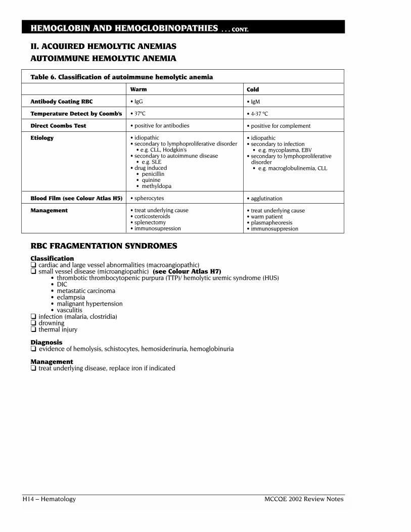
Table 6. Classification of autoimmune hemolytic anemia
Antibody Coating RBC
Temperature Detect by Coomb’s
Direct Coombs Test
Etiology
Blood Film (see Colour Atlas H5)
Management
RBC FRAGMENTATION SYNDROMESClassification❏ cardiac and large vessel abnormalities (macroangiopathic)❏ small vessel disease (microangiopathic) (see Colour Atlas H7)
• thrombotic thrombocytopenic purpura (TTP)/ hemolytic uremic syndrome (HUS)• DIC• metastatic carcinoma• eclampsia• malignant hypertension• vasculitis
❏ infection (malaria, clostridia)❏ drowning❏ thermal injury
Diagnosis❏ evidence of hemolysis, schistocytes, hemosiderinuria, hemoglobinuria
Management❏ treat underlying disease, replace iron if indicated
Warm
• IgG
• 37ºC
• positive for antibodies
• idiopathic• secondary to lymphoproliferative disorder
• e.g. CLL, Hodgkin’s• secondary to autoimmune disease
• e.g. SLE• drug induced
• penicillin• quinine• methyldopa
• spherocytes
• treat underlying cause• corticosteroids• splenectomy• immunosupression
Cold
• IgM
• 4-37 ºC
• positive for complement
• idiopathic• secondary to infection
• e.g. mycoplasma, EBV• secondary to lymphoproliferative
disorder• e.g. macroglobulinemia, CLL
• agglutination
• treat underlying cause• warm patient• plasmapheoresis• immunosuppresion

MCCQE 2002 Review Notes Hematology – H15
HEMOGLOBIN AND HEMOGLOBINOPATHIES . . . CONT.
THROMBOTIC THROMBOCYTOPENIC PURPURA AND HEMOLYTIC UREMIC SYNDROME
Table 7. Thrombotic Thrombocytopenic Purpura (TTP) and Hemolytic Uremic Syndrome (HUS)
TTP HUS
• predominantly adult • predominantly children• neurological symptoms (90%)
• H/A, somnolence, confusion, focal neurological findings,convulsion, stupor, coma
• purpura (90%) due to severe thrombocytopenia • purpura (90-100%) due to severe thrombocytopenia• epistaxis, hematuria, hemoptysis and GI bleed
• epistaxis, hematuria, hemoptysis and GI bleed• microangiopathic hemolytic anemia • microangiopathic hemolytic anemia• fever (90-100%)• GI
• N/V, abdominal pain• renal (40-80%) • renal symptoms (90%)
• abnormal UA, oliguria, ARF • abnormal UA, oliguria, ARF• etiology • etiology
• idiopathic • E. coli serotype O157:H7 virotoxin• familial• secondary TTP
• infection • enterobacteriaceae• viral: flu, HIV
• systemic diseases• SLE and other CVD
• cancer and chemotherapeutic drugs• diagnosis • diagnosis
• by clinical picture • by clinical picture• CBC: anemia, thrombocytopenia • same as TTP• PT, PTT: normal • stool C+S• ESR: normal• negative Coombs’
*Key characteristics bolded
Management❏ plasmapheresis is the treatment of choice❏ steroid is treatment of choice only in mild disease
APLASTIC ANEMIA
❏ destruction of hematopoietic cells of the bone marrow
Etiology❏ radiation❏ drugs
• anticipated (chemotherapy)• idiosyncratic (chloramphenicol, phenylbutazone)
❏ chemicals• benzene and other organic solvents• DDT and insecticides
❏ post viral e.g. hepatitis B, parvovirus❏ idiopathic
• often immune (T-cell mediated)❏ paroxysmal nocturnal hemoglobinuria❏ marrow replacement❏ congenital
Clinical Presentation❏ occurs at any age❏ slightly more common in males❏ can present acutely or insidiously❏ anemia or neutropenia or thrombocytopenia (any combination) +/– pancytopenia❏ thrombocytopenia with bruising, bleeding gums, epistaxis❏ anemia with SOB, pallor and fatigue

H16 – Hematology MCCQE 2002 Review Notes
APLASTIC ANEMIA . . . CONT.
❏ presentation of neutropenia ranges from infection in the mouth to septicemia❏ absence of splenomegaly
Diagnosis❏ serum
• neutrophil count < 5.0 x 10⁹/L• platelet count < 20 x 109/L• corrected reticulocyte count < 1%
❏ blood film• decreased normal RBC
❏ bone marrow• aplasia or hypoplasia of marrow cells with fat replacement
Management❏ removal of offending agents❏ supportive care (red cell and platelet transfusions, antibiotics) ❏ antithymocyte globulin (50-60% patients respond)❏ cyclosporine ❏ allogeneic bone marrow transplantation
• minimize blood products on presentation• only irradiated, leuko-depleted blood products should be used• CMV negative blood for CMV negative patients
HEMOSTASIS
THREE PHASES OF HEMOSTASISPrimary Hemostasis❏ goal is to rapidly stop bleeding❏ vessel injury results in collagen and subendothelial structure exposure and
release of vasoconstrictors❏ blood flow is impeded and platelets come in contact with vessel wall❏ platelets adhere to collagen and are activated resulting in change of shape
and release of ADP and thromboxane A2❏ these factors further recruit and aggregate more platelets resulting in
formation of hemostatic plug
Figure 2. Primary Hemostasis
Secondary Hemostasis❏ platelet plug formed through primary hemostasis is reinforced through
process of secondary hemostasis and a stable plug is formed❏ secondary pathways involved in the activation of coagulation factors
include• intrinsic
• activated when vessel wall remains intact• slow pathway
• extrinsic• activated when there is injury to vessel wall• fast pathway
Adhesion to collagen insubendothelium
Release of ADP and thromboxane A2
Aggregation (platelet plug)

MCCQE 2002 Review Notes Hematology – H17
HEMOSTASIS . . . CONT.
Figure 3. Secondary Hemostasis
Figure 4. Fibrin Stabilization and Fibrinolysis
TESTS OF HEMOSTASISTable 8. Commonly Used Tests of Hemostasis
Type of hemostatis Test Reference Range Purpose
Primary platelet count • to quantitate platelet number bleeding time 2-12 mins • platelet functionplatelet aggregation • platelet function
Secondary PTT - depends on lab 22-35 s • measures intrinsic pathwayfactors VIII, IX, XI, XII
PT - depends on lab 11-24 s • measures extrinsic pathwayfactor VIII in particular
TT - depends on lab 14-16 s • measures deficiency of fibrinogen inactivation of prothrombin
INR 1 is normal • permits determination of coagulation status independentof laboratory performing measurement
Fibrinolysis euglobulin lysis time
Other • fibrinogen• fibrinogen degradation products (FDPs)• specific factor assays• tests of physiological inhibitors
(antithrombins, protein S, protein C,hereditary resistance to APC)
• tests of pathologic inhibitors (e.g. lupus anticoagulant)
PartialThromboplastin
Time(PTT)
ProthrombinTime(PT)
Prothrombin Thrombin
FibrinFibrinogen
X Xa
+V+Ca
Common
VIIa
* tissue factor pathway
*
+ TissueThromboplastin
Extrinsic
Kallikrein:
XIIaXII
XI XIa
IX IXa
Thrombin
XIII
XIIIa
Fibrinogen FibrinMonomer
Cross-linkedFibrin
Fibrin(ogen)Degradation
Products(FDP)
PlasminogenActivators
(Urokinase,Tissue activator,
etc.)
Plasmin Plasminogen
Intrinsic+Ca+VIII
ThrombinTime(TT)
H18 – Hematology MCCQE 2002 Review Notes
HEMOSTASIS . . . CONT.
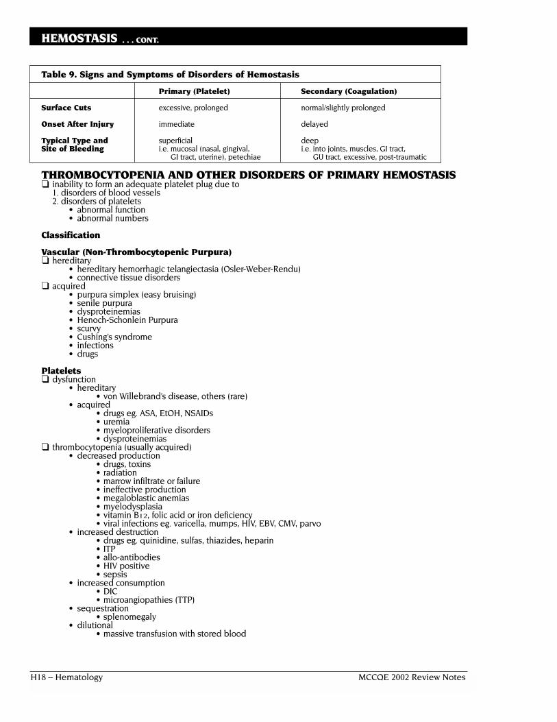
Table 9. Signs and Symptoms of Disorders of Hemostasis
Primary (Platelet) Secondary (Coagulation)
Surface Cuts excessive, prolonged normal/slightly prolonged
Onset After Injury immediate delayed
Typical Type and superficial deepSite of Bleeding i.e. mucosal (nasal, gingival, i.e. into joints, muscles, GI tract,
GI tract, uterine), petechiae GU tract, excessive, post-traumatic
THROMBOCYTOPENIA AND OTHER DISORDERS OF PRIMARY HEMOSTASIS❏ inability to form an adequate platelet plug due to
1. disorders of blood vessels2. disorders of platelets
• abnormal function• abnormal numbers
Classification
Vascular (Non-Thrombocytopenic Purpura)❏ hereditary
• hereditary hemorrhagic telangiectasia (Osler-Weber-Rendu)• connective tissue disorders
❏ acquired• purpura simplex (easy bruising)• senile purpura• dysproteinemias• Henoch-Schonlein Purpura• scurvy• Cushing’s syndrome• infections• drugs
Platelets❏ dysfunction
• hereditary• von Willebrand’s disease, others (rare)
• acquired• drugs eg. ASA, EtOH, NSAIDs• uremia• myeloproliferative disorders• dysproteinemias
❏ thrombocytopenia (usually acquired)• decreased production
• drugs, toxins• radiation• marrow infiltrate or failure• ineffective production• megaloblastic anemias• myelodysplasia• vitamin B12, folic acid or iron deficiency• viral infections eg. varicella, mumps, HIV, EBV, CMV, parvo
• increased destruction• drugs eg. quinidine, sulfas, thiazides, heparin• ITP• allo-antibodies• HIV positive• sepsis
• increased consumption• DIC• microangiopathies (TTP)
• sequestration• splenomegaly
• dilutional• massive transfusion with stored blood

MCCQE 2002 Review Notes Hematology – H19
HEMOSTASIS . . . CONT.
IDIOPATHIC (AUTOIMMUNE) THROMBOCYTOPENIC PURPURA (ITP)
Table 10. Idiopathic Thrombocytopenic Purpura
Features Acute ITP Chronic ITP
Peak Age 2-6 years 20-40 years
Sex Predilection none F > M (3:1)
History of Recent Infection common rare
Onset of Bleed abrupt insidious
Platelet Count < 20 x 109/L 30-80 x 109/L
Duration usually weeks months to years
Spontaneous Remissions 80% or more uncommon
CHRONIC (ADULT-TYPE) ITP❏ most common cause of isolated thrombocytopenia❏ diagnosis of exclusion
Pathophysiology❏ IgG autoantibody❏ spleen
• site of antibody production and platelet destruction• usually not palpable (enlarged in ~ 10%)
Clinical Presentation❏ insidious onset❏ may be seen after mild viral illness or after immunization❏ mucosal or skin bleeding❏ petechiae and easy bruising❏ hematuria❏ melena❏ epistaxis❏ female with menorrhagia
Laboratory Results❏ peripheral blood film: decreased platelets, large platelets❏ bone marrow: plentiful megakaryocytes
• critical test to rule out other causes of thrombocytopenia❏ anti-platelet antibodies present in most❏ increased bleeding time❏ PT and PTT normal
Management❏ conservative if mild
• platelet count > 30,000, no mucosal bleeding❏ steroids: moderate dose, then taper (80% responsive)
• platelet count < 20-30,000 or evidence of mucosal bleeding❏ splenectomy if steroids fail❏ IV gamma globulin if steroids and splenectomy fail or if rapid response is required❏ other: immunosuppressives, platelets, plasma exchange, Danazol
Prognosis❏ fluctuating course❏ overall relatively benign, mortality 1-2%❏ major concern is cerebral hemorrhage at platelet counts < 5 x 10⁹/L
DISORDERS OF SECONDARY HEMOSTASIS
Classification
I. Hereditary❏ Factor VIII: Hemophilia A, von Willebrand’s disease ❏ Factor IX: Hemophilia B (Christmas Disease)❏ Factor XI❏ other factor deficiences are rare
H20 – Hematology MCCQE 2002 Review Notes
HEMOSTASIS . . . CONT.
II. Acquired❏ liver disease❏ DIC❏ vitamin K deficiency❏ circulating anti-coagulants (inhibitors)❏ other e.g. primary fibrinolysis
HEREDITARYI. Hemophilia A (factor VIII)❏ X-linked, 1/5,000 males❏ mild (> 5%), moderate (1-5%), severe (< 1%)
Clinical Presentation❏ hemarthroses, hematomas, GI and GU bleeding❏ bleeding in response to trauma (mild and moderate disease)
• intracranial hemorrhage following head injury❏ spontaneous bleeding (severe disease)
Laboratory Results❏ prolonged PTT, normal INR (PT)❏ decreased factor VIII (< 40% of normal)❏ vWF usually normal or increased
Management❏ minor but not trivial bleeding (eg. hemarthroses)
• heat treated Factor VIII concentrate ❏ major potentially life-threatening bleeding (eg. multiple trauma)
• heat treated Factor VIII concentrate❏ prophylaxis (eg. multiple dental extractions, surgery)
• heat treated Factor VIII concentrate❏ DDAVP in mild or moderate hemophilia A
II. Von Willebrand’s Disease❏ heterogeneous group of defects❏ usually autosomal dominant ❏ qualitative or quantitative abnormality of vWF
• vWF needed for platelet adhesion and acts as carrier for factor VIII• vWF exists as a series of multimers ranging in size
• the largest ones are most active in mediation of platelet adhesion• both large and small complex with factor VIII
❏ both primary and secondary hemostasis affected❏ usually mild to moderate in severity
Classification❏ type I: decreased vWF in platelets and plasma (will see prolonged
bleeding time, decreased factor VIII)❏ type IIA: decreased large and intermediate sized multimers in plasma and
platelets (will see prolonged bleeding time, normal levels of factor VIII)❏ type IIB: largest multimers are missing from plasma but not from platelets
Clinical Presentation❏ mild
• asymptomatic• mucosal and cutaneous bleeding, easy bruising, epistaxis, menorrhagia, gingival bleeding
❏ moderate to severe• as above but worse, occasionally soft-tissue hematomas, petechiae
(rare), GI bleeding, hemarthroses
Course❏ may fluctuate, often improves during pregnancy and with age
Laboratory Results❏ prolonged bleeding time and PTT❏ decreased factor VIII (5-50%) ❏ normal platelet count (except in Type IIB)❏ decreased ristocetin cofactor activity❏ analysis of multimers
Management❏ DDAVP is treatment of choice except in Type IIB
• causes release of vWF and plasminogen activator from endothelial cells• in type IIB, the appearance of the large multimers in the circulation can cause thrombocytopenia
❏ Hemate P in selected cases❏ conjugated estrogens
MCCQE 2002 Review Notes Hematology – H21
HEMOSTASIS . . . CONT.
III. Factor IX Deficiency❏ Christmas disease, Hemophilia B❏ X-linked recessive,1/30,000 males❏ clinical and laboratory features identical to Hemophilia A❏ main treatment is Factor IX concentrate
IV. Factor XI Deficiency (Rosenthal syndrome)❏ autosomal recessive inheritance ❏ usually mild, often diagnosed in adulthood❏ treatment: fresh frozen plasma
ACQUIRED
I. Liver Disease❏ deficient synthesis of all factors except VIII❏ aberrant synthesis: fibrinogen❏ deficient clearance of hemostatic “debris” and fibrinolytic activators❏ accelerated destruction due to dysfibrinogenemias: increased fibrinolysis, DIC❏ thrombocytopenia: hypersplenism, folate deficiency, EtOH intoxication, DIC❏ platelet dysfunction: EtOH abuse❏ miscellaneous: inhibition of secondary hemostasis by FDPs❏ peripheral blood smear: target cells❏ diagnosis
• factor V because it has the shortest half-life• elevated INR (PT), PTT and bleeding time
❏ treatment: fresh frozen plasma, platelets
II. Vitamin K Deficiency
Etiology❏ poor diet (especially in alcoholics)❏ biliary obstruction❏ chronic liver disease❏ malabsorption e.g. celiac disease ❏ drugs
• oral anticoagulants produce inhibition of factors II, VII, IX, X, Protein C & S• antibiotics eradicating gut flora which is 50 % of vitamin K supply
❏ hemorrhagic disease of newborn
Diagnosis❏ INR (PT) is elevated out of proportion to the elevation of the PTT❏ decreased factors II, VII, IX and X (because vitamin K-dependent)
Management❏ vitamin K 10-20 mg SC (not IM)❏ Note: PT should improve within 24 hours, if not search for other causes
III. Disseminated Intravascular Coagulation (DIC)❏ massive uncontrolled intravascular coagulation resulting in depletion of
platelets, coagulation factors and fibrinogen❏ not a primary disorder but a syndrome that complicates a number of other conditions
Clinical Conditions Associated with DIC❏ activation of procoagulant activity
• anti-phospholipid antibody• intravascular hemolysis (incompatible blood, malaria)• tissue factor• tissue injury (obstetric catastrophes, leukemia, tumours, liver disease, trauma, burns)• snakebite• fat embolism• heat stroke
❏ endothelial injury• infections• vasculitis• metastatic disease (adenocarcinoma)• aortic aneurysm• giant hemangioma
❏ reticuloendothelial injury• liver disease• splenectomy

H22 – Hematology MCCQE 2002 Review Notes
HEMOSTASIS . . . CONT.
❏ vascular stasis• hypotension• hypovolemia• pulmonary embolus
❏ other• acute hypoxia/acidosis• extracorporeal circulation
Signs of Microvascular Thrombosis (Early DIC)❏ neurological: multifocal, delirium, coma, seizures❏ skin: focal ischemia, superficial gangrene❏ renal: oliguria, azotemia, cortical necrosis❏ pulmonary: ARDS❏ GI: acute ulceration❏ RBC: microangiopathic hemolysis
Signs of Hemorrhagic Diathesis (Late DIC)❏ neurologic: intracranial bleeding❏ skin: petechiae, eccyhmosis, oozing from puncture sites❏ renal: hematuria❏ mucosal: gingival oozing, epistaxis, massive bleeding
Diagnosis❏ clinical picture❏ laboratory
• primary hemostasis: decreased platelets• secondary hemostasis: prolonged INR (PT), PTT, TT, decreased
fibrinogen and other factors• fibrinolysis increased FDPs, short lysis time• extent of fibrin deposition: urine output, urea, RBC fragmentation
Management❏ recognize early❏ TREAT UNDERLYING DISORDER❏ life support measures, O2, blood transfusion, fluid therapy❏ replacement of hemostatic elements with platelet transfusion, FFP, cryoprecipitate
THROMBOSIS
Virchow’s Triad❏ stasis❏ hypercoaguable state❏ endothelial injury
Etiology ❏ endothelial damage❏ blood flow
• stasis• turbulence• hyperviscosity
❏ blood components• platelets• contact factors• thrombin• Factor VIII• fibrin
❏ hypercoagulable state due to• cancer • pregnancy• birth control pills• DIC• lipids• decreased physiological inhibitors (antithrombin-III, protein C, protein S)• hereditary resistance to activated protein C (Factor V Leiden mutation)• prothrombin variant 20210A• nephrotic syndrome

MCCQE 2002 Review Notes Hematology – H23
HEMOSTASIS . . . CONT.
Management (acute and prophylaxis)❏ hyperhomocysteine anticoagulants
• low molecular weight heparin• no test required• reduced incidence of HIT
• unfractionated heparin• maintain PTT 1.5-2.5 x the normal control
• coumadin (see Table 11)• hirudin
❏ thrombolytics• snake venom enzymes (ancrod)• plasminogen activators (streptokinase, urokinase, tPA)
❏ antiplatelet agents• ASA• sulfinopyrazone• dipyridamole
Table 11. Monitoring Coumadin (Warfarin) Therapy (therapeutic ranges)
INR
Range Target
• pre-operative• surgery 1.5-2.5 2• hip surgery 2-3 2.5
• prevention of venous thrombosis 2-3 2.5
• active venous thrombosis, pulmonary embolism and prevention of recurrent venous thrombosis 2-4 3
• prevention of arterial thrombo-embolism including mechanical heart valves 3-4.5 3.5
• INR should never exceed 5
HEPARIN-INDUCED THROMBOCYTOPENIA (HIT)
HIT-I❏ non-immune❏ decrease in platelet count usually seen early (48-72 hours post
administration) but may take up to 1 week to appear❏ transient thrombocytopenia, returns to normal once heparin discontinued❏ no intravascular thrombosis❏ likely due to platelet aggregation and sequestration
HIT-II❏ immune-mediated❏ typically occurs at day 5-15 of heparin therapy and decline is gradual❏ HIT can begin sooner in patients who have received heparin in the past three months❏ delayed-onset HIT occurs several days after discontinuing heparin❏ typical platelet count in patients with HIT ranges from 25 to 100 x 109/L
Pathogenesis❏ immunoglobulin-mediated adverse drug reaction❏ pathogenic antibody, usually IgG recognizes a multimolecular complex of heparin
and platelet factor 4, resulting in platelet activation via platelet Fc receptors andactivation of the coagulation system
Clinical Complications❏ cases of serious bleeding related to thrombocytopenia have been reported❏ intravascular thrombosis
• both venous (DVT, PE, venous gangrene) and arterial thrombi (MI, stroke, limb vessels) can form❏ heparin-induced skin necrosis❏ unusual thrombotic complications include mesenteric artery or vein
occlusion, adrenal hemorrhage and infarction❏ acute platelet activation syndromes
• acute inflammatory reactions (eg. fever/chills, flushing, etc.), transient global amnesia
Laboratory Tests❏ C-serotonin release assay ❏ ELISA
• measures binding of antibody in patients serum to PF4:heparin complex
H24 – Hematology MCCQE 2002 Review Notes
HEMOSTASIS . . . CONT.
Management❏ discontinuation of heparin❏ platelet count should return to normal in a few days❏ danaparoid (organon) is the preferred agent if anti-thrombic therapy is indicated❏ low-molecular-weight heparin is less likely to cause HIT in de novo use but
still carries an increased risk if previously sensitized with unfractionated heparin ❏ other alternatives include ancrod and hirudin❏ patient may be re-exposed to heparin only under careful supervision
HEMATOLOGIC MALIGNANCIES
OVERVIEWMyeloid ❏ clonal stem cell neoplasms
i. acute myeloid leukemia (clonal proliferation of immature cells)ii. myeloproliferative disorders (proliferation of mature cells)
• polycythemia rubra vera• chronic granulocytic (myelogenous) leukemia• idiopathic myelofibrosis• essential thrombocythemia
iii. myelodysplastic syndromes (defective differentiation)
Lymphoid❏ all cells arise from a single abnormal lymphoid precursor (B or T)
i. acute lymphoblastic leukemia (arise from stem cell)ii. lymphomas (arise from maturing lymphoid cell)
• Hodgkin’s lymphoma• non-Hodgkin’s lymphoma
iii. malignant clonal proliferation of B cells• chronic lymphocytic leukemia• plasma cell dyscrasias• light chain disease• monoclonal gammopathy of unknown significance• macroglobulinemia of Waldenstrom• macroglobulinemia-hyperviscosity syndrome
MYELOID MALIGNANCIES
ACUTE MYELOID LEUKEMIA (AML)❏ failure of myeloid cell to differentiate beyond blast stage ❏ clonal proliferation of immature hematopoietic cells❏ incidence increases with age❏ associated with exposure to benzene, radiation and alkylating agents
Pathophysiology❏ uncontrolled growth of blasts in marrow leads to
• suppression of normal hematopoietic cells • appearance of blasts in peripheral blood• accumulation of blasts in other sites • metabolic consequences of a large tumour mass
❏ chronic myeloproliferative disorders and myelodysplastic syndromes can transform into AML
Clinical Features of AML❏ decrease in normal hematopoiesis
• anemia• pallor, weakness, fatigue, dyspnea on exertion
• thrombocytopenia• purpura• mucosal bleeding• associated with DIC (promyelocytic leukemia- a type of AML)
• neutropenia ––> infections• septicemia• pneumonitis• skin and mucosal infections

MCCQE 2002 Review Notes Hematology – H25
MYELOID MALIGNANCIES . . . CONT.
❏ accumulation of blast cells in marrow• skeletal pain• bony tenderness, especially sternum
❏ accumulation of blast cells at other sites• lymphadenopathy• hepatosplenomegaly• gums• skin - leukemia cutis• CNS - N/V, H/A, papilledema +/– hemorrhage• gonads• eyes - Roth spots (oval retinal hemorrhages surrounding pale spot), blurred vision, diplopia
❏ metabolic effects - aggravated by treatment• increase in uric acid ––> uric acid nephropathy• release of phosphates ––> decrease in Ca2+ and Mg2+
• release of pro-coagulants ––> DIC
Diagnosis❏ peripheral blood film (see Colour Atlas H11)
• decreased hemoglobin (usually normocytic, normochromic anemia) and platelets• variable leukocyte count• decrease in normal granulocytes• presence of blast cells (Auer Rods) – azurophilic granules within lysosomes
❏ bone marrow• usually hypercellular• increased blast cells - > 30% leukemic blasts for definitive diagnosis (normal < 5%)• decrease in normal erythropoiesis, myelopoiesis, megakaryocytes
❏ cytogenetics and molecular analysis❏ INR (PT), PTT, FDP, fibrinogen in case of DIC❏ increased uric acid, LDH and LFTs❏ decreased Ca2+
❏ baseline urea and creatinine❏ chest x-ray to r/o mediastinal compression and infection
Management of AML❏ cure - defined as survival that parallels age-matched population❏ first step is complete remission- defined as normal peripheral blood
smear, normal bone marrow with < 5% blasts, and normal clinical state❏ leukemia will recur after complete remission if no further treatment given❏ aims of treatment
• eliminate abnormal clone - cytotoxic therapy• 1. Induction• 2. Consolidation or BMT
• repopulation of marrow with normal hemopoietic cells• consider acceleration with hematopoetic growth factors
e.g. G-CSF, GM-CSF if increased incidence of severe infection❏ supportive care
• prophylaxis against infection via regular C&S of urine, feces, sputum,oropharynx, catheter sites, perianal area
• antibiotics if fever with C&S of all orifices and chest x-ray• platelet and RBC transfusions - CMV negative products• prevention and treatment of metabolic abnormalities
Prognosis❏ achievement of first remission
• 70-80% if 60 years old, 50% if > 60 years old• median survival 12-24 months• 5 year survival 40%
❏ statistics may be improved by BMT – 50-60% cure rate

H26 – Hematology MCCQE 2002 Review Notes
CHRONIC MYELOPROLIFERATIVE DISORDERS❏ clonal myeloid stem cell abnormalities leading to qualitative and
quantitative changes to erythroid, myeloid, and platelet cells❏ may develop marrow fibrosis with time❏ all disorders may progress to acute myelogenous leukemia❏ mainly middle-aged and older patients
COMMON FEATURES❏ increased
• uric acid• LDH• serum B12• transcobalamin I• eosinophils• basophils • blood histamine (from basophils)
❏ pruritus❏ bruising❏ thrombosis❏ peptic ulcer disease (histamine increases acid secretion)
Table 12. Chronic Myeloproliferative Disorders
PRV CGL (CML) IMF ET
HCT 88 9/N 9 N
WBC 8 88 8/9 N
PLT 8 8/9 8/9 888
LAP 88 9 8/N 8/N
marrow fibrosis ± ± +++ ±
splenomegaly + +++ +++ +
hepatomegaly – + ++ –
PRV = polycythemia rubra vera CGL = chronic granulocytic leukemia IMF = idiopathic myelofibrosis ET = essential thrombocythemiaLAP = leukocyte alkaline phosphatase
POLYCYTHEMIA RUBRA VERA (PRV)❏ autonomous overproduction of erythroid cells
Clinical Features❏ secondary to high red cell mass and hyperviscosity
• headache, dizziness, tinnitus• congestive heart failure• thrombosis
❏ secondary to platelet abnormalities• cerebrovascular accident• myocardial infarction• phlebitis• bleeding, bruising
❏ secondary to high blood histamine (from basophils)• pruritus, especially post-bath or shower• peptic ulcer
❏ secondary to high cell turnover• gout (due to hyperuricemia)
Management❏ phlebotomy
• if symptoms are due to erythrocytosis alone and platelet count normal or only slightly increased
❏ alkylating agents• if symptoms systemic or secondary to splenic enlargement
❏ antihistamines❏ allopurinol❏ 32P
MCCQE 2002 Review Notes Hematology – H27
CHRONIC MYELOPROLIFERATIVE DISORDERS . . . CONT.
Complications❏ vascular complications (thrombosis, hemorrhage)❏ myeloid metaplasia❏ acute leukemia
Causes of Secondary Polycythemia❏ spurious (decrease in plasma volume)❏ poor tissue oxygenation
• high altitude• cyanotic congenital heart disease or pulmonary disease• hemoglobinopathies with increased O2 affinity• carbon monoxide poisoning
❏ local renal hypoxia• renal artery stenosis• renal cysts
❏ ectopic production of erythropoietin• uterine leiomyoma• cerebellar hemangioma• hepatocellular cancer• pheochromocytoma• renal cell cancer
CHRONIC GRANULOCYTIC (MYELOGENOUS) LEUKEMIA (CML)❏ overproduction of myeloid cells, erythoid cells and platelets in peripheral blood ❏ marked myeloid hyperplasia in bone marrow
Clinical Features❏ disorder of middle age❏ 40% asymptomatic❏ secondary to splenic involvement
• splenomegaly (most common physical finding) • shoulder tip pain due to splenic infarction
❏ secondary to high blood histamine• pruritus, peptic ulcer
❏ secondary to rapid cell turnover• fever, weight loss
❏ secondary to anemia• symptoms of anemia - most commonly fatigue
❏ secondary to gross elevation of the WBC (rare)• encephalopathy• priapism
Diagnostic Features❏ Philadelphia (Ph1) chromosome
• translocation between chromosomes 9 and 22• the c-abl proto-oncogene is translocated from chromosome 9 to
“breakpoint cluster region” (bcr) of chromosome 22 to produce bcr-c-abl fusion gene, an active tyrosine kinase
• detection of this fusion gene is a diagnostic test for CML (present inover 90% of patients)
❏ leukocyte alkaline phosphatase (LAP)• normal constituent of secondary neutrophil granules• low or absent (normal or increased in other chronic
myeloproliferative diseases and reactive states)❏ peripheral blood film (see Colour Atlas H10)
• leukocytosis with early myeloid precursors • eosinophils and basophils may be increased• hypogranular basophils
❏ bone marrow• myeloid hyperplasia with a left shift, increased megakaryocytes and
increased reticulin or fibrosis
Course/Outcomes❏ chronic phase
• normal bone marrow function• white blood cells differentiate and function normally
❏ accelerated phase• fever• marked increase in basophils• increased extramedullary hematopoiesis (unusual sites)• transformation ––> disease similar to idiopathic myelofibrosis• pancytopenia secondary to marrow aplasia
H28 – Hematology MCCQE 2002 Review Notes
CHRONIC MYELOPROLIFERATIVE DISORDERS . . . CONT.
❏ acute phase (blast transformation)• 2/3 develop a picture similar to AML
• unresponsive to remission induction• 1/3 develop a picture similar to ALL
• remission induction (return to chronic phase) achievable• sepsis• bleeding • thrombosis
Management❏ symptomatic
• allopurinol and antihistamines❏ chronic phase
• hydroxyurea or occasionally busulfan• interferon • STI 571
❏ only curative treatment is bone marrow transplantation
IDIOPATHIC MYELOFIBROSIS (IMF)❏ marrow replaced by fibrosis - abnormal megakaryocytes stimulate collagen deposition
Clinical Features❏ same as CML except no priapism or encephalopathy
Diagnostic Features❏ significant hemolysis due to hypersplenism and red cell fragmentation❏ peripheral blood film (see Colour Atlas H16)
• tear drop cells• red cell and megakaryocyte fragments• increased polychromasia• nucleated RBCs and poikilocytes • giant abnormal platelets due to early release from marrow• leukoerythroblastic changes i.e. due to the space occupying lesions
in the bone marrow, a variable number of erythroid and myeloidcells are released into the circulation
❏ bone marrow• replaced with fibrosis, difficult to aspirate• megakaryocytes normal or increased
Management❏ transfusion❏ erythropoietin❏ androgens❏ allopurinol and antihistamines❏ folic acid if stores depleted❏ desferoxamine for iron overload (iron and aluminum chelator)❏ hydroxyurea in extremely small doses❏ splenectomy in highly selected cases❏ bone marrow transplant
Complications❏ refractory anemia❏ pancytopenia❏ transformation to AML❏ thrombosis and bleeding
ESSENTIAL THROMBOCYTHEMIA❏ overproduction of platelets in absence of recognizable stimulus❏ invariably above 400,000/mL
Clinical Features❏ asymptomatic most common❏ bleeding - although plentiful, platelets are not working❏ thrombosis ❏ symptoms 2º to splenic enlargement, high blood histamine, and rapid cell
turnover - as per CML and IMF
Laboratory Features❏ defect in platelet function may be present❏ elevation of phosphatase and potassium in plasma sample due to release
of cytoplasmic content from aggregation of platelets
MCCQE 2002 Review Notes Hematology – H29
CHRONIC MYELOPROLIFERATIVE DISORDERS . . . CONT.
Diagnosis❏ exclude other myeloproliferative diseases and 2º thrombocythemia
Management❏ hydroxyurea❏ 32P❏ plateletpheresis❏ avoid splenectomy as spleen is removing unwanted platelets
Complications❏ bleeding❏ thrombosis❏ leukemic transformation❏ transformation to myelofibrosis
Clinical Pearl❏ There is an asymptomatic “benign” form of essential thrombocythemia with a stable or
slowly rising platelet count; treatment includes observation, ASA, sulfinpyrazone or dipyridamole.
Causes of Secondary Thrombocythemia❏ infection❏ inflammation (IBD, arthritis)❏ malignancy❏ hemorrhage❏ Fe deficiency❏ hemolytic anemia❏ post splenectomy❏ post chemotherapy
MYELODYSPLASTIC SYNDROMES
❏ set of clonal disorders characterized by one or more cytopenias with anemia present❏ ineffective hematopoiesis despite presence of adequate numbers of
progenitor cells (bone marrow is usually hyper-cellular)❏ considered preleukemic: 30-70% develop AML❏ most common in elderly, post-chemotherapy, benzene or radiation exposure❏ insidious onset❏ clinical presentation
• fatigue, weakness, pallor, infections, bruising and rarely weight loss,fever, and hepatosplenomegaly
❏ diagnostic triad1. anemia ± thrombocytopenia ± neutropenia2. bone marrow hypercellular or normocellular3. dysmyelopoiesis in bone marrow precursors
❏ hematological changes• RBC: variable morphology with decreased reticulocyte count• WBC: decrease in granulocytes and abnormal function• platelet: either too large or too small and thrombocytopenia
FAB Classification❏ refractory anemia (RA)❏ refractory anemia with ring sideroblasts (RARS)❏ refractory anemia with excess blasts (RAEB)❏ refractory anemia with excess blasts in transformation (RAEB-T)❏ chronic myelomonocytic leukemia (CMML)
Management❏ symptomatic: transfusion, antibiotics❏ hematopoietic growth factors (G-CSF, GM-CSF) may decrease risk of infection❏ erythropoietics❏ AML induction chemotherapy: 50-60% remission, 90% relapse❏ bone marrow transplant may be curative

H30 – Hematology MCCQE 2002 Review Notes
LYMPHOID MALIGNANCIES
ACUTE LYMPHOBLASTIC LEUKEMIA (ALL)Pathophysiology❏ develops from any lymphoid cell blocked at a particular stage of development
Clinical Features❏ see AML❏ 50% present with fever
Diagnosis❏ see AML❏ leukemic lymphoblasts lack specific morphological or cytochemical
features, therefore diagnosis depends on immunophenotyping ❏ immunology (B or T lineage)❏ cytogenetics
Treatment❏ see AML❏ eliminate abnormal clone
1. Induction 2. Consolidation 3. Intensification 4. Maintenance5. Prophylaxis: CNS with XRT or MTX
Prognosis❏ depends upon response to initial induction or if remission is achieved following relapse❏ achievement of first remission: 60-90%❏ childhood ALL: 80% long term remission (> 5 years)❏ adult ALL: 30-40% 5 year survival
Table 13. To Differentiate AML From ALL – Remember Big and Small
AML (see Colour Atlas H11) ALL (see Colour Atlas H13)
big people (adults) small people (kids)
big blasts small blasts
lots of cytoplasm little cytoplasm
lots of nucleoli (3-5) few nucleoli (1-3)
lots of granules and Auer rods no granules
big toxicity of treatment little toxicity of treatment
big mortality rate small mortality rate
myeloperoxidase, sudan black stain PAS (periodic acid schiff)
maturation defect beyond myeloblast or promyelocyte maturation defect beyond lymphoblast
LYMPHOMAS
HODGKIN’S DISEASE AND NON-HODGKIN’S LYMPHOMA STAGING❏ Stage I
• involvement of a single lymph node region or extralymphatic organ or site❏ Stage II
• involvement of two or more lymph node regions OR an extralymphatic site and one or more lymph node regions on SAME side of diaphragm
❏ Stage III• involvement of lymph node regions on BOTH sides of the diaphragm• may or may not be accompanied by single extralymphatic site or splenic involvement
❏ Stage IV• diffuse involvement of one or more extralymphatic organs including bone marrow
MCCQE 2002 Review Notes Hematology – H31
LYMPHOMAS . . . CONT.
Subtypes❏ A = Absence of B symptoms❏ B = Presence of B symptoms
B Symptoms❏ unexplained fever > 38ºC❏ unexplained weight loss (> 10% of body weight in 6 months)❏ night sweats
HODGKIN’S DISEASE❏ substantial number represents monocloncal B cell disorders ❏ bimodal distribution with peaks at the age of 20 years and > 50 years
Clinical Features❏ lymphadenopathy (neck, axilla)❏ B symptoms❏ classical symptoms
• pruritus• painful nodes following alcohol consumption
Diagnosis❏ nodal biopsy (see Colour Atlas H15)❏ bone marrow biopsy for Reed-Sternberg cell – polynucleated cells derived from B-cells
• nodular sclerosis is the most common histological subtype
Work-up❏ CBC
• normocytic normochromic anemia• leukocytosis in 1/3 of patients• eosinophilia• platelet count is normal or increased in early disease but decreased
in advanced disease❏ biochemistry
• RFTs to assess renal excretion of chemotherapeutics• LFTs to r/o liver involvement• uric acid • ESR to monitor disease progress• Ca2+, ALP, phosphate for bone metastasis
❏ chest x-ray to r/o mediastinal masses and lung metastases❏ CT of chest, abdomen and pelvis
Management❏ high cure rate❏ Stage I-II: radiation therapy or chemotherapy plus local field radiation
(less risk of second malignancy)❏ Stage III-IV: combination chemotherapy eg. ABVD or MOPP❏ relapse: high dose chemotherapy, bone marrow transplant
Complications of Treatment❏ diminished fertility
• consider oophoropexy/sperm banking before radiation❏ post-splenectomy sepsis
• immunize pre-splenectomy❏ hypothyroidism❏ secondary malignancies
• < 2% risk of MDS, AML • usually within 4 years after exposure to alkylating agents and radiation• solid tumours in the radiation fields > 10 years after exposure
❏ accelerated cardiovascular disease
NON-HODGKIN’S LYMPHOMA
Clinical Features❏ painless superficial lymphadenopathy usually > 1 lymph region❏ usually presents as widespread disease❏ constitutional symptoms (fever, weight loss, night sweats) not as common as in Hodgkin’s disease❏ cytopenia: anemia +/– neutropenia +/– thrombocytopenia if bone marrow fails❏ abdominal symptoms or signs
• hepatosplenomegaly• retroperitoneal and mesenteric involvement (2nd most common site of involvement)
❏ oropharyngeal involvement in 5-10% with sore throat and obstructive apnea
H32 – Hematology MCCQE 2002 Review Notes
LYMPHOMAS . . . CONT.
Diagnosis❏ lymph node biopsy
• fine needle aspiration occasionally sufficient, core biopsy preferred❏ bone marrow biopsy❏ peripheral blood film sometimes shows lymphoma cells
Work-Up❏ CBC
• normocytic normochromic anemia• autoimmune hemolytic anemia• advanced disease: thrombocytopenia, neutropenia, and leukoerythroblastic anemia
❏ biochemistry• increase in uric acid• abnormal LFTs in liver metastases• elevated LDH (rapidly progressing disease and poor prognostic factor)
❏ chest x-ray + CT for thoracic involvement❏ CT for abdominal and pelvic involvement
Revised European American lymphoma (REAL) Classification for Subtypes of NHL ❏ several classification systems exist and may be used at different centres
1. plasma cell disorders2. Hodgkin’s lymphoma3. indolent lymphoma/leukemia
• good prognosis: median survival 10 years• not curable if stage III/IV• 8 subtypes of NHL
4. aggressive lymphoma/leukemia• shorter natural history• 30-60% cured with intensive combination chemotherapy• 5 year survival 50-60%• 2 main subtypes of NHL
Management of NHL❏ localized disease (e.g. GI, brain, bone, head and neck)
• surgery (if applicable)• radiotherapy to primary site and adjacent nodal areas• adjuvant chemotherapy
❏ indolent lymphoma• watchful waiting• radiation therapy• chemotherapy
❏ aggressive lymphoma• combination chemotherapy• aggressive consolidation with marrow or stem cell support
NHL Complications❏ hypersplenism❏ infection❏ autoimmune hemolytic anemia and thrombocytopenia❏ vascular obstruction (from enlarged nodes)❏ Note: never give live vaccines e.g. MMR and oral polio
Indicators of Poor Prognosis❏ > 60 years old❏ poor response to therapy❏ multiple nodal regions❏ elevated LDH❏ > 5cm nodes❏ previous history of low grade disease or AIDS
MCCQE 2002 Review Notes Hematology – H33
MALIGNANT CLONAL PROLIFERATIONS OF B CELLSCHRONIC LYMPHOCYTIC LEUKEMIA (CLL)❏ indolent disease characterized by the clonal malignancy of poorly functioning B cells❏ accumulation of neoplastic lymphocytes in blood, bone marrow, lymph nodes and spleen❏ most common leukemia in western world❏ mainly older patients❏ up to 60% asymptomatic ❏ 9 year median survival, but varies greatly
Investigations ❏ absolute lymphocytosis > 5.0 x 10⁹/L (usually > 10.0 x 10⁹/L)❏ lymphocytes small and mature❏ smudge cells (see Colour Atlas H12)❏ diffuse or focal infiltration of marrow by lymphocytes
Complications❏ bone marrow failure❏ bulky lymphadenopathy❏ hypersplenism❏ immune hemolytic anemia❏ immune thrombocytopenia❏ hypogammaglobinemia❏ monoclonal gammopathy (often IgM)❏ hyperuricemia with treatment❏ transformation to histiocytic lymphoma
Management❏ the gentlest treatment that will control symptoms
• observation if early, stable, asymptomatic• intermittent chlorambucil• corticosteroids• radiotherapy• chemotherapy
❏ no cure
PLASMA CELL MYELOMA (MULTIPLE MYELOMA)❏ monoclonal malignancy of plasma cells engaged in the production of a specific protein (paraprotein)
characterized by replacement of bone marrow and bone destruction❏ incidence: 3 per 100 000❏ increasing frequency with age❏ the protein produced is monoclonal i.e. one class of heavy chains and one type of light chains (“M” protein)❏ light chains only: 15% (light chain disease)❏ IgD (1%) and IgE are rare
Clinical Features❏ onset between 40-70 years❏ bone pain, tenderness, deformity❏ weakness, fatigue (due to anemia)❏ weight loss, night sweats with advanced disease❏ abnormal bleeding (epistaxis, purpura)❏ infection eg. pneumococcal diseases❏ renal failure ❏ on exam: pallor, bone deformity, pathologic fractures, bone tenderness,
hepato/splenomegaly, petechiae and purpura
Laboratory Features❏ peripheral blood film (see Colour Atlas H14)
• rouleaux • rare plasma cells• normocytic anemia, thrombocytopenia, leukopenia
❏ bone marrow• focal or diffuse increase in plasma cells (see Colour Atlas H9)• primitive plasma cells
❏ biochemistry • hypercalcemia (N/V, apathy, weakness, polydipsia, polyuria)• increased creatinine• increased ESR• narrow anion gap (myeloma protein is a cation)
❏ monoclonal protein on serum protein electrophoresis❏ heavy chain and light chain types identified by serum immunoelectrophoresis❏ decreased normal immunoglobulins❏ urine electrophoresis (Bence-Jones protein, a light chain dimer)
H34 – Hematology MCCQE 2002 Review Notes
MALIGNANT CLONAL PROLIFERATIONS OF B CELLS . . . CONT.
Diagnosis❏ bone pain, anemia, increased ESR or increased rouleaux suggests myeloma❏ classic diagnostic triad: must show increased numbers of atypical immature plasma cells
1. greater than 10% abnormal plasma cells in bone marrow 2. lytic bone lesions3. monoclonal protein spike in serum or urine
Complications❏ bone abnormalities
• osteoporosis, pathological fractures - common due to osteoclastic activating factor and PTHrp
• lytic lesions are classical (skull, spine, proximal long bones, ribs)• osteoclast activating factor (hypercalcemia, normal ALP)
❏ renal failure secondary to• myeloma kidney (intratubular deposition of light chains)• hypercalcemic nephropathy• pyelonephritis • amyloidosis from chronic inflammation• obstructive uropathy• renal infiltration by plasma cells• hyperuricemia• hyperviscosity compromising renal blood flow
❏ recurrent bacterial infections❏ anemia❏ hyperviscosity syndrome (caused by M protein)❏ amyloidosis (CHF, nephrotic syndrome, joint pain, carpal tunnel syndrome)❏ transformation to acute leukemia
Management❏ melphalan, cyclophosphamide or other alkylating agents❏ corticosteroids❏ radiotherapy to local painful lesions❏ bisphosphonates❏ follow serum or urine M protein as indicator of response❏ early identification and treatment of complications❏ treatment of renal failure
• hydration• corticosteroids• plasmapheresis
❏ autologous stem cell transplant ❏ thalidomide
Prognosis❏ median survival 24-30 months
LIGHT CHAIN DISEASE❏ plasma cells produce only light chains❏ 15% of patients with myeloma❏ diagnosis
• urine immunoelectrophoresis• serum studies often non-diagnostic as light chains can pass through glomerulus
❏ renal failure a MAJOR problem❏ prognosis: survival kappa > lambda light chains
MONOCLONAL GAMMOPATHY OF UNKNOWN SIGNIFICANCE (BENIGN MONOCLONAL GAMMOPATHY)❏ incidence: 0.15% in general population, 3% of people > 70 years of age❏ diagnosis
• exclude myeloma• < 10% plasma cells in bone marrow• no rise in the M protein with time
❏ 10% of patients develop multiple myeloma each year in the first 3 years
MACROGLOBULINEMIA OF WALDENSTROM❏ uncontrollable proliferation of lymphoplasmacytoid cells (a hybrid of
lymphocytes and plasma cells)❏ monoclonal IgM para protein is produced❏ symptoms: weakness, fatigue, bleeding (oronasal), recurrent infections, dyspnea, CHF, weight loss,
neurological symptoms peripheral neuropathy, cerebral dysfunction)❏ signs: pallor, splenomegaly, hepatomegaly, lymphadenopathy, retinal lesions

MCCQE 2002 Review Notes Hematology – H35
MALIGNANT CLONAL PROLIFERATIONS OF B CELLS . . . CONT.
❏ bone marrow shows plasmacytoid lymphocytes❏ bone lesions usually not present❏ cold hemagglutinin disease possible❏ normocytic anemia, rouleaux, high ESR if hyperviscosity not present❏ watch for hyperviscosity syndrome
MACROGLOBULINEMIA-HYPERVISCOSITY SYNDROME
Clinical Features❏ hypervolemia causing: CHF, headache, lethargy, dilutional anemia❏ CNS symptoms: headache, vertigo, ataxia, stroke❏ retina shows venous engorgement and hemorrhages❏ bleeding diathesis
• due to impaired platelet function, absorption of soluble coagulation factors e.g. nasal bleeding, oozing gums
❏ ESR usually very low
Management ❏ chlorambucil or melphalan❏ corticosteroids❏ plasmapheresis for hyperviscosity
Table 14. Characteristics of B Cell Malignant Proliferation
CLL Macroglobulinemia Myeloma
Cell Type lymphocyte plasmacytoid plasma celllymphocyte
Protein IgM if IgM IgG, A, D or Epresent
Lymph Nodes very common common rare
Hepatosplenomegaly common common rare
Bone Lesions rare rare common
Hypercalcemia rare rare common
Renal failure rare rare common
Immunoglobulin Autoimmune common infrequent rareComplications
BONE MARROW TRANSPLANTATION❏ allows even more intensive therapy for hematologic malignancies❏ high doses of chemo +/– whole body radiation❏ “marrow rescue”
• autologous: from self • allogeneic: HLA identical sibling (donor must be < 55 years)
❏ complications• cytopenias - especially neutropenia and thrombocytopenia• infections - especially opportunistic• drug toxicity
H36 – Hematology MCCQE 2002 Review Notes
TUMOUR LYSIS SYNDROME❏ more common in diseases with large tumour burden and high proliferative
rate (high grade lymphoma, leukemia)❏ metabolic abnormalities
• hyperuricemia• hyperkalemia• hyperphosphatemia• hypocalcemia
❏ complications❏ lethal cardiac arrhythmia❏ acute renal failure
Management❏ prevention
• aggressive IV hydration• alkalinization of the urine • allopurinol• correction of pre-existing metabolic abnormalities
❏ dialysis
WBC DISORDERS
NEUTROPHILIADefinition❏ absolute neutrophil count (ANC) > 7.5 x 10⁹/liter
Mechanism❏ increased mitosis/proliferation e.g. response to chronic infection❏ decreased marrow storage pool e.g. acute response to infection❏ decreased marginal pool e.g. acute response to infection❏ decreased egress from circulating pool e.g. chronic steroids
Etiology❏ acute infections especially bacterial❏ inflammation❏ metabolic derangement e.g. uremia, acidosis, gout❏ acute hemorrhage or hemolysis❏ malignant neoplasm and myeloproliferative disorders❏ steroid therapy (due to poor migration)
LEUKEMOID REACTIONS❏ blood findings resembling those seen in certain types of leukemia with
immature WBC in the peripheral blood film❏ myeloid leukemia mimicked by
• pneumonia• other acute bacterial infections• intoxications• burns• malignant disease• severe hemorrhage or hemolysis
❏ lymphoid leukemia mimics (see Infectious Diseases Chapter)• pertussis• TB• infectious mononucleosis
❏ monocytic leukemia mimics • TB
NEUTROPENIADefinition❏ ANC < 2.5 x 10⁹/liter
Mechanisms❏ decreased stem cells e.g. aplastic anemia❏ decreased mitosis e.g. marrow hypoplasia secondary to alkylating agents❏ increased ineffective mitosis eg. megaloblastic anemia❏ increased peripheral destruction e.g. hypersplenism❏ combinations e.g. lymphoma❏ increased marginal pool or decreased storage pool egress e.g. viremia

MCCQE 2002 Review Notes Hematology – H37
WBC DISORDERS . . . CONT.
Etiology❏ overwhelming infection
• viral: HIV, hepatitis, EBV• bacterial: typhoid, miliary TB
❏ drugs and chemicals• examples: ionizing radiation, benzene, chemotherapeutic drugs,
anti-inflammatory drugs• dose-dependent predictable e.g. anticonvulsants• dose-dependent idiosyncratic e.g. ASA, phenothiazine, indomethacin• dose-independent hypersensitivity• antibody-mediated eg. penicillins
❏ marrow disease• low B12/folate• bone marrow infiltration (hematologic malignancies > solid tumours)• aplastic anemia
❏ hereditary: cyclic neutropenia❏ hypersplenism
Clinical Features❏ fever, chills❏ infection by opportunistic organisms❏ painful ulceration on skin, anus, mouth and throat by opportunistic organisms❏ septicemia in later stage
Diagnosis❏ CBC❏ bone marrow biopsy to rule out marrow failure
AGRANULOCYTOSIS ❏ virtually complete disappearance of granulocytes from the blood and
granulocyte precursors from the marrow; drugs often implicated❏ abrupt onset of
• fever, chills and weakness• oropharyngeal ulcers
❏ drug induced (eg. clozapine)❏ highly lethal without vigorous treatment
Management❏ discontinue offending drug❏ antimicrobial therapy e.g. TMP-SMX, ciprofloxacin, antifungal❏ Filgrastim (G-CSF) - growth factor that stimulates neutrophil production
APPROACH TO SPLENOMEGALY
Etiology❏ infections
• subacute bacterial endocarditis, TB, salmonella, EBV, CMV, histoplasmosis, malaria, toxoplasmosis, schistosomiasis, HIV/AIDS
❏ hematologic disorders • hemolytic anemia, hemoglobinopathies, Fe deficiency anemia
❏ congestive splenomegaly, portal hypertension: secondary• secondary to portal or splenic vein obstruction• secondary to intrahepatic disease• secondary to CHF
❏ infiltrative or metabolic diseases• lipid storage disease, mucopolysaccharidosis, glycogen storage
disease, amyloidosis, tyrosinemia❏ immunological
• SLE, sarcoidosis❏ neoplastic
• leukemia, lymphoma, Hodgkin’s disease❏ epidermal cysts❏ other
• serum sickness, Felty’s syndrome, osteoperosis
Mild Spleen Enlargement❏ 0-4 cm below costal margin❏ CHF, SBE, SLE, RA, thalassemia minor, acute malaria, typhoid fever

H38 – Hematology MCCQE 2002 Review Notes
WBC DISORDERS . . . CONT.
Moderate Spleen Enlargement❏ 4-8 cm below costal margin❏ hepatitis, cirrhosis, lymphomas, infectious mononucleosis, hemolytic anemias, splenic infarct,
splenic abscess, amyloidosis, acute leukemias, hemolytic anemias
Massive Spleen Enlargement❏ > 8 cm below costal margin❏ chronic leukemias, lymphoma, myelofibrosis, hairy cell leukemia, leishmaniasis, portal vein obstruction,
polycythemia vera (end-stage), primary thrombocythemia, lipid-storage disease, sarcoidosis, thalassemia major
BLOOD PRODUCTS AND TRANSFUSIONS
BLOOD GROUPS
Table 15. Blood Groups
Group Antigen Antibody
O H anti-A, anti-B
A A anti-B
B B anti-A
A B A and B nil
❏ group compatible uncrossmatched blood is safer than O-negative uncrossmatched blood - there is no universal donor
RED CELLS
Packed Cells❏ stored at 4ºC❏ transfuse within 35 days of collection, otherwise hyperkalemia due to cell lysis❏ transfuse within 7 days of collection if renal failure or hepatic failure is present to reduce solute load❏ each unit will raise hematocrit by about 4% or hemoglobin by 10 gm/L (1 g/dL)
Selection of Red Cells for Transfusion❏ donor blood should be crossmatch compatible (by mixing recipient serum with donor RBC)❏ donor blood should be free of irregular blood group antibodies❏ the donor blood should be the same ABO and Rh group as the recipient
PLATELETS
Table 17. Platelet Products
Product Indication
Random Donor (pooled) thrombocytopenia with bleeding
Single Donor Platelets potential BMT recipients
HLA Matched Platelets refractoriness to pooled or single donor platelets
❏ each unit of random donor platelets should increase the platelet count by approximately 10 x 109/L❏ single donor platelets should increase the platelet count by 40-60 x 109/L❏ if an increment in the platelet count is not seen, alloantibodies, bleeding, sepsis or hypersplenism
may be present
Table 16. Red Cells
Product Indication
Packed Cells symptomatic anemiableeding with hypovolemia
Frozen Red Cells rare blood groupsmultiple alloantibodies

MCCQE 2002 Review Notes Hematology – H39
BLOOD PRODUCTS AND TRANSFUSIONS . . . CONT.
COAGULATION FACTORSTable 18. Coagulation Factor Products
Product Indication
Fresh Frozen Plasma Depletion of multiple coagulation factors
Cryoprecipitate Factor VIII deficiencyVon Willebrand’s diseaseHypofibrinogenemiaHemate P
Factor VIII Concentrate Factor VIII deficiency
Factor IX Concentrate Factor IX deficiency
Special Considerations❏ irradiated blood products
• potential BMT recipients• immunocompromised patients
❏ CMV negative blood products• potential transplant recipients• neonates
GROUP AND RESERVE SERUM❏ an alternative to holding crossmatched blood for individuals who may require transfusion
• recipient’s ABO and Rh group is determined• recipient’s serum is tested for the presence of irregular blood group antibodies• serum is kept frozen
❏ compatible blood can be issued immediately in an emergency or within 30 minutes electively
ACUTE COMPLICATIONS OF BLOOD TRANSFUSIONS
Febrile Nonhemolytic Transfusion Reactions❏ due to antibodies stimulated by previous transfusions or pregnancies
against antigens on donor lymphocytes, granulocytes, platelets or to lymphokines that are released with storage of the cells
❏ signs and symptoms: chills, fever❏ management and prevention
• stop transfusion • acetaminophen• steroids• filtered blood• washed blood
Allergic Reactions❏ usually due to interaction between donor plasma proteins and recipient IgE antibodies❏ signs and symptoms: a spectrum from urticaria and generalized itching to wheezing to anaphylaxis
• Note: anaphylaxis is rare, usually in IgA deficient patients reacting against IgA in donor plasma ❏ management and prevention
• antihistamines• slow infusion• steroids• washed blood• anaphylaxis may require IV epinephrine and IgA deficient blood
components in future
Acute Hemolytic Transfusion Reactions❏ most commonly due to incorrect patient identification❏ intravascular hemolytic reaction due to complement activation❏ signs and symptoms
• muscle pain, back pain• fever, N/V, chest pain, wheezing• dyspnea, tachypnea (acute respiratory distress syndrome)• feeling of impending doom• hemoglobinemia• renal failure - DIC• hypotension and vascular collapse• patient under general anesthetic may present with bleeding
H40 – Hematology MCCQE 2002 Review Notes
BLOOD PRODUCTS AND TRANSFUSIONS . . . CONT.
❏ investigations• repeat crossmatch and donor and recipient blood groups• direct antiglobulin test (direct Coombs’ test)
❏ management• stop transfusion• hydrate aggressively• transfuse with compatible blood products
Citrate Toxicity❏ seen with massive transfusion and with liver disease❏ toxicity secondary to hypocalcemia❏ prevented by giving 10 mL of 10% calcium gluconate fo every 2 units of blood
Hyperkalemia
Circulatory Overload❏ signs: dyspnea, orthopnea, cynasosis, sudden anxiety, hemoptysis, crackles in lung bases❏ with prior CHF and in elderly patients❏ minimize the amount of saline given with the blood
Hemorrhagic State due to Dilutional Coagulopathy❏ with massive transfusion❏ packed cells contain no Factor VIII or V or platelets❏ correct with fresh frozen plasma and platelets
Bacterial Infections❏ never give blood > 4 hours after a bag has been entered!❏ signs and symptoms: chills, rigors, fever, hypotension, shock, DIC
(profound symptoms with Gram negatives)❏ management: blood cultures, IV antibiotics, fluids
DELAYED COMPLICATIONS IN TRANSFUSIONS❏ days to weeks❏ viral infection risks
• HIV < 1:500,000• HBV < 1:250 ,000• HCV < 1: 10,000
Delayed Hemolytic Transfusion Reaction❏ may be delayed up to 5 to 10 days❏ extravascular hemolysis due to alloantibodies that are too weak to be
detected by indirect antiglobulin test or by crossmatch❏ may be confused with autoimmune hemolytic anemia❏ signs and symptoms: anemia, fever, history of recent transfusion, jaundice,
positive direct Coombs’ test❏ further transfusion should be avoided
Iron Overload❏ often with repeated transfusion for long periods of time,
e.g. beta-thalassemia major❏ use of iron chelators after transfusion can reduce the chance of iron overload❏ complications include secondary hemochromatosis
• dilated cardiomyopathy• cirrhosis• DM, hypothyroidism, delayed growth and puberty
Transfusion Associated GVHD❏ transfused T-lymphocytes recognize and react against the “host” (recipient)❏ between 4-30 days later❏ most patients with this have severely impaired immune systems
(e.g. Hodgkin’s, NHL, acute leukemias)❏ signs and symptoms: fever, diarrhea, liver function abnormalities, pancytopenia❏ mortality about 90%❏ prevention: gamma irradiation of blood components
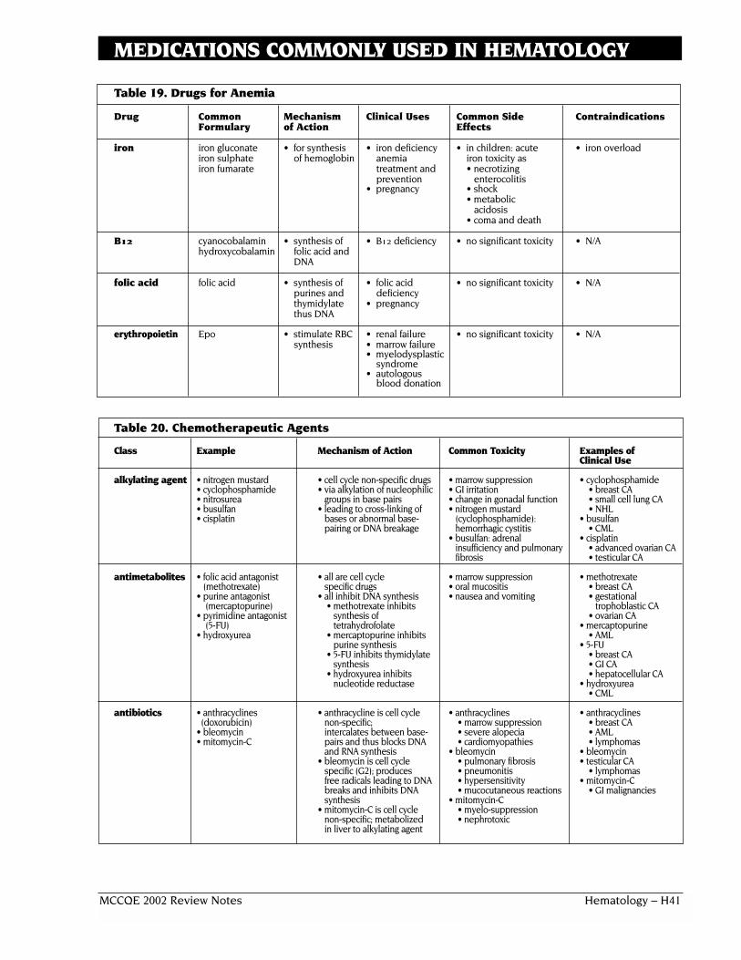
MCCQE 2002 Review Notes Hematology – H41
MEDICATIONS COMMONLY USED IN HEMATOLOGY
Table 19. Drugs for Anemia
Drug Common Mechanism Clinical Uses Common Side ContraindicationsFormulary of Action Effects
iron iron gluconate • for synthesis • iron deficiency • in children: acute • iron overloadiron sulphate of hemoglobin anemia iron toxicity asiron fumarate treatment and • necrotizing
prevention enterocolitis• pregnancy • shock
• metabolic acidosis
• coma and death
B12 cyanocobalamin • synthesis of • B12 deficiency • no significant toxicity • N/Ahydroxycobalamin folic acid and
DNA
folic acid folic acid • synthesis of • folic acid • no significant toxicity • N/Apurines and deficiency thymidylate • pregnancythus DNA
erythropoietin Epo • stimulate RBC • renal failure • no significant toxicity • N/Asynthesis • marrow failure
• myelodysplastic syndrome
• autologousblood donation
Table 20. Chemotherapeutic Agents
Class Example Mechanism of Action Common Toxicity Examples of Clinical Use
alkylating agent • nitrogen mustard • cell cycle non-specific drugs • marrow suppression • cyclophosphamide• cyclophosphamide • via alkylation of nucleophilic • GI irritation • breast CA• nitrosurea groups in base pairs • change in gonadal function • small cell lung CA• busulfan • leading to cross-linking of • nitrogen mustard • NHL• cisplatin bases or abnormal base- (cyclophosphamide): • busulfan
pairing or DNA breakage hemorrhagic cystitis • CML• busulfan: adrenal • cisplatin
insufficiency and pulmonary • advanced ovarian CAfibrosis • testicular CA
antimetabolites • folic acid antagonist • all are cell cycle • marrow suppression • methotrexate(methotrexate) specific drugs • oral mucositis • breast CA
• purine antagonist • all inhibit DNA synthesis • nausea and vomiting • gestational (mercaptopurine) • methotrexate inhibits trophoblastic CA
• pyrimidine antagonist synthesis of • ovarian CA(5-FU) tetrahydrofolate • mercaptopurine
• hydroxyurea • mercaptopurine inhibits • AMLpurine synthesis • 5-FU
• 5-FU inhibits thymidylate • breast CAsynthesis • GI CA
• hydroxyurea inhibits • hepatocellular CAnucleotide reductase • hydroxyurea
• CML
antibiotics • anthracyclines • anthracycline is cell cycle • anthracyclines • anthracyclines (doxorubicin) non-specific; • marrow suppression • breast CA
• bleomycin intercalates between base- • severe alopecia • AML• mitomycin-C pairs and thus blocks DNA • cardiomyopathies • lymphomas
and RNA synthesis • bleomycin • bleomycin• bleomycin is cell cycle • pulmonary fibrosis • testicular CA
specific (G2); produces • pneumonitis • lymphomasfree radicals leading to DNA • hypersensitivity • mitomycin-C breaks and inhibits DNA • mucocutaneous reactions • GI malignanciessynthesis • mitomycin-C
• mitomycin-C is cell cycle • myelo-suppressionnon-specific; metabolized • nephrotoxicin liver to alkylating agent

H42 – Hematology MCCQE 2002 Review Notes
MEDICATIONS COMMONLY USED IN HEMATOLOGY . . . CONT.
Table 20. Chemotherapeutic Agents (continued)
Class Example Mechanism of Action Common Toxicity Examples of Clinical Use
alkaloids • vinblastine • all are cell cycle specific • all have marrow suppression • vincristine and vinblastine• vincristine • vincristine and vinblastine • vincristine and vinblastine • lymphomas• podophyllotoxin inhibit assembly of • neurotoxic with areflexia, • Wilm’s tumour
(etoposide) microtubules therefore peripheral neuritis and • podophyllotoxin• taxol mitotic spindles and M paralytic ileus • small cell lung CA
phase • taxol • prostate CA• podophyllotoxin activates • neurotoxic as above • testicular CA
opoisomerase II therefore • taxolDNA breaks down • advanced breast CA
• taxol inhibits disassembly of • ovarian CAmicrotubules therefore cells are stuck in M phase
hormones • glucocorticoids • tamoxifen • glucocorticoid • glucocorticoids• tamoxifen • as a partial E2 antagonist • refer to Endocrinology • CML• flutamide • flutamide: androgen under Cushing’s syndrome • lymphomas• aminoglutethimide receptor antagonist • tamoxifen • tamoxifen
• aminoglutethimide: • menopausal symptoms • breast CAaromatase inhibitor in E2 • long term: retinopathy • flutamidesynthesis • aminoglutethimide • prostate CA
• menopausal symptoms • aminoglutethimide• skin rashes • metastatic breast CA
others • carboplatin • carboplatin • carboplatin • carboplatin• mitoxantrone • DNA binding • myelo-suppression • ovarian CA
• mitoxantrone • nausea, vomiting • mitoxantrone• ?DNA breaks • nephrotoxicity • AML
• mitoxantrone • NHL• cardiotoxicity • breast CA• alopecia • ovarian CA
• lung CA
REFERENCES
Armitage J.O. Treatment of Non-Hodgkin’s lymphoma. 1993. N Engl J Med. 328: 1023-1030.
Bataiile R, Harousseua J. Multiple myeloma. 1997. N Engl J Med. 336:1657-64.
Castellone DD. Evaluation of Bleeding Disorders. In Saunders Manual of Clinical Laboratory Science. Craig Lehman ED. WB Saunders CO,Philadelphia, PA, 1998.
Cohen K, Scadden D.T. Non-Hodgkin’s lymphoma: pathogenesis, clinical presentation, and treatment. 2001. Cancer Treat Res.104:201-03.
Heaney M.L., Golde D.W. Myelodysplasia. 1999. N Engl J Med. 340:1649-60.
http://cancernet.nci.nih.gov
Lowenberg B. Downing J.R., Burnett A. Acute myeloid leukemia. 1999. N Engl J Med. 341:1051-62.
Mackie IJ, and Bull HA. Normal haemostasis and it regulation. Blood Rev 3:237, 1989.
Mechanisms of severe transfusion reactions. Transfus Clin Biol. 2001 Jun;8(3):278-81.
Pui C., Evans W.E. Acute lymphoblastic leukemia. 1998. N Engl J Med. 339:605-15.
Rozman C., Montserrat E. Chronic lymphocytic leukemia. 1995. N Engl J Med. 333:1052-57.
Sawyers C. Chronic myeloid leukemia. N Engl J Med. 1999. 340:1330-40.
The Merck Manual; Section 11, Chapter 133: platelet disorders.
Related Documents