Nanoparticle Encapsulated Lipopeptide Conjugate of Antitubercular Drug Isoniazid: In Vitro Intracellular Activity and in Vivo Efficacy in a Guinea Pig Model of Tuberculosis Kata Horva ́ ti, † Bernadett Bacsa, † E ́ va Kiss, ‡ Gergő Gyulai, ‡ Kinga Fodor, § Gyula Balka, ∥ Mikló s Rusvai, ∥ Eleonó ra Szabó , ⊥ Ferenc Hudecz, †,# and Szilvia Bő sze* ,† † MTA-ELTE Research Group of Peptide Chemistry, Hungarian Academy of Sciences, ‡ Laboratory of Interfaces and Nanostructures, Institute of Chemistry, and # Department of Organic Chemistry, Eö tvö s L. University, Budapest, 1117 Hungary § Department of State Veterinary Medicine and Agricultural Economics and ∥ Department of Pathology and Forensic Veterinary Medicine, Faculty of Veterinary Science, Szent Istva ́ n University, Budapest, 1078 Hungary ⊥ Laboratory of Bacteriology, Kora ́ nyi National Institute for Tuberculosis and Respiratory Medicine, Budapest, 1122 Hungary ABSTRACT: Considering that Mycobacterium tuberculosis (Mtb) can survive in host phagocytes for decades and currently applied drugs are largely ineffective in killing intracellular Mtb, novel targeted delivery approaches to improve tuberculosis chemotherapy are urgently needed. In order to enhance the efficacy of a clinically used antitubercular agent (isoniazid, INH) a novel lipopeptide carrier was designed based on the sequence of tuftsin, which has been reported as a macrophage-targeting molecule. The conjugate showed relevant in vitro activity on Mtb H 37 Rv culture with low cytotoxicity and hemolytic activity on human cells. The conjugate directly killed intracellular Mtb and shows much greater efficacy than free INH. To improve bioavailability, the conjugate was encapsulated into poly(lactide-co-glycolide) (PLGA) nanoparticles and tested in vivo in a guinea pig infection model. External clinical signs, detectable mycobacterial colonies in the organs, and the histopathological findings substantiate the potent chemotherapeutic effect of orally administered conjugate- loaded nanoparticles. ■ INTRODUCTION Despite the worldwide availability of antituberculosis drugs, long duration of the treatment, serious adverse effects, poor patient compliance, and the emergence of multidrug-resistant strains indicate that the identification of novel antituberculars, the modification of existing drugs, and the development of new delivery systems to shorten tuberculosis (TB) chemotherapy are urgently needed. Many new drug candidates have been designed from known antibacterial drug classes by synthetic tailoring. 1 Re-engineering of an existing chemical scaffold can improve the antimycobacterial efficacy, safety, and tolerability. 2 For more than 60 years, isoniazid (INH) has been a front-line drug in the battle against Mycobacterium tuberculosis (Mtb), and it is used as a component of currently applied multidrug chemotherapy of TB. INH is a small hydrophilic molecule that targets mycolic acid biosynthesis and is activated inside the mycobacterial cell by KatG (catalase-peroxidase) enzyme. 3 Pharmacokinetic/pharmacodynamic properties and the poten- tial to cause neurotoxic and hepatotoxic side effects lead the intensive research on the derivatization of INH (reported INH- derivatives have been reviewed along with their biological activity by V. Judge et al.). 4 In a number of studies, high potency of synthetic analogues of INH with increased lipophilicity was found in vitro and in vivo. 5−7 The incorporation of a lipophilic moiety to the INH scaffold can enhance membrane affinity and penetration into the infected tissues and cells. R. Maccari and her co-workers synthesized and pharmacologically explored numerous different lipophilic analogues of INH with the aim of finding new compounds with activity against TB as well as other AIDS- associated diseases. In the biological assays, general correlation between the lipophilicity and effectiveness of the compounds against intracellular Mtb was found. 8−11 Such a property is considered highly predictive for an effective antitubercular agent, since the causative agent of TB can live and multiply inside macrophages. Exposure to Mtb results in a silent, yet persistent infection in the vast majority, during which the Mtb bacilli are metabolically slowed down and persist within their host cells for years or even decades. Current TB chemotherapy, while effective in killing actively growing Mtb, is largely ineffective in killing persistent or dormant bacilli. The great survival ability of Mtb is based on (i) the capability to transform into a stage of dormancy in which the bacillus is shielded by an extremely robust cell wall and renders itself phenotypically resistant to chemotherapy; (ii) the capacity to modulate macrophages and evade phagocytic digestion mechanisms; (iii) initiating the formation of granulomas (the hallmark of TB) which provide a Received: October 16, 2014 Revised: November 11, 2014 Article pubs.acs.org/bc © XXXX American Chemical Society A dx.doi.org/10.1021/bc500476x | Bioconjugate Chem. XXXX, XXX, XXX−XXX

Welcome message from author
This document is posted to help you gain knowledge. Please leave a comment to let me know what you think about it! Share it to your friends and learn new things together.
Transcript
Nanoparticle Encapsulated Lipopeptide Conjugate of AntitubercularDrug Isoniazid: In Vitro Intracellular Activity and in Vivo Efficacy in aGuinea Pig Model of TuberculosisKata Horvati,† Bernadett Bacsa,† Eva Kiss,‡ Gergo Gyulai,‡ Kinga Fodor,§ Gyula Balka,∥ Miklos Rusvai,∥
Eleonora Szabo,⊥ Ferenc Hudecz,†,# and Szilvia Bosze*,†
†MTA-ELTE Research Group of Peptide Chemistry, Hungarian Academy of Sciences, ‡Laboratory of Interfaces and Nanostructures,Institute of Chemistry, and #Department of Organic Chemistry, Eotvos L. University, Budapest, 1117 Hungary§Department of State Veterinary Medicine and Agricultural Economics and ∥Department of Pathology and Forensic VeterinaryMedicine, Faculty of Veterinary Science, Szent Istvan University, Budapest, 1078 Hungary⊥Laboratory of Bacteriology, Koranyi National Institute for Tuberculosis and Respiratory Medicine, Budapest, 1122 Hungary
ABSTRACT: Considering that Mycobacterium tuberculosis (Mtb) can survive inhost phagocytes for decades and currently applied drugs are largely ineffective inkilling intracellular Mtb, novel targeted delivery approaches to improvetuberculosis chemotherapy are urgently needed. In order to enhance the efficacyof a clinically used antitubercular agent (isoniazid, INH) a novel lipopeptidecarrier was designed based on the sequence of tuftsin, which has been reported asa macrophage-targeting molecule. The conjugate showed relevant in vitro activityon Mtb H37Rv culture with low cytotoxicity and hemolytic activity on humancells. The conjugate directly killed intracellular Mtb and shows much greaterefficacy than free INH. To improve bioavailability, the conjugate was encapsulated into poly(lactide-co-glycolide) (PLGA)nanoparticles and tested in vivo in a guinea pig infection model. External clinical signs, detectable mycobacterial colonies in theorgans, and the histopathological findings substantiate the potent chemotherapeutic effect of orally administered conjugate-loaded nanoparticles.
■ INTRODUCTION
Despite the worldwide availability of antituberculosis drugs,long duration of the treatment, serious adverse effects, poorpatient compliance, and the emergence of multidrug-resistantstrains indicate that the identification of novel antituberculars,the modification of existing drugs, and the development of newdelivery systems to shorten tuberculosis (TB) chemotherapyare urgently needed. Many new drug candidates have beendesigned from known antibacterial drug classes by synthetictailoring.1 Re-engineering of an existing chemical scaffold canimprove the antimycobacterial efficacy, safety, and tolerability.2
For more than 60 years, isoniazid (INH) has been a front-linedrug in the battle against Mycobacterium tuberculosis (Mtb), andit is used as a component of currently applied multidrugchemotherapy of TB. INH is a small hydrophilic molecule thattargets mycolic acid biosynthesis and is activated inside themycobacterial cell by KatG (catalase-peroxidase) enzyme.3
Pharmacokinetic/pharmacodynamic properties and the poten-tial to cause neurotoxic and hepatotoxic side effects lead theintensive research on the derivatization of INH (reported INH-derivatives have been reviewed along with their biologicalactivity by V. Judge et al.).4 In a number of studies, highpotency of synthetic analogues of INH with increasedlipophilicity was found in vitro and in vivo.5−7
The incorporation of a lipophilic moiety to the INH scaffoldcan enhance membrane affinity and penetration into the
infected tissues and cells. R. Maccari and her co-workerssynthesized and pharmacologically explored numerous differentlipophilic analogues of INH with the aim of finding newcompounds with activity against TB as well as other AIDS-associated diseases. In the biological assays, general correlationbetween the lipophilicity and effectiveness of the compoundsagainst intracellular Mtb was found.8−11 Such a property isconsidered highly predictive for an effective antitubercularagent, since the causative agent of TB can live and multiplyinside macrophages.Exposure to Mtb results in a silent, yet persistent infection in
the vast majority, during which the Mtb bacilli are metabolicallyslowed down and persist within their host cells for years oreven decades. Current TB chemotherapy, while effective inkilling actively growing Mtb, is largely ineffective in killingpersistent or dormant bacilli. The great survival ability of Mtb isbased on (i) the capability to transform into a stage ofdormancy in which the bacillus is shielded by an extremelyrobust cell wall and renders itself phenotypically resistant tochemotherapy; (ii) the capacity to modulate macrophages andevade phagocytic digestion mechanisms; (iii) initiating theformation of granulomas (the hallmark of TB) which provide a
Received: October 16, 2014Revised: November 11, 2014
Article
pubs.acs.org/bc
© XXXX American Chemical Society A dx.doi.org/10.1021/bc500476x | Bioconjugate Chem. XXXX, XXX, XXX−XXX
safety niche for the bacteria.12 Different in vitro models ofmycobacterial persistence have been established over the years,based on oxygen depletion,13 nutrient starvation,14 or the use ofstanding cultures (infection of phagocytic cells or cell lines withMtb).15 Under certain circumstances, such as anti-TNF-αtherapy16 or immune weakness (HIV/AIDS patients, organtransplants),17 the dormant Mtb bacilli are able to reactivateand cause TB-related clinical signs. One of the most challengingfactors in fighting TB is the ability to kill dormant Mtb.For such a purpose we established an advanced drug delivery
system for INH: the drug molecule is coupled to a lipopeptidecarrier and the conjugate is encapsulated into PLGA nano-particles. The peptide part of the construct is a tuftsin-relatedsequence. Tuftsin is a natural phagocytosis stimulating peptideand it has been reported as a macrophage targetingmolecule.18,19 It is estimated that there are 72 000 bindingsites available for tuftsin on the surface of phagocytic cells;20
therefore, tuftsin can be a potent chemical vector for targeteddelivery into macrophages through receptor mediated internal-ization.21,22 C. M. Gupta and his co-workers have developedtuftsin-bearing liposomes that not only enhance the host’sresistance against a variety of infections, but also serve as usefulvehicles for the site-specific delivery of drugs in a variety ofmacrophage-based infections, such as leishmaniasis andTB.23−26 During the past decade, a new group of sequentialoligopeptide carriers has been developed in our laboratorybased on the canine tuftsin sequence TKPKG.27,28 Thesecompounds are nontoxic, nonimmunogenic, and exhibit tuftsin-
like biological properties, e.g., receptor-binding and immun-stimulatory activity. In the present study, the TKPKG peptidewas elongated with palmitic acid to enhance the lipophilicity,membrane affinity, and PLGA encapsulation efficacy.Encapsulation of drug molecules into nanoparticles is an
efficient method for sustained controlled release and is beingintensively explored for antitubercular therapy.29−31 Nano-particles have the ability to protect the entrapped drug from theexternal environment, and to delay metabolism and clearance.Over the years, a variety of natural and synthetic polymer-basednanoparticles have been tested as delivery platforms, of whichpoly(lactide-co-glycolide) (PLGA) has been extensively inves-tigated because of its biocompatibility, biodegradability, andwide targeting capability.32,33 PLGA is approved by the USFood and Drug Administration (FDA) and by the EuropeanMedicine Agency (EMA)34 for clinical use and it can be utilizedin various human applications.35 G. K. Khuller and R. Pandeyhave published detailed evaluation of INH, rifampin, andpyrazinamide loaded PLGA nanoparticles bearing significanttherapeutic potential against experimental TB in guinea pigsand mice.36−38
Previously, we reported on a lipopeptide derivative of INHrepresenting enhanced interaction with model lipid mem-brane39 and mycolic acid containing monolayer.40 Theconjugate was successfully entrapped in PLGA nanoparticleswith highly improved encapsulation efficacy and characterizedby high drug content, low polydispersity, and spherical shapewithin nanometer size.39
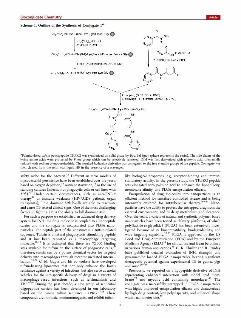
Scheme 1. Outline of the Synthesis of Conjugate 1a
aPalmitoylated tuftsin pentapeptide TKPKG was synthesized on solid phase by Boc/Bzl (gray sphere represents the resin). The side chains of thelysine amino acids were protected by Fmoc group which can be selectively removed. INH was first derivatized with glyoxylic acid, then mildlyreduced with sodium cyanoborohydride. The resulted hydrazide derivative was conjugated to the free ε-amino groups of the peptide. Conjugate wasthen cleaved from the resin with liquid HF in the presence of a scavenger.
Bioconjugate Chemistry Article
dx.doi.org/10.1021/bc500476x | Bioconjugate Chem. XXXX, XXX, XXX−XXXB
In the present work, INH was coupled to a palmitoylatedtuftsin peptide derivative to result the pT5i conjugate (1). Thein vitro selectivity of conjugate 1 was determined by themeasurement of the antitubercular efficacy on Mtb H37Rv andthe hemolytic and cytotoxic effects on human cells. The killingefficacy of 1 on intracellular Mtb was also determined. For invivo evaluation, conjugate 1 was encapsulated into PLGAnanoparticles (2) and the chemotherapeutic effect of 2 wasmeasured on Mtb infected guinea pigs.
■ RESULTSSynthesis and Characterization of pT5i Conjugate (1).
The lipopepitde conjugate of INH was prepared by a strategyoutlined in Scheme 1. The semiprotected TKPKG pentapep-tide [Thr(Bzl)-Lys(Fmoc)-Pro-Lys(Fmoc)-Gly] was synthe-sized and its N-terminal was palmitoylated on solid phase. INHwas incorporated into the side chain of two lysine residuesusing a bifunctional linker entity. After final cleavage, 1 wasisolated and carefully characterized by mass spectrometry,analytical HPLC and amino acid analysis. The overallpercentage yield of the synthesiscalculated to the capacityof the MBHA resinwas up to 64.8%. The peptide-conjugatecontent of the lyophilized product was 60.7%. The mono-isotopic molecular mass of the conjugate was measured by massspectrometry (Mcalc = 1120.6; Mfound = 1120.7). The purity ofthe conjugate, which was checked by analytical RP-HPLC, was≥95%.Comparison of In Vitro Antitubercular Effect, Cyto-
toxicity, and Hemolytic Activity. Antitubercular activity ofconjugate 1 and the free INH was determined against MtbH37Rv strain. The minimal inhibitory concentration (MIC, thelowest concentration of a compound at which no visible growthof the bacteria occurred) of INH in Sula media was 0.02 mg/L(0.15 μM). The MIC value of conjugate 1 was 0.125 mg/L(0.11 μM). It is interesting to observe that the peptideconjugate of INH proved to be slightly more active than thefree drug expressed in molar term (MIC = 0.11 μM forconjugate 1 vs MIC = 0.15 μM for INH).In order to evaluate the in vitro selectivity we also studied the
cytotoxicity and hemolytic activity of the compounds on humanPBMC and erythrocytes, respectively. We found that INH andconjugate 1 were not cytotoxic to human PBMC and expressedno hemolytic activity to human erythrocytes even at the highestconcentration (for both compounds: IC50 > 1000 μM; HC50 >1000 μM).INH-Lipopeptide Conjugate 1 Kills Intracellular Mtb.
In order to evaluate drug delivery and intracellular Mtb killingefficacy of lipopeptide conjugated INH, we infected humanMonoMac6 monocytes with virulent Mtb H37Rv bacteria andthen the infected cells were treated with the compounds at 100and 250 mg/L concentrations. The free INH did not exhibitintracellular antitubercular activity even at the higherconcentration used (250 mg/L, 1.82 mM). In contrast,conjugate 1 significantly reduced the colony forming units(CFU) of intracellular Mtb (Table 1) even at the lower 100mg/L (0.09 mM) concentration. Considering that the drugcontent of 100 mg conjugate 1 is 14.8 mg, the affectivity of 1 ismore than 16-fold higher than an equivalent amount of freeINH.PLGA Encapsulation of the pT5i Conjugate. In order to
improve bioavailability and enable the sustained release, thepT5i conjugate was encapsulated into PLGA nanoparticles aswe have previously described.39 The peptide-conjugate content
of the PLGA-pT5i nanoparticles (2) was 15.5%, whichcorresponds to 3.8% free INH content. The encapsulationefficacy of the conjugate 1 was up to 92 ± 7%.
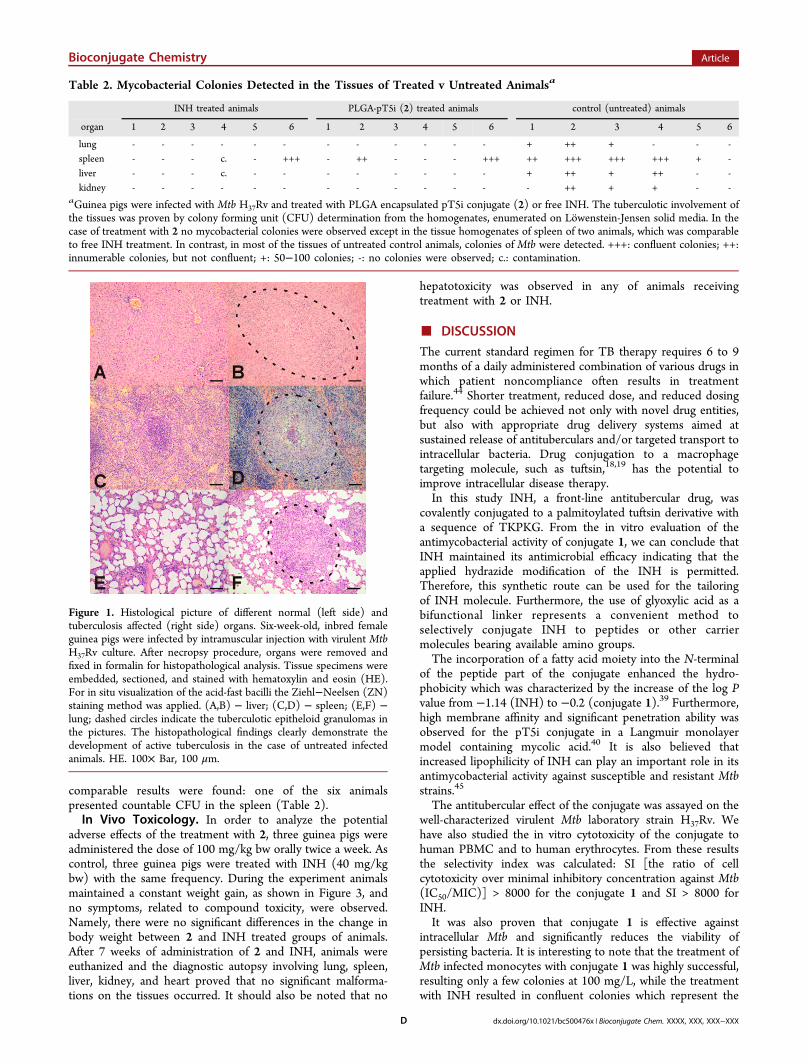
Infection of Guinea Pigs and Signs of Tuberculosis.To analyze the response of guinea pigs to infection with MtbH37Rv, external clinical signs and body weight change weremonitored weekly in untreated animals. Three infected animalsin the control group died at the 13rd, 14th, and 19th week.Signs of the disease, related to TB, such as scuffed fur, bodyweight loss, and lethargy, were also observed in three othercases. These external clinical signs of TB were further provedby mycobacterial CFU count, detected in spleen, lung, liver,and kidney homogenates as documented in Table 2.Representative sections of tissues were taken from Mtb infectedanimals and histopathology revealed serious involvement by thedisease process. The formation of typical tuberculotic lesions,e.g., formation of multiple epitheloid granulomas in the lymphnodes, lungs, liver, and spleen of the infected animals, as a resultof type IV, TH1 mediated hypersensitivity reaction wasobserved. Multinucleated giant cell formation and/or calcifica-tion were also observed in some samples. Ziehl-Neelsenstaining demonstrated the acid-fast bacteria in the cytoplasmof the epitheloid cells, or in the multinucleated giant cells(Figure 1).
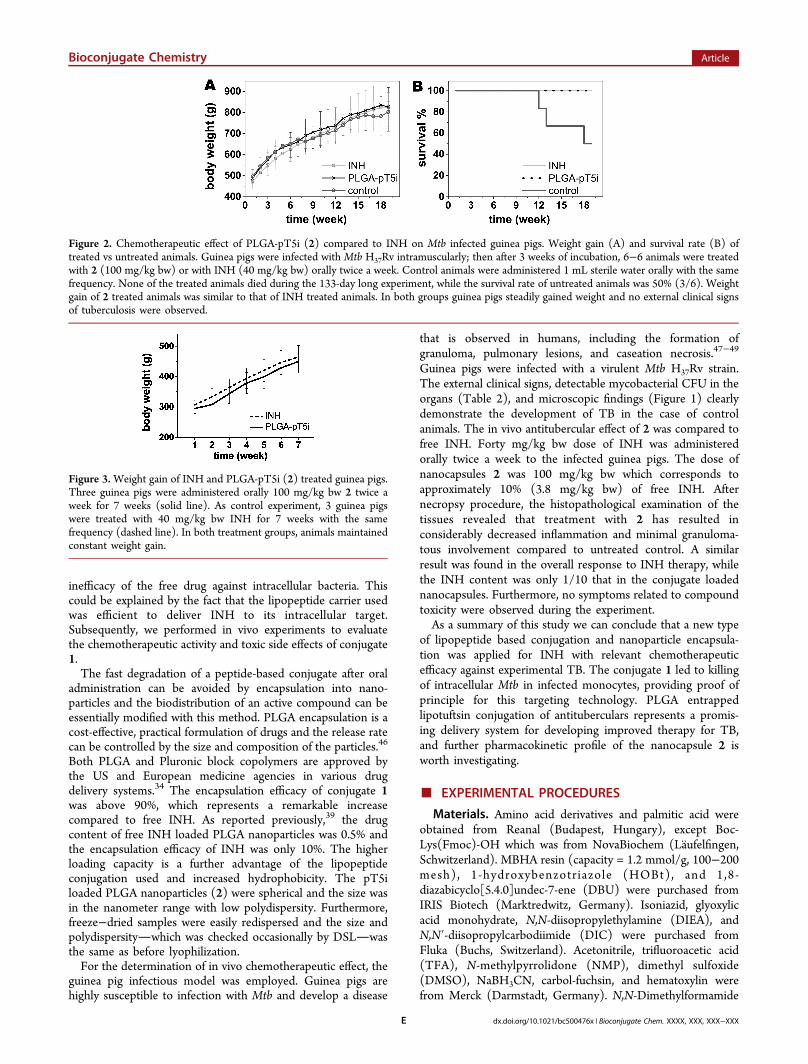
Comparison of the Chemotherapeutic Effect ofEncapsulated INH Conjugate with Free Drug. The effectof PLGA encapsulated INH-conjugate (2) as compared withthat of the free drug was studied. For this experiment six−sixguinea pigs were infected with Mtb H37Rv and after 3 weeks ofincubation animals were treated orally with 2 or INH twice aweek, for a period of 12 weeks. Pharmacokinetics andpharmacodynamics studies suggested that the human-equiv-alent dose of INH in a guinea pig model of TB chemotherapy isbetween 30 and 60 mg/kg bw.41−43 In this study, a 40 mg/kgdose of INH was administered. The dose of 2 was 100 mg/kgbw, which corresponds to approximately 10% (3.8 mg/kg bw)of free INH. As illustrated in Figure 2A, all treated guinea pigssteadily gained weight throughout the whole experiment. Nodeath occurred and no external clinical signs of active TB wereobserved. The overall response to INH therapy was similar tothe treatment with 2 (Figure 2A,B). Oral administration of 2 toMtb infected guinea pigs resulted in undetectable mycobacterialCFU in the lung, liver, and kidney homogenates; however, anumber of viable bacteria remained in the spleens of two out ofthe six animals. In the case of INH treated guinea pigs,
Table 1. Inhibition of Intracellular Mtb by INH and by pT5iConjugate (1)a
CFUb
compound 100 mg/L 250 mg/L
control +++ +++INH +++ +++pT5i + 10
aCultured MonoMac6 cells were infected with Mtb H37Rv and treatedwith the compounds at 0.1 and 0.25 mg/mL final concentration. Ascontrol, untreated cells were used. The conjugate 1 effectively kill theintracellular Mtb bacillus and lower the number of detectablemycobacterial colonies, while free INH was unable to penetrate andkill intracellular bacteria. bColony forming units of Mtb, enumeratedon Lowenstein-Jensen solid media (+++: confluent colonies; +: 50−100 colonies).
Bioconjugate Chemistry Article
dx.doi.org/10.1021/bc500476x | Bioconjugate Chem. XXXX, XXX, XXX−XXXC
comparable results were found: one of the six animalspresented countable CFU in the spleen (Table 2).In Vivo Toxicology. In order to analyze the potential
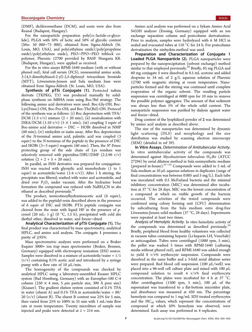
adverse effects of the treatment with 2, three guinea pigs wereadministered the dose of 100 mg/kg bw orally twice a week. Ascontrol, three guinea pigs were treated with INH (40 mg/kgbw) with the same frequency. During the experiment animalsmaintained a constant weight gain, as shown in Figure 3, andno symptoms, related to compound toxicity, were observed.Namely, there were no significant differences in the change inbody weight between 2 and INH treated groups of animals.After 7 weeks of administration of 2 and INH, animals wereeuthanized and the diagnostic autopsy involving lung, spleen,liver, kidney, and heart proved that no significant malforma-tions on the tissues occurred. It should also be noted that no
hepatotoxicity was observed in any of animals receivingtreatment with 2 or INH.
■ DISCUSSION
The current standard regimen for TB therapy requires 6 to 9months of a daily administered combination of various drugs inwhich patient noncompliance often results in treatmentfailure.44 Shorter treatment, reduced dose, and reduced dosingfrequency could be achieved not only with novel drug entities,but also with appropriate drug delivery systems aimed atsustained release of antituberculars and/or targeted transport tointracellular bacteria. Drug conjugation to a macrophagetargeting molecule, such as tuftsin,18,19 has the potential toimprove intracellular disease therapy.In this study INH, a front-line antitubercular drug, was
covalently conjugated to a palmitoylated tuftsin derivative witha sequence of TKPKG. From the in vitro evaluation of theantimycobacterial activity of conjugate 1, we can conclude thatINH maintained its antimicrobial efficacy indicating that theapplied hydrazide modification of the INH is permitted.Therefore, this synthetic route can be used for the tailoringof INH molecule. Furthermore, the use of glyoxylic acid as abifunctional linker represents a convenient method toselectively conjugate INH to peptides or other carriermolecules bearing available amino groups.The incorporation of a fatty acid moiety into the N-terminal
of the peptide part of the conjugate enhanced the hydro-phobicity which was characterized by the increase of the log Pvalue from −1.14 (INH) to −0.2 (conjugate 1).39 Furthermore,high membrane affinity and significant penetration ability wasobserved for the pT5i conjugate in a Langmuir monolayermodel containing mycolic acid.40 It is also believed thatincreased lipophilicity of INH can play an important role in itsantimycobacterial activity against susceptible and resistant Mtbstrains.45
The antitubercular effect of the conjugate was assayed on thewell-characterized virulent Mtb laboratory strain H37Rv. Wehave also studied the in vitro cytotoxicity of the conjugate tohuman PBMC and to human erythrocytes. From these resultsthe selectivity index was calculated: SI [the ratio of cellcytotoxicity over minimal inhibitory concentration against Mtb(IC50/MIC)] > 8000 for the conjugate 1 and SI > 8000 forINH.It was also proven that conjugate 1 is effective against
intracellular Mtb and significantly reduces the viability ofpersisting bacteria. It is interesting to note that the treatment ofMtb infected monocytes with conjugate 1 was highly successful,resulting only a few colonies at 100 mg/L, while the treatmentwith INH resulted in confluent colonies which represent the
Table 2. Mycobacterial Colonies Detected in the Tissues of Treated v Untreated Animalsa
INH treated animals PLGA-pT5i (2) treated animals control (untreated) animals
organ 1 2 3 4 5 6 1 2 3 4 5 6 1 2 3 4 5 6
lung - - - - - - - - - - - - + ++ + - - -spleen - - - c. - +++ - ++ - - - +++ ++ +++ +++ +++ + -liver - - - c. - - - - - - - - + ++ + ++ - -kidney - - - - - - - - - - - - - ++ + + - -
aGuinea pigs were infected with Mtb H37Rv and treated with PLGA encapsulated pT5i conjugate (2) or free INH. The tuberculotic involvement ofthe tissues was proven by colony forming unit (CFU) determination from the homogenates, enumerated on Lowenstein-Jensen solid media. In thecase of treatment with 2 no mycobacterial colonies were observed except in the tissue homogenates of spleen of two animals, which was comparableto free INH treatment. In contrast, in most of the tissues of untreated control animals, colonies of Mtb were detected. +++: confluent colonies; ++:innumerable colonies, but not confluent; +: 50−100 colonies; -: no colonies were observed; c.: contamination.
Figure 1. Histological picture of different normal (left side) andtuberculosis affected (right side) organs. Six-week-old, inbred femaleguinea pigs were infected by intramuscular injection with virulent MtbH37Rv culture. After necropsy procedure, organs were removed andfixed in formalin for histopathological analysis. Tissue specimens wereembedded, sectioned, and stained with hematoxylin and eosin (HE).For in situ visualization of the acid-fast bacilli the Ziehl−Neelsen (ZN)staining method was applied. (A,B) − liver; (C,D) − spleen; (E,F) −lung; dashed circles indicate the tuberculotic epitheloid granulomas inthe pictures. The histopathological findings clearly demonstrate thedevelopment of active tuberculosis in the case of untreated infectedanimals. HE. 100× Bar, 100 μm.
Bioconjugate Chemistry Article
dx.doi.org/10.1021/bc500476x | Bioconjugate Chem. XXXX, XXX, XXX−XXXD
inefficacy of the free drug against intracellular bacteria. Thiscould be explained by the fact that the lipopeptide carrier usedwas efficient to deliver INH to its intracellular target.Subsequently, we performed in vivo experiments to evaluatethe chemotherapeutic activity and toxic side effects of conjugate1.The fast degradation of a peptide-based conjugate after oral
administration can be avoided by encapsulation into nano-particles and the biodistribution of an active compound can beessentially modified with this method. PLGA encapsulation is acost-effective, practical formulation of drugs and the release ratecan be controlled by the size and composition of the particles.46
Both PLGA and Pluronic block copolymers are approved bythe US and European medicine agencies in various drugdelivery systems.34 The encapsulation efficacy of conjugate 1was above 90%, which represents a remarkable increasecompared to free INH. As reported previously,39 the drugcontent of free INH loaded PLGA nanoparticles was 0.5% andthe encapsulation efficacy of INH was only 10%. The higherloading capacity is a further advantage of the lipopeptideconjugation used and increased hydrophobicity. The pT5iloaded PLGA nanoparticles (2) were spherical and the size wasin the nanometer range with low polydispersity. Furthermore,freeze−dried samples were easily redispersed and the size andpolydispersitywhich was checked occasionally by DSLwasthe same as before lyophilization.For the determination of in vivo chemotherapeutic effect, the
guinea pig infectious model was employed. Guinea pigs arehighly susceptible to infection with Mtb and develop a disease
that is observed in humans, including the formation ofgranuloma, pulmonary lesions, and caseation necrosis.47−49
Guinea pigs were infected with a virulent Mtb H37Rv strain.The external clinical signs, detectable mycobacterial CFU in theorgans (Table 2), and microscopic findings (Figure 1) clearlydemonstrate the development of TB in the case of controlanimals. The in vivo antitubercular effect of 2 was compared tofree INH. Forty mg/kg bw dose of INH was administeredorally twice a week to the infected guinea pigs. The dose ofnanocapsules 2 was 100 mg/kg bw which corresponds toapproximately 10% (3.8 mg/kg bw) of free INH. Afternecropsy procedure, the histopathological examination of thetissues revealed that treatment with 2 has resulted inconsiderably decreased inflammation and minimal granuloma-tous involvement compared to untreated control. A similarresult was found in the overall response to INH therapy, whilethe INH content was only 1/10 that in the conjugate loadednanocapsules. Furthermore, no symptoms related to compoundtoxicity were observed during the experiment.As a summary of this study we can conclude that a new type
of lipopeptide based conjugation and nanoparticle encapsula-tion was applied for INH with relevant chemotherapeuticefficacy against experimental TB. The conjugate 1 led to killingof intracellular Mtb in infected monocytes, providing proof ofprinciple for this targeting technology. PLGA entrappedlipotuftsin conjugation of antituberculars represents a promis-ing delivery system for developing improved therapy for TB,and further pharmacokinetic profile of the nanocapsule 2 isworth investigating.
■ EXPERIMENTAL PROCEDURES
Materials. Amino acid derivatives and palmitic acid wereobtained from Reanal (Budapest, Hungary), except Boc-Lys(Fmoc)-OH which was from NovaBiochem (Laufelfingen,Schwitzerland). MBHA resin (capacity = 1.2 mmol/g, 100−200mesh), 1-hydroxybenzotriazole (HOBt), and 1,8-diazabicyclo[5.4.0]undec-7-ene (DBU) were purchased fromIRIS Biotech (Marktredwitz, Germany). Isoniazid, glyoxylicacid monohydrate, N,N-diisopropylethylamine (DIEA), andN,N′-diisopropylcarbodiimide (DIC) were purchased fromFluka (Buchs, Switzerland). Acetonitrile, trifluoroacetic acid(TFA), N-methylpyrrolidone (NMP), dimethyl sulfoxide(DMSO), NaBH3CN, carbol-fuchsin, and hematoxylin werefrom Merck (Darmstadt, Germany). N,N-Dimethylformamide
Figure 2. Chemotherapeutic effect of PLGA-pT5i (2) compared to INH on Mtb infected guinea pigs. Weight gain (A) and survival rate (B) oftreated vs untreated animals. Guinea pigs were infected with Mtb H37Rv intramuscularly; then after 3 weeks of incubation, 6−6 animals were treatedwith 2 (100 mg/kg bw) or with INH (40 mg/kg bw) orally twice a week. Control animals were administered 1 mL sterile water orally with the samefrequency. None of the treated animals died during the 133-day long experiment, while the survival rate of untreated animals was 50% (3/6). Weightgain of 2 treated animals was similar to that of INH treated animals. In both groups guinea pigs steadily gained weight and no external clinical signsof tuberculosis were observed.
Figure 3.Weight gain of INH and PLGA-pT5i (2) treated guinea pigs.Three guinea pigs were administered orally 100 mg/kg bw 2 twice aweek for 7 weeks (solid line). As control experiment, 3 guinea pigswere treated with 40 mg/kg bw INH for 7 weeks with the samefrequency (dashed line). In both treatment groups, animals maintainedconstant weight gain.
Bioconjugate Chemistry Article
dx.doi.org/10.1021/bc500476x | Bioconjugate Chem. XXXX, XXX, XXX−XXXE
(DMF), dichloromethane (DCM), and eosin were also fromReanal (Budapest, Hungary).For the nanoparticle preparation poly(DL-lactide-co-glyco-
lide), PLGA with 50% of lactic and 50% of glycolic content(Mw: 50 000−75 000), obtained from Sigma-Aldrich (St.Louis, MO, USA), and poly(ethylene oxide)/poly(propyleneoxide)/poly(ethylene oxide), PEO−PPO−PEO triblock co-polymer, Pluronic 12700 provided by BASF Hungaria Kft.(Budapest, Hungary), were applied as received.For the in vitro assays RPMI-1640 medium (with or without
phenol red), fetal calf serum (FCS), nonessential amino acids,3-(4,5-dimethyltiazol-2-yl)-2,5-diphenyl tetrazolium bromide(MTT), Lowenstein-Jensen and Sula medium base wereobtained from Sigma-Aldrich (St. Louis, MO, USA).Synthesis of pT5i Conjugate (1). Protected tuftsin
derivate (TKPKG, T5) was produced manually by solid-phase synthesis on MBHA resin using Boc/Bzl strategy. Thefollowing amino acid derivatives were used: Boc-Gly-OH, Boc-Lys(Fmoc)-OH, Boc-Pro-OH, and Boc-Thr(Bzl)-OH. Protocolof the synthesis was as follows: (i) Boc deprotection with TFA/DCM (1:3 v/v) mixture (2 + 20 min); (ii) neutralization withDIEA/DCM (1:10 v/v) (5 × 1 min); (iii) coupling 3 equiv ofBoc-amino acid derivative − DIC − HOBt dissolved in NMP(60 min); (iv) ninhydrin or isatin assay. After Boc deprotectionof the N-terminal amino acid, palmitic acid was coupled (3equiv) to the N-terminus of the peptide in the presence of DICand HOBt (3−3 equiv) reagents (60 min). Then, the Nε-Fmocprotecting group of the side chain of Lys residues wasselectively removed with piperidine/DBU/DMF (2:2:96 v/v)solution (2 + 2 + 5 + 20 min).In parallel, an INH derivative was prepared for conjugation:
INH was reacted with glyoxylic acid monohydrate (1 to 1equiv) in acetonitrile/water (1:4 v/v)). After 1 h stirring, theprecipitate was filtered, washed with water and acetonitrile, anddried over P2O5 under vacuum. After the hydrazine bondformation the compound was reduced with NaBH3CN in absethanol as described previously.50
The product, isonicotinoylhydrazinoacetic acid (6 equiv),was added to the peptidyl-resin described above in the presenceof 6 equiv of DIC and HOBt. PT5i peptide conjugate wascleaved from the resin with liquid HF in the presence of p-cresol (20 mL: 1 g) (0 °C, 1.5 h), precipitated with cold absdiethyl ether, dissolved in water, and freeze−dried.Analytical Characterization of pT5i Conjugate (1). The
final product was characterized by mass spectrometry, analyticalHPLC, and amino acid analysis. The conjugate 1 possesses apurity of ≥95%.Mass spectrometric analyses were performed on a Bruker
Esquire 3000+ ion trap mass spectrometer (Bruker, Bremen,Germany) equipped with electrospray ionization (ESI) source.Samples were dissolved in a mixture of acetonitrile/water = 1/1(v/v) containing 0.1% acetic acid and introduced by a syringepump with a flow rate of 10 μL/min.The homogeneity of the compounds was checked by
analytical HPLC using a laboratory-assembled Knauer HPLCsystem (Bad Homburg, Germany) with an Eurospher-100 C-4column (250 × 4 mm, 5 μm particle size, 300 Å pore size)(Knauer). The gradient elution system consisted of 0.1% TFAin water (eluent A) and 0.1% TFA in acetonitrile/water = 80/20 (v/v) (eluent B). The eluent B content was 25% for 5 min,then varied from 25% to 100% in 35 min with 1 mL/min flowrate at room temperature. Twenty microliters of sample wasinjected and peaks were detected at λ = 214 nm.
Amino acid analysis was performed on a Sykam Amino AcidS433H analyzer (Eresing, Germany) equipped with an ionexchange separation column and postcolumn derivatization.Prior to analysis, samples were hydrolyzed with 6 M HCl insealed and evacuated tubes at 110 °C for 24 h. For postcolumnderivatization the ninhydrin-method was used.
Preparation and Characterization of Conjugate 1Loaded PLGA Nanoparticle (2). PLGA nanoparticles wereprepared by the nanoprecipitation (solvent exchange) methodsimilar to that employed previously.39 Briefly, 85 mg PLGA and60 mg conjugate 1 were dissolved in 8.5 mL acetone and addeddropwise to 34 mL of 2 g/L aqueous solution of Pluronic12700 with magnetic stirring at room temperature. Nano-particles formed and the stirring was continued until completeevaporation of the organic solvent. The resulting particlesuspension was centrifuged at 6000 rpm for 10 min to removethe possible polymer aggregates. The amount of that sedimentwas always less than 5% of the whole solid content. Thenanoparticle suspension was purified by dialysis against waterand freeze−dried.Drug content of the lyophilized powder of 2 was determined
by amino acid analysis as described above.The size of the nanoparticles was determined by dynamic
light scattering (DLS) and morphology and the sizedistribution was studied by scanning electron microscopy(SEM) (detailed in ref 39).
In Vitro Assays. Determination of Antitubercular Activity.In vitro antitubercular activity of the compounds wasdetermined against Mycobacterium tuberculosis H37Rv (ATCC27294) by serial dilution method in Sula semisynthetic medium(prepared in-house).51−53 Compounds were added to 5 mL ofSula medium as 10 μL aqueous solutions in duplicates (range offinal concentrations was between 0.005 and 5 mg/L). Each tubewas inoculated with 0.5 McFarland bacteria and the minimalinhibitory concentration (MIC) was determined after incuba-tion at 37 °C for 28 days. MIC was the lowest concentration ofa compound at which no visible growth of the bacteriaoccurred. The activities of the tested compounds wereconfirmed using colony forming unit (CFU) determinationby subculturing from the Sula medium onto drug-freeLowensten-Jensen solid medium (37 °C, 28 days). Experimentswere repeated at least two times.
Analysis of Hemolytic Activity. In vitro hemolytic activity ofthe compounds was determined as described previously.54
Briefly, peripheral blood from healthy volunteers was collectedin vacuum tubes containing heparin (Li-heparin LH, VenoSafe)as anticoagulant. Tubes were centrifuged (1000 rpm, 5 min),the pellet was washed 3 times with RPMI-1640 (culturingmedia without phenol red), and RPMI-1640 was added enoughto yield 8 v/v% erythrocyte suspension. Compounds weredissolved in the same buffer and a 3-fold serial dilution serieswere prepared. Red blood cell suspension (100 μL/well) wasplaced into a 96-well cell culture plate and mixed with 100 μLcompound solution to result 4 v/v% final erythrocyteconcentration. The plates were incubated for 4 h at 37 °C.After centrifugation (1500 rpm, 5 min), 100 μL of thesupernatant was transferred to a flat-bottom microtiter plate,and absorbance was measured at 450 nm. The percentagehemolysis was compared to 1 mg/mL SDS treated erythrocytesand the HC50 values, which represent the concentrations ofcompound at which 50% hemolysis was observed, weredetermined. Each assay was performed in 4 replicates.
Bioconjugate Chemistry Article
dx.doi.org/10.1021/bc500476x | Bioconjugate Chem. XXXX, XXX, XXX−XXXF
Cell Culturing and Cytotoxicity Assay. Peripheral bloodmononuclear cells (PBMC) were prepared from peripheralblood of healthy volunteers using Ficoll-Hypaque densitygradient centrifugation method. PBMC were cultured incomplete medium prepared from RPMI-1640 supplementedwith 10% FCS, 2 mM L-glutamine, and 160 μg/mL gentamycinat 37 °C in 5% CO2 atmosphere. Twenty-four hours prior totreatment, PBMC cells were plated into a 96-well round-bottom plate (250 000 cell/100 μL complete medium).Prior to treatment, cells were washed with serum-free RPMI-
1640 medium. Compounds to be tested were dissolved inserum-free medium and added to the cells to give 0.5−1000μM final concentration. Cells were incubated with thecompounds for 4 h, then cell viability was tested using theMTT (3-(4,5-dimethyltiazol-2-yl)-2,5-diphenyl tetrazoliumbromide) test.55−57 Briefly, 45 μL MTT solutions were addedto each well (2 mg/mL, solved in serum-free medium).Following 4 h of incubation, plates were centrifuged at 2000rpm for 5 min, and the supernatant was carefully aspirated witha G30 needle. The precipitated purple crystals were dissolved in100 μL DMSO, and after 10 min agitation, the absorbance wasdetermined at λ = 540 and 620 nm using ELISA plate reader(iEMS Reader, Labsystems). The measured cytotoxicity inpercentage as a function of compound concentration wasrepresented graphically using Microcal Origin 8.6 software. IC50value is the mean of 4 parallel experiments and represents theconcentration of the compound which is required for 50%inhibition.Determination of Intracellular Antitubercular Activity.
Infection of MonoMac6 cells with Mtb H37Rv and the efficacyagainst intracellular Mtb bacteria were determined using amodified method based on previously published works.15,58,60
Briefly, MonoMac6 human monocytic cell line (DSMZ no.:ACC 124, Braunschweig, Germany)59 was maintained inRPMI-1640 medium containing 10% FCS, 2 mM L-glutamine,and 160 μg/mL gentamycin at 37 °C in 5% CO2 atmosphere.Prior to experiment, cells were cultured in a 24-well plate for 24h (2 × 10⁵ cells/1 mL medium/well). Adherent cells wereinfected with Mtb H37Rv at a multiplicity of infection (MOI) of10 for 4 h. Nonphagocytized extracellular bacteria wereremoved and the culture was washed three times with serumfree RPMI. The infected monolayer was incubated for 1 daybefore antitubercular treatment. Infected cells were then treatedwith the compounds at 100 and 250 mg/L final concentrations.After 3 days the treatment was repeated with fresh solution ofthe compounds for an additional 3 days. Untreated cells wereconsidered as a negative control. As control compound, INHwas used at 100 and 250 mg/L final concentration. Afterwashing stepsin order to remove the antitubercularsinfected cells were lysed with 2.5% sodium dodecyl sulfatesolution. The CFU of Mtb was enumerated on Lowenstein-Jensen solid media after 4 weeks of incubation. Experimentswere repeated at least twice.In Vivo Evaluation. Animals and Ethics Statement.
Inbred female guinea pigs aged 6 weeks at weight 400−500 gwere housed four per cage and allowed free access to water andto standard pellet diet. All animal experiments were approvedby the Hungarian Scientific Ethical Committee on AnimalExperimentation (No: 22.1/3720/003/2009), and all effortswere made to minimize suffering.Experimental Infection and Chemotherapy. After 4 weeks
of incubation, guinea pigs were infected by intramuscular
injection with 1.5 × 10⁸ viable bacilli of Mtb H37Rv in a volumeof 0.1 mL of Sula medium.Chemotherapy was started 3 weeks after infection. Guinea
pigs were divided into three different groups of 6 animals: INHtreated animals (group 1); 2 treated animals (group 2);untreated controls (group 3). Forty mg/kg bw INH and 100mg/kg bw of 2 were suspended in 1 mL sterile water andadministered orally twice a week. Control animals wereadministered 1 mL of sterile water with the same frequency.
Necropsy Procedure, CFU Determination, and Histopa-thology. After 12 weeks of chemotherapy, animals wereeuthanized by ketamine (40 mg/kg bw) and dexmedetomidine(Dexdomitor, 0.5 mg/kg bw). To count the number of bacteriain the organs, a portion of lung, spleen, liver, and kidney wereresected and homogenized in Sula media. 100 μL aliquots of1:1000 dilution of the homogenates were plated ontoLowenstein-Jensen solid media, and after 8 weeks of incubationat 37 °C in 5% CO2 atmosphere, CFU was enumerated.For histopathological analysis, lung, spleen, liver, kidney,
inguinal lymph nodes, and heart muscle of each animal wereremoved and fixed in 8% neutral buffered formalin for 24 h atroom temperature. Tissue specimens were dehydrated in aseries of ethanol and xylene baths and embedded in paraffinwax. Sections (3−4 μm) were cut, mounted on Super FrostUltra Plus slides (Thermo Scientific, Menzel-Glaser, Braunsch-weig, Germany), deparaffinized in xylene and graded ethanol,and stained with hematoxylin and eosin (HE). For in situvisualization of the acid-fast bacilli Ziehl−Neelsen (ZN)staining method was applied on similarly pretreated sections.This method is based on the highly resistant bacterial wall ofMycobacteria that retain the original red dye (Carbol-Fuchsin)after destaining with 3% hydrochloric acid in alcohol. Acid fastMycobacteria appear as red rods in these sections, whereas allother bacteria and tissue components are blue according to thefinal hematoxylin counterstaining.
In Vivo Toxicology. Six-week-old inbred female guinea pigs(250−350 g) were housed and fed as described above. Animalswere administered 100 mg/kg bw of 2 orally twice a week(group 1), or 40 mg/kg bw of INH (group 2) (n = 3 for eachtreatment group). To monitor the side effects of thechemotherapy, changes in body weight were determinedweekly. After 7 weeks of administration, animals wereeuthanized and diagnostic autopsy was evaluated.
■ AUTHOR INFORMATIONCorresponding Author*Tel: +36-1-372-2500 ext. 1736, fax: +36-1-372-2620, e-mail:[email protected].
NotesThe authors declare no competing financial interest.
■ ACKNOWLEDGMENTSThe authors thank Dr. Hedvig Medzihradszky-Schweiger forthe amino acid analyses and to Mr. Sandor David forperforming the antimycobacterial tests. This work wassupported by the Hungarian Research Fund (T68358,T68285, 104928, 104275); by the Social Renewal OperationalProgramme (TAMOP-4.2.2.B-10/1, Development of a complexeducational assistance/support system for talented students andprospective researchers at the Szent Istvan University” project),and by the National Research and Development Programme(NKFP_07_1-TB_INTER-HU).
Bioconjugate Chemistry Article
dx.doi.org/10.1021/bc500476x | Bioconjugate Chem. XXXX, XXX, XXX−XXXG
■ REFERENCES(1) Fischbach, M. A., and Walsh, C. T. (2009) Antibiotics foremerging pathogens. Science 325, 1089−1093.(2) Koul, A., Arnoult, E., Lounis, N., Guillemont, J., and Andries, K.(2011) The challenge of new drug discovery for tuberculosis. Nature469, 483−490.(3) Zhang, Y., Heym, B., Allen, B., Young, D., and Cole, S. (1992)The catalase-peroxidase gene and isoniazid resistance of mycobacte-rium tuberculosis. Nature 358, 591−593.(4) Judge, V., Narasimhan, B., and Ahuja, M. (2012) Isoniazid: Themagic molecule. Med. Chem. Res. 21, 3940−3957.(5) Rastogi, N., and Goh, K. S. (1990) Action of 1-isonicotinyl-2-palmitoyl hydrazine against the mycobacterium avium complex andenhancement of its activity by m-fluorophenylalanine. Antimicrob.Agents Chemother. 34, 2061−2064.(6) Hearn, M. J., Cynamon, M. H., Chen, M. F., Coppins, R., Davis,J., Kang, H. J. O., Noble, A., Tu-Sekine, B., Terrot, M. S., Trombino,D., Thai, M., Webster, E. R., and Wilson, R. (2009) Preparation andantitubercular activities in vitro and in vivo of novel schiff bases ofisoniazid. Eur. J. Med. Chem. 44, 4169−4178.(7) Sriram, D., Yogeeswari, P., and Priya, D. Y. (2009)Antimycobacterial activity of novel n-(substituted)-2-isonicotinoylhy-drazinocarbothioamide endowed with high activity towards isoniazidresistant tuberculosis. Biomed. Pharmacother. 63, 36−39.(8) Maccari, R., Ottana, R., Bottari, B., Rotondo, E., and Vigorita, M.G. (2004) In vitro advanced antimycobacterial screening of cobalt(ii)and copper(ii) complexes of fluorinated isonicotinoylhydrazones.Bioorg. Med. Chem. Lett. 14, 5731−5733.(9) Maccari, R., Ottana, R., Monforte, F., and Vigorita, M. G. (2002)In vitro antimycobacterial activities of 2′-monosubstituted isonicoti-nohydrazides and their cyanoborane adducts. Antimicrob. AgentsChemother. 46, 294−299.(10) Maccari, R., Ottana, R., and Vigorita, M. G. (2005) In vitroadvanced antimycobacterial screening of isoniazid-related hydrazones,hydrazides and cyanoboranes: Part 14. Bioorg. Med. Chem. Lett. 15,2509−2513.(11) Vigorita, M. G., Ottana, R., Maccari, R., Monforte, F., Bisignano,G., and Pizzimenti, F. C. (1998) Synthesis and in vitro antimicrobialand antitumoral screening of novel lipophilic isoniazid analogues. Vi.Boll. Chim. Farm. 137, 267−276.(12) Gengenbacher, M., and Kaufmann, S. H. (2012) Mycobacteriumtuberculosis: Success through dormancy. FEMS Microbiol. Rev. 36,514−532.(13) Wayne, L. G. (2001) In vitro model of hypoxically inducednonreplicating persistence of mycobacterium tuberculosis. MethodsMol. Med. 54, 247−269.(14) Betts, J. C., Lukey, P. T., Robb, L. C., McAdam, R. A., andDuncan, K. (2002) Evaluation of a nutrient starvation model ofmycobacterium tuberculosis persistence by gene and proteinexpression profiling. Mol. Microbiol. 43, 717−731.(15) Wright, E. L., Quenelle, D. C., Suling, W. J., and Barrow, W. W.(1996) Use of mono mac 6 human monocytic cell line and j774murine macrophage cell line in parallel antimycobacterial drug studies.Antimicrob. Agents Chemother. 40, 2206−2208.(16) Gardam, M. A., Keystone, E. C., Menzies, R., Manners, S.,Skamene, E., Long, R., and Vinh, D. C. (2003) Anti-tumour necrosisfactor agents and tuberculosis risk: Mechanisms of action and clinicalmanagement. Lancet Infect. Dis. 3, 148−155.(17) Maartens, G., and Wilkinson, R. J. (2007) Tuberculosis. Lancet370, 2030−2043.(18) Siemion, I. Z., and Kluczyk, A. (1999) Tuftsin: On the 30-yearanniversary of Victor Najjar’s discovery. Peptides 20, 645−674.(19) Tripathi, S. K., Goyal, R., Kashyap, M. P., Pant, A. B., Haq, W.,Kumar, P., and Gupta, K. C. (2012) Depolymerized chitosansfunctionalized with bpei as carriers of nucleic acids and tuftsin-tethered conjugate for macrophage targeting. Biomaterials 33, 4204−4219.
(20) Barshavit, Z., Goldman, R., Stabinsky, Y., and Fridkin, M.(1979) Tuftsin-macrophage interaction - specific binding andaugmentation of phagocytosis. J. Cell. Physiol. 100, 55−62.(21) Amoscato, A. A., Davies, P. J., Babcock, G. F., and Nishioka, K.(1983) Receptor-mediated internalization of tuftsin. Ann. N.Y. Acad.Sci. 419, 114−134.(22) Gottlieb, P., Stabinsky, Y., Hiller, Y., Beretz, A., Hazum, E.,Tzehoval, E., Feldman, M., Segal, S., Zakuth, V., Spirer, Z., and Fridkin,M. (1983) Tuftsin receptors. Ann. N.Y. Acad. Sci. 419, 93−106.(23) Agarwal, A., Kandpal, H., Gupta, H. P., Singh, N. B., and Gupta,C. M. (1994) Tuftsin-bearing liposomes as rifampin vehicles intreatment of tuberculosis in mice. Antimicrob. Agents Chemother. 38,588−593.(24) Agrawal, A. K., Agrawal, A., Pal, A., Guru, P. Y., and Gupta, C.M. (2002) Superior chemotherapeutic efficacy of amphotericin b intuftsin-bearing liposomes against leishmania donovani infection inhamsters. J. Drug Targeting 10, 41−45.(25) Gupta, C. M., and Haq, W. (2005) Tuftsin-bearing liposomes asantibiotic carriers in treatment of macrophage infections. MethodsEnzymol. 391, 291−304.(26) Guru, P. Y., Agrawal, A. K., Singha, U. K., Singhal, A., andGupta, C. M. (1989) Drug targeting in leishmania-donovani infectionsusing tuftsin-bearing liposomes as drug vehicles. FEBS Lett. 245, 204−208.(27) Mezo, G., Kalaszi, A., Remenyi, J., Majer, Z., Hilbert, A., Lang,O., Kohidai, L., Barna, K., Gaal, D., and Hudecz, F. (2004) Synthesis,conformation, and immunoreactivity of new carrier molecules basedon repeated tuftsin-like sequence. Biopolymers 73, 645−656.(28) Bai, K. B., Lang, O., Orban, E., Szabo, R., Kohidai, L., Hudecz,F., and Mezo, G. (2008) Design, synthesis, and in vitro activity ofnovel drug delivery systems containing tuftsin derivatives andmethotrexate. Bioconjugate Chem. 19, 2260−2269.(29) Pandey, R., and Ahmad, Z. (2011) Nanomedicine andexperimental tuberculosis: Facts, flaws, and future. Nanomedicine 7,259−272.(30) Gelperina, S., Kisich, K., Iseman, M. D., and Heifets, L. (2005)The potential advantages of nanoparticle drug delivery systems inchemotherapy of tuberculosis. Am. J. Resp. Crit. Care Med. 172, 1487−1490.(31) Sosnik, A., Carcaboso, A. M., Glisoni, R. J., Moretton, M. A., andChiappetta, D. A. (2010) New old challenges in tuberculosis:Potentially effective nanotechnologies in drug delivery. Adv. DrugDelivery Rev. 62, 547−559.(32) Bala, I., Hariharan, S., and Kumar, M. N. (2004) Plgananoparticles in drug delivery: The state of the art. Crit. Rev. Ther.Drug Carrier Syst. 21, 387−422.(33) Shive, M. S., and Anderson, J. M. (1997) Biodegradation andbiocompatibility of PLA and PLGA microspheres. Adv. Drug DeliveryRev. 28, 5−24.(34) Danhier, F., Ansorena, E., Silva, J. M., Coco, R., Le Breton, A.,and Preat, V. (2012) PLGA-based nanoparticles: An overview ofbiomedical applications. J. Controlled Release 161, 505−522.(35) Ul-Ain, Q., Sharma, S., and Khuller, G. K. (2003) Chemo-therapeutic potential of orally administered poly(lactide-co-glycolide)microparticles containing isoniazid, rifampin, and pyrazinamide againstexperimental tuberculosis. Antimicrob. Agents Chemother. 47, 3005−3007.(36) Pandey, R., Sharma, A., Zahoor, A., Sharma, S., Khuller, G. K.,and Prasad, B. (2003) Poly (dl-lactide-co-glycolide) nanoparticle-based inhalable sustained drug delivery system for experimentaltuberculosis. J. Antimicrob. Chemother. 52, 981−986.(37) Pandey, R., Zahoor, A., Sharma, S., and Khuller, G. K. (2003)Nanoparticle encapsulated antitubercular drugs as a potential oral drugdelivery system against murine tuberculosis. Tuberculosis (Edinburgh)83, 373−378.(38) Sharma, A., Pandey, R., Sharma, S., and Khuller, G. K. (2004)Chemotherapeutic efficacy of poly (dl-lactide-co-glycolide) nano-particle encapsulated antitubercular drugs at sub-therapeutic dose
Bioconjugate Chemistry Article
dx.doi.org/10.1021/bc500476x | Bioconjugate Chem. XXXX, XXX, XXX−XXXH
against experimental tuberculosis. Int. J. Antimicrob. Agents 24, 599−604.(39) Kiss, E., Schnoller, D., Pribranska, K., Hill, K., Penzes, C. B.,Horvati, K., and Bosze, S. (2011) Nanoencapsulation of antituberculardrug isoniazid and its lipopeptide conjugate. J. Dispersion Sci. Technol.32, 1728−1734.(40) Penzes, C. B., Schnoller, D., Horvati, K., Bosze, S., Mezo, G.,and Kiss, E. (2012) Membrane affinity of antituberculotic drugconjugate using lipid monolayer containing mycolic acid. Colloids Surf.,A 413, 142−148.(41) Ahmad, Z., Klinkenberg, L. G., Pinn, M. L., Fraig, M. M.,Peloquin, C. A., Bishai, W. R., Nuermberger, E. L., Grosset, J. H., andKarakousis, P. C. (2009) Biphasic kill curve of isoniazid reveals thepresence of drug-tolerant, not drug-resistant, mycobacterium tuber-culosis in the guinea pig. J. Infect. Dis. 200, 1136−1143.(42) Dutta, N. K., Alsultan, A., Gniadek, T. J., Belchis, D. A., Pinn, M.L., Mdluli, K. E., Nuermberger, E. L., Peloquin, C. A., and Karakousis,P. C. (2013) Potent rifamycin-sparing regimen cures guinea pigtuberculosis as rapidly as the standard regimen. Antimicrob. AgentsChemother. 57, 3910−3916.(43) Jayaram, R., Shandil, R. K., Gaonkar, S., Kaur, P., Suresh, B. L.,Mahesh, B. N., Jayashree, R., Nandi, V., Bharath, S., Kantharaj, E., andBalasubramanian, V. (2004) Isoniazid pharmacokinetics-pharmacody-namics in an aerosol infection model of tuberculosis. Antimicrob.Agents Chemother. 48, 2951−2957.(44) Chan, E. D., and Iseman, M. D. (2002) Current medicaltreatment for tuberculosis. BMJ 325, 1282−1286.(45) Rodrigues, M. O., Cantos, J. B., D’Oca, C. R., Soares, K. L.,Coelho, T. S., Piovesan, L. A., Russowsky, D., da Silva, P. A., andD’Oca, M. G. (2013) Synthesis and antimycobacterial activity ofisoniazid derivatives from renewable fatty acids. Bioorg. Med. Chem. 21,6910−6914.(46) Makino, K., Nakajima, T., Shikamura, M., Ito, F., Ando, S.,Kochi, C., Inagawa, H., Soma, G., and Terada, H. (2004) Efficientintracellular delivery of rifampicin to alveolar macrophages usingrifampicin-loaded PLGA microspheres: Effects of molecular weightand composition of PLGA on release of rifampicin. Colloids Surf., B:Biointerfaces 36, 35−42.(47) Dharmadhikari, A. S., and Nardell, E. A. (2008) What animalmodels teach humans about tuberculosis. Am. J. Respir. Cell Mol. Biol.39, 503−508.(48) McMurray, D. N. (1994) Guinea pig model of tuberculosis. InTuberculosis: Pathogenesis, protection and control (Bloom, B. R., Ed.) pp135−147, American Society for Microbiology, Washington, DC.(49) Ahmad, Z., Nuermberger, E. L., Tasneen, R., Pinn, M. L.,Williams, K. N., Peloquin, C. A., Grosset, J. H., and Karakousis, P. C.(2010) Comparison of the ’denver regimen’ against acute tuberculosisin the mouse and guinea pig. J. Antimicrob. Chemother. 65, 729−734.(50) Horvati, K., Mezo, G., Szabo, N., Hudecz, F., and Bosze, S.(2009) Peptide conjugates of therapeutically used antitubercularisoniazid-design, synthesis and antimycobacterial effect. J. Pept. Sci. 15,385−391.(51) Sula, L. (1963) Who co-operative studies on a simple culturetechnique for the isolation of mycobacteria. 1. Preparation,lyophilization and reconstitution of a simple semi-synthetic concen-trated liquid medium; culture technique; growth pattern of differentmycobacteria. Bull. World Health Org. 29, 589−606.(52) Sula, L., and Sundaresan, T. K. (1963) Who co-operative studieson a simple culture technique for the isolation of mycobacteria. 2.Comparison of the efficacy of lyophilized liquid medium with that ofloewenstein-jensen (l-j) medium. Bull. World Health Org. 29, 607−625.(53) Vinsova, J., Cermakova, K., Tomeckova, A., Ceckova, M.,Jampilek, J., Cermak, P., Kunes, J., Dolezal, M., and Staud, F. (2006)Synthesis and antimicrobial evaluation of new 2-substituted 5,7-di-tert-butylbenzoxazoles. Bioorg. Med. Chem. 14, 5850−5865.(54) Helmerhorst, E. J., Reijnders, I. M., van ’t Hof, W., Veerman, E.C., and Nieuw Amerongen, A. V. (1999) A critical comparison of the
hemolytic and fungicidal activities of cationic antimicrobial peptides.FEBS Lett. 449, 105−110.(55) Liu, Y., Peterson, D. A., Kimura, H., and Schubert, D. (1997)Mechanism of cellular 3-(4,5-dimethylthiazol-2-yl)-2,5-diphenyltetra-zolium bromide (mtt) reduction. J. Neurochem. 69, 581−593.(56) Mosmann, T. (1983) Rapid colorimetric assay for cellulargrowth and survival: Application to proliferation and cytotoxicityassays. J. Immunol. Methods 65, 55−63.(57) Slater, T. F., Sawyer, B., and Straeuli, U. (1963) Studies onsuccinate-tetrazolium reductase systems. III. Points of coupling of fourdifferent tetrazolium salts. Biochim. Biophys. Acta 77, 383−393.(58) Tomioka, H., Sato, K., Kajitani, H., Akaki, T., and Shishido, S.(2000) Comparative antimicrobial activities of the newly synthesizedquinolone wq-3034, levofloxacin, sparfloxacin, and ciprofloxacinagainst mycobacterium tuberculosis and mycobacterium aviumcomplex. Antimicrob. Agents Chemother. 44, 283−286.(59) Ziegler-Heitbrock, H. W., Thiel, E., Futterer, A., Herzog, V.,Wirtz, A., and Riethmuller, G. (1988) Establishment of a human cellline (mono mac 6) with characteristics of mature monocytes. Int. J.Cancer 41, 456−461.(60) Horvati, K., Bacsa, B., Szabo, N., David, S., Mezo, G., Grolmusz,V., Vertessy, B., Hudecz, F., and Bosze, S. (2012) Enhanced cellularuptake of a new, in silico identified antitubercular candidate by peptideconjugation. Bioconjug. Chem. 23, 900−907.
Bioconjugate Chemistry Article
dx.doi.org/10.1021/bc500476x | Bioconjugate Chem. XXXX, XXX, XXX−XXXI
Related Documents