Citation: Venugopala, K.N.; Al-Shar’i, N.A.; Dahabiyeh, L.A.; Hourani, W.; Deb, P.K.; Pillay, M.; Abu-Irmaileh, B.; Bustanji, Y.; Chandrashekharappa, S.; Tratrat, C.; et al. Antitubercular, Cytotoxicity, and Computational Target Validation of Dihydroquinazolinone Derivatives. Antibiotics 2022, 11, 831. https:// doi.org/10.3390/antibiotics11070831 Academic Editor: Martina Hrast Received: 30 May 2022 Accepted: 17 June 2022 Published: 21 June 2022 Publisher’s Note: MDPI stays neutral with regard to jurisdictional claims in published maps and institutional affil- iations. Copyright: © 2022 by the authors. Licensee MDPI, Basel, Switzerland. This article is an open access article distributed under the terms and conditions of the Creative Commons Attribution (CC BY) license (https:// creativecommons.org/licenses/by/ 4.0/). antibiotics Article Antitubercular, Cytotoxicity, and Computational Target Validation of Dihydroquinazolinone Derivatives Katharigatta N. Venugopala 1,2, * , Nizar A. Al-Shar’i 3 , Lina A. Dahabiyeh 4 , Wafa Hourani 5 , Pran Kishore Deb 5, * , Melendhran Pillay 6 , Bashaer Abu-Irmaileh 7 , Yasser Bustanji 7,8 , Sandeep Chandrashekharappa 9 , Christophe Tratrat 1 , Mahesh Attimarad 1 , Anroop B. Nair 1 , Nagaraja Sreeharsha 1,10 , Pottathil Shinu 11 , Michelyne Haroun 1 , Mahmoud Kandeel 12,13 , Abdulmalek Ahmed Balgoname 1 , Rashmi Venugopala 14 and Mohamed A. Morsy 1,15 1 Department of Pharmaceutical Sciences, College of Clinical Pharmacy, King Faisal University, Al-Ahsa 31982, Saudi Arabia; [email protected] (C.T.); [email protected] (M.A.); [email protected] (A.B.N.); [email protected] (N.S.); [email protected] (M.H.); [email protected] (A.A.B.); [email protected] (M.A.M.) 2 Department of Biotechnology and Food Science, Faculty of Applied Sciences, Durban University of Technology, Durban 4000, South Africa 3 Department of Medicinal Chemistry and Pharmacognosy, Faculty of Pharmacy, Jordan University of Science and Technology, P.O. Box 3030, Irbid 22110, Jordan; [email protected] 4 Department of Pharmaceutical Sciences, School of Pharmacy, The University of Jordan, Amman 11942, Jordan; [email protected] 5 Department of Pharmaceutical Sciences, Faculty of Pharmacy, Philadelphia University, Amman 19392, Jordan; [email protected] 6 Department of Microbiology, National Health Laboratory Services, KZN Academic Complex, Inkosi Albert Luthuli Central Hospital, Durban 4001, South Africa; [email protected] 7 Hamdi Mango Center for Scientific Research, The University of Jordan, Amman 11942, Jordan; [email protected] (B.A.-I.); [email protected] (Y.B.) 8 Department of Basic Medical Sciences, College of Medicine, University of Sharjah, Sharjah 27272, United Arab Emirates 9 Department of Medicinal Chemistry, National Institute of Pharmaceutical Education and Research (NIPER-R) Raebareli, Lucknow 226002, India; [email protected] 10 Department of Pharmaceutics, Vidya Siri College of Pharmacy, Off Sarjapura Road, Bangalore 560035, India 11 Department of Biomedical Sciences, College of Clinical Pharmacy, King Faisal University, Al-Ahsa 31982, Saudi Arabia; [email protected] 12 Department of Biomedical Sciences, College of Veterinary Medicine, King Faisal University, Al-Ahsa 31982, Saudi Arabia; [email protected] 13 Department of Pharmacology, Faculty of Veterinary Medicine, Kafrelsheikh University, Kafrelsheikh 33516, Egypt 14 Department of Public Health Medicine, Howard College Campus, University of KwaZulu-Natal, Durban 4001, South Africa; [email protected] 15 Department of Pharmacology, Faculty of Medicine, Minia University, El-Minia 61511, Egypt * Correspondence: [email protected] (K.N.V.); [email protected] (P.K.D.) Abstract: A series of 2,3-dihydroquinazolin-4(1H)-one derivatives (3a–3m) was screened for in vitro whole-cell antitubercular activity against the tubercular strain H37Rv and multidrug-resistant (MDR) Mycobacterium tuberculosis (MTB) strains. Compounds 3l and 3m with di-substituted aryl moiety (halogens) attached to the 2-position of the scaffold showed a minimum inhibitory concentration (MIC) of 2 μg/mL against the MTB strain H37Rv. Compound 3k with an imidazole ring at the 2-position of the dihydroquinazolin-4(1H)-one also showed significant inhibitory action against both the susceptible strain H37Rv and MDR strains with MIC values of 4 and 16 μg/mL, respectively. The computational results revealed the mycobacterial pyridoxal-5 0 -phosphate (PLP)-dependent aminotransferase (BioA) enzyme as the potential target for the tested compounds. In vitro, ADMET calculations and cytotoxicity studies against the normal human dermal fibroblast cells indicated the safety and tolerability of the test compounds 3k–3m. Thus, compounds 3k–3m warrant further optimization to develop novel BioA inhibitors for the treatment of drug-sensitive H37Rv and drug- resistant MTB. Antibiotics 2022, 11, 831. https://doi.org/10.3390/antibiotics11070831 https://www.mdpi.com/journal/antibiotics

Welcome message from author
This document is posted to help you gain knowledge. Please leave a comment to let me know what you think about it! Share it to your friends and learn new things together.
Transcript
Citation: Venugopala, K.N.;
Al-Shar’i, N.A.; Dahabiyeh, L.A.;
Hourani, W.; Deb, P.K.; Pillay, M.;
Abu-Irmaileh, B.; Bustanji, Y.;
Chandrashekharappa, S.;
Tratrat, C.; et al. Antitubercular,
Cytotoxicity, and Computational
Target Validation of
Dihydroquinazolinone Derivatives.
Antibiotics 2022, 11, 831. https://
doi.org/10.3390/antibiotics11070831
Academic Editor: Martina Hrast
Received: 30 May 2022
Accepted: 17 June 2022
Published: 21 June 2022
Publisher’s Note: MDPI stays neutral
with regard to jurisdictional claims in
published maps and institutional affil-
iations.
Copyright: © 2022 by the authors.
Licensee MDPI, Basel, Switzerland.
This article is an open access article
distributed under the terms and
conditions of the Creative Commons
Attribution (CC BY) license (https://
creativecommons.org/licenses/by/
4.0/).
antibiotics
Article
Antitubercular, Cytotoxicity, and Computational TargetValidation of Dihydroquinazolinone DerivativesKatharigatta N. Venugopala 1,2,* , Nizar A. Al-Shar’i 3, Lina A. Dahabiyeh 4 , Wafa Hourani 5,Pran Kishore Deb 5,* , Melendhran Pillay 6, Bashaer Abu-Irmaileh 7, Yasser Bustanji 7,8,Sandeep Chandrashekharappa 9 , Christophe Tratrat 1 , Mahesh Attimarad 1 , Anroop B. Nair 1 ,Nagaraja Sreeharsha 1,10 , Pottathil Shinu 11 , Michelyne Haroun 1, Mahmoud Kandeel 12,13 ,Abdulmalek Ahmed Balgoname 1, Rashmi Venugopala 14 and Mohamed A. Morsy 1,15
1 Department of Pharmaceutical Sciences, College of Clinical Pharmacy, King Faisal University,Al-Ahsa 31982, Saudi Arabia; [email protected] (C.T.); [email protected] (M.A.);[email protected] (A.B.N.); [email protected] (N.S.); [email protected] (M.H.);[email protected] (A.A.B.); [email protected] (M.A.M.)
2 Department of Biotechnology and Food Science, Faculty of Applied Sciences,Durban University of Technology, Durban 4000, South Africa
3 Department of Medicinal Chemistry and Pharmacognosy, Faculty of Pharmacy, Jordan University of Scienceand Technology, P.O. Box 3030, Irbid 22110, Jordan; [email protected]
4 Department of Pharmaceutical Sciences, School of Pharmacy, The University of Jordan, Amman 11942, Jordan;[email protected]
5 Department of Pharmaceutical Sciences, Faculty of Pharmacy, Philadelphia University, Amman 19392, Jordan;[email protected]
6 Department of Microbiology, National Health Laboratory Services, KZN Academic Complex, Inkosi AlbertLuthuli Central Hospital, Durban 4001, South Africa; [email protected]
7 Hamdi Mango Center for Scientific Research, The University of Jordan, Amman 11942, Jordan;[email protected] (B.A.-I.); [email protected] (Y.B.)
8 Department of Basic Medical Sciences, College of Medicine, University of Sharjah,Sharjah 27272, United Arab Emirates
9 Department of Medicinal Chemistry, National Institute of Pharmaceutical Education and Research (NIPER-R)Raebareli, Lucknow 226002, India; [email protected]
10 Department of Pharmaceutics, Vidya Siri College of Pharmacy, Off Sarjapura Road, Bangalore 560035, India11 Department of Biomedical Sciences, College of Clinical Pharmacy, King Faisal University,
Al-Ahsa 31982, Saudi Arabia; [email protected] Department of Biomedical Sciences, College of Veterinary Medicine, King Faisal University,
Al-Ahsa 31982, Saudi Arabia; [email protected] Department of Pharmacology, Faculty of Veterinary Medicine, Kafrelsheikh University,
Kafrelsheikh 33516, Egypt14 Department of Public Health Medicine, Howard College Campus, University of KwaZulu-Natal,
Durban 4001, South Africa; [email protected] Department of Pharmacology, Faculty of Medicine, Minia University, El-Minia 61511, Egypt* Correspondence: [email protected] (K.N.V.); [email protected] (P.K.D.)
Abstract: A series of 2,3-dihydroquinazolin-4(1H)-one derivatives (3a–3m) was screened for in vitrowhole-cell antitubercular activity against the tubercular strain H37Rv and multidrug-resistant (MDR)Mycobacterium tuberculosis (MTB) strains. Compounds 3l and 3m with di-substituted aryl moiety(halogens) attached to the 2-position of the scaffold showed a minimum inhibitory concentration(MIC) of 2 µg/mL against the MTB strain H37Rv. Compound 3k with an imidazole ring at the2-position of the dihydroquinazolin-4(1H)-one also showed significant inhibitory action against boththe susceptible strain H37Rv and MDR strains with MIC values of 4 and 16 µg/mL, respectively.The computational results revealed the mycobacterial pyridoxal-5′-phosphate (PLP)-dependentaminotransferase (BioA) enzyme as the potential target for the tested compounds. In vitro, ADMETcalculations and cytotoxicity studies against the normal human dermal fibroblast cells indicatedthe safety and tolerability of the test compounds 3k–3m. Thus, compounds 3k–3m warrant furtheroptimization to develop novel BioA inhibitors for the treatment of drug-sensitive H37Rv and drug-resistant MTB.
Antibiotics 2022, 11, 831. https://doi.org/10.3390/antibiotics11070831 https://www.mdpi.com/journal/antibiotics
Antibiotics 2022, 11, 831 2 of 20
Keywords: dihydroquinazolin-4(1H)-ones; anti-TB activity; MTT assay; molecular docking studies;molecular dynamic simulations studies
1. Introduction
Tuberculosis (TB) is one of the top 10 leading causes of mortality worldwide and isthe leading cause of death from infectious disease among adults [1]. It is a communicabledisease caused by the bacterium Mycobacterium tuberculosis (MTB) that primarily affectsthe lungs, resulting in pulmonary TB [2]. TB can also infect other sites of the body, causingextrapulmonary TB [3,4].
To date, no major changes in the treatment of TB have been made, and the currentstandard treatment still involves a combination of four antibiotics (isoniazid, rifampin,pyrazinamide, and ethambutol) given for two months followed by isoniazid and rifampicinfor an additional four months [2]. This anti-TB regimen has been successful in the treat-ment of MTB H37Rv. However, the emergence of multidrug-resistant TB (MDR-TB) andextensively drug-resistant TB (XDR-TB), as well as HIV/TB co-infection cases, have madeTB control more difficult [5,6]. Moreover, treating resistant TB can take up to 24 monthsand might be associated with side effects and a low chance of cure. This, in turn, can leadto poor patient compliance, which can also contribute to the development of resistance [7].
Several drug leads from natural products [7,8] and marine organisms [9] and var-ious chemical entities and repurposed drugs, such as linezolid and clofazimine [10,11],have been suggested for the treatment of resistant TB [2]. Some of them have been condi-tionally approved and recommended by the World Health Organization (WHO), includingbedaquiline, delamanid, and pretomanid. However, the majority are still undergoingclinical trials. Despite their favorable anti-TB action, resistance to both bedaquiline anddelamanid, due to prolonged duration of therapy, has been reported [12,13]. Conversely,safety concerns were raised about the use of pretomanid, linezolid, and clofazimine for thetreatment of TB [2,14,15]. Despite the efforts to discover new anti-TB compounds, currenttherapies are still facing the development of resistance and poor compliance due to longtreatment duration. Therefore, it is evident that there is an urgent need for the developmentof new potential anti-TB compounds that can act on new molecular targets to overcomedrug-resistant MTB strains and to control the wide spread of TB. 2,3-Dihydroquinazolin-4(1H)-ones (2,3-DHQs) are fused heterocyclic compounds that exist in natural products suchas luotonins A, B, E and F [16,17], tryptanthrin [18], rutaecarpine [19]. 2,3-DHQs possess abroad range of pharmacological properties such as anti-cancer [20–24], antidepressant [25],antidiabetic [26], antifungal [27], antihypertensive [28,29], anti-inflammatory [30,31], an-tibacterial [32], antioxidant [33], antiviral [34], bronchodilator [35], centrally acting musclerelaxant [36], diuretic [37], sedative and hypnotic [38] agents (Figure 1). Quinazolin deriva-tives have also been reported as bactericides [39], fungicides [40] and insecticides [41].
We have been interested in identifying potential novel anti-TB compounds from natu-ral sources [42,43], cyclic depsipeptides [44], and synthetic heterocyclic compounds havingpharmacophores, such as benzothiazoles, triazolyl 1,2,3,4-tetrahydropyrimidines, dihy-dropyrimidines, and substituted indolizines as potential antitubercular agents [45–52].In continuation of our efforts in identifying promising heterocyclic compounds for pharma-cological activity [53–63], this study aims to identify potential anti-TB compounds. Herein,we screened a set of our recently reported substituted 2,3-dihydroquinazolin-4(1H)-oneanalogues (except the novel compound 3l) to assess their potency against different strainsof MTB. These compounds were also further investigated to evaluate their cytotoxicityagainst normal human dermal fibroblast cells. Moreover, we attempted to identify theputative mycobacterial target of those compounds by applying a computational approach.
Antibiotics 2022, 11, 831 3 of 20Antibiotics 2021, 10, x FOR PEER REVIEW 3 of 22
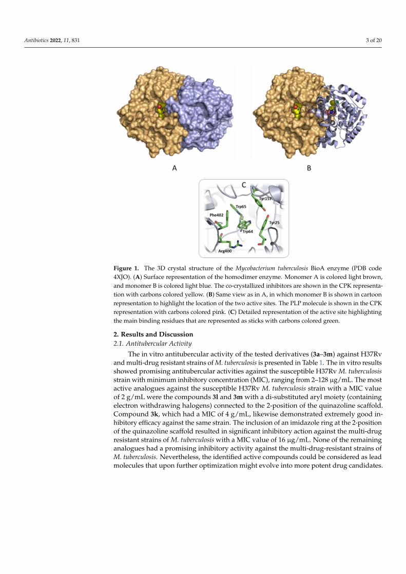
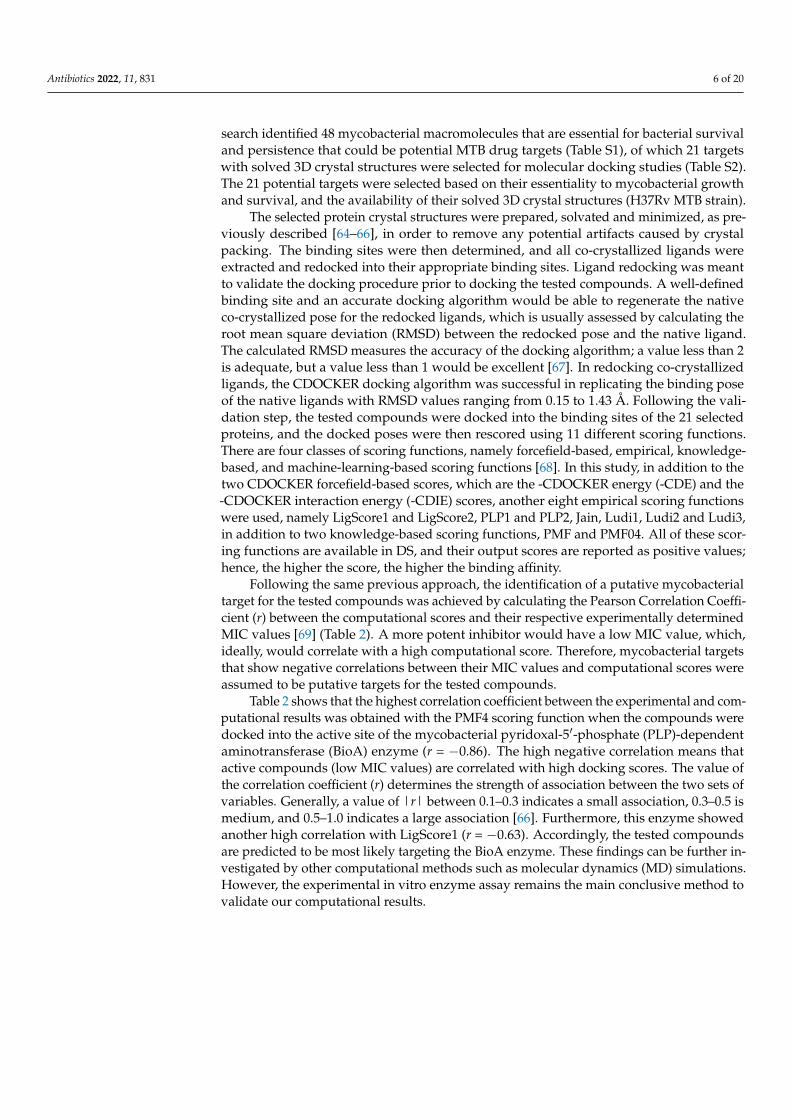
Figure 1. The 3D crystal structure of the Mycobacterium tuberculosis BioA enzyme (PDB code 4XJO). (A) Surface representation of the homodimer enzyme. Monomer A is colored light brown, and mon-omer B is colored light blue. The co-crystallized inhibitors are shown in the CPK representation with carbons colored yellow. (B) Same view as in A, in which monomer B is shown in cartoon represen-tation to highlight the location of the two active sites. The PLP molecule is shown in the CPK repre-sentation with carbons colored pink. (C) Detailed representation of the active site highlighting the main binding residues that are represented as sticks with carbons colored green.
We have been interested in identifying potential novel anti-TB compounds from nat-ural sources [42,43], cyclic depsipeptides [44], and synthetic heterocyclic compounds hav-ing pharmacophores, such as benzothiazoles, triazolyl 1,2,3,4-tetrahydropyrimidines, di-hydropyrimidines, and substituted indolizines as potential antitubercular agents [45–52]. In continuation of our efforts in identifying promising heterocyclic compounds for phar-macological activity [53–63], this study aims to identify potential anti-TB compounds. Herein, we screened a set of our recently reported substituted 2,3-dihydroquinazolin-4(1H)-one analogues (except the novel compound 3l) to assess their potency against dif-ferent strains of MTB. These compounds were also further investigated to evaluate their cytotoxicity against normal human dermal fibroblast cells. Moreover, we attempted to identify the putative mycobacterial target of those compounds by applying a computa-tional approach.
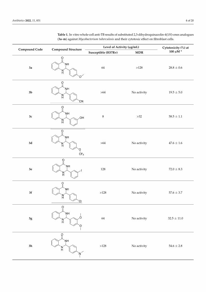
2. Results and Discussion 2.1. Antitubercular Activity
The in vitro antitubercular activity of the tested derivatives (3a–3m) against H37Rv and multi-drug resistant strains of M. tuberculosis is presented in Table 1. The in vitro re-sults showed promising antitubercular activities against the susceptible H37Rv M. tuber-culosis strain with minimum inhibitory concentration (MIC), ranging from 2–128 µg/mL. The most active analogues against the susceptible H37Rv M. tuberculosis strain with a MIC value of 2 g/mL were the compounds 3l and 3m with a di-substituted aryl moiety (con-taining electron withdrawing halogens) connected to the 2-position of the quinazoline scaffold. Compound 3k, which had a MIC of 4 g/mL, likewise demonstrated extremely good inhibitory efficacy against the same strain. The inclusion of an imidazole ring at the 2-position of the quinazoline scaffold resulted in significant inhibitory action against the multi-drug resistant strains of M. tuberculosis with a MIC value of 16 µg/mL. None of the
Figure 1. The 3D crystal structure of the Mycobacterium tuberculosis BioA enzyme (PDB code4XJO). (A) Surface representation of the homodimer enzyme. Monomer A is colored light brown,and monomer B is colored light blue. The co-crystallized inhibitors are shown in the CPK representa-tion with carbons colored yellow. (B) Same view as in A, in which monomer B is shown in cartoonrepresentation to highlight the location of the two active sites. The PLP molecule is shown in the CPKrepresentation with carbons colored pink. (C) Detailed representation of the active site highlightingthe main binding residues that are represented as sticks with carbons colored green.
2. Results and Discussion2.1. Antitubercular Activity
The in vitro antitubercular activity of the tested derivatives (3a–3m) against H37Rvand multi-drug resistant strains of M. tuberculosis is presented in Table 1. The in vitro resultsshowed promising antitubercular activities against the susceptible H37Rv M. tuberculosisstrain with minimum inhibitory concentration (MIC), ranging from 2–128 µg/mL. The mostactive analogues against the susceptible H37Rv M. tuberculosis strain with a MIC valueof 2 g/mL were the compounds 3l and 3m with a di-substituted aryl moiety (containingelectron withdrawing halogens) connected to the 2-position of the quinazoline scaffold.Compound 3k, which had a MIC of 4 g/mL, likewise demonstrated extremely good in-hibitory efficacy against the same strain. The inclusion of an imidazole ring at the 2-positionof the quinazoline scaffold resulted in significant inhibitory action against the multi-drugresistant strains of M. tuberculosis with a MIC value of 16 µg/mL. None of the remaininganalogues had a promising inhibitory activity against the multi-drug-resistant strains ofM. tuberculosis. Nevertheless, the identified active compounds could be considered as leadmolecules that upon further optimization might evolve into more potent drug candidates.
Antibiotics 2022, 11, 831 4 of 20
Table 1. In vitro whole-cell anti-TB results of substituted 2,3-dihydroquinazolin-4(1H)-ones analogues(3a–m) against Mycobacterium tuberculosis and their cytotoxic effect on fibroblast cells.
Compound Code Compound StructureLevel of Activity (µg/mL) Cytotoxicity (%) at
100 µM *Susceptible (H37Rv) MDR
3a
Antibiotics 2021, 10, x FOR PEER REVIEW 4 of 22
remaining analogues had a promising inhibitory activity against the multi-drug-resistant strains of M. tuberculosis. Nevertheless, the identified active compounds could be consid-ered as lead molecules that upon further optimization might evolve into more potent drug candidates.
Table 1. In vitro whole-cell anti-TB results of substituted 2,3-dihydroquinazolin-4(1H)-ones ana-logues (3a–m) against Mycobacterium tuberculosis and their cytotoxic effect on fibroblast cells.
Compound Code Compound Structure Level of Activity (µg/mL) Cytotoxicity (%) at 100
µM * Susceptible (H37Rv) MDR
3a
64 >128 28.8 ± 0.6
3b
>64 No activity 19.5 ± 5.0
3c
8 >32 58.5 ± 1.1
3d
>64 No activity 47.6 ± 1.6
3e
128 No activity 72.0 ± 8.3
3f
>128 No activity 57.6 ± 3.7
3g
64 No activity 32.5 ± 11.0
3h
>128 No activity 54.6 ± 2.8
NH
NH
O
OH
64 >128 28.8 ± 0.6
3b
Antibiotics 2021, 10, x FOR PEER REVIEW 4 of 22
remaining analogues had a promising inhibitory activity against the multi-drug-resistant strains of M. tuberculosis. Nevertheless, the identified active compounds could be consid-ered as lead molecules that upon further optimization might evolve into more potent drug candidates.
Table 1. In vitro whole-cell anti-TB results of substituted 2,3-dihydroquinazolin-4(1H)-ones ana-logues (3a–m) against Mycobacterium tuberculosis and their cytotoxic effect on fibroblast cells.
Compound Code Compound Structure Level of Activity (µg/mL) Cytotoxicity (%) at 100
µM * Susceptible (H37Rv) MDR
3a
64 >128 28.8 ± 0.6
3b
>64 No activity 19.5 ± 5.0
3c
8 >32 58.5 ± 1.1
3d
>64 No activity 47.6 ± 1.6
3e
128 No activity 72.0 ± 8.3
3f
>128 No activity 57.6 ± 3.7
3g
64 No activity 32.5 ± 11.0
3h
>128 No activity 54.6 ± 2.8
NH
NH
O
OH
>64 No activity 19.5 ± 5.0
3c
Antibiotics 2021, 10, x FOR PEER REVIEW 4 of 22
remaining analogues had a promising inhibitory activity against the multi-drug-resistant strains of M. tuberculosis. Nevertheless, the identified active compounds could be consid-ered as lead molecules that upon further optimization might evolve into more potent drug candidates.
Table 1. In vitro whole-cell anti-TB results of substituted 2,3-dihydroquinazolin-4(1H)-ones ana-logues (3a–m) against Mycobacterium tuberculosis and their cytotoxic effect on fibroblast cells.
Compound Code Compound Structure Level of Activity (µg/mL) Cytotoxicity (%) at 100
µM * Susceptible (H37Rv) MDR
3a
64 >128 28.8 ± 0.6
3b
>64 No activity 19.5 ± 5.0
3c
8 >32 58.5 ± 1.1
3d
>64 No activity 47.6 ± 1.6
3e
128 No activity 72.0 ± 8.3
3f
>128 No activity 57.6 ± 3.7
3g
64 No activity 32.5 ± 11.0
3h
>128 No activity 54.6 ± 2.8
NH
NH
O
OH 8 >32 58.5 ± 1.1
3d
Antibiotics 2021, 10, x FOR PEER REVIEW 4 of 22
remaining analogues had a promising inhibitory activity against the multi-drug-resistant strains of M. tuberculosis. Nevertheless, the identified active compounds could be consid-ered as lead molecules that upon further optimization might evolve into more potent drug candidates.
Table 1. In vitro whole-cell anti-TB results of substituted 2,3-dihydroquinazolin-4(1H)-ones ana-logues (3a–m) against Mycobacterium tuberculosis and their cytotoxic effect on fibroblast cells.
Compound Code Compound Structure Level of Activity (µg/mL) Cytotoxicity (%) at 100
µM * Susceptible (H37Rv) MDR
3a
64 >128 28.8 ± 0.6
3b
>64 No activity 19.5 ± 5.0
3c
8 >32 58.5 ± 1.1
3d
>64 No activity 47.6 ± 1.6
3e
128 No activity 72.0 ± 8.3
3f
>128 No activity 57.6 ± 3.7
3g
64 No activity 32.5 ± 11.0
3h
>128 No activity 54.6 ± 2.8
NH
NH
O
OH
>64 No activity 47.6 ± 1.6
3e
Antibiotics 2021, 10, x FOR PEER REVIEW 4 of 22
remaining analogues had a promising inhibitory activity against the multi-drug-resistant strains of M. tuberculosis. Nevertheless, the identified active compounds could be consid-ered as lead molecules that upon further optimization might evolve into more potent drug candidates.
Table 1. In vitro whole-cell anti-TB results of substituted 2,3-dihydroquinazolin-4(1H)-ones ana-logues (3a–m) against Mycobacterium tuberculosis and their cytotoxic effect on fibroblast cells.
Compound Code Compound Structure Level of Activity (µg/mL) Cytotoxicity (%) at 100
µM * Susceptible (H37Rv) MDR
3a
64 >128 28.8 ± 0.6
3b
>64 No activity 19.5 ± 5.0
3c
8 >32 58.5 ± 1.1
3d
>64 No activity 47.6 ± 1.6
3e
128 No activity 72.0 ± 8.3
3f
>128 No activity 57.6 ± 3.7
3g
64 No activity 32.5 ± 11.0
3h
>128 No activity 54.6 ± 2.8
NH
NH
O
OH
128 No activity 72.0 ± 8.3
3f
Antibiotics 2021, 10, x FOR PEER REVIEW 4 of 22
remaining analogues had a promising inhibitory activity against the multi-drug-resistant strains of M. tuberculosis. Nevertheless, the identified active compounds could be consid-ered as lead molecules that upon further optimization might evolve into more potent drug candidates.
Table 1. In vitro whole-cell anti-TB results of substituted 2,3-dihydroquinazolin-4(1H)-ones ana-logues (3a–m) against Mycobacterium tuberculosis and their cytotoxic effect on fibroblast cells.
Compound Code Compound Structure Level of Activity (µg/mL) Cytotoxicity (%) at 100
µM * Susceptible (H37Rv) MDR
3a
64 >128 28.8 ± 0.6
3b
>64 No activity 19.5 ± 5.0
3c
8 >32 58.5 ± 1.1
3d
>64 No activity 47.6 ± 1.6
3e
128 No activity 72.0 ± 8.3
3f
>128 No activity 57.6 ± 3.7
3g
64 No activity 32.5 ± 11.0
3h
>128 No activity 54.6 ± 2.8
NH
NH
O
OH
>128 No activity 57.6 ± 3.7
3g
Antibiotics 2021, 10, x FOR PEER REVIEW 4 of 22
remaining analogues had a promising inhibitory activity against the multi-drug-resistant strains of M. tuberculosis. Nevertheless, the identified active compounds could be consid-ered as lead molecules that upon further optimization might evolve into more potent drug candidates.
Table 1. In vitro whole-cell anti-TB results of substituted 2,3-dihydroquinazolin-4(1H)-ones ana-logues (3a–m) against Mycobacterium tuberculosis and their cytotoxic effect on fibroblast cells.
Compound Code Compound Structure Level of Activity (µg/mL) Cytotoxicity (%) at 100
µM * Susceptible (H37Rv) MDR
3a
64 >128 28.8 ± 0.6
3b
>64 No activity 19.5 ± 5.0
3c
8 >32 58.5 ± 1.1
3d
>64 No activity 47.6 ± 1.6
3e
128 No activity 72.0 ± 8.3
3f
>128 No activity 57.6 ± 3.7
3g
64 No activity 32.5 ± 11.0
3h
>128 No activity 54.6 ± 2.8
NH
NH
O
OH
64 No activity 32.5 ± 11.0
3h
Antibiotics 2021, 10, x FOR PEER REVIEW 4 of 22
remaining analogues had a promising inhibitory activity against the multi-drug-resistant strains of M. tuberculosis. Nevertheless, the identified active compounds could be consid-ered as lead molecules that upon further optimization might evolve into more potent drug candidates.
Table 1. In vitro whole-cell anti-TB results of substituted 2,3-dihydroquinazolin-4(1H)-ones ana-logues (3a–m) against Mycobacterium tuberculosis and their cytotoxic effect on fibroblast cells.
Compound Code Compound Structure Level of Activity (µg/mL) Cytotoxicity (%) at 100
µM * Susceptible (H37Rv) MDR
3a
64 >128 28.8 ± 0.6
3b
>64 No activity 19.5 ± 5.0
3c
8 >32 58.5 ± 1.1
3d
>64 No activity 47.6 ± 1.6
3e
128 No activity 72.0 ± 8.3
3f
>128 No activity 57.6 ± 3.7
3g
64 No activity 32.5 ± 11.0
3h
>128 No activity 54.6 ± 2.8
NH
NH
O
OH
>128 No activity 54.6 ± 2.8
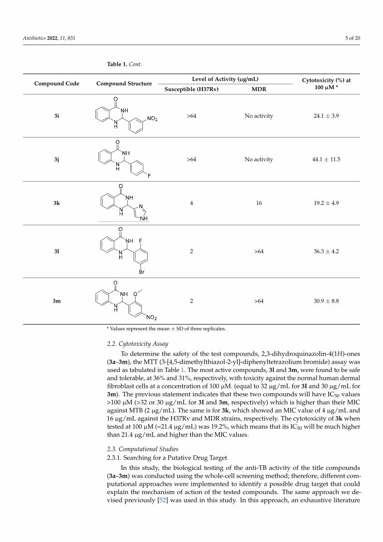
Antibiotics 2022, 11, 831 5 of 20
Table 1. Cont.
Compound Code Compound StructureLevel of Activity (µg/mL) Cytotoxicity (%) at
100 µM *Susceptible (H37Rv) MDR
3i
Antibiotics 2021, 10, x FOR PEER REVIEW 5 of 22
3i
>64 No activity 24.1 ± 3.9
3j
>64 No activity 44.1 ± 11.5
3k
4 16 19.2 ± 4.9
3l
2 >64 36.3 ± 4.2
3m 2 >64 30.9 ± 8.8
* Values represent the mean ± SD of three replicates.
2.2. Cytotoxicity Assay To determine the safety of the test compounds, 2,3-dihydroquinazolin-4(1H)-ones
(3a–3m), the MTT (3-[4,5-dimethylthiazol-2-yl]-diphenyltetrazolium bromide) assay was used as tabulated in Table 1. The most active compounds, 3l and 3m, were found to be safe and tolerable, at 36% and 31%, respectively, with toxicity against the normal human dermal fibroblast cells at a concentration of 100 µM. (equal to 32 µg/mL for 3I and 30 µg/mL for 3m). The previous statement indicates that these two compounds will have IC50 values > 100 µM (>32 or 30 µg/mL for 3I and 3m, respectively) which is higher than their MIC against MTB (2 µg/mL). The same is for 3k, which showed an MIC value of 4 µg/mL and 16 µg/mL against the H37Rv and MDR strains, respectively. The cytotoxicity of 3k when tested at 100 µM (=21.4 µg/mL) was 19.2%, which means that its IC50 will be much higher than 21.4 µg/mL and higher than the MIC values.
2.3. Computational Studies 2.3.1. Searching for a Putative Drug Target
In this study, the biological testing of the anti-TB activity of the title compounds (3a–3m) was conducted using the whole-cell screening method; therefore, different computa-tional approaches were implemented to identify a possible drug target that could explain the mechanism of action of the tested compounds. The same approach we devised previ-ously [52] was used in this study. In this approach, an exhaustive literature search identi-fied 48 mycobacterial macromolecules that are essential for bacterial survival and persis-tence that could be potential MTB drug targets (Table S1), of which 21 targets with solved 3D crystal structures were selected for molecular docking studies (Table S2). The 21 po-tential targets were selected based on their essentiality to mycobacterial growth and sur-vival, and the availability of their solved 3D crystal structures (H37Rv MTB strain).
>64 No activity 24.1 ± 3.9
3j
Antibiotics 2021, 10, x FOR PEER REVIEW 5 of 22
3i
>64 No activity 24.1 ± 3.9
3j
>64 No activity 44.1 ± 11.5
3k
4 16 19.2 ± 4.9
3l
2 >64 36.3 ± 4.2
3m 2 >64 30.9 ± 8.8
* Values represent the mean ± SD of three replicates.
2.2. Cytotoxicity Assay To determine the safety of the test compounds, 2,3-dihydroquinazolin-4(1H)-ones
(3a–3m), the MTT (3-[4,5-dimethylthiazol-2-yl]-diphenyltetrazolium bromide) assay was used as tabulated in Table 1. The most active compounds, 3l and 3m, were found to be safe and tolerable, at 36% and 31%, respectively, with toxicity against the normal human dermal fibroblast cells at a concentration of 100 µM. (equal to 32 µg/mL for 3I and 30 µg/mL for 3m). The previous statement indicates that these two compounds will have IC50 values > 100 µM (>32 or 30 µg/mL for 3I and 3m, respectively) which is higher than their MIC against MTB (2 µg/mL). The same is for 3k, which showed an MIC value of 4 µg/mL and 16 µg/mL against the H37Rv and MDR strains, respectively. The cytotoxicity of 3k when tested at 100 µM (=21.4 µg/mL) was 19.2%, which means that its IC50 will be much higher than 21.4 µg/mL and higher than the MIC values.
2.3. Computational Studies 2.3.1. Searching for a Putative Drug Target
In this study, the biological testing of the anti-TB activity of the title compounds (3a–3m) was conducted using the whole-cell screening method; therefore, different computa-tional approaches were implemented to identify a possible drug target that could explain the mechanism of action of the tested compounds. The same approach we devised previ-ously [52] was used in this study. In this approach, an exhaustive literature search identi-fied 48 mycobacterial macromolecules that are essential for bacterial survival and persis-tence that could be potential MTB drug targets (Table S1), of which 21 targets with solved 3D crystal structures were selected for molecular docking studies (Table S2). The 21 po-tential targets were selected based on their essentiality to mycobacterial growth and sur-vival, and the availability of their solved 3D crystal structures (H37Rv MTB strain).
>64 No activity 44.1 ± 11.5
3k
Antibiotics 2021, 10, x FOR PEER REVIEW 5 of 22
3i
>64 No activity 24.1 ± 3.9
3j
>64 No activity 44.1 ± 11.5
3k
4 16 19.2 ± 4.9
3l
2 >64 36.3 ± 4.2
3m 2 >64 30.9 ± 8.8
* Values represent the mean ± SD of three replicates.
2.2. Cytotoxicity Assay To determine the safety of the test compounds, 2,3-dihydroquinazolin-4(1H)-ones
(3a–3m), the MTT (3-[4,5-dimethylthiazol-2-yl]-diphenyltetrazolium bromide) assay was used as tabulated in Table 1. The most active compounds, 3l and 3m, were found to be safe and tolerable, at 36% and 31%, respectively, with toxicity against the normal human dermal fibroblast cells at a concentration of 100 µM. (equal to 32 µg/mL for 3I and 30 µg/mL for 3m). The previous statement indicates that these two compounds will have IC50 values > 100 µM (>32 or 30 µg/mL for 3I and 3m, respectively) which is higher than their MIC against MTB (2 µg/mL). The same is for 3k, which showed an MIC value of 4 µg/mL and 16 µg/mL against the H37Rv and MDR strains, respectively. The cytotoxicity of 3k when tested at 100 µM (=21.4 µg/mL) was 19.2%, which means that its IC50 will be much higher than 21.4 µg/mL and higher than the MIC values.
2.3. Computational Studies 2.3.1. Searching for a Putative Drug Target
In this study, the biological testing of the anti-TB activity of the title compounds (3a–3m) was conducted using the whole-cell screening method; therefore, different computa-tional approaches were implemented to identify a possible drug target that could explain the mechanism of action of the tested compounds. The same approach we devised previ-ously [52] was used in this study. In this approach, an exhaustive literature search identi-fied 48 mycobacterial macromolecules that are essential for bacterial survival and persis-tence that could be potential MTB drug targets (Table S1), of which 21 targets with solved 3D crystal structures were selected for molecular docking studies (Table S2). The 21 po-tential targets were selected based on their essentiality to mycobacterial growth and sur-vival, and the availability of their solved 3D crystal structures (H37Rv MTB strain).
4 16 19.2 ± 4.9
3l
Antibiotics 2021, 10, x FOR PEER REVIEW 5 of 22
3i
>64 No activity 24.1 ± 3.9
3j
>64 No activity 44.1 ± 11.5
3k
4 16 19.2 ± 4.9
3l
2 >64 36.3 ± 4.2
3m 2 >64 30.9 ± 8.8
* Values represent the mean ± SD of three replicates.
2.2. Cytotoxicity Assay To determine the safety of the test compounds, 2,3-dihydroquinazolin-4(1H)-ones
(3a–3m), the MTT (3-[4,5-dimethylthiazol-2-yl]-diphenyltetrazolium bromide) assay was used as tabulated in Table 1. The most active compounds, 3l and 3m, were found to be safe and tolerable, at 36% and 31%, respectively, with toxicity against the normal human dermal fibroblast cells at a concentration of 100 µM. (equal to 32 µg/mL for 3I and 30 µg/mL for 3m). The previous statement indicates that these two compounds will have IC50 values > 100 µM (>32 or 30 µg/mL for 3I and 3m, respectively) which is higher than their MIC against MTB (2 µg/mL). The same is for 3k, which showed an MIC value of 4 µg/mL and 16 µg/mL against the H37Rv and MDR strains, respectively. The cytotoxicity of 3k when tested at 100 µM (=21.4 µg/mL) was 19.2%, which means that its IC50 will be much higher than 21.4 µg/mL and higher than the MIC values.
2.3. Computational Studies 2.3.1. Searching for a Putative Drug Target
In this study, the biological testing of the anti-TB activity of the title compounds (3a–3m) was conducted using the whole-cell screening method; therefore, different computa-tional approaches were implemented to identify a possible drug target that could explain the mechanism of action of the tested compounds. The same approach we devised previ-ously [52] was used in this study. In this approach, an exhaustive literature search identi-fied 48 mycobacterial macromolecules that are essential for bacterial survival and persis-tence that could be potential MTB drug targets (Table S1), of which 21 targets with solved 3D crystal structures were selected for molecular docking studies (Table S2). The 21 po-tential targets were selected based on their essentiality to mycobacterial growth and sur-vival, and the availability of their solved 3D crystal structures (H37Rv MTB strain).
2 >64 36.3 ± 4.2
3m
Antibiotics 2021, 10, x FOR PEER REVIEW 5 of 22
3i
>64 No activity 24.1 ± 3.9
3j
>64 No activity 44.1 ± 11.5
3k
4 16 19.2 ± 4.9
3l
2 >64 36.3 ± 4.2
3m 2 >64 30.9 ± 8.8
* Values represent the mean ± SD of three replicates.
2.2. Cytotoxicity Assay To determine the safety of the test compounds, 2,3-dihydroquinazolin-4(1H)-ones
(3a–3m), the MTT (3-[4,5-dimethylthiazol-2-yl]-diphenyltetrazolium bromide) assay was used as tabulated in Table 1. The most active compounds, 3l and 3m, were found to be safe and tolerable, at 36% and 31%, respectively, with toxicity against the normal human dermal fibroblast cells at a concentration of 100 µM. (equal to 32 µg/mL for 3I and 30 µg/mL for 3m). The previous statement indicates that these two compounds will have IC50 values > 100 µM (>32 or 30 µg/mL for 3I and 3m, respectively) which is higher than their MIC against MTB (2 µg/mL). The same is for 3k, which showed an MIC value of 4 µg/mL and 16 µg/mL against the H37Rv and MDR strains, respectively. The cytotoxicity of 3k when tested at 100 µM (=21.4 µg/mL) was 19.2%, which means that its IC50 will be much higher than 21.4 µg/mL and higher than the MIC values.
2.3. Computational Studies 2.3.1. Searching for a Putative Drug Target
In this study, the biological testing of the anti-TB activity of the title compounds (3a–3m) was conducted using the whole-cell screening method; therefore, different computa-tional approaches were implemented to identify a possible drug target that could explain the mechanism of action of the tested compounds. The same approach we devised previ-ously [52] was used in this study. In this approach, an exhaustive literature search identi-fied 48 mycobacterial macromolecules that are essential for bacterial survival and persis-tence that could be potential MTB drug targets (Table S1), of which 21 targets with solved 3D crystal structures were selected for molecular docking studies (Table S2). The 21 po-tential targets were selected based on their essentiality to mycobacterial growth and sur-vival, and the availability of their solved 3D crystal structures (H37Rv MTB strain).
2 >64 30.9 ± 8.8
* Values represent the mean ± SD of three replicates.
2.2. Cytotoxicity Assay
To determine the safety of the test compounds, 2,3-dihydroquinazolin-4(1H)-ones(3a–3m), the MTT (3-[4,5-dimethylthiazol-2-yl]-diphenyltetrazolium bromide) assay wasused as tabulated in Table 1. The most active compounds, 3l and 3m, were found to be safeand tolerable, at 36% and 31%, respectively, with toxicity against the normal human dermalfibroblast cells at a concentration of 100 µM. (equal to 32 µg/mL for 3I and 30 µg/mL for3m). The previous statement indicates that these two compounds will have IC50 values>100 µM (>32 or 30 µg/mL for 3I and 3m, respectively) which is higher than their MICagainst MTB (2 µg/mL). The same is for 3k, which showed an MIC value of 4 µg/mL and16 µg/mL against the H37Rv and MDR strains, respectively. The cytotoxicity of 3k whentested at 100 µM (=21.4 µg/mL) was 19.2%, which means that its IC50 will be much higherthan 21.4 µg/mL and higher than the MIC values.
2.3. Computational Studies2.3.1. Searching for a Putative Drug Target
In this study, the biological testing of the anti-TB activity of the title compounds(3a–3m) was conducted using the whole-cell screening method; therefore, different com-putational approaches were implemented to identify a possible drug target that couldexplain the mechanism of action of the tested compounds. The same approach we de-vised previously [52] was used in this study. In this approach, an exhaustive literature
Antibiotics 2022, 11, 831 6 of 20
search identified 48 mycobacterial macromolecules that are essential for bacterial survivaland persistence that could be potential MTB drug targets (Table S1), of which 21 targetswith solved 3D crystal structures were selected for molecular docking studies (Table S2).The 21 potential targets were selected based on their essentiality to mycobacterial growthand survival, and the availability of their solved 3D crystal structures (H37Rv MTB strain).
The selected protein crystal structures were prepared, solvated and minimized, as pre-viously described [64–66], in order to remove any potential artifacts caused by crystalpacking. The binding sites were then determined, and all co-crystallized ligands wereextracted and redocked into their appropriate binding sites. Ligand redocking was meantto validate the docking procedure prior to docking the tested compounds. A well-definedbinding site and an accurate docking algorithm would be able to regenerate the nativeco-crystallized pose for the redocked ligands, which is usually assessed by calculating theroot mean square deviation (RMSD) between the redocked pose and the native ligand.The calculated RMSD measures the accuracy of the docking algorithm; a value less than 2is adequate, but a value less than 1 would be excellent [67]. In redocking co-crystallizedligands, the CDOCKER docking algorithm was successful in replicating the binding poseof the native ligands with RMSD values ranging from 0.15 to 1.43 Å. Following the vali-dation step, the tested compounds were docked into the binding sites of the 21 selectedproteins, and the docked poses were then rescored using 11 different scoring functions.There are four classes of scoring functions, namely forcefield-based, empirical, knowledge-based, and machine-learning-based scoring functions [68]. In this study, in addition to thetwo CDOCKER forcefield-based scores, which are the -CDOCKER energy (-CDE) and the-CDOCKER interaction energy (-CDIE) scores, another eight empirical scoring functionswere used, namely LigScore1 and LigScore2, PLP1 and PLP2, Jain, Ludi1, Ludi2 and Ludi3,in addition to two knowledge-based scoring functions, PMF and PMF04. All of these scor-ing functions are available in DS, and their output scores are reported as positive values;hence, the higher the score, the higher the binding affinity.
Following the same previous approach, the identification of a putative mycobacterialtarget for the tested compounds was achieved by calculating the Pearson Correlation Coeffi-cient (r) between the computational scores and their respective experimentally determinedMIC values [69] (Table 2). A more potent inhibitor would have a low MIC value, which,ideally, would correlate with a high computational score. Therefore, mycobacterial targetsthat show negative correlations between their MIC values and computational scores wereassumed to be putative targets for the tested compounds.
Table 2 shows that the highest correlation coefficient between the experimental and com-putational results was obtained with the PMF4 scoring function when the compounds weredocked into the active site of the mycobacterial pyridoxal-5′-phosphate (PLP)-dependentaminotransferase (BioA) enzyme (r = −0.86). The high negative correlation means thatactive compounds (low MIC values) are correlated with high docking scores. The value ofthe correlation coefficient (r) determines the strength of association between the two sets ofvariables. Generally, a value of |r| between 0.1–0.3 indicates a small association, 0.3–0.5 ismedium, and 0.5–1.0 indicates a large association [66]. Furthermore, this enzyme showedanother high correlation with LigScore1 (r = −0.63). Accordingly, the tested compoundsare predicted to be most likely targeting the BioA enzyme. These findings can be further in-vestigated by other computational methods such as molecular dynamics (MD) simulations.However, the experimental in vitro enzyme assay remains the main conclusive method tovalidate our computational results.
Antibiotics 2022, 11, 831 7 of 20
Table 2. The overall correlation coefficients matrix between the minimum inhibitory concentrationvalues of the tested compounds and their computational scores obtained from different scoringfunctions for each of the selected target enzymes.
Index Name LS1 LS2 PLP1 PLP2 Jain PMF PMF4 CDE CDIE Ludi-1 Ludi-2 Ludi-3
1 Alanine racemase −0.71 0.29 −0.16 −0.12 0 −0.06 −0.51 0.38 0.31 −0.15 −0.19 −0.352 CamA −0.57 −0.39 −0.12 −0.05 −0.09 −0.07 −0.45 −0.3 −0.38 0.44 0.19 −0.063 DprE1 −0.3 0.62 0.1 −0.01 −0.12 0.28 −0.82 0.51 0.38 0.33 0.2 0.34 FabH −0.67 0.26 −0.42 −0.44 −0.02 −0.24 −0.75 0.35 0.32 −0.24 −0.3 −0.315 InhA −0.59 −0.5 0.12 0.32 0.32 0.12 −0.29 0.02 −0.08 0.4 0.49 −0.056 MabA −0.4 0.35 −0.3 −0.23 −0.51 −0.46 −0.66 0.28 0.16 −0.13 −0.15 −0.347 AspS −0.68 −0.32 −0.14 −0.05 0.05 0.08 −0.51 0.06 −0.16 0.06 0.01 −0.038 LeuRS −0.82 0.54 0.3 −0.06 −0.15 −0.27 −0.62 0.41 0.08 0.58 0.54 0.139 GlmU −0.3 0.52 −0.32 0.11 −0.36 0.05 −0.81 0.43 0.2 0.17 0.17 0.3310 PanK −0.02 0.75 −0.14 −0.12 −0.08 −0.15 −0.31 0.25 0.25 0.56 0.38 0.0811 PknB −0.36 −0.58 −0.56 −0.3 0.03 −0.03 −0.67 0.03 −0.2 0.1 0.12 −0.1212 PknA −0.42 0.4 −0.07 0.11 −0.76 0.13 −0.52 0.17 0.01 0.25 0.05 0.0313 KasA −0.28 −0.08 −0.62 −0.48 −0.38 −0.24 −0.38 −0.04 −0.26 −0.27 −0.25 −0.4314 KasA (BS2) * −0.27 0.38 0.17 0.06 0.32 0.43 −0.44 0.14 0.43 −0.02 0.06 0.1615 Pks13 −0.1 0.32 0.3 0.39 0.66 −0.12 −0.66 0.59 0.29 0.1 0.22 0.2516 BioA # −0.63 0.1 0.14 0.28 0.65 −0.42 −0.86 0.74 0.55 0.52 0.58 0.6517 EchA6 −0.33 0.17 −0.04 0.3 0.31 −0.17 −0.69 0.38 0.31 0.46 0.05 0.1818 MmpL3 −0.77 −0.38 0.25 0.31 0.14 0.32 −0.39 0.04 −0.07 0.1 0.1 0.4219 AspAT −0.41 0.67 −0.24 0.24 0.16 0.01 −0.62 0.23 0.14 0.15 0.41 0.4420 MurE −0.83 −0.05 0.25 0.35 0.6 −0.31 −0.38 0.31 0.15 0.33 0.29 0.221 EthR −0.39 0.17 0.06 0.19 −0.41 −0.24 −0.78 0.48 0.13 0.13 0.22 0.1222 PrpC −0.4 0.29 −0.44 −0.46 0.08 0.04 −0.56 0.37 0.3 0.08 0.16 0.05
* KasA enzyme has two binding sites; an acyl chain-binding site (BS1), and the catalytic active site (BS2). Both wereused for docking. This refers to the second binding site. # The highlighted values, in bold red, represent thehighest obtained correlation coefficient.
2.3.2. Analysis of the Binding Interactions with the Putative Target (BioA)
The identified putative target was the BioA enzyme. This enzyme catalyzes thesecond step in biotin biosynthesis and is essential for bacterial survival and persistence.Contrary to humans, MTB de novo synthesizes biotin to be utilized by carboxylases in fattyacid metabolism and gluconeogenesis pathways; thereby, BioA is an ideal target for thedevelopment of potential antitubercular agents [70].
Structurally, the functional form of the mycobacterial BioA enzyme is a homodimer,in which the monomer structure is composed of two domains, the small domain (aminoacid residues 1–60 and 339–437) and the large domain (residues 61–338). Two active sitesare located at the interface between the two monomers, 18 Å apart, and are composedof residues Pro24-Ser34, Ser62-Ala67, Arg156-Asp160, His171-Arg181, Gln224-Gly228,and Arg400-Arg403 from one chain, and Met′87-His′97 and Ala′307-Asn′322 from the otherchain [70–72]. Among these residues, Tyr25, Trp64, Trp65, Tyr157, Arg400, and Phe402were reported to be of high relevance to ligand binding [72,73] (Figure 1).
The structural model of the MTB BioA enzyme was downloaded from the proteindata bank (PDB code, 4XJO), which corresponds to the BioA enzyme in complex with aninhibitor, 5-[4-(3-chlorobenzoyl)piperazin-1-yl]-1H-inden-1-one (41O), and the coenzymepyridoxal-5′-phosphate (PLP) at a resolution of 1.50 Å; then, it was prepared as detailed inthe Methods section. The docking protocol was validated prior to docking the designedcompounds via extracting and redocking the co-crystallized ligand (41O). The redockedpose was in perfect agreement with the native co-crystallized ligand pose with a RMSDof 0.15 Å. Then, the designed compounds were docked into the active site of the enzyme.Figure 2 shows the binding mode and binding interactions of compound 3m within theactive site of the BioA enzyme compared to that of the co-crystallized ligand (41O).
Antibiotics 2022, 11, 831 8 of 20Antibiotics 2021, 10, x FOR PEER REVIEW 8 of 22
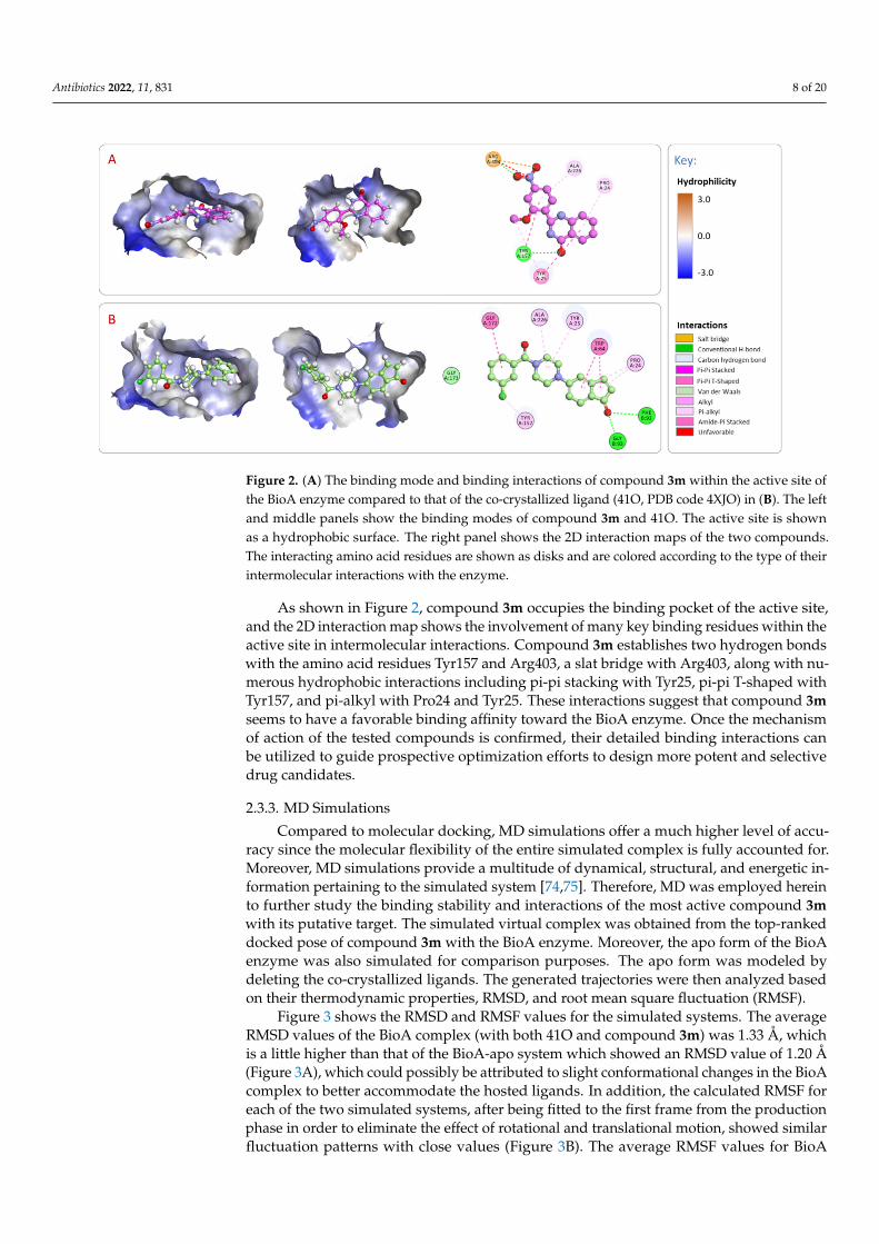
Figure 2. (A) The binding mode and binding interactions of compound 3m within the active site of the BioA enzyme compared to that of the co-crystallized ligand (41O, PDB code 4XJO) in (B). The left and middle panels show the binding modes of compound 3m and 41O. The active site is shown as a hydrophobic surface. The right panel shows the 2D interaction maps of the two compounds. The interacting amino acid residues are shown as disks and are colored according to the type of their intermolecular interactions with the enzyme.
As shown in Figure 2, compound 3m occupies the binding pocket of the active site, and the 2D interaction map shows the involvement of many key binding residues within the active site in intermolecular interactions. Compound 3m establishes two hydrogen bonds with the amino acid residues Tyr157 and Arg403, a slat bridge with Arg403, along with numerous hydrophobic interactions including pi-pi stacking with Tyr25, pi-pi T-shaped with Tyr157, and pi-alkyl with Pro24 and Tyr25. These interactions suggest that compound 3m seems to have a favorable binding affinity toward the BioA enzyme. Once the mechanism of action of the tested compounds is confirmed, their detailed binding in-teractions can be utilized to guide prospective optimization efforts to design more potent and selective drug candidates.
2.3.3. MD Simulations Compared to molecular docking, MD simulations offer a much higher level of accu-
racy since the molecular flexibility of the entire simulated complex is fully accounted for. Moreover, MD simulations provide a multitude of dynamical, structural, and energetic information pertaining to the simulated system [74,75]. Therefore, MD was employed herein to further study the binding stability and interactions of the most active compound 3m with its putative target. The simulated virtual complex was obtained from the top-ranked docked pose of compound 3m with the BioA enzyme. Moreover, the apo form of the BioA enzyme was also simulated for comparison purposes. The apo form was mod-eled by deleting the co-crystallized ligands. The generated trajectories were then analyzed based on their thermodynamic properties, RMSD, and root mean square fluctuation (RMSF).
Figure 3 shows the RMSD and RMSF values for the simulated systems. The average RMSD values of the BioA complex (with both 41O and compound 3m) was 1.33 Å, which is a little higher than that of the BioA-apo system which showed an RMSD value of 1.20 Å (Figure 3A), which could possibly be attributed to slight conformational changes in the BioA complex to better accommodate the hosted ligands. In addition, the calculated RMSF for each of the two simulated systems, after being fitted to the first frame from the pro-duction phase in order to eliminate the effect of rotational and translational motion,
Figure 2. (A) The binding mode and binding interactions of compound 3m within the active site ofthe BioA enzyme compared to that of the co-crystallized ligand (41O, PDB code 4XJO) in (B). The leftand middle panels show the binding modes of compound 3m and 41O. The active site is shownas a hydrophobic surface. The right panel shows the 2D interaction maps of the two compounds.The interacting amino acid residues are shown as disks and are colored according to the type of theirintermolecular interactions with the enzyme.
As shown in Figure 2, compound 3m occupies the binding pocket of the active site,and the 2D interaction map shows the involvement of many key binding residues within theactive site in intermolecular interactions. Compound 3m establishes two hydrogen bondswith the amino acid residues Tyr157 and Arg403, a slat bridge with Arg403, along with nu-merous hydrophobic interactions including pi-pi stacking with Tyr25, pi-pi T-shaped withTyr157, and pi-alkyl with Pro24 and Tyr25. These interactions suggest that compound 3mseems to have a favorable binding affinity toward the BioA enzyme. Once the mechanismof action of the tested compounds is confirmed, their detailed binding interactions canbe utilized to guide prospective optimization efforts to design more potent and selectivedrug candidates.
2.3.3. MD Simulations
Compared to molecular docking, MD simulations offer a much higher level of accu-racy since the molecular flexibility of the entire simulated complex is fully accounted for.Moreover, MD simulations provide a multitude of dynamical, structural, and energetic in-formation pertaining to the simulated system [74,75]. Therefore, MD was employed hereinto further study the binding stability and interactions of the most active compound 3mwith its putative target. The simulated virtual complex was obtained from the top-rankeddocked pose of compound 3m with the BioA enzyme. Moreover, the apo form of the BioAenzyme was also simulated for comparison purposes. The apo form was modeled bydeleting the co-crystallized ligands. The generated trajectories were then analyzed basedon their thermodynamic properties, RMSD, and root mean square fluctuation (RMSF).
Figure 3 shows the RMSD and RMSF values for the simulated systems. The averageRMSD values of the BioA complex (with both 41O and compound 3m) was 1.33 Å, whichis a little higher than that of the BioA-apo system which showed an RMSD value of 1.20 Å(Figure 3A), which could possibly be attributed to slight conformational changes in the BioAcomplex to better accommodate the hosted ligands. In addition, the calculated RMSF foreach of the two simulated systems, after being fitted to the first frame from the productionphase in order to eliminate the effect of rotational and translational motion, showed similarfluctuation patterns with close values (Figure 3B). The average RMSF values for BioA
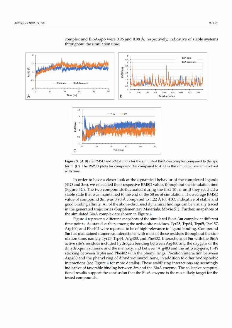
Antibiotics 2022, 11, 831 9 of 20
complex and BioA-apo were 0.96 and 0.98 Å, respectively, indicative of stable systemsthroughout the simulation time.
Antibiotics 2021, 10, x FOR PEER REVIEW 9 of 22
showed similar fluctuation patterns with close values (Figure 3B). The average RMSF val-ues for BioA complex and BioA-apo were 0.96 and 0.98 Å, respectively, indicative of stable systems throughout the simulation time.
Figure 3. (A,B) are RMSD and RMSF plots for the simulated BioA-3m complex compared to the apo form. (C). The RMSD plots for compound 3m compared to 41O as the simulated system evolved with time.
In order to have a closer look at the dynamical behavior of the complexed ligands (41O and 3m), we calculated their respective RMSD values throughout the simulation time (Figure 3C). The two compounds fluctuated during the first 10 ns until they reached a stable state that was maintained to the end of the 50 ns of simulation. The average RMSD value of compound 3m was 0.90 Å compared to 1.22 Å for 41O, indicative of stable and good binding affinity. All of the above-discussed dynamical findings can be visually traced in the generated trajectories (Supplementary Materials; Movie S1). Further, snap-shots of the simulated BioA complex are shown in Figure 4.
Figure 3. (A,B) are RMSD and RMSF plots for the simulated BioA-3m complex compared to the apoform. (C). The RMSD plots for compound 3m compared to 41O as the simulated system evolvedwith time.
In order to have a closer look at the dynamical behavior of the complexed ligands(41O and 3m), we calculated their respective RMSD values throughout the simulation time(Figure 3C). The two compounds fluctuated during the first 10 ns until they reached astable state that was maintained to the end of the 50 ns of simulation. The average RMSDvalue of compound 3m was 0.90 Å compared to 1.22 Å for 41O, indicative of stable andgood binding affinity. All of the above-discussed dynamical findings can be visually tracedin the generated trajectories (Supplementary Materials; Movie S1). Further, snapshots ofthe simulated BioA complex are shown in Figure 4.
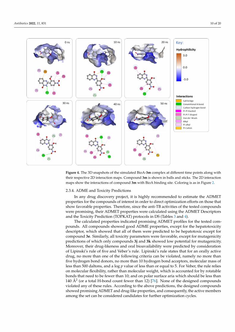
Figure 4 represents different snapshots of the simulated BioA-3m complex at differenttime points. As stated earlier, among the active site residues, Tyr25, Trp64, Trp65, Tyr157,Arg400, and Phe402 were reported to be of high relevance to ligand binding. Compound3m has maintained numerous interactions with most of those residues throughout the sim-ulation time, namely Tyr25, Trp64, Arg400, and Phe402. Interactions of 3m with the BioAactive site’s residues included hydrogen bonding between Arg400 and the oxygens of thedihydroquinazolinone and the methoxy, and between Arg403 and the nitro oxygens; Pi-Pistacking between Trp64 and Phe402 with the phenyl rings; Pi-cation interaction betweenArg400 and the phenyl ring of dihydroquinazolinone; in addition to other hydrophobicinteractions (see Figure 4 for more details). These stabilizing interactions are seeminglyindicative of favorable binding between 3m and the BioA enzyme. The collective computa-tional results support the conclusion that the BioA enzyme is the most likely target for thetested compounds.
Antibiotics 2022, 11, 831 10 of 20Antibiotics 2021, 10, x FOR PEER REVIEW 10 of 22
Figure 4. The 3D snapshots of the simulated BioA-3m complex at different time points along with their respective 2D interaction maps. Compound 3m is shown in balls and sticks. The 2D interaction maps show the interactions of compound 3m with BioA binding site. Coloring is as in Figure 2.
Figure 4 represents different snapshots of the simulated BioA-3m complex at differ-ent time points. As stated earlier, among the active site residues, Tyr25, Trp64, Trp65, Tyr157, Arg400, and Phe402 were reported to be of high relevance to ligand binding. Com-pound 3m has maintained numerous interactions with most of those residues throughout the simulation time, namely Tyr25, Trp64, Arg400, and Phe402. Interactions of 3m with the BioA active site’s residues included hydrogen bonding between Arg400 and the ox-ygens of the dihydroquinazolinone and the methoxy, and between Arg403 and the nitro oxygens; Pi-Pi stacking between Trp64 and Phe402 with the phenyl rings; Pi-cation inter-action between Arg400 and the phenyl ring of dihydroquinazolinone; in addition to other hydrophobic interactions (see Figure 4 for more details). These stabilizing interactions are seemingly indicative of favorable binding between 3m and the BioA enzyme. The collec-tive computational results support the conclusion that the BioA enzyme is the most likely target for the tested compounds.
2.3.4. ADME and Toxicity Predictions In any drug discovery project, it is highly recommended to estimate the ADMET
properties for the compounds of interest in order to direct optimization efforts on those that show favorable properties. Therefore, since the anti-TB activities of the tested com-pounds were promising, their ADMET properties were calculated using the ADMET De-scriptors and the Toxicity Prediction (TOPKAT) protocols in DS (Tables 3 and 4).
Figure 4. The 3D snapshots of the simulated BioA-3m complex at different time points along withtheir respective 2D interaction maps. Compound 3m is shown in balls and sticks. The 2D interactionmaps show the interactions of compound 3m with BioA binding site. Coloring is as in Figure 2.
2.3.4. ADME and Toxicity Predictions
In any drug discovery project, it is highly recommended to estimate the ADMETproperties for the compounds of interest in order to direct optimization efforts on those thatshow favorable properties. Therefore, since the anti-TB activities of the tested compoundswere promising, their ADMET properties were calculated using the ADMET Descriptorsand the Toxicity Prediction (TOPKAT) protocols in DS (Tables 3 and 4).
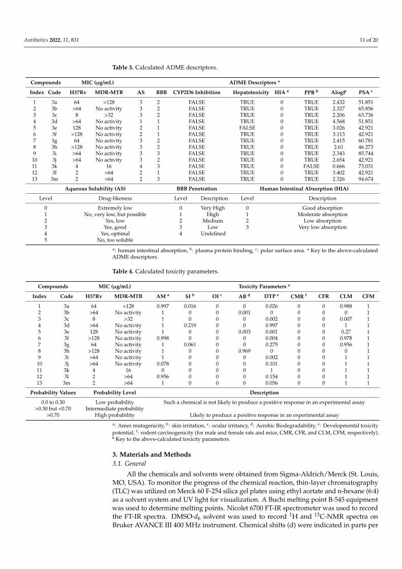
The calculated properties indicated promising ADMET profiles for the tested com-pounds. All compounds showed good ADME properties, except for the hepatotoxicitydescriptor, which showed that all of them were predicted to be hepatotoxic except forcompound 3e. Similarly, all toxicity parameters were favorable, except for mutagenicitypredictions of which only compounds 3j and 3k showed low potential for mutagenicity.Moreover, their drug-likeness and oral bioavailability were predicted by considerationof Lipinski’s rule of five and Veber’s rule. Lipinski’s rule states that for an orally activedrug, no more than one of the following criteria can be violated, namely no more thanfive hydrogen bond donors, no more than 10 hydrogen bond acceptors, molecular mass ofless than 500 daltons, and a log p value of less than or equal to 5. For Veber, the rule relieson molecular flexibility, rather than molecular weight, which is accounted for by rotatablebonds that need to be fewer than 10; and on polar surface aria which should be less than140 Å2 (or a total H-bond count fewer than 12) [76]. None of the designed compoundsviolated any of these rules. According to the above predictions, the designed compoundsshowed promising ADMET and drug-like properties, and consequently, the active membersamong the set can be considered candidates for further optimization cycles.
Antibiotics 2022, 11, 831 11 of 20
Table 3. Calculated ADME descriptors.
Compounds MIC (µg/mL) ADME Descriptors *
Index Code H37Rv MDR-MTB AS BBB CYP2D6 Inhibition Hepatotoxicity HIA a PPB b AlogP PSA c
1 3a 64 >128 3 2 FALSE TRUE 0 TRUE 2.432 51.8512 3b >64 No activity 3 2 FALSE TRUE 0 TRUE 2.327 65.8563 3c 8 >32 3 2 FALSE TRUE 0 TRUE 2.206 63.7364 3d >64 No activity 1 1 FALSE TRUE 0 TRUE 4.568 51.8515 3e 128 No activity 2 1 FALSE FALSE 0 TRUE 3.026 42.9216 3f >128 No activity 2 1 FALSE TRUE 0 TRUE 3.113 42.9217 3g 64 No activity 3 2 FALSE TRUE 0 TRUE 2.415 60.7818 3h >128 No activity 3 2 FALSE TRUE 0 TRUE 2.61 46.2739 3i >64 No activity 3 3 FALSE TRUE 0 TRUE 2.343 85.744
10 3j >64 No activity 3 2 FALSE TRUE 0 TRUE 2.654 42.92111 3k 4 16 4 3 FALSE TRUE 0 FALSE 0.666 73.03112 3l 2 >64 2 1 FALSE TRUE 0 TRUE 3.402 42.92113 3m 2 >64 2 3 FALSE TRUE 0 TRUE 2.326 94.674
Aqueous Solubility (AS) BBB Penetration Human Intestinal Absorption (HIA)
Level Drug-likeness Level Description Level Description
0 Extremely low 0 Very High 0 Good absorption1 No, very low, but possible 1 High 1 Moderate absorption2 Yes, low 2 Medium 2 Low absorption3 Yes, good 3 Low 3 Very low absorption4 Yes, optimal 4 Undefined5 No, too soluble
a: human intestinal absorption, b: plasma protein binding, c: polar surface area. * Key to the above-calculatedADME descriptors.
Table 4. Calculated toxicity parameters.
Compounds MIC (µg/mL) Toxicity Parameters *
Index Code H37Rv MDR-MTB AM a SI b OI c AB d DTP e CMR f CFR CLM CFM
1 3a 64 >128 0.997 0.016 0 0 0.026 0 0 0.988 12 3b >64 No activity 1 0 0 0.001 0 0 0 0 13 3c 8 >32 1 0 0 0 0.002 0 0 0.007 14 3d >64 No activity 1 0.219 0 0 0.997 0 0 1 15 3e 128 No activity 1 0 0 0.003 0.001 0 0 0.27 16 3f >128 No activity 0.998 0 0 0 0.004 0 0 0.978 17 3g 64 No activity 1 0.061 0 0 0.275 0 0 0.956 18 3h >128 No activity 1 0 0 0.969 0 0 0 0 19 3i >64 No activity 1 0 0 0 0.002 0 0 1 110 3j >64 No activity 0.078 0 0 0 0.101 0 0 1 111 3k 4 16 0 0 0 0 1 0 0 1 112 3l 2 >64 0.956 0 0 0 0.154 0 0 1 113 3m 2 >64 1 0 0 0 0.056 0 0 1 1
Probability Values Probability Level Description
0.0 to 0.30 Low probability Such a chemical is not likely to produce a positive response in an experimental assay>0.30 but <0.70 Intermediate probability
>0.70 High probability Likely to produce a positive response in an experimental assay
a: Ames mutagenicity, b: skin irritation, c: ocular irritancy, d: Aerobic Biodegradability, e: Developmental toxicitypotential, f: rodent carcinogenicity (for male and female rats and mice, CMR, CFR, and CLM, CFM, respectively).* Key to the above-calculated toxicity parameters.
3. Materials and Methods3.1. General
All the chemicals and solvents were obtained from Sigma-Aldrich/Merck (St. Louis,MO, USA). To monitor the progress of the chemical reaction, thin-layer chromatography(TLC) was utilized on Merck 60 F-254 silica gel plates using ethyl acetate and n-hexane (6:4)as a solvent system and UV light for visualization. A Buchi melting point B-545 equipmentwas used to determine melting points. Nicolet 6700 FT-IR spectrometer was used to recordthe FT-IR spectra. DMSO-d6 solvent was used to record 1H and 13C-NMR spectra onBruker AVANCE III 400 MHz instrument. Chemical shifts (d) were indicated in parts per
Antibiotics 2022, 11, 831 12 of 20
million downfield from tetramethylsilane, and the coupling constant (J) is recorded inHertz. The splitting pattern is abbreviated as follows: s, singlet; d, doublet; t, triplet; m,multiplet. Mass spectra were recorded using LC-MS-Agilent 1100 series with MSD (Iontrap) using 0.1% aqueous trifluoroacetic acid in acetonitrile system on C18-BDS column.Elemental analysis was performed on Thermo Finnigan FLASH EA 1112 CHN analyzer.
3.2. Chemistry
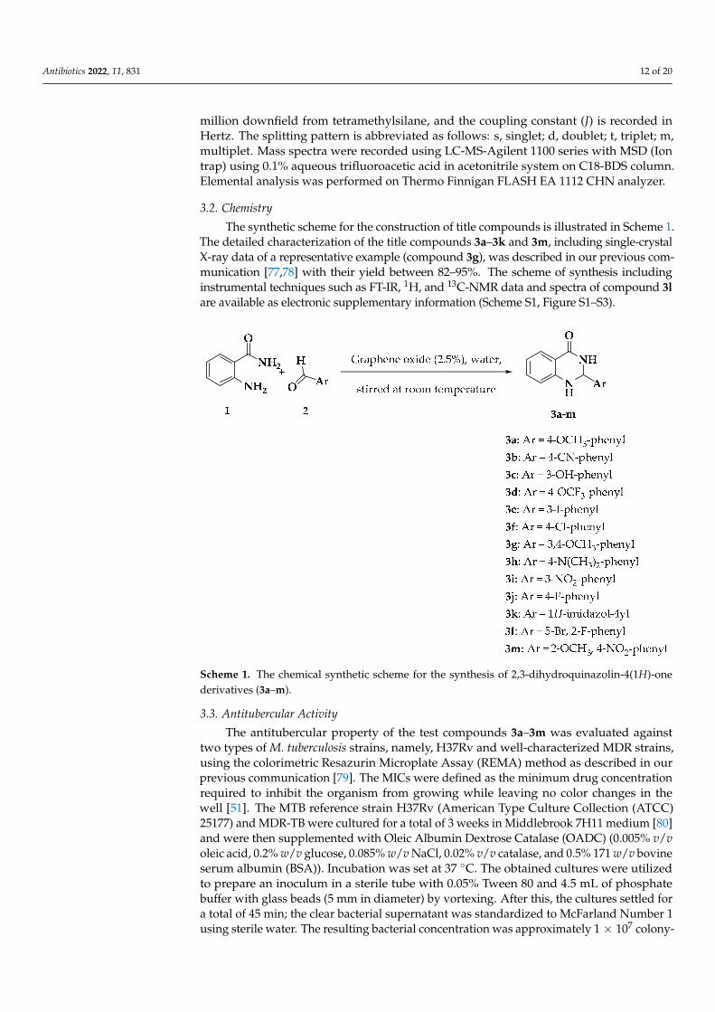
The synthetic scheme for the construction of title compounds is illustrated in Scheme 1.The detailed characterization of the title compounds 3a–3k and 3m, including single-crystalX-ray data of a representative example (compound 3g), was described in our previous com-munication [77,78] with their yield between 82–95%. The scheme of synthesis includinginstrumental techniques such as FT-IR, 1H, and 13C-NMR data and spectra of compound 3lare available as electronic supplementary information (Scheme S1, Figure S1–S3).
Antibiotics 2021, 10, x FOR PEER REVIEW 13 of 22
Scheme 1. The chemical synthetic scheme for the synthesis of 2,3-dihydroquinazolin-4(1H)-one de-rivatives (3a–m).
3.3. Antitubercular Activity The antitubercular property of the test compounds 3a–3m was evaluated against two
types of M. tuberculosis strains, namely, H37Rv and well-characterized MDR strains, using the colorimetric Resazurin Microplate Assay (REMA) method as described in our previ-ous communication [79]. The MICs were defined as the minimum drug concentration re-quired to inhibit the organism from growing while leaving no color changes in the well [51]. The MTB reference strain H37Rv (American Type Culture Collection (ATCC) 25177) and MDR-TB were cultured for a total of 3 weeks in Middlebrook 7H11 medium [80] and were then supplemented with Oleic Albumin Dextrose Catalase (OADC) (0.005% v/v oleic acid, 0.2% w/v glucose, 0.085% w/v NaCl, 0.02% v/v catalase, and 0.5% 171 w/v bovine se-rum albumin (BSA)). Incubation was set at 37 °C. The obtained cultures were utilized to prepare an inoculum in a sterile tube with 0.05% Tween 80 and 4.5 mL of phosphate buffer with glass beads (5 mm in diameter) by vortexing. After this, the cultures settled for a total of 45 min; the clear bacterial supernatant was standardized to McFarland Number 1 using sterile water. The resulting bacterial concentration was approximately 1 × 10⁷ colony-forming units (CFU)/mL, which was then diluted with sterile water. Overall, 100 µL of the dilution was added to Middlebrook 7H10 agar plates containing 8–0.125 µg/mL of the agent. The test compounds (8 µg/mL) were dissolved in distilled water and diluted to the required concentration before being added to the agar medium. The test compound MICs were read 3 weeks following 37 °C incubation and were regarded as the minimum drug concentration that could inhibit >99% growth of the bacterial culture when compared to controls.
3.4. Cell Line The cell line under investigation was normal human skin fibroblast cells (CCD-
1064SK) purchased from American Type Culture Collection (ATCC) (Manassas, VA, USA). Fibroblast cells were cultured in Iscove’s Modified Dulbecco’s Medium (Euro Clone, Italy) as recommended by ATCC. The media was supplemented with 10% heat-
Scheme 1. The chemical synthetic scheme for the synthesis of 2,3-dihydroquinazolin-4(1H)-onederivatives (3a–m).
3.3. Antitubercular Activity
The antitubercular property of the test compounds 3a–3m was evaluated againsttwo types of M. tuberculosis strains, namely, H37Rv and well-characterized MDR strains,using the colorimetric Resazurin Microplate Assay (REMA) method as described in ourprevious communication [79]. The MICs were defined as the minimum drug concentrationrequired to inhibit the organism from growing while leaving no color changes in thewell [51]. The MTB reference strain H37Rv (American Type Culture Collection (ATCC)25177) and MDR-TB were cultured for a total of 3 weeks in Middlebrook 7H11 medium [80]and were then supplemented with Oleic Albumin Dextrose Catalase (OADC) (0.005% v/voleic acid, 0.2% w/v glucose, 0.085% w/v NaCl, 0.02% v/v catalase, and 0.5% 171 w/v bovineserum albumin (BSA)). Incubation was set at 37 ◦C. The obtained cultures were utilizedto prepare an inoculum in a sterile tube with 0.05% Tween 80 and 4.5 mL of phosphatebuffer with glass beads (5 mm in diameter) by vortexing. After this, the cultures settled fora total of 45 min; the clear bacterial supernatant was standardized to McFarland Number 1using sterile water. The resulting bacterial concentration was approximately 1 × 10⁷ colony-
Antibiotics 2022, 11, 831 13 of 20
forming units (CFU)/mL, which was then diluted with sterile water. Overall, 100 µL ofthe dilution was added to Middlebrook 7H10 agar plates containing 8–0.125 µg/mL of theagent. The test compounds (8 µg/mL) were dissolved in distilled water and diluted tothe required concentration before being added to the agar medium. The test compoundMICs were read 3 weeks following 37 ◦C incubation and were regarded as the minimumdrug concentration that could inhibit >99% growth of the bacterial culture when comparedto controls.
3.4. Cell Line
The cell line under investigation was normal human skin fibroblast cells (CCD-1064SK)purchased from American Type Culture Collection (ATCC) (Manassas, VA, USA). Fibroblastcells were cultured in Iscove’s Modified Dulbecco’s Medium (Euro Clone, Italy) as recom-mended by ATCC. The media was supplemented with 10% heat-inactivated fetal bovineserum (FBS) (Ebsdorfergrund, Germany), 1% of 2 mM L-glutamine, 100 U/mL penicillin,and 100 µg/mL streptomycin. According to the cells’ growth profile, fibroblast-seedingdensity was 1 × 10⁵ cell/well. Cell viability was determined by trypan blue exclusionusing a hemocytometer.
3.5. MTT Cytotoxicity Assay
The MTT colorimetric assay was used to assess the effect of the synthesized compoundson the viability of the fibroblast cell lines. Initially, cells were washed with phosphate buffersaline (PBS) followed by decantation of PBS and cells detachment with 0.25% trypsin-EDTA(Euro Clone). A volume of 10 mL of the culture media was added, and the cell suspensionwas centrifuged at 1000 rpm for 10 min. The pellets were resuspended in a 10 mL mediumto make a single-cell suspension. The viability of the cells was determined by trypan blueexclusion, and it exceeded 90% as counted in a hemocytometer. The cell suspension wasdiluted to give the optimal seeding density, and 100 µL of the cell suspension was platedin a 96-well plate. Cells were cultured at 37 ◦C in a humidified atmosphere of 5% CO2.After 24 h incubation, the cells were treated with 100 µM of the synthesized compounds(diluted in culture media to yield the required concentration) and then incubated for 72 h.At the end of the exposure time, 15 µL of MTT stock solution (5 mg/mL in sterile PBS,pH 7.4) (Promega, Madison, WI, USA) was added to each well and incubated for 3 h.After that, 100 µL of solubilizing stop solution was added to each well to solubilize thedark violet formazan crystals. The optical densities at 570 nm were then measured using amicro-plate reader (Biotek, Winooski, VT, USA), and the percentage of cell viability wascalculated with respect to a control corresponding to untreated cells.
3.6. Computational Methods
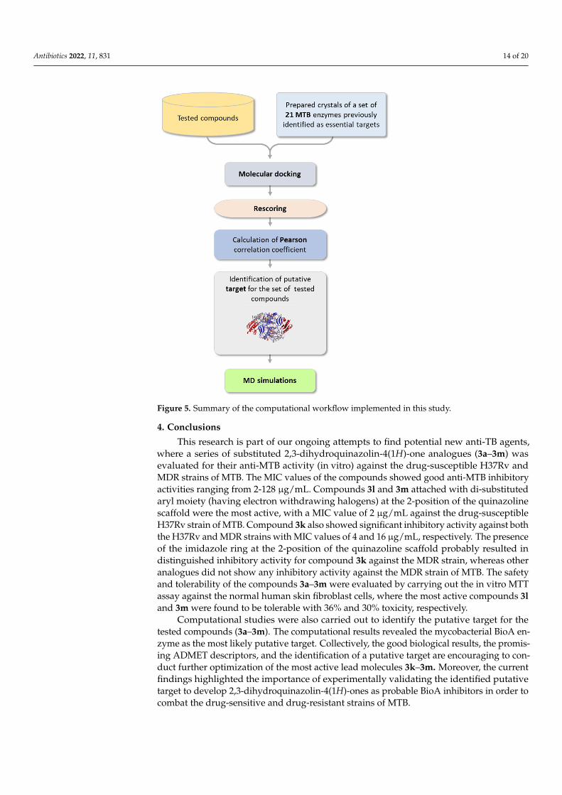
The same computational approach we implemented previously to identify the putativemycobacterial targets for the screened compounds was used in this study [52] (Figure 5).Briefly, an extensive literature review revealed 48 macromolecular targets that are essentialfor mycobacterial survival, of which 21 have solved 3D crystal structures (deposited in theProtein Data Bank) that were used in molecular docking studies [81–83]. All crystal struc-tures were prepared, solvated, and minimized using Biovia Discovery Studio 2020 [52,84].Then, the tested compounds were also prepared using DS and were docked into the bindingsites of the 21 proteins using the CDOCKER algorithm in DS. All docked poses were thenrescored using different scoring functions, and their Pearson correlation coefficients withthe experimental MIC values were calculated. The enzyme with the highest correlationvalue was identified as a putative target for the tested compounds. Finally, the bindingmode and binding stability of a virtual complex of compound 3m with its putative targetwere simulated using Amber 12 relative to the native co-crystallized inhibitor. Moreover,a range of pharmacokinetic and toxicity descriptors for the tested compounds (3a–3m) wascalculated using the ADMET Descriptors protocol and the Toxicity Prediction (TOPKAT)protocol in DS [85].
Antibiotics 2022, 11, 831 14 of 20Antibiotics 2021, 10, x FOR PEER REVIEW 15 of 22
Figure 5. Summary of the computational workflow implemented in this study.
4. Conclusions This research is part of our ongoing attempts to find potential new anti-TB agents,
where a series of substituted 2,3-dihydroquinazolin-4(1H)-one analogues (3a–3m) was evaluated for their anti-MTB activity (in vitro) against the drug-susceptible H37Rv and MDR strains of MTB. The MIC values of the compounds showed good anti-MTB inhibi-tory activities ranging from 2-128 µg/mL. Compounds 3l and 3m attached with di-substi-tuted aryl moiety (having electron withdrawing halogens) at the 2-position of the quinazoline scaffold were the most active, with a MIC value of 2 µg/mL against the drug-susceptible H37Rv strain of MTB. Compound 3k also showed significant inhibitory activ-ity against both the H37Rv and MDR strains with MIC values of 4 and 16 µg/mL, respec-tively. The presence of the imidazole ring at the 2-position of the quinazoline scaffold probably resulted in distinguished inhibitory activity for compound 3k against the MDR strain, whereas other analogues did not show any inhibitory activity against the MDR strain of MTB. The safety and tolerability of the compounds 3a–3m were evaluated by carrying out the in vitro MTT assay against the normal human skin fibroblast cells, where the most active compounds 3l and 3m were found to be tolerable with 36% and 30% tox-icity, respectively.
Computational studies were also carried out to identify the putative target for the tested compounds (3a–3m). The computational results revealed the mycobacterial BioA enzyme as the most likely putative target. Collectively, the good biological results, the promising ADMET descriptors, and the identification of a putative target are encouraging to conduct further optimization of the most active lead molecules 3k–3m. Moreover, the current findings highlighted the importance of experimentally validating the identified putative target to develop 2,3-dihydroquinazolin-4(1H)-ones as probable BioA inhibitors in order to combat the drug-sensitive and drug-resistant strains of MTB.
Figure 5. Summary of the computational workflow implemented in this study.
4. Conclusions
This research is part of our ongoing attempts to find potential new anti-TB agents,where a series of substituted 2,3-dihydroquinazolin-4(1H)-one analogues (3a–3m) wasevaluated for their anti-MTB activity (in vitro) against the drug-susceptible H37Rv andMDR strains of MTB. The MIC values of the compounds showed good anti-MTB inhibitoryactivities ranging from 2-128 µg/mL. Compounds 3l and 3m attached with di-substitutedaryl moiety (having electron withdrawing halogens) at the 2-position of the quinazolinescaffold were the most active, with a MIC value of 2 µg/mL against the drug-susceptibleH37Rv strain of MTB. Compound 3k also showed significant inhibitory activity against boththe H37Rv and MDR strains with MIC values of 4 and 16 µg/mL, respectively. The presenceof the imidazole ring at the 2-position of the quinazoline scaffold probably resulted indistinguished inhibitory activity for compound 3k against the MDR strain, whereas otheranalogues did not show any inhibitory activity against the MDR strain of MTB. The safetyand tolerability of the compounds 3a–3m were evaluated by carrying out the in vitro MTTassay against the normal human skin fibroblast cells, where the most active compounds 3land 3m were found to be tolerable with 36% and 30% toxicity, respectively.
Computational studies were also carried out to identify the putative target for thetested compounds (3a–3m). The computational results revealed the mycobacterial BioA en-zyme as the most likely putative target. Collectively, the good biological results, the promis-ing ADMET descriptors, and the identification of a putative target are encouraging to con-duct further optimization of the most active lead molecules 3k–3m. Moreover, the currentfindings highlighted the importance of experimentally validating the identified putativetarget to develop 2,3-dihydroquinazolin-4(1H)-ones as probable BioA inhibitors in order tocombat the drug-sensitive and drug-resistant strains of MTB.
Antibiotics 2022, 11, 831 15 of 20
Supplementary Materials: The following supporting information can be downloaded at: https://www.mdpi.com/article/10.3390/antibiotics11070831/s1, Scheme S1: Synthetic scheme, proce-dure, characterization details of 2-(5-bromo-2-fluorophenyl)-2,3-dihydroquinazolin-4(1H)-one (3l);Figure S1: FT-IR of 2-(5-bromo-2-fluorophenyl)-2,3-dihydroquinazolin-4(1H)-one (3l); Figure S2: 1H-NMR of 2-(5-bromo-2-fluorophenyl)-2,3-dihydroquinazolin-4(1H)-one (3l); Figure S3: 13C-NMRof 2-(5-bromo-2-fluorophenyl)-2,3-dihydroquinazolin-4(1H)-one (3l); Table S1: Reported essen-tial and potential mycobacterial drug targets. Table S2: The selected 20 essential mycobacterialdrug targets that we used for molecular modeling studies. References [86–119] are cited in theSupplementary Materials.
Author Contributions: Conceptualization, K.N.V., C.T., P.K.D., L.A.D. and S.C.; Methodology, K.N.V.,N.A.A.-S., P.S., C.T., P.K.D., L.A.D., W.H., S.C., M.P., M.A., A.B.N., N.S., B.A.-I., Y.B., M.K., M.H.,R.V., A.A.B. and M.A.M.; Software, K.N.V., N.A.A.-S., C.T., P.K.D. and M.K.; Validation, K.N.V.,N.A.A.-S., C.T., S.C., Y.B. and M.A.M.; Formal analysis, K.N.V., P.S., C.T., S.C., M.P., M.A., N.S. andW.H.; Investigation, K.N.V., C.T., P.K.D., L.A.D., S.C. and A.B.N.; Resources, K.N.V., N.A.A.-S., P.K.D.,S.C., R.V. and M.P.; Data curation, K.N.V., C.T., S.C. and M.A.M.; Writing—original draft preparation,K.N.V., N.A.A.-S., P.S., C.T., P.K.D., L.A.D., W.H., M.A., N.S., B.A.-I., Y.B., A.A.B., M.H., R.V. andM.A.M.; Writing—review and editing, K.N.V., N.A.A.-S., P.K.D., C.T., L.A.D., S.C., M.P., W.H., A.B.N.,B.A.-I., M.K., M.H., R.V., A.A.B. and M.A.M.; Visualization, K.N.V., C.T. and P.K.D.; Supervision,K.N.V.; Project administration, K.N.V.; Funding acquisition, K.N.V. All authors have read and agreedto the published version of the manuscript.
Funding: This work was supported by the Deanship of Scientific Research, Vice Presidency forGraduate Studies and Scientific Research, King Faisal University, Saudi Arabia (grant numberAN000168).
Institutional Review Board Statement: Not applicable.
Informed Consent Statement: Not applicable.
Data Availability Statement: Not applicable.
Acknowledgments: The authors thank the Deanship of Scientific Research, Vice Presidency for Gradu-ate Studies and Scientific Research, King Faisal University, Saudi Arabia, for support and encouragement.
Conflicts of Interest: The authors declare no conflict of interest.
References1. World Health Organization. Global Tuberculosis Report 2019 (Executive Summary). Available online: https://www.who.int/tb/
publications/global_report/tb19_Exec_Sum_12Nov2019.pdf?ua=1 (accessed on 9 September 2020).2. Furin, J.; Cox, H.; Pai, M. Tuberculosis. Lancet 2019, 393, 1642–1656. [CrossRef]3. Lee, J.Y. Diagnosis and Treatment of Extrapulmonary Tuberculosis. Tuberc. Respir. Dis. 2015, 78, 47–55. [CrossRef] [PubMed]4. Tetali, S.R.; Kunapaeddi, E.; Mailavaram, R.P.; Singh, V.; Borah, P.; Deb, P.K.; Venugopala, K.N.; Hourani, W.; Tekade, R.K. Current
advances in the clinical development of anti-tubercular agents. Tuberculosis 2020, 125, 101989. [CrossRef] [PubMed]5. Udwadia, Z.F. MDR, XDR, TDR tuberculosis: Ominous progression. Thorax 2012, 67, 286–288. [CrossRef]6. Dheda, K.; Shean, K.; Zumla, A.; Badri, M.; Streicher, E.M.; Page-Shipp, L.; Willcox, P.; John, M.A.; Reubenson, G.; Govindasamy,
D.; et al. Early treatment outcomes and HIV status of patients with extensively drug-resistant tuberculosis in South Africa: Aretrospective cohort study. Lancet 2010, 375, 1798–1807. [CrossRef]
7. Quan, D.; Nagalingam, G.; Payne, R.; Triccas, J.A. New tuberculosis drug leads from naturally occurring compounds. Int. J. Infect.Dis. 2017, 56, 212–220. [CrossRef]
8. Yang, D.; Taylor, Z.E.; Handy, S.; Li, S.J.; Liu, J.W.; Stabenow, J.; Zalduondo, L.; Jonsson, C.B.; Altman, E.; Kong, Y. Identification ofAnti-tuberculosis Compounds From Aurone Analogs. Front. Microbiol. 2020, 11, 1004. [CrossRef]
9. Dasyam, N.; Munkacsi, A.B.; Fadzilah, N.H.; Senanayake, D.S.; O’Toole, R.F.; Keyzers, R.A. Identification and Bioactivityof 3-epi-Xestoaminol C Isolated from the New Zealand Brown Alga Xiphophora chondrophylla. J. Nat. Prod. 2014, 77,1519–1523. [CrossRef]
10. Tang, S.J.; Yao, L.; Hao, X.H.; Liu, Y.D.; Zeng, L.H.; Liu, G.; Li, M.W.; Li, F.J.; Wu, M.Y.; Zhu, Y.S.; et al. Clofazimine for theTreatment of Multidrug-Resistant Tuberculosis: Prospective, Multicenter, Randomized Controlled Study in China. Clin. Infect.Dis. 2015, 60, 1361–1367. [CrossRef]
11. Lee, M.; Lee, J.; Carroll, M.W.; Choi, H.; Min, S.; Song, T.; Via, L.E.; Goldfeder, L.C.; Kang, E.; Jin, B.; et al. Linezolid for Treatmentof Chronic Extensively Drug-Resistant Tuberculosis. N. Engl. J. Med. 2012, 367, 1508–1518. [CrossRef]
Antibiotics 2022, 11, 831 16 of 20
12. Nguyen, T.V.A.; Anthony, R.; Bañuls, A.-L.; Vu, D.H.; Alffenaar, J. Bedaquiline resistance: Its emergence, mechanism andprevention. Clin. Infect. Dis. 2018, 66, 1625–1630. [CrossRef] [PubMed]
13. Fujiwara, M.; Kawasaki, M.; Hariguchi, N.; Liu, Y.; Matsumoto, M. Mechanisms of resistance to delamanid, a drug for Mycobac-terium tuberculosis. Tuberculosis 2018, 108, 186–194. [CrossRef] [PubMed]
14. Cox, H.; Ford, N. Linezolid for the treatment of complicated drug-resistant tuberculosis: A systematic review and meta-analysis.Int. J. Tuberc. Lung Dis. 2012, 16, 447–454. [CrossRef] [PubMed]
15. Dalcolmo, M.; Gayoso, R.; Sotgiu, G.; D’Ambrosio, L.; Rocha, J.L.; Borga, L.; Fandinho, F.; Braga, J.U.; Galesi, V.M.N.;Barreira, D.; et al. Effectiveness and safety of clofazimine in multidrug-resistant tuberculosis: A nationwide report from Brazil.Eur. Respir. J. 2017, 49, 1602445. [CrossRef] [PubMed]
16. Liang, J.L.; Cha, H.C.; Jahng, Y. Recent advances in the studies on luotonins. Molecules 2011, 16, 4861–4883. [CrossRef] [PubMed]17. Xu, J.; Li, Z.; Luo, J.; Yang, F.; Liu, T.; Liu, M.; Qiu, W.-W.; Tang, J. Synthesis and Biological Evaluation of Heterocyclic Ring-Fused
Betulinic Acid Derivatives as Novel Inhibitors of Osteoclast Differentiation and Bone Resorption. J. Med. Chem. 2012, 55,3122–3134. [CrossRef]
18. Lee, S.; Kim, D.-C.; Baek, H.Y.; Lee, K.-D.; Kim, Y.-C.; Oh, H. Anti-neuroinflammatory effects of tryptanthrin from Polygonumtinctorium Lour. in lipopolysaccharide-stimulated BV2 microglial cells. Arch. Pharmacal Res. 2018, 41, 419–430. [CrossRef]
19. Tian, K.-M.; Li, J.-J.; Xu, S.-W. Rutaecarpine: A promising cardiovascular protective alkaloid from Evodia rutaecarpa (Wu ZhuYu). Pharmacol. Res. 2019, 141, 541–550. [CrossRef]
20. Estlin, E.J.; Pinkerton, C.R.; Lewis, I.J.; Lashford, L.; McDowell, H.; Morland, B.; Kohler, J.; Newell, D.R.; Boddy, A.V.;Taylor, G.A.; et al. A phase I study of nolatrexed dihydrochloride in children with advanced cancer. A United Kingdom Children’sCancer Study Group Investigation. Br. J. Cancer 2001, 84, 11–18. [CrossRef]
21. Gudimella, K.K.; Bonige, K.B.; Gundla, R.; Katari, N.K.; Yamajala, B.; Battula, V.R. 2,4-Diphenyl-1,2-dihydroquinazolineDerivatives: Synthesis, Anticancer Activity and Docking Studies. ChemistrySelect 2019, 4, 12528–12533. [CrossRef]
22. Kamal, A.; Bharathi, E.V.; Reddy, J.S.; Ramaiah, M.J.; Dastagiri, D.; Reddy, M.K.; Viswanath, A.; Reddy, T.L.; Shaik, T.B.; Push-pavalli, S.N.C.V.L.; et al. Synthesis and biological evaluation of 3,5-diaryl isoxazoline/isoxazole linked 2,3-dihydroquinazolinonehybrids as anticancer agents. Eur. J. Med. Chem. 2011, 46, 691–703. [CrossRef] [PubMed]
23. Madasu, C.; Xu, Y.-M.; Wijeratne, E.M.K.; Liu, M.X.; Molnár, I.; Gunatilaka, A.A.L. Semi-synthesis and cytotoxicity eval-uation of pyrimidine, thiazole, and indole analogues of argentatins A–C from guayule (Parthenium argentatum) resin.Med. Chem. Res. 2022. [CrossRef]
24. Mallavadhani, U.V.; Chandrashekhar, M.; Nayak, V.L.; Ramakrishna, S. Synthesis and anticancer activity of novel fusedpyrimidine hybrids of myrrhanone C, a bicyclic triterpene of Commiphora mukul gum resin. Mol. Divers. 2015, 19, 745–757.[CrossRef] [PubMed]
25. Iyer, K.A.; Alix, K.; Eltit, J.M.; Solis, E., Jr.; Pan, X.; Argade, M.D.; Khatri, S.; De Felice, L.J.; Sweet, D.H.; Schulte, M.K.; et al.Multi-modal antidepressant-like action of 6- and 7-chloro-2-aminodihydroquinazolines in the mouse tail suspension test. Psy-chopharmacology 2019, 236, 2093–2104. [CrossRef] [PubMed]
26. Barmak, A.; Niknam, K.; Mohebbi, G. Synthesis, Structural Studies, and α-Glucosidase Inhibitory, Antidiabetic, and Antioxi-dant Activities of 2,3-Dihydroquinazolin-4(1H)-ones Derived from Pyrazol-4-carbaldehyde and Anilines. ACS Omega 2019, 4,18087–18099. [CrossRef] [PubMed]
27. Guillon, R.; Pagniez, F.; Picot, C.; Hédou, D.; Tonnerre, A.; Chosson, E.; Duflos, M.; Besson, T.; Logé, C.; Le Pape, P. Discovery of anovel broad-spectrum antifungal agent derived from albaconazole. ACS Med. Chem. Lett. 2013, 4, 288–292. [CrossRef]
28. Mujeeb Ur, R.; Rathore, A.; Siddiqui, A.A.; Parveen, G.; Yar, M.S. Synthesis and characterization of quinazoline derivatives:Search for hybrid molecule as diuretic and antihypertensive agents. J. Enzym. Inhib. Med. Chem. 2014, 29, 733–743. [CrossRef]
29. Obase, H.; Takai, H.; Teranishi, M.; Nakamizo, N. Synthesis of (1-substituted piperidin-4-yl)-1H-benzimidazoles and (1-substitutedpiperidin-4-yl)-3,4-dihydroquinazolines as possible antihypertensive agents. J. Heterocycl. Chem. 1983, 20, 565–573. [CrossRef]
30. El-Sabbagh, O.I.; Ibrahim, S.M.; Baraka, M.M.; Kothayer, H. Synthesis of new 2,3-dihydroquinazolin-4(1H)-one derivatives foranalgesic and anti-inflammatory evaluation. Arch. Pharm. 2010, 343, 274–281. [CrossRef]
31. Clissold, S.P.; Beresford, R. Proquazone. A review of its pharmacodynamic and pharmacokinetic properties, and therapeuticefficacy in rheumatic diseases and pain states. Drugs 1987, 33, 478–502. [CrossRef]
32. Kamal, A.; Babu, K.S.; Poornachandra, Y.; Nagaraju, B.; Ali Hussaini, S.M.; Shaik, S.P.; Ganesh Kumar, C.; Alarifi, A. Efficient andgreen sulfamic acid catalyzed synthesis of new 1,2-dihydroquinazoline derivatives with antibacterial potential. Arab. J. Chem.2015, 12, 3546–3554. [CrossRef]
33. Salehi, P.; Ayyari, M.; Bararjanian, M.; Ebrahimi, S.N.; Aliahmadi, A. Synthesis, antibacterial and antioxidant activity of novel2,3-dihydroquinazolin-4(1H)-one derivatives of dehydroabietylamine diterpene. J. Iran. Chem. Soc. 2014, 11, 607–613. [CrossRef]
34. Chung, C.K.; Liu, Z.; Lexa, K.W.; Andreani, T.; Xu, Y.; Ji, Y.; DiRocco, D.A.; Humphrey, G.R.; Ruck, R.T. Asymmetric HydrogenBonding Catalysis for the Synthesis of Dihydroquinazoline-Containing Antiviral, Letermovir. J. Am. Chem. Soc. 2017, 139,10637–10640. [CrossRef] [PubMed]
35. Mehta, D.R.; Naravane, J.S.; Desai, R.M. Vasicinone. A Bronchodilator Principle from Adhatoda Vasica Nees (N. O. Acanthaceae).J. Org. Chem. 1963, 28, 445–448. [CrossRef]
36. Yamamura, M.; Ochiai, T.; Ishida, R. Effects of afloqualone, a new centrally acting muscle relaxant, on DRL response and CER inrats (author’s transl). Nihon Yakurigaku Zasshi 1981, 78, 381–392. [CrossRef] [PubMed]
Antibiotics 2022, 11, 831 17 of 20
37. Ferrando, C.; Foy, J.M.; Pratt, C.N.F.W.; Purvis, J.R. On the pharmacological actions of a diuretic, fenquizone, with particularreference to its site of action. J. Pharm. Pharmacol. 1981, 33, 219–222. [CrossRef] [PubMed]
38. van Zyl, E.F. A survey of reported synthesis of methaqualone and some positional and structural isomers. Forensic Sci. Int. 2001,122, 142–149. [CrossRef]
39. Jafari, E.; Khajouei, M.R.; Hassanzadeh, F.; Hakimelahi, G.H.; Khodarahmi, G.A. Quinazolinone and quinazoline derivatives:Recent structures with potent antimicrobial and cytotoxic activities. Res. Pharm. Sci. 2016, 11, 1–14.
40. Li, W.J.; Li, Q.; Liu, D.L.; Ding, M.W. Synthesis, fungicidal activity, and sterol 14alpha-demethylase binding interaction of2-azolyl-3,4-dihydroquinazolines on Penicillium digitatum. J. Agric. Food Chem. 2013, 61, 1419–1426. [CrossRef]
41. Zhou, Y.; Feng, Q.; Di, F.; Liu, Q.; Wang, D.; Chen, Y.; Xiong, L.; Song, H.; Li, Y.; Li, Z. Synthesis and insecticidal activities of2,3-dihydroquinazolin-4(1H)-one derivatives targeting calcium channel. Bioorganic Med. Chem. 2013, 21, 4968–4975. [CrossRef]
42. Alveera, S.; Venugopala, K.N.; Khedr, M.A.; Pillay, M.; Nwaeze, K.U.; Coovadia, Y.; Shode, F.; Odhav, B. Antimycobacterial,docking and molecular dynamic studies of pentacyclic triterpenes from Buddleja saligna leaves. J. Biomol. Struct. Dyn. 2017, 35,2654–2664. [CrossRef]
43. Venugopala, K.N. Posological review of dose extrapolation methodologies in animal studies, early-phase clinical trials and specialpopulations. Int. J. Res. Pharm. Sci. 2020, 11, 6079–6084. [CrossRef]
44. Venugopala, K.N.; Albericio, F.; Coovadia, Y.M.; Kruger, H.G.; Maguire, G.E.M.; Pillay, M.; Govender, T. Total synthesis of adepsidomycin analogue by convergent solid-phase peptide synthesis and macrolactonization strategy for antitubercular activity.J. Pept. Sci. 2011, 17, 683–689. [CrossRef]
45. Venugopala, K.N.; Nayak, S.K.; Pillay, M.; Prasanna, R.; Coovadia, Y.M.; Odhav, B. Synthesis and antitubercular activity of2-(substituted phenyl/benzyl-amino)-6-(4-chlorophenyl)-5-(methoxycarbonyl)-4-methyl-3,6-dihydropyrimidin-1-ium chlorides.Chem. Biol. Drug Des. 2013, 81, 219–227. [CrossRef]
46. Venugopala, K.N.; Dharma Rao, G.B.; Bhandary, S.; Pillay, M.; Chopra, D.; Aldhubiab, B.E.; Attimarad, M.; Alwassil, O.I.; Harsha,S.; Mlisana, K. Design, synthesis, and characterization of (1-(4-aryl)- 1H-1,2,3-triazol-4-yl)methyl, substituted phenyl-6-methyl-2-oxo-1,2,3,4-tetrahydropyrimidine-5-carboxylates against Mycobacterium tuberculosis. Drug Des. Dev. Ther. 2016, 10, 2681–2690.[CrossRef] [PubMed]
47. Venugopala, K.N.; Chandrashekharappa, S.; Pillay, M.; Bhandary, S.; Kandeel, M.; Mahomoodally, F.M.; Morsy, M.A.; Chopra, D.;Aldhubiab, B.E.; Attimarad, M.; et al. Synthesis and structural elucidation of novel benzothiazole derivatives as anti-tubercularagents: In-silico screening for possible target identification. Med. Chem. 2019, 15, 311–326. [CrossRef] [PubMed]
48. Venugopala, K.N.; Chandrashekharappa, S.; Pillay, M.; Abdallah, H.H.; Mahomoodally, F.M.; Bhandary, S.; Chopra, D.; Attimarad,M.; Aldhubiab, B.E.; Nair, A.B. Computational, crystallographic studies, cytotoxicity and anti-tubercular activity of substituted7-methoxy-indolizine analogues. PLoS ONE 2019, 14, e0217270. [CrossRef] [PubMed]
49. Venugopala, K.N.; Tratrat, C.; Pillay, M.; Mahomoodally, F.M.; Bhandary, S.; Chopra, D.; Morsy, M.A.; Haroun, M.; Aldhubiab,B.E.; Attimarad, M.; et al. Anti-tubercular activity of substituted 7-methyl and 7-formylindolizines and in silico study forprospective molecular target identification. Antibiotics 2019, 8, 247. [CrossRef]
50. Khedr, M.A.; Pillay, M.; Chandrashekharappa, S.; Chopra, D.; Aldhubiab, B.E.; Attimarad, M.; Alwassil, O.I.; Mlisana, K.; Odhav,B.; Venugopala, K.N. Molecular modeling studies and anti-TB activity of trisubstituted indolizine analogues; molecular dockingand dynamic inputs. J. Biomol. Struct. Dyn. 2018, 36, 2163–2178. [CrossRef]
51. Venugopala, K.N.; Chandrashekharappa, S.; Deb, P.K.; Tratrat, C.; Pillay, M.; Chopra, D.; Al-Shar’i, N.A.; Hourani, W.; Dahabiyeh,L.A.; Borah, P.; et al. Anti-tubercular activity and molecular docking studies of indolizine derivatives targeting mycobacterialInhA enzyme. J. Enzym. Inhib. Med. Chem. 2021, 36, 1472–1487. [CrossRef]
52. Venugopala, K.; Tratrat, C.; Chandrashekharappa, S.; Attimarad, M.; SreeHarsha, N.; Nair, A.; Pottathil, S.; Venugopala, R.;Al-Attraqchi, O.; Morsy, M.; et al. Anti-tubercular Potency and Computationallyassessed Drug-likeness and Toxicology ofDiversely Substituted Indolizines. Indian J. Pharm. Educ. 2019, 53, 545–552. [CrossRef]
53. Venugopala, K.N.; Chandrashekharappa, S.; Tratrat, C.; Deb, P.K.; Nagdeve, R.D.; Nayak, S.K.; Morsy, M.A.; Borah, P.; Maho-moodally, F.M.; Mailavaram, R.P. Crystallography, Molecular Modeling, and COX-2 Inhibition Studies on Indolizine Derivatives.Molecules 2021, 26, 3550. [CrossRef] [PubMed]
54. Uppar, V.; Chandrashekharappa, S.; Shivamallu, C.; Kollur, S.P.; Ortega-Castro, J.; Frau, J.; Flores-Holguín, N.; Basarikatti, A.I.;Chougala, M.; Mohan, M.M.; et al. Investigation of antifungal properties of synthetic dimethyl-4-bromo-1-(substituted benzoyl)pyrrolo[1,2-a] quinoline-2,3-dicarboxylates analogues: Molecular docking studies and conceptual DFT-based chemical reactivitydescriptors and pharmacokinetics evaluation. Molecules 2021, 26, 2722. [CrossRef] [PubMed]
55. Venugopala, K.N.; Al-Attraqchi, O.H.; Tratrat, C.; Nayak, S.K.; Morsy, M.A.; Aldhubiab, B.E.; Attimarad, M.; Nair, A.B.;Sreeharsha, N.; Venugopala, R. Novel series of methyl 3-(substituted benzoyl)-7-substituted-2-phenylindolizine-1-carboxylates aspromising anti-inflammatory agents: Molecular modeling studies. Biomolecules 2019, 9, 661. [CrossRef] [PubMed]
56. Nefisath, P.; Dasappa, J.P.; Haripriya, B.; Chopra, D.; Venugopala, K.N.; Deb, P.K.; Gleiser, R.M.; Mohanlall, V.; Maharaj, R.;Shashiprabha; et al. Synthesis, structural elucidation and larvicidal activity of novel arylhydrazones. J. Mol. Struct. 2021,1236, 130305. [CrossRef]
57. Nefisath, P.; Dasappa, J.P.; Haripriya, B.; Chopra, D.; Venugopala, K.N.; Deb, P.K.; Gleiser, R.M.; Mohanlall, V.; Maharaj, R.;Shashiprabha, S.; et al. Synthesis, characterization and larvicidal activity of novel benzylidene derivatives of fenobam and its thioanalogues with crystal insight. J. Mol. Struct. 2021, 1226, 129386. [CrossRef]
Antibiotics 2022, 11, 831 18 of 20
58. Venugopala, K.N.; Habeebuddin, M.; Aldhubiab, B.E.; Asif, A.H. Design, Synthesis, and In Vitro Evaluation of Novel IndolylDiHydropyrazole Derivatives as Potential Anticancer Agents. Molecules 2021, 26, 5235. [CrossRef]
59. Haroun, M.; Tratrat, C.; Petrou, A.; Geronikaki, A.; Ivanov, M.; Ciric, A.; Sokovic, M.; Nagaraja, S.; Venugopala, K.N.; BalachandranNair, A. Exploration of the antimicrobial effects of benzothiazolylthiazolidin-4-one and in silico mechanistic investigation.Molecules 2021, 26, 4061. [CrossRef]
60. Tratrat, C.; Haroun, M.; Paparisva, A.; Kamoutsis, C.; Petrou, A.; Gavalas, A.; Eleftheriou, P.; Geronikaki, A.; Venugopala, K.N.;Kochkar, H. New Substituted 5-Benzylideno-2-Adamantylthiazol [3, 2-b][1, 2, 4] Triazol-6 (5H) ones as Possible Anti-InflammatoryAgents. Molecules 2021, 26, 659. [CrossRef]
61. Morsy, M.A.; Ali, E.M.; Kandeel, M.; Venugopala, K.N.; Nair, A.B.; Greish, K.; El-Daly, M. Screening and molecular docking ofnovel benzothiazole derivatives as potential antimicrobial agents. Antibiotics 2020, 9, 221. [CrossRef]
62. Venugopala, K.N.; Khedr, M.A.; Girish, Y.R.; Bhandary, S.; Chopra, D.; Morsy, M.A.; Aldhubiab, B.E.; Deb, P.K.; Attimarad,M.; Nair, A.B. Crystallography, in silico studies, and In vitro antifungal studies of 2,4,5 trisubstituted 1,2,3-triazole analogues.Antibiotics 2020, 9, 350. [CrossRef] [PubMed]
63. Haroun, M.; Tratrat, C.; Kolokotroni, A.; Petrou, A.; Geronikaki, A.; Ivanov, M.; Kostic, M.; Sokovic, M.; Carazo, A.; Mladenka, P.5-Benzyliden-2-(5-methylthiazol-2-ylimino) thiazolidin-4-ones as Antimicrobial Agents. Design, Synthesis, Biological Evaluationand Molecular Docking Studies. Antibiotics 2021, 10, 309. [CrossRef] [PubMed]
64. Al-Shar’i, N.A.; Al-Balas, Q.A.; Al-Waqfi, R.A.; Hassan, M.A.; Alkhalifa, A.E.; Ayoub, N.M. Discovery of a nanomolar inhibitor ofthe human glyoxalase-I enzyme using structure-based poly-pharmacophore modelling and molecular docking. J. Comput.-AidedMol. Des. 2019, 33, 799–815. [CrossRef] [PubMed]
65. Al-Shar’i, N.A.; Al-Balas, Q.A.; Hassan, M.A.; El-Elimat, T.M.; Aljabal, G.A.; Almaaytah, A.M. Ellagic acid: A potent glyoxalase-Iinhibitor with a unique scaffold. Acta Pharm. 2021, 71, 115–130. [CrossRef]
66. Deb, P.K.; Al-Shar’i, N.A.; Venugopala, K.N.; Pillay, M.; Borah, P. In vitro anti-TB properties, in silico target validation, moleculardocking and dynamics studies of substituted 1,2,4-oxadiazole analogues against Mycobacterium tuberculosis. J. Enzym. Inhib.Med. Chem. 2021, 36, 869–884. [CrossRef]
67. Al-Shar’i, N.A.; Musleh, S.S. Identification of CHK1 Kinase Inhibitors Using Structure Based Pharmacophore Modelling andMolecular Docking. Indian J. Pharm. Sci. 2020, 82, 472–482. [CrossRef]
68. Li, J.; Fu, A.; Zhang, L. An Overview of Scoring Functions Used for Protein–Ligand Interactions in Molecular Docking. Interdiscip.Sci. Comput. Life Sci. 2019, 11, 320–328. [CrossRef]
69. Al-Balas, Q.A.; Hassan, M.A.; Al-Shar’i, N.A.; El-Elimat, T.; Almaaytah, A.M. Computational and experimental exploration of thestructure–activity relationships of flavonoids as potent glyoxalase-I inhibitors. Drug Dev. Res. 2018, 79, 58–69. [CrossRef]
70. Liu, F.; Dawadi, S.; Maize, K.M.; Dai, R.; Park, S.W.; Schnappinger, D.; Finzel, B.C.; Aldrich, C.C. Structure-Based Optimization ofPyridoxal 5′-Phosphate-Dependent Transaminase Enzyme (BioA) Inhibitors that Target Biotin Biosynthesis in Mycobacteriumtuberculosis. J. Med. Chem. 2017, 60, 5507–5520. [CrossRef]
71. Dey, S.; Lane, J.M.; Lee, R.E.; Rubin, E.J.; Sacchettini, J.C. Structural Characterization of the Mycobacterium tuberculo-sis Biotin Biosynthesis Enzymes 7,8-Diaminopelargonic Acid Synthase and Dethiobiotin Synthetase. Biochemistry 2010, 49,6746–6760. [CrossRef]
72. Singh, S.; Khare, G.; Bahal, R.K.; Ghosh, P.C.; Tyagi, A.K. Identification of Mycobacterium tuberculosis BioA inhibitors by usingstructure-based virtual screening. Drug Des. Dev. Ther. 2018, 12, 1065–1079. [CrossRef] [PubMed]
73. Dai, R.; Geders, T.W.; Liu, F.; Park, S.W.; Schnappinger, D.; Aldrich, C.C.; Finzel, B.C. Fragment-based exploration of binding siteflexibility in Mycobacterium tuberculosis BioA. J. Med. Chem. 2015, 58, 5208–5217. [CrossRef] [PubMed]
74. Al-Shar’i, N.A.; Alnabulsi, S.M. Explaining the autoinhibition of the SMYD enzyme family: A theoretical study. J. Mol. Graph.Model. 2016, 68, 147–157. [CrossRef] [PubMed]
75. Al-Shar’i, N.A.; Al-Balas, Q.A. Molecular Dynamics Simulations of Adenosine Receptors: Advances, Applications and Trends.Curr. Pharm. Des. 2019, 25, 783–816. [CrossRef]
76. Hou, T.; Wang, J.; Zhang, W.; Xu, X. ADME Evaluation in Drug Discovery. 6. Can Oral Bioavailability in Humans Be EffectivelyPredicted by Simple Molecular Property-Based Rules? J. Chem. Inf. Model. 2007, 47, 460–463. [CrossRef]
77. Venugopala, K.N.; Ramachandra, P.; Tratrat, C.; Gleiser, R.M.; Bhandary, S.; Chopra, D.; Morsy, M.A.; Aldhubiab, B.E.; Attimarad,M.; Nair, A.B.; et al. Larvicidal activities of 2-aryl-2,3-dihydroquinazolin -4-ones against malaria vector Anopheles arabiensis, Insilico ADMET prediction and molecular target investigation. Molecules 2020, 25, 1316. [CrossRef]
78. Bairagi, K.M.; Venugopala, K.N.; Alwassil, O.I.; Mohanlall, V.; Chandrashekharappa, S.; Nayak, S.K. Synthesis, crystalstructure and Hirshfeld surface analysis of 2-(4-fluorophenyl)-2,3–dihydroquinazolin-4(1H)-one. Chem. Data Collect. 2020,26, 100355. [CrossRef]
79. Martin, A.; Morcillo, N.; Lemus, D.; Montoro, E.; Telles, M.A.; Simboli, N.; Pontino, M.; Porras, T.; Leon, C.; Velasco, M.; et al.Multicenter study of MTT and resazurin assays for testing susceptibility to first-line anti-tuberculosis drugs. Int. J. Tuberc. LungDis. 2005, 9, 901–906.
80. Middlebrook, G.; Reggiards, Z.; Tigertt, W.D. Automable radiometric detection of growth of Mycobacterium tuberculosis in selectivemedia. Am. Rev. Respir. Dis. 1977, 115, 1067–1069.
Antibiotics 2022, 11, 831 19 of 20
81. Deb, P.K.; Mailavaram, R.; Chandrasekaran, B.; Kaki, V.R.; Kaur, R.; Kachler, S.; Klotz, K.N.; Akkinepally, R.R. Synthesis, adenosinereceptor binding and molecular modelling studies of novel thieno[2,3-d]pyrimidine derivatives. Chem. Biol. Drug Des. 2018, 91,962–969. [CrossRef]
82. Dhingra, M.S.; Deb, P.K.; Chadha, R.; Singh, T.; Karan, M. Synthesis, evaluation, and molecular docking studies of cycloalkyl/aryl-3,4,5-trimethylgallates as potent non-ulcerogenic and gastroprotective anti-inflammatory agents. Med. Chem. Res. 2014, 23,87–106. [CrossRef]
83. Deb, P.K.; Sharma, A.; Piplani, P.; Akkinepally, R.R. Molecular docking and receptor-specific 3D-QSAR studies of acetyl-cholinesterase inhibitors. Mol. Divers. 2012, 16, 803–823. [CrossRef]
84. Deb, P.K.; Junaid, A.; El-Rabie, D.; Hon, T.; Nasr, E.; Pichika, M. Molecular Docking Studies and Comparative Binding ModeAnalysis of FDA Approved HIV Protease Inhibitors. Asian J. Chem. 2014, 26, 6227–6232. [CrossRef]
85. Al-Jaidi, B.A.; Telfah, S.T.; Bardaweel, S.K.; Deb, P.K.; Borah, P.; Venugopala, K.N.; Bataineh, Y.A.; Al Khames Aga, Q.A.Anticancer Activity and in Silico ADMET Properties of 2,4,5-Trisubstitutedthiazole Derivatives. Curr. Drug Metab. 2021, 22,532–536. [CrossRef] [PubMed]
86. Wang, F.; Sambandan, D.; Halder, R.; Wang, J.; Batt, S.M.; Weinrick, B.; Ahmad, I.; Yang, P.; Zhang, Y.; Kim, J.; et al. Identificationof a small molecule with activity against drug-resistant and persistent tuberculosis. Proc. Natl. Acad. Sci. USA 2013, 110,E2510–E2517. [CrossRef] [PubMed]
87. Torfs, E.; Piller, T.; Cos, P.; Cappoen, D. Opportunities for Overcoming Mycobacterium tuberculosis Drug Resistance: EmergingMycobacterial Targets and Host-Directed Therapy. Int. J. Mol. Sci. 2019, 20, 2868. [CrossRef]
88. Piton, J.; Foo, C.S.-Y.; Cole, S.T. Structural studies of Mycobacterium tuberculosis DprE1 interacting with its inhibitors. DrugDiscov. Today 2017, 22, 526–533. [CrossRef]
89. Kamsri, P.; Hanwarinroj, C.; Phusi, N.; Pornprom, T.; Chayajarus, K.; Punkvang, A.; Suttipanta, N.; Srimanote, P.; Suttisintong, K.;Songsiriritthigul, C.; et al. Discovery of New and Potent InhA Inhibitors as Antituberculosis Agents: Structure-Based VirtualScreening Validated by Biological Assays and X-ray Crystallography. J. Chem. Inf. Model. 2019, 60, 226–234. [CrossRef]
90. Huang, C.-C.; Smith, C.V.; Glickman, M.S.; Jacobs, W.R.; Sacchettini, J.C. Crystal Structures of Mycolic Acid CyclopropaneSynthases from Mycobacterium tuberculosis. J. Biol. Chem. 2002, 277, 11559–11569. [CrossRef]
91. LeMagueres, P.; Im, H.; Ebalunode, J.; Strych, U.; Benedik, M.J.; Briggs, J.M.; Kohn, A.H.; Krause, K.L. The 1.9 Å Crystal Structureof Alanine Racemase from Mycobacterium tuberculosis Contains a Conserved Entryway into the Active Site. Biochemistry 2005,44, 1471–1481. [CrossRef]
92. Shetye, G.S.; Franzblau, S.G.; Cho, S. New tuberculosis drug targets, their inhibitors, and potential therapeutic impact. Transl. Res.2020, 220, 68–97. [CrossRef] [PubMed]
93. Cohen-Gonsaud, M.; Ducasse, S.; Hoh, F.; Zerbib, D.; Labesse, G.; Quemard, A. Crystal Structure of MabA from Mycobacteriumtuberculosis, a Reductase involved in Long-chain Fatty Acid Biosynthesis. J. Mol. Biol. 2002, 320, 249–261. [CrossRef]
94. Gurcha, S.S.; Usha, V.; Cox, J.; Fütterer, K.; Abrahams, K.; Bhatt, A.; Alderwick, L.; Reynolds, R.C.; Loman, N.; Nataraj, V.; et al.Biochemical and Structural Characterization of Mycobacterial Aspartyl-tRNA Synthetase AspS, a Promising TB Drug Target.PLoS ONE 2014, 9, e113568. [CrossRef]
95. Yuan, T.; Sampson, N.S. Hit Generation in TB Drug Discovery: From Genome to Granuloma. Chem. Rev. 2018, 118, 1887–1916.[CrossRef] [PubMed]
96. Chen, C.; Han, X.; Yan, Q.; Wang, C.; Jia, L.; Taj, A.; Zhao, L.; Ma, Y. The Inhibitory Effect of GlmU Acetyltransferase InhibitorTPSA on Mycobacterium tuberculosis May Be Affected Due to Its Methylation by Methyltransferase Rv0560c. Front. Cell. Infect.Microbiol. 2019, 9, 251. [CrossRef]
97. Chiarelli, L.R.; Mori, G.; Orena, B.S.; Esposito, M.; Lane, T.; Ribeiro, A.L.D.J.L.; Degiacomi, G.; Zemanová, J.; Szádocka, S.; Huszar,S.; et al. A multitarget approach to drug discovery inhibiting Mycobacterium tuberculosis PyrG and PanK. Sci. Rep. 2018,8, 3187. [CrossRef]
98. Wlodarchak, N.; Teachout, N.; Beczkiewicz, J.; Procknow, R.; Schaenzer, A.J.; Satyshur, K.; Pavelka, M.; Zuercher, W.; Drewry, D.;Sauer, J.-D.; et al. In Silico Screen and Structural Analysis Identifies Bacterial Kinase Inhibitors which Act with β-Lactams ToInhibit Mycobacterial Growth. Mol. Pharm. 2018, 15, 5410–5426. [CrossRef]
99. Kang, C.-M.; Abbott, D.W.; Park, S.T.; Dascher, C.C.; Cantley, L.C.; Husson, R.N. The Mycobacterium tuberculosis serine/threoninekinases PknA and PknB: Substrate identification and regulation of cell shape. Genes Dev. 2005, 19, 1692–1704. [CrossRef]
100. Ioerger, T.R.; O’Malley, T.; Liao, R.; Guinn, K.M.; Hickey, M.J.; Mohaideen, N.; Murphy, K.C.; Boshoff, H.I.M.; Mizrahi, V.; Rubin,E.J.; et al. Identification of New Drug Targets and Resistance Mechanisms in Mycobacterium tuberculosis. PLoS ONE 2013,8, e75245. [CrossRef]
101. Aggarwal, A.; Parai, M.K.; Shetty, N.; Wallis, D.; Woolhiser, L.; Hastings, C.; Dutta, N.; Galaviz, S.; Dhakal, R.C.; Shrestha, R.; et al.Development of a Novel Lead that Targets M. tuberculosis Polyketide Synthase 13. Cell 2017, 170, 249–259. [CrossRef]
102. Jansen, R.S.; Mandyoli, L.; Hughes, R.; Wakabayashi, S.; Pinkham, J.T.; Selbach, B.; Guinn, K.M.; Rubin, E.J.; Sacchettini, J.C.; Rhee,K.Y. Aspartate aminotransferase Rv3722c governs aspartate-dependent nitrogen metabolism in Mycobacterium tuberculosis. Nat.Commun. 2020, 11, 1960. [CrossRef] [PubMed]
103. Cox, J.; Abrahams, K.; Alemparte, C.; Ghidelli-Disse, S.; Rullas, J.; Angulo-Barturen, I.; Singh, A.; Gurcha, S.S.; Nataraj, V.; Bethell,S.; et al. THPP target assignment reveals EchA6 as an essential fatty acid shuttle in mycobacteria. Nat. Microbiol. 2016, 1, 15006.[CrossRef] [PubMed]
Antibiotics 2022, 11, 831 20 of 20
104. Willand, N.; Dirié, B.; Carette, X.; Bifani, P.; Singhal, A.; Desroses, M.; Leroux, F.; Willery, E.; Mathys, V.; Déprez-Poulain, R.; et al.Synthetic EthR inhibitors boost antituberculous activity of ethionamide. Nat. Med. 2009, 15, 537–544. [CrossRef]
105. Munshi, T.; Gupta, A.; Evangelopoulos, D.; Guzman, J.D.; Gibbons, S.; Keep, N.H.; Bhakta, S. Characterisation of ATP-DependentMur Ligases Involved in the Biogenesis of Cell Wall Peptidoglycan in Mycobacterium tuberculosis. PLoS ONE 2013, 8, e60143.[CrossRef] [PubMed]
106. VanderVen, B.C.; Fahey, R.J.; Lee, W.; Liu, Y.; Abramovitch, R.B.; Memmott, C.; Crowe, A.M.; Eltis, L.D.; Perola, E.; Deininger,D.D.; et al. Novel Inhibitors of Cholesterol Degradation in Mycobacterium tuberculosis Reveal How the Bacterium’s MetabolismIs Constrained by the Intracellular Environment. PLoS Pathog. 2015, 11, e1004679. [CrossRef]
107. Mori, G.; Chiarelli, L.R.; Esposito, M.; Makarov, V.; Bellinzoni, M.; Hartkoorn, R.C.; Degiacomi, G.; Boldrin, F.; Ekins, S.; Ribeiro,A.L.D.J.L.; et al. Thiophenecarboxamide Derivatives Activated by EthA Kill Mycobacterium tuberculosis by Inhibiting the CTPSynthetase PyrG. Chem. Biol. 2015, 22, 917–927. [CrossRef] [PubMed]
108. Baugh, L.; Phan, I.; Begley, D.W.; Clifton, M.C.; Armour, B.; Dranow, D.M.; Taylor, B.M.; Muruthi, M.M.; Abendroth, J.; Fairman,J.W.; et al. Increasing the structural coverage of tuberculosis drug targets. Tuberculosis 2014, 95, 142–148. [CrossRef]
109. Connor, S.E.; Capodagli, G.C.; Deaton, M.K.; Pegan, S.D. Structural and functional characterization ofMycobacterium tuberculo-sistriosephosphate isomerase. Acta Crystallogr. Sect. D Biol. Crystallogr. 2011, 67, 1017–1022. [CrossRef] [PubMed]
110. Chaturvedi, S.; Bhakuni, V. Unusual Structural, Functional, and Stability Properties of Serine Hydroxymethyltransferase fromMycobacterium tuberculosis. J. Biol. Chem. 2003, 278, 40793–40805. [CrossRef]
111. Fivian-Hughes, A.S.; Houghton, J.; Davis, E.O. Mycobacterium tuberculosis thymidylate synthase gene thyX is essential and po-tentially bifunctional, while thyA deletion confers resistance to p-aminosalicylic acid. Microbiology 2012, 158, 308–318. [CrossRef]
112. Singh, V.; Mizrahi, V. Identification and validation of novel drug targets in Mycobacterium tuberculosis. Drug Discov. Today 2017,22, 503–509. [CrossRef]
113. Fu, G.; Wu, J.; Liu, W.; Zhu, D.; Hu, Y.; Deng, J.; Zhang, X.-E.; Bi, L.; Wang, D.-C. Crystal structure of DNA gyrase B′ domainsheds lights on the mechanism for T-segment navigation. Nucleic Acids Res. 2009, 37, 5908–5916. [CrossRef] [PubMed]
114. Krieger, I.V.; Freundlich, J.S.; Gawandi, V.B.; Roberts, J.P.; Gawandi, V.B.; Sun, Q.; Owen, J.L.; Fraile, M.T.; Huss, S.I.; Lavandera,J.-L.; et al. Structure-Guided Discovery of Phenyl-diketo Acids as Potent Inhibitors of M. tuberculosis Malate Synthase. Chem.Biol. 2012, 19, 1556–1567. [CrossRef] [PubMed]
115. Bruning, J.B.; Murillo, A.C.; Chacon, O.; Barletta, R.G.; Sacchettini, J.C. Structure of the Mycobacterium tuberculosis d -Alanine:D -Alanine Ligase, a Target of the Antituberculosis Drug d -Cycloserine. Antimicrob. Agents Chemother. 2011, 55, 291–301.[CrossRef] [PubMed]
116. Sundaramurthi, J.C.; Hanna, L.E.; Selvaraju, S.; Brindha, S.; Gnanadoss, J.J.; Vincent, S.; Singh, H.; Swaminathan, S. TBDRUGS—Database of drugs for tuberculosis. Tuberculosis 2016, 100, 69–71. [CrossRef]
117. Goude, R.; Amin, A.G.; Chatterjee, D.; Parish, T. The Critical Role of embC in Mycobacterium tuberculosis. J. Bacteriol. 2008, 190,4335–4341. [CrossRef] [PubMed]
118. Huszár, S.; Singh, V.; Polcicová, A.; Baráth, P.; Barrio, M.B.; Lagrange, S.; Leblanc, V.; Nacy, C.A.; Mizrahi, V.; Mikušová, K. N-Acetylglucosamine-1-Phosphate Transferase, WecA, as a Validated Drug Target in Mycobacterium tuberculosis. Antimicrob.Agents Chemother. 2017, 61, e01310-17. [CrossRef]
119. Venugopala, K.; Kandeel, M.; Pillay, M.; Deb, P.; Abdallah, H.; Mahomoodally, M.; Chopra, D. Anti-Tubercular Properties of4-Amino-5-(4-Fluoro-3- Phenoxyphenyl)-4H-1,2,4-Triazole-3-Thiol and Its Schiff Bases: Computational Input and MolecularDynamics. Antibiotics 2020, 9, 559. [CrossRef]
Related Documents