12 Myogenic Differentiation of ES Cells for Therapies in Neuromuscular Diseases: Progress to Date Camila F. Almeida, Danielle Ayub-Guerrieri and Mariz Vainzof University of São Paulo, Human Genome Research Center Brazil 1. Introduction The neuromuscular disorders are a heterogeneous group of genetic diseases characterized by progressive degeneration and impaired regeneration of skeletal muscle, resulting in weakness. The mobility of patients is very reduced, leading, depending on the disease severity, to wheelchair dependency and reduced life expectancy and quality. Currently there are no proven treatments for these diseases, except for palliative measures to improve a patient’s quality of life. Nevertheless, cell therapy using embryonic or somatic stem cells is considered to offer the best potential for success, and many projects are now being undertaken to evaluate the therapeutic possibilities of this approach. Theoretically, due to their pluripotency, embryonic stem cells can give rise to any type of tissue, and raises the possibility of successfully treating many diseases. However, a simple injection of ES cells into various body locations in model organisms often leads to formation of undesirable teratomas and not to healthy new tissues. Accordingly, ES cells must be partially differentiated and selected prior to injection to increase the likelihood of implantation and growth of tissue exhibiting differentiation of the desired type. Although some modest advances have been achieved, to date the use of ES cells for therapy of neuromuscular disorders still remains a distant goal. In this review we mainly consider the role of embryonic stem cells in neuromuscular therapeutic approaches. 2. Muscular dystrophies The neuromuscular disorders form a heterogeneous group of genetic diseases characterized by progressive loss of muscular strength caused by defects in or absence of muscle proteins and also to imbalance between rates of tissue degeneration and regeneration. There is great clinical variability, ranging from extremely mild to severe forms (Emery, 2002). More than 30 genetically defined forms are recognized, and in the last decade, mutations in several genes coding for the sarcolemmal, sarcomeric, cytosolic or nuclear muscle proteins have been reported. Deficiencies or loss of function of these proteins leads to variable degrees of progressive muscle degeneration, which in turn results in progressive loss of motor ability (Vainzof et al., 2003). The principle proteins involved occupy specific niches in muscle cells: dystrophin (Dmd), sarcoglycan (Sgca) and dysferlin (Dysf) are sarcolemmal or peri-sarcolemmal proteins; laminin alpha 2 (Lama2) and collagen type VI (Col6) are

Welcome message from author
This document is posted to help you gain knowledge. Please leave a comment to let me know what you think about it! Share it to your friends and learn new things together.
Transcript

12
Myogenic Differentiation of ES Cells for Therapies in Neuromuscular
Diseases: Progress to Date
Camila F. Almeida, Danielle Ayub-Guerrieri and Mariz Vainzof
University of São Paulo, Human Genome Research Center Brazil
1. Introduction
The neuromuscular disorders are a heterogeneous group of genetic diseases characterized by progressive degeneration and impaired regeneration of skeletal muscle, resulting in weakness. The mobility of patients is very reduced, leading, depending on the disease severity, to wheelchair dependency and reduced life expectancy and quality. Currently there are no proven treatments for these diseases, except for palliative measures to improve a patient’s quality of life. Nevertheless, cell therapy using embryonic or somatic stem cells is considered to offer the best potential for success, and many projects are now being undertaken to evaluate the therapeutic possibilities of this approach. Theoretically, due to their pluripotency, embryonic stem cells can give rise to any type of tissue, and raises the possibility of successfully treating many diseases. However, a simple injection of ES cells into various body locations in model organisms often leads to formation of undesirable teratomas and not to healthy new tissues. Accordingly, ES cells must be partially differentiated and selected prior to injection to increase the likelihood of implantation and growth of tissue exhibiting differentiation of the desired type. Although some modest advances have been achieved, to date the use of ES cells for therapy of neuromuscular disorders still remains a distant goal. In this review we mainly consider the role of embryonic stem cells in neuromuscular therapeutic approaches.
2. Muscular dystrophies
The neuromuscular disorders form a heterogeneous group of genetic diseases characterized by progressive loss of muscular strength caused by defects in or absence of muscle proteins and also to imbalance between rates of tissue degeneration and regeneration. There is great clinical variability, ranging from extremely mild to severe forms (Emery, 2002). More than 30 genetically defined forms are recognized, and in the last decade, mutations in several genes coding for the sarcolemmal, sarcomeric, cytosolic or nuclear muscle proteins have been reported. Deficiencies or loss of function of these proteins leads to variable degrees of progressive muscle degeneration, which in turn results in progressive loss of motor ability (Vainzof et al., 2003). The principle proteins involved occupy specific niches in muscle cells: dystrophin (Dmd), sarcoglycan (Sgca) and dysferlin (Dysf) are sarcolemmal or peri-sarcolemmal proteins; laminin alpha 2 (Lama2) and collagen type VI (Col6) are

Embryonic Stem Cells – Differentiation and Pluripotent Alternatives 228
extracellular matrix proteins; telethonin (Tcap) and actin alpha 1 (Acta1) are sarcomeric proteins; calpain 3 (Capn3) and FKRP (Fkrp) are cytosolic enzymes; and emerin (Emd) and lamin A/C (Lmna/c)are nuclear proteins (Figure 1).
Fig. 1. Schematic representation of proteins involved in the process of muscle degeneration in neuromuscular disorders, localized at the sarcolemma, the sarcomere, the cytosol and the nucleus. The boxes contain the disease name associated with the protein. LGMD – limb-girdle muscular dystrophy; EDMD- Emery-Dreifuss muscular dystrophy; FKRP – fukutin-related protein; NOS – nitric oxide synthase.
Defects in components of the dystrophin-glycoprotein complex (DGC) are known to be an important cause of different forms of muscular dystrophies (Ervasti & Campbell, 1993; Yoshida & Ozawa, 1990). The DGC is an oligomeric complex connecting the subsarcolemmal cytoskeleton to the extracellular matrix. The DGC consists of dystroglycan (- and -DG), sarcoglycan (, -, -, - and -SG) and syntrophin/dystrobrevin sub-complexes. The intracellular link of the DGC is the protein dystrophin that plays an important structural role in muscle fibers. Mutations in the dystrophin gene cause the most common form of neuromuscular disorder, namely X-linked Duchenne muscular dystrophy (DMD) (Hoffman et al., 1987) with a frequency of 1 in 3000 males in most populations studied. Further, this disease epitomises the severe need for efficient treatment modalities, since 1/3rd of all patients arise from new mutations and the disease frequency can never be modulated efficiently by genetic counselling approaches.

Myogenic Differentiation of ES Cells for Therapies in Neuromuscular Diseases: Progress to Date 229
Mutations in the genes coding the four SG proteins cause severe forms of limb-girdle muscular dystrophies type LGMD2D, 2E, 2C and 2F. The peripheral membrane glycoprotein -DG, a receptor for the heterotrimeric basement membrane protein laminin-2, binds to -DG and so completes the connection from the inside to the outside of the cell (Straub and Campbell, 1997). Mutations in the Lama2 gene, encoding the 2 chain of laminin-2, cause 2-laminin deficiency, and a severe form of congenital muscular dystrophy (CMD1A) linked to human chromosome 6q (Tomé et al., 1994). In addition, some forms of muscular dystrophy have recently been associated with genes encoding putative or known glycosyltransferases. Muscle protein analysis in these patients shows a hypoglycosilation of -dystroglycan and a consequent reduction of numerous ligands components of the extracellular matrix, such as laminin 2 (Muntoni et al., 2004). Other milder forms of muscular dystrophy are caused by mutations in genes coding the enzyme calpain 3 (Capn3), the sarcolemmal protein dysferlin (Dysf), and the sarcomeric protein telethonin (Tcap) (Vainzof & Zatz, 2003).
2.1 Animal models for neuromuscular diseases Several animal models, manifesting phenotypes observed in neuromuscular diseases have been identified in nature or generated in laboratory. These models generally present physiological alterations observed in human patients, and can be used as important tools for genetic, clinical and histopathological studies (Vainzof et al., 2008). The mdx mouse is the most widely used animal model for Duchenne muscular dystrophy (DMD) (Bulfield et al., 1984). Although it is a good genetic and biochemical model, presenting total deficiency of dystrophin in muscle, this mouse is not useful for clinical comparisons, because of its very mild phenotype. The canine golden retriever MD model presents a more clinically relevant model for DMD in humans due to the much larger size of the animals, significant muscle weakness progression and premature lethality. Models for autosomal recessive limb-girdle MD include the SJL/J mice that develop spontaneous myopathy resulting from a mutation in the Dysferlin gene, which is a specific model for LGMD2B (Bittner et al., 1999). For the human sarcoglycanopathies (SG), the BIO14.6 hamster is the spontaneous animal model for -SG deficiency, while some canine models with deficiency of SG proteins have also been identified (Straub et al., 1998). More recently, using homologous recombination in embryonic stem cells, several mouse models have been developed with null mutations in each one of the four SG genes. All sarcoglycan-null animals display a progressive muscular dystrophy of variable severity, and share the property of a significant secondary reduction in the expression of the other members of the sarcoglycan subcomplex, and other components of the Dystrophin-glycoprotein complex. Mouse models for congenital MD include the dy/dy (dystrophia-muscularis) mouse, and the allelic mutant dy2J/dy2J mouse, both presenting a significant reduction of 2-laminin in the muscle and a severe phenotype. The myodystrophy mouse (Largemyd), harbors a mutation in the glycosyltransferase Large, which leads to altered glycosylation of -DG, and a severe phenotype (Grewal et al., 2001). Other informative models for muscle proteins include the knockout mouse for myostatin, demonstrating that this protein is a negative regulator of muscle growth (Patel & Amthor, 2005). Additionally, the stress syndrome in pigs, caused by mutations in the porcine Ryr1 gene, helped to localize the gene causing malignant hyperthermia and Central Core myopathy in humans (Yang et al., 2006).

Embryonic Stem Cells – Differentiation and Pluripotent Alternatives 230
The study of animal models for genetic neuromuscular diseases, in spite of some differences with their equivalent human disease phenotypes, can provide important clues to understanding the pathogenesis of these disorders in humans and are also very valuable for testing strategies for cellular therapeutic approaches.
3. Muscle development
Activating key genes in a sequence similar to that occurring in the normal organism is a reasonable approach to obtain differentiated muscle cells in vitro. This depends on understanding the gene pathways leading to myogenic differentiation. Skeletal muscle development can be divided into a number of principal stages: determination of the cell fate (myoblast formation); myoblasts proliferation; alignment and fusion of myoblasts; formation of myotubes; maturation of myotubes and muscle fibre formation (Figure 2). Different molecular factors regulate each step in a particular and very ordered manner.
Fig. 2. Schematic of the myogenic cascade
3.1 Anatomy of embryonic myogenesis – Myotome formation The majority of skeletal myogenic progenitors arise from somites, which are transitory condensations of paraxial mesoderm on either side of the neural tube and notochord. As the maturation of somites progresses, myogenic progenitor cells are confined to the epithelium of the dermomyotome, which give rise to the dermis and the skeletal muscle of the trunk and limbs (Buckingham, 2006). The dermomyotome is subdivided into the hypaxial dermomyotome – the source of the lateral trunk muscles and limb muscles – and the epaxial dermomyotome – the source of the deep back musculature (Parker et al., 2003). In this structure, it is possible to distinguish two lips: the hypaxial and epaxial lips, from which cells delaminate and migrate under the dermomyotome, forming the myotome, an intermediate structure (Buckingham, 2001) (Figure 3).
3.2 Molecular markers and regulatory factors Pax3 and Pax7 are markers for cells derived from the dermomyotome and recently formed muscle masses. Pax3 expression is involved in progenitor muscle cell formation and is essential for the definition and migration of these cells to their proper location in the body. Pax3 acts mainly during embryogenesis, while Pax7 is more important in adult myogenesis.

Myogenic Differentiation of ES Cells for Therapies in Neuromuscular Diseases: Progress to Date 231
Fig. 3. The origin of different muscles in the embryo. VLL – ventral lateral lip. DML – dorsal medial lip. (Adapted from Buckingham, 2001).
The induction of myogenesis in the cells of somites is conducted by factors secreted by the notochord and neural tube: various members of the Wnt family and Shh, are responsible for the activation of MRFs (myogenic regulatory factors). Expression of Ctnnb1 (β-catenin) dependent Wnt6 signalling is important for the maintenance of the epithelial structure of the dermomyotome that is essential for the ordered progression of myogenesis. In epaxial muscle, Wnt family members are involved in Myf5 and MyoD regulation through a complex cascade of gene regulation that includes the action of Shh as a positive regulator of Myf5 (Parker et al., 2003) (Figure 4 A). The MRFs concerned are MyoD, Myf5, myogenin and Myf6 (Mrf4) and each one has a defined role in regulating skeletal muscle development and differentiation, directing the expression of genes responsible for the formation of the contractile machinery of the muscle (Bryson-Richardson & Currie, 2008). All have a homologous bHLH domain, required for DNA binding and dimerization with transcription factors of the E-protein family. The complexes of MRF-E proteins bind to a specific consensus sequence found in the promoters of many muscle-specific genes. In hypaxial muscle, the MRFs are up-regulated by Pax3, which in turn, is regulated by family members of the sine oculis homeobox (Six) and eyes absent (Eya) genes (Bryson-Richardson & Currie, 2008) (Figure 4 B).

Embryonic Stem Cells – Differentiation and Pluripotent Alternatives 232
Fig. 4. Gene regulatory networks in A- epaxial muscle (left) and in B- hypaxial muscle (right).
Another important family of transcription factors is the myocyte enhancer factor-2 (Mef2), also involved in the expression of many muscle-protein genes in mouse (Naya & Olson, 1999). The Mef2 family acts in conjunction with MRFs, especially Myf6 and Myog (myogenin), to coordinate myoblast terminal differentiation.
3.3 Myogenesis in the adult – muscle regeneration The cells responsible for regeneration in adult muscle are the satellite cells, localized under the basal lamina of muscle fibers. The satellite cells are partially undifferentiated myogenic precursor cells capable of both self-renew and differentiation into new myogenic cells (Relaix & Marcelle, 2009). In response to injury, and under the stimulus of several myogenic factors, these cells are activated, start to proliferate and differentiate, fuse to pre-existing fibers (hypertrophy), or generate new fibers (hyperplasia) in a process recapitulating muscle development (Hawke & Garry, 2001). Myogenic determinants involve the components of the family of transcription factors called muscle regulatory factors (MRFs), include: a) Myf5 and Myod1, responsible for muscle-cell type determination and satellite-cells activation; b) Myf6 (also called Mrf4) and Myog, responsible for muscle differentiation (Brand-Saberi & Christ, 1999). In addition to satellite cells, there are other cell types that contribute to muscle regeneration: bone-marrow derived cells (Ferrari et al., 1998), muscle side-population cells (Gussoni et al., 1999), CD34+/Sca1+ cells (Torrente et al., 2001; Lee et al., 2000) and cells of vascular origin (Figure 5). The regenerative capacity of the satellite cells, however, is finite, and the exhaustion of the pool of precursor cells is an important factor contributing to the progressive muscle deterioration observed in human and murine muscular dystrophy. In fact, the exhaustion of satellite cells is the primary cause of onset of symptoms in Duchenne muscular dystrophy.
A B

Myogenic Differentiation of ES Cells for Therapies in Neuromuscular Diseases: Progress to Date 233
Fig. 5. Adult myogenesis and precursors cell types. Adapted from Parker et al., 2003.
Thus, cell therapies for muscular dystrophies can also focus on the re-establishment of the muscle’s satellite cell pool, which reduces rapidly when there are excessive cycles of degeneration and regeneration.
4. ES cells in myogenic differentiation
Any attempt to direct the differentiation of ES cells must be designed so that the majority of cells will start to differentiate, as far as possibly, simultaneously into the desired cell type with avoidance of teratoma formation caused by non-committed pluripotent stem cells. Induction of appropriate myogenesis is a relatively difficult task given the unique architecture of muscle tissue. There are three main potential approaches to directed differentiation of ES cells into muscle: the use of muscle specific growth and differentiation factors, genetic modifications and use of genetically modified feeder cells (Grivennikov, 2008). The first two have already been tested for myogenic induction, but to date, there are no reports on the use of modified feeder cells for this purpose. For the differentiation of ES cells into different cell lineages, the cells must be cultivated in aggregates called embryoid bodies (EBs) by the hanging drop method (Figure 6), in which the 3-dimensional structure of the embryoid bodies, in combination with application of growth factors favouring myogenesis, encourages the stem cells to differentiate into myoblasts. The ES cells are first cultivated in drops of medium containing an exact number of cells leading to formation of EBs within 2 days, following which, the EBs are transferred into suspension cultures for some additional days resulting in adhesion of the EBs onto the bottom of tissue culture plates. The medium for EB cultivation is changed for one supporting myogenic differentiation at the time of EB adhesion. Studies suggest that the EBs need to be cultivated for five days in the suspension phase to obtain maximal differentiation into skeletal muscle cells.

Embryonic Stem Cells – Differentiation and Pluripotent Alternatives 234
Fig. 6. Hanging drop method
4.1 In vitro induction of myogenesis The use of chemical compounds to induce differentiation is a quite simple approach based on media supplementation. In some cases, different cell types can be obtained from the same compound according to the substance’s concentration. For instance, 1µM retinoic acid causes neuronal differentiation, while 25nM retinoic acid enhances skeletal myogenesis (Kennedy et al., 2009). One of the first reports of myogenic differentiation of ES cells was reported by Rohwedel et al. (1994), based on protocols developed by Wobus et al. in 1988 (2002), which depend on a prior differentiation in embryoid bodies followed by a posterior treatment with 1% DMSO (dimethyldisulfoxide) or 10-8M retinoic acid. The myogenic cells obtained in this work were identified as myoblasts and myotubes by hematoxylin-eosin staining. The first myosin-positive and desmin-positive skeletal muscle cells appeared four days after EB adhesion. Myogenin-positive myocytes were identified after six days and fusion into myotubes on the seventh day. Neuronal cells also appeared mostly before the differentiation of skeletal muscle cells, which reached a maximum in nine days. However, some EB outgrowths containing ES cells, which decreased with prolonged time of differentiation. Using RT-PCR the authors detected expression of the myogenic regulatory factors myogenin, Myf5, Myf6 and Myod1, indicating that ES myogenic differentiation in vitro resembles myogenesis in vivo (Rohwedel et al., 1994). Retinoic acid (RA) is a derivate of vitamin A and has different roles in various processes in embryonic development and regulates the expression of several hundreds of genes (Blomhoff & Blomhoff, 2006), including MRFs. In stem cells and myoblast cells lines, RA enhances skeletal myogenesis at low concentrations (Edwards & McBurney, 1983; Halevy & Lerman, 1993; Albagli-Curiel et al., 1993, as cited in Kennedy et al., 2009). In the work of Kennedy et al. (2009), mouse ES cells and P19 cells (pluripotent embryonal carcinoma cells) were differentiated with various RA concentrations, ranging from zero to 50 nM and 1%DMSO, that promotes skeletal myogenesis, but not cardiogenesis. They detected

Myogenic Differentiation of ES Cells for Therapies in Neuromuscular Diseases: Progress to Date 235
increasing transcript levels of Meox1, Pax3 and Myod1 (all skeletal muscle markers) in the presence of various concentrations of RA by RT-PCR and showed that transcription peaked at a concentration of 25nM. Prelle et al. (2000) overexpressed the Igf2 gene (insulin-like growth factor 2) in mouse embryonic stem cells to evaluate this protein as a stimulator of myogenesis. Igf2 was identified as an autocrine differentiation factor in myoblasts (Stewart et al., 1996, as cited in Prelle et al., 2000). It is also a survival factor during the transition from proliferation to differentiation in myoblasts and overexpression of Igf2 in myoblasts results in enhanced differentiation characterized by accelerated expression of myogenin mRNA and extensive myotube formation (Stewart & Rotwein, 1996, as cited in Prelle et al., 2000). Embryoid bodies were formed by overexpression of Igf2 in ES cells and myocytes were observed three days after adhesion, with myotube formation four days later and commencement of myotube contraction after ten days. They also detected the expression of the myogenic proteins titin and sarcomeric Myhc (myosin heavy chain). On the last day of differentiation (day 23), the large contracting myotubes showed a regular sarcomeric organization of both titin and myosin proteins. By semi-quantitative RT-PCR, the expression onset and intensity of the skeletal muscle-specific genes Myf5, Myod1 and Myog showed an increase in EB outgrowths overexpressing Igf2, with a similar pattern of expression to that occurring in vivo. Compared with non-transformed cells, the cells overexpressing Igf2 showed an accelerated myogenic differentiation, associated with enhanced expression of MRFs, without effects in the sarcomeric structural organization. We recently tested different factors for differentiating murine embryonic stem cells into muscle. In terms of morphology, the cells obtained are very similar to myoblasts in primary culture. The cells obtained also express mRNA from some proteins typical of the process of myogenesis, but proteins of mature muscle were not detected. To induce myogenesis in mES cells, EBs were cultured in media with 1%DMSO or 5µL of 10-7M Igf2. After 13 days, cells were harvested for mRNA and protein analysis. Cells treated with Igf2 visually seemed to differentiate more rapidly and with a larger proportion of cells morphologically similar to myoblasts than the cells treated with DMSO (data not published) (Figure 7).
Fig. 7. Embryoid body from mES cells treated with Igf2 twelve days after adhesion. Cells indicated by the arrows are similar in morphology to myoblasts.

Embryonic Stem Cells – Differentiation and Pluripotent Alternatives 236
4.2 In vivo experiments and potential of therapies for NMD Due to the lack of any available therapy for NMDs, cell therapy has been suggested as a promising alternative. Several attempts to use adult stem cells have been carried out, but with limited results. However, due to their greater pluripotency, ES cells show a greater potential for cell therapy, but can also form teratomas, when simply injected into the organism without predifferentiation, which must be avoided. Methods must be developed for inducing differentiation of the ES cells into the desired cell type before therapy, in such a way as to obtain a uniform population of differentiated cells, and, ideally, without the presence of undifferentiated ES cells. Although there are a reasonable number of articles describing derivation of cell lines with some skeletal muscle features, the functional results are far from ideal and more basic research into in vitro culturing procedures is required. The skeletal muscle tissue has a unique architecture, and it is very complicated to reproduce this in vitro, rendering it impossible to obtain totally differentiated cells prior to transplantation. Accordingly, transplants are best made with cells that are already committed to differentiating into the myogenic lineage but have not yet completed the process, which will occur in vivo following transplantation. Co-culturing of muscle stem/precursor cells from skeletal muscle with EB outgrowths can induce myogenic differentiation in vivo. This was achieved by Bhagavati & Xu, (2005) by obtaining muscle from normal mice and deriving ES cells by pre-plating muscle fragments and culturing EBs over them. The myogenic differentiation obtained in this manner is probably due to the myogenic stem/precursor cells being provided an optimal developmental environment by inductive signals for the EBs. Alternatively, it is possible that the occasional cell fusion between ES cells and myogenic precursor cells, results in the reprogramming of a limited number of ES cells. To test the potential of these cells to form skeletal muscle in vivo, the ES cells derived from co-culturing were injected into mdx mice via intramuscular injection. The muscles were analyzed after two weeks by immunohistochemical and in situ hybridization analyses. Dystrophin positive fibers that were lying on the surface of recipient muscle fibres were observed in 2 out of 8 injected mice. However, no functional evaluation was performed to test whether this newly derived muscle tissue was functionally normal (Bhagavati & Xu, 2005). Zheng et al. (2006) used three different media, and treatment with 10mM 5-azacytidine in some experiments, to induce myogenic differentiation in EBs derived from hES cells. The 5-azacytidine treatment reduced cell proliferation and caused the cells to elongate. The expression analysis showed that the drug decreased the expression of Met and Pax3, but increased the expression of Pax7 and Myod1. The expression of Myf5, Des (desmin), Myhc, Tnni1 (troponin I), Ncam1 was observed under all culture conditions, but occurred in the absence of myotube and myofiber formation. This indicates that, although the treated cells had the potential to initiate myogenic gene expression, they were not yet committed to complete the process and form muscle cells. The transplantation of the human ES-derived precursors to NOD-SCID mice injured with cardiotoxin and irradiation resulted in the incorporation of approximately 28% of cells into host myofibers. In the adult environment, hES cells derived precursors followed the same sequence of muscle development as during embryogenesis: myoblasts expressed muscle-specific structural proteins, fused together to form myotubes and to mature into myofibers. Hybrid regenerated myofibers displayed striated myofibrils and expressed desmin, actinin, troponin I, dystrophin (human) and myosin heavy chain. The transplanted hES cells also gave rise to satellite cells, which can provide a semi-permanent source of donor cells (Zheng et al., 2006).

Myogenic Differentiation of ES Cells for Therapies in Neuromuscular Diseases: Progress to Date 237
A mouse ES cell line (ZHTc6-MyoD) was established by Ozasa et al. (2007) by introducing a Myod1 transgene controlled by a Tet-Off system. This cell line is feeder-free, proliferates indefinitely and has the potential to differentiate almost exclusively into the myogenic lineage in the absence of doxycycline, and without pre-differentiation into embryoid bodies. To start the differentiation process, doxycycline is removed and a differentiation medium containing 4% fetal bovine serum (FBS) is used. Although other FBS concentrations were tested, the best one for differentiation was found to be 4%. The morphology of ZHTc6-MyoD cells in an undifferentiated state is round, but after changing to differentiation medium, they became elongated. After seven days, they started to fuse into myotubes, and occasional light muscle contractions were observed. Besides Myod1 expression, the expression of desmin (day 4), myogenin (day 4) and dystrophin (day 8) was detected by Western blotting. Pax7, Myod1, desmin, myosin heavy chain and dystrophin expression was assayed by immuno-histochemical analysis. The cell’s potential to differentiate into myofibers in vivo was also investigated by intramuscular injections into mdx mice and clusters of dystrophin-positive myofibers were detected in the injected area (Ozasa et al., 2007). The use of hES cells in regenerative medicine requires pure cell populations, so that specific precursors are isolated; this is important to control cell differentiation into the desired lineage and avoid problems, such as teratomas. Barberi et al. (2007) developed a feeder-free induction system in monolayer culture to derive mesenchymal precursors from hES cells. The isolation of mesenchymal precursors was made using FACS (fluorescent activated cell sorting) of CD73+ cells, from which skeletal myoblasts were isolated by selecting for the expression of Ncam1 (neural cell adhesion molecule 1) in another round of cell sorting. When induced with N2 medium, these skeletal myoblasts underwent terminal differentiation and formed contractile myotubes. The majority of Ncam1+ cells expressed Myod1 and Myog in both undifferentiated and differentiated condition; however, the expression of mature muscle protein markers, such as myosin heavy chain 2a, desmin, skeletal muscle actin and sarcomeric myosin were present only in cells in terminal differentiation. After transplantation into a muscle injury model, long-term engraftment of hES cell-derived skeletal myoblasts was observed (Barberi et al., 2007). One of the reasons for the difficulty in obtaining skeletal muscle progenitors from ES cells is the scarcity of paraxial mesoderm formation in the embryoid body, due to the lack of signals from the neural tube and notochord that are only present in the embryo. Enhancement of paraxial mesoderm formation was achieved by Darabi et al. (2008) by inducing Pax3 by doxycyclin and sorting EBs for the presence of the PDGF-α receptor (Pdgfr1) a paraxial mesoderm marker, and absence of Flk1 (synonymous with VEGFR in humans), which is a lateral plate mesoderm marker. The PDGFαR+Flk1- (Pdgfr1+/Flk1-) cell population is enriched under Pax3 induction for doxycycline, once paraxial mesoderm expansion is Pax3 dependent in the embryo; this population shows proliferative capacity and myogenic potential. However, terminal muscle differentiation occurs only when Pax3 expression is removed and the cells exposed to a differentiation medium containing 2% horse serum. When transplanted into the tibialis anterior muscle of Rag2-/-γc-/- mice injured with cardiotoxin, the Pdgfr1+/Flk1- cells do not form teratomas and show muscle regeneration without the necessity of continuous Pax3 induction. In mdx mice, these cells exhibited significant engraftment and better functional properties (Darabi et al., 2008). The majority of transplantation experiments have been carried out in recessive models of muscular dystrophy, so it is unknown whether the same cellular therapies would produce equivalent results in dominant models. To address this question, Darabi et al. (2009) tested the therapeutic potential of the Pax3 induced Pdgfr1+/Flk1- cells in Frg1 transgenic mice, a
Embryonic Stem Cells – Differentiation and Pluripotent Alternatives 238
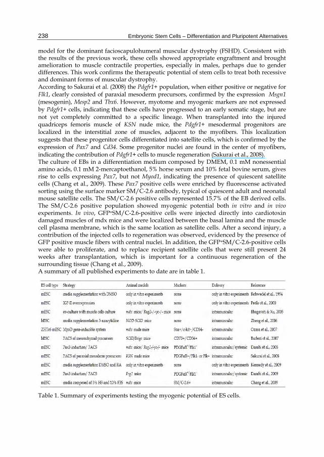
model for the dominant facioscapulohumeral muscular dystrophy (FSHD). Consistent with the results of the previous work, these cells showed appropriate engraftment and brought amelioration to muscle contractile properties, especially in males, perhaps due to gender differences. This work confirms the therapeutic potential of stem cells to treat both recessive and dominant forms of muscular dystrophy. According to Sakurai et al. (2008) the Pdgfr1+ population, when either positive or negative for Flk1, clearly consisted of paraxial mesoderm precursors, confirmed by the expression Msgn1 (mesogenin), Mesp2 and Tbx6. However, myotome and myogenic markers are not expressed by Pdgfr1+ cells, indicating that these cells have progressed to an early somatic stage, but are not yet completely committed to a specific lineage. When transplanted into the injured quadriceps femoris muscle of KSN nude mice, the Pdgfr1+ mesodermal progenitors are localized in the interstitial zone of muscles, adjacent to the myofibers. This localization suggests that these progenitor cells differentiated into satellite cells, which is confirmed by the expression of Pax7 and Cd34. Some progenitor nuclei are found in the center of myofibers, indicating the contribution of Pdgfr1+ cells to muscle regeneration (Sakurai et al., 2008). The culture of EBs in a differentiation medium composed by DMEM, 0.1 mM nonessential amino acids, 0.1 mM 2-mercaptoethanol, 5% horse serum and 10% fetal bovine serum, gives rise to cells expressing Pax7, but not Myod1, indicating the presence of quiescent satellite cells (Chang et al., 2009). These Pax7 positive cells were enriched by fluorescense activated sorting using the surface marker SM/C-2.6 antibody, typical of quiescent adult and neonatal mouse satellite cells. The SM/C-2.6 positive cells represented 15.7% of the EB derived cells. The SM/C-2.6 positive population showed myogenic potential both in vitro and in vivo experiments. In vivo, GFP+SM/C-2.6-positive cells were injected directly into cardiotoxin damaged muscles of mdx mice and were localized between the basal lamina and the muscle cell plasma membrane, which is the same location as satellite cells. After a second injury, a contribution of the injected cells to regeneration was observed, evidenced by the presence of GFP positive muscle fibers with central nuclei. In addition, the GFP+SM/C-2.6-positive cells were able to proliferate, and to replace recipient satellite cells that were still present 24 weeks after transplantation, which is important for a continuous regeneration of the surrounding tissue (Chang et al., 2009). A summary of all published experiments to date are in table 1.
Table 1. Summary of experiments testing the myogenic potential of ES cells.

Myogenic Differentiation of ES Cells for Therapies in Neuromuscular Diseases: Progress to Date 239
5. Conclusion
Several attempts to achieve appropriate differentiation of ES cells and implantation into model organisms have been tested with mixed results. The differentiation into the muscular lineage could be successfully obtained in vitro, as evidenced by the expression of myogenic markers. However, despite attempts to direct in vivo differentiation, to date few reports have documented successful long term therapeutic results using these procedures. Strategies, such as selecting subpopulations of undifferentiated or partially differentiated embryonic stem cells before any kind of implantation treatment is undertaken, are emerging and should result in more homogenous cell populations and, diminish the frequency of maturation of stem cells into undesired cell types. In fact, the combination of several approaches, including cell sorting, genetic modification, and prior in vitro induction will probably be necessary to obtain successful therapeutic outcomes with ES cells.
6. Acknowledgment
The authors would like to thank the following researchers, for scientific and technical support: Dr. Mayana Zatz, Dr. Lydia Yamamoto, Dr. Lygia da Veiga Pereira. The authors also thank Dr P. L. Pearson, a visiting professor to Brazil for his contributions to both the grammar and scientific content of this work. This work was supported by Fundação de Amparo a Pesquisa do Estado de São Paulo - Centro de Pesquisa, Inovação e Difusão (FAPESP-CEPID), Conselho Nacional de Desenvolvimento Científico e Tecnológico (CNPq), and Instituto Nacional de Ciencia e Tecnologia (INCT), FINEP, and Associação Brasileira de Distrofia Muscular (ABDIM).
7. References
Barberi, T.; Bradbury, M.; Dincer, Z.; Panagiotakus, G.; Socci, N. D. & Studer, L. (2007). Derivation of engraftable skeletal myoblasts from human embryonic stem cells. Nature Medicine, Vol.13, No.5, (May, 2007), pp.642-648
Bhagavati, S. & Xu, W. (2005). Generation of skeletal muscle from transplanted embryonic stem cells in dystrophic mice. Biochemical and Biophysical Research Communications, Vol.333, No.2, (June, 2005), pp.644-649
Bittner, R. E.; Anderson, L. V.; Burkhardt, E.; Bashir, R.; Vafiadaki, E.; Ivanova, S.; Raffelsberger, T.; Maerk, I.; Höger, H.; Jung, M.; Karbasiyan, M.; Storch, M.; Lassmann, H.; Moss, J. A.; Davison, K.; Harrison, R.; Bushby, K.M. & Reis, A. (1999). Dysferlin deletion in SJL mice (SJL-Dysf) defines a natural model for limb girdle muscular dystrophy 2B. Nature Genetics, Vol.23, No.2, (October, 1999), pp.141-142
Blomhoff, R. & Blomhoff, H.F. (2006). Overview of retinoid metabolism and function. Journal of Neurobiology, Vol.66, No.7, (June, 2006), pp.606-630
Brand-Saberi, B. & Christ, B. (1999). Genetic and epigenetic control of muscle development in vertebrates. Cell Tissue Research, Vol.296, No.1, (April, 1999), pp.199-212
Bryson-Richardson, R.J. & Currie, P. D. (2008). The genetics of vertebrate myogenesis. Nature Reviews Genetics, Vol.9, No.8, (August, 2008), pp.632-646

Embryonic Stem Cells – Differentiation and Pluripotent Alternatives 240
Buckingham, M. (2006). Myogenic progenitor cells and skeletal myogenesis in vertebrates. Current Opinion in Genetics & Development, Vol.16, No.5, (October, 2006), pp. 525-532
Buckingham, M. (2001). Skeletal muscle formation in vertebrates. Current Opinion in Genetics & Development, Vol.11, No.4, (August, 2001), pp.440-448
Bulfield, G.; Siller W. G.; Wight, P. A. & Moore, K. J. (1984). X Chromosome-Linked Muscular Dystrophy (mdx) in the Mouse. Proceedings of the National Academy of Sciences, Vol.81, No.4, (February, 1984), pp.1189-1192
Chang, H.; Yoshimoto, M.; Umeda, K.; Iwasa, T.; Mizuno, Y.; Fukada, S.; Yamamoto, H.; Motobashi, N.; Suzuki, Y. M.; Takeda, S.; Heike, T. & Nakahata, T. (2009). Generation of transplantable, functional satellite-like cells from mouse embryonic stem cells. The FASEB Journal, Vol.23, No.6, (June, 2009), pp.1907-1919
Darabi, R.; Gehlbach, K.; Bachoo, R. M.; Kamath, S.; Osawa, M.; Kamm, E. K.; Kyba, M. & Perlingeiro, R. C. R. (2008). Functional skeletal muscle regeneration from differentiating embryonic stem cells. Nature Medicine, Vol.14, No.2, (February, 2008), pp.134-143
Darabi, R.; Baik, J.; Clee, M.; Kyba, M.; Tupler, R. & Perlingeiro, R. C. R. (2009). Engraftment of embryonic stem cell-derived myogenic progenitors in a dominant model of muscular dystrophy. Experimental Neurology, Vol.220, No.1, (August, 2009), pp.212-216
Emery, A. E. H. (2002). The muscular dystrophies. The Lancet, Vol.359, No.9307, (February, 2002), pp.687-695
Ervasti, J. M. & Campbell, K. P. (1993). A role for the dystrophin-glycoprotein complex as a transmembrane linker between laminin and actin. Journal of Cell Biology, Vol.122, No.4, (August, 1993), pp.809-823
Ferrari, G.; Cusella-De Angelis, G.; Coletta, M.; Paolucci, E.; Stornaiuolo, A.; Cossu, G. & Mavilio, F. (1998). Muscle regeneration by bone marrow-derived myogenic progenitors. Science, Vol.279, No.5356, (March, 1998), pp.1528-1530
Grivennikov, I. A. (2008). Embryonic stem cells and the problem of directed differentiation. Biochemistry (Moscow), Vol.73, No.13, (February, 2008), pp.1438-1452
Gussoni, E.; Soneoka, Y.; Strickland, C. D.; Buzney, E. A.; Khan, M. K.; Flint, A. F.; Kunkel, L. M. & Mulligan R. C. (1999). Dystrophin expression in the mdx mouse restored by stem cell transplantation. Nature, Vol.401, No.6751, (September, 1999), pp.390-394
Hawke, T.J. & Garry, D.J. (2001). Myogenic satellite cells: physiology to molecular biology. Journal of Applied Physiology, Vol.91, No.2, (August, 2001), pp.534-551
Hoffman, E. P.; Brown, R. H. Jr & Kunkel, L. M. (1987). Dystrophin: the protein product of the Duchenne muscular dystrophy locus. Cell, Vol.51, No.6, (December, 1987), pp.919-928
Kennedy, K. A. M.; Porter, T.; Mehta, V.; Ryan, S. D.; Price, F.; Peshdary, V.; Karamboulas, C.; Savage, J.; Drysdale, T. A.; Li, S.; Bennett, S. A. L. & Skerjanc, I. L. (2009). Retinoic acid enhances skeletal muscle progenitor formation and bypasses inhibition by bone morphogenetic protein 4 but not dominant negative β-catenin. BMC Biology, Vol.7, (October, 2009), pp.67-88
Lee, J. Y, Qu-Petersen, Z.; Cao, B. ; Kimura, S. ; Jankowski, R. ; Cummins, J. ; Usas, A. ; Gates, C. ; Robbins, P. ; Wernig, A. & Huard, J. (2000). Clonal isolation of muscle-derived

Myogenic Differentiation of ES Cells for Therapies in Neuromuscular Diseases: Progress to Date 241
cells capable of enhancing muscle regeneration and bone healing. Journal of Cell Biology, Vol.150, No.5, (September, 2000), pp.1085-1100
Muntoni, F.; Brockington, M.; Torelli, S. & Brown, S. C. (2004). Defective glycosylation in congenital muscular dystrophies. Current Opinion in Neurology, Vol.17, No.2, (April, 2004), pp.205-209
Naya, F. S. & Olson, E. (1999). MEF2: a transcriptional target for signaling pathways controlling skeletal muscle growth and differentiation. Current Opinion in Cell Biology, Vol.11, No.6, (December, 1999), pp.683-688
Ozasa, S.; Kimura, S.; Ito, K.; Ueno, H.; Ikezawa, M.; Matsukura, M.; Yoshioka, K.; Araki, K.; Yamamura, K.; Abe, K.; Niwa, H. & Miike, T. (2007). Efficient conversion of ES cells into myogenic lineage using the gene-inducible system. Biochemical and Biophysical Research Communications, Vol.357, No.4, (April, 2007), pp.957-963
Parker, M. H.; Seale, P. & Rudnicki, M.A. (2003). Looking back to the embryo: defining transcriptional networks in adult myogenesis. Nature Reviews Genetics, Vol.4, No.7, (July 2003), pp. 495-507
Patel, K. & Amthor, H. (2005). The function of Myostatin and strategies of Myostatin blockade - new hope for therapies aimed at promoting growth of skeletal muscle. Neuromuscular Disorders, Vol.15, No.2, (February, 2005), pp.117-126
Prelle, K.; Wobus, A. M.; Krebs, O.; Blum, W. F. & Wolf, E. (2000). Overexpression of insulin-like growth factor-II in mouse embryonic stem cells promotes myogenic differentiation. Biochemical and Biophysical Research Communications, Vol.277, No.3, (September, 2000), pp.631-638
Relaix, F. & Marcelle, C. (2009). Muscle stem cells. Current Opinion in Cell Biology, Vol.21, No.6, (December, 2009), pp.748-753
Rohwedel, J.; Maltsev, V.; Bober, E.; Arnold, H. H.; Hescheler, J. & Wobus, A. M. (1994). Muscle cell differentiation of embryonic stem cells reflects myogenesis in vivo: developmentally regulated expression of myogenic determination genes and functional expression of ionic currents. Developmental Biology, Vol.164, No.1, (February, 1994), pp.87-101
Sakurai, H.; Okawa, Y.; Inami, Y.; Nishio, N. & Isobe, K. (2008). Paraxial mesodermal progenitors derived from mouse embryonic stem cells contribute to muscle regeneration via differentiation into muscle satellite cells. Stem Cells, Vol.26, No.7, (May, 2008), pp.1865-1873
Straub, V. & Campbell, K. P. (1997). Muscular dystrophies and the dystrophin-glycoprotein complex. Current Opinion in Neurology, Vol.10, No.2, (April, 1997), pp.168-175
Straub, V.; Duclos, F.; Venzke, D. P.; Lee, J. C.; Cutshall, S.; Leveille, C. J. & Campbell, K. P. (1998). Molecular pathogenesis of muscle degeneration in the delta-sarcoglycan-deficient hamster. American Journal of Pathology, Vol.153, No.5, (November, 1998), pp.1623-1630
Tome, F. M.; Evangelista, T.; Leclerc, A.; Sunada, Y.; Manole, E., Estournet, B.; Barois, A.; Campbell, K. P. & Fardeau, M. (1994). Congenital muscular dystrophy with merosin deficiency. Comptes Rendus De l’Academie des Sciences III, Vol.317, No.4, (April, 1994), pp.351-357
Torrente, Y.; Tremblay, J. P.; Pisati, F.; Belicchi, M.; Rossi, B.; Sironi, M.; Fortunato, F.; El Fahime, M.; D’Angelo, M. G.; Caron, N. J.; Constantin, G.; Paulin, D.; Scarlato, G. & Bresolin, N. (2001). Intraarterial injection of muscle-derived CD34(+) Sca-1(+) stem

Embryonic Stem Cells – Differentiation and Pluripotent Alternatives 242
cells restores dystrophin in mdx mice. Journal of Cell Biology, Vol.152, No.2, (January, 2001), pp.335-348
Vainzof, M.; Passos-Bueno, M.R. & Zatz, M. (2003). Immunological methods for the analysis of protein expression in neuromuscular diseases. Methods Molecular Biology, Vol.217, pp.355–378.
Vainzof, M. & Zatz, M. (2003).Protein defects in neuromuscular diseases. Brazilian Journal of Medical and Biological Research, Vol.36, No.5, (May, 2003), pp.543-555
Vainzof, M.; Ayub-Guerrieri, D.; Onofre, P.C.G.; Martins, P.C.M.; Lopes, V.F.; Zilberztajn, D.; Maia, L.S.; Sell, K. & Yamamoto, L.U. (2008). Animal Models for genetic neuromuscular diseases. Journal of Molecular Neuroscience, Vol.34, No.3, (March, 2008), pp.241-248
Wobus, A. M.; Guan, K.; Yang, H. T. & Boheler, K. R. (2002). Embryonic stem cells as a model to study cardiac, skeletal muscle and vascular smooth muscle cell differentiation. Methods in Molecular Biology, Vol.185, pp.127-156
Yang, T.; Riehl, J.; Esteve, E.; Matthael, K. I.; Goth, S.; Allen, P. D.; Pessah, I. N. & Lopez, J. R. (2006). Pharmacologic and functional characterization of malignant hyperthermia in the R163C RyR1 knock-in mouse. Anestesiology, Vol.105, No.6, (December, 2006),pp.1164-1175
Yoshida, M. & Ozawa, E. (1990). Glycoprotein complex anchoring dystrophin to sarcolemma. Journal of Biochemistry, Vol.108, No.5, (November, 1990), pp.748-752
Zheng, J. K.; Wang, Y.; Karandikar, A.; Wang, Q.; Gai, H.; Liu, A. L.; Peng, C. & Sheng, H. Z. (2006). Skeletal myogenesis by human embryonic stem cells. Cell Research, Vol.16, No.8, (August, 2006), pp.713-722
Related Documents