Mycobacterium tuberculosis Characteristics Mycobacterium tuberculosis is a rod-shaped (bacillus) bacterium that causes the disease tuberculosis in humans, as well as other primates, hamsters, dogs, and guinea pigs (Figure 1). Since the organism is non-motile, it travels through the air on particles called droplet nuclei. Droplet nuclei, which range in size from 1 to 5 μm , are introduced into the air when an infected person sneezes, coughs, etc. Normal air currents keep the particles airborne so that they can spread throughout an area. (See the “Pathogenesis” section for information on how an infection progresses.) In addition to being non-motile, M. tuberculosis is an obligate aerobe, meaning that the bacterium can only survive in an environment that contains oxygen. (See the “Pathogenesis” section to learn more about the environment in which M. tuberculosis lives.) Figure 1. Scanning electron micrograph of Mycobacterium tuberculosis bacilli. Although Mycobacterium tuberculosis possesses a Gram-positive type cell wall, a cell wall with extensive peptidoglycan and no outer membrane, the bacterium does not stain with Gram stain reagents. Gram stain reagents are unable to penetrate the cell wall of the bacillus because layers of lipids surround the peptidoglycan in mycobacterium. Unlike most Gram-negative bacteria, which have a 5-20% lipid content by weight, M. tuberculosis and other mycobacterium are composed of up to 60% lipids. Many of these lipids are in the form of mycolic acids. (See the “Adaptations” section for more on the importance of high lipid content.) Since the Gram stain method proves ineffective on Mycobacterium tuberculosis , acid-fast staining must be used to make the bacilli visible under a microscope. In the Ziehl-Neelsen procedure, bacteria from a sputum (mucus coughed up from the lungs) sample are flooded with a basic solution of carbolfuchsin, a magenta dye. After heating the slide in a flame, the sample is washed with water and treated with acid-alcohol to decolorize the bacteria. Then, a counter stain of methylene blue is applied to the sample. When the process is complete, M. tuberculosis bacilli

Welcome message from author
This document is posted to help you gain knowledge. Please leave a comment to let me know what you think about it! Share it to your friends and learn new things together.
Transcript
8/3/2019 Mycobacterium Tuberculosis Official)
http://slidepdf.com/reader/full/mycobacterium-tuberculosis-official 1/4
Mycobacterium tuberculosis
Characteristics
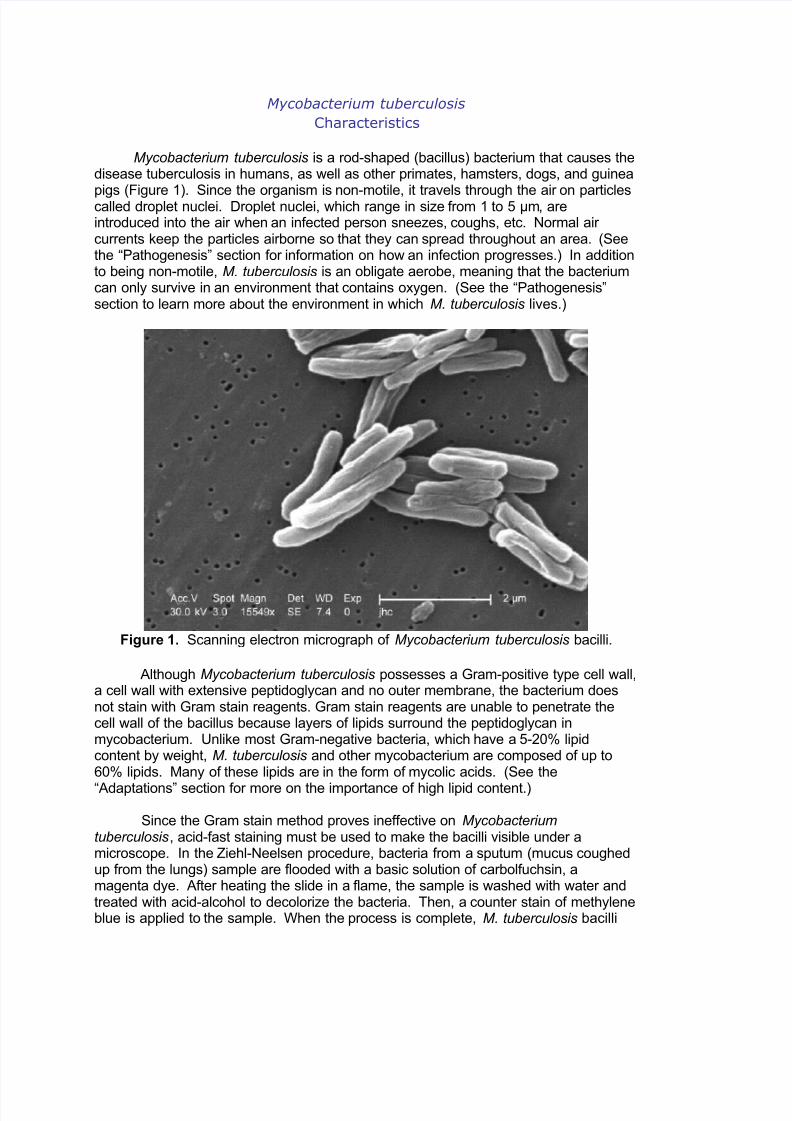
Mycobacterium tuberculosis is a rod-shaped (bacillus) bacterium that causes thedisease tuberculosis in humans, as well as other primates, hamsters, dogs, and guinea
pigs (Figure 1). Since the organism is non-motile, it travels through the air on particlescalled droplet nuclei. Droplet nuclei, which range in size from 1 to 5 μm, areintroduced into the air when an infected person sneezes, coughs, etc. Normal air currents keep the particles airborne so that they can spread throughout an area. (Seethe “Pathogenesis” section for information on how an infection progresses.) In additionto being non-motile, M. tuberculosis is an obligate aerobe, meaning that the bacteriumcan only survive in an environment that contains oxygen. (See the “Pathogenesis”section to learn more about the environment in which M. tuberculosis lives.)
Figure 1. Scanning electron micrograph of Mycobacterium tuberculosis bacilli.
Although Mycobacterium tuberculosis possesses a Gram-positive type cell wall,a cell wall with extensive peptidoglycan and no outer membrane, the bacterium doesnot stain with Gram stain reagents. Gram stain reagents are unable to penetrate thecell wall of the bacillus because layers of lipids surround the peptidoglycan inmycobacterium. Unlike most Gram-negative bacteria, which have a 5-20% lipidcontent by weight, M. tuberculosis and other mycobacterium are composed of up to60% lipids. Many of these lipids are in the form of mycolic acids. (See the
“Adaptations” section for more on the importance of high lipid content.)
Since the Gram stain method proves ineffective on Mycobacteriumtuberculosis, acid-fast staining must be used to make the bacilli visible under amicroscope. In the Ziehl-Neelsen procedure, bacteria from a sputum (mucus coughedup from the lungs) sample are flooded with a basic solution of carbolfuchsin, amagenta dye. After heating the slide in a flame, the sample is washed with water andtreated with acid-alcohol to decolorize the bacteria. Then, a counter stain of methyleneblue is applied to the sample. When the process is complete, M. tuberculosis bacilli
8/3/2019 Mycobacterium Tuberculosis Official)
http://slidepdf.com/reader/full/mycobacterium-tuberculosis-official 2/4
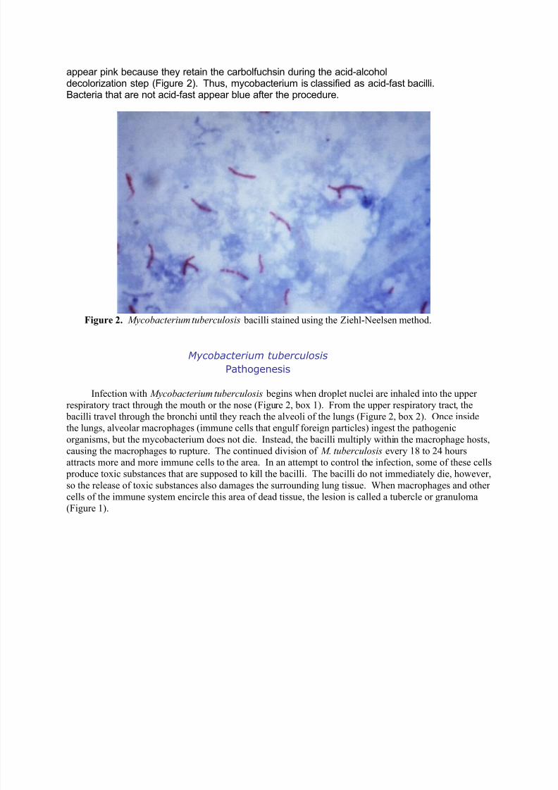
appear pink because they retain the carbolfuchsin during the acid-alcoholdecolorization step (Figure 2). Thus, mycobacterium is classified as acid-fast bacilli.Bacteria that are not acid-fast appear blue after the procedure.
Figure 2. Mycobacterium tuberculosis bacilli stained using the Ziehl-Neelsen method.
Mycobacterium tuberculosis
Pathogenesis
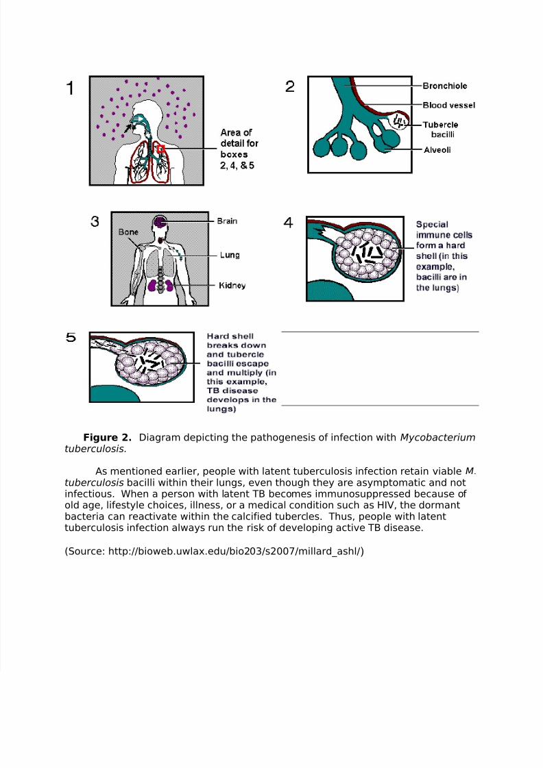
Infection with Mycobacterium tuberculosis begins when droplet nuclei are inhaled into the upper
respiratory tract through the mouth or the nose (Figure 2, box 1). From the upper respiratory tract, the
bacilli travel through the bronchi until they reach the alveoli of the lungs (Figure 2, box 2). Once insidethe lungs, alveolar macrophages (immune cells that engulf foreign particles) ingest the pathogenic
organisms, but the mycobacterium does not die. Instead, the bacilli multiply within the macrophage hosts,
causing the macrophages to rupture. The continued division of M. tuberculosis every 18 to 24 hours
attracts more and more immune cells to the area. In an attempt to control the infection, some of these cells
produce toxic substances that are supposed to kill the bacilli. The bacilli do not immediately die, however,
so the release of toxic substances also damages the surrounding lung tissue. When macrophages and other
cells of the immune system encircle this area of dead tissue, the lesion is called a tubercle or granuloma
(Figure 1).

8/3/2019 Mycobacterium Tuberculosis Official)
http://slidepdf.com/reader/full/mycobacterium-tuberculosis-official 3/4
Figure 1. Mycobacterium tuberculosis bacilli visible within granuloma. This tissue sample wastaken from the endometrial layer of the uterus. This is an example of tuberculosis diseasepersisting in an area of the body outside of the lungs.
The interior of a tubercle consists of a gelatinous mass of host cells and bacilli that givesthe damaged tissue a cheese-like consistency. Therefore, this type of tissue death is referred toas cessation necrosis. If the immune system is successful in preventing the M.
tuberculosis bacilli from multiplying further, the caseous tubercles become walled-off andcalcified (Figure 2, box 4). Although calcified lesions still contain viable bacteria, the bacteriacannot be spread to other individuals. When Mycobacterium tuberculosis lies dormant in thelungs, a person is said to have a latent tuberculosis infection. Such persons, who represent 85-95% of infected individuals, show no overt symptoms of disease.
In 5-15% of infected individuals, the immune system fails to prevent the infection fromprogressing and the interiors of the caseous lesions become liquefied. Liquefaction allowsviable M. tuberculosis bacilli to spill out of the tubercles, leaving behind a cavity in the lungs(Figure 2, box 5). When these bacilli infect lower portions of the lungs or enter the bronchi theresult is an active case of pulmonary tuberculosis disease. People with pulmonary tuberculosisare capable of spreading the disease to others through the bacteria in their sputum. They also
manifest symptoms such as weight loss, weakness, night sweats, chest pain, and coughing upblood. If viable bacilli enter the bloodstream, M. tuberculosis can travel to organs of the bodyoutside of the lungs (Figure 1, above and Figure 2, box 3). Known as extra-pulmonarytuberculosis, this form of the disease is rarely contagious.
8/3/2019 Mycobacterium Tuberculosis Official)
http://slidepdf.com/reader/full/mycobacterium-tuberculosis-official 4/4
Figure 2. Diagram depicting the pathogenesis of infection with Mycobacterium
tuberculosis.
As mentioned earlier, people with latent tuberculosis infection retain viableM.tuberculosis bacilli within their lungs, even though they are asymptomatic and notinfectious. When a person with latent TB becomes immunosuppressed because of old age, lifestyle choices, illness, or a medical condition such as HIV, the dormantbacteria can reactivate within the calcified tubercles. Thus, people with latenttuberculosis infection always run the risk of developing active TB disease.
(Source: http://bioweb.uwlax.edu/bio203/s2007/millard_ashl/)
Related Documents


![[Micro] mycobacterium tuberculosis](https://static.cupdf.com/doc/110x72/55d6fc67bb61ebfa2a8b47ea/micro-mycobacterium-tuberculosis.jpg)