mSin3A corepressor regulates diverse transcriptional networks governing normal and neoplastic growth and survival Jan-Hermen Dannenberg, 1,4 Gregory David, 1,3,4 Sheng Zhong, 2 Jaco van der Torre, 1 Wing H. Wong, 2 and Ronald A. DePinho 1,5 1 Department of Medical Oncology, Dana Farber Cancer Institute; Departments of Medicine and Genetics, Harvard Medical School, Boston, Massachusetts 02115, USA; 2 Department of Statistics, Stanford University, Stanford, California 94305, USA mSin3A is a core component of a large multiprotein corepressor complex with associated histone deacetylase (HDAC) enzymatic activity. Physical interactions of mSin3A with many sequence-specific transcription factors has linked the mSin3A corepressor complex to the regulation of diverse signaling pathways and associated biological processes. To dissect the complex nature of mSin3A’s actions, we monitored the impact of conditional mSin3A deletion on the developmental, cell biological, and transcriptional levels. mSin3A was shown to play an essential role in early embryonic development and in the proliferation and survival of primary, immortalized, and transformed cells. Genetic and biochemical analyses established a role for mSin3A/HDAC in p53 deacetylation and activation, although genetic deletion of p53 was not sufficient to attenuate the mSin3A null cell lethal phenotype. Consistent with mSin3A’s broad biological activities beyond regulation of the p53 pathway, time-course gene expression profiling following mSin3A deletion revealed deregulation of genes involved in cell cycle regulation, DNA replication, DNA repair, apoptosis, chromatin modifications, and mitochondrial metabolism. Computational analysis of the mSin3A transcriptome using a knowledge-based database revealed several nodal points through which mSin3A influences gene expression, including the Myc-Mad, E2F, and p53 transcriptional networks. Further validation of these nodes derived from in silico promoter analysis showing enrichment for Myc-Mad, E2F, and p53 cis-regulatory elements in regulatory regions of up-regulated genes following mSin3A depletion. Significantly, in silico promoter analyses also revealed specific cis-regulatory elements binding the transcriptional activator Stat and the ISWI ATP-dependent nucleosome remodeling factor Falz, thereby expanding further the mSin3A network of regulatory factors. Together, these integrated genetic, biochemical, and computational studies demonstrate the involvement of mSin3A in the regulation of diverse pathways governing many aspects of normal and neoplastic growth and survival and provide an experimental framework for the analysis of essential genes with diverse biological functions. [Keywords: Histone modifications; knock-out; mSin3 complex; mSin3A; transcriptional regulation; tumorigenesis] Supplemental material is available at http://www.genesdev.org. Received December 6, 2004; revised version accepted May 20, 2005. Chromatin dynamics impact virtually all known biologi- cal processes governing normal mammalian develop- ment and the growth and survival of primary and cancer cells. Histones are the central structural elements of chromatin, and their N-terminal tails are subject to co- valent modifications by the opposing actions of histone acetyltransferases (HATs) and histone deacetylases (HDACs) as well as other enzymatic activities. This re- versible acetylation, along with other histone modifica- tions, alters either focal or global chromatin domains and thereby influences the activation or repression of gene transcription, activities of the repair DNA machin- ery, formation of structures required for proper chromo- some segregation among other biological processes (for review, see Fischle et al. 2003 and references therein). 3 Present address: Department of Pharmacology, New York University, School of Medicine, New York, NY, 10016 USA. 4 These authors equally contributed to this work. 5 Corresponding author. E-MAIL [email protected]; FAX (617) 632-6069. Article and publication are at http://www.genesdev.org/cgi/doi/10.1101/ gad.1286905. GENES & DEVELOPMENT 19:1581–1595 © 2005 by Cold Spring Harbor Laboratory Press ISSN 0890-9369/05; www.genesdev.org 1581

Welcome message from author
This document is posted to help you gain knowledge. Please leave a comment to let me know what you think about it! Share it to your friends and learn new things together.
Transcript
mSin3A corepressor regulates diversetranscriptional networks governing normaland neoplastic growthand survivalJan-Hermen Dannenberg,1,4 Gregory David,1,3,4 Sheng Zhong,2 Jaco van der Torre,1 Wing H. Wong,2
and Ronald A. DePinho1,5
1Department of Medical Oncology, Dana Farber Cancer Institute; Departments of Medicine and Genetics, Harvard MedicalSchool, Boston, Massachusetts 02115, USA; 2Department of Statistics, Stanford University, Stanford, California 94305, USA
mSin3A is a core component of a large multiprotein corepressor complex with associated histone deacetylase(HDAC) enzymatic activity. Physical interactions of mSin3A with many sequence-specific transcriptionfactors has linked the mSin3A corepressor complex to the regulation of diverse signaling pathways andassociated biological processes. To dissect the complex nature of mSin3A’s actions, we monitored the impactof conditional mSin3A deletion on the developmental, cell biological, and transcriptional levels. mSin3A wasshown to play an essential role in early embryonic development and in the proliferation and survival ofprimary, immortalized, and transformed cells. Genetic and biochemical analyses established a role formSin3A/HDAC in p53 deacetylation and activation, although genetic deletion of p53 was not sufficient toattenuate the mSin3A null cell lethal phenotype. Consistent with mSin3A’s broad biological activities beyondregulation of the p53 pathway, time-course gene expression profiling following mSin3A deletion revealedderegulation of genes involved in cell cycle regulation, DNA replication, DNA repair, apoptosis, chromatinmodifications, and mitochondrial metabolism. Computational analysis of the mSin3A transcriptome using aknowledge-based database revealed several nodal points through which mSin3A influences gene expression,including the Myc-Mad, E2F, and p53 transcriptional networks. Further validation of these nodes derived fromin silico promoter analysis showing enrichment for Myc-Mad, E2F, and p53 cis-regulatory elements inregulatory regions of up-regulated genes following mSin3A depletion. Significantly, in silico promoter analysesalso revealed specific cis-regulatory elements binding the transcriptional activator Stat and the ISWIATP-dependent nucleosome remodeling factor Falz, thereby expanding further the mSin3A network ofregulatory factors. Together, these integrated genetic, biochemical, and computational studies demonstrate theinvolvement of mSin3A in the regulation of diverse pathways governing many aspects of normal andneoplastic growth and survival and provide an experimental framework for the analysis of essential genes withdiverse biological functions.
[Keywords: Histone modifications; knock-out; mSin3 complex; mSin3A; transcriptional regulation;tumorigenesis]
Supplemental material is available at http://www.genesdev.org.
Received December 6, 2004; revised version accepted May 20, 2005.
Chromatin dynamics impact virtually all known biologi-cal processes governing normal mammalian develop-ment and the growth and survival of primary and cancercells. Histones are the central structural elements of
chromatin, and their N-terminal tails are subject to co-valent modifications by the opposing actions of histoneacetyltransferases (HATs) and histone deacetylases(HDACs) as well as other enzymatic activities. This re-versible acetylation, along with other histone modifica-tions, alters either focal or global chromatin domainsand thereby influences the activation or repression ofgene transcription, activities of the repair DNA machin-ery, formation of structures required for proper chromo-some segregation among other biological processes (forreview, see Fischle et al. 2003 and references therein).
3Present address: Department of Pharmacology, New York University,School of Medicine, New York, NY, 10016 USA.4These authors equally contributed to this work.5Corresponding author.E-MAIL [email protected]; FAX (617) 632-6069.Article and publication are at http://www.genesdev.org/cgi/doi/10.1101/gad.1286905.
GENES & DEVELOPMENT 19:1581–1595 © 2005 by Cold Spring Harbor Laboratory Press ISSN 0890-9369/05; www.genesdev.org 1581
These biological processes are affected through his-tone-modifying enzymes whose activities are targeted tospecific sites on the DNA via their association with co-repression scaffolding molecules that can be recruited bysequence-specific binding proteins such as transcriptionfactors. The mammalian Sin3 proteins, mSin3A andmSin3B, represent prototypical corepressor moleculesthat were discovered initially through their direct inter-action with the sequence-specific transcriptional repres-sors of the Myc family network, Mad1 and Mxi1 (Ayer etal. 1995; Schreiber-Agus et al. 1995; Laherty et al. 1997)and later shown to associate with HDAC1 and HDAC2within a large multiprotein complex, the Sin3/HDACcorepressor complex (for review, see Schreiber-Agus andDePinho 1998; Ayer 1999; Knoepfer and Eisenman 1999;Silverstein and Ekwall 2004). Subsequent efforts havealso revealed that the mSin3/HDAC proteins are re-cruited by an expanding array of factors involved prima-rily in transcriptional regulation including p53 (Murphyet al. 1999), Ikaros (Koipally et al. 1999), REST (Huang etal. 1999), and E2F4 (Rayman et al. 2002), contributing torepression of their target genes.
mSin3A and mSin3B proteins share extensive similar-ity within their four paired amphipathic helix (PAH) pro-tein-interaction domains. The presence of two highly re-lated mSin3 homologs in mammals and other species(Dang et al. 1999; Silverstein and Ekwall 2004), suggeststhat each mSin3 homolog serves separable functions.The mSin3 corepressor complex’s central enzymaticactivity is imparted through its Class I HDAC constitu-ents, HDAC1 and HDAC2. The maintenance of mSin3-associated HDAC activity is dependent on another inte-gral component, mSds3, whose ortholog in Saccharo-mycese cerevisiae is epistatic to the yeast sin3p andHDAC orthologs (Vannier et al. 1996; Alland et al. 2002).Additional core mSin3/HDAC proteins include the reti-noblastoma-associated proteins, RbAp46 and RbAp48,which are thought to stabilize interactions with themSin3 complex and the nucleosome, mSin3-associatedproteins (SAPs: SAP18, SAP30, SAP130, and SAP180),RBP1, p33ING1b, and BRMS1, which have been isolatedthrough biochemical means and remain the focus of on-going functional investigations (for review, see Ayer1999; Kuzmichev et al. 2002; Lai et al. 2001; Skowyra etal. 2001; Fleischer et al. 2003; Meehan et al. 2004).
As initially reported for the Mad family, the mSin3/HDAC complex also drives the repression activity of nu-merous other sequence-specific transcription factorswhose list has expanded considerably in the past fewyears. Among those factors that recruit the mSin3/HDAC complex, several are involved in cell survival andapoptosis, thereby linking this complex activity to can-cer-relevant processes. For instance, tumor-suppressorproteins, including p53 (Murphy et al. 1999), pRb (Brehmet al. 1998; Luo et al. 1998; Magnaghi-Jaulin et al. 1998;Lai et al. 2001), and Menin (Kim et al. 2003) have beenshown to repress their target genes through interactionwith the mSin3/HDAC complex. Moreover, aberrant re-cruitment of this complex by altered transcription fac-tors has been shown to be pathogenetic in several human
cancers, such as Acute Promyelocytic Leukemia andAcute Myeloid Leukemia (for review, see Melnick andLicht 2002).
In addition to its role in transcriptional repression,new activities for the mSin3/HDAC complex are con-tinually uncovered in diverse model organisms. For ex-ample, S. cerevisiae sin3p has been shown to participatein the transcriptional activation (as opposed to repres-sion) of MAPK Hog1 target genes upon osmotic stress(De Nadal et al. 2004), and recent links have been forgedbetween S. cerevisiae sin3 and DNA replication (Apari-cio et al. 2004). S. cerevisiae and Caenorhabditis elegansSin3 molecules have also been implicated in the DNAdamage-repair process in a transcription-independentmanner (Pothof et al. 2003; Jazayeri et al. 2004). Finally,several Sin3-associated molecules have been shown toparticipate in activities other than transcriptional regu-lation including a role for the Sin3 complex in centro-mere function and genomic stability in both yeast andmammals. Along these lines, genetic inactivation of Pst1(the homolog of mSin3A in fission yeast) or mSds3 (anessential component of the mSin3/HDAC complex inmouse cells) results in loss of specific chromatin modi-fications at centromeric or pericentromeric loci, and ul-timately, impaired chromosomal segregation (David etal. 2003; Nakayama et al. 2003; Silverstein et al. 2003).
Here, against this backdrop of significant knowledge ofthe mSin3 corepressor complex, we have sought to betterdefine the biological roles of mSin3A in normal mam-malian development, the growth and survival of primaryand cancer cells, and the regulation of transcriptionalnetworks. To this end, a conditional mSin3A knock-outmodel system, biochemical analyses, and computationalmethods were used in an integrated manner to under-stand the biological impact of zygotic and somatic dele-tion of mSin3A in vivo and in cultured primary andtransformed cells.
Results
mSin3A is essential for early embryonic developmentand for cellular proliferation and survival
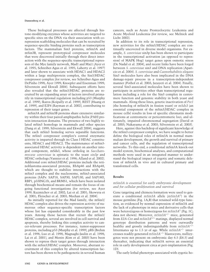
Gene targeting and chimera formation were used to gen-erate a conditional mSin3A allele (mSin3AL) in themouse germline (Fig. 1A,B) that retained wild-type func-tion, as evidenced by normal expression of mSin3A andthe lack of a phenotype in mice and derivative cells thatwere heterozygous or homozygous for mSin3AL (Fig. 1C;data not shown). Moreover, mSin3A+/− mice, generatedfrom E2A-Cre and mSin3AL/+ matings, displayed normalgenotype distribution patterns and were outwardlyhealthy and grossly indistinguishable from mSin3A+/+
littermates up to 1.5 yr of age. While mSin3A+/− inter-crosses readily generated mSin3A−/− blastocysts, nullizy-gotes were not present on embryonic day 6.5 (E6.5) andthereafter, indicating that mSin3A serves an essentialrole in early development circa at peri-implantation (Fig.1D).
The early lethal phenotype associated with zygotic ho-
Dannenberg et al.
1582 GENES & DEVELOPMENT
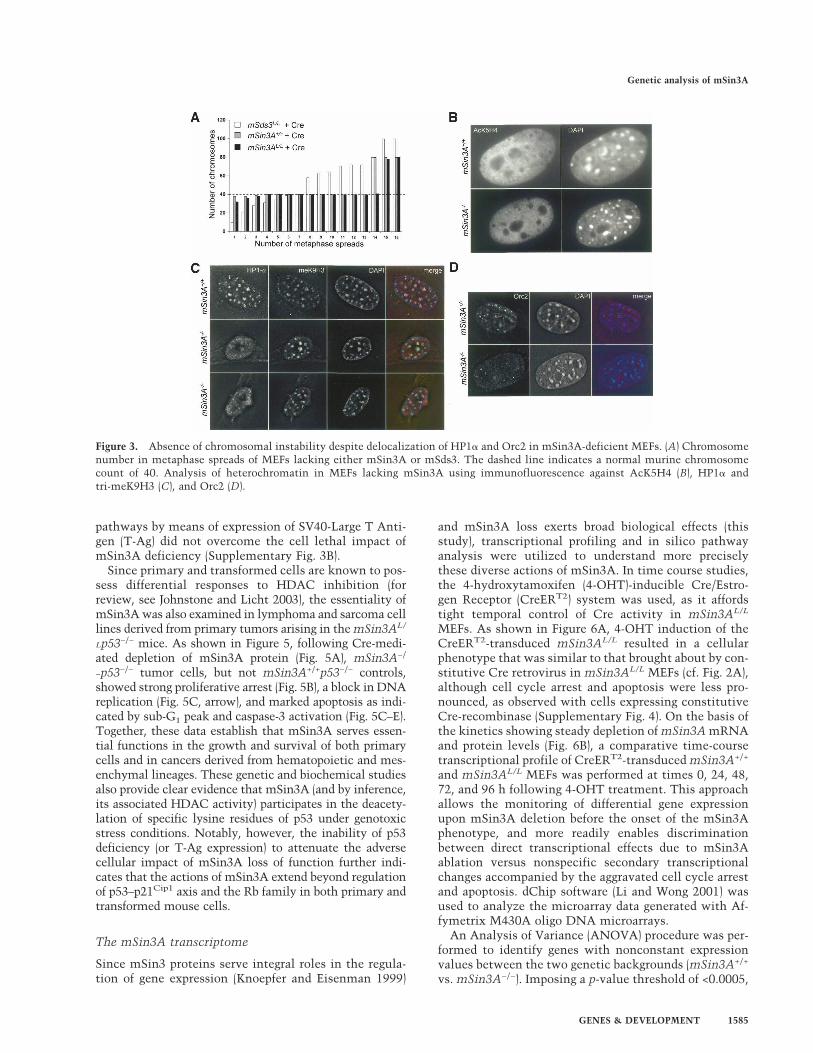
mozygous deletion of mSin3A prompted somatic condi-tional mSin3A deletion studies in primary cell cultures.To that end, Cre-encoding retroviruses were used to in-fect mSin3AL/L mouse embryonic fibroblasts (MEFs), re-sulting in steady and complete depletion of mSin3A pro-tein only in the mSin3AL/L MEFs over a 5-d period (Fig.1C). While the well-recognized modest Cre-induced sta-sis was evident in mSin3A+/+ MEFs, Cre expression inmSin3AL/L MEFs was associated with profound growtharrest and increased apoptosis, as revealed by micro-scopic inspection, TUNEL assay, and by FACS profileswith a sub-G1 peak (Fig. 2; Supplementary Fig. 1). Inaddition to a G2/M arrest, the BrdU-PI FACS profiles ofmSin3A−/− cultures revealed a subpopulation withS-phase DNA content, yet no BrdU incorporation (Fig.2C, arrow)—a profile consistent with unscheduled DNAreplication with subsequent triggering of an S-phasecheckpoint.
To solidify the link between mSin3A deficiency andthe above cellular defects, ectopic expression of wild-type mSin3A, but not an irrelevant gene (luciferase), wasable to eliminate the aforementioned cellular pheno-types associated with deletion of endogenous mSin3A(Supplementary Fig. 2). Finally, Western blot analyses ofmSin3A-deficient cultures showed no appreciable up-regulation of mSin3B or HDAC1 (Fig. 1C), although tran-scriptional up-regulation of several genes encoding othermSin3A-associated factors was detected, suggesting po-tential gene regulatory interactions (see transcriptionalprofiles below). The cell lethal phenotype upon mSin3Adepletion, coupled with continued expression of thestructurally related mSin3B, underscores that mSin3Aserves separable and essential cell physiological func-tions.
Distinctive roles for mSin3A and mSds3in chromosome biology
Previous yeast studies have suggested a strong epistaticrelationship between Sin3 and its associated HDAC and
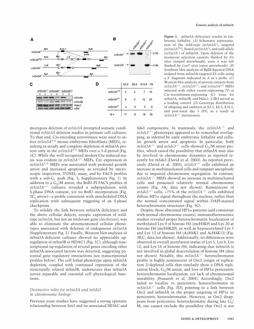
Sds3 components. In mammals, the mSin3A−/− andmSds3−/− phenotypes appeared to be somewhat overlap-ping, as inferred by early embryonic lethality and cellu-lar growth arrest and apoptosis. In particular, bothmSin3A−/− and mSds3−/− cells showed G2/M arrest pro-files, which raised the possibility that mSin3A may alsobe involved in chromosome dynamics as reported re-cently for mSds3 (David et al. 2003). As reported previ-ously (David et al. 2003), mSds3−/− cells exhibited anincrease in multinucleated cells and rampant aneuploidydue to impaired chromosome segregation. In contrast,mSin3A−/− MEFs showed no increase in multinucleatedcells and possessed relatively normal chromosomecounts (Fig. 3A; data not shown). Reminiscent ofmSds3−/− cells, >75% of the mSin3A−/− cells exhibiteddiffuse HP1� signal throughout the nucleus, rather thanthe normal concentrated signal within DAPI-stainedheterochromatin structures (Fig. 3C).
Despite these abnormal HP1� patterns (and consistentwith normal chromosome counts), immunofluorescencestudies revealed proper heterochromatin localization ofmethylated Lys 9 of histone H3 (meH3K9) and Lys 20 athistone H4 (meH4K20), as well as hypoacetylated Lys 5and Lys 12 of histone H4 (AcH4K5 and AcH4K12) (Fig.3B,C; data not shown). Additionally, no differences wereobserved in overall acetylation status of Lys 5, Lys 8, Lys12, and Lys 16 of histone H4, indicating that mSin3A isnot involved in global deacetylation of histone H4 (datanot shown). Notably, this mSin3A−/− heterochromatinprofile is highly reminiscent of Orc2 (origin of replica-tion 2)-depleted cells that similarly show a DNA repli-cation block, G2/M arrest, and loss of HP1� pericentricheterochromatin localization, yet lack of chromosomalinstability (Prasanth et al. 2004). Accordingly, Orc2failed to localize to pericentric heterochromatin inmSin3A−/− cells (Fig. 3D), pointing to a link betweenOrc2 and mSin3A in the proper targeting of HP1� topericentric heterochromatin. However, as Orc2 disap-pears from pericentric heterochromatic during late G2/M, one cannot exclude the possibility that Orc2 is not
Figure 1. mSin3A deficiency results in em-bryonic lethality. (A) Schematic representa-tion of the wild-type (mSin3A+), targeted(mSin3ATgt), floxed (mSin3AL), and null-allele(mSin3A−) of mSin3A. Upon deletion of theneomycin selection cassette flanked by Frtsites (striped arrowheads), exon 4 was leftflanked by LoxP sites (open arrowheads). (B)Southern blot analysis of BglII-digested DNAisolated from mSin3A-targeted ES cells usinga 3� fragment indicated in A as a probe. (C)Western blot analysis of protein extracts frommSin3A+/+, mSin3AL/+, and mSin3AL/L MEFsinfected with either vector-expressing (V) orCre-recombinase-expressing (C) virus formSin3A, mSin3B, and Hdac1. Cdk4 served asa loading control. (D) Genotype distributionof offspring and embryos at E3.5, E6.5, E10.5,and post-natal day 5 (P5), as a result ofmSin3A+/− intercrosses.
Genetic analysis of mSin3A
GENES & DEVELOPMENT 1583

present at pericentric heterochromatin as mSin3A−/−
cells are constrained at the late G2/M phase. In sum-mary, mSin3A and mSds3 deficiencies differentially im-pact on cell cycle kinetics, as evidenced by the specificlink of mSin3A to DNA replication processes and theseminal role of Sds3 in chromosome segregation. Thesedifferences in chromosome dynamics suggest that eithermSds3 functions independently of mSin3A, or thatmSin3A and B homologs serve overlapping functions inthese specific cellular processes.
mSin3A regulation of the p53–p21Cip1 axis
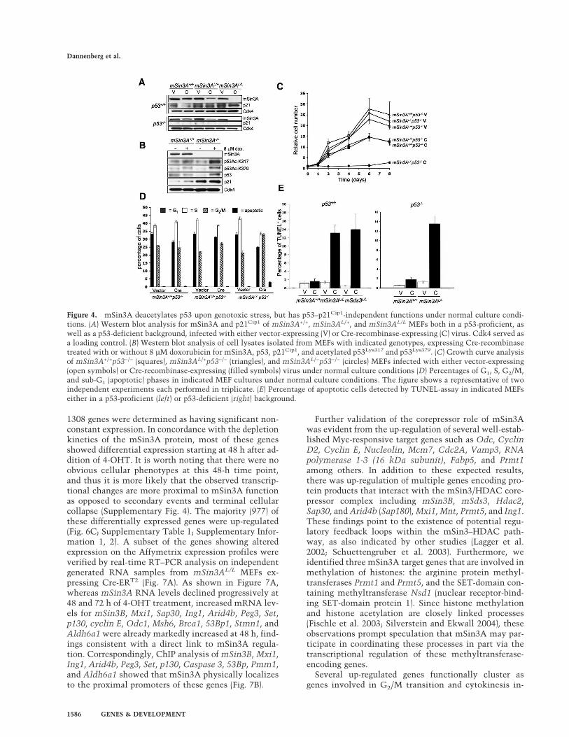
The cell cycle arrest and apoptosis phenotypes ofmSin3A deficiency prompted an in-depth genetic analy-sis of the p53–p21Cip1 pathway, particularly in the lightof previous reports revealing (1) mSin3A–p53 physicalassociation (Murphy et al. 1999), (2) modulation of p53acetylation and transcriptional activity upon genotoxicstress (Sakaguchi et al. 1998), and (3) capacity of HDACinhibitors or genetic deletion of Hdac1 to induce the p53transcriptional target p21Cip1 (Lagger et al. 2002, 2003;for review, see Johnstone and Licht 2003). In line withthese observations, we observed consistent induction ofp21Cip1 expression upon Cre-mediated deletion ofmSin3A (Fig. 4A,B). Indeed, chromatin immunoprecipi-tation (ChIP) experiments revealed the presence of
mSin3A on the promoter of p21Cip1 (data not shown).While mSin3AL/L MEFs infected with Cre retrovirusesshowed no significant additional increase in p21Cip1 pro-tein levels, independent experiments revealed hyper-acetylation of p53 on Lys 317 and Lys 379, upon treat-ment with doxorubicin (Fig. 4B). These data strongly sug-gest that mSin3A regulates the p53–p21Cip1 axis on twolevels; it represses p21Cip1 transcription by regulatingacetylation of histones in the promoter of p21Cip1 andregulates p53 activity via deacetylation upon genotoxicstress.
To substantiate the mSin3A–p53–p21Cip1 inter-relationship on the genetic level, mSin3A+/+p53−/−,mSin3AL/+p53−/− and mSin3AL/−p53−/− MEFs were in-fected with either Cre or control retrovirus, and sub-sequently analyzed for DNA damage-induced p21Cip1
induction and associated effects on proliferation andcell cycle. p21Cip1 induction was found to be robustin mSin3A−/−p53+/+ cells and totally abolished inmSin3A−/−p53−/− cells (Fig. 4A), indicating that p53 isresponsible for genotoxin-induced p21Cip1 up-regulationin the setting of mSin3A deficiency. Despite this, p53deficiency did not alleviate the mSin3A−/− phenotypes ofpoor cell proliferation, reduced S-phase and increasedG2/M fractions, block in DNA replication, and markedapoptosis (Fig. 4C–E; Supplementary Fig. 3A). Addition-ally, functional inactivation of both the p53 and pRb
Figure 2. mSin3A deficiency reduces proliferative capacity in MEFs by increased apoptosis, block in DNA replication, and G2/Marrest. (A) Growth curve analysis of mSin3A+/+ (squares), mSin3AL/+ (triangles), and mSin3AL/L (circles) MEFs infected with eithervector-expressing (open) or Cre-recombinase-expressing (filled) virus. The figure shows a representative of three independent experi-ments, each performed in triplicate. Error bars indicate the standard deviation of the mean. Note that time-point 0 indicates 4 d afterthe beginning of the infection. (B) MEF cell cultures of indicated genotypes 5 d after infection with either vector- or Cre-recombinase-expressing viruses. (C) BrdU-PI FACS analysis of mSin3A+/+, mSin3AL/L, and mSds3L/L MEFs infected with vector- or Cre-recombinase-expressing viruses. Notice the presence of apoptotic cells (arrowhead) and S-phase-arrested cells (arrow) in mSin3AL/L-expressingCre-recombinase. (D) Percentages of G1, S, G2/M, and sub-G1 (apoptotic) phases in indicated MEF cultures under normal cultureconditions. The figure shows a representative of three independent experiments, each performed in triplicate.
Dannenberg et al.
1584 GENES & DEVELOPMENT
pathways by means of expression of SV40-Large T Anti-gen (T-Ag) did not overcome the cell lethal impact ofmSin3A deficiency (Supplementary Fig. 3B).
Since primary and transformed cells are known to pos-sess differential responses to HDAC inhibition (forreview, see Johnstone and Licht 2003), the essentiality ofmSin3A was also examined in lymphoma and sarcoma celllines derived from primary tumors arising in the mSin3AL/
Lp53−/− mice. As shown in Figure 5, following Cre-medi-ated depletion of mSin3A protein (Fig. 5A), mSin3A−/
−p53−/− tumor cells, but not mSin3A+/+p53−/− controls,showed strong proliferative arrest (Fig. 5B), a block in DNAreplication (Fig. 5C, arrow), and marked apoptosis as indi-cated by sub-G1 peak and caspase-3 activation (Fig. 5C–E).Together, these data establish that mSin3A serves essen-tial functions in the growth and survival of both primarycells and in cancers derived from hematopoietic and mes-enchymal lineages. These genetic and biochemical studiesalso provide clear evidence that mSin3A (and by inference,its associated HDAC activity) participates in the deacety-lation of specific lysine residues of p53 under genotoxicstress conditions. Notably, however, the inability of p53deficiency (or T-Ag expression) to attenuate the adversecellular impact of mSin3A loss of function further indi-cates that the actions of mSin3A extend beyond regulationof p53–p21Cip1 axis and the Rb family in both primary andtransformed mouse cells.
The mSin3A transcriptome
Since mSin3 proteins serve integral roles in the regula-tion of gene expression (Knoepfer and Eisenman 1999)
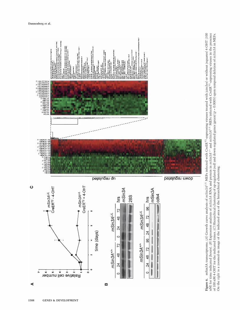
and mSin3A loss exerts broad biological effects (thisstudy), transcriptional profiling and in silico pathwayanalysis were utilized to understand more preciselythese diverse actions of mSin3A. In time course studies,the 4-hydroxytamoxifen (4-OHT)-inducible Cre/Estro-gen Receptor (CreERT2) system was used, as it affordstight temporal control of Cre activity in mSin3AL/L
MEFs. As shown in Figure 6A, 4-OHT induction of theCreERT2-transduced mSin3AL/L resulted in a cellularphenotype that was similar to that brought about by con-stitutive Cre retrovirus in mSin3AL/L MEFs (cf. Fig. 2A),although cell cycle arrest and apoptosis were less pro-nounced, as observed with cells expressing constitutiveCre-recombinase (Supplementary Fig. 4). On the basis ofthe kinetics showing steady depletion of mSin3A mRNAand protein levels (Fig. 6B), a comparative time-coursetranscriptional profile of CreERT2-transduced mSin3A+/+
and mSin3AL/L MEFs was performed at times 0, 24, 48,72, and 96 h following 4-OHT treatment. This approachallows the monitoring of differential gene expressionupon mSin3A deletion before the onset of the mSin3Aphenotype, and more readily enables discriminationbetween direct transcriptional effects due to mSin3Aablation versus nonspecific secondary transcriptionalchanges accompanied by the aggravated cell cycle arrestand apoptosis. dChip software (Li and Wong 2001) wasused to analyze the microarray data generated with Af-fymetrix M430A oligo DNA microarrays.
An Analysis of Variance (ANOVA) procedure was per-formed to identify genes with nonconstant expressionvalues between the two genetic backgrounds (mSin3A+/+
vs. mSin3A−/−). Imposing a p-value threshold of <0.0005,
Figure 3. Absence of chromosomal instability despite delocalization of HP1� and Orc2 in mSin3A-deficient MEFs. (A) Chromosomenumber in metaphase spreads of MEFs lacking either mSin3A or mSds3. The dashed line indicates a normal murine chromosomecount of 40. Analysis of heterochromatin in MEFs lacking mSin3A using immunofluorescence against AcK5H4 (B), HP1� andtri-meK9H3 (C), and Orc2 (D).
Genetic analysis of mSin3A
GENES & DEVELOPMENT 1585
1308 genes were determined as having significant non-constant expression. In concordance with the depletionkinetics of the mSin3A protein, most of these genesshowed differential expression starting at 48 h after ad-dition of 4-OHT. It is worth noting that there were noobvious cellular phenotypes at this 48-h time point,and thus it is more likely that the observed transcrip-tional changes are more proximal to mSin3A functionas opposed to secondary events and terminal cellularcollapse (Supplementary Fig. 4). The majority (977) ofthese differentially expressed genes were up-regulated(Fig. 6C; Supplementary Table 1; Supplementary Infor-mation 1, 2). A subset of the genes showing alteredexpression on the Affymetrix expression profiles wereverified by real-time RT–PCR analysis on independentgenerated RNA samples from mSin3AL/L MEFs ex-pressing Cre-ERT2 (Fig. 7A). As shown in Figure 7A,whereas mSin3A RNA levels declined progressively at48 and 72 h of 4-OHT treatment, increased mRNA lev-els for mSin3B, Mxi1, Sap30, Ing1, Arid4b, Peg3, Set,p130, cyclin E, Odc1, Msh6, Brca1, 53Bp1, Stmn1, andAldh6a1 were already markedly increased at 48 h, find-ings consistent with a direct link to mSin3A regula-tion. Correspondingly, ChIP analysis of mSin3B, Mxi1,Ing1, Arid4b, Peg3, Set, p130, Caspase 3, 53Bp, Pmm1,and Aldh6a1 showed that mSin3A physically localizesto the proximal promoters of these genes (Fig. 7B).
Further validation of the corepressor role of mSin3Awas evident from the up-regulation of several well-estab-lished Myc-responsive target genes such as Odc, CyclinD2, Cyclin E, Nucleolin, Mcm7, Cdc2A, Vamp3, RNApolymerase 1-3 (16 kDa subunit), Fabp5, and Prmt1among others. In addition to these expected results,there was up-regulation of multiple genes encoding pro-tein products that interact with the mSin3/HDAC core-pressor complex including mSin3B, mSds3, Hdac2,Sap30, and Arid4b (Sap180), Mxi1, Mnt, Prmt5, and Ing1.These findings point to the existence of potential regu-latory feedback loops within the mSin3–HDAC path-way, as also indicated by other studies (Lagger et al.2002; Schuettengruber et al. 2003). Furthermore, weidentified three mSin3A target genes that are involved inmethylation of histones: the arginine protein methyl-transferases Prmt1 and Prmt5, and the SET-domain con-taining methyltransferase Nsd1 (nuclear receptor-bind-ing SET-domain protein 1). Since histone methylationand histone acetylation are closely linked processes(Fischle et al. 2003; Silverstein and Ekwall 2004), theseobservations prompt speculation that mSin3A may par-ticipate in coordinating these processes in part via thetranscriptional regulation of these methyltransferase-encoding genes.
Several up-regulated genes functionally cluster asgenes involved in G2/M transition and cytokinesis in-
Figure 4. mSin3A deacetylates p53 upon genotoxic stress, but has p53–p21Cip1-independent functions under normal culture condi-tions. (A) Western blot analysis for mSin3A and p21Cip1 of mSin3A+/+, mSin3AL/+, and mSin3AL/L MEFs both in a p53-proficient, aswell as a p53-deficient background, infected with either vector-expressing (V) or Cre-recombinase-expressing (C) virus. Cdk4 served asa loading control. (B) Western blot analysis of cell lysates isolated from MEFs with indicated genotypes, expressing Cre-recombinasetreated with or without 8 µM doxorubicin for mSin3A, p53, p21Cip1, and acetylated p53Lys317 and p53Lys379. (C) Growth curve analysisof mSin3A+/+p53−/− (squares), mSin3AL/+p53−/− (triangles), and mSin3AL/−p53−/− (circles) MEFs infected with either vector-expressing(open symbols) or Cre-recombinase-expressing (filled symbols) virus under normal culture conditions (D) Percentages of G1, S, G2/M,and sub-G1 (apoptotic) phases in indicated MEF cultures under normal culture conditions. The figure shows a representative of twoindependent experiments each performed in triplicate. (E) Percentage of apoptotic cells detected by TUNEL-assay in indicated MEFseither in a p53-proficient (left) or p53-deficient (right) background.
Dannenberg et al.
1586 GENES & DEVELOPMENT
cluding 53Bp1, p130, Gadd45-�, Mcm4, Mcm5, Mcm7,Securin, Set, Tacc3, and Cdc2A. None of these geneshave been reported to be specifically up-regulated in G2
or mitosis, suggesting that their up-regulation is notmerely a consequence of the G2/M cell cycle arrest im-posed by mSin3A deficiency (Whitfield et al. 2002).Therefore, the fact that some of these genes, when over-expressed, give rise to a G2/M arrest (e.g., Gadd45� and53Bp1) provides a possible explanation for the observedG2/M cell cycle arrest in mSin3A−/− cells. Similarly, theapoptosis associated with mSin3A depletion could relatein part to transcriptional up-regulation of Caspase-3,Ing1, Peg3, Pdcd5, Trim27, Mcl1, Pin1, and sphingosinephosphate lyase 1, all encoding pro-apoptotic activities(Nicholson et al. 1995; Helbing et al. 1997; Reiss et al.2004).
Several genes identified in the mSin3A transcriptomeare known E2F target genes, including Cyclin E, CyclinD3, Mcm5, Mcm7, Replication Protein A, Rad51, Se-curin, Ribonucleotide Reductase M1, RibonucleotideReductase M2, Caspase 3, cell division cycle 2A, andcell division cycle 6. The changes in E2F target generegulation are consistent with their regulation by E2Fcomplexes containing retinoblastoma family proteinsand class I HDACs (Brehm et al. 1998; Luo et al. 1998;Magnaghi-Jaulin et al. 1998; Lai et al. 2001; Rayman etal. 2002). In a similar manner, the up-regulation ofknown p53 target genes (Stathmin 1, Fk506bp3) is con-sistent with documented p53–mSin3A physical andfunctional interactions (Murphy et al. 1999; Zilfou et al.2001; this study).
A prominent group of genes present in the mSin3Atranscriptome participate in DNA-repair processes suchas Rpa1, Msh6, Rad17, Lig1, Lig3, Apex1, and Nsep1.These observations are in line with work in yeast sup-
porting a role for Sin3 in the nonhomologous end-joiningpathway (Jazayeri et al. 2004). In this regard, there is anup-regulation of several DNA repair genes involved inthe homologous recombination pathway includingBrca1, 53Bp1, and Rad51. The up-regulation of homolo-gous recombination genes in a context of impaired NHEJactivity raises the intriguing possibility that mSin3Amay be involved in coordinating the relative levels ofNHEJ versus homologous recombination repair activi-ties in the cell.
Consistent with findings in Drosophila, mSin3A dele-tion resulted in the up-regulation of genes involved incytosolic and mitochondrial energy-generating pathways(Pdk1, Mrsp23, Oghd, Adac9, Bdh, Got2, Hadh2, Cs,Fxc1, Prdx3), strengthening the provocative role formSin3A in the regulation of mitochondrial respiratoryactivity (Pile et al. 2003; see below). Finally, mSin3Aregulates the expression of several genes involved inDNA replication (Rpa1, Lig1, Mcm4, Mcm5, Mcm7,Nap1l1) providing a basis for the observed defects inDNA replication upon deletion of mSin3A, and strength-ening the case for a direct role for mSin3A in the DNAreplication process.
Identification of known and novel mSin3Atranscriptional nodes
Enrichment of known Myc, E2F, and p53 targets in themSin3A transcriptome prompted us to search the regu-latory regions of all genes regulated by mSin3A for addi-tional Myc, E2F, and p53 target genes, as well as forunanticipated links other transcription factors and theirbinding sites. In the case of E2F, 66 mSin3A−/− up-regu-lated genes contained E2F-binding sites within 500 bpupstream or downstream of the transcriptional start site.
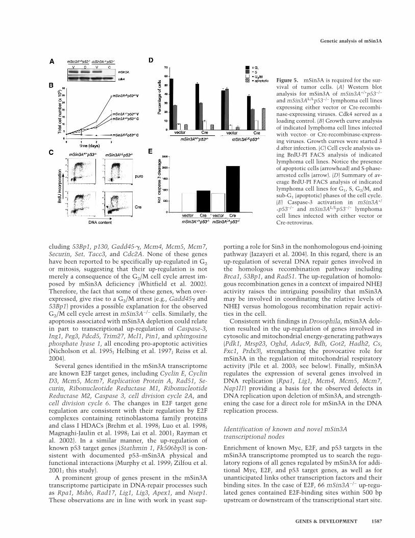
Figure 5. mSin3A is required for the sur-vival of tumor cells. (A) Western blotanalysis for mSin3A of mSin3A+/+p53−/−
and mSin3AL/Lp53−/− lymphoma cell linesexpressing either vector or Cre-recombi-nase-expressing viruses. Cdk4 served as aloading control. (B) Growth curve analysisof indicated lymphoma cell lines infectedwith vector- or Cre-recombinase-express-ing viruses. Growth curves were started 3d after infection. (C) Cell cycle analysis us-ing BrdU-PI FACS analysis of indicatedlymphoma cell lines. Notice the presenceof apoptotic cells (arrowhead) and S-phase-arrested cells (arrow). (D) Summary of av-erage BrdU-PI FACS analysis of indicatedlymphoma cell lines for G1, S, G2/M, andsub-G1 (apoptotic) phases of the cell cycle.(E) Caspase-3 activation in mSin3A+/
+p53−/− and mSin3AL/Lp53−/− lymphomacell lines infected with either vector orCre-retrovirus.
Genetic analysis of mSin3A
GENES & DEVELOPMENT 1587
Figu
re6.
mSi
n3A
tran
scri
ptom
e.(A
)Gro
wth
curv
ean
alys
isof
mSi
n3A
L/L
ME
Fsin
fect
edw
ith
Cre
ER
T2-e
xpre
ssin
gvi
ruse
str
eate
dw
ith
(cir
cles
)or
wit
hou
t(s
quar
es)4
-OH
T(1
00n
M)
for
tim
ein
dica
ted
(hou
rs).
(B)
Exp
ress
ion
anal
ysis
ofm
Sin
3AR
NA
and
prot
ein
inm
Sin
3A+/+
and
mSi
n3A
L/L
ME
Fsin
fect
edw
ith
Cre
ER
T2-e
xpre
ssin
gvi
ruse
sin
the
pres
ence
of10
0n
M4-
OH
Tfo
rth
ein
dica
ted
tim
es.(
C)H
iera
rch
ical
clu
ster
ing
ofu
p-re
gula
ted
(red
)an
ddo
wn
-reg
ula
ted
gen
es(g
reen
)(p
=0.
0005
)upo
nte
mpo
rald
elet
ion
ofm
Sin
3Ain
ME
Fs.
On
the
righ
tis
azo
omed
-in
imag
eof
the
indi
cate
dar
eaof
the
hie
rarc
hic
alcl
ust
erin
g.
Dannenberg et al.
1588 GENES & DEVELOPMENT
Compared with a random collection of 1161 genes, therewas a 1.95-fold enrichment (4.40% vs. 8.61%; p = 0.044)in E2F-binding sites in the collection of induced genesupon deletion of mSin3A. Scanning for p53-binding ele-ments (which are typically positioned at greater dis-tances from the transcriptional start site), 64 mSin3A−/−
up-regulated genes were found to contain p53-bindingsites within 5000 bp of the transcriptional start site ineither upstream or downstream direction. A 1.85-fold en-richment was observed for p53-binding sites in themSin3A−/− up-regulated genes compared with the ran-dom genes (8.47% vs. 4.57%, p = 0.046). Finally, a searchfor Myc target genes by scanning the promoters of theup-regulated genes for E-box sequences identified 189genes and 1.34-fold enrichment (24.6% vs. 18.33%,p = 0.183) for E-box sequences over a set of randomgenes. The relatively low enrichment for E-box se-quences is likely due to the reduced information presentin E-box sequences compared with the longer E2F- andp53-binding sequences, as well as the known ability ofthe Myc oncoprotein to regulate a significant fraction ofthe genome, as revealed by studies in human and Dro-sophila (Fernandez et al. 2003; Orian et al. 2003). To-gether, these findings show that gene expression profil-ing and in silico promoter analysis can lead to the iden-tification of E2F-, Myc-, and p53-responsive targets.
Next, we sought to validate further the above linksbetween mSin3A and the E2F, Myc, and p53 transcrip-tional networks, and to search for new mSin3A interac-tions. Additionally, since mSin3A does not directly bindto DNA, we sought to identify DNA-binding transcrip-tion factors like E2F, Myc, and p53 that are used bymSin3A to regulate gene expression. To this end, aknowledge-based database (Ingenuity Pathways Knowl-edge Base, http://www.ingenuity.com) was used to inte-grate the mSin3A transcriptome (particularly the up-regulated genes) into various pathways. As expected, this
network analysis identified the transcription factorsE2F1, E2F4, Myc, and p53 as nodal points that connectnumerous mSin3A-modulated genes (SupplementaryFig. 5), thus providing a measure of validation for thecis-regulatory element results above. In addition to theseanticipated networks, several other networks emerged,including those governed by the nodal transcription fac-tors Fos and peroxisome proliferative activated receptor(PPAR). The latter transcription factor is of special inter-est, since the mSin3A transcriptome contains manygenes involved in mitochondrial metabolism/respiration(Acsl1, Nr2f2, Ctps, Pmm1, Tpm2, Pc, Scarb1, Hsd17b4,Cs, Fabp5, and Umpk among others) (see SupplementaryTable 1; Supplementary Fig. 6), and the PPAR pathwayhas been implicated in mitochondrial respiration andmetabolism (Puigserver et al. 1998). Furthermore, regu-lation of gene expression through the PPAR pathway ap-pears subject to regulation by histone deacetylation (Fanet al. 2004). Similarly, Fos protein heterodimerizes withJun, which has been shown to repress transcription byrecruiting mSin3A/HDAC complex (Lee et al. 2000). Fi-nally, the Pathway Knowledge Base also revealed links tovarious genes involved in DNA repair, reinforcing thedata from yeast of a role for the otholog in DNA repair(Supplementary Fig. 6; Jazayeri et al. 2004).
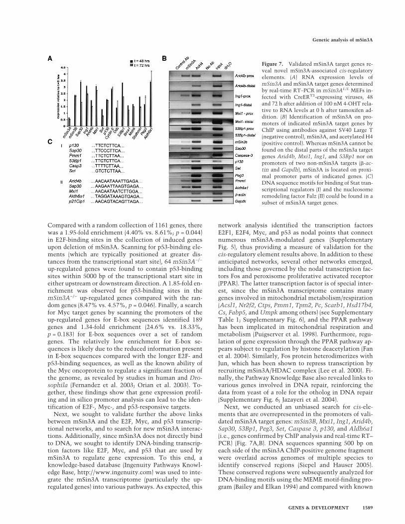
Next, we conducted an unbiased search for cis-ele-ments that are overrepresented in the promoters of vali-dated mSin3A target genes: mSin3B, Mxi1, Ing1, Arid4b,Sap30, 53Bp1, Peg3, Set, Caspase 3, p130, and Aldh6a1(i.e., genes confirmed by ChIP analysis and real-time RT–PCR) (Fig. 7A,B). DNA sequences spanning 500 bp oneach side of the mSin3A ChIP-positive genome fragmentwere overlaid across genomes of multiple species toidentify conserved regions (Siepel and Hauser 2005).These conserved regions were subsequently analyzed forDNA-binding motifs using the MEME motif-finding pro-gram (Bailey and Elkan 1994) and compared with known
Figure 7. Validated mSin3A target genes re-veal novel mSin3A-associated cis-regulatoryelements. (A) RNA expression levels ofmSin3A and mSin3A target genes determinedby real-time RT–PCR in mSin3AL/L MEFs in-fected with CreERT2-expressing viruses, 48and 72 h after addition of 100 nM 4-OHT rela-tive to RNA levels at 0 h after tamoxifen ad-dition. (B) Identification of mSin3A on pro-moters of indicated mSin3A target genes byChIP using antibodies against SV40 Large T(negative control), mSin3A, and acetylated H4(positive control). Whereas mSin3A cannot befound on the distal parts of the mSin3a targetgenes Arid4b, Mxi1, Ing1, and 53Bp1 nor onpromoters of two non-mSin3A targets (�-ac-tin and Gapdh), mSin3A is located on proxi-mal promoter parts of indicated genes. (C)DNA sequence motifs for binding of Stat tran-scriptional regulators (I) and the nucleosomeremodeling factor Falz (II) could be found in asubset of mSin3A target genes.
Genetic analysis of mSin3A
GENES & DEVELOPMENT 1589
transcription factor-binding motifs using the Match Pro-gram in Transfac (Matys et al. 2003). As shown in Figure7C, two cis-regulatory elements were identified that aresimilar to DNA-binding motifs for the transcriptionalactivator Stat, whose DNA-binding activity has beenshown to be acetylation dependent (Wang et al. 2005;Yuan et al. 2005) and Falz (also know as FAC1/BPTF/Nurf301), a nucleosome remodeling factor that is used inthe ISWI ATP-dependent chromatin remodeling com-plex NURF (Bowser et al. 1995; Jordan-Sciutto et al.1999; Jones et al. 2000; Xiao et al. 2001; Badenhorst et al.2002). Together, these data suggest that the combinedcomputational and ChIP analyses provides a means forthe validation of known mSin3A cofactors and an entrypoint for the identification of novel mSin3A cofactors.
Discussion
This study provides genetic evidence that mSin3A playsa critical role in the regulation of several prominent tran-scriptional networks and is essential for the proliferationand survival of both normal and neoplastic cells. Con-sistent with roles for mSin3A in diverse physiologicalprocesses, loss of mSin3A function provokes an earlyembryonic lethal condition, generates defects at allphases of the cell cycle including the DNA synthesisphase, enhances apoptotic responses, particularly in thesetting of genotoxic stress, and alters the expression ofmany genes linked to cell cycle regulation, DNA repli-cation, DNA repair, mitochondrial metabolism, and his-tone modification. Consistent with antecedent bio-chemical data, computational analysis of the mSin3Atranscriptome and the promoter regions of up-regulatedgenes provided genetic confirmation of the critical roleof mSin3A in the regulation of the Myc-Mad network,the pRB–E2F axis, and the p53 pathway. In addition, ourcomputational efforts have also forged putative links togene networks regulated by PPAR, FOS, STAT, andFALZ.
Despite up-regulation of mSin3A partners, as well asubiquitous coexpression of the highly related mSin3Bhomolog, the loss of mSin3A function exacted profoundphysiological consequences, underscoring the essentialand separable roles of the mSin3 homologs. ThatmSin3A and mSin3B possess nonoverlapping functionsis in line with the capacity of mSin3A and mSin3B tooccupy different promoters in mammalian cells (Ray-man et al. 2002), as well as the assignment of distinctfunctions for the two yeast Sin3 orthologs (Nakayama etal. 2003; Silverstein et al. 2003). The nature and extent ofthe common and distinct functions of mSin3A andmSin3B will require additional direct genetic study.Along these lines, it will be paramount to determinewhether there is either functional redundancy amongmSin3A and mSin3B in the establishment of functionalcentromeric heterochromatin, or whether there is indeeda unique mSin3A/B-independent function for mSds3 inthis aspect of chromosome biology.
At the same time, although mSds3 appears to be criti-cal for maintenance of HDAC-associated enzymatic ac-
tivity in the mSin3A and mSin3B corepressor complex(Alland et al. 2002), it is important to note thatmSin3A−/− cells exhibit phenotypes that are not ob-served in the mSds3−/− cells. Specifically, mSin3A−/−
MEF cultures harbor a subpopulation containing S-phaseDNA content with no ongoing DNA replication (noBrdU incorporation). Given that this DNA contentanomaly cannot be attributed to chromosomal instabil-ity and that there is prominent representation of DNAreplication genes in the mSin3A transcriptome, we pro-pose that mSin3A plays a direct role in regulation ofDNA replication in mammalian cells. This hypothesis isin line with previous studies in S. cerevisiae, showing arole for Sin3 and Rpd3 (ortholog of HDAC1) in the acti-vation of late origins of replication independent relatingto their role in transcriptional repression (Aparicio et al.2004). Additionally, in Drosophila, activation of origin ofreplication is dependent on Rpd3 as evidenced by theloss of Rpd3 function, resulting in genome-wide hyper-acetylation, genomic replication, and redistribution ofthe origin binding protein ORC2 (Aggarwal and Calvi2004). Together, these collective insights have shapedour view of the mSin3 corepressor complex. Rather thanthe existence of a monolithic collection of components,the data are consistent with the ability of the mSin3complex to operate in a more modular fashion, such thatvarious transcription factors and other DNA-binding el-ements may recruit only a subset of the mSin3/HDACcomponents. This speculation is consistent with emerg-ing observation of the existence of several biochemicallydistinct forms of the mSin3 multiprotein complex(Fleischer et al. 2003).
In the context of cancer, a strong consensus hasemerged that modulators of chromatin architecture areintimately linked to key cancer-relevant pathways. Par-ticularly relevant to this issue is the previously docu-mented physical and biochemical interactions betweenthe mSin3A corepressor and the p53 tumor suppressor.Here, our genetic analysis has substantiated the abilityof mSin3A to modulate p53 activation in the setting ofDNA damage, as evidenced by Lys 317 and Lys 379 hy-peracetylation and increased p53 protein levels upon lossof mSin3A function. Thus, we propose that an importantfunction of the mSin3A/HDAC complex is the regula-tion of p53 acetylation status and cell cycle control fol-lowing genotoxin-induced injury. In addition, it is worthnoting that several deacetylases and HDAC-containingcomplexes have shown the capacity to deacetylate p53,including the NuRD complex (Luo et al. 2000), all ClassI HDACs (Juan et al. 2000), and the class III deacetylaseSIRT1 (Luo et al. 2001; Vaziri et al. 2001; Cheng et al.2003). These observations, together with the findings ofthis report, raise the interesting possibility that multipleprotein complexes, and therefore, multiple biochemicalsignaling pathways, converge on the p53 protein to regu-late its activity via acetylation in various physiologicalsettings.
In silico promoter scanning revealed significant en-richment of Myc, E2F, and p53 cis-elements in themSin3A transcriptome. It is interesting to note, how-
Dannenberg et al.
1590 GENES & DEVELOPMENT
ever, that not all Myc, E2F, and p53 target genes areup-regulated, which implies that mSin3A might regulateonly a subset of these targets, that mSin3A may onlyengage these transcription factors under certain physi-ological conditions or cell types, and/or that mSin3Bmay serve overlapping functions on a subset of thesegene targets. One important caveat to these suppositionsis the fact that only a limited region of the promotersequences of the mSin3A transcriptome genes wasscanned, and it is possible that Myc, E2F, and p53 cis-regulatory elements were not captured in our analyses,thereby resulting in an underestimation of the mSin3Atarget genes coregulated by Myc, p53, and E2F. The iden-tification of the Stat and Falz cis-regulatory elements ina subset of the mSin3A target genes shows that mSin3Auses various DNA-binding transcription factors to conferselectivity and specificity in transcriptional regulation.Recently, it was shown that acetylation of Stat3 is criti-cal for the protein to form stable dimers required forcytokine-stimulated DNA-binding and transcriptionalregulation and to enhance transcription of cell growth-related genes (Wang et al. 2005; Yuan et al. 2005). Thesestudies, together with our finding that mSin3A can re-press Stat target gene transcription, suggests a model inwhich mSin3A-associated HDAC activity inhibits Stat3dimerization and neutralizes its transcriptional poten-tial. Various studies have implied collaborative interac-tions between different chromatin remodeling com-plexes, including mSin3A/HDAC and SWI/SNF (Stern-berg et al. 1987; Sif et al. 2001; Kuzmichev et al. 2002). Inthis context, our finding that mSin3A regulates genetranscription through Falz appears relevant, as Falz is acomponent of the ISWI ATP-dependent nucleosome re-modeling complex NURF. Falz contains a bromodomain,which are known to bind acetylated lysines in histonesand nonhistone proteins (for review, see Yang 2004), sug-gesting that Falz might recruit mSin3A/HDAC com-plexes to such targets to deacetylate specific promoterregions or modify the transcriptional activity of nonhis-tone proteins. A human NURF complex was shown tocontain, beside FALZ, also RbAp46 and RbAp48, twoproteins that are often found in complex with mSin3A(Barak et al. 2003). Notably, Nurf301 can act as a nega-tive regulator of the JAK/STAT signaling pathway,downstream of the STAT proteins, suggesting thatmSin3A may regulate gene transcription through bothDNA-binding transcription factors (Badenhorst et al.2002). Along these lines, our computational and ChIPanalyses reveal that the mSin3A target gene Sap30 con-tains Stat- as well as Falz-binding motifs in its promoter.
In summary, the integrated genetic, biochemical, andcomputational approaches of this study has enabled us togain insight into the diverse functions of mSin3A. Theapplication of this integrated approach and conditionalsomatic inactivation may find utility in the analysis ofmultifunctional complexes serving cell essential func-tions. In the case of mSin3A, this study confirmed theessential role of mSin3A, and by extension, the mSin3Acorepressor complex in numerous biological processesrelevant to normal physiology and to cancer, and served
to uncover known and novel cofactors regulating variousbiochemical processes (e.g., DNA replication, nucle-somal remodeling, etc.).
The observations of this report suggest a general strat-egy for assessment of the other components of the mSin3corepressor complex in normal cells and in cancer.
Materials and methods
Generation of mSin3A conditional null allele
A conditional mSin3A-targeting construct was generated byflanking mSin3A exon 4 with a 5� loxP-Frt-PGK-neomcyin ex-pression cassette (Aguirre et al. 2003) and a 3� loxP site. Uponelectroporation of 129/SvOla embryonic stem (ES) cells and se-lection at 200 µg/mL G418, homologous recombinants wereidentified by Southern blot analysis. Correctly targeted ESclones were injected into C57Bl/6 blastocysts and germlinetransmitting chimeric mice were bred to CAGG-Flpe (Rod-riguez et al. 2000) and EIIa-Cre transgenic mice (Lakso et al.1996) to generate the mSin3AL and mSin3A− alleles, respec-tively. Mice were genotyped by Southern analysis and multiplexPCR (primers and conditions are available upon request).
Preparation of MEFs, cell culture, and retroviral infection
MEFs were isolated from 13.5-d-post-coitum (dpc) embryos us-ing standard procedures (Dannenberg et al. 2000) and cultured inDMEM supplemented with 10% fetal bovine serum (FBS), 0.1mM �-mercapto-ethanol, and antibiotics. Ecotropic retroviralsupernatants were produced by cotransfection of 293T packag-ing cells with 6 µg viral DNA and 6 µg pCL vector (Naviaux etal. 1996) using lipofectamin 2000 (Invitrogen) according to themanufacturer’s instructions. Viral supernatant was collectedand filtered through a 0.45-µm filter 48, 56, and 72 h post-trans-fection. Low-density cultures of early passage MEFs were in-fected two times for at least 6 h with viral supernatants con-taining 4 µg/mL polybrene (Sigma) with an 8-h interval. After arecovery period of 36 h, the MEFs were selected for at least 48 husing medium containing 2.5 µg/mL puromycin (Sigma) beforethe cells were subjected to any of the analyses reported in thisstudy.
To study p53 acetylation, MEFs were infected with eithercontrol or Cre-expressing viruses and treated with doxorubicin72 h post-infection and processed as described in Cheng et al.(2003).
Proliferation curves, cell cycle, and apoptosis analyses
Proliferation curves were carried out as described in Dannen-berg et al. (2000). Cell cycle analysis was performed by incubat-ing MEFs on 10-cm tissue culture plates with 10 µM 5-Bro-modeoxy uridine (Sigma) for 1 h and processed as described inDannenberg et al. (2000). Assessment of apoptosis was per-formed by growing MEFs on eight-well chamber slides for 24–48h, and subsequently analyzed using an In Situ Cell DetectionKit (Roche) according to the manufacturer’s instructions. Quan-tification of apoptotic cells was performed by counting thenumber of TUNEL-positive nuclei in 100 DAPI-stained nucleiin three different fields at 100× magnification. All experimentswere performed at 5–7 d after retroviral infection with pBabe-puro or pBabe-Cre virus.
Metaphase spreads and immunofluoresence
Metaphase spreads from MEFs were generated as described inDavid et al. (2003). Immunofluoresence analysis of MEFs was
Genetic analysis of mSin3A
GENES & DEVELOPMENT 1591
performed 5–7 d after retroviral infection with pBabe-puro orpBabe-Cre virus. MEFs were cultured for 24–48 h on eight-wellchamber slides, fixed with 2% fresh paraformaldehyde (Pierce)and permeabilized with 0.1% Triton X-100. Antibodies againstthe following proteins were used: AcK12H4, AcK5H4, meK9H3,meK20H4 (Upstate), HP1� (Chemicon). For the Orc2 (BD Bio-sciences)-staining, Triton extraction was performed prior tofixation as described in Prasanth et al. (2004). Immunofluores-cent images were acquired using either a Zeiss Axiovert 200-Minverted microscope or a CoolSnap HQ CCD camera (Photo-metrics) driven by the Slidebook 4.0 b 2.5 software (intelligentImaging Innovations). Pictures were acquired with 1 × 1 binningand a 0.1-mm Z-step. Deconvolution was performed by thenearest neighbor method with 20 iterations.
Protein analyses
Protein levels were determined by Western blot analyses usingroutine protocols. Antibodies against mSin3A (K-20), mSin3B(AK-12), p130 (C-20), Cdk4 (C-22), and p21Cip1 (C-19) were ob-tained from Santa Cruz, HDAC1 from Upstate, p53 antibody(Ab-7) from Oncogene Science, and p53AcK317 and p53AcK379from Trevigen. Peroxidase-conjugated goat anti-rabbit and goatanti-mouse secondary antibodies were from Pierce. To detectanti-p53 antibodies, we used peroxidase-conjugated bovine anti-sheep antibodies from Santa Cruz.
Generation and culturing of mSin3AL/Lp53−/− sarcomas andlymphomas
mSin3A+/+p53−/− and mSin3AL/Lp53−/− mice were generatedby mSin3AL/+p53−/− intercrosses, and subsequently allowed toage. On average, at 10–15 wk of age, these mice developed (an-gio)sarcoma and T-cell lymphomas. Single-cell suspensions ofsarcomas and lymphomas were prepared and taken into culture.Sarcomas were maintained in culture using exactly the sameconditions as for MEFs. Lymphomas were cultured in cultureflasks in RPMI medium (Gibco), supplemented with 10% FBS,0.1 mM �-mercapto ethanol, and antibiotics.
Gene expression profiling
Subconfluent mSin3A+/+ and mSin3AL/L MEFs cultures on 10-cm culture dishes were twice infected with retroviruses ex-pressing the CreERT2 recombinase and selected for puromycin(2.5 µg/mL) for the rest of the experiment. After reaching con-fluency, the cells were passaged onto 15-cm culture dishes andselected for an additional 2 d. Subsequently, each plate was splitinto 5 × 3 10-cm culture dishes. For five consecutive days, 4-hy-droxy tamoxifen was added to a new set of three 10-cm dishes ata final concentration of 100 mM. RNA and protein were isolatedat the end of the experiment to document mSin3A status. Two10-cm plates for each time point were used to isolate RNA usingTrizol (Invitrogen) and an RNeasy mini kit (Qiagen). Gene ex-pression profiling was performed on the Affymetrix M430Aplatform.
Real time RT–PCR
Real-time PCR was performed using the Quantitec SYBR kitfrom Qiagen according to the manufacturer’s prescription.Primer sequences are available on request. RNA was isolatedfrom mSin3AL/L MEFs expressing CreERT2 and treated withTamoxifen for either 0, 48, or 72 h using Rneasy kits (Qiagen).To calculate RNA quantities, we established standard curveswith serial dilutions of known RNA concentrations. One-hun-
dred nanograms of total RNA was used for each RT–PCR (Qia-gen). All RT–PCRs were performed in triplicate.
ChIP and identification of cis-regulatory elements
ChIP experiments were performed using confluent early passage(passage 4) primary MEFs (pMEFs). For each ChIP assay, we used3 × 107 cells and followed a published protocol (Ren et al. 2002).Antibodies used for ChIP are anti-mSin3A (K-20; Santa Cruz),anti-SV40 LargeT (gift of J. DeCaprio, Dana-Farber Cancer In-stitute, Boston, MA), anti-acetylated histone H4 (Upstate). PCRprimers were designed for each kilobase of a 5-kb promoter frag-ment, and are available on request. Information on the meth-odology used in the identification of novel cis-regulatory ele-ments in promoters of mSin3A target genes can be found in theSupplemental Material.
Bioinformatic analyses of the gene expression profiling data
Analysis of the raw gene expression profiling was performedusing dChip software (Li and Wong 2001). In order to identifypotential E2f-binding, sites in the promoters of 1182 probe setsthat showed significant (p = 0.0005) up-regulation upon mSin3Adeletion, we mapped the genes onto the genome and determinedthe transcriptional start site (TSS). Promoters as large as 500 bpupstream and 500 bp down stream of the TSS were scanned witha known E2F matrix for potential E2f-binding sites. Only thetwo best hits per promoter were recorded for location and score.p53-binding sites in the same promoters were found by follow-ing the same procedure, except that the scan region for the p53matrix was expanded to 5000 bp upstream and 5000 bp downstream of the TSS. E-box sequences were identified in a regionspanning 1000 bp upstream and 200 bp downstream of the TSS.Sequences for the E2F, p53, Myc, and AP-1-binding sites used toscan the promoter regions of the genes in the mSin3A transcrip-tome are documented in Transfac (Matys et al. 2003). To iden-tify gene networks within the mSin3A transcriptome, we usedthe Ingenuity Pathways Knowledge Base (http://www.ingenuity.com).
Acknowledgments
We are grateful to James Horner and Michelle Neptune of thetransgenic and gene-targeting facility for generating the targetedES cells and the germline mouse strains. We acknowledge YaoYao for advice on the use of recombineering to generate thetargeting construct. We are thankful for the expert assistance ofthe animal facility staff; Shan Jiang, Karen Marmon, Alice Yu,and Yan Zhang. We thank Pedro Carvalho for assistance withdeconvolution microscopy. We thank Erguen Sahin, GiovanniTonon, and Ruben Carrasco for helpful discussions. We thankNabeel El-Bardeesy for the Cre- and Cre-ERT2 retroviral vectors.We thank Giovanni Tonon for assistance with the IngenuityPathways Knowledge Base analysis. We thank Alex Stegh forhelp in the analysis of caspase-3 activity. We thank Omar Kab-barah for help with the real-time RT–PCR. We thank RubenCarrasco for expert histological examination of tissue slides. Wethank J. DeCaprio for kindly providing the SV40 Large T anti-body and Darrell Borger for the retroviral Large T SV40 expres-sion construct. We are grateful to Robert N. Eisenman for com-municating data prior to publication. J-H.D. was a Damon-Ru-nyon Fellow supported by the Damon Runyon Cancer ResearchFoundation and is currently a Dutch Cancer Society Fellow.This work was funded by the National Institutes of Healthgrants GM67250 (W.H.W. and S.Z.) and RO1CA86379 (R.A.D.)
Dannenberg et al.
1592 GENES & DEVELOPMENT
and by grants from the American Cancer Society. R.A.D. is anAmerican Cancer Society Research Professor and an Ellison Se-nior Scholar.
Note added in proof
A study by Cowley et al. (2005) using an independently gener-ated mSin3A conditional mouse shows similar effects ofmSin3A deficiency on embryonic development, MEF prolifera-tion, and chromosomal dynamics.
References
Aggarwal, B.D. and Calvi, B.R. 2004. Chromatin regulates originactivity in Drosophila follicle cells. Nature 430: 372–376.
Aguirre, A.J., Bardeesy, N., Sinha, M., Lopez, L., Tuveson, D.A.,Horner, J., Redston, M.S., and DePinho, R.A. 2003. ActivatedKras and Ink4a/Arf deficiency cooperate to produce meta-static pancreatic ductal adenocarcinoma. Genes & Dev.17: 3112–3126.
Alland, L., David, G., Shen-Li, H., Potes, J., Muhle, R., Lee, H.C.,Hou, H.J., Chen, K., and DePinho, R.A. 2002. Identificationof mammalian Sds3 as an integral component of the Sin3/histone deacetylase corepressor complex. Mol. Cell. Biol.22: 2743–2750.
Aparicio, J.G., Viggiani, C.J., Gibson, D.G., and Aparicio, O.M.2004. The Rpd3–Sin3 histone deacetylase regulates replica-tion timing and enables intra-S origin control in Saccharo-myces cerevisiae. Mol. Cell. Biol. 24: 4769–4780.
Ayer, D.E. 1999. Histone deacetylases: Transcriptional repres-sion with siners and nurds. Trends Cell. Biol. 9: 193–198.
Ayer, D.E., Lawrence, Q.A., and Eisenman, R.N. 1995. Mad-Max transcriptional repression is mediated by ternary com-plex formation with mammalian homologs of yeast repres-sor Sin3 Cell 80: 767–776.
Badenhorst, P., Voas, M., Rebay, I., and Wu, C. 2002. Biologicalfunctions of the ISWI chromatin remodeling complexNURF. Genes & Dev. 16: 3186–3198.
Bailey, T.L. and Elkan, C. 1994. Fitting a mixture model byexpectation maximization to discover motifs in biopoly-mers. Proc. Int. Conf. Intell. Syst. Mol. Biol. 2: 28–36.
Barak, O., Lazzaro, M.A., Lane, W.S., Speicher, D.W., Picketts,D.J., and Shiekhattar, R. 2003. Isolation of human NURF: Aregulator of Engrailed gene expression. EMBO J. 22: 6089–6100.
Bowser, R., Giambrone, A., and Davies, P. 1995. FAC1, a novelgene identified with the monoclonal antibody Alz50, is de-velopmentally regulated in human brain. Dev. Neurosci.17: 20–37.
Brehm, A., Miska, E.A., McCance, D.J., Reid, J.L., Bannister,A.J., and Kouzarides, T. 1998. Retinoblastoma protein re-cruits histone deacetylase to repress transcription. Nature391: 597–601.
Cheng, H.L., Mostoslavsky, R., Saito, S., Manis, J.P., Gu, Y.,Patel, P., Bronson, R., Appella, E., and Alt, F.W. 2003. De-velopmental defects and p53 hyperacetylation in Sir2 homo-log (SIRT1)-deficient mice. Proc. Natl. Acad. Sci. 100:10794–10799.
Cowley, S.M., Iritani, B.M., Mendrysa, S.M., Xu, T., Cheng, P.F.,Yada, J., Liggitt, H.D., and Eisenman, R.N. 2005. ThemSin3A chromatin modifying complex is essential for em-bryogenesis and T cell development. Mol. Cell. Biol. (inpress).
Dang, V.D., Benedik, M.J., Ekwall, K., Choi, J., Allshire, R.C,
and Levin, H.L. 1999. A new member of the Sin3 family ofcorepressors is essential for cell viability and required forretroelement propagation in fission yeast. Mol. Cell. Biol.19: 2351–2365.
Dannenberg, J.-H., van Rossum, A., Schuijff, L., and te Riele, H.2000. Ablation of the retinoblastoma gene family deregu-lates G(1) control causing immortalization and increased cellturnover under growth-restricting conditions. Genes & Dev.14: 3051–3064.
David, G., Turner, G.M., Yao, Y., Protopopov, A., and DePinho,R.A. 2003. mSin3-associated protein, mSds3, is essential forpericentric heterochromatin formation and chromosomesegregation in mammalian cells. Genes & Dev. 17: 2396–2405.
De Nadal, E., Zapater, M., Alepuz, P.M., Sumoy, L., Mas, G.,and Posas, F. 2004. The MAPK Hog1 recruits Rpd3 histonedeacetylase to activate osmoresponsive genes. Nature427: 370–374.
Fan, M., Rhee, J., St-Pierre, J., Handschin, C., Puigserver, P., Lin,J., Jaeger, S., Erdjument-Bromage, H., Tempst, P., andSpiegelman, B.M. 2004. Suppression of mitochondrial respi-ration through recruitment of p160 myb binding protein toPGC-1�: Modulation by p38 MAPK. Genes & Dev. 18: 278–289.
Fernandez, P.C., Frank, S.R., Wang, L., Schroeder, M., Liu, S.,Greene, J., Cocito, A., and Amati, B. 2003. Genomic targetsof the human c-Myc protein. Genes & Dev. 17: 1115–1129.
Fischle, W., Wang, Y., and Allis, C.D. 2003. Histone and chro-matin cross-talk. Curr. Opin. Cell. Biol. 15: 172–183.
Fleischer, T.C., Yun, U.J., and Ayer, D.E. 2003. Identificationand characterization of three new components of themSin3A corepressor complex. Mol. Cell. Biol. 23: 3456–3467.
Helbing, C.C., Veillette, C., Riabowol, K., Johnston, R.N., andGarkavtsev, I. 1997. A novel candidate tumor suppressor,ING1, is involved in the regulation of apoptosis. Cancer Res.57: 1255–1258.
Huang, Y., Myers, S.J., and Dingledine, R. 1999. Transcriptionalrepression by REST: Recruitment of Sin3A and histone de-actylase to neuronal genes. Nat. Neurosci. 8: 67–72.
Jazayeri, A., McAinsh, A.D., and Jackson, S.P. 2004. Saccharo-myces cerevisiae Sin3p facilitates DNA double-strand breakrepair. Proc. Natl. Acad. Sci. 101: 1644–1649.
Johnstone, R.W. and Licht, J.D. 2003. Histone deacetylase in-hibitors in cancer therapy: Is transcription the primary tar-get? Cancer Cell 4: 13–18.
Jones, M.H., Hamana, N., and Shimane, M. 2000. Identificationand characterization of BPTF, a novel bromodomain tran-scription factor. Genomics 63: 35–39.
Jordan-Sciutto, K.L., Dragich, J.M., Rhodes, J.L., and Bowser, R.1999. Fetal Alz-50 clone 1, a novel zinc finger protein, bindsa specific DNA sequence and acts as a transcriptional regu-lator. J. Biol. Chem. 274: 35262–35268.
Juan, L.J., Shia, W.J., Chen, M.H., Yang, W.M., Seto, E., Lin, Y.S.,and Wu, C.W. 2000. Histone deacetylases specifically down-regulate p53-dependent gene activation. J. Biol. Chem.275: 20436–20443.
Kim, H., Lee, J.E., Cho, E.J., Liu, J.O., and Youn, H.D. 2003.Menin, a tumor suppressor, represses JunD-mediated tran-scriptional activity by association with an mSin3A–histonedeacetylase complex. Cancer Res. 63: 6135–6139.
Knoepfler, P.S. and Eisenman, R.N. 1999. Sin meets NuRD andother tails of repression. Cell 99: 447–450.
Koipally, J., Renold, A., Kim, J., and Georgopoulos, K. 1999.Repression by Ikaros and Aiolos is mediated through histonedeacetylase complexes. EMBO J. 18: 3090–3100.
Genetic analysis of mSin3A
GENES & DEVELOPMENT 1593
Kuzmichev, A., Zhang, Y., Erdjument-Bromage, H., Tempst, P.,and Reinberg, D. 2002. Role of the Sin3–histone deacetylasecomplex in growth regulation by the candidate tumor sup-pressor p33(ING1). Mol. Cell. Biol. 22: 835–848.
Lagger, G., O’Carroll, D., Rembold, M., Khier, H., Tischler, J.,Weitzer, G., Schuettengruber, B., Hauser, C., Brunmeir, R.,Jenuwein, T., et al. 2002. Essential function of histonedeacetylase 1 in proliferation control and CDK inhibitor re-pression. EMBO J. 21: 2672–2681.
Lagger, G., Doetzlhofer, A., Schuettengruber, B., Haidweger, E.,Simboeck, E., Tischler, J., Chiocca, S., Suske, G.,Rotheneder, H., Wintersberger, E., et al. 2003. The tumorsuppressor p53 and histone deacetylase 1 are antagonisticregulators of the cyclin-dependent kinase inhibitor p21/WAF1/CIP1 gene. Mol. Cell. Biol. 23: 2669–2679.
Laherty, C.D., Yang, W.M., Sun, J.M., Davie, J.R., Seto, E., andEisenman, R.N. 1997. Histone deacetylases associated withthe mSin3 corepressor mediate mad transcriptional repres-sion. Cell 89: 349–356.
Lai, A, Kennedy, B.K., Barbie, D.A., Bertos, N.R., Yang, X.J.,Theberge, M.C., Tsai, S.C., Seto, E., Zhang, Y., Kuzmichev,A., et al. 2001. RBP1 recruits the mSIN3–histone deacetylasecomplex to the pocket of retinoblastoma tumor suppressorfamily proteins found in limited discrete regions of thenucleus at growth arrest. Mol. Cell. Biol. 8: 2918–2932.
Lakso, M., Pichel, J.G., Gorman, J.R., Sauer, B., Okamoto, Y.,Lee, E., Alt, F.W., and Westphal, H. 1996. Efficient in vivomanipulation of mouse genomic sequences at the zygotestage. Proc. Natl. Acad. Sci. 93: 5860–5865.
Lee, S.K., Kim, J.H., Lee, Y.C., Cheong, J., and Lee, J.W. 2000.Silencing mediator of retinoic acid and thyroid hormone re-ceptors, as a novel transcriptional corepressor molecule ofactivating protein-1, nuclear factor-�B, and serum responsefactor. J. Biol. Chem. 275: 12470–12474.
Li, C. and Wong, W. 2001. Model-based analysis of oligonucleo-tide arrays: Expression index computation and outlier detec-tion. Proc. Natl. Acad. Sci. 98: 31–36.
Luo, R.X., Postigo, A.A., and Dean, D.C. 1998. Rb interacts withhistone deacetylase to repress transcription. Cell 92: 463–473.
Luo, J., Su, F., Chen, D., Shiloh, A., and Gu, W. 2000. Deacety-lation of p53 modulates its effect on cell growth and apopto-sis. Nature 408: 377–381.
Luo, J., Nikolaev, A.Y., Imai, S., Chen, D., Su, F., Shiloh, A.,Guarente, L., and Gu, W. 2001. Negative control of p53 bySir2� promotes cell survival under stress. Cell 107: 137–148.
Magnaghi-Jaulin, L., Groisman, R., Naguibneva, I., Robin, P.,Lorain, S., Le Villain, J.P., Troalen, F., Trouche, D., andHarel-Bellan, A. 1998. Retinoblastoma protein repressestranscription by recruiting a histone deacetylase. Nature391: 601–605.
Matys, V., Fricke, E., Geffers, R., Gossling, E., Haubrock, M.,Hehl, R., Hornischer, K., Karas, D., Kel, A.E., Kel-Margoulis,O.V., et al. 2003. TRANSFAC®: Transcriptional regulation,from patterns to profiles. Nucleic Acids Res. 31: 374–378.
Meehan, W.J., Samant, R.S., Hopper, J.E., Carrozza, M.J.,Shevde, L.A., Workman, J.L., Eckert, K.A., Verderame, M.F.,and Welch, D.R. 2004. Breast cancer metastasis suppressor 1(BRMS1) forms complexes with retinoblastoma-binding pro-tein 1 (RBP1) and the mSin3 histone deacetylase complexand represses transcription. J. Biol. Chem. 279: 1562–1569.
Melnick, A. and Licht, J.D. 2002. Histone deacetylases as thera-peutic targets in hematologic malignancies. Curr. Opin. He-matol. 9: 322–332.
Murphy, M., Ahn, J., Walker, K.K., Hoffman, W.H., Evans, R.M.,Levine, A.J., and George, D.L. 1999. Transcriptional repres-
sion by wild-type p53 utilizes histone deacetylases, medi-ated by interaction with mSin3a. Genes & Dev. 13: 2490–2501.
Nakayama, J., Xiao, G., Noma, K., Malikzay, A., Bjerling, P.,Ekwall, K., Kobayashi, R., and Grewal, S.I. 2003. Alp13, anMRG family protein, is a component of fission yeast Clr6histone deacetylase required for genomic integrity. EMBO J.22: 2776–2787.
Naviaux, R.K., Costanzi, E., Haas, M., and Verma, I.M. 1996.The pCL vector system: Rapid production of helper-free,high-titer, recombinant retroviruses. J. Virol. 70: 5701–5705.
Nicholson, D.W., Ali, A., Thornberry, N.A., Vaillancourt, J.P.,Ding, C.K., Gallant, M., Gareau, Y., Griffin, P.R., Labelle,M., Lazebnik, Y.A., et al. 1995. Identification and inhibi-tion of the ICE/CED-3 protease necessary for mammalianapoptosis. Nature 376: 37–43.
Orian, A., van Steensel, B., Delrow, J., Bussemaker, H.J., Li, L.,Sawado, T., Williams, E., Loo, L.W., Cowley, S.M., Yost, C.,et al. 2003. Genomic binding by the Drosophila Myc, Max,Mad/Mnt transcription factor network. Genes & Dev.17: 1101–1114.
Pile, L.A., Spellman, P.T., Katzenberger, R.J., and Wassarman,D.A. 2003. The SIN3 deacetylase complex represses genesencoding mitochondrial proteins: Implications for the regu-lation of energy metabolism. J. Biol. Chem. 278: 37840–37848.
Pothof, J., van Haaften, G., Thijssen, K., Kamath, R.S., Fraser,A.G., Ahringer, J., Plasterk, R.H., and Tijsterman, M. 2003.Identification of genes that protect the C. elegans genomeagainst mutations by genome-wide RNAi. Genes & Dev.17: 443–448.
Prasanth, S.G., Prasanth, K.V., Siddiqui, K., Spector, D.L., andStillman, B. 2004. Human Orc2 localizes to centrosomes,centromeres and heterochromatin during chromosome in-heritance. EMBO J. 23: 2651–2663.
Puigserver, P., Wu, Z., Park, C.W., Graves, R., Wright, M., andSpiegelman, B.M. 1998. A cold-inducible coactivator ofnuclear receptors linked to adaptive thermogenesis. Cell92: 829–839.
Rayman, J.B., Takahashi, Y., Indjeian, V.B., Dannenberg, J.-H.,Catchpole, S., Watson, R.J., te Riele, H., and Dynlacht, B.D.2002. E2F mediates cell cycle-dependent transcriptional re-pression in vivo by recruitment of an HDAC1/mSin3B core-pressor complex. Genes & Dev. 16: 933–947.
Reiss, U., Oskouian, B., Zhou, J., Gupta, V., Sooriyakumaran, P.,Kelly, S., Wang, E., Merrill Jr., A.H., and Saba, J.D. 2004.Sphingosine-phosphate lyase enhances stress-induced ce-ramide generation and apoptosis. J. Biol. Chem. 279: 1281–1290.
Ren, B., Cam, H., Takahashi, Y., Volkert, T., Terragni, J., Young,R.A., and Dynlacht, B.D. 2002. E2F integrates cell cycle pro-gression with DNA repair, replication, and G(2)/M check-points. Genes & Dev. 16: 245–256.
Rodriguez, C.I., Buchholz, F., Galloway, J., Sequerra, R., Kasper,J., Ayala, R., Stewart, A.F., and Dymecki, S.M. 2000. High-efficiency deleter mice show that FLPe is an alternative toCre-loxP. Nat. Genet. 2: 139–140.
Sakaguchi, K., Herrera, J.E., Saito, S., Miki, T., Bustin, M., Vas-silev, A., Anderson, C.W., and Appella, E. 1998. DNA dam-age activates p53 through a phosphorylation-acetylation cas-cade. Genes & Dev. 12: 2831–2841.
Schreiber-Agus, N. and DePinho, R.A. 1998. Repression by themad(mxi1)–sin3 complex. Bioessays 20: 808–818.
Schreiber-Agus, N., Chin, L., Chen, K., Torres, R., Rao, G.,Guida, P., Skoultchi, A.I., and DePinho, R.A. 1995. Anamino-terminal domain of Mxi1 mediates anti-Myc onco-
Dannenberg et al.
1594 GENES & DEVELOPMENT
genic activity and interacts with a homolog of the yeast re-pressor SIN3. Cell 80: 777–786.
Schuettengruber, B., Simboeck, E., Khier, H., and Seiser, C.2003. Autoregulation of mouse histone deacetylase 1 expres-sion. Mol. Cell. Biol. 19: 6993–7004.
Siepel, A. and Haussler, D. 2005. Phylogenetic hidden Markovmodels. In Statistical methods in molecular evolution (ed.R. Nielsen), pp. 325–351. Springer, New York.
Sif, S., Saurin A.J., Imbalzano, A.N., and Kingston, R.E. 2001.Purification and characterization of mSin3A-containingBrg1 and hBrm chromatin remodeling complexes. Genes &Dev. 15: 603–618.
Silverstein, R.A. and Ekwall, K. 2004. Sin3: A flexible regulatorof global gene expression and genome instability. Curr.Genet. 47: 1–17.
Silverstein, R.A., Richardson, W., Levin, H., Allshire, R., andEkwall, K. 2003. A new role for the transcriptional corepres-sor SIN3; regulation of centromeres. Curr. Biol. 13: 68–72.
Skowyra, D., Zeremski, M., Neznanov, N., Li, M., Choi, Y.,Uesugi, M., Hauser, C.A., Gu, W., Gudkov, A.V., and Qin, J.2001. Differential association of products of alternative tran-scripts of the candidate tumor suppressor ING1 with themSin3/HDAC1 transcriptional corepressor complex. J. Biol.Chem. 276: 8734–8739.
Sternberg, P.W., Stern, M.J., Clark, I., and Herskowitz, I. 1987.Activation of the yeast HO gene by release from multiplenegative controls. Cell 48: 567–577.
Vannier, D., Balderes, D., and Shore, D. 1996. Evidence that thetranscriptional regulators SIN3 and RPD3, and a novel gene(SDS3) with similar functions, are involved in transcrip-tional silencing in S. cerevisiae. Genetics 144: 1343–1353.
Vaziri, H., Dessain, S.K., Ng Eaton, E., Imai, S.I., Frye, R.A.,Pandita, T.K., Guarente, L., and Weinberg, R.A. 2001.hSIR2(SIRT1) functions as an NAD-dependent p53 deacety-lase. Cell 107: 149–159.
Wang, R., Cherukuri, P., and Luo, J. 2005. Activation of Stat3sequence-specific DNA binding and transcription by p300/CREB-binding protein-mediated acetylation. J. Biol. Chem.280: 11528–11534.
Whitfield, M.L., Sherlock, G., Saldanha, A.J., Murray, J.I., Ball,C.A., Alexander, K.E., Matese, J.C., Perou, C.M., Hurt,M.M., Brown, P.O., et al. 2002. Identification of genes peri-odically expressed in the human cell cycle and their expres-sion in tumors. Mol. Biol. Cell. 6: 1977–2000.
Xiao, H., Sandaltzopoulos, R., Wang, H.M., Hamiche, A.,Ranallo, R., Lee, K.M., Fu, D., and Wu, C. 2001. Dual func-tions of largest NURF subunit NURF301 in nucleosome slid-ing and transcription factor interactions. Mol. Cell. 8: 531–543.
Yang, X.J. 2004. Lysine acetylation and the bromodomain: Anew partnership for signaling. Bioessays 26: 1076–1087.
Yuan, Z.L., Guan, Y.J., Chatterjee, D., and Chin, Y.E. 2005.Stat3 dimerization regulated by reversible acetylation of asingle lysine residue. Science 307: 269–273.
Zilfou, J.T., Hoffman, W.H., Sank, M., George, D.L., and Mur-phy, M. 2001. The corepressor mSin3a interacts with theproline-rich domain of p53 and protects p53 from protea-some-mediated degradation. Mol. Cell. Biol. 21: 3974–3985.
Genetic analysis of mSin3A
GENES & DEVELOPMENT 1595
Related Documents