ORIGINAL RESEARCH ARTICLE published: 15 January 2015 doi: 10.3389/fnhum.2014.01033 Movement related slow cortical potentials in severely paralyzed chronic stroke patients Ozge Yilmaz 1,2 *, Niels Birbaumer 1,3 and Ander Ramos-Murguialday 1,4 1 Institute of Medical Psychology and Behavioral Neurobiology, University of Tuebingen, Tuebingen, Germany 2 Brain and Mind Studies Lab, Department of Psychology, Bahcesehir University, Istanbul, Turkey 3 Ospedale San Camillo, Istituto di Ricovero e Cura a Carattere Scientifico, Lido di Venezia, Italy 4 Health Technologies Department, Tecnalia, San Sebastian, Spain Edited by: Richard A. P. Roche, The National University of Ireland Maynooth, Ireland Reviewed by: Patricia Mary Gough, Maynooth University, Ireland Sarah J. Casey, Acquired Brain Injury Ireland, Ireland *Correspondence: Ozge Yilmaz, Brain and Mind Studies Lab, Bahcesehir University, Ciragan Cad. 2-4 34353 Besiktas, Istanbul, Turkey e-mail: [email protected] Movement-related slow cortical potentials (SCPs) are proposed as reliable and immediate indicators of cortical reorganization in motor learning. SCP amplitude and latency have been reported as markers for the brain’s computational effort, attention and movement planning. SCPs have been used as an EEG signature of motor control and as a main feature in Brain-Machine-Interfaces (BMIs). Some reports suggest SCPs are modified following stroke. In this study, we investigated movement-related SCPs in severe chronic stroke patients with no residual paretic hand movements preceding and during paretic (when they try to move) and healthy hand movements. The aim was to identify SCP signatures related to cortex integrity and complete paralysis due to stroke in the chronic stage. Twenty severely impaired (no residual finger extension) chronic stoke patients, of whom ten presented subcortical and ten cortical and subcortical lesions, underwent EEG and EMG recordings during a cue triggered hand movement (open/close) paradigm. SCP onset appeared and peaked significantly earlier during paretic hand movements than during healthy hand movements. Amplitudes were significantly larger over the midline (Cz, Fz) for paretic hand movements while contralateral (C4, F4) and midline (Cz, Fz) amplitudes were significantly larger than ipsilateral activity for healthy hand movements. Dividing the participants into subcortical only and mixed lesioned patient groups, no significant differences observed in SCP amplitude and latency between groups. This suggests lesions in the thalamocortical loop as the main factor in SCP changes after stroke. Furthermore, we demonstrated how, after long-term complete paralysis, post-stroke intention to move a paralyzed hand resulted in longer and larger SCPs originating in the frontal areas. These results suggest SCP are a valuable feature that should be incorporated in the design of new neurofeedback strategies for motor neurorehabilitation. Keywords: stroke, movement related slow cortical potentials, EEG, movement preparation, intention to move INTRODUCTION Stroke occurs as a consequence of cardiovascular flow distur- bances damaging neural networks in the brain. Following stroke, reorganization of cortical networks occurs (Grefkes et al., 2008; Ward, 2011). Reorganization consists among other consequences of enhanced neural activity of the healthy hemisphere (Chollet et al., 1991; Murase et al., 2004; Bashir et al., 2010). Movement-related slow cortical potentials (SCPs) recorded with EEG can be divided into two main components: (a) poten- tials occurring during intention or anticipation of an upcoming movement which is also called the Bereitschaftspotential (BP) for self-paced movements (Barrett et al., 1986), (b) the motor potential (MP) occurring at the time of the execution (Deecke et al., 1969). A BP is a bilateral low frequency (0–5 Hz) nega- tive shift (NS) occurring seconds before the movement onset. The MP peak rises primarily contralateral to the movement side around the onset of a voluntary movement (Birbaumer et al., 1991), which is also referred to as peak NS (Barrett et al., 1986; Shibasaki and Hallett, 2006). According to the excitation thresh- old theory, during the preparation of a movement SCPs serve as regulatory mechanisms that facilitate neuronal firing of the involved networks (Elbert and Rockstroh, 1987; Birbaumer et al., 1991). Cortical activity preceding voluntary movements is well- documented (Kornhuber and Deecke, 1965). The main cortical generators of SCPs are the premotor cortices (PMC), supple- mentary motor areas (SMAs) and cingulate cortices (Deecke, 1987; Cui et al., 1999). There is emerging evidence that subcor- tical structures, particularly the basal ganglia, also contribute to movement preparation, execution and control. The thalamus is connected to cortex and both the basal ganglia and the cerebellar pathways and the role of these connections in movement prepa- ration has recently been studied extensively in humans (Rektor, 2002; Paradiso et al., 2004). A lesion in any of these structures or connections (e.g., corticothalamic loop) could affect move- ment preparation and planning and should be reflected in SCPs, Frontiers in Human Neuroscience www.frontiersin.org January 2015 | Volume 8 | Article 1033 | 1 HUMAN NEUROSCIENCE

Welcome message from author
This document is posted to help you gain knowledge. Please leave a comment to let me know what you think about it! Share it to your friends and learn new things together.
Transcript
ORIGINAL RESEARCH ARTICLEpublished: 15 January 2015
doi: 10.3389/fnhum.2014.01033
Movement related slow cortical potentials in severelyparalyzed chronic stroke patientsOzge Yilmaz1,2*, Niels Birbaumer1,3 and Ander Ramos-Murguialday1,4
1 Institute of Medical Psychology and Behavioral Neurobiology, University of Tuebingen, Tuebingen, Germany2 Brain and Mind Studies Lab, Department of Psychology, Bahcesehir University, Istanbul, Turkey3 Ospedale San Camillo, Istituto di Ricovero e Cura a Carattere Scientifico, Lido di Venezia, Italy4 Health Technologies Department, Tecnalia, San Sebastian, Spain
Edited by:
Richard A. P. Roche, The NationalUniversity of Ireland Maynooth,Ireland
Reviewed by:
Patricia Mary Gough, MaynoothUniversity, IrelandSarah J. Casey, Acquired Brain InjuryIreland, Ireland
*Correspondence:
Ozge Yilmaz, Brain and MindStudies Lab, Bahcesehir University,Ciragan Cad. 2-4 34353 Besiktas,Istanbul, Turkeye-mail: [email protected]
Movement-related slow cortical potentials (SCPs) are proposed as reliable and immediateindicators of cortical reorganization in motor learning. SCP amplitude and latency havebeen reported as markers for the brain’s computational effort, attention and movementplanning. SCPs have been used as an EEG signature of motor control and as a mainfeature in Brain-Machine-Interfaces (BMIs). Some reports suggest SCPs are modifiedfollowing stroke. In this study, we investigated movement-related SCPs in severe chronicstroke patients with no residual paretic hand movements preceding and during paretic(when they try to move) and healthy hand movements. The aim was to identify SCPsignatures related to cortex integrity and complete paralysis due to stroke in the chronicstage. Twenty severely impaired (no residual finger extension) chronic stoke patients,of whom ten presented subcortical and ten cortical and subcortical lesions, underwentEEG and EMG recordings during a cue triggered hand movement (open/close) paradigm.SCP onset appeared and peaked significantly earlier during paretic hand movements thanduring healthy hand movements. Amplitudes were significantly larger over the midline (Cz,Fz) for paretic hand movements while contralateral (C4, F4) and midline (Cz, Fz) amplitudeswere significantly larger than ipsilateral activity for healthy hand movements. Dividingthe participants into subcortical only and mixed lesioned patient groups, no significantdifferences observed in SCP amplitude and latency between groups. This suggests lesionsin the thalamocortical loop as the main factor in SCP changes after stroke. Furthermore,we demonstrated how, after long-term complete paralysis, post-stroke intention to movea paralyzed hand resulted in longer and larger SCPs originating in the frontal areas. Theseresults suggest SCP are a valuable feature that should be incorporated in the design ofnew neurofeedback strategies for motor neurorehabilitation.
Keywords: stroke, movement related slow cortical potentials, EEG, movement preparation, intention to move
INTRODUCTIONStroke occurs as a consequence of cardiovascular flow distur-bances damaging neural networks in the brain. Following stroke,reorganization of cortical networks occurs (Grefkes et al., 2008;Ward, 2011). Reorganization consists among other consequencesof enhanced neural activity of the healthy hemisphere (Cholletet al., 1991; Murase et al., 2004; Bashir et al., 2010).
Movement-related slow cortical potentials (SCPs) recordedwith EEG can be divided into two main components: (a) poten-tials occurring during intention or anticipation of an upcomingmovement which is also called the Bereitschaftspotential (BP)for self-paced movements (Barrett et al., 1986), (b) the motorpotential (MP) occurring at the time of the execution (Deeckeet al., 1969). A BP is a bilateral low frequency (0–5 Hz) nega-tive shift (NS) occurring seconds before the movement onset.The MP peak rises primarily contralateral to the movement sidearound the onset of a voluntary movement (Birbaumer et al.,1991), which is also referred to as peak NS (Barrett et al., 1986;
Shibasaki and Hallett, 2006). According to the excitation thresh-old theory, during the preparation of a movement SCPs serveas regulatory mechanisms that facilitate neuronal firing of theinvolved networks (Elbert and Rockstroh, 1987; Birbaumer et al.,1991).
Cortical activity preceding voluntary movements is well-documented (Kornhuber and Deecke, 1965). The main corticalgenerators of SCPs are the premotor cortices (PMC), supple-mentary motor areas (SMAs) and cingulate cortices (Deecke,1987; Cui et al., 1999). There is emerging evidence that subcor-tical structures, particularly the basal ganglia, also contribute tomovement preparation, execution and control. The thalamus isconnected to cortex and both the basal ganglia and the cerebellarpathways and the role of these connections in movement prepa-ration has recently been studied extensively in humans (Rektor,2002; Paradiso et al., 2004). A lesion in any of these structuresor connections (e.g., corticothalamic loop) could affect move-ment preparation and planning and should be reflected in SCPs,
Frontiers in Human Neuroscience www.frontiersin.org January 2015 | Volume 8 | Article 1033 | 1
HUMAN NEUROSCIENCE
Yilmaz et al. Slow cortical potentials in chronic stroke
especially if motor recovery does not take place and chronic stageis reached.
It has been shown that the latency of SCP [i.e., the timebetween the onset of SCP and the movement onset recordedwith electromyography (EMG)] indicates the preparation timeof the required action, with longer the latencies indicating morecomplex is the motor tasks (Tarkka and Hallett, 1990; Langet al., 1992). Repetitive simple movements, which do not requirehigher order cognitive pre-planning and preparation, are relatedto shorter SCP latency and smaller SCP amplitudes (Libet et al.,1982). It has been suggested that the amplitude of the nega-tive peak may indicate the brain’s computational “demand” toperform the movement (Libet et al., 1982; Lang et al., 1990;Simonetta et al., 1991; Libet, 1992).
Several groups have demonstrated altered SCP features aftera brain disease or injury (Sasaki and Gemba, 1984; Cunningtonet al., 1999). Kitamura et al. (1996), in a study involving two sub-cortical stroke patients performing synergistic movements of theparetic arm, report that the early SCP component remained bilat-eral as in healthy participants. However, MP was also distributedbilaterally as opposed to its dominant contralateral distribu-tion in healthy individuals, indicating a stronger involvement ofthe healthy hemisphere during the movements of paretic side.Additionally, stroke patients’ recovering motor function spon-taneously shows an increase in fMRI BOLD activity toward thenon-lesioned hemisphere when executing paretic hand move-ments. These are shifted back to ipsilesional areas once recoverytakes place, reaching a “normal” bilateral activation with a peakat hand movement’s contralateral hemisphere (Rossini et al.,2003; Murphy and Corbett, 2009). Green et al. (1999) offer sup-port for these findings using multimodal neuroimaging methodswith EEG and fMRI demonstrating that the intact hemispherebecomes more active after stroke in participants with varyingdegrees of recovery. Similar findings regarding contralesional acti-vation were observed by other groups (Lang et al., 1988; Verlegeret al., 2003; Jankelowitz and Colebatch, 2005). It has also recentlybeen shown sensorimotor-rhythm (SMR) based EEG Brain-Machine-Interfaces (BMIs) can be used to recover motor functionin chronic severely paretic stroke patients (Ramos-Murguialdayet al., 2013). Furthermore, SCPs have been extensively used asfeatures for neurofeedback and BMI control (Birbaumer et al.,1999).
In this study, we investigated the effects of cortex integrityand stroke severity on SCPs (i.e., neural reorganization in par-ticipants with severe hand weakness in the chronic stage). Thefinal goal is to identify relevant features that can be used andoptimized (e.g., toward normal potentials’ characteristics) in SCPbased BMI neurofeedback therapy for motor rehabilitation inparalyzed chronic stroke patients. We studied SCPs of 20 severelyimpaired chronic stroke patients, who suffered from subcorticaland mixed (cortical and subcortical) lesions, during paretic andhealthy hand movements. The aim was to investigate changes inSCPs in severe chronic stroke comparing the SCP amplitudes andlatencies induced by the subcortical vs. cortical lesions and pareticvs. healthy hand movements. We used the healthy hand move-ment related SCPs as reference because it is related to healthymotor output. Although brain activity might not be the same as in
a healthy person, the motor output is normal. Due to the severityof motor impairment in our participants, we expected to observewidespread (i.e., bilateral) SCP activity and earlier SCP onset inparetic compared to the healthy hand movements (compensatorymovement planning). An ipsilateral over-activation (i.e., highernegative amplitudes) and a contralateral lower activation wereexpected during motor preparation of the paretic compared tohealthy hand movements (maladaptive higher involvement of theintact hemisphere). Furthermore, we hypothesized that frontaland premotor areas in the participants presenting mixed lesions(subcortical and cortical) would show higher levels of activa-tion, due to increased compensatory efforts of the secondarymotor areas, compared to the participants with subcorticallesions.
MATERIALS AND METHODSPARTICIPANTSTwenty hemiparetic (none of the participants had bilaterallesions) participants 51.4 ± 11.1 years old and 5.9 ± 5.5 yearssince stroke participated in the study. Ten participants (5 male, 5female) presented subcortical lesions (Sub-L) only and 10 par-ticipants (7 male, 3 female) presented mixed lesions (Mix-L)(subcortical and cortical areas). Selection criteria were no residualfinger extension and time since stroke of at least 12 months. Thedegree of functional severity was measured using a modified ver-sion of the Fugl-Meyer Assessment (FMA) scale. (for participantinformation and detailed selection criteria see the SupplementaryInformation). The study was conducted at the University ofTuebingen, Germany. Informed consent was obtained from allparticipants involved. The study was approved by the ethics com-mittee of the Faculty of Medicine of the University of Tuebingen(Germany).
DATA ACQUISITIONParticipants underwent a 16-channel EEG recording (Acticap,BrainProducts GmbH, Germany) session [Fp1, Fp2, F3, Fz, F4,T7, C3, Cz, C4, T8, CP3, CP4, P3, Pz, P4, Oz, AFz (Ground) andFCz (Reference)]. Surface electromyographic (EMG) activity wasrecorded from both arms using eight bipolar Ag/AgCl electrodesfrom Myotronics-Noromed (Tukwila, WA, USA) on four differ-ent muscle groups (extensor carpi ulnaris, extensor digitorum,external head of the biceps and external head of the triceps) inorder to detect movement onset and involuntary muscle contrac-tions. Electrooculography (EOG) recordings were also carried outfor ocular corrections.
Participants performed an audiovisual task. The imperativecue was visual (an arrow pointing right or left appearing on thescreen for 5 s) and auditory (a sound indicating right or left) givenconcurrently. This protocol was tried to resemble movementsduring a standard rehabilitation session. Participants either exe-cuted a hand opening and closing movement with their healthyhand (HM) or tried to open and close the paretic hand (PM)at a comfortable personal pace for 5 s according to the audio-visual imperative cues. Participants were trained and instructedto avoid compensatory movements during the intention to openand close the paretic hand. The inter-trial-interval was random-ized between 3 and 4 s and a fixation cross appeared on the screen
Frontiers in Human Neuroscience www.frontiersin.org January 2015 | Volume 8 | Article 1033 | 2
Yilmaz et al. Slow cortical potentials in chronic stroke
during this inter-trial resting interval. The data acquired in onesession, which was lasted around 30 min.
DATA ANALYSISData were analyzed using Brain Vision Analyzer 2.0 signal pro-cessing software (BrainProducts GmbH, Germany). During theEEG preprocessing a 50 Hz notch filter was applied. Data wereseparated from ocular artifacts using the Gratton and Colesmethod (Gratton et al., 1983). Participants performed 68 trialsper condition (resting, healthy and paretic hand movements).After the artifact eliminations the mean number of epochs aver-aged was 58 and 46, for the healthy hand and paretic hand,respectively.
We used current source density (CSD) to analyze reference freedata. Although 16 channels might not be sufficient to accuratelyestimate the CSD, we assume that the configuration used in thisstudy permitted the CSD calculation in the channels used in theanalysis (C3, F3, Cz, Fz, C4, F4: for more on CSD method seethe Supplementary Information). EEG data were filtered between0.1 and 2.5 Hz to detect SCPs. Data were segmented from −2500to 2000 ms, aligned to the EMG onset and to the cue onset sep-arately. The first 500 ms of each segment were used for baselinecorrection.
Left- and right-sided lateralized scalp sites were swapped inthe participants with the right hemispheric lesion, in order to beable to make statistical comparisons between all patients’ lesionedhemisphere and intact hemisphere data (e.g., F3 for the left lesionsand F4 for the right lesion were pooled) (Rosahl and Knight,1995). Thus, in this text, when we mention paretic movements,this refers to right hand movement and when we mention healthyhand movements, this refers to left hand movements. For paretichand movement condition, F3 and C3 will be contralateral andF4 and C4 will be ipsilateral to the movement and for healthyhand condition F3 and C3 will be ipsilateral and F4 and C4 willbe contralateral (Table 1).
Six frontal and central electrodes were used for statisticalanalysis (F3, Fz, F4, C3, Cz, C4) because the activity of fronto-central cortices is the major source for SCPs (Deecke et al., 1969;Libet et al., 1982). SCP onset time (Onset) and peak amplitude(Peak-Amp) features were extracted, while lesion (subcorticaland mixed) location and hand movement condition (paretic andhealthy) were independent variables. One-Way ANOVA for groupcomparisons and repeated measures ANOVA with paired t-test
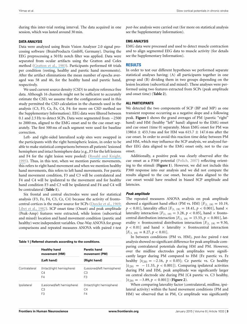
Table 1 | Referred channels according to the conditions.
Healthy hand
movement (HM)
Paretic hand
movement (PM)
(Left hand) (Right hand)
Contralateral (Intact/right hemisphere)C4F4
(Lesioned/left hemisphere)C3F3
Ipsilateral (Lesioned/left hemisphere)C3F3
(Intact/right hemisphere)C4F4
post-hoc analysis were carried out (for more on statistical analysissee the Supplementary Information).
EMG ANALYSISEMG data were processed and used to detect muscle contractionand to align segmented EEG data to muscle activity (for detailssee the Supplementary Information).
RESULTSIn order to test our different hypotheses we performed separatestatistical analyses having; (A) all participants together in onegroup and (B) dividing them in two groups depending on thelesion location (subcortical and mixed). These analyses were per-formed using two features extracted from SCPs (peak amplitudeand onset time) (Table 2).
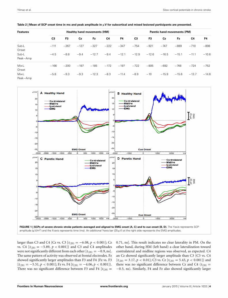
ALL PARTICIPANTSWe detected the two components of SCP (BP and MP) as onetransient response occurring as a negative slope and a followingpeak. Figure 1 shows the grand averages of PM (paretic “right”hand) and HM (healthy “left” hand) aligned to the EMG onsetand cue onset (time 0) separately. Mean EMG onset for PM was1380.6 ± 453.3 ms and for HM was 615.7 ± 147.6 ms after thecue onset. In order to avoid this reaction time delay between PMand HM, which may influence the SCP analysis, we analyzed fur-ther EEG data aligned to the EMG onset only, not to the cueonset.
Additionally, a positive peak was clearly observed after thecue onset as a P300 potential (Polich, 2007) reflecting orient-ing to the stimuli (Figure 1). However, we did not include thisP300 response into our analysis and we did not compare theresults aligned to the cue onset, because data aligned to thecue onset would have resulted in biased SCP amplitude andlatencies.
Peak amplitudeThe repeated measures ANOVA analysis on peak amplitudeshowed a significant hand effect (PM vs. HM) [F(1, 19) = 10.19,p < 0.01], laterality effect [F(1, 19) = 18.41, p < 0.001], hand ×laterality interaction [F(1, 19) = 3.28, p < 0.05], hand × fronto-central distribution interaction [F(1, 19) = 15.55, p < 0.001], lat-erality × frontocentral distribution interaction [F(1, 19) = 9.50,p < 0.01] and hand × laterality × frontocentral interaction[F(1, 19) = 8.27, p < 0.01].
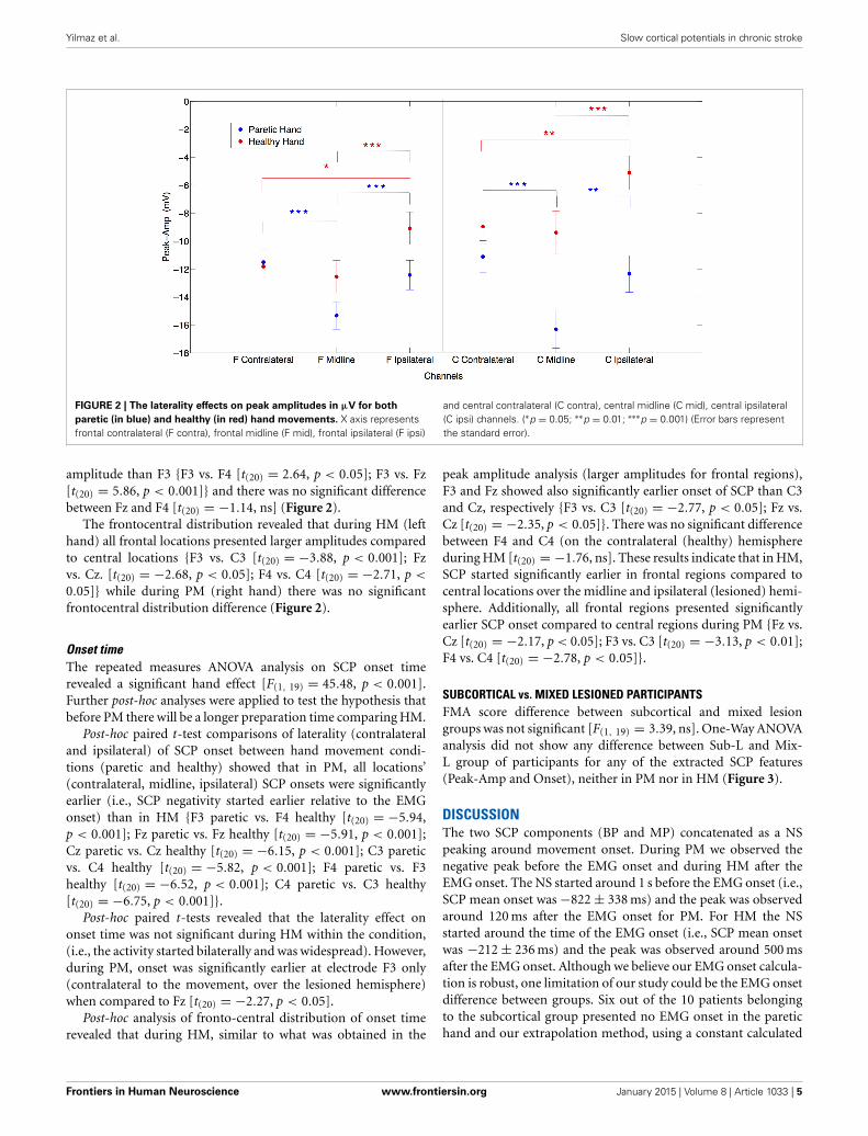
In between conditions (PM vs. HM), post-hoc paired t-testanalysis showed no significant difference for peak amplitude com-paring contralateral potentials during HM and PM. However,over the midline electrodes peak amplitudes were signifi-cantly larger during PM compared to HM {Fz paretic vs. Fzhealthy [t(20) = −2.16, p < 0.05]; Cz paretic vs. Cz healthy[t(20) = − 11.33, p < 0.001]}. Comparing ipsilateral activitiesduring PM and HM, peak amplitude was significantly largeron central electrode site during PM {C4 paretic vs. C3 healthy,[t(20) = −3.89, p < 0.001]} (Figure 2).
When comparing laterality factor (contralateral, midline, ipsi-lateral activity) within the hand movement conditions (PM andHM) we observed that in PM, Cz amplitude was significantly
Frontiers in Human Neuroscience www.frontiersin.org January 2015 | Volume 8 | Article 1033 | 3
Yilmaz et al. Slow cortical potentials in chronic stroke
Table 2 | Mean of SCP onset time in ms and peak amplitude in µV for subcortical and mixed lesioned participants are presented.
Features Healthy hand movements (HM) Paretic hand movements (PM)
C3 F3 Cz Fz C4 F4 C3 F3 Cz Fz C4 F4
Sub-LOnset
−111 −267 −127 −327 −222 −347 −754 −921 −747 −889 −710 −898
Sub-LPeak−Amp
−4.5 −8.8 −9.4 −12.7 −9.4 −12.1 −12.9 −12.6 −16.5 −15.1 −11.1 −10.6
Mix-LOnset
−166 −200 −187 −185 −172 −197 −722 −805 −692 −748 −724 −752
Mix-LPeak−Amp
−5.8 −9.3 −9.3 −12.3 −8.3 −11.4 −8.9 −10 −15.9 −15.6 −13.7 −14.6
FIGURE 1 | SCPs of severe chronic stroke patients averaged and aligned to EMG onset (A, C) and to cue onset (B, D). The Y-axis represents SCPamplitude (µV/m2) and the X-axis represents time (ms). An additional Y-axis bar (25 µV) at the right side represents the EMG amplitudes.
larger than C3 and C4 {Cz vs. C3 [t(20) = −6.08, p < 0.001]; Czvs. C4 [t(20) = −5.89, p < 0.001]} and C3 and C4 amplitudeswere not significantly different from each other [t(20) = −0.9, ns].The same pattern of activity was observed at frontal electrodes. Fzshowed significantly larger amplitudes than F3 and F4 {Fz vs. F3[t(20) = −5.51, p < 0.001]; Fz vs. F4 [t(20) = −4.06, p < 0.001]}.There was no significant difference between F3 and F4 [t(20) =
0.71, ns]. This result indicates no clear laterality in PM. On theother hand, during HM (left hand) a clear lateralization towardcontralateral and midline regions was observed, as expected. C4an Cz showed significantly larger amplitude than C3 {C3 vs. C4[t(20) = 3.17, p < 0.01]; C3 vs. Cz [t(20) = 5.43, p < 0.001]} andthere was no significant difference between Cz and C4 (t(20) =−0.5, ns). Similarly, F4 and Fz also showed significantly larger
Frontiers in Human Neuroscience www.frontiersin.org January 2015 | Volume 8 | Article 1033 | 4
Yilmaz et al. Slow cortical potentials in chronic stroke
FIGURE 2 | The laterality effects on peak amplitudes in µV for both
paretic (in blue) and healthy (in red) hand movements. X axis representsfrontal contralateral (F contra), frontal midline (F mid), frontal ipsilateral (F ipsi)
and central contralateral (C contra), central midline (C mid), central ipsilateral(C ipsi) channels. (∗p = 0.05; ∗∗p = 0.01; ∗∗∗p = 0.001) (Error bars representthe standard error).
amplitude than F3 {F3 vs. F4 [t(20) = 2.64, p < 0.05]; F3 vs. Fz[t(20) = 5.86, p < 0.001]} and there was no significant differencebetween Fz and F4 [t(20) = −1.14, ns] (Figure 2).
The frontocentral distribution revealed that during HM (lefthand) all frontal locations presented larger amplitudes comparedto central locations {F3 vs. C3 [t(20) = −3.88, p < 0.001]; Fzvs. Cz. [t(20) = −2.68, p < 0.05]; F4 vs. C4 [t(20) = −2.71, p <
0.05]} while during PM (right hand) there was no significantfrontocentral distribution difference (Figure 2).
Onset timeThe repeated measures ANOVA analysis on SCP onset timerevealed a significant hand effect [F(1, 19) = 45.48, p < 0.001].Further post-hoc analyses were applied to test the hypothesis thatbefore PM there will be a longer preparation time comparing HM.
Post-hoc paired t-test comparisons of laterality (contralateraland ipsilateral) of SCP onset between hand movement condi-tions (paretic and healthy) showed that in PM, all locations’(contralateral, midline, ipsilateral) SCP onsets were significantlyearlier (i.e., SCP negativity started earlier relative to the EMGonset) than in HM {F3 paretic vs. F4 healthy [t(20) = −5.94,p < 0.001]; Fz paretic vs. Fz healthy [t(20) = −5.91, p < 0.001];Cz paretic vs. Cz healthy [t(20) = −6.15, p < 0.001]; C3 pareticvs. C4 healthy [t(20) = −5.82, p < 0.001]; F4 paretic vs. F3healthy [t(20) = −6.52, p < 0.001]; C4 paretic vs. C3 healthy[t(20) = −6.75, p < 0.001]}.
Post-hoc paired t-tests revealed that the laterality effect ononset time was not significant during HM within the condition,(i.e., the activity started bilaterally and was widespread). However,during PM, onset was significantly earlier at electrode F3 only(contralateral to the movement, over the lesioned hemisphere)when compared to Fz [t(20) = −2.27, p < 0.05].
Post-hoc analysis of fronto-central distribution of onset timerevealed that during HM, similar to what was obtained in the
peak amplitude analysis (larger amplitudes for frontal regions),F3 and Fz showed also significantly earlier onset of SCP than C3and Cz, respectively {F3 vs. C3 [t(20) = −2.77, p < 0.05]; Fz vs.Cz [t(20) = −2.35, p < 0.05]}. There was no significant differencebetween F4 and C4 (on the contralateral (healthy) hemisphereduring HM [t(20) = −1.76, ns]. These results indicate that in HM,SCP started significantly earlier in frontal regions compared tocentral locations over the midline and ipsilateral (lesioned) hemi-sphere. Additionally, all frontal regions presented significantlyearlier SCP onset compared to central regions during PM {Fz vs.Cz [t(20) = −2.17, p < 0.05]; F3 vs. C3 [t(20) = −3.13, p < 0.01];F4 vs. C4 [t(20) = −2.78, p < 0.05]}.
SUBCORTICAL vs. MIXED LESIONED PARTICIPANTSFMA score difference between subcortical and mixed lesiongroups was not significant [F(1, 19) = 3.39, ns]. One-Way ANOVAanalysis did not show any difference between Sub-L and Mix-L group of participants for any of the extracted SCP features(Peak-Amp and Onset), neither in PM nor in HM (Figure 3).
DISCUSSIONThe two SCP components (BP and MP) concatenated as a NSpeaking around movement onset. During PM we observed thenegative peak before the EMG onset and during HM after theEMG onset. The NS started around 1 s before the EMG onset (i.e.,SCP mean onset was −822 ± 338 ms) and the peak was observedaround 120 ms after the EMG onset for PM. For HM the NSstarted around the time of the EMG onset (i.e., SCP mean onsetwas −212 ± 236 ms) and the peak was observed around 500 msafter the EMG onset. Although we believe our EMG onset calcula-tion is robust, one limitation of our study could be the EMG onsetdifference between groups. Six out of the 10 patients belongingto the subcortical group presented no EMG onset in the paretichand and our extrapolation method, using a constant calculated
Frontiers in Human Neuroscience www.frontiersin.org January 2015 | Volume 8 | Article 1033 | 5

Yilmaz et al. Slow cortical potentials in chronic stroke
FIGURE 3 | Grand average SCPs of the participants with mixed
(subcortical and cortical) lesion (A, C) and with subcortical lesion (A,
D). Contralateral (green), midline (black) and ipsilateral (red) electrodes
were shown for each movement condition. EMG activity is in blue. Anadditional Y-axis bar (25 µV) at the right side represents the EMGamplitudes.
on all patients from both groups presenting EMG onset, may beinappropriate.
The SCP NS started significantly earlier and lasted longer inpreparation for PM than HM and negligible preparation timewas detected before HM, indicating a significantly longer move-ment preparatory phase during PM. We assume earlier SCP onsetrepresents a longer neural computational effort to evoke the nec-essary brain excitation to induce a motor top-down commandduring PM. This result supports previous findings suggesting thatthe time between the onset of the SCP and the EMG onset mayindicate extended preparation (Tarkka and Hallett, 1990)—thebrain starts exciting motor networks (facilitating the neural fir-ing) for a significantly longer time before an EMG onset can beproduced on the paretic side compared to healthy hand motoractions.
Longer planning time was needed for PM. Individuals withsevere paralysis lack contingencies between volitional actionsand consequences. Such contingencies cannot therefore beused to drive reorganization within functional brain networks.Consequently, such individuals are prone to devolution toward
a maladaptive state indicative of learned disuse (Taub et al.,1999; Krakauer, 2006; Pomeroy et al., 2011). Therefore, motorintention contingency delivered by a BMI driven orthosis mayexplain the positive results of Ramos-Murguialday et al. workand should reduce the time lapse between SCP peak and EMGonset. Furthermore, a combination of SMR and SCP feed-back could improve BMI feedback optimizing the rehabilitationintervention.
Due to the severity of motor impairment in our partici-pants we expected to observe compensatory over-activation (i.e.,larger peak amplitudes) during motor preparation for executionattempts of PM compared to HM generated SCPs. The peakamplitude of SCP was significantly larger during PM over themidline (Fz, Cz) and ipsilateral central (C4) locations than dur-ing HM. It has been previously suggested that the amplitude of thenegativity indicates the brain’s computational demand to performthe movement (Jankelowitz and Colebatch, 2005). Therefore, ourresults denote a higher effort during PM. However, this higheractivity occurs over ipsilateral central areas and medial-fronto-central areas, which may be due to maladaptive (since participants
Frontiers in Human Neuroscience www.frontiersin.org January 2015 | Volume 8 | Article 1033 | 6

Yilmaz et al. Slow cortical potentials in chronic stroke
did not recover) over-activation of the healthy hemisphere and/orto the lack of cross-hemispheric inhibition from lesioned tonon-lesioned hemisphere.
On the other hand, between conditions we did not observeany significant difference in SCP amplitude at the electrodes con-tralateral to the movement during PM (C3) compared to HM(C4). We expected a decrease in amplitude of SCPs due to thelesion in the contralateral (ipsilesional) hemisphere during PMbut this was not the case. Cortical generators of SCP might notbe compromised and the recorded SCP amplitude during PMcould remain at similar levels if the SCP generated in subcorti-cal structures does not contribute significantly (due to volumeconduction) to the activity recorded with the EEG electrodes.
We observed that preparation of movement comes fromfrontal and not from central areas. The larger response on thehealthy side could reflect the non-use or atrophy of the lesionedside or it may indicate a compensatory over-activation on thehealthy side.
Following previous findings (Kitamura et al., 1996) we didnot find any difference in peak amplitude between contralateral(ipsilesional) and ipsilateral (contralesional) hemispheres dur-ing PM, while during HM the amplitude was significantly largerin contralateral compared to ipsilateral electrodes (Green et al.,1999). Since peak amplitude has been related with the computa-tional demand, one could expect either an increase in the peakamplitude on the ipsilesional side if cortex is intact (subcorticallesion), which is what happened in all participants, or a decreasein peak amplitude in primary motor areas and an increase inother non-lesioned secondary motor areas if cortex is lesioned(mixed lesion). We hypothesized that frontal and premotor areasin participants presenting mixed lesions (subcortical and corti-cal) would be more activated, due to the compensatory role ofthe secondary motor areas to overcome the loss of primary motorcortical structures in the participants presenting cortical lesions.However, we did not observe any significant difference betweenSub-L and Mix-L groups, neither in the peak amplitude nor inSCP onset during PM or HM. This may be due to the fact thatin participants presenting mixed lesions, the cortex was not heav-ily affected and peri-lesional areas could induce similar SCP peakamplitudes. However, since the top-down control of the motorneurons is not efficient and there is no afferent return confirm-ing the delivery of the efferent signal, other areas of the cortex(medial and ipsilateral) are excited (facilitation of neural firing)to exploit their connection to the motor neurons of the affectedmuscles, resulting in significantly higher SCPs amplitudes and inmaladaptive compensatory functional brain changes.
CONCLUSIONWe describe SCP changes in chronic severe stroke patients.Paralyzed participants showed significantly longer movementplanning time and significantly larger SCP peak amplitudes inmedial and ipsilateral sites during PM, indicating larger compu-tational demand during paretic hand intention to move. Non-use of the paretic hand induces over-activation of the healthyhemisphere with larger SCP amplitudes and, probably, strongermaladaptive inhibition of the lesioned side impeding corticalreorganization and motor rehabilitation. These changes were
independent of cortex integrity. SCP latency and peak amplitudein both hemispheres appear to be appropriate features to be usedand optimized in BMI-like neurorehabilitation interventions.
ACKNOWLEDGMENTSWe would like to thank our participants and the Stroke team. Thiswork was funded by the European Research Council (ERC) Grant227632, the Werner Reichardt Centre of Integrative Neuroscience(CIN), by the German Federal Ministry of Education andResearch (BMBF, Förderzeichen 01GQ0831) as well as theDeutsche Forschungsgemeinschaft (DFG), DAAD (DeutscherAkademischer Austauschdienst) and the Baden-WuerttembergStiftung (ROB-1), Baden-Württemberg Stiftung (ROB-1), theIndian-European collaborative research and technological devel-opment projects (INDIGO-DTB2-051), the Natural ScienceFundation of China (NSFC 31450110072).
SUPPLEMENTARY MATERIALThe Supplementary Material for this article can be foundonline at: http://www.frontiersin.org/journal/10.3389/fnhum.
2014.01033/abstract
REFERENCESBarrett, G., Shibasaki, H., and Neshige, R. (1986). Cortical potentials pre-
ceding voluntary movement: evidence for three periods of preparation inman. Electroencephalogr. Clin. Neurophysiol. 63, 327–339. doi: 10.1016/0013-4694(86)90017-9
Bashir, S., Mizrahi, I., Weaver, K., Fregni, F., and Pascual-Leone, A. (2010).Assessment and modulation of neural plasticity in rehabilitation with transcra-nial magnetic stimulation. PM &R 2, 253–268. doi: 10.1016/j.pmrj.2010.10.015
Birbaumer, N., Elbert, T., Canavan, A. G., and Rockstroh, B. (1991). Slow potentialsof the cerebral cortex and behavior. Physiol. Rev. 70, 1–41.
Birbaumer, N., Ghanayim, N., Hinterberger, T., Iversen, I., Kotchoubey, B., Kübler,A., et al. (1999). A spelling device for the paralysed. Nature 398, 297–298. doi:10.1038/18581
Chollet, F., DiPiero, V., Wise, R. J., Brooks, D. J., Dolan, R. J., and Frackowiak, R.S. (1991). The functional anatomy of motor recovery after stroke in humans:a study with positron emission tomography. Ann. Neurol. 29, 63–71. doi:10.1002/ana.410290112
Cui, R. Q., Huter, D., Lang, W., and Deecke, L. (1999). Neuroimage of voluntarymovement: topography of the Bereitschaftspotential, a 64-channel DC currentsource density study. Neuroimage 1, 124–134. doi: 10.1006/nimg.1998.0388
Cunnington, R., Iansek, R., and Bradshaw, J. L. (1999). Movement-related poten-tials in Parkinson’s disease: external cues and attentional strategies. Mov. Disord.14, 63–68.
Deecke, L. (1987). Bereitschaftspotential as an indicator of movement prepara-tion in supplementary motor area and motor cortex. Ciba Found. Symp. 132,231–250.
Deecke, L., Scheid, P., and Kornhuber, H. H. (1969). Distribution of readinesspotential, pre-motion positivity, and motor potential of the human cerebralcortex preceding voluntary finger movements. Exp. Brain Res. 7, 158–168. doi:10.1007/BF00235441
Elbert, T., and Rockstroh, B. (1987). Threshold regulation- a key to understandingof the combined dynamics of EEG and event-related potentials. J. Psychophysiol.4, 317–333.
Gratton, G., Coles, M. G., and Donchin, E. (1983). A new method for off-lineremoval of ocular artifact. Electroencephalogr. Clin. Neurophysiol. 55, 468–484.doi: 10.1016/0013-4694(83)90135-9
Green, J. B., Bialy, Y., Sora, E., and Ricamato, A. (1999). High-resolution EEG inpoststroke hemiparesis can identify ipsilateral generators during motor tasks.Stroke 30, 2659–2665. doi: 10.1161/01.STR.30.12.2659
Grefkes, C., Nowak, D. A., Eickhoff, S. B., Dafotakis, M., Küst, J., Karbe, H., et al.(2008). Cortical connectivity after subcortical stroke assessed with functionalmagnetic resonance imaging. Ann. Neurol. 63, 236–246. doi: 10.1002/ana.21228
Frontiers in Human Neuroscience www.frontiersin.org January 2015 | Volume 8 | Article 1033 | 7

Yilmaz et al. Slow cortical potentials in chronic stroke
Jankelowitz, S. K., and Colebatch, J. K. (2005). Movement related potentialsin acutely induced weakness and stroke. Exp. Brain Res. 161, 104–113. doi:10.1007/s00221-004-2051-6
Kitamura, J., Shibasaki, H., and Takeuchi, T. (1996). Cortical potentials precedingvoluntary elbow movement in recovered hemiparesis. Electroencephalogr. Clin.Neurophysiol. 98, 149–156. doi: 10.1016/0013-4694(95)00218-9
Kornhuber, H. H., and Deecke, L. (1965). Hirnpotentialeanderungen beiWillkuerbewegungen und passiven Bewegungen des Menschen: bere-itschaftspotential und reafferente Potentiale. Pfluegers Arch. 284, 1–17.doi: 10.1007/BF00412364
Krakauer, J. W. (2006). Motor learning: its relevance to stroke recovery and neu-rorehabilitation. Curr. Opin. Neurol. 19, 84–90. doi: 10.1097/01.wco.0000200544.29915.cc
Lang, W., Beisteiner, R., Lindinger, G., and Deecke, L. (1992). Changes of corticalactivity when executing learned motor sequences. Exp. Brain Res. 89, 435–440.doi: 10.1007/BF00228259
Lang, W., Lang, M., Podreka, I., Steiner, M., Uhl, F., Suess, E., et al. (1988).DC-potential shifts and regional cerebral blood flow reveal frontal cortexinvolvement in human visuomotor learning. Exp. Brain Res. 71, 353–364. doi:10.1007/BF00247495
Lang, W., Obrig, H., Lindinger, G., Cheyne, D., and Deecke, L. (1990).Supplementary motor area activation while tapping bimanually differentrhythms in musicians. Exp. Brain Res. 79, 504–514. doi: 10.1007/BF00229320
Libet, B. (1992). Voluntary acts and readiness potentials. Electroencephalogr. Clin.Neurophysiol. 82, 85–86. doi: 10.1016/0013-4694(92)90186-L
Libet, B., Wright, E. W., and Gleason, C. A. (1982). Readiness-potentials precedingunrestricted ‘spontaneous’ vs. pre-planned voluntary acts. Electroencephalogr.Clin. Neurophysiol. 54, 322–335. doi: 10.1016/0013-4694(82)90181-X
Murase, N., Duque, J., Mazzocchio, R., and Cohen, L. G. (2004). Influence of inter-hemispheric interactions on motor function in chronic stroke. Annu. Neurol.55, 400–409. doi: 10.1002/ana.10848
Murphy, T. H., and Corbett, D. (2009). Plasticity during stroke recovery: fromsynapse to behavior. Nat. Rev. Neurosci. 10, 861–872. doi: 10.1038/nrn2735
Paradiso, G., Cunic, D., Saint-Cyr, J. A., Hoque, T., Lozano, A. M., Lang, A. E.,et al. (2004). Involvement of human thalamus in the preparation of self-pacedmovement. Brain 127, 2717–2731. doi: 10.1093/brain/awh288
Polich, J. (2007). Updating P300: an integrative theory of P3a and P3b. Clin.Neurophysiol. 118, 2128–2148. doi: 10.1016/j.clinph.2007.04.019
Pomeroy, V., Aglioti, S. M., Mark, V. W., McFarland, D., Stinear, C., Wolf, S.L., et al. (2011). Neurological principles and rehabilitation of action disor-ders: rehabilitation interventions. Neurorehabil. Neural Repair. 25, 33–43. doi:10.1177/1545968311410942
Ramos-Murguialday, A., Broetz, D., Rea, M., Läer, L., Yilmaz, Ö., Brasil, F. L., et al.(2013). Brain–machine interface in chronic stroke rehabilitation: a controlledstudy. Ann. Neurol. 74, 100–108. doi: 10.1002/ana.23879
Rektor, I. (2002). Scalp-recorded Bereitschaftspotential is the result of the activ-ity of cortical and subcortical generators–a hypothesis. Clin. Neurophysiol. 113,1998–2005. doi: 10.1016/S1388-2457(02)00286-9
Rosahl, S. K., and Knight, R. T. (1995). Role of prefrontal cortex in generation ofthe contingent negative variation. Cereb. Cortex 5, 123–134. doi: 10.1093/cer-cor/5.2.123
Rossini, P. M., Calautti, C., Pauri, F., and Baron, J. C. (2003). Post-strokeplastic reorganisation in the adult brain. Lancet Neurol. 2, 493–502. doi:10.1016/S1474-4422(03)00485-X
Sasaki, K., and Gemba, H. (1984). Compensatory motor function of thesomatosensory cortex for the motor cortex temporarily impaired by cooling inthe monkey. Exp. Brain Res. 55, 60–68. doi: 10.1007/BF00240498
Shibasaki, H., and Hallett, M. (2006). What is the Bereitschaftspotential?. Clin.Neurophysiol. 117, 2341–2356. doi: 10.1016/j.clinph.2006.04.025
Simonetta, M., Clanet, M., and Rascol, O. (1991). Bereitschaftspotential in asimple movement or in a motor sequence starting with the same simple move-ment. Electroencephalogr. Clin. Neurophysiol. 81, 129–134. doi: 10.1016/0168-5597(91)90006-J
Tarkka, I. M., and Hallett, M. (1990). Cortical topography of premotor andmotor potentials preceding self-paced, voluntary movement of dominant andnon-dominant hands. Electroencephalogr. Clin. Neurophysiol. 75, 36–43. doi:10.1016/0013-4694(90)90150-I
Taub, E., Uswatte, G., and Pidikiti, R. (1999). Constraint-InducedMovement Therapy: a new family of techniques with broad applica-tion to physical rehabilitation–a clinical review. J. Rehabil. Res. Dev. 36,237–251.
Verleger, R., Adam, S., Rose, M., Vollmer, C., Wauschkuhn, B., and Kömpf,D. (2003). Control of hand movements after striatocapsular stroke:high-resolution temporal analysis of the function of ipsilateral activa-tion. Clin. Neurophysiol. 114, 1468–1476. doi: 10.1016/S1388-2457(03)00125-1
Ward, N. S. (2011). Assessment of cortical reorganisation for hand func-tion after stroke. J. Physiol. 589, 5625–5632. doi: 10.1113/jphysiol.2011.220939
Conflict of Interest Statement: The authors declare that the research was con-ducted in the absence of any commercial or financial relationships that could beconstrued as a potential conflict of interest.
Received: 16 October 2014; accepted: 09 December 2014; published online: 15 January2015.Citation: Yilmaz O, Birbaumer N and Ramos-Murguialday A (2015) Movementrelated slow cortical potentials in severely paralyzed chronic stroke patients. Front.Hum. Neurosci. 8:1033. doi: 10.3389/fnhum.2014.01033This article was submitted to the journal Frontiers in Human Neuroscience.Copyright © 2015 Yilmaz, Birbaumer and Ramos-Murguialday. This is an open-access article distributed under the terms of the Creative Commons Attribution License(CC BY). The use, distribution or reproduction in other forums is permitted, providedthe original author(s) or licensor are credited and that the original publication in thisjournal is cited, in accordance with accepted academic practice. No use, distribution orreproduction is permitted which does not comply with these terms.
Frontiers in Human Neuroscience www.frontiersin.org January 2015 | Volume 8 | Article 1033 | 8
Related Documents


