Formation of Cortical Fields on a Reduced Cortical Sheet Kelly J. Huffman, 1 Zolta ´ n Molna ´ r, 2,3 Anton Van Dellen, 2 Dianna M. Kahn, 1 Colin Blakemore, 2 and Leah Krubitzer 1 1 Center for Neuroscience and Department of Psychology, University of California, Davis, Davis, California 95616, 2 University Laboratory of Physiology, University of Oxford, England OX1 3PT, and 3 Institut de Biologie Cellulaire et de Morphologie ´ Faculte ´ de Medecine, 1005 Lausanne, Switzerland Theories of both cortical field development and cortical evolu- tion propose that thalamocortical projections play a critical role in the differentiation of cortical fields (O’Leary, 1989; Krubitzer, 1995). In the present study, we examined how changing the size of the immature neocortex before the establishment of thalamocortical connections affects the subsequent develop- ment and organization of the adult neocortex. This alteration in cortex is consistent with one of the most profound changes made to the mammalian neocortex throughout evolution: cor- tical size. Removing the caudal one-third to three-fourths of the cortical neuroepithelial sheet unilaterally at an early stage of development in marsupials resulted in normal spatial relation- ships between visual, somatosensory, and auditory cortical fields on the remaining cortical sheet. Injections of neuroana- tomical tracers into the reduced cortex revealed in an altered distribution of thalamocortical axons; this alteration allowed the maintenance of their original anteroposterior distribution. These results demonstrate the capacity of the cortical neuroepithe- lium to accommodate different cortical fields at early stages of development, although the anteroposterior and mediolateral relationships between cortical fields appear to be invariant. The shifting of afferents and efferents with cortical reduction or expansion at very early stages of development may have oc- curred naturally in different lineages over time and may be sufficient to explain much of the phenotypic variation in cortical field number and organization in different mammals. Key words: cerebral cortex; visual cortex; cortical organiza- tion; development; evolution; electrophysiology; Monodelphis domestica The mammalian cerebral cortex is composed of separate cortical fields that have distinct architectonic appearances, patterns of connectivity, and physiological properties (Brodmann, 1909; Kaas, 1982). How these fields develop and how development is altered in different mammalian lineages to generate the remark- able variation in brain size and cortical field number is funda- mental to understanding how brains are constructed in evolution. One approach to unraveling this mystery is to examine the prod- ucts of the evolutionary process to determine common patterns of brain organization as well as to identify modifications to this common plan. By comparing neocortical organization across spe- cies, it is possible to gain insights into the constraints imposed on the developing nervous system that limit the types of modifica- tions that can be made to existing patterns of organization (Kru- bitzer, 1995). A second approach to understanding brain evolution is to examine the developmental process itself (Killackey, 1990; Wray, 1994). By investigating the developmental mechanisms that ac- count for the existing organization of the neocortex in extant mammals, we can infer how homologous developmental processes have been modified to yield the differences in cortical organiza- tion observed across mammals. Comparative work has demonstrated that one of the most dramatic changes to the neocortex is an increase in the size of the cortical sheet (Kaas, 1982; Ridgway, 1986; Kaas, 1988; Stephan et al., 1988; Krubitzer, 1995; Manger et al., 1998). This change is associated with an increase in cortical field number and has led us to question how changing the size of the developing cortical sheet would affect the subsequent cortical organization. Currently, there is some dispute over when cortical fields are differentiated in development and how cortical fields are speci- fied. One proposition is that cortical fields can begin the process of compartmentalization very early in development in the ven- tricular zone, well before cortical neurons are born and before any connections are made (Rakic, 1988; DeHay et al., 1993; Polleux et al., 1997a,b) (for review, see Levitt et al., 1995; Rakic, 1995b). A second view is that the developing neocortex is unspec- ified, to a large extent, and that connections from the thalamus and other sources contribute to cortical field generation (Chang et al., 1986; O’Leary, 1989; Killackey, 1990; Roe et al., 1990; Molna ´r and Blakemore, 1991, 1995; Schlaggar and O’Leary, 1991; O’Leary et al., 1994). Although these theories are historically relevant and allow us to formulate testable hypotheses regarding the laminar and tangential differentiation of the cerebral cortex, neither explanation of cortical field specification is likely to be exclusive. Rather, some combination of early regional differenti- ation as well as thalamic afferent contribution is ultimately re- sponsible for cortical field differentiation in development and the resultant cortical organization in adults (for review, see O’Leary et al., 1994). Received June 30, 1999; revised Sept. 3, 1999; accepted Sept. 3, 1999. This project was supported by grants to Leah Krubitzer from National Institutes of Health (1R01 NS35103-01A1) and the Whitehall Foundation (M97-20), and by a Novartis Bursary award, a McDonnell Pew Centre for Cognitive Neuroscience Oxford, North American Network grant to Zolta ´n Molna ´r, and a Leverhulme research grant and a Medical Research Council Grant (G9320738) to Colin Blake- more. We thank Ken Britten, Barbara Chapman, Bruno Olshausen, and Gregg Recanzone for helpful comments on this manuscript. Correspondence should be addressed to Leah Krubitzer, Center for Neuroscience, 1544 Newton Court, Davis, California 95616. E-mail: [email protected]. Copyright © 1999 Society for Neuroscience 0270-6474/99/199939-14$05.00/0 The Journal of Neuroscience, November 15, 1999, 19(22):9939–9952

Welcome message from author
This document is posted to help you gain knowledge. Please leave a comment to let me know what you think about it! Share it to your friends and learn new things together.
Transcript

Formation of Cortical Fields on a Reduced Cortical Sheet
Kelly J. Huffman,1 Zoltan Molnar,2,3 Anton Van Dellen,2 Dianna M. Kahn,1 Colin Blakemore,2 andLeah Krubitzer1
1Center for Neuroscience and Department of Psychology, University of California, Davis, Davis, California 95616,2University Laboratory of Physiology, University of Oxford, England OX1 3PT, and 3Institut de Biologie Cellulaire et deMorphologie Faculte de Medecine, 1005 Lausanne, Switzerland
Theories of both cortical field development and cortical evolu-tion propose that thalamocortical projections play a critical rolein the differentiation of cortical fields (O’Leary, 1989; Krubitzer,1995). In the present study, we examined how changing the sizeof the immature neocortex before the establishment ofthalamocortical connections affects the subsequent develop-ment and organization of the adult neocortex. This alteration incortex is consistent with one of the most profound changesmade to the mammalian neocortex throughout evolution: cor-tical size. Removing the caudal one-third to three-fourths of thecortical neuroepithelial sheet unilaterally at an early stage ofdevelopment in marsupials resulted in normal spatial relation-ships between visual, somatosensory, and auditory corticalfields on the remaining cortical sheet. Injections of neuroana-tomical tracers into the reduced cortex revealed in an altered
distribution of thalamocortical axons; this alteration allowed themaintenance of their original anteroposterior distribution. Theseresults demonstrate the capacity of the cortical neuroepithe-lium to accommodate different cortical fields at early stages ofdevelopment, although the anteroposterior and mediolateralrelationships between cortical fields appear to be invariant. Theshifting of afferents and efferents with cortical reduction orexpansion at very early stages of development may have oc-curred naturally in different lineages over time and may besufficient to explain much of the phenotypic variation in corticalfield number and organization in different mammals.
Key words: cerebral cortex; visual cortex; cortical organiza-tion; development; evolution; electrophysiology; Monodelphisdomestica
The mammalian cerebral cortex is composed of separate corticalfields that have distinct architectonic appearances, patterns ofconnectivity, and physiological properties (Brodmann, 1909;Kaas, 1982). How these fields develop and how development isaltered in different mammalian lineages to generate the remark-able variation in brain size and cortical field number is funda-mental to understanding how brains are constructed in evolution.One approach to unraveling this mystery is to examine the prod-ucts of the evolutionary process to determine common patterns ofbrain organization as well as to identify modifications to thiscommon plan. By comparing neocortical organization across spe-cies, it is possible to gain insights into the constraints imposed onthe developing nervous system that limit the types of modifica-tions that can be made to existing patterns of organization (Kru-bitzer, 1995).
A second approach to understanding brain evolution is toexamine the developmental process itself (Killackey, 1990; Wray,1994). By investigating the developmental mechanisms that ac-count for the existing organization of the neocortex in extantmammals, we can infer how homologous developmental processes
have been modified to yield the differences in cortical organiza-tion observed across mammals.
Comparative work has demonstrated that one of the mostdramatic changes to the neocortex is an increase in the size of thecortical sheet (Kaas, 1982; Ridgway, 1986; Kaas, 1988; Stephan etal., 1988; Krubitzer, 1995; Manger et al., 1998). This change isassociated with an increase in cortical field number and has led usto question how changing the size of the developing cortical sheetwould affect the subsequent cortical organization.
Currently, there is some dispute over when cortical fields aredifferentiated in development and how cortical fields are speci-fied. One proposition is that cortical fields can begin the processof compartmentalization very early in development in the ven-tricular zone, well before cortical neurons are born and beforeany connections are made (Rakic, 1988; DeHay et al., 1993;Polleux et al., 1997a,b) (for review, see Levitt et al., 1995; Rakic,1995b). A second view is that the developing neocortex is unspec-ified, to a large extent, and that connections from the thalamusand other sources contribute to cortical field generation (Changet al., 1986; O’Leary, 1989; Killackey, 1990; Roe et al., 1990;Molnar and Blakemore, 1991, 1995; Schlaggar and O’Leary, 1991;O’Leary et al., 1994). Although these theories are historicallyrelevant and allow us to formulate testable hypotheses regardingthe laminar and tangential differentiation of the cerebral cortex,neither explanation of cortical field specification is likely to beexclusive. Rather, some combination of early regional differenti-ation as well as thalamic afferent contribution is ultimately re-sponsible for cortical field differentiation in development and theresultant cortical organization in adults (for review, see O’Learyet al., 1994).
Received June 30, 1999; revised Sept. 3, 1999; accepted Sept. 3, 1999.This project was supported by grants to Leah Krubitzer from National Institutes
of Health (1R01 NS35103-01A1) and the Whitehall Foundation (M97-20), and by aNovartis Bursary award, a McDonnell Pew Centre for Cognitive NeuroscienceOxford, North American Network grant to Zoltan Molnar, and a Leverhulmeresearch grant and a Medical Research Council Grant (G9320738) to Colin Blake-more. We thank Ken Britten, Barbara Chapman, Bruno Olshausen, and GreggRecanzone for helpful comments on this manuscript.
Correspondence should be addressed to Leah Krubitzer, Center for Neuroscience,1544 Newton Court, Davis, California 95616. E-mail: [email protected] © 1999 Society for Neuroscience 0270-6474/99/199939-14$05.00/0
The Journal of Neuroscience, November 15, 1999, 19(22):9939–9952

To examine the contribution of developing thalamocorticalafferents to cortical field specification, we removed a large portionof the cortical neuroepithelium in Monodelphis domestica beforethe arrival of thalamocortical afferents, which occurs at postnatalday 7 (P7) (Molnar et al., 1998). On the basis of our observationsfrom comparative studies, along with a wealth of studies oncortical development, we predicted that the removal of a rela-tively large portion of the cortical neuroepithelial sheet would notabolish cortical fields that normally reside in the removed cortex.Rather, such a manipulation might result in the emergence ofcortical fields and thalamic afferents in appropriate geometricalignment on the remaining cortical sheet. A different possibleoutcome would be the loss of certain cortical fields, withoutsubstantially affecting any other cortical fields or related thalamicnuclei (Cunningham et al., 1987).
In the series of experiments reported here, we removed a largeportion of the immature neocortex in Monodelphis domestica atP4 (see Fig. 1b), which corresponds to embryonic day 14 (E14) inrats (Molnar et al., 1998). In the adult manipulated animals,electrophysiological recordings, neuroanatomical connections,and architectonic analyses were used to assess the organizationand thalamocortical connections of the remaining cortex.
Preliminary results from this study have been published previ-ously (Huffman et al., 1998; Krubitzer et al., 1998).
MATERIALS AND METHODSAnimals. Four days after birth (P4), two adult Monodelphis domesticamothers (Fig. 1a) were immobilized with an initial dose of alphaxalone(45 mg/kg) and alphadolone (15 mg/kg); this anesthetic does not passinto the mother’s milk. Subsequent doses of one-half of the initial dosewere given as needed to maintain anesthesia. The individual young (Fig.1b) were anesthetized hypothermically by placing ice on each pup justbefore surgery. Once anesthetized, the skin over the skull was cut andretracted (Fig. 1c), the developing skull was cut, and between one-thirdand three-fourths of the posterior cortical neuroepithelium was manuallyexcised unilaterally under microscopic guidance using microsurgical in-struments (Fig. 1d). The skull flap was repositioned, and the skin washeld in place with Nexaband (Veterinary Products Laboratories, Phoe-nix, AZ). The initial manipulations were performed under aseptic con-ditions, and the chronic surgery in adults was performed under standardsterile conditions. All experimental protocols were approved by theAnimal Use and Care Administrative Advisory Committee of the Uni-versity of California, Davis.
Of the two litters in which P4 animals were manipulated, one mothercannibalized the young, a behavior that is common in these animals incaptivity. In the one remaining litter, four animals were used for elec-trophysiological recording, and one animal was used for studyingthalamocortical connections. Experiments were conducted 8–12 monthsafter the initial P4 manipulations. A total of 9 normal animals were usedto study the functional organization (n 5 6) and connections (n 5 3) ofthe cortex, and three normal animals were used for volumetric measure-ments of the diencephalon (see below).
After the animals reached adulthood, the neocortex was surveyedusing multiunit electrophysiological recording techniques, similar tothose used to identify the location and internal organization of corticalfields in various mammals (Krubitzer et al., 1993). By delineating stim-ulus preference, receptive field size, and receptive field configuration, wecould subdivide the neocortex into separate cortical fields (Huffman etal., 1999). In one of the animals, the thalamocortical connections of thereduced neocortex were investigated by placing the fluorescent tracersdiamidino yellow (DY) 1 nuclear yellow (NY), fluororuby (FR), and
Figure 1. Photograph of an adult short-tailed opossum, Monodelphisdomestica (a), and a P4 infant (b). To document the surgical procedures,a CCD camera (Optronics) was used to produce digital images of the P4infants while they were attached to the mother’s nipple during our
4
procedure (c). A midline incision was made, and the skin and skull wereretracted in preparation for the cortical neuroepithelial reduction. Afterthe lesion was made with the excision tool (d), the skin was repositionedwith forceps and held together with glue. Scale bar, 1 mm.
9940 J. Neurosci., November 15, 1999, 19(22):9939–9952 Huffman et al. • Formation of Cortical Fields

fluoroemerald (FE) into the neocortex and identifying retrogradelylabeled cell bodies in the thalamus.
For electrophysiological recording experiments the animals were anes-thetized with ketamine hydrochloride (40 mg/kg, i.m.), xylazine (5mg/kg, i.m.), and sodium pentobarbital (20 mg/kg, i.p.). Once anesthe-tized, the dorsal surface of the neocortex was completely exposed, anddigital images of the exposed cortex were printed so that recording sitescould be matched to blood vessel patterns. Tungsten electrodes (5 MV)designed to record from small cell clusters were lowered into the brain ata depth of 200–400 mm at a number of closely spaced recording sites. Ateach site, sensory stimulation consisted of small indentations of theglabrous and hairy skin with fine probes, light brushing of the glabrousskin, displacing of hairs, and hard taps to the skin and body for somato-sensory stimulation. For visual stimulation, full-field flashes of light,moving bars of light, and flickering light were used to evoke neuralresponses. Clicks and high-frequency taps produced by tapping metal orwood objects together were used for auditory stimulation. After thecompletion of experiments, probes were placed in the cortex at severallocations for later identification in histologically processed tissue.
For neuroanatomical tracing experiments, the animals were anesthe-tized as describe above, and then 0.3 ml of 7% FE and of 7% FR wereinjected into the neocortex. The DY plus NY were placed in the neo-cortex as crystals (Krubitzer et al., 1993). The animals were allowed tosurvive for 1 week for transport of tracers.
After the completion of both the electrophysiological recording exper-iments and neuroanatomical tracing experiments, the animals were per-fused with 0.9% saline, followed by 3% paraformaldehyde in phosphatebuffer, pH 7.4, and then 3% paraformaldehyde in 10% sugar phosphate
buffer. When perfusion was complete, the brains were removed from thecranium and immersed in 30% sugar phosphate buffer overnight. Thebrains were sectioned coronally on a freezing microtome into 50–60 mmsections and alternately stained for Nissl substance, cytochrome oxidase(CO) (Carroll and Wong-Riley, 1984), and in some cases myelin (Gall-yas, 1979). In the case in which injections of neuroanatomical tracerswere made, an alternate series of sections was mounted for fluorescentmicroscopy.
Histolog ical and electrophysiolog ical data analysis. In all cases, digitalimages of the brain in which electrode tracks and probes were markedwere correlated with histologically processed tissue. By examining recep-tive field progressions for neurons in somatosensory cortex and delineat-ing different territories that represented different types of sensory input,maps of the cortex were made, and in normal animals they were relatedto architectonic boundaries (Huffman et al., 1999). For the four caseswith early lesions, the physiological maps alone were used to determinethe location and organization of sensory fields because the corticalarchitecture was difficult to identify after the numerous electrode pene-trations, and the architecture was often irregular (see below). In the onecase in which anatomical tracers were placed in the neocortex, theinjection site locations and spread were reconstructed from serial sectionsand transposed onto a digital image of the cortex. From the series ofsections mounted for fluorescent microscopy, the thalamic cell bodieslabeled with FE, DY 1 NY, and FR were plotted in x–y coordinatesusing a fluorescent microscope equipped with a digitizer attached to apersonal computer. Using blood vessels and other tissue landmarks, thelocations of plotted cell bodies were determined by transposing thethalamic nuclear boundaries from tissue stained for Nissl and CO.
To determine the relative contribution from each major sensory pro-jection nucleus in the thalamus to a circumscribed location of the cortex,back-labeled cells in the thalamus were counted, and percentages werecomputed for each of the tracers used. All of the labeled cells in thethalamus for each tracer were counted in nine alternate sections (onesection every 150 mm) where all labeled cells were plotted, and nuclearboundaries were determined. For each tracer, the total number of cellsfor the series within the dorsal division of the lateral geniculate nucleus(LGd), the medial geniculate nucleus (MG), and the ventral posteriornucleus (VP) was determined, and the percentage of the total number oflabeled cells was computed (see Table 1).
To determine whether the changes in the size of the diencephalonipsilateral to the lesion were significantly different from normal, volumet-ric measurements of the diencephalon in three normal animals and threeof the animals that underwent cortical removals were made. Two malesand one female were used in each group. Weights ranged from 80 to 130gm. Volume estimates were determined based on samples from everythird section (stained for Nissl). To make these volumetric measure-ments, it was necessary to appoint equal anterior and posterior levels ofthe diencephalon across cases. The anterior level was assigned as thelevel at which the habenula began. The posterior level was assigned as thelevel at which the superior colliculus (SC) and the MG were both presentin the same section. The area of the ipsilateral diencephalon was mea-sured using the software NIH image 1.61. Volume estimates were cal-culated by summing the product of the surface area ( A) and the distance( D) between sampled sections, across all sections sampled. This isexpressed as
V 5 (i 5 1
n
(Ai 3 D).
n 5 the total number of sections sampled. After all volumes weredetermined, a one-way ANOVA was performed to assess groupdifferences.
RESULTSReductions in the size of the neuroepithelial sheet before thearrival of thalamic afferents had five effects on the adult neocortexand subcortical structures. First, visual and auditory fields formedon the remaining cortical sheet in a position not normally occu-pied by these fields. Second, the size of some of the cortical fieldson the remaining cortical sheet was reduced. Third, the thalamo-cortical afferents retained their normal relative spatial relation-ships with the diminished cortex. Fourth, the laminar architectureof the cortex, as defined using Nissl, CO, and myelin stains
Figure 2. a, Photograph of a lateral view of the brain of a normal adultshort-tailed opossum and ( b) an adult short-tailed opossum that had thecaudal one-third of the right developing neocortex removed at P4. Thearrows in both figures indicate the caudal pole of each hemisphere in thenormal cortex and the manipulated cortex. Note that the caudal pole ofthe remaining cortex (b) is far rostral to its location in the normal animaland compared with the opposite hemisphere. Rostral is to the right, anddorsal is to the top. Scale bar, 1 mm.
Huffman et al. • Formation of Cortical Fields J. Neurosci., November 15, 1999, 19(22):9939–9952 9941

appeared irregular in most of the regions of cortex. Fifth, theentire dorsal thalamus and superior colliculus on the side of thebrain in which the cortex was removed were reduced.
Electrophysiological recordingAfter the removal of one-third to three-fourths of the neuroepi-thelial sheet, electrophysiological recording techniques were usedto determine the gross functional organization of the remainingneocortex in the adult (Figs. 2, 3). In normal marsupials, theprimary visual field (V1) occupies approximately one-third of thecortex and is located at the caudomedial pole (Kahn et al., 1999;Rosa et al., 1999). The primary somatosensory field (S1) islocated rostral to V1 and is somewhat smaller in size than V1(Huffman et al., 1999). Although the primary auditory field (A1)has not been described fully in Monodelphis, a densely myelinatedregion of cortex in which neurons respond only to auditorystimulation has been identified in the location of A1 in othermarsupials (Gates and Aitkin, 1982; Aitkin et al., 1986). We term
the field A because of uncertainties in homology with A1 in othermammals. Cortex just rostral and lateral to V1 (termed here V) isdominated by visual inputs, whereas cortex medial to A androstral to V is dominated by auditory inputs (A), although a fewsites contained neurons that responded to visual stimulation.Cortex lateral to S1 contains neurons responsive to both somato-sensory and auditory stimulation (Fig. 3a,b).
In the cases in which the cortical neuroepithelium was reducedby approximately one-third to one-half [97-22, data not shown;97-18, moderate removal (Fig. 3c,d)], some aspects of organiza-tion appeared similar to that of normally organized brains ofMonodelphis, and other aspects were different. For example, wewere able to identify three major sensory regions that exclusivelyrepresented visual, auditory, or somatosensory inputs, which weterm V, A, and S1, respectively. Because we did not obtain aretinotopic map of visual cortex, we cannot say that this region isV1, like that described in normal animals. However, despite the
Figure 3. Electrophysiological recording results from a right cortical hemisphere of a normal adult (a, b) and two right cortical hemispheres of adultsthat underwent removal of a portion of the cortical neuroepithelium at P4 (c–f ). The illustrations to the lef t depict recording sites (black dots); thin linesrepresent physiological boundaries that enclose regions of the cortex in which neurons responded to the same sensory modality. The illustrations at theright denote the primary sensory fields, including the primary visual, primary somatosensory, and presumptive primary auditory field (V1, S1, A) in thenormal animal (b), and the pure visual, somatosensory, and auditory fields (V, S1, and A) in the animals with moderate (d) and large ( f) cortical removals.In the normal animal, thick dark lines (a, b) denote architectonic boundaries. Despite the large removals of the developing neocortex, the pattern ofgeneral rostrocaudal and mediolateral organization of cortical fields, although compressed, was relatively normal. A noteworthy change in the neocortexwas that as the extent of the reduction increased, the relative amount of multimodal cortex increased. Scale bar, 1 mm. V or Vis, Visual; A or Aud,auditory; S, somatosensory; A1S or A/S, auditory and somatosensory; A1V or V/A, auditory and visual; V/A/S, visual, auditory, and somatosensory; wA,weak auditory; wS, weak somatosensory; V1, visual 1 other sensory input; x, no response; CT, caudal temporal field; m, medial; r, rostral.
9942 J. Neurosci., November 15, 1999, 19(22):9939–9952 Huffman et al. • Formation of Cortical Fields

fact that all or most of what would be visual cortex was removed,a region of cortex containing neurons that responded exclusivelyto visual stimulation was located in the caudomedial portion ofthe remaining cortex. Thus, the relative position of fields on theremaining cortical sheet was similar to normal animals. Unlike innormal animals, there appeared to be more cortex that wasdominated by mixed inputs such as auditory and visual. Also, asecond zone of pure auditory inputs was identified caudally, in alocation that was separate from A described in normal animals.Finally, the relative size of V was substantially reduced, whereasthe relative size of S1 appeared only slightly smaller than innormal animals. The relative amount of cortical space occupiedby pure auditory inputs appeared larger.
In cases in which substantially more cortex was removed [e.g.,approximately three-fourths of the cortical neuroepithelial sheet;98-4, data not shown, and 98-3 (Fig. 3e, f, Large Removal)],including all of the putative visual neuroepithelium and much ofthe putative auditory neuroepithelium, we still identified regionsof the cortex that were dominated by visual, auditory, or somato-sensory inputs. However, the region in which neurons respondedonly to visual stimulation, and no other modality, was identifiedonly at a single recording site. Although the presence of a purelyvisual cortical field is questionable, the caudal portion of theremaining cortex did contain neurons that responded to visualstimulation in addition to somatosensory and auditory stimula-tion (Fig. 3e, f). This “visual cortex” is located in a rostralposition in comparison to normals. Cortex rostral to this con-tained neurons that responded to auditory stimulation alone,auditory and somatosensory stimulation, or somatosensory stim-ulation alone. As in the case of the moderate removal, the cortexthat resided between the major sensory domains was multimodal.
Thalamocortical connectionsInjections of anatomical tracers into rostral, middle, and caudallocations in the cortex in which approximately one-half of the
neuroepithelium was removed demonstrated that thalamocorticalafferents maintain a normal relative pattern in terms of theirgross rostrocaudal organization (Fig. 4). Specific patterns of tha-lamic afferents could not be ascertained because injections werenot placed under electrophysiological guidance. We purposelyinjected large amounts of tracer to ensure success in backlabelingcells in the thalamus and to allow us to examine large scalethalamocortical topographic relationships. An injection of DY 1NY in the far rostral pole of the neocortex (Fig. 4a) resulted in alarge number (72%) of retrogradely labeled cells in the VP of thethalamus (Figs. 4c, 5; Table 1). VP is one of the major thalamicnuclei associated with processing somatic inputs. Twenty-onepercent of the retrogradely labeled DY 1 NY cells were alsoobserved just dorsal to VP (in the posterior nucleus), and only7% were identified in the lateral geniculate nucleus (Fig. 5, LGN;Table 1). This injection was in the expected location of S1 aselectrophysiological studies in other cases demonstrate, but wasvery large and likely to have spread into portions of visual cortexas well. No labeled cell bodies were identified in the MG from thisinjection.
The injection of FR into a location just caudal to the DY 1 NYinjection resulted in 63% of the labeled cells in VP, 14% in MG,and 6% in LGd (Table 1). This injection was in the expectedlocation of somatosensory cortex, although it spread into theexpected locations of auditory and multimodal cortex. The threesmall injections of FE in the caudal pole of cortex were placed inthe expected location of visual cortex, or multimodal cortex,which includes neurons responsive to visual stimulation. Forty-eight percent of the retrogradely labeled cell bodies were found inthe LGd (Figs. 4b, 5), 40% were in VP, and 4% were in MG. Theinjections of bi-directional tracers FE and FR also labeled axonsin the cerebral peduncle. The remaining labeled neurons in thethalamus that were not found in the major projection nuclei werefound dorsal to the posterior portion of VP (in the posterior
Figure 4. a, Digital image of a brain(case 98-31) that underwent removal ofthe cortical neuroepithelium at P4, and asuperimposition, using the softwareAdobe Photoshop 4.0, of the center andspread of the injection sites on the leftcortical hemisphere. Scale bar, 1 mm. Theinjections included DY 1 NY ( yellow)into a rostral portion of cortex, FR (red)into a middle portion of cortex, and threeinjections of FE ( green) into the caudalpole of the cortex. b, Digital image ofretrogradely labeled cells in the dorsaldivision of the LGN resulting from aninjection of FE into the caudal pole of theremaining cortex, in the expected locationof visual cortex. Scale bar for b, c, 50 mm.c, Digital image of retrogradely labeledcells in the VP resulting from an injectionof DY 1 NY in the presumptive somato-sensory/multimodal cortex.
Huffman et al. • Formation of Cortical Fields J. Neurosci., November 15, 1999, 19(22):9939–9952 9943

Figure 5. A series of sections from anterior to posterior through the thalamus of case 98-31 (a–f ). Each dot represents a retrogradely labeled cell bodyfrom a cortical injection in the left hemisphere. Yellow dots are cells labeled with DY 1 NY, red dots are cells labeled with FR, and dark green dots arecells labeled with FE. Thin lines represent nuclear boundaries determined by architectonic analyses of alternate, neighboring sections stained for Nisslor CO. Throughout the thalamus, most of the DY 1 NY-labeled cells were located in VP, although some of these cells were also found in the LGd.Most FR cells were observed in the VP, although some were seen in the MG and a few were observed in LGd. Labeled cells resulting from the mostcaudal set of injections in the cortex (FE) were mostly found in the LGd, demonstrating that although the entire occipital lobe was absent in this case,the LGd maintained substantial projections to the caudal portion of the remaining cortex, where, in other cases, visually responsive neurons were found.Portions of axons (small dots) labeled with FR and FE were found in the cerebral peduncle (CP). Some of the thalamic boundaries are taken fromTurlejski et al. (1994). Scale bar, 1 mm. Hb, Habenula; MD, mediodorsal nucleus; LGv, ventral division of the lateral geniculate nucleus; OT, optic tract;IML, internal medullary lamina; CeM, central medial nucleus; Pr, pretectum; SC, superior colliculus; CG, central gray. Dorsal is to the top, and lateralis to the lef t and right of each section.
9944 J. Neurosci., November 15, 1999, 19(22):9939–9952 Huffman et al. • Formation of Cortical Fields

nucleus) and ventral to the anterior aspect of VP (in the ventrallateral nucleus).
Cortical architectureOur laboratory routinely combines architectonic analysis withphysiological recording results (Huffman et al., 1999). In thenormal Monodelphis, S1, A, and V1 correspond to myelin denseregions with a granular layer IV (Huffman et al., 1999; Kahn etal., 1999). In the animals that received cortical ablations veryearly in development, these types of architectonic distinctionswere not possible. In some regions, the cortex was thinner than innormal animals (Fig. 6). Nissl stains allowed us to identify someof the superficial cortical layers, although field distinctions werenot possible (Fig. 6). In other regions of cortex, particularly in thecaudalmost region, the cortex was thicker than in normal animals(Fig. 6), and although different layers could be recognized, theoverall appearance of the cortex was irregular (Fig. 6).
Subcortical structuresExamination of tissue stained for Nissl and CO revealed thatnuclei in the thalamus, ipsilateral to the cortical removal, ap-peared normal in both their relative location in the dorsal thala-mus and their architectonic appearance (Figs. 7, 8). Thus, VPstained darkly for CO and contained densely packed cells. TheLGd was a darkly CO-stained, cell-dense nucleus in the lateraland dorsal aspect of the thalamus [see Turlejski et al. (1994) andKahn et al. (1998) for normal descriptions of thalamic nuclei inMonodelphis]. The MG was in a caudal location, just ventral to theSC and LGd. Other nuclei such as the mediodorsal nucleus (MD),the pretectal nucleus (Pr), the central medial nucleus (CeM), andthe ventral division of the lateral geniculate nucleus (LGv) alsoappeared normal in cell staining and CO densities (Figs. 7, 8).
The most notable difference in the dorsal thalamus ipsilateralto the cortical lesion was the overall reduction in its size. In thecases in which larger portions of the cortex were removed, thethalamic reduction was more dramatic (Fig. 7c–f). Volumetricmeasurements for three cases in which the cortex was removed at
Figure 6. a, Photomicrograph of normal adult neocortex that has beencut coronally and stained for Nissl. The photomicrographs in b and c arefrom an adult neocortex where a moderate cortical neuroepithelial re-moval was made at P4. Cortex in this region appears normal in terms oflaminar organization (b), although this was not the case for the entire
Table 1. Percentage of labeled cells in thalamic nuclei from threetracers
DY 1 NY FR FE
% of cells in VP 72 63 40% of cells in MG 0 14 4% of cells in LGd 7 6 48% of cells in other nuclei 21 17 8
The percentages of labeled cells in the three major thalamic projection nuclei werecalculated for three tracer injections into cortices of animals in which approximatelyone-third to one-half of the neocortex was removed at P4. Diamidino yellow 1nuclear yellow (DY 1 NY) was injected into the presumptive somatosensory cortex,but the injection site was large and spread into visual cortex as well. Most of thelabeled cells were found in the ventral posterior nucleus of the thalamus (VP). Aninjection of flouroruby (FR) was placed just caudal to DY 1 NY and spread into thepresumptive auditory cortex and multimodal cortex. Most of the labeled cells werein VP. Labeled cells were also found in the medial geniculate nucleus (MG) and thedorsal division of the lateral geniculate nucleus (LGd). Three small injections offlouroemerald (FE) were placed at the caudal pole of the remaining cortex. Mostlabeled cells were found in LGd, almost as many were found in VP, and a few cellswere found in MG.
4
extent of the reduced cortex. In some portions of the cortex, particularlythe region toward the caudal end of cortex (c), the laminar organizationof the cortex was more disrupted. Dorsal is up; lateral is to the right. Scalebar, 1 mm.
Huffman et al. • Formation of Cortical Fields J. Neurosci., November 15, 1999, 19(22):9939–9952 9945

Figure 7. I llustrations of a dorsolateral view of the brain in the normal animal (a) and of one that has undergone a moderate (c) and a large (e) corticalremoval. Photomicrographs of coronally cut CO-stained sections from the corresponding thalamus (b, d, f ). Although the overall size of the thalamushas decreased, nuclear boundaries were still discrete. The LGd (arrows) can be seen in all cases. Scale bar, 1 mm. Cb, Cerebellum; SC, superior colliculus;IC, inferior colliculus; RH, right hemisphere; Pyr, pyriform cortex; OB, olfactory bulb; mLH, medial wall of the left hemisphere.
9946 J. Neurosci., November 15, 1999, 19(22):9939–9952 Huffman et al. • Formation of Cortical Fields
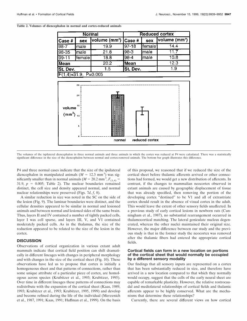
P4 and three normal cases indicate that the size of the ipsilateraldiencephalon in manipulated animals (M 5 12.3 mm3) was sig-nificantly smaller than in normal animals (M 5 20.2 mm3, F(1,4) 531.9, p 5 0.005; Table 2). The nuclear boundaries remaineddistinct, the cell size and density appeared normal, and normalnuclear relationships were preserved (Figs. 7d, f, 8).
A similar reduction in size was noted in the SC on the side ofthe lesion (Fig. 9). The laminar boundaries were distinct, and thecellular densities appeared to be similar in normal and lesionedanimals and between normal and lesioned sides of the same brain.Thus, layers II and IV contained a number of tightly packed cells,layer I was cell sparse, and layers III, V, and VI containedmoderately packed cells. As in the thalamus, the size of thereduction appeared to be related to the size of the lesion in thecortex.
DISCUSSIONObservations of cortical organization in various extant adultmammals indicate that cortical field position can shift dramati-cally in different lineages with changes in peripheral morphologyand with changes in the size of the cortical sheet (Fig. 10). Theseobservations have led us to propose that cortex is initially ahomogeneous sheet and that patterns of connections, rather thansome unique attribute of a particular piece of cortex, are homol-ogous across species (Krubitzer et al., 1993; Krubitzer, 1995).Over time in different lineages these patterns of connections mayredistribute with the expansion of the cortical sheet (Kaas, 1989,1993; Krubitzer et al., 1993; Krubitzer, 1995, 1999) and developand become refined during the life of the individual (Merzenichet al., 1987, 1991; Kaas, 1991; Huffman et al., 1999). On the basis
of this proposal, we reasoned that if we reduced the size of thecortical sheet before thalamic afferents arrived or other connec-tions had formed, we would get a new distribution of afferents. Incontrast, if the changes to mammalian neocortex observed inextant animals are caused by geographic displacement of tissuethat was already specified, then removing the portion of thedeveloping cortex “destined” to be V1 and all of extrastriatecortex should result in the absence of visual cortex in the adult.This would leave the extent of other sensory fields unaffected. Ina previous study of early cortical lesions in newborn rats (Cun-ningham et al., 1987), no substantial rearrangement occurred inthalamocortical matching. The lateral geniculate nucleus degen-erated, whereas the other nuclei maintained their original size.However, the major difference between our study and the previ-ous study is that in the former study the neocortex was removedafter the thalamic fibers had entered the appropriate corticalfields.
Cortical fields can form in a new location on portionsof the cortical sheet that would normally be occupiedby a different sensory modalityOur findings that all sensory inputs are represented on a cortexthat has been substantially reduced in size, and therefore havearrived in a new location compared to that which they normallywould occupy, suggest that the cells of the early neural sheet arecapable of remarkable plasticity. However, the relative rostrocau-dal and mediolateral relationships of cortical fields and thalamicafferents appear to be highly conserved. What are the mecha-nisms that determine these relationships?
Currently, there are several different views on how cortical
Table 2. Volumes of diencephalon in normal and cortex-reduced animals
The volumes of the ispilateral diencephalon in three normal animals and three animals in which the cortex was reduced at P4 were calculated. There was a statisticallysignificant difference in the size of the diencephalon between normal and cortex-removed animals. The bottom bar graph illustrates this difference.
Huffman et al. • Formation of Cortical Fields J. Neurosci., November 15, 1999, 19(22):9939–9952 9947

fields develop. One proposal is that a protomap exists within theventricular zone, and cortical field organization is specified veryearly in development (Rakic, 1988; Barbe and Levitt, 1995; Levittet al., 1997). In this scenario, there is something inherent in aparticular piece of the cortical neuroepithelium that directs it toproduce a particular field. Support for this theory comes fromstudies in monkeys and rats in which differential laminar histo-genesis is observed for different regions of the ventricular zonedestined to become particular cortical fields (Dehay et al., 1993;Polleux et al., 1997a,b). Further support comes from studies thatdemonstrate differential molecular expression patterns in differ-ent domains during corticogenesis (Cohen-Tannoudji et al., 1994;for review, see Rakic, 1995b; Levitt et al., 1997). Finally, Gbx-2-deficient mice in which the thalamus fails to innervate the devel-oping cortex still possess region-specific gene expression in the
neocortex at P0 (Miyashita-Lin et al., 1999). Although sharpboundaries of gene expression do exist in the absence of thalamicinput [boundary ii in Miyashita-Lin et al. (1999)], it is unclear howthey relate to explicitly defined cortical field boundaries. Indeed,the authors state in a previous study that examined the patterns ofexpression of Id-2 (boundary i) that “. . . Id-2 expression in layer5 ends abruptly, clearly demonstrating another intracortical ros-trocaudal boundary. . . . The position of this boundary may be thetransition between frontal (motor) and parietal (sensory) corticalareas . . .” as defined architectonically in adult rats by Zilles andWree (1995) [also see Bulfone et al. (1995) and Suzuki et al.(1997)]. It would be useful to examine the exact relationship ofthese patterns to cortical fields and to determine whether thesepatterns shift in our experimental animals.
A less stringent version of the protomap hypothesis is that
Figure 8. a, c, Reconstructions of the Nissl sections photographed in b and d. These sections of the thalamus are from normal animals ( a) and thosewith a moderate cortical removal ( c). Thin lines mark nuclear boundaries determined by architectonic analyses. The corresponding photomicrographs(b, d) demonstrate that although the thalamus has been reduced on the side of the lesion, nuclear architecture is still distinct. Scale bar, 1 mm.
9948 J. Neurosci., November 15, 1999, 19(22):9939–9952 Huffman et al. • Formation of Cortical Fields

there is differential gene expression in the developing cortex thatreflects subsequent specialization (Levitt et al., 1997) and sets upthe anteroposterior and mediolateral axis of the cortex, which inturn allows thalamocortical relationships to be maintained. Forinstance, differential expression of regulatory homeobox genessuch as Emx2 and Pax6 sets up a general rostrocaudal moleculargradient that may control the ordered growth of thalamic affer-ents (Guilisano et al., 1996; for review, see Chenn et al., 1997).
Pioneering studies by Sperry suggested that topographic rela-tionships in the developing nervous system are initially instigatedby chemoaffinities between the incoming afferents and the targettissue (Sperry, 1963). A modern synthesis of Sperry’s initialformulation is that chemoaffinities are not specific but that mo-lecular gradients in different structures set up a directional axisand specify regional identity (O’Leary et al., 1994; Rubensteinand Beachy, 1998) and promote ordered afferent ingrowth (Rich-ards et al., 1997; Frisen et al., 1998; Mann et al., 1998; for review,see O’Leary et al., 1999). Although results from the present studydo not directly address this issue, they are consistent with the
view that these gradients may provide relative positional cues ormay be capable of rapidly changing their patterns if the environ-ment in which they reside has been altered dramatically.
Although the present results as well as comparative studies ofcortical organization in different mammals (Fig. 10) demonstratethat thalamocortical relationships and relative geographic loca-tion of cortical fields appear to be highly conserved, a number ofimportant questions regarding area specification still remain un-answered. For example, what promotes differential gene expres-sion and consequent molecular gradients? It has been proposedthat the microenvironment in which the developing cells findthemselves plays a large role in this process (Ferri and Levitt,1995; Lillien, 1998), but the boundaries of the environment stillneed to be defined. It may be useful to examine the role of thestructures that border the cerebral cortex, such as the superiorcolliculus/cerebellum (caudal), the olfactory bulb (rostral), andthe pyriform cortex (lateral), in defining its boundaries.
A second view of cortical field differentiation holds that corticalfields are specified late in development and that thalamocortical
Figure 9. I llustrations of reconstructed Nissl sections (a, c) through the SC in the normal animal (a) and an animal that had undergone a moderatecortical removal at P4 ( c). Thin lines mark boundaries between layers of the SC and of the CG. Stipple indicates cell-dense layers II and IV. b, d,Photomicrographs of those sections in the normal (b) and moderate removal ( d) animals. Although the SC in the moderate cortical removal animal isreduced in size, its laminar pattern appears to be maintained. CA, Cerebral aqueduct. Scale bar, 1 mm. Conventions are as in previous Figures.
Huffman et al. • Formation of Cortical Fields J. Neurosci., November 15, 1999, 19(22):9939–9952 9949

connections play an important role in assigning cortical fields(O’Leary, 1989; Killackey, 1990; Roe et al., 1990; Molnar andBlakemore, 1991; Windrem and Finlay, 1991; Killackey et al.,1994). This proposal is supported by studies in which the devel-oping neocortex from one region is transplanted into anotherregion and then takes on the properties of the host (Schlaggar andO’Leary, 1991). Other studies that support this idea demonstratethat in vitro thalamocortical axons will grow toward any portion ofthe cortex, regardless of whether it is a region of cortex that theywould normally innervate (Molnar and Blakemore, 1991, 1999).Finally, studies of cell lineage and dispersion demonstrate thatclonally related neurons can disperse over a wide region of cortexand span several architectonic fields (Walsh and Cepko, 1992,1993).
Our findings demonstrate the ability of early thalamocorticalprojections to innervate a novel location of the neuroepitheliumon the reduced cortical sheet. These results complement previousstudies in hamsters, in which visual inputs were rewired to ulti-mately innervate somatosensory cortex (Frost and Metin, 1985),and in ferrets, in which visual inputs were rerouted into auditorycortex (Roe et al., 1990, 1992; Pallas et al., 1990; Pallas and Sur,1993). In the latter study, the “auditory” cortex was dominated byvisual inputs but had connections that were consistent with audi-tory cortex (Pallas et al., 1990; Pallas and Sur, 1993). However,
the topography of the maps that formed was like those in visualcortex. The authors suggest that cortical area specification occurslate in development, and a cortical area can be induced to supportdifferent types of maps (Roe et al., 1990, 1992).
Taken together, the consistencies across data sets indicate thatthe early embryonic cortical neuroepithelium is plastic, and itsspecification depends on the spatially and temporally regulatedenvironmental signals that can alter the potential of its cells(Lillien, 1998). Thus, the cortex can be reassigned in develop-ment. The way in which the reassignment occurs is constrainedanteroposteriorly and mediolaterally, perhaps by highly con-served but not immutable patterns of graded gene expression inthese two axes. Another possibility, not mutually exclusive fromthe former, is that the overall geometry of the neocortex mayreflect the spatial relationship between thalamic nuclei, which ishighly conserved across mammals.
Reduction in the size of the cortical neuroepitheliumresults in a reduction in the dorsal thalamus andsuperior colliculusOur observation of a decrease in size in the dorsal thalamus maybe explained in two ways. One possibility is that there is aretrograde effect on the developing thalamic afferents resultingfrom a decrease in the size of the target. Regulation of cell death
Figure 10. The organization of major sensory fieldsincluding S1, A, and V1 in various mammals. Althoughcommon areas can be identified across species, the geo-graphic placement of these fields has shifted dramati-cally in different lineages with changes in cortical sheetsize and peripheral specializations. With the expansionof the cortical sheet, cortical fields get larger, but thisenlargement is not linear. Scale bar, 1 mm. Conventionsare as in previous Figures.
9950 J. Neurosci., November 15, 1999, 19(22):9939–9952 Huffman et al. • Formation of Cortical Fields
by the target (Cowan et al., 1984; for review, see Oppenheim,1999) and target-induced changes in cellular morphology of de-veloping afferents (Erzurumlu et al., 1994; Erzurumlu and Jhav-eri, 1995; Ling et al., 1997) have been well established. Thesecond possibility is that corticothalamic afferents from the targetare reduced, and this reduction promotes thalamic cell death. Thetwo possibilities are not exclusive. Although it is possible thatdecreasing target space has a retrograde effect on developingthalamic neurons (Cunningham et al., 1987; Rennie et al., 1994;Lotto and Price, 1995; for review, see Oppenheim, 1999), andincreases cell death in the thalamus, this would not explain thedecrease in size of the superior colliculus, which does not projectdirectly to the cortex. However, because the thalamus has de-creased in size and is a target for a number of collicular projec-tions, there may have been a retrograde effect on developingcollicular neurons promoting increased cell death within thecolliculus.
ConclusionsThe present results demonstrate that the location of a corticalfield is not strictly dependent on a predetermined location on theneuroepithelial sheet. Second, and equally important, very simplechanges in the developing nervous system, such as changes in thesize of the cortical sheet, can trigger a cascade of events that alterthe rest of the nervous system in a manner consistent with whatwe observe in mammals with different sized brains.
One can assume that a simple change in the timing of horizon-tal proliferation of cells in the ventricular zone (Rakic, 1995a;Kornack and Rakic, 1998), or decreased cell death of progenitorcells (Kuida et al., 1998), could have significant consequences forthe organization of the entire CNS. It is unknown whether suchchanges alone are sufficient to explain the emergence of newcortical fields. It is possible that an enlargement of the corticalsheet would simply result in larger cortical fields rather than morecortical fields. However, comparative studies demonstrate thatthe relationship between cortical sheet size and cortical field sizeis nonlinear. It is possible that enlargements of the cortical sheet,in addition to resulting in larger cortical fields, may also promotenew interactions between thalamic inputs as well as corticocorti-cal and interhemispheric interconnections (Krubitzer et al.,1998). Thus, combining retained elements in novel ways on alarger target may increase the information processing capacity ofthe cortex.
REFERENCESAitkin LM, Irvine DRF, Nelson JE, Merzenich MM, Clarey JC (1986)
Frequency representation in the auditory midbrain and forebrain of amarsupial, the northern native cat (Dasyurus hallucatus). Brain BehavEvol 29:17–28.
Barbe MF, Levitt R (1995) Age-dependent specification of the cortico-cortical connections of cerebral grafts. J Neurosci 15:1819–1834.
Brodmann, K (1909) Vergleichende Lokalisationslehre der Grosshirn-rinde in ihren Prinzipien dargestellt auf Grund des Zellenbaues.Leipzig: J. A. Barth.
Bulfone A, Smiga SM, Shimamura K, Peterson A, Puelles L, RubensteinJLR (1995) T-Brain-1: a homolog of Brachyury whose expression de-fines molecularly distinct domains within the cerebral cortex. Neuron15:63–78.
Carroll EW, Wong-Riley MTT (1984) Quantitative light and electronmicroscopic analysis of cytochrome oxidase-rich zones in the striatecortex of the squirrel monkey. J Comp Neurol 222:1–17.
Chang F-LF, Steedman JG, Lund RD (1986) The lamination and con-nectivity of embryonic cerebral cortex transplanted into newborn ratcortex. J Comp Neurol 244:401–411.
Chenn A, Braisted JE, McConnell SK, O’Leary DDM (1997) Develop-ment of the cerebral cortex: mechanisms controlling cell fate, laminar
and areal patterning, and axonal connectivity. In: Molecular and cel-lular approaches to neural development (Cowan WM, Jessell TM,Sipursky SL, eds), pp 440–473. New York: Oxford UP.
Cohen-Tannoudji M, Babinet C, Marion W (1994) Early determinationof a mouse somatosensory cortex marker. Nature 368:460–463.
Cowan WM, Fawcett JW, O’Leary DD, Stanfield BB (1984) Regressiveevents in neurogenesis. Science 225:1258–1265.
Cunningham TJ, Haun F, Chantler PD (1987) Diffusible proteins pro-long survival of dorsal lateral geniculate neurons following occipitalcortex lesions in newborn rats. Dev Brain Res 37:133–141.
DeHay C, Giroud P, Berland M, Smart I, Kennedy H (1993) Modulationof the cell cycle contributes to the parcellation of the primate visualcortex. Nature 366:464–466.
Erzurumlu RS, Jhaveri S (1995) Target influences on the morphology oftrigeminal axons. Exp Neurol 135:1–16.
Erzurumlu RS, McKay RD, Jhaveri S (1994) Morphological specifica-tion of trigeminal neurites depends on target fields. Brain Res DevBrain Res 83:132–137.
Ferri RT, Levitt P (1995) Regulation of region differences in the differ-entiation of cerebral cortical neurons by EGF family-matrix interac-tions. Development 21:1151–1160.
Frisen J, Yates PA, McLaughlin T, Friedman GC, O’Leary DDM, Bar-bacid M (1998) Ephrin-A5 (AL-1/RAGS) is essential for proper ret-inal axon guidance and topographical mapping in the mammalianvisual system. Neuron 20:235–243.
Frost DO, Metin C (1985) Induction of functional retinal projections tothe somatosensory system. Nature 317:162–164.
Gallyas F (1979) Silver staining of myelin by means of physical develop-ment. Neurology 1:203–209.
Gates GR, Aitkin LM (1982) Auditory cortex in the marsupial possumTrichosurus vulpecula. Hear Res 7:1–11.
Guilisano MV, Broccoli V, Pardini C, Boncinelli E (1996) Emx1 andEmx2 show different patterns of expression during proliferation anddifferentiation of the developing cerebral cortex in the mouse. EurJ Neurosci 8:1037–1050.
Huffman K, Molnar Z, Van Dellen A, Krubitzer LA (1998) Formationof cortical maps on a reduced cortical sheet. Soc Neurosci Abstr 24:59.
Huffman KJ, Nelson J, Clarey J, Krubitzer L (1999) Organization ofsomatosensory cortex in three species of marsupials, Dasyurus halluca-tus, Dactylopsila trivirgata, and Monodelphis domestica: neural correlatesof morphological specializations. J Comp Neurol 403:5–32.
Kaas JH (1982) The segregation of function in the nervous system: whydo the sensory systems have so many subdivisions? Contrib SensPhysiol 7:201–240.
Kaas JH (1988) Why does the brain have so many visual areas? J CognitNeurosci 1:121–135.
Kaas JH (1989) The evolution of complex sensory systems in mammals.J Exp Biol 146:165–176.
Kaas JH (1991) Plasticity of sensory and motor maps in adult mammals.Annu Rev Neurosci 14:137–167.
Kaas JH (1993) Evolution of multiple areas and modules within neocor-tex. Perspect Dev Neurobiol 1:101–107.
Kahn DM, Huffman KJ, Le C, Krubitzer L (1998) Retinal projections ina South American marsupial. Soc Neurosci Abstr 24:1393.
Kahn DM, Slutsky DA, Krubitzer LA (1999) Receptive field organiza-tion of primary visual cortex in a metatherian mammal (Monodelphisdomestica). Soc Neurosci Abstr 25:1931.
Killackey HP (1990) Neocortical expansion: an attempt toward relatingphylogeny and ontogeny. J Cognit Neurosci 2:1–17.
Killackey HP, Chiaia NL, Bennett-Clarke CA, Eck M, Rhoades RW(1994) Peripheral influences on the size and organization of somato-topic representations in the fetal rat cortex. J Neurosci 14:1496–1506.
Kornack DR, Rakic P (1998) Changes in cell-cycle kinetics during thedevelopment and evolution of the primate neocortex. Proc Natl AcadSci USA 95:1242–1246.
Krubitzer L (1995) The organization of neocortex in mammals: arespecies differences really so different? Trends Neurosci 18:408–417.
Krubitzer L (1998) What can monotremes tell us about brain evolution?Philos Trans R Soc Lond B Biol Sci 353:1127–1146.
Krubitzer L (1999) How does evolution build a complex brain? In:Evolutionary developmental biology of the cerebral cortex (Bock G,Cardew G, eds). Novartis Foundation Symposium.
Krubitzer LA, Calford MB, Schmid LM (1993) Connections of somato-sensory cortex in megachiropteran bats: the evolution of cortical fieldsin mammals. J Comp Neurol 327:473–506.
Huffman et al. • Formation of Cortical Fields J. Neurosci., November 15, 1999, 19(22):9939–9952 9951

Krubitzer L, Huffman K, Molnar Z (1998) Constructing the neocortex:influences on the pattern of organization in mammals In: Brain andmind: evolutionary perspectives (Gazzaniga MS, Altman J, eds), pp19–34. Strasbourg: Human Frontier Science Program.
Kuida K, Haydar T, Kuan C-Y, Gu Y, Taya C, Karasuyama H, Su MS-S,Rakic P, Flavell RA (1998) Reduced apoptosis and cytochromec-mediated caspase activation in mice lacking caspase 9. Cell94:325–337.
Levitt P, Ferri R, Eagleson K (1995) Molecular contributions to cerebralcortical specification. In: Development of the cerebral cortex (CibaFoundation Symposium) (Bock GR, Cardew G, eds), pp 203–213.Chichester: Wiley.
Levitt P, Barbe MF, Eagleson KL (1997) Patterning and specification ofthe cerebral cortex. Annu Rev Neurosci 20:1–24.
Lillien L (1998) Neural progenitors and stem cells: mechanisms of pro-genitor heterogeneity. Curr Opin Neurobiol 8:37–44.
Ling C, Jhaveri S, Schneider GE (1997) Target as well as source-derivedfactors direct the morphogenesis of anomalous retino-thalamic projec-tions. J Comp Neurol 388:454–466.
Lotto RB, Price DJ (1995) The stimulation of thalamic neurite out-growth by cortex-derived growth factors in vitro: the influence ofcortical age and activity. Eur J Neurosci 7:318–328.
Manger P, Sum M, Szymanski M, Ridgway S, Krubitzer L (1998) Mod-ular subdivisions of dolphin insular cortex: does evolutionary historyrepeat itself. J Cognit Neurosci 10:153–156.
Mann F, Zhukareva Z, Pimenta A, Levitt P, Bolz J (1998) Membrane-associated molecules guide limbic and nonlimbic thalamocortical pro-jections. J Neurosci 18:9409–9419.
Merzenich MM, Nelson RJ, Kaas JH, Stryker MP, Jenkins WM, SookJM, Cynader MS, Schoppmann A (1987) Variability in hand surfacerepresentations in areas 3b and 1 in adult owl and squirrel monkeys.J Comp Neurol 258:281–296.
Merzenich MM, Grajski KA, Jenkins WM, Recanzone GH, Peterson B(1991) Functional cortical plasticity. Cortical network origins of rep-resentation changes. Cold Spring Harb Symp Quant Biol 55:873–887.
Miyashita-Lin EM, Hevner R, Wassarman KM, Martinez S, RubensteinJ (1999) Neocortical regionalization in the absence of thalamic inner-vation. Science 285:906–909.
Molnar Z, Blakemore C (1991) Lack of regional specificity for connec-tions formed between thalamus and cortex in coculture. Nature351:475–477.
Molnar Z, Blakemore C (1995) How do thalamic axons find their way tothe cortex? Trends Neurosci 18:389–397.
Molnar Z, Blakemore C (1999) Development of signals influencing thegrowth and termination of thalamocortical axons in organotypic cul-ture. Exp Neurol 156:363–393.
Molnar Z, Knott GW, Blakemore C, Saunders NR (1998) Developmentof thalamocortical projections in the South American grey short-tailedopossum (Monodelphis domestica). J Comp Neurol 398:491–514.
O’Leary DDM (1989) Do cortical areas emerge from a protocortex?Trends Neurosci 12:400–406.
O’Leary DDM, Schlaggar BL, Tuttle R (1994) Specification of neocor-tical areas and thalamocortical connections. Annu Rev Neurosci17:419–439.
O’Leary DDM, Yates PA, McLaughlin T (1999) Molecular developmentof sensory maps: representing sights and smell in the brain. Cell96:255–269.
Oppenheim RW (1999) Programmed cell death. In: Fundamental neu-roscience (Zigmond MJ, Bloom FE, Roberts JL, Landis SC, SquireLR, eds), pp 581–609. San Diego: Academic.
Pallas SL, Sur M (1993) Visual projections induced into the auditorypathway of ferrets: II. Corticocortical connections of primary auditorycortex. J Comp Neurol 337:317–333.
Pallas SL, Roe AW, Sur M (1990) Visual projections induced into theauditory pathway of ferrets. I. Novel inputs to primary auditory cortex(AI) from the LP/pulvinar complex and the topography of theMGN-AI projection. J Comp Neurol 298:50–68.
Polleux F, DeHay C, Kennedy H (1997a) The timetable of laminarneurogenesis contributes to the specification of cortical areas in mouseneocortex. J Comp Neurol 385:95–116.
Polleux F, DeHay C, Moraillon B, Kennedy H (1997b) Regulation ofneuroblast cell-cycle kinetics plays a crucial role in the generation ofunique features of neocortical areas. J Neurosci 17:7763–7783.
Rakic P (1988) Specification of cerebral cortical areas. Science241:170–176.
Rakic P (1995a) A small step for the cell, a giant leap for mankind: ahypothesis of neocortical expansion during evolution. Trends Neurosci18:383–388.
Rakic P (1995b) Radial versus tangential migration of neuronal clones inthe developing cerebral cortex. Proc Natl Acad Sci USA92:11323–11327.
Rennie S, Lotto RB, Price DJ (1994) Growth-promoting interactionsbetween the murine neocortex and thalamus in organotypic co-cultures.J Neurosci 61:547–564.
Richards LJ, Koester SE, Tuttle R, O’Leary DDM (1997) Directedgrowth of early cortical axons is influenced by a chemoattractantreleased from an intermediate target. J Neurosci 17:2245–2458.
Ridgway SH (1986) Dolphin brain size. In: Research on dolphins (Bry-den MM, Darrison RJ, eds), pp 59–70. Oxford: Oxford UP.
Roe AW, Pallas SL, Hahm JO, Sur M (1990) A map of visual spaceinduced in primary auditory cortex. Science 250:818–820.
Roe AW, Pallas SL, Kwon YH, Sur M (1992) Visual projections routedto the auditory pathway in ferrets: receptive fields of visual neurons inprimary auditory cortex. J Neurosci 12:3651–3664.
Rosa MGP, Krubitzer LA, Molnar Z, Nelson JE (1999) Organization ofvisual cortex in the northern quoll, Dasyurus hallucatus: evidence for ahomologue of the second visual area in marsupials. Eur J Neurosci11:907–915.
Rubenstein JLR, Beachy PA (1998) Patterning of the embryonic fore-brain. Curr Opin Neurobiol 8:18–26.
Schlaggar BL, O’Leary DDM (1991) Potential of visual cortex to de-velop an array of functional units unique to somatosensory cortex.Science 252:1556–1560.
Sperry RW (1963) Chemoaffinity in the orderly growth of nerve fiberpatterns and connections. Proc Natl Acad Sci USA 50:703–710.
Stephan H, Baron G, Frahm HD (1988) Comparative size of brains andbrain components. Comp Primate Biol 4:1–38.
Suzuki SC, Inoue T, Kimura Y, Tanaka T, Takeichi M (1997) Neuronalcircuits are subdivided by differential expression of type-II classiccadherins in postnatal mouse brains. Mol Cell Neurosci 9:433–447.
Turlejski K, Djavadian RL, Saunders NR (1994) Projection of visuo-topically organized afferents to the dorsal thalamus on the opossum,Monodelphis domestica. Acta Neurobiol Exp 54:307–319.
Walsh C, Cepko CL (1992) Widespread dispersion of neuronal clonesacross functional regions of the cerebral cortex. Science 255:434–440.
Walsh C, Cepko CL (1993) Clonal dispersion in proliferative layers ofdeveloping cerebral cortex. Nature 362:632–635.
Windrem MS, Finlay BL (1991) Thalamic ablations and neocortical de-velopment: alterations of cortical cytoarchitecture and cell number.Cereb Cortex 1:230–240.
Wray GA (1994) Developmental evolution: new paradigms and para-doxes. Dev Genet 15:1–6.
Zilles K, Wree A (1995) Cortex: areal and laminar structure. In: The ratnervous system, Ed 2 (Paxinos G, ed), pp 649–685. San Diego: Aca-demic.
9952 J. Neurosci., November 15, 1999, 19(22):9939–9952 Huffman et al. • Formation of Cortical Fields
Related Documents