Preservation of motor programs in paraplegics as demonstrated by attempted and imagined foot movements Sabina Hotz-Boendermaker, a Marion Funk, b Paul Summers, c Peter Brugger, b Marie-Claude Hepp-Reymond, d Armin Curt, e and Spyros S. Kollias c, ⁎ a Spinal Cord Injury Centre, Balgrist University Hospital Zurich, Switzerland b Department of Neurology, University Hospital Zurich, Switzerland c Institute of Neuroradiology, University Hospital Zurich, Frauenklinikstr. 10, CH-8091 Zurich, Switzerland d Institute of Neuroinformatics, University and ETH Zurich, Switzerland e ICORD and Division of Neurology/Department of Medicine, The University of British Columbia, Canada Received 21 May 2007; revised 26 July 2007; accepted 29 July 2007 Available online 23 August 2007 Execution and imagination of movement activate distinct neural circuits, partially overlapping in premotor and parietal areas, basal ganglia and cerebellum. Can long-term deafferented/deefferented patients still differentiate attempted from imagined movements? The attempted execution and motor imagery network of foot movements have been investigated in nine chronic complete spinal cord-injured (SCI) patients using fMRI. Thorough behavioral assessment showed that these patients were able to differentiate between attempted execution and motor imagery. Supporting the outcome of the behavioral assessment, fMRI disclosed specific patterns of activation for movement attempt and for motor imagery. Compared with motor execution data of healthy controls, movement attempt in SCI patients revealed reduced primary motor cortex activation at the group level, although activation was found in all single subjects with a high variability. Further comparisons with healthy subjects revealed that during attempt and motor imagery, SCI patients show enhanced activation and recruitment of additional regions in the parietal lobe and cerebellum that are important in sensorimotor integration. These findings reflect central plastic changes due to altered input and output and suggest that SCI patients may require additional cognitive resources to perform these tasks that may be one and the same phenomenon, or two versions of the same phenomenon, with quantitative differences between the two. Nevertheless, the retained integrity of movement attempt and motor imagery networks in SCI patients demonstrates that chronic para- plegics can still dispose of the full motor programs for foot movements and that therefore, attempted and imagined movements should be integrated in rehabilitative strategies. © 2007 Elsevier Inc. All rights reserved. Keywords: Spinal cord injury; Attempt to move; Motor imagery; Functional MRI Introduction The human motor system generates accurate movements, which are centrally stored and can be modified and retrieved under various conditions. The complexity of the processes involved in any motor action has led to the concept that the central nervous system contains internal models representing these processes and optimiz- ing motor control (Kawato, 1999; Wolpert and Ghahramani, 2000). Among these models, forward models predict the relationship between issued motor commands and the resulting changes in the sensorimotor system, monitored by the reafferent sensory inflow which supplies information about the state of the body. In this context, patients with complete spinal cord injury (SCI) provide a unique human model for studying the effects of deafferentation on motor control, and on the sensorimotor system in general. We have recently used functional magnetic resonance imaging (fMRI) to investigate the activation patterns during motor imagery in chronic SCI patients (Alkadhi et al., 2005). This study provided evidence that in this patient group motor imagery still engages the central machinery of movements as suggested by Jeannerod (1995). Studies in healthy subjects revealed that internal simulation of a movement induces similar physiological reactions as its execution (Decety and Jeannerod, 1995; Jeannerod and Decety, 1995). A number of imaging studies disclosed functional circuits shared by both movement execution and imagination (Jackson et al., 2001; Lafleur et al., 2002), although subtle differences in the localization of activation foci between the two tasks have also been reported (Stephan et al., 1995; Gerardin et al., 2000; Hanakawa et al., 2003; Nair et al., 2003). While it is generally accepted that “overt” or executed motor behavior and “covert” or simulated behavior are intimately related (Jeannerod, 2001), the ability to physically execute a movement is not necessarily required for its mental performance. This is well recognized in patients with hemiplegia who are still able after a cerebrovascular insult to mentally move their limbs, even after years of disuse (Johnson, 2000; Johnson-Frey, 2004). In a case www.elsevier.com/locate/ynimg NeuroImage 39 (2008) 383 – 394 ⁎ Corresponding author. Fax: +41 255 45 04. E-mail address: [email protected] (S.S. Kollias). Available online on ScienceDirect (www.sciencedirect.com). 1053-8119/$ - see front matter © 2007 Elsevier Inc. All rights reserved. doi:10.1016/j.neuroimage.2007.07.065

Welcome message from author
This document is posted to help you gain knowledge. Please leave a comment to let me know what you think about it! Share it to your friends and learn new things together.
Transcript

www.elsevier.com/locate/ynimg
NeuroImage 39 (2008) 383–394Preservation of motor programs in paraplegics as demonstrated byattempted and imagined foot movements
Sabina Hotz-Boendermaker,a Marion Funk,b Paul Summers,c Peter Brugger,b
Marie-Claude Hepp-Reymond,d Armin Curt,e and Spyros S. Kolliasc,⁎
aSpinal Cord Injury Centre, Balgrist University Hospital Zurich, SwitzerlandbDepartment of Neurology, University Hospital Zurich, Switzerlandc Institute of Neuroradiology, University Hospital Zurich, Frauenklinikstr. 10, CH-8091 Zurich, Switzerlandd Institute of Neuroinformatics, University and ETH Zurich, Switzerlande ICORD and Division of Neurology/Department of Medicine, The University of British Columbia, Canada
Received 21 May 2007; revised 26 July 2007; accepted 29 July 2007Available online 23 August 2007
Execution and imagination of movement activate distinct neuralcircuits, partially overlapping in premotor and parietal areas, basalganglia and cerebellum. Can long-term deafferented/deefferentedpatients still differentiate attempted from imagined movements? Theattempted execution and motor imagery network of foot movementshave been investigated in nine chronic complete spinal cord-injured(SCI) patients using fMRI. Thorough behavioral assessment showedthat these patients were able to differentiate between attemptedexecution andmotor imagery. Supporting the outcome of the behavioralassessment, fMRI disclosed specific patterns of activation for movementattempt and for motor imagery. Compared with motor execution dataof healthy controls, movement attempt in SCI patients revealed reducedprimary motor cortex activation at the group level, although activationwas found in all single subjects with a high variability. Furthercomparisons with healthy subjects revealed that during attempt andmotor imagery, SCI patients show enhanced activation and recruitmentof additional regions in the parietal lobe and cerebellum that areimportant in sensorimotor integration. These findings reflect centralplastic changes due to altered input and output and suggest that SCIpatients may require additional cognitive resources to perform thesetasks that may be one and the same phenomenon, or two versions of thesame phenomenon, with quantitative differences between the two.Nevertheless, the retained integrity of movement attempt and motorimagery networks in SCI patients demonstrates that chronic para-plegics can still dispose of the full motor programs for foot movementsand that therefore, attempted and imagined movements should beintegrated in rehabilitative strategies.© 2007 Elsevier Inc. All rights reserved.
Keywords: Spinal cord injury; Attempt to move; Motor imagery; FunctionalMRI
⁎ Corresponding author. Fax: +41 255 45 04.E-mail address: [email protected] (S.S. Kollias).Available online on ScienceDirect (www.sciencedirect.com).
1053-8119/$ - see front matter © 2007 Elsevier Inc. All rights reserved.doi:10.1016/j.neuroimage.2007.07.065
Introduction
The human motor system generates accurate movements, whichare centrally stored and can be modified and retrieved under variousconditions. The complexity of the processes involved in any motoraction has led to the concept that the central nervous systemcontains internal models representing these processes and optimiz-ing motor control (Kawato, 1999; Wolpert and Ghahramani, 2000).Among these models, forward models predict the relationshipbetween issued motor commands and the resulting changes in thesensorimotor system, monitored by the reafferent sensory inflowwhich supplies information about the state of the body. In thiscontext, patients with complete spinal cord injury (SCI) provide aunique human model for studying the effects of deafferentation onmotor control, and on the sensorimotor system in general.
We have recently used functional magnetic resonance imaging(fMRI) to investigate the activation patterns during motor imagery inchronic SCI patients (Alkadhi et al., 2005). This study providedevidence that in this patient group motor imagery still engages thecentral machinery of movements as suggested by Jeannerod (1995).Studies in healthy subjects revealed that internal simulation of amovement induces similar physiological reactions as its execution(Decety and Jeannerod, 1995; Jeannerod and Decety, 1995). Anumber of imaging studies disclosed functional circuits shared by bothmovement execution and imagination (Jackson et al., 2001; Lafleur etal., 2002), although subtle differences in the localization of activationfoci between the two tasks have also been reported (Stephan et al.,1995; Gerardin et al., 2000; Hanakawa et al., 2003; Nair et al., 2003).
While it is generally accepted that “overt” or executed motorbehavior and “covert” or simulated behavior are intimately related(Jeannerod, 2001), the ability to physically execute a movement isnot necessarily required for its mental performance. This is wellrecognized in patients with hemiplegia who are still able after acerebrovascular insult to mentally move their limbs, even afteryears of disuse (Johnson, 2000; Johnson-Frey, 2004). In a case

384 S. Hotz-Boendermaker et al. / NeuroImage 39 (2008) 383–394
study using fMRI, a woman with congenitally absent limbs wasable to cortically command movements of her phantom limbs,suggesting that body parts that have never been physicallydeveloped can be represented in sensory and motor cortical areas(Brugger et al., 2000). In the investigation of Alkadhi et al. (2005),paraplegic patients mentally moving their paralyzed feet stronglyactivated brain areas corresponding to both the execution network,including the primary sensorimotor cortex, and the imagerynetwork described in healthy subjects.
In complete SCI patients, both intended overt movements andcovert movements remain without obvious motor responses.Therefore, only attempted (MA) and imagined (MI) movementscan be compared. The ability of SCI patients to distinguish betweenattempted and imagined movements has up to now not beenassessed behaviorally and only a few imaging studies haveinvestigated brain activity in these patients during attempted andimagined movements of the disconnected body parts (Sabbah et al.,2002; Cramer et al., 2005). These investigations with hetero-geneous patient groups reported reduced activation in primary andsecondary cortical motor regions for both MA and MI, thus being atodd with our previous experience (Alkadhi et al., 2005).
To address this issue, we undertook further neuroimaginginvestigations in a homogeneous group of chronic paraplegics, allafter at least two years post injury, with complete SCI lesions as-certained by standardized neurophysiological methods. We con-sider this time interval as a chronic state and thus, appropriate toinvestigate the influence of long-term deafferentation on attemptingto move the feet and generating mental images of the same move-ment. In addition, the ability of the patients to perform MI and MAand to distinguish between the two was quantitatively assessed. Weexpected that in SCI patients able to distinguish between MI andMA these two tasks will generate distinct brain activation patterns.
Material and methods
Subjects
Nine paraplegic patients were recruited from the outpatientclinic of our institution (3 females and 6 males; mean age 35 years,
Table 1Individual clinical and behavioral data for the SCI patients with means
Subject Level of complete motorimpairment/ASIAa
Age/Sexb Time sinceinjury (years)
Vim
S1 Th6/A 40/M 7 5S2 L1/A 28/M 11 3S3 Th5/A 42/M 20 2S4 L3/B 29/M 2 2S5 Th3/A 38/M 5 10S6 Th6/A 29/F 11 2S7 Th9/A 27/M 13 3S8 Th8/A 41/F 9 3S9 Th11/A 39/F 10 2Group mean 34.8 9.8 3SD 6.3 5.1 2
a ASIA impairment scale: A: no sensory or motor function is preserved; B: senb M: male; F: female.c Kinesthetic MI assessed with Vividness of Motor Imagery Questionnaire (VQI
score at 24).d Ability of attempt to move the right foot with intensity of the feeling (1: very w
rare; 6: very often).
SD 6). The Edinburgh Handedness Inventory revealed clear righthand dominance for all subjects. Table 1 gives the age, sex, etiologyof the SCI, the level of complete motor deficit, and the time sinceSCI. For the nine patients, the mean period following traumatic SCIwas 9 years (range 2–20 years). All had clinically complete motorSCI between Th3 and L3, as assessed with the impairment scale ofthe American Spinal Injury Association (ASIA; Maynard et al.,1997), transcranial magnetic stimulation (TMS), and somatosen-sory evoked potentials (SSEP; Curt and Dietz, 1999). All subjectshad repeated clinical examinations and SSEP of the posterior tibialnerves and MEP (motor evoked potentials) of the anterior tibialmuscles. The measures were performed at the outpatient clinic andwere repeated within 6 months to assure the completeness of SCI.Only one subject (S4) reported some clinical sensation (light touch)at the sacral dermatomes but had complete paralysis of the lowerlimbs and the SSEP and MEP were completely abolished. Twelveage-matched healthy right-handed volunteers (5 females and 7males; mean age 29 years, SD 3.7) were recruited as controls.
None of the participants had suffered a brain lesion or had ahistory of neurological or psychiatric illness. Informed consent wasobtained from all subjects and they were reimbursed for theirparticipation in the study. The experimental protocol was approvedby the Ethics Committee of the Balgrist University Hospital ofZurich, Switzerland.
Assessment of movement attempt and execution
The motor task studied in the fMRI experiments consisted ofrepetitive alternating dorsal and plantar flexion of the right foot(30°–0°–45°) at a self-paced rhythm of approximate of 0.5 Hz. Theability to attempt moving the foot (motor attempt, MA) wasassessed as follows. The perceived intensity and frequency of at-tempted movements was rated in a structured interview on phantomsensations, which had been developed for evaluating phantom bodyphenomena, paresthesia and movement sensations in SCI patients.Of particular relevance was rating the intensity of the feeling tomove the right foot and the frequency of spontaneous attempts indaily life. Answers were noted as qualitative descriptors and boththe phenomena’s frequency and intensity were individually rated
vidness of kinestheticotor imagery (VQIM)c
Ability for attemptedmovement intensityd
Ability for attemptedmovement frequencyd
4 3 33 4 44 5 56 6 66 4 15 5 41 3 24 5 54 5 39.7 4.5 3.66.6 3.6 1.7
sory is preserved below the level, but not motor.
M), range 1–5 (1: high, 5: low imagination, range of 24–120, with the best
eak; 6: very high) and frequency of spontaneous attempt in daily life (1: very

385S. Hotz-Boendermaker et al. / NeuroImage 39 (2008) 383–394
using a 6-point scale (see Table 1). The verbal instruction for MA inSCI patients was: “Try to move your right foot up and down at anapproximate speed of 0.5 Hz”. Correct performance was controlledusing an adapted version of the controllability of motor imagery(CMI) described by Naito et al. (2002). With eyes closed, thesubjects were required to try moving their right foot as describedabove and, on command, to promptly give a verbal description ofthe foot position (flexed or extended). In healthy volunteers, attemptto move was not required as the MA task is difficult to performwithout generating isometric muscle contractions. Instead, they hadto execute the foot movement (motor execution, ME). Followinginstruction, the ability of the controls to move their right foot up anddown was visually verified.
Assessment of motor imagery
The ability of the subjects to perform MI was assessed with theVividness of Motor Imagery Questionnaire (VMIQ; Isaac et al.,1986). The VMIQ consists of 24 items specific to movement. Itrefers to the visual imagery of the movement and to the imagery ofmovement kinaesthetic sensations. The interesting feature of thisquestionnaire is that each item refers to two imagery perspectives;the ‘internal’ or first person perspective and the ‘external’ or thirdperson perspective. Responders were required to imagine each itemboth with respect to themselves (first person, kineasthetic sensation)and with respect to someone else (third person, visualisation). Foreach item, participants were asked to indicate the vividness of animagined movement on a 5-point scale: 1 (excellent imagination ofthe movement performance as lively as actual performance), 2 (agood capacity to imagine movement performance), 3 (moderatecapacity to imagine the performance of a movement), 4 (a vague orunclear image) or 5 (no image at all). This questionnaire has apossible range of 24–120 for both, first and third person imagin-ation, with the best score at 24. The lower the score, the more vividthe imagery.
To achieve consistent performance of MI in both groups andavoid muscle activity in the healthy subjects, all were trained witheyes closed in the first person motor imagery to mentally move theirright foot (dorsal and plantar flexion) outside of the scanner. Theinstruction for MI in both controls and SCI patients was “Imagineyourself performing the same foot movement without actuallyexecuting it”. To control for proper task performance, the CMI wasapplied here as in the MA task (see above, Naito et al., 2002). Thetraining was continued up to the point where subjects could fulfillthe requirements of the CMI and felt comfortable with the task.
Experimental protocol
The brain activation patterns underlying execution and imagina-tion of foot movements were investigated with fMRI. Experimentalconditions were presented in a fixed-order sequence consisting ofattempted (SCI patients) or overt (controls) movements followed bymotor imagery. The eyes were kept closed in both conditions. Eachexperimental condition was administered in a standard block designconsisting of three 21-s periods of baseline alternating with three21-s periods of motor task. For the ME/MA condition, the baselinewas rest, for the MI condition the baseline condition consisted ofsilent automatic upwards counting starting from number six. Thisrest condition was chosen to make a clear distinction between themental motor task and the rest condition (i.e. to ensure that subjectsstopped imagery). Starting with six avoids the tendency of subjects
to imagine counting with their fingers. All execution and imagerytasks were self-paced at a rate of approximately 0.5 Hz. Thebeginning and end of each activation period were signalled withverbal commands “go” and “stop” for ME and MA and “go” and“six” for MI, transmitted over the MR scanner’s intercom system.Correct task performance during data acquisition was visuallycontrolled, with the observer monitoring of any movements orapparent change in the resting state of the non-moving limbs, andverifying performance of the ME task by the controls. No overtmovements were observed during the MI task in healthy controls, orduring MI or MA by the SCI patients.
Scanning procedure
Blood oxygenation level-dependent (BOLD) sensitive fMRIwas carried on a 1.5 Twhole body scanner equipped with a standard6-channel head coil using a single-shot, gradient-echo, echo-planarimaging (EPI) sequence (TE=55 ms, TR=3000 ms, flip angle 90°).For each task, 126 time points were acquired consisting of 30contiguous, axial slices (resolution 5×3.4×3.4 mm3) covering theentire brain. A T1-weighted whole-brain anatomical referencevolume data with an isotropic spatial resolution of 1.2 mm was alsoacquired with a 3D spoiled, gradient-echo sequence (TE (echotime)=9 ms, TR (repetition time)=50 ms).
Imaging analysis
Image analysis was performed using SPM99 (Welcome Depart-ment of Cognitive Neurology, London, http://fil.ion.ucl.ac.uk/spm)under MATLAB 6.1 (Mathworks Inc., Natiek, MA, USA). The firsttwo volumes of each fMRI time series were discarded. For eachsubject, all remaining EPI volumes were realigned to the tenthvolume of the first time series. A mean image was then created andthe anatomical image was co-registered with this mean image. Afterco-registration, the structural image was spatially normalized intothe reference system of a representative brain template (MontrealNeurological Institute, MNI) using an affine and nonlinear trans-formation. The normalization parameters were subsequently ap-plied to the functional images. Finally, the EPI images were re-sampled to a voxel size of 3×3×3 mm3 and smoothed with aGaussian kernel of 10 mm full-width at half-maximum (FWHM).The statistical analysis was performed at two levels in the context ofthe General Linear Model. Each single condition was modeledusing a delayed boxcar function convolved with the hemodynamicresponse function. This data analysis was performed on a subject-by-subject basis to identify the general network involved in thetasks by comparing the activation with the rest condition.
Group analyses were performed according to the random effectsprocedure, using the single subject contrast images as input (Fristonet al., 1996). Four group-wise parametric maps were generated usinga one-sample t-test as ME and MI of foot movements in healthysubjects and as MA and MI of foot in the SCI patients. Additionallyfor the second level analysis four contrasts were defined: (i) MA inSCI patients compared with ME in healthy controls; (ii) MI in SCIcompared withMI in healthy controls; (iii) ME compared withMI inhealthy controls; and (iv) MA compared with MI in SCI patients.
Region of interest analysis
To analyze the acquired data a region of interest (ROI) approachwas used. Based on the known functional neuroanatomy of the
386 S. Hotz-Boendermaker et al. / NeuroImage 39 (2008) 383–394
human sensorimotor system (Jackson et al., 2001; Lafleur et al.,2002), the following ROIs were defined for both hemispheres:precentral and postcentral gyrus, paracentral lobule, supplementarymotor area (SMA), cingulate motor area (CMA), frontal operculum,superior and inferior parietal regions, thalamus, basal ganglia andcerebellum. The anatomical ROIs were defined according to theautomated anatomic atlas (Tzourio-Mazoyer et al., 2002). For eachactivated cluster, the volume of activation and the maximal signalintensity were determined and the localization in MNI coordinateswas obtained using the “WFU-Pickatlas”, a web-based interactiveprogram which provides the coordinates of a specified ROI afterimplementation of small volume correction (Maldjian et al., 2003).The chosen threshold was set at pb0.01 because of the relativelyweak activation expected for foot movements as already describedin other fMRI investigations (Dobkin et al., 2004; MacIntosh et al.,2004).
Results
Behavioral data
In the structured interview, all SCI subjects claimed to be able toattempt moving their foot and to differentiate between attemptedand internally simulated movements. The ability to perform bothtasks was further confirmed by the test for controllability of motorimagery (CMI; Naito et al., 2002) since all subjects were able toindicate the posture of their foot during both tasks. The patientswere able to rate the intensity of their feeling during attemptedmovements on the 6-point scale, as well as the frequency of spon-taneous daily performance (Table 1). The intensity was described asmedium to very high during task performance (mean 4.5, SD 3.6,range from 3 to 6). In contrast, the daily performance was lower(mean 3.6, SD 1.7, range from 3 to 6). The intensity and frequencyof task performance were significantly correlated (r=0.77,pb0.05). In the Vividness of Motor Imagery Questionnaire(VMIQ), the performance of the SCI patients did not significantlydiffer from that of the healthy controls (mean 39.7, SD 26.6 and44.3, SD 16.3, respectively).
fMRI study
Motor execution (ME) in healthy controls and movement attempt(MA) in SCI patients
In the controls, dorsal and plantar flexion of the right foot ac-tivated the left primary sensorimotor cortex (M1/S1) and bilaterallymesial (SMA, pre-SMA, CMA, CMAr), dorsal premotor (PMd) andventral premotor (PMv) areas. Further, left-sided activation wasobserved in the superior (SP) and inferior (IP) parietal lobules, inthalamus, posterior putamen, and in anterior cerebellum (Table 2;Fig. 1).
When the SCI patients attempted to move their foot, the patternof activated regions was very similar to that found in the controlsduring execution. In addition, new significant clusters were foundbilaterally in the prefrontal (PF) and SP cortex, in the right PMvregion and the posterior putamen (Table 2).
The single-subject analysis revealed activation in the primarymotor cortex in all 9 SCI patients (Table 3). In this analysis, aconsiderable variation in volumes and t-values was found in theprimary motor and somatosensory (S1) foot representations of theSCI patients during MA. Fig. 2 displays for the individual subjectsthe activation maxima in the foot motor region. The greater scatter
of the individual SCI data is most probably responsible for thesmaller activation extent and intensity found in the group analysisfor the patients during MA compared to ME in healthy subjects.Fig. 2 also displays the activation maxima of each subject in PMv,SP, and IP with some scatter in all three regions.
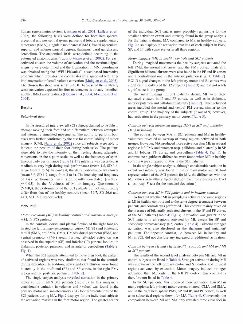
Motor imagery (MI) in healthy controls and SCI patientsDuring imagined movements the healthy subjects activated the
left PMd, the mesial PM areas, and the PMv cortex bilaterally.Significant bilateral clusters were also found in the PF and IP cortexand a contralateral one in the anterior putamen (Fig. 3; Table 2).BOLD signal changes in the left primary motor and S1 cortex wassignificant in only 3 of the 12 subjects (Table 3) and did not reachsignificance in the group.
The main findings in SCI patients during MI were largeactivated clusters in IP and PF cortex, as well as in thalamus,anterior putamen and pallidum bilaterally (Table 2). Other activatedareas included the mesial and ventral PM cortex, similar to thecontrol group. The majority of the subjects (7 out of 9) however,had activation in the primary motor cortex (Table 3).
Contrast between movement attempt (MA) in SCI and execution(ME) in healthy
The contrast between MA in SCI patients and ME in healthyvolunteers revealed an overlap of many regions activated in bothgroups. However, MA produced more activation than ME in severalregions: left PMv and putamen resp. pallidum, and bilaterally in SPand IP lobules, PF cortex and cerebellum (Table 4; Fig. 1). Incontrast, no significant differences were found when ME in healthycontrols were compared to MA in the SCI patients.
In the single-subject analysis, although considerable variation inextent and intensity was found in the primary motor and S1 footrepresentations of the SCI patients for MA, the differences with theME values in healthy subjects did not reach the significance level(t-test, resp. F-test for the standard deviations).
Contrast between MI in SCI patients and in healthy controlsTo find out whether MI in paraplegia activates the same regions
as MI in healthy controls and to the same degree, a contrast betweenpatients and controls was performed. This contrast mainly revealedthe presence of bilaterally activated clusters in the IP and PF cortexof the SCI patients (Table 4; Fig. 3). Activation was greater in theSCI patients in all regions activated by MI, except for SP andsecondary somatosensory (S2) cortex (Table 4). Bilateral strongeractivation was also disclosed in the thalamus and putamen/pallidum. The opposite contrast, i.e. between MI in healthy andMI in SCI, did not disclose any increased or additional activation.
Contrast between MI and ME in healthy controls and MA and MIin SCI patients
The results of the second level analysis between ME and MI incontrol subjects are listed in Table 4. Stronger activation during MEwas shown in the left primary motor and S1 cortex and in mostregions activated by execution. Motor imagery induced strongeractivation than ME only in the left PF cortex. This contrast istherefore not listed in Table 4.
In the SCI patients, MA produced more activation than MI inmany regions: left primary motor cortex, bilateral CMA and SMA,and in the right hemisphere PMv, SP and IP, and PF cortex, as wellas in subcortical regions shown for MA (Table 4). Conversely, thecomparison between MI and MA only revealed three clear foci in

Table 2Coordinates (in MNI standard brain space) of significant cluster maxima, t-values, and volumes in the ROI group analysis for executed, attempted, and imagined movements versus baseline in healthy controls andSCI patients (threshold pb0.01, corrected)
Functional Movement execution healthy Movement attempt SCI Motor imagery healthy Motor imagery SCIROI x y z Max.
t valueVolume(voxel)
x y z Max.t value
Volume(voxel)
x y z Max.t value
Volume(voxel)
x y z Max.t value
Volume(voxel)
M1 Left −6 −36 60 11.51 266 −12 −33 60 9.75 95S1 Left −15 −39 75 5.55 26 −30 −45 66 3.26 8S2 Left −57 −21 18 4.77 42 −60 −21 15 9.75 35 −63 −21 30 4.05 9SMA Left −9 −18 57 7.91 300 3 −21 57 6.88 124 −18 −6 66 3.08 5Pre-SMA Right 0 0 48 10.62 241 6 6 51 4.19 40
Left −9 18 45 3.26 14CMA Left −6 −30 48 5.22 29 −12 −36 54 8.74 77CMAr Right 0 0 45 12.18 229 6 9 39 3.55 21
Left −3 −3 42 5.47 105 −6 0 36 6.63 47PMd Right 45 −3 48 7.68 51
Left −36 −3 57 7.98 72 −36 −6 54 3.31 8PMv Right 60 9 9 8.92 157 51 3 0 4.77 67 60 15 −3 3.87 13 42 3 30 4.41 14
Left −57 3 6 8.85 161 −45 3 9 8.76 193 −48 3 0 5.01 35 −48 6 33 5.62 161SP Right 15 −63 66 5.26 37
Left −27 −48 69 5.39 69 −30 −63 57 7.55 165 −30 −51 69 4.66 5IP Right 66 −27 30 7.03 170 54 −30 24 6.56 111 54 −30 24 3.94 18 66 −33 33 6.65 216
Left −54 −36 27 9.28 239 −57 −39 39 5.15 319 −60 −33 24 3.34 7 −54 −48 30 10.47 527PF Right 42 39 3 3.8 20 30 33 −15 5.12 11 54 42 0 8.40 148
Left −51 15 30 4.14 8 −45 15 −6 5.69 235 −54 30 9 6.73 471TH Right 24 0 3 3.96 48
Left −9 −18 −3 8.80 106 −21 −15 6 5.81 101 −21 −12 3 3.37 13PU/PA Right 30 −15 6 4.56 77 −30 9 3 7.85 18
Left −30 −15 6 6.46 87 −30 −21 3 9.66 129 −24 −6 −6 8.09 17 −21 −3 0 4.33 51CB Right 27 −42 −27 7.26 99 9 −45 −18 20.86 553
Left −33 −57 −30 6.41 44 −18 −72 −24 7 146Right 27 −45 −45 7.71 10Left −30 −54 −45 4.81 14 −9 −84 −27 6.11 7
L=left, R=right, L/R=bilateral; ROI, region of interest; M1, primary motor cortex; S1, primary somatosensory cortex; S2, secondary somatosensory cortex; SMA, supplementary motor area; CMA, cingulate motorarea; PMd, premotor dorsal cortex; PMv premotor ventral cortex; SP, superior parietal cortex; IP, inferior parietal cortex; PF, prefrontal cortex; TH, thalamus; PU/PA, putamen/pallidum; CB, cerebellum.
387S.
Hotz-B
oendermaker
etal.
/NeuroIm
age39
(2008)383–394

Fig. 1. Activation patterns (group analysis) in SCI patients and controls displayed on mean anatomic T1-weighted images. Left column: movement attempt (MA)in SCI patients.Middle column: movement execution (ME) in controls. Right column: contrasts between movement attempt in SCI patients (MA) and movementexecution in controls (ME). (a) central region, superior and inferior parietal areas (SP and IP); (b) IP area, premotor ventral (PMv) and prefrontal cortex (PF); (c)premotor ventral (PMv), putamen/pallidum, thalamus; (d) cerebellum. Coordinates of significant regions listed in Tables 2 and 4.
388 S. Hotz-Boendermaker et al. / NeuroImage 39 (2008) 383–394
the left hemisphere: one in left PMd and two maxima in the parietalcortex, one in the SP and the other in the IP lobules.
Correlation of behavioural data and fMRI dataFor the SCI patients correlation coefficients were computed
between the quantitative aspects of the BOLD signal in all ROIs(max t-values and volumes of activation) and the clinical andbehavioral data of the individual subjects (number of disconnectedsegments, time since injury, VMIQ scores, intensity and frequencyin the 6-point rating scale for MA). No correlation coefficientreached the significance level, neither for MA nor for MI.
Discussion
The present study assessed the ability of chronic SCI patients tointernally distinguish between attempted and imagined movementsof their paralyzed feet and how these differ from executed andsimulated movements in healthy controls. Four main findings
summarize our results. First, the behavioural data clearly demon-strate that chronic complete SCI patients retain their ability tosubjectively differentiate between the executive features requiredfor MA and the cognitive ones necessary for MI. Secondly, thisbehavioural finding was confirmed by fMRI data revealingdistinctly differential patterns of activation for the two conditions.Moreover, when SCI patients attempted to move their paralyzedfoot the same network was recruited as when healthy subjectsactually executed the foot movement. The same was true for theinternal simulation of the movements, which activated the regionspreviously described for MI in healthy subjects and also seen in thecontrols of the present study. Third, our study confirms that duringMA, cortical motor areas, in particular the primary sensorimotorcortex, are functionality preserved in SCI patients, though withreduced activation due to a long period of disconnection. Finally,the enhanced activation in most secondary motor areas and theadditional recruitment of prefrontal and parietal areas both duringMA and MI in SCI patients suggests that the paraplegic condition

Table 3Frequency of single subject activation in specified ROIs
FunctionalROI
ExecutioncontrolsN=10
Movementattempt SCIN=9
Motor imagerycontrolsN=10
Motor imagerySCI N=9
M1 10/– 9/– 3./− 7/–S1 10/5 6/4 3/– 3/1S2 9/5 6/4 3/3 5/3SMA 10 7 5 8CMA 10 7 5 7PMd 8/7 5/4 5/2 3/2PMv 8/7 5/4 5/5 5/7SP 9/5 9/7 7/1 4/3IP 6/4 6/4 7/6 9/9PU/PA 4/0 3/2 –/– –/–CB 9/4 8/7 4/3 3/3
Number contralateral/Number ipsilateral. Abbreviations: see Table 2.
Fig. 2. Local maxima of the single-subject activations for movement attemptin SCI patients and execution in controls after normalization for primarymotor cortex (M1), superior parietal cortex (SP), inferior parietal cortex (IP),premotor ventral (PMv). Yellow: SCI patients. Green: controls. Left column:x, y coordinates projected onto a coronal section of a representative MNIstandard brain through the most anterior local maxima. Right column: x, zcoordinates projected onto a transverse section through the most inferiorlocal maxima. Note that the general scatter is partially because several higherand lower sections have been projected onto one single section.
389S. Hotz-Boendermaker et al. / NeuroImage 39 (2008) 383–394
may require an increase in attention allocation to perform the tasksand/or have induced some adaptive changes in the functionalnetworks involved.
Movement attempt in SCI patients
Few neuroimaging studies have addressed MA in chronic spinalcord-injured patients (Sabbah et al., 2002; Cramer et al., 2005;Halder et al., 2006; Fallani et al., 2007). The most recent fMRIinvestigation (Cramer et al., 2005) reported an activation patternduring MA similar to that observed during execution in healthycontrols, though with decreased volumes in most cortical regionsexamined. The present study replicated this activation patternhowever, with the exception of the primary motor cortex, equivalentor greater BOLD activation was found in all other areas, as well asrecruitment of several additional regions (PMv, SP, IP, and PFcortex). In addition, in the present investigation, the basal gangliawere always activated, in the healthy subjects as well as in thechronic SCI patients. This is in contrast with Cramer et al. (2005)who reported a significant BOLD signal in the pallidum only fortheir SCI population and who interpreted this finding as theemergence of pathological activation. Differences in experimentaldesigns most likely account for the discrepancy between thesefindings. In the study by Cramer et al. (2005), attempted movementwas initiated by a video of the target motion shown before andduring the fMRI session, and the foot task used in theirinvestigation, an attempt to crush a displayed object every 3 s,was more complex than our self initiated, simple, repetitive dorsaland plantar foot flexion. Furthermore, healthy subjects in their studyperformed also a movement attempt task, which is difficult toperform without isometric muscle contractions, as opposed to thesimple motor execution used in our study.
The fact that no significant differences in BOLD signal betweenMA in the SCI patients and ME in healthy controls were found inprimary sensorimotor and PM mesial cortex, supports ourassumption that these are two corresponding conditions, whichcan be contrasted with each other, despite the fact that attempt tomove can only be indirectly controlled through behavioural tests, asthe movements are not visible. The similarity between the networkactivated in SCI patients during MA and the execution network ofhealthy subjects additionally provides the neural and thus “visible”evidence for task performance. In fact, this finding in chronic para-plegics, who were all neurophysiologically tested for completeness
of the disconnection, reveals their retained potential to initiate andcontrol foot movements, even after a long period of non-use, assuggested by the behavioural assessment. Persistence of motornetworks in long-term deafferented subjects has also been reportedin amputees who showed fast recovery of sensory motor functionsfollowing reafferentation. fMRI studies in these subjects revealedfollowing hand-grafting a reversal of cortical reorganization to anormal activation pattern (Giraux et al., 2001; Neugroschl et al.,2005). Consistent with results of earlier investigations (Lacourse etal., 1999; Halder et al., 2006) in the group analysis, the activation inthe primary motor cortex during MA was reduced as compared toME of healthy controls, but this did not reach the significance level.At the single subject level however, the size and intensity of signalchanges in the primary motor cortex did not differ significantly

Fig. 3. Activation patterns (group analysis) in SCI patients and controls displayed on mean anatomic T1-weighted images. Left column: motor imagery in SCIpatients (MISCI). Middle column: motor imagery in controls (MIcontrol). Right column: contrasts between motor imagery in SCI patients (MISCI) and in controls(MIcontrol). (a) central region, superior and inferior parietal areas (SP and IP); (b) IP area, premotor ventral (PMv) and prefrontal cortex (PF); (c) premotor ventral(PMv), putamen/pallidum, thalamus. Coordinates of significant regions listed in Tables 2 and 4.
390 S. Hotz-Boendermaker et al. / NeuroImage 39 (2008) 383–394
when controls and patients were compared, suggesting that thesmaller cluster observed in the SCI patients at group level wasprobably due to the scatter of the individual data during theaveraging process. In spite of the electrophysiological assessedinterruption of the sensory afferent pathway from the periphery, asmall BOLD signal has been observed in the postcentral regionconfirming our earlier findings of a primary somatosensory (S1)foot representation recruitment in complete SCI patients (Alkadhi etal., 2005). This postcentral activation can be attributed to anefference copy of the ongoing movement in sensory regions (Holstand Mittelstaedt, 1950). A recent fMRI investigation with ischemicnerve block on the lower limb also disclosed activation in S1 givingfurther support to this hypothesis (Christensen et al., 2007).
Two present findings suggest that MA is a more demanding taskthan ME. First, both the additionally activated focus in the PFcortex and the activation enhancement in the parietal lobe suggestthe existence of a stronger cognitive component during MA. Thismay reflect the intense attention allocation required from thechronic SCI patients to perform a considered easy over-learned task(Allen et al., 1997; Rowe et al., 2002; Rushworth et al., 2003).Second, the comparison between attempted and performed foot taskrevealed stronger activity specific for MA in the parietal cortex, incerebellar regions, and in the putamen. One cannot exclude that thechronic paraplegic condition has also induced adaptive changes inthese key structures yielding sensorimotor transformations andmovement guidance (Catalan et al., 1998; Allen et al., 2005). The
scattered activation seen in the individual activation in the SCIindividual data in parietal and premotor regions also points to thepresence of adaptive changes. Recent EEG data strongly suggestmodifications in connectivity between cortical regions during MAin SCI patients compared to healthy subjects (Fallani et al., 2007).
Motor imagery in SCI patients
In our earlier study (Alkadhi et al., 2005), SCI patients wereasked to mentally move their right foot. This instruction led toenhanced activation of an extensive network of brain areascomprising regions activated both during motor imagery andduring execution in healthy controls (Lafleur et al., 2002). In thepresent investigation, MI in SCI patients recruited areas that werespatially more restricted to frontal, mesial and premotor ventralcortex, parietal regions, thalamus and striatum. These are regionsthat normally activate during MI in healthy subjects (Gerardin et al.,2000). Compared to our former study where the primary motorcortex was significantly activated during MI, activation in thepresent study was inconsistently observed in the individualsubjects, in accordance with previous investigations using similartasks (Porro et al., 1996; Gerardin et al., 2000). In contrast, duringMA, the primary motor cortex was consistently activated, though ata reduced intensity. These fMRI findings clearly confirmed theresults in the behavioral assessments namely that the SCI patientswere performing distinct MA and MI tasks.

Table 4Coordinates (in MNI standard brain space) of significant cluster maxima, t-values, and volumes for the contrasts in healthy controls and SCI patients (threshold pb0.01, corrected)
Functional Controls ME vs. MI SCI MA vs. MI SCI MI vs. MA SCI MA vs. ME controls SCI MI vs. controls MIROI x y z Max.
t valueVolume(voxel)
x y z Max.t value
Volume(voxel)
x y z Max.t value
Volume(voxel)
x y z Max.t value
Volume(voxel)
x y z Max.t value
Volume(voxel)
M1 Left −3 −36 60 10.87 262 −12 −33 60 7.34 111S1 Right 18 −45 75 5.07 40
Left −15 −39 75 6.05 29SMA Bilat 9 −15 66 10.97 501 6 −24 57 5.94 45 12 −12 54 3.26 5CMA Bilat −3 −36 51 6.00 53 −12 −36 54 6.63 70CMAr Bilat −3 0 39 11.82 169 −6 0 39 3.26 12PMd Right 15 −21 66 5.56 12
Left −15 −15 69 3.63 13 −39 −27 60 3.76 9PMv Right 45 −30 18 6.61 41 51 12 27 5.24 37 54 12 30 2.91 10
Left −48 −27 18 8.14 46 −45 3 12 3.75 56 −45 12 6 2.99 22SP Right 15 −72 51 3.99 24 30 −60 63 3.75 12
Left −18 −42 63 5.46 41 −42 −30 63 5.38 85 −30 −63 57 4.92 62IP Right 63 −24 18 7.98 152 42 −63 27 4.86 88 39 −72 36 3.41 30 63 −45 33 3.67 91
Left −51 −27 18 8.14 100 −57 −24 48 5.26 19 −36 −63 54 4.13 17 −60 −51 33 3.53 109Right 45 −42 54 3.64 23 42 −48 45 4.38 23Left −39 −51 60 6.1 5 −42 −72 36 3.37 42 −42 −60 54 3.44 30
PF Right 42 42 18 5.88 112 30 12 −21 3.84 6 51 30 6 4.08 152Left −30 45 −15 3.97 39 −36 33 39 3.80 41
TH Right 21 −21 9 3.02 7Left −21 −24 6 3.83 14 −21 −24 −3 4.94 21 −9 −6 9 3.1 18
PU/PA Right 21 −3 −6 3.3 81Left −30 −12 6 5.03 46 −27 −15 6 6.85 48 −30 −6 −6 3.47 29 −27 6 3 3.14 53
CB Right 15 −42 −24 8.22 81 9 −45 −21 6.21 96 12 −48 −21 7.92 384 21 −45 −27 3.01 9Left −36 −57 −39 7.09 98 −15 −48 −15 4.27 24 −9 −63 −24 6.34 314Right 27 −69 −27 4.21 17 27 −45 −45 4.36 14 9 −57 −15 3.01 11Left −24 −36 −30 3.73 10 −12 −84 −27 3.86 43 −30 −81 −33 4.43 43
Abbreviations: see Table 2.
391S.
Hotz-B
oendermaker
etal.
/NeuroIm
age39
(2008)383–394

392 S. Hotz-Boendermaker et al. / NeuroImage 39 (2008) 383–394
Prefrontal and parietal areas showed enhanced activation duringMI in the SCI patients when compared to the control group. Thisincreased activity confirms our previous findings (Alkadhi et al.,2005), but is not in line with those of Cramer and colleagues (2005)who, in a similar contrast, did not observe significant changes inthese regions. In their study, the only cortical area showingincreased activation during MI was the superior temporal gyrus, aregion important for the visual perception of biological motion,which never activated in our investigation. These conflictingfindings between the two studies can be attributed to differences inthe experimental protocols used. Videos of the required complexmovement were shown in their study with the instruction to imaginemovement completion, which may have induced unconsciousstrategies leading to 3rd person motor imagery. As recentlydemonstrated, kinesthetic (1st person) and visual (3rd person)motor imagery are supported by different neural networks(Solodkin et al., 2004). In our experiment, no visual stimuli werepresented and the subjects with eyes closed were specificallyinstructed and trained to prevent developing a strategy leading tovisualization of their limb. Accordingly, activation in visual regionswas not observed during attempted or imagined movements.
Central motor control in paraplegia
The present investigation indicates that in chronic paraplegicpatients the central programs for execution of foot movements andtheir internal simulation remain preserved, activating severalcommon regions and, in addition, other distinct ones specific toeither task. MA and MI in a status of chronic deafferentation anddeefferentation are complex tasks, which recruit cortical regionsinvolved in higher cognitive processes. Despite every effort in thisstudy to distinguish between the two tasks, taking into considera-tion the single subjects’ activations, as well as their contrasts, wecannot completely rule out the likelihood of a contamination ofeither task by the other. This possibility may explain the activationof the PF cortex during MA and the large number of SCI patientswho had some activation in the primary motor cortex during MI.These two tasks may be one and the same phenomenon, or twoversions of the same phenomenon, with quantitative differencesbetween the two.
How the control of virtual foot movements can be preservedafter a prolonged period of complete disconnection? In patients withchronic hemiplegia, the ability to construct internal action repre-sentations of the upper limbs can be robust even after years of limbnon-use (Johnson-Frey, 2004). The process of matching the finalposition of one’s limbs with an intended movement is achievedthrough a comparison process between the predicted sensory con-sequences of the action and the actual sensory feedback (Desmurgetand Grafton, 2000; Wolpert and Ghahramani, 2000). Since peri-pheral cutaneous and proprioceptive afferents of the lower limbs areunavailable in complete SCI patients, this process can be accom-plished solely by means of stored motor programs and the resultingstream of motor commands with their sensory signals generatedthrough corollary discharge (Blakemore and Sirigu, 2003). Theadditional fact that the SCI patients have continuous daily visualcontrol of their body may also play a role in maintaining an internalrepresentation of their limbs through a continuous updating bysimply looking at them (Wolpert et al., 1998). These speculationsare supported by the retained integrity of the internal action repre-sentation in our patients as revealed by both the structured interviewand the fMRI data.
It has been suggested that parietal areas constitute the neuralsubstrate for the storage of visual and kinaesthetic limb postures,which are subsequently mapped onto corresponding motor regions(Sirigu et al., 1996). Damage to the parietal cortex leads to theinability both of maintaining an internal representation of the body(Wolpert et al., 1998) and of internal movement simulation (Siriguet al., 1996). These findings indicate that the parietal cortex is a keystructure in sensorimotor integration and, together with its inter-actions with the cerebellum, plays an important role in acquisitionand recall of skilled movements (Allen et al., 1997; Shadmehr andHolcomb, 1997; Andersen and Buneo, 2003; Blakemore and Sirigu,2003). The enhanced parietal and cerebellar activations observed inchronic SCI patients during MA and MI in our study suggests thatsome adaptive changes have occurred in these regions. The absenceof sensory input may have modified the functionality of these areasin order to maintain an intact body representation and organizemotor plans accordingly.
Clinical significance
The present study demonstrates in chronic paraplegics theretained functionality of neuronal networks that in healthy subjectsare responsible for dorsal and plantar flexions of the foot and theirinternal simulation. This finding may have important clinical valuewhen considering new treatment approaches aiming at functionalrecovery following spinal cord damage. If reconnection of the brainto the paralyzed limbs through the spinal cord is successful,according to our present data, the still functional motor programsshould allow a certain degree of motor control. It further providesthe principal physiological requirements for the development of abrain–computer interface device that uses intention-driven neuronalactivity to be converted into a control signal that enables usefultasks (Hochberg et al., 2006). The apparent integrity of MI in SCIpatients and the resemblance of their MA network with the MEnetwork of healthy subjects suggest that the paraplegics still disposeof the full motor programs for overt and covert foot movements.Recent reports provide convincing evidence that mental practicebased on motor imagery might be beneficial for learning newmovements and/or strengthening memorized ones (Jackson et al.,2003; Lacourse et al., 2005; Cramer et al., 2007). We thereforesuggest that MA and MI may be useful adjuncts to traditionalrehabilitation strategies for improving motor functions after spinalcord injury particularly for incomplete patients with residual motorfunctions.
Acknowledgments
We thank Kai Lutz for his assistance in fMRI data analysis. Thiswork was supported by the Swiss National Science Foundation(SNF) Project # 3100-67168.01.
References
Alkadhi, H., Brugger, P., Boendermaker, S.H., Crelier, G., Curt, A., Hepp-Reymond, M.C., Kollias, S.S., 2005. What disconnection tells aboutmotor imagery: evidence from paraplegic patients. Cereb. Cortex 15,131–140.
Allen, G., Buxton, R.B., Wong, E.C., Courchesne, E., 1997. Attentionalactivation of the cerebellum independent of motor involvement. Science275, 1940–1943.
Allen, G., McColl, R., Barnard, H., Ringe, W.K., Fleckenstein, J., Cullum,

393S. Hotz-Boendermaker et al. / NeuroImage 39 (2008) 383–394
C.M., 2005. Magnetic resonance imaging of cerebellar–prefrontal andcerebellar–parietal functional connectivity. NeuroImage 28, 39–48.
Andersen, R.A., Buneo, C.A., 2003. Sensorimotor integration in posteriorparietal cortex. Adv. Neurol. 93, 159–177.
Blakemore, S.J., Sirigu, A., 2003. Action prediction in the cerebellum and inthe parietal lobe. Exp. Brain Res. 153, 239–245.
Brugger, P., Kollias, S.S., Muri, R.M., Crelier, G., Hepp-Reymond, M.C.,Regard, M., 2000. Beyond remembering: phantom sensations of con-genitally absent limbs. Proc. Natl. Acad. Sci. U. S. A. 97, 6167–6172.
Catalan, M.J., Honda, M., Weeks, R.A., Cohen, L.G., Hallett, M., 1998. Thefunctional neuroanatomy of simple and complex sequential fingermovements: a PET study. Brain 121, 253–264.
Christensen, M.S., Lundbye-Jensen, J., Geertsen, S.S., Petersen, T.H.,Paulson, O.B., Nielsen, J.B., 2007. Premotor cortex modulates soma-tosensory cortex during voluntary movements without proprioceptivefeedback. Nat. Neurosci. 10, 417–419.
Cramer, S.C., Lastra, L., Lacourse, M.G., Cohen, M.J., 2005. Brain motorsystem function after chronic, complete spinal cord injury. Brain 128,2941–2950.
Cramer, S.C., Orr, E.L., Cohen, M.J., Lacourse, M.G., 2007. Effects ofmotor imagery training after chronic, complete spinal cord injury. Exp.Brain Res. 177, 233–242.
Curt, A., Dietz, V., 1999. Electrophysiological recordings in patients withspinal cord injury: significance for predicting outcome. Spinal Cord 37,157–165.
Decety, J., Jeannerod, M., 1995. Mentally simulated movements in virtualreality: does Fitts’s law hold in motor imagery? Behav. Brain Res. 72,127–134.
Desmurget, M., Grafton, S., 2000. Forward modeling allows feedbackcontrol for fast reaching movements. Trends Cogn. Sci. 4, 423–431.
Dobkin, B.H., Firestine, A., West, M., Saremi, K., Woods, R., 2004. Ankledorsiflexion as an fMRI paradigm to assay motor control for walkingduring rehabilitation. NeuroImage 23, 370–381.
Fallani, F.D., Astolfi, L., Cincotti, F., Mattia, D., Marciani, M.G., Salinari,S., Kurths, J., Gao, S., Cichocki, A., Colosimo, A., Babiloni, F., 2007.Cortical functional connectivity networks in normal and spinal cordinjured patients: evaluation by graph analysis. Hum. Brain Mapp.(Electronic publication ahead of print).
Friston, K.J., Holmes, A., Poline, J.B., Price, C.J., Frith, C.D., 1996.Detecting activations in PET and fMRI: levels of inference and power.NeuroImage 4, 223–235.
Gerardin, E., Sirigu, A., Lehericy, S., Poline, J.B., Gaymard, B., Marsault,C., Agid, Y., Le Bihan, D., 2000. Partially overlapping neural networksfor real and imagined hand movements. Cereb. Cortex 10, 1093–1104.
Giraux, P., Sirigu, A., Schneider, F., Dubernard, J.M., 2001. Corticalreorganization in motor cortex after graft of both hands. Nat. Neurosci. 4,691–692.
Halder, P., Curt, A., Brem, S., Lang-Dullenkopf, A., Bucher, K., Kollias, S.,Brandeis, D., 2006. Preserved aspects of cortical foot control inparaplegia. NeuroImage 31, 692–698.
Hanakawa, T., Immisch, I., Toma, K., Dimyan, M.A., Van Gelderen, P.,Hallett, M., 2003. Functional properties of brain areas associated withmotor execution and imagery. J. Neurophysiol. 89, 989–1002.
Hochberg, L.R., Serruya, M.D., Friehs, G.M., Mukand, J.A., Saleh, M.,Caplan, A.H., Branner, A., Chen, D., Penn, R.D., Donoghue, J.P., 2006.Neuronal ensemble control of prosthetic devices by a human withtetraplegia. Nature 442, 164–171.
Holst, E.v., Mittelstaedt, H., 1950. Das Reafferenzprincip (Wechselwirkun-gen zwischen Zentralnervensystem und Periferie). Naturwissenschaft37, 464–476.
Isaac, A., Marks, D.F., Russel, D.G., 1986. An instrument for assessingimagery of movements: the Vividness of Movements Imagery Ques-tionnaire (VMIQ). J. Ment. Imag. 10, 23–30.
Jackson, P.L., Lafleur, M.F., Malouin, F., Richards, C., Doyon, J., 2001.Potential role of mental practice using motor imagery in neurologicrehabilitation. Arch. Phys. Med. Rehabil. 82, 1133–1141.
Jackson, P.L., Lafleur, M.F., Malouin, F., Richards, C.L., Doyon, J.,
2003. Functional cerebral reorganization following motor sequencelearning through mental practice with motor imagery. NeuroImage 20,1171–1180.
Jeannerod, M., 1995. Mental imagery in the motor context. Neuropsycho-logia 33, 1419–1432.
Jeannerod, M., 2001. Neural simulation of action: a unifying mechanism formotor cognition. NeuroImage 14, S103–S109.
Jeannerod, M., Decety, J., 1995. Mental motor imagery: a window into therepresentational stages of action. Curr. Opin. Neurobiol. 5, 727–732.
Johnson, S.H., 2000. Imagining the impossible: intact motor representationsin hemiplegics. NeuroReport 11, 729–732.
Johnson-Frey, S.H., 2004. Stimulation through simulation? Motor imageryand functional reorganization in hemiplegic stroke patients. Brain Cogn.55, 328–331.
Kawato, M., 1999. Internal models for motor control and trajectory plan-ning. Curr. Opin. Neurobiol. 9, 718–727.
Lacourse, M.G., Cohen, M.J., Lawrence, K.E., Romero, D.H., 1999. Cor-tical potentials during imagined movements in individuals with chronicspinal cord injuries. Behav. Brain Res. 104, 73–88.
Lacourse, M.G., Orr, E.L., Cramer, S.C., Cohen, M.J., 2005. Brain activ-ation during execution and motor imagery of novel and skilled sequen-tial hand movements. NeuroImage 27, 505–519.
Lafleur, M.F., Jackson, P.L., Malouin, F., Richards, C.L., Evans, A.C.,Doyon, J., 2002. Motor learning produces parallel dynamic functionalchanges during the execution and imagination of sequential footmovements. NeuroImage 16, 142–157.
MacIntosh, B.J., Mraz, R., Baker, N., Tam, F., Staines, W.R., Graham, S.J.,2004. Optimizing the experimental design for ankle dorsiflexion fMRI.NeuroImage 22, 1619–1627.
Maldjian, J.A., Laurienti, P.J., Kraft, R.A., Burdette, J.H., 2003. Anautomated method for neuroanatomic and cytoarchitectonic atlas-basedinterrogation of fMRI data sets. NeuroImage 19, 1233–1239.
Maynard Jr., F.M., Bracken, M.B., Creasey, G., Ditunno Jr., J.F., Donovan,W.H., Ducker, T.B., Garber, S.L., Marino, R.J., Stover, S.L., Tator, C.H.,Waters, R.L., Wilberger, J.E., Young, W., 1997. International standardsfor neurological and functional classification of spinal cord injury.American Spinal Injury Association. Spinal Cord 35, 266–274.
Nair, D.G., Purcott, K.L., Fuchs, A., Steinberg, F., Kelso, J.A., 2003.Cortical and cerebellar activity of the human brain during imagined andexecuted unimanual and bimanual action sequences: a functional MRIstudy. Brain Res. Cogn. Brain Res. 15, 250–260.
Naito, E., Kochiyama, T., Kitada, R., Nakamura, S., Matsumura, M.,Yonekura, Y., Sadato, N., 2002. Internally simulated movementsensations during motor imagery activate cortical motor areas and thecerebellum. J. Neurosci. 22, 3683–3691.
Neugroschl, C., Denolin, V., Schuind, F., Van Holder, C., David, P.,Balériaux, D., Metens, T., 2005. Functional MRI activation of soma-tosensory and motor cortices in a hand-grafted patient with early clinicalsensorimotor recovery. Eur. Radiol. 15, 1806–1814.
Porro, C.A., Francescato, M.P., Cettolo, V., Diamond, M.E., Baraldi, P.,Zuiani, C., Bazzocchi, M., di Prampero, P.E., 1996. Primary motor andsensory cortex activation during motor performance and motor imagery:a functional magnetic resonance imaging study. J. Neurosci. 16,7688–7698.
Rowe, J., Friston, K., Frackowiak, R., Passingham, R., 2002. Attention toaction: specific modulation of corticocortical interactions in humans.NeuroImage 17, 988–998.
Rushworth, M.F., Johansen-Berg, H., Gobel, S.M., Devlin, J.T., 2003. Theleft parietal and premotor cortices: motor attention and selection.NeuroImage 20 (Suppl 1), S89–S100.
Sabbah, P., de, S.S., Leveque, C., Gay, S., Pfefer, F., Nioche, C., Sarrazin,J.L., Barouti, H., Tadie, M., Cordoliani, Y.S., 2002. Sensorimotorcortical activity in patients with complete spinal cord injury: a functionalmagnetic resonance imaging study. J. Neurotrauma 19, 53–60.
Shadmehr, R., Holcomb, H.H., 1997. Neural correlates of motor memoryconsolidation. Science 277, 821–825.
Sirigu, A., Duhamel, J.R., Cohen, L., Pillon, B., Dubois, B., Agid, Y., 1996.

394 S. Hotz-Boendermaker et al. / NeuroImage 39 (2008) 383–394
The mental representation of hand movements after parietal cortexdamage. Science 273, 1564–1568.
Solodkin, A., Hlustik, P., Chen, E.E., Small, S.L., 2004. Fine modulation innetwork activation during motor execution and motor imagery. Cereb.Cortex 14, 1246–1255.
Stephan, K.M., Fink, G.R., Passingham, R.E., Silbersweig, D., Ceballos-Baumann, A.O., Frith, C.D., Frackowiak, R.S., 1995. Functional ana-tomy of the mental representation of upper extremity movements inhealthy subjects. J. Neurophysiol. 73, 373–386.
Tzourio-Mazoyer, N., Landeau, B., Papathanassiou, D., Crivello, F., Etard, O.,Delcroix, N., Mazoyer, B., Joliot, M., 2002. Automated anatomicallabeling of activations in SPM using a macroscopic anatomical parcella-tion of the MNI MRI single-subject brain. NeuroImage 15, 273–289.
Wolpert, D.M., Ghahramani, Z., 2000. Computational principles ofmovement neuroscience. Nat. Neurosci. 3, 1212–1217 (Suppl).
Wolpert, D.M., Goodbody, S.J., Husain, M., 1998. Maintaining internalrepresentations: the role of the human superior parietal lobe. Nat.Neurosci. 1, 529–533.
Related Documents