Morphometric analysis of the association of primary shoulder reconstruction procedures with scapular growth in obstetric brachial plexus paralysis patients Julia K. Terzis & Dimitrios Karypidis & Ricardo Mendoza & Zinon T. Kokkalis & Norou Diawara Published online: 14 May 2014 # American Association for Hand Surgery 2014 Abstract Background Obstetric brachial plexus paralysis (OBPP) has been associated with shoulder deformities, scapular growth, and shoulder function impairment. The absence of balanced muscular forces acting on the scapula has been considered responsible for scapula dysplasia and impaired growth as compared with the normal side. Scapula growth impairment may also lead to shoulder and upper extremity dysfunction. This study aims at showing the association of primary nerve reconstruction with the restoration of scapular bone growth potential. Methods This is a retrospective review of 73 patients with OBPP who underwent primary shoulder reconstruction. Pa- tients were categorized for assessment and analysis into group A, global paralysis; group B, Erb’ s palsy; and group C, Erb’ s palsy with C7 root involvement. Scapular posteroanterior and lateral X-rays were obtained in which four scapula dimensions were manually measured. The growth discrepancy depending on the applied treatment was investigated. Results The highest improvement was noted in scapular height in the Erb’s palsy group who underwent simultaneous neurotization of the suprascapular and axillary nerves. The oblique axis was more improved in the Erb’ s palsy group while both big and small widths were more improved in the Erb’ s palsy with C7 root involvement group in patients who underwent concomitant neurotization of the suprascapular and the axillary nerves. Functional improvement correlated positively with growth improvement in all groups and scapular dimensions. Conclusion Scapula growth and shoulder function improve- ment were higher in patients with Erb’ s palsy. Simultaneous axillary and suprascapular nerve neurotization provided the best outcome in both functional and growth restoration. Keywords Scapula . Bone growth . Obstetric brachial plexus paralysis . Primary plexus reconstruction . Shoulder Introduction Most of the patients presenting with obstetric brachial plexus paralysis (OBPP) experience gradual spontaneous recovery. About 10 % of cases with C5–C6 lesion, 35 % with C5–C7 lesion, 50 % with C5–T1, and nearly all cases with C5–T1 with positive Horner sign most commonly report dysfunction of shoulder abduction (SA) and external rotation (SER) [5]. Sjoberg et al. [32] studied the prognosis of children with OBPP among 25,000 newborns in a 10-year period and emphasized prevention regarding obstetric and pediatric management. Early in the twentieth century, osseous deformities in pa- tients with OBPP were frequently observed in plain X-rays and reported but little or no evidence was provided regarding J. K. Terzis International Institute of Reconstructive Microsurgery, 27-28 Thomson Ave., Long Island City, NY 11101, USA D. Karypidis : R. Mendoza International Institute of Reconstructive Microsurgery, 27-28 Thomson Ave., Long Island City, NY 11101, USA Z. T. Kokkalis First Department of Orthopaedics, Athens University School of Medicine, “Attikon” University Hospital, 1 Rimini, 12462, Chaidari, Athens 12462, Greece N. Diawara Mathematics and Statistics Department, Old Dominion University, 4700 Elkhorn Ave, Norfolk, VA 23529, USA J. K. Terzis (*) Department of Plastic Surgery, New York University Medical Center, Long Island City, NY 11101, USA e-mail: [email protected] J. K. Terzis e-mail: [email protected] HAND (2014) 9:292–302 DOI 10.1007/s11552-014-9637-7

Welcome message from author
This document is posted to help you gain knowledge. Please leave a comment to let me know what you think about it! Share it to your friends and learn new things together.
Transcript

Morphometric analysis of the association of primary shoulderreconstruction procedures with scapular growth in obstetricbrachial plexus paralysis patients
Julia K. Terzis & Dimitrios Karypidis &Ricardo Mendoza & Zinon T. Kokkalis & Norou Diawara
Published online: 14 May 2014# American Association for Hand Surgery 2014
AbstractBackground Obstetric brachial plexus paralysis (OBPP) hasbeen associated with shoulder deformities, scapular growth,and shoulder function impairment. The absence of balancedmuscular forces acting on the scapula has been consideredresponsible for scapula dysplasia and impaired growth ascompared with the normal side. Scapula growth impairmentmay also lead to shoulder and upper extremity dysfunction.This study aims at showing the association of primary nervereconstruction with the restoration of scapular bone growthpotential.Methods This is a retrospective review of 73 patients withOBPP who underwent primary shoulder reconstruction. Pa-tients were categorized for assessment and analysis into groupA, global paralysis; group B, Erb’s palsy; and group C, Erb’s
palsy with C7 root involvement. Scapular posteroanterior andlateral X-rays were obtained in which four scapula dimensionswere manually measured. The growth discrepancy dependingon the applied treatment was investigated.Results The highest improvement was noted in scapular heightin the Erb’s palsy group who underwent simultaneousneurotization of the suprascapular and axillary nerves. Theoblique axis was more improved in the Erb’s palsy group whileboth big and small widthsweremore improved in the Erb’s palsywith C7 root involvement group in patients who underwentconcomitant neurotization of the suprascapular and the axillarynerves. Functional improvement correlated positively withgrowth improvement in all groups and scapular dimensions.Conclusion Scapula growth and shoulder function improve-ment were higher in patients with Erb’s palsy. Simultaneousaxillary and suprascapular nerve neurotization provided thebest outcome in both functional and growth restoration.
Keywords Scapula . Bone growth . Obstetric brachial plexusparalysis . Primary plexus reconstruction . Shoulder
Introduction
Most of the patients presenting with obstetric brachial plexusparalysis (OBPP) experience gradual spontaneous recovery.About 10 % of cases with C5–C6 lesion, 35 % with C5–C7lesion, 50%with C5–T1, and nearly all cases with C5–T1withpositive Horner sign most commonly report dysfunction ofshoulder abduction (SA) and external rotation (SER) [5].Sjoberg et al. [32] studied the prognosis of children with OBPPamong 25,000 newborns in a 10-year period and emphasizedprevention regarding obstetric and pediatric management.
Early in the twentieth century, osseous deformities in pa-tients with OBPP were frequently observed in plain X-raysand reported but little or no evidence was provided regarding
J. K. TerzisInternational Institute of Reconstructive Microsurgery, 27-28Thomson Ave., Long Island City, NY 11101, USA
D. Karypidis : R. MendozaInternational Institute of Reconstructive Microsurgery, 27-28Thomson Ave., Long Island City, NY 11101, USA
Z. T. KokkalisFirst Department of Orthopaedics, Athens University School ofMedicine, “Attikon”University Hospital, 1 Rimini, 12462, Chaidari,Athens 12462, Greece
N. DiawaraMathematics and Statistics Department, Old Dominion University,4700 Elkhorn Ave, Norfolk, VA 23529, USA
J. K. Terzis (*)Department of Plastic Surgery, NewYork UniversityMedical Center,Long Island City, NY 11101, USAe-mail: [email protected]
J. K. Terzise-mail: [email protected]
HAND (2014) 9:292–302DOI 10.1007/s11552-014-9637-7
the etiology of these deformities. According to Ebaug et al.[7], the upper and lower portions of the trapezius and theserratus anterior muscles are important for scapulothoracicmotion. In patients with OBPP, the anatomical integrity andfunction of these muscles are impaired due to the develop-mental irregularities of the scapula. Therefore, it was observedthat the aforementioned long-standingmuscle imbalance leadsto glenohumeral deformity which further deteriorates the re-sidual weakness and functional limitation of the shoulder.Nevertheless, they did not analyze specific anatomic irregu-larities of the scapula and they did not investigate the effect oftreatment on the latter. Pearl and Edgerton [25] investigatedthe pathological changes that occur in the glenohumeral jointduring growth in 25 patients with OBPP, but their findingswere not associated with the anatomic characteristics of thescapula and the scapulothoracic joint. Waters et al. [41] de-scribed glenohumeral and other skeletal deformities, such asglenoid hypoplasia, posterior subluxation and flattening of thehumeral head, and hooking of the coracoid, in 42 childrenwith OBPP evaluated with computerized tomography or mag-netic resonance imaging. Although they noted improvementin both function and growth of the glenohumeral joint andscapula, they did not provide an evaluation of a potentialassociation between improvement and specific treatment.
In their study including 58 children with unilateral OBPP,van Gelein Vitringa et al. [40] showed that the affected scapulawas significantly smaller compared to that of the normal side.This reduction of growth was more noted on the dorsal surfaceof the affected scapula than on its ventral surface, and it wassignificantly associatedwith the subluxation of the humeral headdue to the brachial plexus lesion. However, their study includedthe measurement of only the horizontal distance between themedial side of the scapula and the subchondral glenoid.
Russo et al. [28] reported the significance of scapulothoracicand glenohumeral contribution to shoulder and upper extremitymotion and the degree that this contribution is decreased inchildren with OBPP. This decrease was significantly associatedwith the scapulothoracic displacement due to abnormal scapu-lar morphology and dysplasia. However, these results were notclearly associated with specific scapular deformities or treat-ment histories.
Terzis et al. [39] also reported the radiologic characteristicsof shoulder deformities in OBPP such as the elevated andhypoplastic scapula, the shallow and flattened glenoid fossa,the inferiorly directed coracoid process, and the abnormallytapered acromion. However, the analysis of scapular growthand the association of the improved developmental and func-tional potential of the shoulder with the treatment were notincluded.
It is therefore evident that the absence of appropriate mus-cle force on the bones in OBPP patients leads to bone growthand size deficiency compared to the unaffected side. Appar-ently, there is a paucity of literature on scapula growth
impairment in particular which comprises both a functionaland cosmetic deformity that needs to be addressed due to therole of the scapula in shoulder function [5, 32].
The aim of this study is to report the association of primaryshoulder reconstruction procedures with scapular growth inOBPP patients.
Methods and Material
This is a retrospective review of 73 patients with OBPP whounderwent primary shoulder reconstruction procedures be-tween March 1979 and March 2007. Detailed patient historyand a follow-up time of 2 years or longer were prerequisitesfor patient inclusion. Table 1 shows patients’ demographicsand OBPP diagnosis.
Primary shoulder reconstruction procedures (n=125) in-cluded neurolysis and axillary (Ax) and/or suprascapularnerve (Ss) neurotization using extraplexus and intraplexusmotor donors. The term neurotization refers to primary nerverepair using interposition nerve grafts, most often harvestedsural and saphenous nerves. The proximal end of the interpo-sition nerve graft is coapted to the motor donor and the distalend to the nerve being neurotized, in an end-to-end fashion.Nerve regeneration is enabled along the nerve grafts. Inintraplexus neurotization, donor options include the undam-aged roots or available branches or fascicles of functioningdonors. In global root avulsions where intraplexus donors are
Table 1 Patients’ demographics and OBPP diagnosis
Total patients included 73
Female 37 patients
Male 36 patients
Mean denervation time 0.39±0.58 year old
Global paralysis in 48 patients
Erb’s paralysis 10 patients
Erb’s paralysis with C7 root involvement in 15 patients
Mean follow-up time 5.85±2.54 years
Gestation (weeks)
<37 8
37–42 64
>42 1
Forceps 15
Suction 9
Birth weight (g)
<4,500 54
>4,500 19
Right 34
Left 37
Bilateral 2
OBPP Obstetric brachial plexus paralysis
HAND (2014) 9:292–302 293

few, reconstruction is carried out by a combination of the fewintraplexus donors available and available extraplexus donors.Table 2 shows the times of Ax and Ss neurotization and themotor donors that were used. Postoperative functional im-provement in SA andSER and10 additional variables (Table 3)were included in data analysis. Patients were categorizedinto three groups: group A, patients with global paralysis;group B, Erb’s palsy; and group C, Erb’s palsy with C7 rootinvolvement.
Detailed physical examination; myelography and cervicalspine CT scans under general anesthesia; inspiratory andexpiratory films; clavicle, shoulder, arm, and chest radio-graphs (for phrenic nerve paralysis detection); nerve conduc-tion studies; and needle electromyography were performedduring preoperative evaluation. Clinical evaluation in infantsalso included muscle contraction determination by palpatingthe muscles and check for possible color and trophic changesof the upper extremity. Shoulder function evaluation includedSA and SER measurements in degrees. Sensation was testedin the different dermatomes. The association of primary shoul-der reconstruction procedures with scapula growth was theprimary factor of investigation in this study.
Scapular X-rays were obtained according to a standardizedprotocol which included the following criteria:
(a) Each patient had the standard anatomical position so asthe medial ends of the clavicles were equidistant from themidline and the distance from the X-ray unit was alwaysthe same. The distance (150 cm) could be read from thescale on the rotating arm of the unit.
(b) The same standard three posterior–anterior views wereincluded, one with anterior flexion of the shoulder withthe patient pushing against a steady surface without anymedial or lateral rotation, one with the upper limbs in fulladduction without any medial or lateral rotation, and onewith the scapula fully protracted and the upper limbmedially rotated.
(c) The same standard X-ray system, settings, housing andparts, film quality, and development were used.
(d) The ratio of each dimension on the paralyzed side to thatof the scapula on the nonparalyzed side was calculated.Thus, X-ray interpretation and scapula dimensions com-parisons were all converted into ratios (Fig. 1) so as toeliminate any errors due to differences in distances be-tween the X-ray system and the patient or among the X-ray views despite the aforementioned standardizedprotocol.
All procedures followed were in accordance with the eth-ical standards of the responsible committee on human exper-imentation (institutional and national) and with the HelsinkiDeclaration of 1975, as revised in 2000 and 2008 (5).
Table 2 Primary axillary and suprascapular nerve reconstruction
Motor donor Ax Motordonor
SS Roots Neurolysis
Acc 2 Acc 25 C5,C6,C7,C8,T1
4
C5 9 Acc, C5 1 C5,C6 2
C6 4 Acc, Phr 1 C6, T1 2
T1 1 C3 and C5 1
T5 1 C5 21
T6 and T7 2 C5 and C6 1
T6, T7, and T8 1 C6 6
cC7 1 C7 1
cC7 and T10 1
cC7 and PC from C5 1
PC from C5 16
PC from C6 11
PC from C5 and C6 4
PC from C7 4
PC from C6 and C7 1
PC from cC7 1
Total 60 Total 57 Total 8
The times of neurotization of the axillary (Ax) and the suprascapularnerve (SS) and the motor donors that were used are shown. The axillarynerve was neurotized directly by specific roots or indirectly byreconstructing the posterior cord.
C5, C6, C7, T1, T5, T6, T7, T8, T10, motor donors from the third, fifth,sixth, and seventh cervical and first, fifth, sixth, seventh, eighth, and tenththoracic roots, respectively; cC7, contralateral (to the side of the brachialplexus lesion) seventh cervical root; PC, posterior cord of the brachialplexus; Acc, accessory nerve; Phr, phrenic nerve
Table 3 The variables that have been included in the data analysis
Independent variables Dependent variables
Gender Scapular height ratio improvementa
Side of brachialplexus paralysis
Scapular big width ratio improvementb
Gestation Scapular small width ratio improvementc
Birth weight Scapular oblique axis ratio improvementd
Apgar score
Denervation time
Intraoperative diagnosis
Degrees of improvement inshoulder abduction
Degrees of improvement inshoulder external rotation
Primary procedures
a Post‐op scapular height paralyzed sidePost‐op scapular height normal side −
Pre‐op scapular height paralyzed sidePre‐op scapular height normal side
b Post‐op scapular big width paralyzed sidePost‐op scapular big width normal side −
Pre‐op scapular big width paralyzed sidePre‐op scapular big width normal side
c Post‐op scapular small width paralyzed sidePost‐op scapular small width normal side −
Pre‐op scapular small width paralyzed sidePre‐op scapular small width normal side
d Post‐op scapular oblique axis paralyzed sidePost‐op scapular oblique axis normal side −
Pre‐op scapular oblique axis paralyzed sidePre‐op scapular oblique axis normal side
294 HAND (2014) 9:292–302

Data Collection Tool and Methodology
A data collection instrument was designed to promote accu-racy of data transcription, to limit the likelihood of missingdata items, and to enable efficient and accurate data entry intothe computerized database which was used for the analysis.The validity of the format of the instrument has beenestablished in similar previous studies conducted in our centerand in the same population and is shown in Table 4. Thespecific reliability of the instrument was evaluated before itsimplementation using (a) intrarater reliability which was de-termined when an experienced abstractor (DK) used the ab-straction forms twice to collect data from the same records,and the two sets of results were then compared, and (b)interrater reliability which involved the comparison of theresults from the two independent investigators of equal skillwho performed the evaluation each using the same data col-lection form. Cohen’s kappa was used for nominal variables(all-or-nothing agreement between raters and ratings, e.g.,gender, side of paralysis) and intraclass correlation coefficient(ICCs) average (of measurements from two raters) for ratiovariables (magnitude of the disagreement between raters andratings, with larger-magnitude disagreements resulting in low-er ICCs than smaller-magnitude disagreements, e.g., degreesof improvement, preoperative and postoperative scapular di-mensions ratios). Eventually, Cohen’s kappa was 0.88 indi-cating a very high degree of agreement between raters andratings and the average measure intraclass correlation was0.91 with a lower value of 0.8089 and an upper value of0.94 at 95.00 % confidence interval.
Fig. 1 The scapular dimensions that were measured. Height (H), thedistance between the medial and the inferior scapular angle; big width(W), the distance between the tip of the acromion and the medial end ofthe scapular spine; small width (w), the distance between the glenoid andmedial edge of the spine; oblique axis (O), the distance between the tip ofthe acromion and the inferior angle of the scapula. The correspondingratios that were calculated based on the aforementioned dimensions werethe preoperative and postoperative scapular height (Hpre and Hpost), bigwidth (Wpre and Wpost), small width (wpre and wpost), and oblique axis(Opre and Opost) ratios between the paralyzed and the normal side
Table 4 Morphometric analysis of the association of primary shoulder reconstruction procedures in obstetrical brachial plexus paralysis patients. Datacollection form
Field Entry (standardized data/information filled in by the investigators)
Gender The gender of the patient, male or female
Side of brachial plexus paralysis Right or left or bilateral
Gestation Number of weeks of gestation (38–42 weeks)
Birth weight The weight of the newborn in grams (2,500–4,000 g)
Apgar score The established five-criteria evaluation of the newborn baby (0–10)
Denervation time The time between birth and first-stage reconstruction, when innervation is restored
Intraoperative diagnosis The exact diagnosis made during the operation, when nerve root lesions are identifiedfollowing meticulous intraoperative investigation
Age of presentation The age of the patient in the first office visit
Last postoperative age The age of the patient in the last follow-up visit
Hpre and Hpost Preoperative and postoperative scapular height ratio between paralyzed and normal side
Wpre and Wpost Preoperative and postoperative scapular big width ratio between paralyzed and normal side
wpre and wpost Preoperative and postoperative scapular small width between paralyzed and normal side
Opre and Opost Preoperative and postoperative scapular oblique axis ratio between paralyzed and normal side
Degrees of improvement in shoulder abduction The postoperatively gained angle of rotation during shoulder abduction in degrees
Degrees of improvement in shoulder external rotation The postoperatively gained angle of rotation during shoulder external rotation in degrees
Primary procedures The specific primary reconstruction procedures the patient has undergone
HAND (2014) 9:292–302 295
Scapula Measurements Comparison
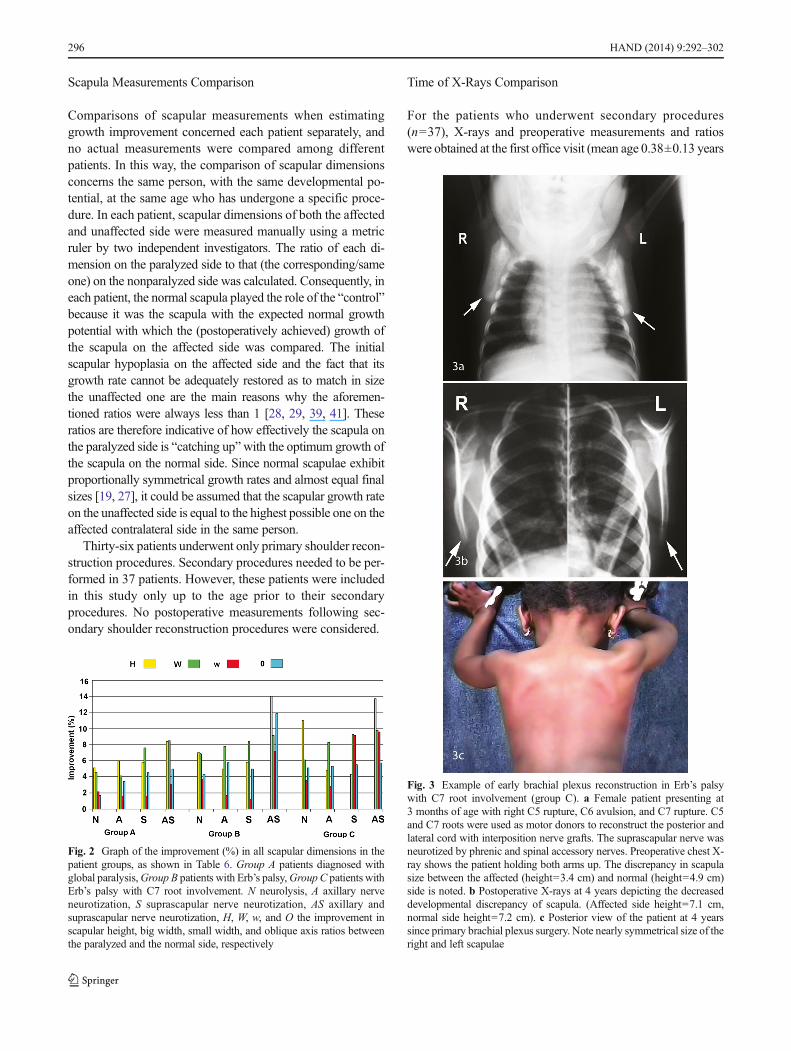
Comparisons of scapular measurements when estimatinggrowth improvement concerned each patient separately, andno actual measurements were compared among differentpatients. In this way, the comparison of scapular dimensionsconcerns the same person, with the same developmental po-tential, at the same age who has undergone a specific proce-dure. In each patient, scapular dimensions of both the affectedand unaffected side were measured manually using a metricruler by two independent investigators. The ratio of each di-mension on the paralyzed side to that (the corresponding/sameone) on the nonparalyzed side was calculated. Consequently, ineach patient, the normal scapula played the role of the “control”because it was the scapula with the expected normal growthpotential with which the (postoperatively achieved) growth ofthe scapula on the affected side was compared. The initialscapular hypoplasia on the affected side and the fact that itsgrowth rate cannot be adequately restored as to match in sizethe unaffected one are the main reasons why the aforemen-tioned ratios were always less than 1 [28, 29, 39, 41]. Theseratios are therefore indicative of how effectively the scapula onthe paralyzed side is “catching up”with the optimum growth ofthe scapula on the normal side. Since normal scapulae exhibitproportionally symmetrical growth rates and almost equal finalsizes [19, 27], it could be assumed that the scapular growth rateon the unaffected side is equal to the highest possible one on theaffected contralateral side in the same person.
Thirty-six patients underwent only primary shoulder recon-struction procedures. Secondary procedures needed to be per-formed in 37 patients. However, these patients were includedin this study only up to the age prior to their secondaryprocedures. No postoperative measurements following sec-ondary shoulder reconstruction procedures were considered.
Time of X-Rays Comparison
For the patients who underwent secondary procedures(n=37), X-rays and preoperative measurements and ratioswere obtained at the first office visit (mean age 0.38±0.13 years
Fig. 2 Graph of the improvement (%) in all scapular dimensions in thepatient groups, as shown in Table 6. Group A patients diagnosed withglobal paralysis,Group B patients with Erb’s palsy,Group C patients withErb’s palsy with C7 root involvement. N neurolysis, A axillary nerveneurotization, S suprascapular nerve neurotization, AS axillary andsuprascapular nerve neurotization, H, W, w, and O the improvement inscapular height, big width, small width, and oblique axis ratios betweenthe paralyzed and the normal side, respectively
Fig. 3 Example of early brachial plexus reconstruction in Erb’s palsywith C7 root involvement (group C). a Female patient presenting at3 months of age with right C5 rupture, C6 avulsion, and C7 rupture. C5and C7 roots were used as motor donors to reconstruct the posterior andlateral cord with interposition nerve grafts. The suprascapular nerve wasneurotized by phrenic and spinal accessory nerves. Preoperative chest X-ray shows the patient holding both arms up. The discrepancy in scapulasize between the affected (height=3.4 cm) and normal (height=4.9 cm)side is noted. b Postoperative X-rays at 4 years depicting the decreaseddevelopmental discrepancy of scapula. (Affected side height=7.1 cm,normal side height=7.2 cm). c Posterior view of the patient at 4 yearssince primary brachial plexus surgery. Note nearly symmetrical size of theright and left scapulae
296 HAND (2014) 9:292–302

of age) and postoperative measurements at the last follow-upvisit prior to secondary procedures (mean age 10.26±1.14 yearsof age).
In patients who underwent primary shoulder reconstructionprocedures alone (n=36), the first preoperative X-rays, mea-surements, and ratios were obtained at the first office visit. Themean age was 0.41±0.15 years. Postoperative measurementsand ratios were obtained at around 10 years of age in order tocomprise comparable age-related values with patients whounderwent secondary procedures. The mean postoperativeage was 10.11±0.45 years of age.
Statistical Analysis
The SAS system (Cary, NC) was used in data analysis. Sta-tistical significance was considered at p<0.05. Linear regres-sion analysis of each scapula dimension based on all the othervariables was performed in order to determine the factor withthe most statistically significant association with it. The
rationale for using regression analysis include the need toidentify and explore the relationships among several variables,to use a simple and credible method of organizing observa-tional data, and if possible, to identify valid predictor variablesso as to be able to make predictions such as the expectedscapular growth improvement following a specific procedure.
Data (logarithmic) transformation was considered whenthere was the need to stabilize variance, to improve the inter-pretability and uniformity of the results. Logarithmic transfor-mation and normality check were performed using normal Q–Q plots for each one of the following independent variables:gestation, birth weight, Apgar score, denervation time, anddegrees of improvement in shoulder abduction and shoulderexternal rotation with each one of the independent variables(improvement in scapular height, big width, small width, andoblique axis). The resulting plots were approximately linearsuggesting that it is plausible that the data come from a nearlynormal distribution. The paired t test was used between the
Fig. 4 a Example of early brachial plexus reconstruction in global palsy(group A). A boy presented at 2 months of age with right Erb’s+C7 palsy(C5, C6, C7 roots avulsion and C8, T1 roots traction). Primary recon-struction included microneurolysis of C8, T1 roots, suprascapular nerveneurotization by distal spinal accessory, neurotization of themusculocutaneous nerve by intraplexus transfer from the lower trunk,transfer of the medial antebrachial cutaneous to the ipsilateral mediannerve by direct repair, and intercostal neurotization of the axillary nerve.Preoperative chest X-ray in which the right (affected) side with height=4.2 cm and left (normal) height=4.5 cm scapular bones are shown. Theinitial discrepancy of the scapular dimension between the scapular bonesof the normal and the affected side is noted. b Scapula X-rays 5 yearsfollowing primary brachial plexus reconstruction. The discrepancy isconsiderably decreased upon reinnervation of the scapular muscles show-ing that the affected scapula recovered its developmental potential (heightof the affected=5.4 cm, height of the normal=5.4 cm)
Fig. 5 a Example of late brachial plexus reconstruction at 12 months. Afemale patient presenting at 1 year of age with left global OBPP. Sheunderwent microneurolysis of the upper trunk, suprascapular nerve, andlower trunk. Intraplexus neurotization from C6 root to themusculocutaneous and from C7 to the middle trunk and posterior cordusing interposition nerve grafts. Preoperative X-rays show scapular di-mensions discrepancy between the left and right side (normal height (R)=4.8 cm, affected height (L)=4.3 cm). b Postoperative X-rays showingnormal height (R)=10.02 cm and affected height (L)=9.9 cm. The de-creased dimensional discrepancy is notable
HAND (2014) 9:292–302 297

pre-and postoperative degrees of shoulder abduction and ex-ternal rotation, respectively, in each group of different diag-nosis. The observed power for the regression study with a pvalue of 0.05, a sample size of 73 patients, and 10 independentvariables as shown in Table 3 is 0.90.
Results
Scapular Growth Improvement
The highest improvement was noted in scapular height (H) inthe Erb’s palsy group who underwent simultaneousneurotization of the Ss and the Ax. The oblique axis (O) wasmore improved in the Erb’s palsy group while both big (W)
and small (w) widths were more improved in the Erb’s palsywith C7 root involvement group in patients who underwentconcomitant neurotization of the Ss and the Ax. The graph inFig. 2 shows the improvement (%) in all scapular dimensionsin the patient groups.
The postoperative decrease of discrepancy in each dimen-sion was significantly correlated with that of the other dimen-sions. The strongest correlation was between the w and W ofthe scapula (0.58), showing that the scapula retained an anal-ogous growth rate in all the measured dimensions, which is astrong indication of symmetrical bone growth. However, theaffected side never reaches fully the size of the contralateralunaffected scapula, despite its documented developmentalattainment. There was no significant correlation between de-nervation time and scapular growth in all groups.
Table 5 The mean preoperative and postoperative shoulder abduction and external rotation and the improvement in all groups
Group Pre SA Post SA Imp p value Pre SER Post SER Imp p value
A 36.92 109.90 72.98 0.031* 0.48 45.19 44.71 0.04*
B 39.16 138.33 99.16 0.17 5 65 60 0.30
C 50.29 145 94.70 0.045* 2.05 60.58 58.52 0.05*
Mean values are in degrees
Group A patients diagnosed with global paralysis, Group B patients with Erb’s palsy, Group C patients with Erb’s palsy with C7 root involvement; SAshoulder abduction; SER shoulder external rotation; Pre SA preoperative shoulder abduction; Post SA postoperative shoulder abduction; Pre SERpreoperative shoulder external rotation; Post SER postoperative shoulder external rotation; IMP improvement
*p<0.05, statistically significant results
Table 6 The preoperative and postoperative scapular dimensions ratios and the improvement in all groups, on account of the type of primary procedurethat was performed
Group Primary procedures Heightpreop
Heightpostop
Imp Big widthpreop
Big widthpostop
Imp Smallwidthpreop
Small widthpostop
Imp Obliqueaxispreop
Obliqueaxis postop
Imp
A Neurolysis 88.1 93.2 5.1 86 90.5 4.5 88.6 90.7 2.1 92.4 94.1 1.7
Axillary n. 88.6 94.6 6 89 93.1 4.1 86.8 88.4 1.6 93.3 96.7 3.4
Suprascapular n. 86.1 91.9 5.8 84.8 92.4 7.6 83.1 84.7 1.6 90.1 94.6 4.5
Axillary+suprascapular n. 86.6 95 8.4 89.8 98.3 8.5 83 86 3 88.6 93.6 5
B Neurolysis 91 98 7 89.9 96.8 6.9 94.3 98 3.7 92.7 97 4.3
Axillary n. 92 97 5 89.8 97.6 7.8 94.1 95.8 1.7 84.6 90.4 5.8
Suprascapular n. 89 94.8 5.8 87.8 96.2 8.4 95 96.2 1.2 90.1 95.1 5
Axillary+suprascapular n. 83.9 97.9 14 90.5 99.6 9.1 90.7 97.9 7.2 87.3 99.2 11.9
C Neurolysis 85 96 11 88.9 95 6.1 92.5 96 3.5 93.8 98.9 5.1
Axillary n. 89.6 94.4 4.8 87.5 95.8 8.3 91.3 94.1 2.8 88.7 94 5.3
Suprascapular n. 89.5 93.8 4.3 86.8 96.1 9.3 86.7 95.8 9.1 89.7 95.2 5.5
Axillary+suprascapular n. 83.5 97.2 13.7 88.1 97.9 9.8 89.2 98.8 9.6 93.6 99.3 5.7
Patient group A, global paralysis; group B, Erb’s palsy; group C, Erb’s palsy with C7 root involvement. Primary procedures that were performed:Neurolysis, Axillary n. (Axillary nerve neurotization), Suprascapular n. (Suprascapular nerve neurotization), Axillary+Suprascapular n. (Axillary andsuprascapular nerve neurotization). Height preop and Height postop, the pre- and postoperative scapular height ratios between the paralyzed and thenormal side; Big width preop and Big width postop, the pre- and postoperative scapular big width ratios between the paralyzed and the normal side;Small width preop and Small width postop, the pre- and postoperative scapular small width ratios between the paralyzed and the normal side; Obliqueaxis preop and Oblique postop, the pre- and postoperative scapular oblique axis ratios between the paralyzed and the normal side; Imp, the improvementin the aforementioned ratios. All numbers are percentages
298 HAND (2014) 9:292–302

Functional Improvement
Functional improvement correlated positively with growth im-provement in all groups and scapular dimensions. Exemplarycases are presented in Figs. 3, 4, and 5. The improvement in SAand SER showed a significant negative correlation with thedenervation time (−0.4, p<0.03, and −0.38, p<0.05). Primaryshoulder reconstructive procedures resulted in amean improve-ment of 88.9° in SA and of 54.41° in SER (p<0.02). The meanpreoperative and postoperative SA and SER in degrees and theimprovement in each group are shown in Table 5.
Preoperative and postoperative ratios of each dimensionand mean improvement on account of the primary shoulderreconstructive procedures that were performed are shown inTable 6. Functional results and the rationale for primary re-construction are shown in Table 7.
Other Variables
Birth weight, gestation period, the side of the paralysis,and Apgar scores did not have a statistically significant
association with scapula growth nor with functionalresults of all the procedures and in all groups. Genderhad a statistically significant association with H andamong patients with global paralysis, females obtainingbetter results from primary shoulder reconstruction proceduresthan males (means 96.5 and 94.6 %, respectively, p<0.05).Gender was not statistically significant regarding the otherdimensions in all groups.
Discussion
This study showed that scapular height and big width had thehighest improvement (mean values 7.57 and 7.53 %, respec-tively) and small width had the lowest (mean improvement3.92 %). It was also shown that scapula growth and shoulderfunction improvement were higher in patients with Erb’spalsy. Patients with global palsy had the lowest improvementin both scapula growth and shoulder function. SimultaneousAx and Ss neurotization provided the best outcome in bothfunctional and growth restoration which is consistent with
Table 7 The mean preoperative and postoperative shoulder abduction and external rotation and the improvement in all groups
Anatomicaldiagnosis,N=numberof patients
Rationale for primary reconstruction Shoulder abductionmean improvement(p value)
Shoulder externalrotation meanimprovement(p value)
Quality of life
Global paralysis(C5–T1), N=48
Usually: rupture of the upper roots with avulsion of thelower plexus.
1. Neurotize the two lower avulsed roots (C8 and T1) fromthe C5 root and use the C6 root for reconstruction of thelateral and posterior cords
2. If C7 available (rare): C7 motor fibers to neurotize C8and T1 roots directly by end-to-end repair, due toproximity (faster reinnervation of the hand musculature)
3. Restore shoulder stability, elbow flexion (reinnervationof the supraspinatus, deltoid, and biceps)
4. Restore hand sensation (supraclavicular sensory nervesand ipsilateral sensory intercostals)
72.98° (0.031*) 44.71° (0.04*) Average–good
Erb’s palsy(C5, C6), N=10
1. Restore shoulder stability (reconstruction of thesupraspinatus and deltoid muscles through reinnervationof the axillary nerve through elements of the proximalupper plexus or direct neurotization of the suprascapularnerve from the ipsilateral distal accessory nerve)
2. Restore elbow flexion (biceps reconstruction throughmusculocutaneous nerve reinnervation through:-C6 (if not avulsed) or-Anterior division of C7 transfer to the lateral corddirectly by end-to-end repair or-Contralateral C7 via saphenous or sural cross-chestnerve grafts or-Ipsilateral intercostal nerves
3. Triceps reinnervation with reinnervation of the posteriorcord from intraplexus donors or from the ipsilateralintrecostals (unless used to neurotize musculocutaneous)
99.16° (0.17) 60° (0.30) Good–Excellent
(C5–C7) N=15 94.70° (0.045*) 58.52° (0.05*) Good
Quality of life report is based on the clinical examination of each child and on the impression of the physician who performed the evaluation. The rangeincluded poor, fair, average, good, and excellent
*statistically significant (p<0.05)
HAND (2014) 9:292–302 299

several previous studies [2, 3, 24]. Early surgical interventionwas attempted (earlier than 3 months) in global paralysis andyielded the best functional results.
Scapular stabilizing synergists comprise a group of mus-cles which act together producing opposing actions that resultin the dynamic stability of the scapula (Fig. 6) [18, 19, 27, 31,42]. Our reconstructive strategy depended on the denervationtime, the type of plexus lesion, and the availability of motordonors. In early cases (mean denervation time=0.39 year old),the entire plexus reconstruction was undertaken using multi-ple interposition nerve grafts (mean length=3.2±1.1 cm) toreinnervate peripheral targets. In later cases (mean denervationtime=0.9 year old), Ax and/or Ss neurotization was performedusing interposition nerve grafts (mean length=7.7±2.1 cm) or
via direct coaptation with intercostal nerves (T5–T8) whichwere used as motor donors when intraplexus donors were notavailable (Table 2).
Consequently, reinnervation of the rhomboids (C4, C5),serratus anterior (C5–7), trapezius (CNXI: accessory nerve),levator scapulae (C3–5), and pectoralis minor (C8, T1) isachieved. Therefore, the balance of forces which is responsi-ble for scapular dynamic stability around a horizontal axisperpendicular to its plane (upward, downward rotation) andaround a horizontal axis in its plane (posterior, anterior tilt) isrestored [21, 33]. Additionally, optimal muscle activation ofthe shoulder joint muscles is achieved due to functional scap-ular retraction and external rotation. This position is a prereq-uisite for a normal scapula–humeral rhythm maximizing theactivation of all scapular muscles and making the scapula astable base for the origin of all the rotator cuff muscles [21].
Additionally, with the aforementioned procedures, reinner-vation of the rotator cuff muscles (supraspinatus, infraspinatus,subscapularis, and teres minor from C5, C6), teres major (seg-mental levels C5, C6), latissimus dorsi (LD) (C6–C8), deltoid(C5, C6), and coracobrachialis (C5–C7) muscles was alsoaddressed. Supraspinatus fibers create a horizontal pull whichcounteracts the vertical pull of the deltoid muscle and stabilizethe scapular position when the arm is raised in flexion andabduction [18, 21]. The diagonal arrangement of subscapularisfibers produces a diagonal line of pull which stabilizes thescapula during internal rotation of the arm. Similarly, theinfraspinatusmuscle produces a diagonal stabilizing pull duringshoulder external rotation [18, 21, 42]. The teres minor exter-nally rotates the shoulder and creates a concavity compressionmechanism which pulls the humeral head into the glenoidfossa. The secondary stabilizer LD adducts, internally rotates,and extends the shoulder joint while it indirectly depresses thelateral angle of the scapula and retracts it downwards, alsofacilitating scapular anterior tilt along with the teres major.Similarly, the pectoralis major (C5–T1), responsible for adduc-tion, horizontal flexion, and internal rotation at theglenohumeral joint, as a whole indirectly depresses and pro-tracts the scapula. Additionally, the coracobrachialis facilitatesscapular posterior tilt along with the anterior and medial deltoid[18, 21, 33, 42].
Secondary shoulder deformities in children with OBPPhave been described in 1913 by Fairbank [8] and subsequentlyby several other authors [2, 7, 23, 25, 32, 41, 45]. However,there is no detailed knowledge of the factors that are respon-sible for the development of secondary shoulder deformities ininfants with OBPP since most of the available studies havebeen based on children over 1 year of age [3, 25, 41].
Clarke et al. [7] reported the return of useful function ininfants with Erb’s palsy treated with neurolysis but not in thetotal palsy cases. There was no comparative analysis of theirresults with those of conservative treatment and nerve repair.Subsequently, Capek et al. [4] reported the early outcome of
Fig. 6 Scapular muscles and their effect on scapular dynamic stability. aUpward rotation-posterior tilt;UTupper trapezius, LT lower trapezius, SAserratus anterior. b Downward rotation-anterior tilt; LS levator scapulae,RM rhomboid major, Rm rhomboid minor, Pm pectoralis minor. c Del-toid–supraspinatus interaction during arm raising; Ssp supraspinatus, DTdeltoid. d Stabilization during shoulder external rotation–scapular anteri-or posterior tilt; Isp infraspinatus, Tm teres minor, TM teres major (scap-ular anterior tilt), CB coracobrachialis (scapular posterior tilt). e Sscsubscapularis (stabilization during shoulder internal rotation), LDlatissimus dorsi (retraction, anterior tilt, angle depression), TC triceps-long head (shoulder stabilization). The serratus anterior and upper andlower trapezius muscles are the primary stabilizing synergists whichproduce upward scapular rotation and posterior tilt, whereas the rhom-boids, levator scapulae, and pectoralis minor muscles are responsible fordownward scapular rotation and anterior tilt. The serratus anterior pro-tracts the scapula while the rhomboids and midtrapezius muscles retract it
300 HAND (2014) 9:292–302

neuroma-in-continuity resection and the superior results ofinterposition nerve grafting to neurolysis. Xu et al. [44] com-pared the effects of conservative treatment, neurolysis, andnerve transfer and grafting in 31 patients with OBPP. Theyfound no statistically significant difference betweenneurolysis and conservative treatment. Gilbert et al. [9–13]published their successful outcomes of neuroma resection andbrachial plexus surgery.
Gu et al. [17] reported better results with phrenic nervetransfers, and Gruber et al. [16] first reported a case of live-donor nerve transplantation in an 8-month-old infant withglobal OBPP without, however, clinically significant func-tional improvement. Amr et al. [1] investigated the effect ofend-to-side neurotization of partially regenerated recipientnerves in improving motor power in late OBPP cases. Chenet al. [6] determined the advantages of contralateral C7 nerveroot transfer in their series of 12 infants and children. Terzisand Kokkalis [35] published their outcomes in OBPP treat-ment concluding that reconstruction of the axillary nerve (Ax)should always be performed to maximize the final outcome ofshoulder function. The best results were seen in cases operatedon earlier than 3 months, where the posterior cord was recon-structed from intraplexus donors. Reconstruction of the Axdirectly from the intercostal nerves was a reliable option in latecases. Scapula size increases proportionally with age more inwidth than in length, and the infrascapular height increasesfaster than suprascapular height [14, 24]. Postnatal growth isinitially linear followed by an acceleration at shortly over13 years of age which leads to the gradual decrease in growthrate near the age of 17 and finally to the completion of growtharound 20 years of age [14, 28]. The inferior angle, the medialborder of the scapula, and the acromion fuse around 23 and20 years of age, respectively [15, 43].
The likelihood and the timing of early surgical interventionremain an area of controversy. According to Tassin [34],spontaneous recovery of shoulder function using the Malletscale was observed in babies with deltoid and biceps contrac-tions by 3 months of age. Recovery was less than average(Mallet grade III) when contractions had started after the thirdmonth of age. Shenaq et al. [30] agree with the previousobservation and suggest that the ideal time for early surgicalintervention in such patients is the fourth month of age. Gilbert[13] has incorporated the aforementioned time limit of 3monthsof age in his suggested indications for surgery. Clarke et al. [7]also consider patients without improvement at 3 months of ageto be candidates for early reconstruction. Conversely, Zancolli[45] delayed the decision for early intervention until 6–8monthsand Laurent et al. [22] reported adequate arm functional returnwith attempted repair as late as 24 months. In our center [38], ina previous series of 80 infants who had undergone surgery forobstetrical brachial plexus injuries, those with denervation timemore than 3 months needed secondary procedures such asmuscle transfers, rotational osteotomies, or wrist fusions.
Additionally, in global paralysis cases with denervation timeless than 3 months, adequate hand function was also possible torestore [35–38].
In OBPP patients, the lack of the normal forces applied tothe scapula by the weakened muscles leads to a decreased rateof bone growth [20, 26]. Growth impairment has been report-ed to concern primarily the total width and the height of thescapula in addition to several inconsistently observed types ofdeformities such as the anterior rotation of the scapular spinewhich is responsible for a narrower scapuloclavicular spaceand the downward rotational deformity of the spine [39]. Inour study, it has been shown that primary plexus reconstruc-tion restores regional muscle function and biomechanics,which recovers the growth potential of the scapula. The pos-itive correlation between functional and growth improvementsupports the fact that muscular function plays a significant rolein scapular growth and that impairment of the former affectsnegatively the latter.
Conflict of Interest and Financial Disclosure Statements Julia K.Terzis declares that she has no conflict of interest and that she has notreceived any kind of financial support/funds from any source.
Dimitrios Karypidis declares that he has no conflict of interest and thathe has not received any kind of financial support/funds from any source.
Ricardo Mendoza declares that he has no conflict of interest and thathe has not received any kind of financial support/funds from any source.
Zinon T. Kokkalis declares that he has no conflict of interest and thathe has not received any kind of financial support/funds from any source.
Norou Diawara declares that he has no conflict of interest and that hehas not received any kind of financial support/funds from any source.
Statement of Human and Animal Rights All procedures followedwere in accordance with the ethical standards of the responsible commit-tee on human experimentation (institutional and national) and with theHelsinki Declaration of 1975, as revised in 2000 and 2008 (5).
Statement of Informed Consent Informed consent was obtained fromall patients for being included in the study. Additional informed consentwas obtained from all patients for whom identifying information isincluded in this article.
References
1. Amr SM, Moharram AN, Abdel-Meguid KM. Augmentation ofpartially regenerated nerves by end-to-side side-to-side graftingneurotization: experience based on eight late obstetric brachial plexuscases. J Brachial Plex Peripher Nerve Inj. 2006;5:1–6.
2. Beischer AD, Simmons TD, Torode IP. Glenoid version in childrenwith obstetric brachial plexus palsy. J Pediatr Orthop. 1999;19:359–61.
3. Birch R. Birth lesions of the brachial plexus. In: Birch R, Bonney G,Wynn Parry CB, editors. Surgical disorders of the peripheral nerves.London: Churchill Livingstone; 1998. p. 209–34.
4. Capek L, Clarke HM, Curtis CG. Neuroma-in-continuity resection:early outcome in obstetrical brachial plexus palsy. Plast ReconstrSurg. 1998;102:1555.
5. Chen L, Gu YD, Hu SN. Applying transfer of trapezius and/orlatissimus dorsi with teres major for reconstruction of abduction
HAND (2014) 9:292–302 301

and external rotation of the shoulder in obstetrical brachial plexuspalsy. J Reconstr Microsurg. 2002;18(4):275–80.
6. Chen L, Gu YD, Hu SN, et al. Contralateral C7 transfer for thetreatment of brachial plexus root avulsions in children—a report of12 cases. J Hand Surg [Am]. 2007;32(1):96–103.
7. Ebaugh DD, McClure PW, Karduna AR. Three-dimensionalscapulothoracic motion during active and passive arm elevation.Clin Biomech (Bristol, Avon). 2005;20(7):700–9.
8. Fairbank HAT. Birth palsy: a lecture on subluxation of the shoulderjoint in infants and young children. Lancet. 1913;i:1217–23.
9. Gilbert A, Brockman R, Carlioz H. Surgical treatment of brachialplexus birth palsy. Clin Orthop. 1991;264:39.
10. Gilbert A, Razaboni R, Amar-Khodja S. Indications and results ofbrachial plexus surgery in obstetrical palsy. Orthop Clin N Am.1988;19:91.
11. Gilbert A, Tassin JL. Obstetrical palsy: a clinical, pathologic, andsurgical review. In: Terzis JK, editor. Microreconstruction of nerveinjuries. Philadelphia: WB Saunders; 1987. p. 529.
12. Gilbert A. Obstetrical brachial plexus palsy. In: Tubiana R, editor.The hand. Philadelphia: WB Saunders; 1993. p. 575.
13. Gilbert A, Whitaker I. Obstetrical brachial plexus lesions. J HandSurg (Br). 1991;16(5):489–91.
14. GravesWW. Observations on age changes in the scapula. Am J PhysAnthropol. 1922;5:21–33.
15. Gray DJ. Variations in human scapulae. Am J Phys Anthropol.1942;29:57–72.
16. Gruber SA, Mancias P, Swinford RD, et al. Living-donor nervetransplantation for global obstetric brachial plexus palsy. J ReconstrMicrosurg. 2006;22(4):245–54.
17. Gu YD,WuMM, Zheng YL, et al. Phrenic nerve transfer for brachialplexus motor neurotization. Microsurgery. 1989;10:1.
18. Halder AM, Itoi E, An KN. Anatomy and biomechanics of theshoulder. Orthop Clin N Am. 2000;31:159.
19. Hollinshead WH. The back and limbs. In: Hollinshead WH, editor.Anatomy for surgeons, vol. III. Philadelphia: Harper &Row; 1982. p.300–8.
20. Kattan KR, Spitz HB. Roentgen findings in obstetrical injuries to thebrachial plexus. Radiology. 1968;91:462–7.
21. Laumann U. Kinesiology of the shoulder joint. In: Kolbel R, HelbigB, Blauth W, et al., editors. Shoulder replacement. Berlin: Springer-Verlag; 1987.
22. Laurent JP, Lee RT. Birth-related upper brachial plexus injuries ininfants: operative and nonoperative approaches. J Child Neurol.1994;9(2):111–7.
23. Narakas AO. Obstetrical brachial plexus injuries. In: Lamb DW,editor. The paralysed hand. The hand and upper limb, vol. 2.Edinburgh: Churchill Livingstone; 1987.
24. Ogden JA, Phillips SB. Radiology of postnatal skeletal development.VII. The scapula. Skelet Radiol. 1983;9(3):157–69.
25. Pearl ML, Edgerton BW. Glenoid deformity secondary to brachialplexus birth palsy. J Bone Joint Surg Am. 1998;80-A:659–67.
26. Pollock AN, Reed MH. Shoulder deformities from obstetrical bra-chial plexus paralysis. Skelet Radiol. 1989;18(4):295–7.
27. Ruland III LJ, Ruland CM, Matthews LS. Scapulothoracic anatomyfor the arthroscopist. Arthroscopy. 1995;11:52.
28. Russo SA, Kozin SH, Zlotolow DA, Thomas KF, Hulbert RL,Mattson JM, et al. Scapulothoracic and glenohumeral contributionsto motion in children with brachial plexus birth palsy. J ShoulderElbow Surg. 2013;23(3):327–38.
29. Scheuer JL, Black SM. The juvenile skeleton. London: Elsevier;2004.
30. Shenaq SM, Berzin E, Lee R, et al. Brachial plexus birth injuries andcurrent management. Clin Plast Surg. 1998;25(4):527–36.
31. Sisto DJ, Jobe FW. The operative treatment of scapulothoracic bur-sitis in professional pitchers. Am J Sports Med. 1986;14:192.
32. Sjoberg I, Erichs K, Bjerre I. Cause and effect of obstetric (neonatal)brachial plexus palsy. Acta Paediatr Scand. 1988;77:357–64.
33. Smith MJ, Sparkes V, Enright S. Scapular rotator muscle imbalancein swimmers with subacromial impingement symptoms. Br J SportsMed. 2007;41:118–24.
34. Tassin JL. Paralysies obstetricales du plexus brachial. Evolutionspontanee, Resultats des interventions reparatrices precoses Univ.Paris VII; 1983.
35. Terzis JK, Kokkalis ZT. Shoulder function following primary axillarynerve reconstruction in obstetrical brachial plexus patients. PlastReconstr Surg. 2008;122(5):1457–69.
36. Terzis JK, Kokkalis ZT. Primary and secondary shoulder reconstruc-tion in obstetric brachial plexus palsy. Injury. 2008;39 Suppl 3:S5–14.
37. Terzis JK, Kokkalis ZT. Outcomes of hand reconstruction in obstetricbrachial plexus palsy. Plast Reconstr Surg. 2008;122(2):516–26.
38. Terzis JK, Papakonstantinou KC. Management of obstetric brachialplexus palsy. Hand Clin. 1999;15(4):717–36.
39. Terzis JK, Vekris MD, Okajima S, et al. Shoulder deformities inobstetric brachial plexus paralysis: a computed tomography study. JPediatr Orthop. 2003;23(2):254–60.
40. van Gelein Vitringa VM, van Royen BJ, van der Sluijs JA. Scapulardeformity in obstetric brachial plexus palsy and the Hueter-Volkmannlaw; a retrospective study. BMC Musculoskelet Disord. 2013;14:107.
41. Waters PM, Smith GR, Jaramillo D. Glenohumeral deformity sec-ondary to brachial plexus birth palsy. J Bone Joint Surg Am. 1998;80-A:668–77.
42. Williams Jr GR, Shakil M, Klimkiewicz J, et al. Anatomy of thescapulothoracic articulation. Clin Orthop. 1999;359:237.
43. Wolffson DM. Scapula shape and muscle function with specialreference to vertebral border. Am J Phys Anthropol. 1950;8:331–42.
44. Xu J, Cheng X, Gu Y. Different methods and results in the treatmentof obstetrical brachial plexus palsy. J Reconstr Microsurg.2000;16(6):417–20.
45. Zancolli EA. Classification and management of the shoulder in birthpalsy. Orthop Clin N Am. 1981;12(2):433–57.
302 HAND (2014) 9:292–302
Related Documents