Downloaded from www.microbiologyresearch.org by IP: 54.237.107.209 On: Thu, 30 Jun 2016 10:37:02 Morphological changes and proteome response of Corynebacterium glutamicum to a partial depletion of FtsI Noelia Valbuena, 1 Michal Letek, 1 Angelina Ramos, 1 3 Juan Ayala, 2 Diana Nakunst, 3 Joern Kalinowski, 3 Luis M. Mateos 1 and Jose ´ A. Gil 1 Correspondence Jose ´ A. Gil [email protected] 1 Departamento de Ecologı ´a, Gene ´ tica y Microbiologı ´a, A ´ rea de Microbiologı ´a, Facultad de Biologı ´a, Universidad de Leo ´ n, 24071 Leo ´ n, Spain 2 Centro Biologı ´a Molecular ‘Severo Ochoa’, Consejo Superior de Investigaciones Cientı ´ficas, CSIC-UAM, Campus Universidad Auto ´ noma, Cantoblanco, 28049 Madrid, Spain 3 Institut fur Genomforschung, Universitat Bielefeld, Universitatsstrasse 25, D-33615 Bielefeld, Germany Received 15 December 2005 Revised 10 March 2006 Accepted 30 March 2006 In Corynebacterium glutamicum, as in many Gram-positive bacteria, the cell division gene ftsI is located at the beginning of the dcw cluster, which comprises cell division- and cell wall-related genes. Transcriptional analysis of the cluster revealed that ftsI is transcribed as part of a polycistronic mRNA, which includes at least mraZ, mraW, ftsL, ftsI and murE, from a promoter that is located upstream of mraZ. ftsI appears also to be expressed from a minor promoter that is located in the intergenic ftsL–ftsI region. It is an essential gene in C. glutamicum, and a reduced expression of ftsI leads to the formation of larger and filamentous cells. A translational GFP-FtsI fusion protein was found to be functional and localized to the mid-cell of a growing bacterium, providing evidence of its role in cell division in C. glutamicum. This study involving proteomic analysis (using 2D SDS-PAGE) of a C. glutamicum strain that has partially depleted levels of FtsI reveals that at least 20 different proteins were overexpressed in the organism. Eight of these overexpressed proteins, which include DivIVA, were identified by MALDI-TOF. Overexpression of DivIVA was confirmed by Western blotting using anti-DivIVA antibodies, and also by fluorescence microscopy analysis of a C. glutamicum RESF1 strain expressing a chromosomal copy of a divIVA-gfp transcriptional fusion. Overexpression of DivIVA was not observed when FtsI was inhibited by cephalexin treatment or by partial depletion of FtsZ. INTRODUCTION Corynebacteria are Gram-positive, pleomorphic asporo- genous bacteria that are widely distributed in nature. While some of them are pathogenic to plants, animals and humans, the non-pathogenic corynebacteria, such as Corynebac- terium glutamicum, are widely used in the industrial pro- duction of amino acids and nucleotides (Gourdon & Lindley, 1999; Nakayama et al., 1978). In recent years, different approaches have been used to characterize cell division genes in corynebacteria, and the possible relationship between amino acid production and growth inhibition (Honrubia et al., 1998; Kobayashi et al., 1997; Ramos et al., 2003b; Wachi et al., 1999). Recent studies have shown that corynebacteria might follow an archaic pattern of mycelial growth that involves an asymmetric mechanism of division (Ramos et al., 2005), with cell elongation occurring at the tip of the daughter cells (Daniel & Errington, 2003). As a member of the order Actino- mycetales, corynebacteria show apical growth, similar to that seen in Corynebacterium diphtheriae (Umeda & Amako, 1983). The availability of the complete genome sequence of C. glutamicum (GenBank accession nos NC_003450 and BX927154) has enabled us to study the expression profile/ regulation of the cell division gene ftsI, and perform proteomic studies in the organism. FtsI homologues have been described in different bacteria, such as Escherichia coli (Begg et al., 1992; Botta & Park, 1981) or Bacillus subtilis (Daniel et al., 1996; Marston et al., 1998). FtsI (also called penicillin-binding protein 3, PBP3) is a well-characterized protein that has been reported to be expressed in very low amounts in the cell (about 100 3Present Address: Departamento de Biologı ´a Funcional, A ´ rea de Microbiologı ´a, Facultad de Medicina, Universidad de Oviedo, 33006 Oviedo, Spain. Abbreviations: Q-PCR, quantitative PCR; RACE, rapid amplification of cDNA ends. 0002-8773 G 2006 SGM Printed in Great Britain 2491 Microbiology (2006), 152, 2491–2503 DOI 10.1099/mic.0.28773-0

Welcome message from author
This document is posted to help you gain knowledge. Please leave a comment to let me know what you think about it! Share it to your friends and learn new things together.
Transcript
Downloaded from www.microbiologyresearch.org by
IP: 54.237.107.209
On: Thu, 30 Jun 2016 10:37:02
Morphological changes and proteome response ofCorynebacterium glutamicum to a partial depletionof FtsI
Noelia Valbuena,1 Michal Letek,1 Angelina Ramos,13 Juan Ayala,2
Diana Nakunst,3 Joern Kalinowski,3 Luis M. Mateos1 and Jose A. Gil1
Correspondence
Jose A. Gil
1Departamento de Ecologıa, Genetica y Microbiologıa, Area de Microbiologıa, Facultad deBiologıa, Universidad de Leon, 24071 Leon, Spain
2Centro Biologıa Molecular ‘Severo Ochoa’, Consejo Superior de Investigaciones Cientıficas,CSIC-UAM, Campus Universidad Autonoma, Cantoblanco, 28049 Madrid, Spain
3Institut fur Genomforschung, Universitat Bielefeld, Universitatsstrasse 25, D-33615 Bielefeld,Germany
Received 15 December 2005
Revised 10 March 2006
Accepted 30 March 2006
In Corynebacterium glutamicum, as in many Gram-positive bacteria, the cell division gene ftsI
is located at the beginning of the dcw cluster, which comprises cell division- and cell
wall-related genes. Transcriptional analysis of the cluster revealed that ftsI is transcribed as part of
a polycistronic mRNA, which includes at least mraZ, mraW, ftsL, ftsI and murE, from a promoter
that is located upstream of mraZ. ftsI appears also to be expressed from a minor promoter that
is located in the intergenic ftsL–ftsI region. It is an essential gene in C. glutamicum, and a reduced
expression of ftsI leads to the formation of larger and filamentous cells. A translational GFP-FtsI
fusion protein was found to be functional and localized to the mid-cell of a growing bacterium,
providing evidence of its role in cell division in C. glutamicum. This study involving proteomic
analysis (using 2D SDS-PAGE) of a C. glutamicum strain that has partially depleted levels of
FtsI reveals that at least 20 different proteins were overexpressed in the organism. Eight of
these overexpressed proteins, which include DivIVA, were identified by MALDI-TOF.
Overexpression of DivIVA was confirmed by Western blotting using anti-DivIVA antibodies, and
also by fluorescence microscopy analysis of a C. glutamicum RESF1 strain expressing a
chromosomal copy of a divIVA-gfp transcriptional fusion. Overexpression of DivIVA was not
observed when FtsI was inhibited by cephalexin treatment or by partial depletion of FtsZ.
INTRODUCTION
Corynebacteria are Gram-positive, pleomorphic asporo-genous bacteria that are widely distributed in nature. Whilesome of them are pathogenic to plants, animals and humans,the non-pathogenic corynebacteria, such as Corynebac-terium glutamicum, are widely used in the industrial pro-duction of amino acids and nucleotides (Gourdon &Lindley, 1999; Nakayama et al., 1978).
In recent years, different approaches have been used tocharacterize cell division genes in corynebacteria, and thepossible relationship between amino acid production andgrowth inhibition (Honrubia et al., 1998; Kobayashi et al.,
1997; Ramos et al., 2003b; Wachi et al., 1999). Recent studieshave shown that corynebacteria might follow an archaicpattern of mycelial growth that involves an asymmetricmechanism of division (Ramos et al., 2005), with cellelongation occurring at the tip of the daughter cells (Daniel& Errington, 2003). As a member of the order Actino-mycetales, corynebacteria show apical growth, similar to thatseen in Corynebacterium diphtheriae (Umeda & Amako,1983). The availability of the complete genome sequenceof C. glutamicum (GenBank accession nos NC_003450 andBX927154) has enabled us to study the expression profile/regulation of the cell division gene ftsI, and performproteomic studies in the organism.
FtsI homologues have been described in different bacteria,such as Escherichia coli (Begg et al., 1992; Botta & Park, 1981)or Bacillus subtilis (Daniel et al., 1996; Marston et al., 1998).FtsI (also called penicillin-binding protein 3, PBP3) is awell-characterized protein that has been reported to beexpressed in very low amounts in the cell (about 100
3Present Address: Departamento de Biologıa Funcional, Area deMicrobiologıa, Facultad de Medicina, Universidad de Oviedo, 33006Oviedo, Spain.
Abbreviations: Q-PCR, quantitative PCR; RACE, rapid amplification ofcDNA ends.
0002-8773 G 2006 SGM Printed in Great Britain 2491
Microbiology (2006), 152, 2491–2503 DOI 10.1099/mic.0.28773-0
Downloaded from www.microbiologyresearch.org by
IP: 54.237.107.209
On: Thu, 30 Jun 2016 10:37:02
molecules) (Dougherty et al., 1996). The protein consists ofa short cytoplasmic domain, a single membrane-spanningsegment and a large periplasmic domain that encodes atranspeptidase activity that is involved in the biosynthesis ofseptal peptidoglycan. Immunofluorescence microscopy hasshown that in E. coli during the later stages of cell growth FtsIlocalizes to the division site at the septum. The septallocalization of FtsI, however, depends upon prior localiza-tion of the other cell division proteins, such as FtsZ, FtsA,FtsK, FtsQ, FtsL and FtsW (Mercer & Weiss, 2002; Weiss etal., 1999), and therefore, it appears that FtsI is a late recruitto the division site. In B. subtilis, the septal localization ofPBP3/FtsI is also delayed, but is needed for the localizationof the division inhibitor MinC (Marston & Errington, 1999).
DivIVA is another cell division protein that has beenextensively studied in B. subtilis. It enables cell division in theorganism by sequestering the cell division inhibitors MinCand MinD at the cell poles (Cha & Stewart, 1997; Edwards &Errington, 1997; Marston et al., 1998). In this respect, its roleis similar to that of MinE of E. coli, which repels MinCDinhibitors at the cell poles (Marston et al., 1998). DivIVAalso acts at the cell pole by interacting with the chromosomesegregation machinery, and it is involved in the correctlocalization of the oriC region at the cell pole, a step thatprecedes asymmetric division during sporulation (Thomaideset al., 2001). More recently, Harry & Lewis (2003) found thatDivIVA localizes at the poles of germinated and outgrowncells in B. subtilis without prior assembly of the divisionapparatus at this site, suggesting that its localization doesnot occur by direct interaction with components of thedivision machinery, as proposed by Edwards et al. (2000).
In C. glutamicum, as in many other bacteria, divIVA islocated downstream from the dcw cluster. Its encoded pro-duct (DivIVA) appears to be an essential protein playingan important role at the cell poles in the organism (Ramoset al., 2003b). Overexpression of DivIVA-GFP translationalfusion in C. glutamicum leads to an altered morphologyshowing rounder larger and swollen cells, with the DivIVA-GFP product being preferentially localized at the cell poles.It has been previously suggested that DivIVA participatesin the maintenance of cell morphology in C. glutamicum(Ramos et al., 2003b).
In the present study we show that ftsI in C. glutamicum, asin E. coli, is an essential gene that is required for the main-tenance of cell shape and morphology. We further show thata partial depletion of FtsI induces an increased concentra-tion of DivIVA, opening new questions about the regulationof cell division and polar growth in this micro-organism.
METHODS
Bacterial strains, plasmids and culture conditions. All thebacterial strains and plasmids used in the study are described inTable 1. E. coli strains were grown at 37 uC in Luria–Bertani broth(Hanahan, 1983), supplemented with agar where appropriate. Whennecessary, the antibiotics kanamycin, apramycin and ampicillin were
used at a final concentration of 50 mg ml21. C. glutamicum cells weregrown in trypticase soy broth (TSB; Difco) or trypticase soy agar(TSA) (TSB containing 2 % agar) at 30 uC.
DNA manipulation. Plasmid DNA was isolated from E. coli accord-ing to the method of Holmes & Quigley (1981). E. coli cells (DH5a
and S17-1) were transformed by the method of Hanahan (1983).
All the mobilizable plasmids (integrative and bifunctional) wereintroduced into the donor strain (E. coli S17-1) and then transferred toC. glutamicum RES167 (or additional recombinant strains) followingthe method described previously (Mateos et al., 1996).
Purification of DNA fragments was carried out using a GENECLEANkit (Bio 101). Restriction enzymes were purchased from Promega andNew England Biolabs.
Total DNA from C. glutamicum was isolated using the Kirby methoddescribed for Streptomyces (Kieser et al., 2000), except that the cellswere treated with 5 mg lysozyme ml21 for 4 h at 30 uC.
DNA probes for Southern blots were labelled with DIG-High Prime,according to the manufacturer’s (Roche) instructions.
RNA isolation, RT-PCR analysis, Q-PCR and RACE-PCR. For totalRNA isolation from C. glutamicum, cells were grown in TSB mediumto OD600 1?5. RNA was isolated using the RNeasy kit (Qiagen).
RT-PCR analysis of the total RNA preparation was carried out in orderto detect the presence of a polycistronic transcript originating from theupstream mraZ that includes ftsI; 1 mg total RNA was used as thetemplate to generate single strand cDNA using a first strand cDNAsynthesis kit (Roche), essentially according to the manufacturer’srecommendations. Primers F6, P2, P4, P6, P8 and P10 (Table 2), whichwere used to generate cDNA corresponding to the upstream regions ofthe dcw genes (Fig. 1b), were designed using Primer Express, v2.0(Applied Biosystems). The generated cDNAs were used as templates forsubsequent PCR amplification using the primer pairs P0/F6 (for theupstream mraZ), P1/P2 (for the intergenic mraZ–mraW region), P3/P4(for the intergenic mraW–ftsL region), P5/P6 (for the intergenic ftsL–ftsI region), P7/P8 (for the intragenic ftsI region) and P9/P10 (for theintergenic ftsI–murE region) (Table 2, Fig. 1b). The PCR amplifiedproducts were analysed by electrophoresis on 2 % agarose gels. Theabsence of DNA contamination of the RNA samples was ascertained byPCR using appropriate primers as negative and positive controls.
For Q-PCR analysis of ftsI, the first RT-PCR to generate the cDNA wasperformed using only the forward primer (primer P11, Table 2). A1/20 volume of the generated cDNA sample was then used as a templatein the second step of the PCR in which both the forward and the reverseprimers (P11 and P12, respectively; Table 2) were used in the reactionmixture, in a total volume of 25 ml. Reactions were performed usingan ABI Prism 7000 sequence detection system (Applied Biosystems).Results are indicated relative to the Ct (cycle threshold) value. Ct isdefined as the cycle at which fluorescence is determined to be statis-tically significant compared to the background, being inverselyproportional to the log of the initial copy number; this value wascalculated automatically by the ABI Prism 7000 SDS software.
RACE-PCR experiments were performed according to the 59/39 RACEkit, 2nd generation (Roche), protocol. In order to identify promotersthat were located upstream from mraZ and ftsI, 2 mg total RNApreparation was used as a template to generate single strand cDNAusing primers F5 and P11, respectively (Table 2). A homopolymeric Atail was added to the 39 end of the purified cDNA preparation usingterminal transferase, the dA-tailed cDNA that was obtained was used intwo further PCR amplifications steps; the first one using the primerpair dT-primer/F5 (for mraZ) and dT-primer/F3 (for ftsI) (Table 2).
2492 Microbiology 152
N. Valbuena and others
Downloaded from www.microbiologyresearch.org by
IP: 54.237.107.209
On: Thu, 30 Jun 2016 10:37:02
Table 1. Bacterial strains and plasmids
Strain or plasmid Relevant genotype or description* Source or referenceD
Strains
E. coli DH5a Strain used for general cloning Hanahan (1983)
E. coli JM109 (DE3) E. coli JM109 derivative containing a chromosomal copy of the gene for the
T7 RNA polymerase
Promega
E. coli S17-1 Mobilizing donor strain, pro recA, which has an RP4 derivative integrated into
the chromosome
Schafer et al. (1990)
E. coli AX655 ftsI temperature-sensitive mutant Walker et al. (1975)
C. glutamicum 13032 Wild-type strain ATCC
C. glutamicum RES167 13032 derivative used as a host for transformation, electroporation or conjugation Tauch et al. (2002)
C. glutamicum MAPF RES167 derivative containing a complete copy of ftsI in its chromosomal position
and a copy of gfp-ftsI under the control of Pkan obtained by integration of
plasmid pNV4A
This work (Fig. 2c)
C. glutamicum APF RES167 derivative containing a deleted copy of ftsI in its chromosomal position
and a copy of gfp-ftsI under the control of Pkan obtained by integration of
plasmid pNV5A
This work (Fig. 2b)
C. glutamicum RESF1 RES167 derivative containing a complete copy of ftsI under the control of Plac
obtained by integration of plasmid pOJPB
This work (Fig. 3b)
C. glutamicum AR200 RES167 derivative carrying a deleted copy of divIVA and a copy of divIVA-gfp
obtained by integration of plasmid pKAG1
This work (Fig. 3c)
C. glutamicum RESF12 RESF1 derivative carrying a deleted copy of divIVA and a copy of divIVA-gfp
obtained by integration of plasmid pKAG1
This work (Fig. 3d)
C. glutamicum AR2 Strain containing an incomplete copy of ftsZ under its own promoter and a
complete copy of ftsZ under the control of Plac
Ramos et al. (2005)
C. glutamicum AR20 AR2 derivative containing pECKX99E, a bifunctional E. coli/C. glutamicum
plasmid containing the lacIq gene
Ramos et al. (2005)
Plasmids
pBS KS/SK E. coli vectors containing bla, lacZ, orif1 Stratagene
pGEM-T Easy E. coli vector used to clone PCR-amplified products using Taq enzyme Promega
pFtsI pGEM derivative containing a 1?9 kb fragment from C. glutamicum 13032
amplified by PCR and carrying the ftsI gene
This work
pEGFP Bifunctional E. coli/C. glutamicum promoter-probe vector containing kan as a
selective marker and the promoterless gfp gene as a reporter
Letek et al. (2006)
pEGFP-FtsI pEGFP derivative containing gfp under the control of the 150 bp PftsI (PftsI-gfp) This work
pEMel-1 Bifunctional E. coli/C. glutamicum promoter-probe vector containing kan as a
selective marker and the melC operon from Streptomyces glaucescens as a reporter
Adham et al. (2003)
pEMel-FtsI pEMel-1 derivative containing the melC operon under the control of the 150 bp
PftsI (PftsI-melC1)
This work
pXHis1-Npro E. coli vector containing the xysA gene from Streptomyces halstedii under the
control of Pkan
Adham et al. (2001)
pKFtsI pXHis1-Npro derivative containing ftsI under the control of Pkan This work
pXEGFP2 Plasmid containing a gfp derivative gene under the control of xysA promoter
(Pxys-gfp)
Letek et al. (2006)
pNV3 pKFtsI derivative containing gfp-ftsI under the control of Pkan This work
pOJ260 E. coli mobilizable plasmid with oriT, lacZ and apr as a marker Bierman et al. (1992)
pNV4A pOJ260 derivative containing gfp-ftsI under the control of Pkan This work
pNV5A pNV4A derivative containing gfp-DftsI under the control of Pkan This work
pK18mob E. coli mobilizable plasmid containing oriT, lacZ and kan as a marker Schafer et al. (1990)
pKInt1 pK18mob derivative containing a 340 bp BamHI–SmaI internal fragment of the
C. glutamicum 13032 ftsI gene
This work
pOJPB pOJ260 carrying a 462 bp EcoRI–BamHI fragment from the pFtsI plasmid encoding
the first 154 amino acids from FtsI
This work
http://mic.sgmjournals.org 2493
Partial depletion of FtsI in C. glutamicum
Downloaded from www.microbiologyresearch.org by
IP: 54.237.107.209
On: Thu, 30 Jun 2016 10:37:02
The amplified DNA product was used again in a second round PCRamplification using the primer pair dT-primer/F6 (for mraZ) anddT-primer/F4 (for ftsI) (Table 2). The amplified fragments were clonedinto pGEM-T Easy vector (Table 1), utilizing a T–A cloning technique,and used to transform E. coli DH5a; five plasmids isolated fromdifferent clones (per assay) were sequenced.
Plasmid constructions. In order to subclone the complete ftsIgene from C. glutamicum using PCR-amplified DNA, primers F1
and F2 were designed (Table 2). These primers amplify ftsI including
the second GTG (position 2 293 165) but not any upstream elements
that are likely to contain the RBS and promoter. The 1?9 kb Taq-
amplified PCR product was subcloned into pGEM-T Easy vector,
creating plasmid pFtsI (Table 1). This plasmid was digested with
NdeI and XhoI (target sites of which are present in F1 and F2
primers, respectively, Table 2), and the 1?9 kb fragment (corres-
ponding to the ftsI gene) was used to replace the xysA gene from
pXHis1-Npro, yielding plasmid pKFtsI (Table 1).
pECXK99E Bifunctional E. coli/C. glutamicum vector containing lacIq Kirchner & Tauch
(2003)
pBL1 C. glutamicum endogenous plasmid Santamaria et al. (1984)
pABK Mobilizable plasmid containing an E. coli origin of replication, pBL1 origin of
replication and kan as a marker
Ana B. Campelo,
unpublishedd
pALacI pABK derivative containing the lacIq gene from plasmid pECXK99E This work
pKAG1 pK18mob derivative containing the DdivIVA-gfp gene fusion Ramos et al. (2003b)
*kan, apr and bla are the genes for kanamycin, apramycin and ampicillin resistance, respectively; oriT is the transfer origin for mobilization.
DATCC, American Type Culture Collection.
dDepartamento de Ecologıa, Genetica y Microbiologıa, Universidad de Leon.
Table 1. cont.
Strain or plasmid Relevant genotype or description* Source or referenceD
Table 2. Primers
Use Name and sequence
Cloning of ftsICG F1 59-GGAATTCCATATGACCTACCGGCCTAAATCTTC-39 (NdeI site underlined)
F2 59-CCGCTCGAGTTATTGAGCTTGAAGGATGATC-39 (XhoI site underlined)
RACE PftsI F3 59-ACATACGCGTTGCCTTCTTC-39
F4 59-CAAACGCAAGGCCAAATCAG-39
RACE PmraZ F5 59-TCCGCGCTTGCTGCAAGGTT-39
F6 59-AACCGCGAGACTGTGGTCTT-39
Cloning the ftsI promoter F7 59-CGGAATTCGTGACTTTCCCCAGCAATGG-39 (EcoRI site underlined)
F8 59-GGAATTCCATATGTCCTTCTTCTTGGTTTACG-39 (NdeI site underlined)
Fusion of gfp-ftsI genes F9 59-CGCGTTCGAACTGCCCGGCAA-39
F10 59-GGGAATTCCATATGCTTGTACAGCTCGTCCATG-39 (NdeI site underlined)
RT-PCR* P0 59-GTGTGGGTGACCTGCATGGA-39
P1 59-TCGGTTCGGTGGATTT-39
P2 59-TGTCTTGTACTTATGTTTGTGGAT-39
P3 59-GAAAGAATCGGCAACAACTCAGGA-39
P4 59-CATACCGTGGATCAGCGTCAGC-39
P5 59-ATGCTGCAACCACCGGACAA-39
P6 59-CCGGTAGGTCACTCCTTCTT-39
P7 59-CGCACCACGGTGGATATGTT-39
P8 59-TTCTGCGCCGTACCTGTCTT-39
P9 59-CATGCTTGATGAGCCAGAAC-39
P10 59-AGACCAATAGCCGCGATTGC-39
Q-PCR of ftsI* P11 59-GCCGTCCATGCTGATTCG-39
P12 59-ATCCGCCAATACCCAAACG-39
Cloning lacIq from E. coli L1 59-CGGGATCCATTTTCTCCTTACGCATCTG-39 (BamHI site underlined)
L2 59-CGGGATCCTTCGCGCTAACTCACATTAATTGC-39 (BamHI site underlined)
*Primers F6 and P11 were also used for RT-PCR and PftsI RACE assays, respectively.
2494 Microbiology 152
N. Valbuena and others
Downloaded from www.microbiologyresearch.org by
IP: 54.237.107.209
On: Thu, 30 Jun 2016 10:37:02
In order to detect the presence of the promoter of ftsI, a 150 bp DNA
fragment immediately upstream of the gene was PCR amplified using
the primer pair F7/F8 (Table 2); the PCR product was digested with
EcoRI and NdeI, the sites of which were included in the forward and
reverse primers, respectively, and subcloned in the promoter probe
vectors pEMel-1 and pEGFP, creating pEMel-FtsI and pEGFP-FtsI
plasmids, respectively (Table 1).
To construct a GFP-FtsI translational fusion, we used a variant of GFP
(Clontech) that includes the V163A and S175G mutations introduced
by Siemering et al. (1996); this variant was found to be efficiently
expressed in ‘Brevibacterium lactofermentum’/C. glutamicum (Ramos
et al., 2003a). The whole gfp gene was amplified from plasmid
pXEGFP2 (Table 1) using primers F9 and F10 (Table 2). These primers
were designed to replace the stop codon (TAA) of the gfp gene with
CAT (His), which after NdeI digestion and ligation with the
aforementioned ftsI gene will be immediately followed by the ATG
start of ftsI. Because of the presence of two NdeI sites in the gfp gene,
one at the primer F10 region and the other one at the start of the gene,
the amplified fragment was digested with NdeI and cloned into NdeI-
digested pKFtsI (see above) yielding plasmid pNV3 (Table 1), which
contained the gfp-ftsI gene fusion flanked by BglII sites.
Plasmid pNV4A was obtained by cloning the BglII cassette from pNV3
(BglII-Pkan-gfp-ftsI-BglII) into plasmid pOJ260 (BamHI digested);
pNV4A was then transferred by conjugation to C. glutamicum RES167,
giving rise to the merodiploid strain MAPF (Table 1, Fig. 1), which
carries the normal ftsI gene copy and an additional copy of the gfp-ftsI
gene fusion under Pkan on the chromosome.
Plasmid pNV4A was digested with StuI and EcoRV (to remove the 39
end of ftsI) and autoligated, affording plasmid pNV5A, which carries
gfp-DftsI under the control of Pkan (Table 1). When pNV5A was
transferred by conjugation to C. glutamicum RES167, the resulting
transconjugant strain C. glutamicum APF contained an incomplete
copy of ftsI and the fused gfp-ftsI under Pkan on the chromosome
(Table 1, Fig. 2b).
pKInt1 vector was designed to create an ftsI : : null mutant of C.
glutamicum. In this vector an internal fragment of ftsI that was obtained
as a 340 bp BamHI–SmaI fragment from pFtsI (Table 1, Fig. 1a) was
subcloned at the SmaI and BamHI sites of pK18mob (Table 1). A single
crossover integration of pKInt1 in the chromosome at the ftsI locus
would create two deleted FtsI versions: one lacking 385 amino acids
from its C-terminus and the other lacking 154 amino acids from its
N-terminus.
pOJPB vector was designed to disrupt the chromosomal copy of ftsI
and place a second functional copy of ftsI under the control of the Plac
promoter. This was achieved by subcloning the 462 bp EcoRI–BamHI
59 region of ftsI (which encodes the first 154 amino acids) from
plasmid pFtsI into the EcoRI and BamHI sites of pOJ260, downstream
of the Plac promoter (Table 1).
Vector pALacI was constructed as follows: the E. coli lacIq gene present
in plasmid pECXK99E (Table 1) was PCR amplified using the primer
pair L1/L2 (Table 2), digested with BamHI and subcloned at the same
site in pABK (Table 1).
All of the aforementioned plasmid constructs were confirmed by DNA
sequencing.
DNA sequencing. DNA sequencing was carried out by the dideoxy
nucleotide chain termination method of Sanger et al. (1977). Com-
puter analysis was performed with DNASTAR (DNAstar); database
similarity searches were carried out using the BLAST and FASTA
public servers (National Center for Biotechnology Information,NCBI, and European Bioinformatics Institute, EBI), and multiplealignments of sequences were achieved using CLUSTAL W (EBI).
Preparation of cell-free extracts, SDS-PAGE, Western blot-ting and 2D electrophoresis. Cell-free extracts of C. glutamicumcells were disrupted by sonication as follows. One gram wet weightcells was suspended in 5 ml ice-cold TES buffer (25 mM Tris/HCl,25 mM EDTA, 10?3 % sucrose, pH 8). Sonication was carried outfor periods of 30 s with 1 min intervals in an ice-cooled tube usinga Branson sonicator (model B-12) at 75–100 W, until the cells werecompletely disrupted, as observed microscopically. Cell debris wasremoved by centrifugation (8000 g), and supernatants were used ascell extracts.
SDS-PAGE was carried out essentially as described by Laemmli (1970).Electrophoresis was performed at room temperature in a verticalslab gel (170613061?5 mm), using 10 % (w/v) polyacrylamide at100 V and 60 mA. After electrophoresis, proteins were stained withCoomassie blue or electroblotted to PVDF membranes (Millipore),and immunostained using the following antibodies: mouse mono-clonal antibodies (F126-2) raised against purified DivIVA/Ag84 fromMycobacterium kansasii (provided by Professor A. H. J. Kolk, RoyalTropical Institute, Amsterdam, The Netherlands) and goat anti-mouse IgG alkaline phosphatase-conjugated antibodies (Santa CruzBiotechnology), or rabbit polyclonal anti-GFP and goat anti-rabbit IgGalkaline phosphatase-conjugated antibodies (both from Santa CruzBiotechnology).
2D gel electrophoresis was performed as described by Vohradsky et al.(1997). The IEF of proteins in the first dimension was carried outusing 24 cm Immobiline DryStrips, pH 4–7 (Amersham Pharmacia)followed by electrophoretic separation on 10 % SDS-PAGE gels in thesecond dimension. Preparative gels were loaded with 400 mg totalprotein and stained with Coomassie blue. Protein sizes and isoelectricpoint ranges of the 2D gels were determined using 2D-gel markerproteins (Bio-Rad). The 2D gels were matched and quantified by imageanalysis using the Z3 2D-gel analysis system (Compugen). Proteinspots were identified by peptide mass fingerprinting after they wereexcised from Coomassie blue-stained preparative gels, destained, gelpurified and digested with trypsin. The mass spectra of the peptidesafter proteolytic digestion were determined with a MALDI-TOF massspectrometer (Bruker Biflex III).
Fluorescence microscopy. C. glutamicum cells expressing GFP-FtsI or DivIVA-GFP were observed under a Nikon E400 fluorescencemicroscope. Images were captured with a DN100 Nikon digitalcamera and assembled using Corel Draw, Adobe Photoshop andMetamorph.
RESULTS
Molecular analysis of the ftsI gene fromC. glutamicum
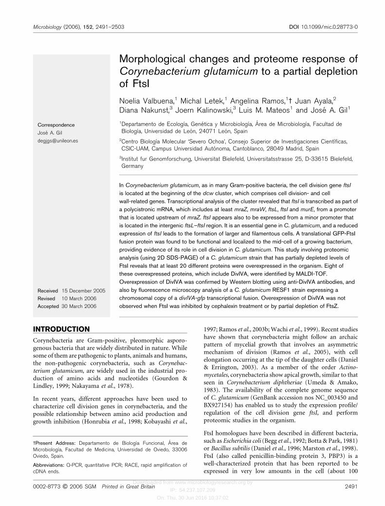
Complete genome sequence information of two strains ofC. glutamicum ATCC 13032 is available, NC_003450 (Ikeda& Nakagawa, 2003) and BX927154 (Kalinowski et al., 2003),and the region of the dcw cluster is shown in Fig. 1(a). Thestructural similarity in the organization of the genes that are59 to the dcw cluster around the ftsI region in both strainssuggests that mraZ, mraW, ftsL, ftsI and murE might becotranscribed. In order to test this possibility, total RNAwas extracted from C. glutamicum cells that were grownin TSB to exponential phase, and used in RT-PCR using
http://mic.sgmjournals.org 2495
Partial depletion of FtsI in C. glutamicum
Downloaded from www.microbiologyresearch.org by
IP: 54.237.107.209
On: Thu, 30 Jun 2016 10:37:02
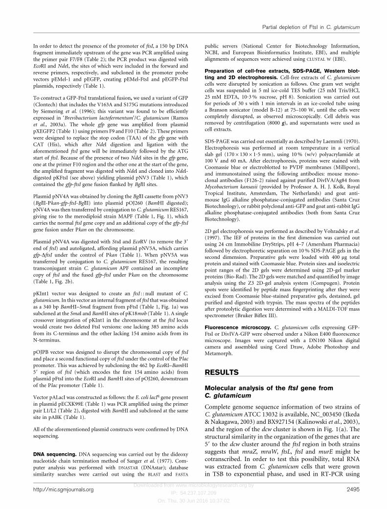
specific primers for the intergenic mraZ–mraW, mraW–ftsL,ftsL–ftsI, ftsI–murE regions, as well as for an internal regionof ftsI (see Fig. 1b). RT-PCR products of the expected sizeswere obtained in all cases, strongly suggesting that there isa transcriptional relationship between these five genes(Fig. 1c). No amplification band was seen when primer pairP0/F6 was used, indicating a lack of cotranscription withmraZ and upstream genes (Fig. 1c, lanes 11 and 12).
The main promoter of the dcw cluster was located by RACEPCR to be at the G residue, at position 2 295 904, which is7 nucleotides downstream from the putative 210 promoterregion TGCAGC (Fig. 1b) (Patek et al., 2003) and 99 ntupstream from the C. glutamicum mraZ gene (at the begin-ning of the mra/dcw cluster of cell division and cell envelopebiosynthesis genes) as seen in E. coli (Hara et al., 1997;Mengin-Lecreulx et al., 1998).
Two different initiation codons for ftsI were suggested forC. glutamicum; the first being a GTG codon at position
2 293 318, which would encode a 704 amino acid proteinwith a calculated molecular mass of 75?7 kDa (Wijayarathnaet al., 2001). The second being the other GTG at position2 293 165, which would encode a 651 amino acid proteinwith a calculated molecular mass of 69?7 kDa (Kalinowskiet al., 2003). In order to determine which of the two proteinproducts might be correct, and also to find a possibletranscriptional start point, additional RACE-PCR experi-ments, using different primers upstream from ftsI, wereperformed. The results showed the presence of an mRNAstarting at C (at position 2 293 257), which is located13 nucleotides downstream from a putative 210 pro-moter region (GAAGAT) in the intergenic ftsL–ftsI region(Fig. 1a). A 150 bp fragment containing the above-mentioned region was PCR amplified using primers F7and F8 (Table 2), and subcloned into the promoter probevectors pEMel-1 and pEGFP (Table 1), creating pEMel-FtsI and pEGFP-FtsI, respectively. These vectors weremobilized into C. glutamicum, and the detection of melanin
(a)
(b)
(c)
Fig. 1. (a) Physical map of the dcw cluster from C. glutamicum ATCC 13032. Arrows represent ORFs found in thesequenced DNA (accession no. NC_003450). (b) The relevant part of the C. glutamicum ATCC 13032 chromosome aroundftsI. Black boxes represent the DNA amplified by RT-PCR using the indicated primer pairs. The upper sequences show thetranscription start point (ts) (large G or C) and the hypothetical ”10 and ”35 boxes (large letters) of Pmra and PftsI. TheBamHI and SmaI sites used for ftsI gene disruption are indicated. (c) Agarose gel electrophoresis of different DNA fragmentsobtained after RT-PCR using C. glutamicum mRNA from a culture in exponential phase (odd lanes). Samples containing onlymRNA were used as negative controls (even lanes). The primer pairs used for RT-PCR were as follows: lanes 1–2, primer pairP1/P2; lanes 3–4, primer pair P3/P4; lanes 5–6, primer pair P5/P6; lanes 7–8, primer pair P7/P8; lanes 9–10, primer pairP9/P10; lanes 11–12, primer pair P0/F6. l, Lambda DNA digested with PstI.
2496 Microbiology 152
N. Valbuena and others
Downloaded from www.microbiologyresearch.org by
IP: 54.237.107.209
On: Thu, 30 Jun 2016 10:37:02
production and GFP activity in the corynebacteria con-firmed the presence of a promoter activity in that region.Therefore, it can be concluded that ftsI is mainly cotran-scribed along with the mraZ, mraW, ftsL and murE genes ofthe operon probably from the Pmra promoter, as describedin E. coli and B. subtilis (Hara et al., 1997), and probablyalso from a minor promoter (PftsI) as in B. subtilis (Danielet al., 1996). This result also suggests that the second GTG(at position 2 293 165) most probably is the start codon offtsI.
Taking into account the above results, a 1?9 kb fragmentcontaining the gene encoding FtsI (Kalinowski et al., 2003)was PCR amplified using specific primers, and ligated intothe pGEM-T Easy E. coli vector using the A–T strategy, tocreate pFtsI (Table 1). However, all of the E. coli trans-formants that were tested for the recombinant plasmid werefound to have the ftsI insert in the opposite orientation tothe Plac promoter. This observation strongly suggests thatoverexpression of FtsI might be toxic in E. coli. Furthermore,we also observed that expression of ftsI was unable to com-plement the temperature-sensitive ftsIts E. coli AX655 strain,as has been described for similar complementation assays(Wijayarathna et al., 2001).
Visualization of FtsICG-GFP fusions
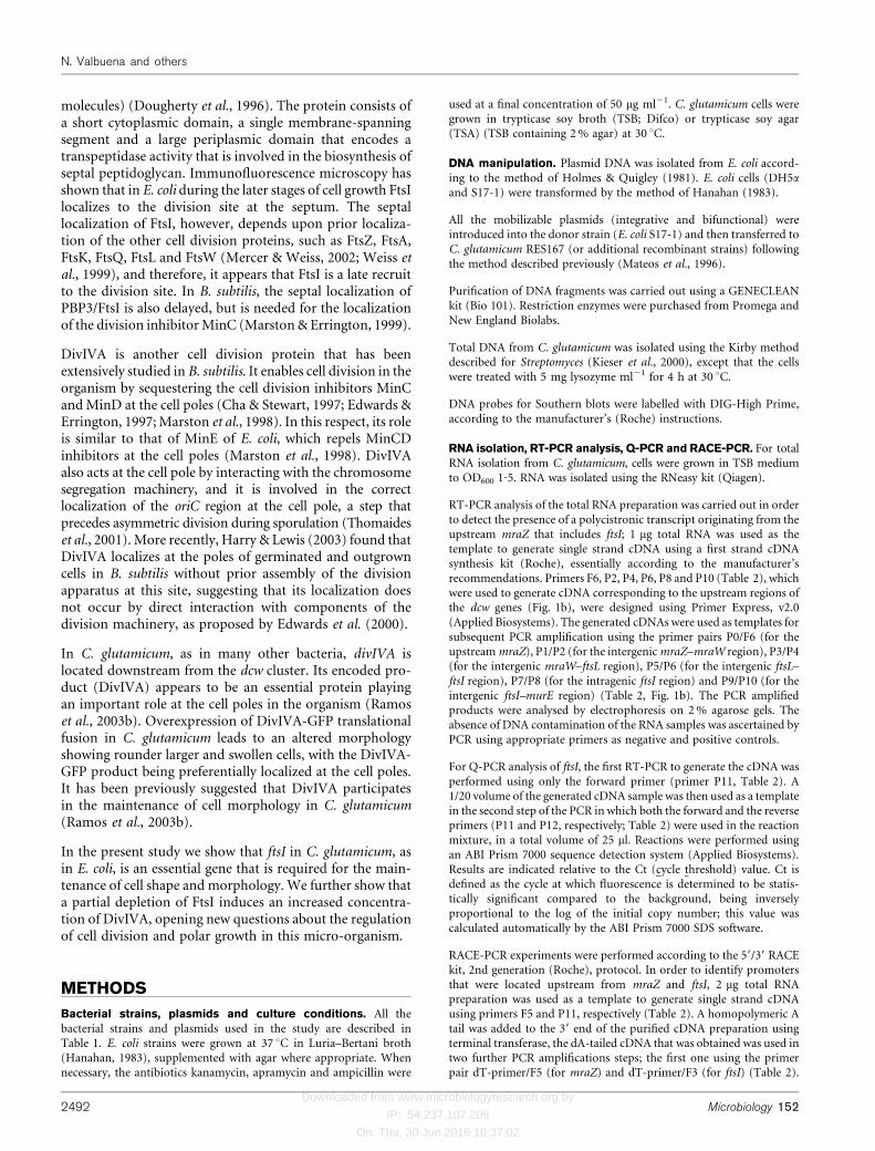
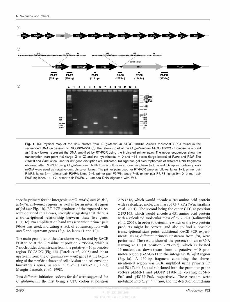
In order to analyse the role of FtsI in cell division in C.glutamicum, various strains of the organism were con-structed in which expression of the gfp-ftsI fusion wasstudied. The Pkan promoter of the kan gene from Tn5 isefficiently expressed in corynebacteria (Cadenas et al., 1991).C. glutamicum transformed with pNV4A vector (MAPFstrain) contains a wild-type copy of ftsI in addition to thegfp-ftsI cassette, which is under the control of the Pkanpromoter and inserted as a single copy in the chromosome(Fig. 2c); in C. glutamicum transformed with pNV5A vector(APF strain) gfp-ftsI expressed under the control of the Pkanpromoter is the only functional copy of the gene since thisstrain carries a truncated form of ftsI (Fig. 2b). Phase-contrast microscopy analysis of C. glutamicum APF(Fig. 2b) revealed typical corynebacterial cell morphologysimilar to the parental strain. Despite the chromosomalintegration, no polar effects on the downstream genes wereperceptible. The GFP-FtsI fluorescence signal was mainlylocated as foci in the mid-cell region (Fig. 2b, indicated byarrowheads). However, although the GFP signal in themerodiploid strain MAPF (Fig. 2c) clearly shows a reducedlevel of fluorescence, this is probably due to the competitionbetween the original FtsI and GFP-FtsI. Nevertheless, thesignal is well above the background autofluorescence thatis seen in the parent C. glutamicum RES167 strain, which hasno GFP fusion (not shown). Furthermore, the localizationof GFP-FtsI at the mid-cell is strongly indicative of theinvolvement of FtsI in C. glutamicum cell division, mostlikely in the biosynthesis of septum peptidoglycan asdescribed for other bacteria.
(a)
(b)
(c)
(d)
Fig. 2. Fluorescence microscopy images of C. glutamicum
cells carrying gfp-ftsI under the control of Pkan: (a) phase-con-trast microscopy image of RES167, (b, c, d) overlays combin-ing phase-contrast and fluorescence microscopy of the strainsAPF (b), MAPF (c) and MAPF transformed with pKInt1 (d).
http://mic.sgmjournals.org 2497
Partial depletion of FtsI in C. glutamicum
Downloaded from www.microbiologyresearch.org by
IP: 54.237.107.209
On: Thu, 30 Jun 2016 10:37:02
ftsI seems to be an essential gene inC. glutamicum
ftsI has been shown to be an essential gene in E. coli (Begget al., 1992) and in B. subtilis (Daniel et al., 1996). Therefore,in order to determine whether ftsI was also necessary for theviability of C. glutamicum, we performed gene disruptionexperiments using the suicide plasmid pKInt1 (Table 1); allattempts to inactivate the ftsI gene using internal fragmentswere unsuccessful, similar to our earlier studies involvingmurC, another essential gene in C. glutamicum (Ramos et al.,2004).
Disruption of ftsI was only possible in the merodiploidstrain C. glutamicum MAPF using plasmid pKInt1. Cellmorphology and growth rate of the transconjugantsexpressing the GFP-FtsI fusion (Fig. 2d) were found to besimilar to the host MAPF strain. Disruption of the originalchromosomal copy of ftsI was confirmed by Southernblotting using DNA isolated from ten fluorescent trans-conjugants (not shown). These results unambiguouslysubstantiate that ftsI is essential in C. glutamicum, as it isin E. coli and B. subtilis.
Decreased expression of ftsI causes severedefects in cell morphology
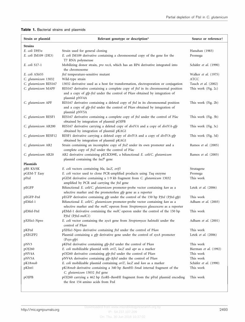
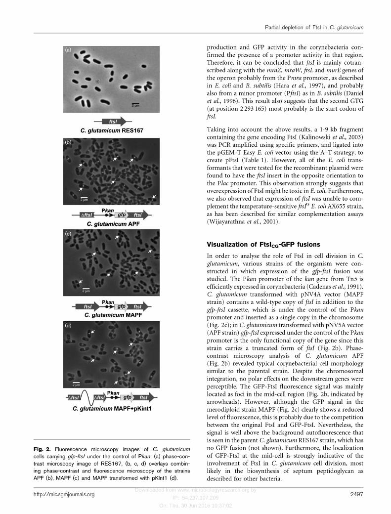
It has been reported previously that the promoter of thelactose operon of E. coli (Plac) is not well recognized by theC. glutamicum RNA polymerase (Ramos et al., 2005). As ftsIappears to be an essential gene in C. glutamicum, and sinceno ftsI null mutants could be obtained, the function of theFtsI was investigated by partial depletion of FtsI levels in theorganism. To do this, C. glutamicum transformed withthe plasmid pOJPB (Table 1) was used. Southern blottingof the transconjugant strain (C. glutamicum RESF1) revealedthe pattern expected of Campbell integration of pOJPB atthe ftsI locus (Fig. 3b). The strain carries a disrupted non-functional copy of ftsI and a functional copy under thecontrol of Plac, and has a distinctive phenotype (seeFig. 3b). This may be due to a reduced expression of ftsI(2?9 times less than the wild-type strain as quantified byQ-PCR analysis); furthermore, cells were found to beirregularly shaped, swollen and larger than the parentC. glutamicum RES167 strain (Fig. 3a). However, thepossibility exists that the aberrant phenotype may be dueto the expression of the truncated ftsI or to polar effects
(a) (c)
(b)(d)
Fig. 3. Fluorescence microscopy images of C. glutamicum cells carrying the ftsI gene under the control of Plac and/or thegene fusion divIVA-gfp. The images are as follows: phase-contrast microscopy images of RES167 (a) and the partiallydepleted FtsI strain RESF1 (b), overlaid phase-contrast and fluorescence microscopy images of AR200 (divIVA-gfp as asingle copy on the chromosome) (c) and RESF12 (d) obtained by integration of plasmid pKAG1 into the chromosome ofRESF1.
2498 Microbiology 152
N. Valbuena and others
Downloaded from www.microbiologyresearch.org by
IP: 54.237.107.209
On: Thu, 30 Jun 2016 10:37:02
on expression of other genes in the dcw cluster locateddownstream from ftsI (see Fig. 1a).
In order to further test the effect of a much reduced FtsI levelin the organism, we carried out a similar experiment as usedfor the study of ftsZ levels in C. glutamicum (Ramos et al.,2005). To reduce the levels of FtsI, we attempted tointroduce the lacIq gene of E. coli, which is present in theplasmid pALacI (Table 1), into C. glutamicum RESF1. All ofour attempts to obtain viable kanamycin- and apramycin-resistant transconjugants using the vector pALacI wereunsuccessful, while control plasmid pABK readily yieldedtransconjugants under the same conditions. These resultsseem to indicate that a more reduced ftsI expression due tothe possible effect of the lacZ repressor (lacIq) is lethal forC. glutamicum.
Decreased expression of ftsI induces theexpression of several genes in C. glutamicum
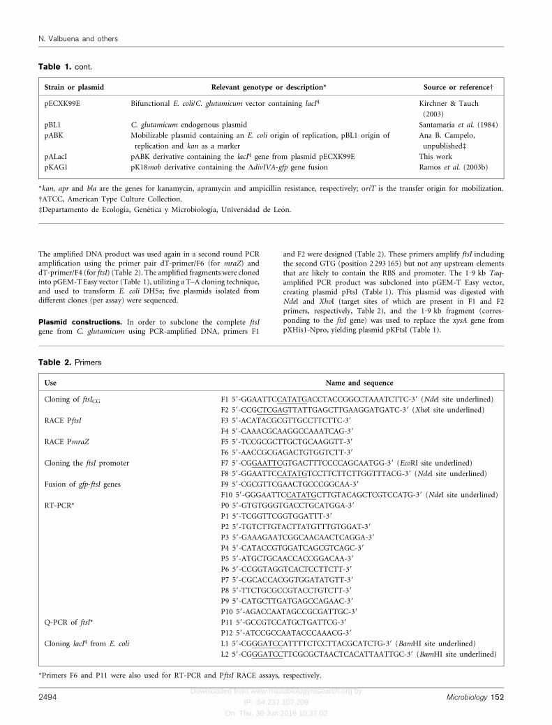
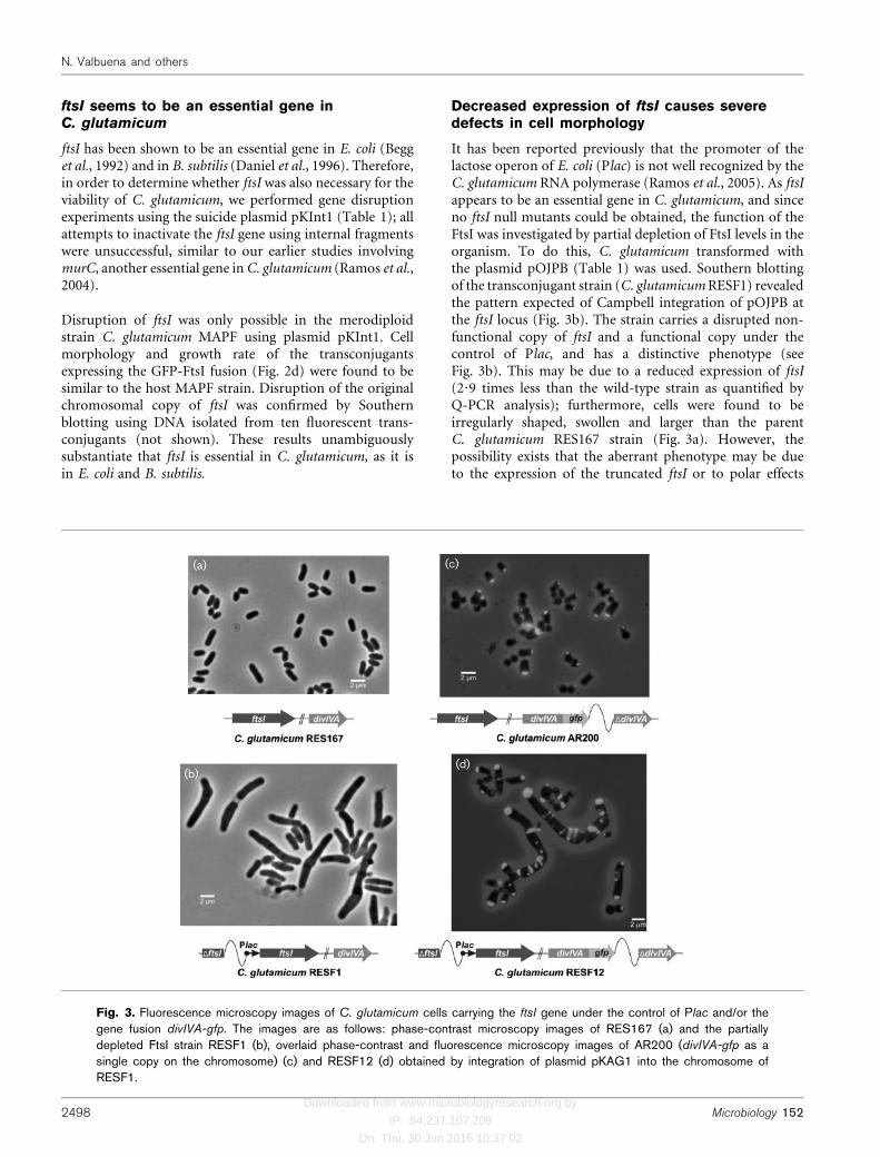
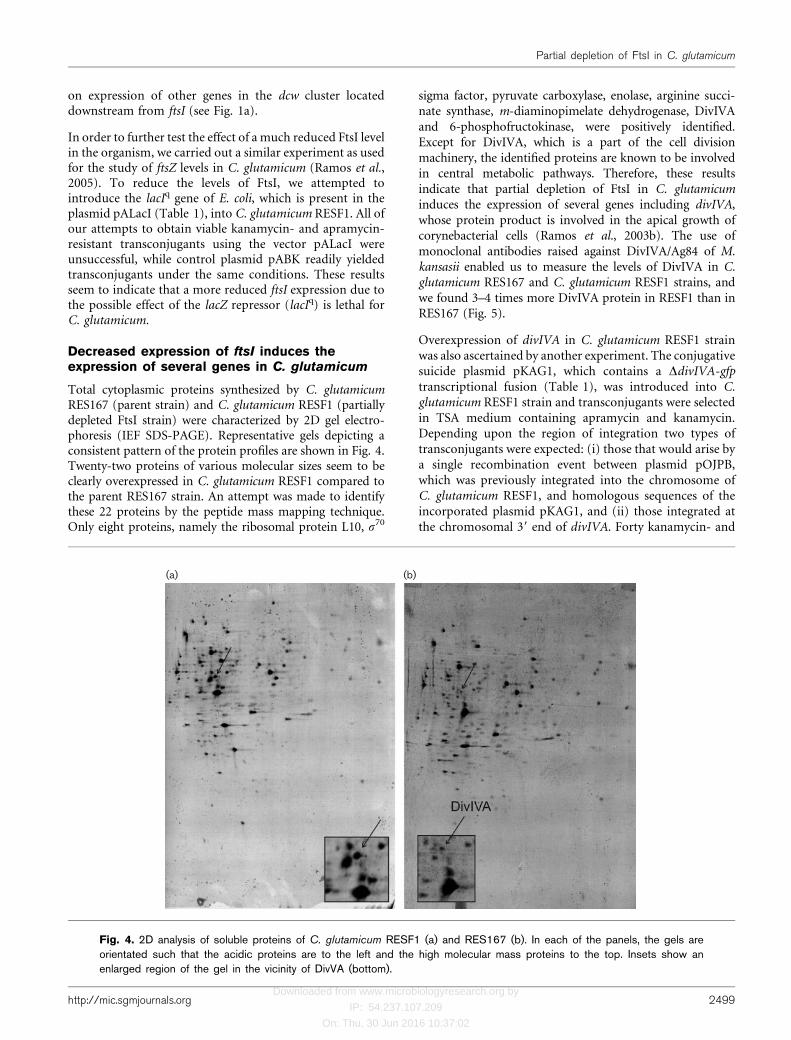
Total cytoplasmic proteins synthesized by C. glutamicumRES167 (parent strain) and C. glutamicum RESF1 (partiallydepleted FtsI strain) were characterized by 2D gel electro-phoresis (IEF SDS-PAGE). Representative gels depicting aconsistent pattern of the protein profiles are shown in Fig. 4.Twenty-two proteins of various molecular sizes seem to beclearly overexpressed in C. glutamicum RESF1 compared tothe parent RES167 strain. An attempt was made to identifythese 22 proteins by the peptide mass mapping technique.Only eight proteins, namely the ribosomal protein L10, s70
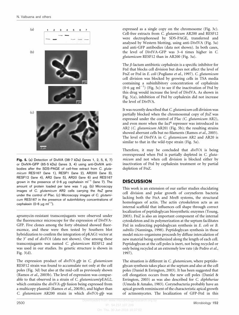
sigma factor, pyruvate carboxylase, enolase, arginine succi-nate synthase, m-diaminopimelate dehydrogenase, DivIVAand 6-phosphofructokinase, were positively identified.Except for DivIVA, which is a part of the cell divisionmachinery, the identified proteins are known to be involvedin central metabolic pathways. Therefore, these resultsindicate that partial depletion of FtsI in C. glutamicuminduces the expression of several genes including divIVA,whose protein product is involved in the apical growth ofcorynebacterial cells (Ramos et al., 2003b). The use ofmonoclonal antibodies raised against DivIVA/Ag84 of M.kansasii enabled us to measure the levels of DivIVA in C.glutamicum RES167 and C. glutamicum RESF1 strains, andwe found 3–4 times more DivIVA protein in RESF1 than inRES167 (Fig. 5).
Overexpression of divIVA in C. glutamicum RESF1 strainwas also ascertained by another experiment. The conjugativesuicide plasmid pKAG1, which contains a DdivIVA-gfptranscriptional fusion (Table 1), was introduced into C.glutamicum RESF1 strain and transconjugants were selectedin TSA medium containing apramycin and kanamycin.Depending upon the region of integration two types oftransconjugants were expected: (i) those that would arise bya single recombination event between plasmid pOJPB,which was previously integrated into the chromosome ofC. glutamicum RESF1, and homologous sequences of theincorporated plasmid pKAG1, and (ii) those integrated atthe chromosomal 39 end of divIVA. Forty kanamycin- and
(a) (b)
Fig. 4. 2D analysis of soluble proteins of C. glutamicum RESF1 (a) and RES167 (b). In each of the panels, the gels areorientated such that the acidic proteins are to the left and the high molecular mass proteins to the top. Insets show anenlarged region of the gel in the vicinity of DivVA (bottom).
http://mic.sgmjournals.org 2499
Partial depletion of FtsI in C. glutamicum
Downloaded from www.microbiologyresearch.org by
IP: 54.237.107.209
On: Thu, 30 Jun 2016 10:37:02
apramycin-resistant transconjugants were observed underthe fluorescence microscope for the expression of DivIVA-GFP. Five clones among the forty obtained showed fluor-escence, and these were then tested by Southern blothybridization to confirm the integration of pKAG1 vector atthe 39 end of divIVA (data not shown). One among thesetransconjugants was named C. glutamicum RESF12 andwas used in our studies. Its genetic structure is shown inFig. 3(d).
The expression product of divIVA-gfp in C. glutamicumRESF12 strain was found to accumulate not only at the cellpoles (Fig. 3d) but also at the mid-cell as previously shown(Ramos et al., 2003b). The level of expression was compar-able to that observed in a strain of C. glutamicum/pEAG2,which contains the divIVA-gfp fusion being expressed froma multicopy plasmid (Ramos et al., 2003b), and higher thanC. glutamicum AR200 strain in which divIVA-gfp was
expressed as a single copy on the chromosome (Fig. 3c).Cell-free extracts from C. glutamicum AR200 and RESF12were electrophoresed by SDS-PAGE, transferred andanalysed by Western blotting, using anti-DivIVA (Fig. 5a)and anti-GFP antibodies (data not shown). In both cases,the level of DivIVA-GFP was 3–4 times higher in C.glutamicum RESF12 than in AR200 (Fig. 5a).
The b-lactam antibiotic cephalexin is a specific inhibitor forFtsI that blocks cell division but does not affect the level ofFtsZ or FtsI in E. coli (Pogliano et al., 1997). C. glutamicumcell division was blocked by growing cells in TSA mediacontaining a subinhibitory concentration of cephalexin(0?6 mg ml21) (Fig. 5c) to see if the inactivation of FtsI bythis drug would increase the level of DivIVA. As shown inFig. 5(a), inhibition of FtsI by cephalexin did not increasethe level of DivIVA.
It was recently described that C. glutamicum cell division waspartially blocked when the chromosomal copy of ftsZ wasexpressed under the control of Plac (C. glutamicum AR2),and even more when the lacIq repressor was introduced inAR2 (C. glutamicum AR20) (Fig. 5b); the resulting strainsshowed aberrant cells but no filaments (Ramos et al., 2005).The level of DivIVA in C. glutamicum AR2 and AR20 issimilar to that in the wild-type strain (Fig. 5a).
Therefore, it may be concluded that divIVA is beingoverexpressed when FtsI is partially depleted in C. gluta-micum and not when cell division is blocked either byinactivation of FtsI by cephalexin treatment or by partialdepletion of FtsZ.
DISCUSSION
This work is an extension of our earlier studies elucidatingcell division and polar growth of coryneform bacterialacking both the FtsA and MreB systems, the structuralhomologues of actin. The actin cytoskeleton acts as aninternal scaffold that influences cell shape through correctlocalization of peptidoglycan biosynthetic enzymes (Young,2003). FtsZ is also an important component of the internalcytoskeleton and its polymerization at the septum facilitatesFtsI in redirecting peptidoglycan synthesis in E. coli or B.subtilis (Nanninga, 1998). Peptidoglycan synthesis in thosemodel micro-organisms proceeds by diffuse intercalation ofnew material being synthesized along the length of each cell.Peptidoglycan at the cell poles is inert, not being recycled oronly being recycled at an extremely low rate (de Pedro et al.,1997).
The situation is different in C. glutamicum, where peptido-glycan synthesis takes place at the septum and also at the cellpoles (Daniel & Errington, 2003). It has been suggested thatcell elongation occurs from the new cell poles (Daniel &Errington, 2003) as was also described for C. diphtheriae(Umeda & Amako, 1983). Corynebacteria probably have anapical growth reminiscent of the characteristic apical growthof actinomycetes. The localization of GFP-FtsI in this
(a)
(b)
(c)
Fig. 5. (a) Detection of DivIVA (38?7 kDa) (lanes 1, 2, 5, 6, 7)or DivIVA-GFP (65?5 kDa) (lanes 3, 4) using anti-DivIVA anti-bodies after the SDS-PAGE of cell-free extract from C. gluta-
micum RES167 (lane 1), RESF1 (lane 2), AR200 (lane 3),RESF12 (lane 4), AR2 (lane 5), AR20 (lane 6) and RES167grown in the presence of 0?6 mg cephalexin ml”1 (lane 7). Theamount of protein loaded per lane was 1 mg. (b) Microscopyimages of C. glutamicum AR2 cells carrying the ftsZ geneunder the control of Plac. (c) Microscopy images of C. glutami-
cum RES167 in the presence of subinhibitory concentrations ofcephalexin (0?6 mg ml”1).
2500 Microbiology 152
N. Valbuena and others
Downloaded from www.microbiologyresearch.org by
IP: 54.237.107.209
On: Thu, 30 Jun 2016 10:37:02
organism appears to be similar to that seen in E. coli (Weisset al., 1999) and B. subtilis (Daniel et al., 2000), and thissuggests the participation of FtsI in the biosynthesis ofpeptidoglycan for septum formation. Sometimes, and in anon-repetitive way, it was also possible to see accumulationof GFP-FtsI at the cell poles, which is reminiscent of pre-vious septa or artefacts due to non-specific accumulation ofGFP-FtsI as described in E. coli (Weiss et al., 1999).
Our results suggest that ftsI is transcribed both from a minorpromoter (PftsI), as in B. subtilis (Daniel et al., 1996), andalso as a part of the polycistronic mraZ, mraW, ftsL andmurE transcript from an upstream promoter (Pmra), asdescribed for E. coli and B. subtilis (Hara et al., 1997;Mengin-Lecreulx et al., 1998). The ftsI gene seems to beessential for the viability of C. glutamicum since genedisruption was possible only in the merodiploid strainC. glutamicum MAPF. No transformants were recoveredwhen we attempted to elicit a stronger reduction in theexpression of ftsI under the control of Plac in C. glutamicumRESF1 transformed with a plasmid carrying lacIq.
When ftsI was expressed as a single copy in the chromosomeunder the control of Plac (strain C. glutamicum RESF1)morphologically abnormal cells (filamentous or branchedfilaments) were obtained. The shape of RESF1 cells contrastswith the bulky and elongated cells obtained when celldivision is inhibited by cephalexin treatment or by partialFtsZ depletion (Fig. 5). These abnormal cells were con-sidered as a general strategy by the bacteria for increasing cellmass when division is blocked in rod-shaped micro-organisms that lack actin homologues as was suggestedby Latch & Margolin (1997) and Ramos et al. (2005). Thecephalexin treatment has no effect on the levels of ftsI or ftsZ(Pogliano et al., 1997), suggesting that the characteristicshape of C. glutamicum RESF1, as a consequence of a severereduction (2?9-fold) in the expression of ftsI (Fig. 3b), isnot only due to a block in cell division. This possibilityprompted us to compare the proteome of C. glutamicumRESF1 with that of the wild-type. It was noticeable from ourresults that the level of DivIVA, among other proteins,increases in the strains that are partially depleted for FtsI.
Several hypotheses can be proposed to explain why a reduc-tion in the levels of FtsI could account for an increase in thelevels of DivIVA, a cell division-associated protein with apossible structural function at C. glutamicum growing cellpoles (Ramos et al., 2003b). The first possibility is that thefilamentous phenotype may be due to the expression of thetruncated ftsI in C. glutamicum RESF1 (Fig. 3b), althoughno filaments were observed in C. glutamicum APF (having atruncated form of ftsI and a unique copy of gfp-ftsI under thecontrol of Pkan) (Fig. 2b). The observed effect of DivIVAoverexpression could also be due to polar effects on theexpression of other genes in the dcw cluster located down-stream from ftsI, such as murE (encoding the tripeptidesynthetase MurE) or murF (encoding pentapeptide synthaseMurF) (Fig. 1a). It has been described for E. coli that thebalance between pentapeptide and tripeptide precursors
determines whether the cells will divide or elongate (Begget al., 1990). We cannot rule out this option, but no effect onthe morphology was observed when different C. glutamicumstrains were obtained by simple recombination in thechromosome (Fig. 2). A third possibility is that depletion ofFtsI, and not the inhibition of FtsI by cephalexin, alters theturnover or stability of DivIVA, or even the expression of thedivIVA gene. Because the amount of FtsI protein, and notthe activity of FtsI, could be the start point of a mechanismleading to DivIVA overexpression, it is possible that abalance between FtsI and DivIVA would be needed for celldivision in C. glutamicum. The requirement of a specificratio between two proteins of the dcw cluster has beendescribed by Dewar et al. (1992) and Flardh et al. (1998).
The filamentous phenotype observed when ftsI is under thecontrol of Plac seems to be the result of a lack of FtsI tosynthesize peptidoglycan at the septum and an increase inthe concentration of enzymes involved in central metabo-lism and DivIVA, which might form a scaffold structure thatguides cell wall biosynthesis and maintains the cell diameterin MreB-lacking rod-shaped corynebacteria.
ACKNOWLEDGEMENTS
This work was funded by grants from the Junta de Castilla y Leon (ref.LE 24/01 and LE14-04), the University of Leon (ULE 2001-08B) andthe Ministerio de Ciencia y Tecnologıa (BIO2002-03223). NoeliaValbuena and Michal Letek are recipients of fellowships from the FPU(Ministry of Education) and Angelina Ramos from the Junta de Castillay Leon. We acknowledge Dr Ramon I. Santamarıa and Dr Jose M.Fernandez-Abalos (CSIC, University of Salamanca, Spain) for the egfp2gene, Dr A. H. J. Kolk (Royal Tropical Institute, Amsterdam, TheNetherlands) for the monoclonal antibody against Ag84/DivIVA, andDr Richard Daniel (Department of Pathology, University of Oxford,Oxford, UK) for his help with the fluorescence microscopy. The criticalreading of the manuscript by Dr Vassant K. Chary (Department ofMicrobiology and Immunology, Temple University School of Medicine,Philadelphia, Pennsylvania, USA) is gratefully acknowledged.
REFERENCES
Adham, S. A., Honrubia, P., Diaz, M., Fernandez-Abalos, J. M.,Santamaria, R. I. & Gil, J. A. (2001). Expression of the genes codingfor the xylanase Xys1 and the cellulase Cel1 from the straw-decomposing Streptomyces halstedii JM8 cloned into the amino-acidproducer Brevibacterium lactofermentum ATCC13869. Arch Microbiol177, 91–97.
Adham, S. A., Rodriguez, S., Ramos, A., Santamaria, R. I. & Gil, J. A.(2003). Improved vectors for transcriptional/translational signalscreening in corynebacteria using the melC operon from Streptomycesglaucescens as reporter. Arch Microbiol 180, 53–59.
Begg, K. J., Takasuga, A., Edwards, D. H., Dewar, S. J., Spratt, B. G.,Adachi, H., Ohta, T., Matsuzawa, H. & Donachie, W. D. (1990). Thebalance between different peptidoglycan precursors determineswhether Escherichia coli cells will elongate or divide. J Bacteriol172, 6697–6703.
Begg, K. J., Tomoyasu, T., Donachie, W. D., Khattar, M., Niki, H.,Yamanaka, K., Hiraga, S. & Ogura, T. (1992). Escherichia coli mutantY16 is a double mutant carrying thermosensitive ftsH and ftsImutations. J Bacteriol 174, 2416–2417.
http://mic.sgmjournals.org 2501
Partial depletion of FtsI in C. glutamicum
Downloaded from www.microbiologyresearch.org by
IP: 54.237.107.209
On: Thu, 30 Jun 2016 10:37:02
Bierman, M., Logan, R., O’Brien, K., Seno, E. T., Rao, R. N. & Schoner,B. E. (1992). Plasmid cloning vectors for the conjugal transfer of DNA
from Escherichia coli to Streptomyces spp. Gene 116, 43–49.
Botta, G. A. & Park, J. T. (1981). Evidence for involvement of
penicillin-binding protein 3 in murein synthesis during septation but
not during cell elongation. J Bacteriol 145, 333–340.
Cadenas, R. F., Martin, J. F. & Gil, J. A. (1991). Construction and
characterization of promoter-probe vectors for corynebacteria using
the kanamycin-resistance reporter gene. Gene 98, 117–121.
Cha, J. H. & Stewart, G. C. (1997). The divIVA minicell locus of
Bacillus subtilis. J Bacteriol 179, 1671–1683.
Daniel, R. A. & Errington, J. (2003). Control of cell morphogenesis in
bacteria: two distinct ways to make a rod-shaped cell. Cell 113, 767–776.
Daniel, R. A., Williams, A. M. & Errington, J. (1996). A complex four-
gene operon containing essential cell division gene pbpB in Bacillus
subtilis. J Bacteriol 178, 2343–2350.
Daniel, R. A., Harry, E. J. & Errington, J. (2000). Role of penicillin-
binding protein PBP 2B in assembly and functioning of the division
machinery of Bacillus subtilis. Mol Microbiol 35, 299–311.
de Pedro, M. A., Quintela, J. C., Holtje, J. V. & Schwarz, H. (1997).Murein segregation in Escherichia coli. J Bacteriol 179, 2823–2834.
Dewar, S. J., Begg, K. J. & Donachie, W. D. (1992). Inhibition of cell
division initiation by an imbalance in the ratio of FtsA to FtsZ.
J Bacteriol 174, 6314–6316.
Dougherty, T. J., Kennedy, K., Kessler, R. E. & Pucci, M. J. (1996).Direct quantitation of the number of individual penicillin-binding
proteins per cell in Escherichia coli. J Bacteriol 178, 6110–6115.
Edwards, D. H. & Errington, J. (1997). The Bacillus subtilis DivIVA
protein targets to the division septum and controls the site specificity
of cell division. Mol Microbiol 24, 905–915.
Edwards, D. H., Thomaides, H. B. & Errington, J. (2000). Pro-
miscuous targeting of Bacillus subtilis cell division protein DivIVA
to division sites in Escherichia coli and fission yeast. EMBO J 19,
2719–2727.
Flardh, K., Palacios, P. & Vicente, M. (1998). Cell division genes
ftsQAZ in Escherichia coli require distant cis-acting signals upstream
of ddlB for full expression. Mol Microbiol 30, 305–315.
Gourdon, P. & Lindley, N. D. (1999). Metabolic analysis of glutamate
production by Corynebacterium glutamicum. Metab Eng 1, 224–231.
Hanahan, D. (1983). Studies on transformation of Escherichia coli
with plasmids. J Mol Biol 166, 557–580.
Hara, H., Yasuda, S., Horiuchi, K. & Park, J. T. (1997). A promoter
for the first nine genes of the Escherichia coli mra cluster of cell
division and cell envelope biosynthesis genes, including ftsI and ftsW.
J Bacteriol 179, 5802–5811.
Harry, E. J. & Lewis, P. J. (2003). Early targeting of Min proteins to
the cell poles in germinated spores of Bacillus subtilis: evidence for
division apparatus-independent recruitment of Min proteins to the
division site. Mol Microbiol 47, 37–48.
Holmes, D. S. & Quigley, M. (1981). A rapid boiling method for the
preparation of bacterial plasmids. Anal Biochem 114, 193–197.
Honrubia, M. P., Fernandez, F. J. & Gil, J. A. (1998). Identification,
characterization, and chromosomal organization of the ftsZ gene
from Brevibacterium lactofermentum. Mol Gen Genet 259, 97–104.
Ikeda, M. & Nakagawa, S. (2003). The Corynebacterium glutamicum
genome: features and impacts on biotechnological processes. Appl
Microbiol Biotechnol 62, 99–109.
Kalinowski, J., Bathe, B., Bartels, D. & 24 other authors (2003).The complete Corynebacterium glutamicum ATCC 13032 genome
sequence and its impact on the production of L-aspartate-derived
amino acids and vitamins. J Biotechnol 104, 5–25.
Kieser, T., Bibb, M. J., Buttner, M. J., Chater, K. F. & Hopwood, D. A.
(2000). Practical Streptomyces Genetics. Norwich: John Innes
Foundation.
Kirchner, O. & Tauch, A. (2003). Tools for genetic engineering in the
amino acid-producing bacterium Corynebacterium glutamicum.
J Biotechnol 104, 287–299.
Kobayashi, M., Asai, Y., Hatakeyama, K., Kijima, N., Wachi, M.,
Nagai, K. & Yukawa, H. (1997). Cloning, sequencing, and char-
acterization of the ftsZ gene from coryneform bacteria. Biochem
Biophys Res Commun 236, 383–388.
Laemmli, U. K. (1970). Cleavage of structural proteins during the
assembly of the head of bacteriophage T4. Nature 227, 680–685.
Latch, J. N. & Margolin, W. (1997). Generation of buds, swellings,
and branches instead of filaments after blocking the cell cycle of
Rhizobium meliloti. J Bacteriol 179, 2373–2381.
Letek, M., Valbuena, N., Ramos, A., Ordonez, E., Gil, J. A. & Mateos,
L. M. (2006). Characterization and use of catabolite-repressed pro-
moters from gluconate genes in Corynebacterium glutamicum.
J Bacteriol 188, 409–423.
Marston, A. L. & Errington, J. (1999). Selection of the midcell
division site in Bacillus subtilis through MinD-dependent polar
localization and activation of MinC. Mol Microbiol 33, 84–96.
Marston, A. L., Thomaides, H. B., Edwards, D. H., Sharpe, M. E. &
Errington, J. (1998). Polar localization of the MinD protein of
Bacillus subtilis and its role in selection of the mid-cell division site.
Genes Dev 12, 3419–3430.
Mateos, L. M., Schafer, A., Kalinowski, J., Martin, J. F. & Puhler, A.
(1996). Integration of narrow-host-range vectors from Escherichia
coli into the genomes of amino acid-producing corynebacteria after
intergeneric conjugation. J Bacteriol 178, 5768–5775.
Mengin-Lecreulx, D., Ayala, J., Bouhss, A., van Heijenoort, J.,
Parquet, C. & Hara, H. (1998). Contribution of the Pmra promoter
to expression of genes in the Escherichia coli mra cluster of cell
envelope biosynthesis and cell division genes. J Bacteriol 180,
4406–4412.
Mercer, K. L. & Weiss, D. S. (2002). The Escherichia coli cell division
protein FtsW is required to recruit its cognate transpeptidase, FtsI
(PBP3), to the division site. J Bacteriol 184, 904–912.
Nakayama, K., Araki, K. & Kase, H. (1978). Microbial production of
essential amino acid with Corynebacterium glutamicum mutants. Adv
Exp Med Biol 105, 649–661.
Nanninga, N. (1998). Morphogenesis of Escherichia coli. Microbiol
Mol Biol Rev 62, 110–129.
Patek, M., Nesvera, J., Guyonvarch, A., Reyes, O. & Leblon, G.
(2003). Promoters of Corynebacterium glutamicum. J Biotechnol 104,
311–323.
Pogliano, J., Pogliano, K., Weiss, D. S., Losick, R. & Beckwith, J.
(1997). Inactivation of FtsI inhibits constriction of the FtsZ
cytokinetic ring and delays the assembly of FtsZ rings at potential
division sites. Proc Natl Acad Sci U S A 94, 559–564.
Ramos, A., Adham, S. A. & Gil, J. A. (2003a). Cloning and expression
of the inorganic pyrophosphatase gene from the amino acid pro-
ducer Brevibacterium lactofermentum ATCC 13869. FEMS Microbiol
Lett 225, 85–92.
Ramos, A., Honrubia, M. P., Valbuena, N., Vaquera, J., Mateos, L. M.
& Gil, J. A. (2003b). Involvement of DivIVA in the morphology
of the rod-shaped actinomycete Brevibacterium lactofermentum.
Microbiology 149, 3531–3542.
Ramos, A., Honrubia, M. P., Vega, D., Ayala, J. A., Bouhss, A.,
Mengin-Lecreulx, D. & Gil, J. A. (2004). Characterization and
chromosomal organization of the murD–murC–ftsQ region of
2502 Microbiology 152
N. Valbuena and others

Downloaded from www.microbiologyresearch.org by
IP: 54.237.107.209
On: Thu, 30 Jun 2016 10:37:02
Corynebacterium glutamicum ATCC 13869. Res Microbiol 155,
174–184.
Ramos, A., Letek, M., Campelo, A. B., Vaquera, J., Mateos, L. M. &Gil, J. A. (2005). Altered morphology produced by ftsZ expression
in Corynebacterium glutamicum ATCC 13869. Microbiology 151,
2563–2572.
Sanger, F., Nicklen, S. & Coulson, A. R. (1977). DNA sequencing with
chain-terminating inhibitors. Proc Natl Acad Sci U S A 74, 5463–5467.
Santamaria, R. I., Gil, J. A., Mesas, J. M. & Martin, J. F. (1984).Characterization of an endogenous plasmid and development of
cloning vectors and a transformation system in Brevibacterium
lactofermentum. J Gen Microbiol 130, 2237–2246.
Schafer, A., Kalinowski, J., Simon, R., Seep-Feldhaus, A.-H. &Puhler, A. (1990). High-frequency conjugal plasmid transfer from
Gram-negative Escherichia coli to various Gram-positive coryneform
bacteria. J Bacteriol 172, 1663–1666.
Siemering, K. R., Golbik, R., Sever, R. & Haseloff, J. (1996).Mutations that suppress the thermosensitivity of green fluorescent
protein. Curr Biol 6, 1653–1663.
Tauch, A., Kirchner, O., Loffler, B., Gotker, S., Puhler, A. & Kalinowski,J. (2002). Efficient electrotransformation of Corynebacterium diphther-
iae with a mini-replicon derived from the Corynebacterium glutamicum
plasmid pGA1. Curr Microbiol 45, 362–367.
Thomaides, H. B., Freeman, M., El Karoui, M. & Errington, J. (2001).Division site selection protein DivIVA of Bacillus subtilis has asecond distinct function in chromosome segregation during sporul-tion. Genes Dev 15, 1662–1673.
Umeda, A. & Amako, K. (1983). Growth of the surface of Coryne-bacterium diphtheriae. Microbiol Immunol 27, 663–671.
Vohradsky, J., Li, X. M. & Thompson, C. J. (1997). Identificationof procaryotic developmental stages by statistical analyses of two-dimensional gel patterns. Electrophoresis 18, 1418–1428.
Wachi, M., Wijayarathna, C. D., Teraoka, H. & Nagai, K. (1999). AmurC gene from coryneform bacteria. Appl Microbiol Biotechnol 51,223–228.
Walker, J. R., Kovaric, A., Allen, J. S. & Gustafson, R. A. (1975).Regulation of bacterial cell division: temperature-sensitive mutantsof Escherichia coli that are defective in septum formation. J Bacteriol123, 693–703.
Weiss, D. S., Chen, J. C., Ghigo, J. M., Boyd, D. & Beckwith, J. (1999).Localization of FtsI (PBP3) to the septal ring requires its membraneanchor, the Z ring, FtsA, FtsQ, and FtsL. J Bacteriol 181, 508–520.
Wijayarathna, C. D., Wachi, M. & Nagai, K. (2001). Isolation offtsI and murE genes involved in peptidoglycan synthesis fromCorynebacterium glutamicum. Appl Microbiol Biotechnol 55, 466–470.
Young, K. D. (2003). Bacterial shape. Mol Microbiol 49, 571–580.
http://mic.sgmjournals.org 2503
Partial depletion of FtsI in C. glutamicum
Related Documents