ONLINE FIRST This is a provisional PDF only. Copyedited and fully formatted version will be made available soon. ISSN: 0015-5659 e-ISSN: 1644-3284 Morphological and morphometric features of sacral hiatus and its clinical significance in caudal epidural anesthesia Authors: E. Nastoulis, D. Tsiptsios, P. Chloropoulou, V. Karapepera, V. Thomaidis, P. Pavlidis, A. Fiska DOI: 10.5603/FM.a2022.0078 Article type: Original article Submitted: 2022-04-16 Accepted: 2022-08-16 Published online: 2022-08-25 This article has been peer reviewed and published immediately upon acceptance. It is an open access article, which means that it can be downloaded, printed, and distributed freely, provided the work is properly cited. Articles in "Folia Morphologica" are listed in PubMed. Powered by TCPDF (www.tcpdf.org)

Welcome message from author
This document is posted to help you gain knowledge. Please leave a comment to let me know what you think about it! Share it to your friends and learn new things together.
Transcript

ONLINE FIRST
This is a provisional PDF only. Copyedited and fully formatted version will be made available soon.
ISSN: 0015-5659
e-ISSN: 1644-3284
Morphological and morphometric features of sacral hiatus andits clinical significance in caudal epidural anesthesia
Authors: E. Nastoulis, D. Tsiptsios, P. Chloropoulou, V. Karapepera, V. Thomaidis,P. Pavlidis, A. Fiska
DOI: 10.5603/FM.a2022.0078
Article type: Original article
Submitted: 2022-04-16
Accepted: 2022-08-16
Published online: 2022-08-25
This article has been peer reviewed and published immediately upon acceptance.It is an open access article, which means that it can be downloaded, printed, and distributed freely,
provided the work is properly cited.Articles in "Folia Morphologica" are listed in PubMed.
Powered by TCPDF (www.tcpdf.org)

Morphological and morphometric features of sacral hiatus and its clinical
significance in caudal epidural anesthesia
E. Nastoulis et al., Morphological and morphometric features of sacral hiatus
E. Nastoulis1, D. Tsiptsios1, P. Chloropoulou2, V. Karapepera1, V. Thomaidis1, P.
Pavlidis3, A. Fiska1
1Department of Anatomy, School of Medicine, Democritus University of Thrace,
Alexandroupolis, Greece
2Department of Anesthesiology, School of Medicine, Democritus University of
Thrace, Alexandroupolis, Greece
3Laboratory of Forensic Sciences, School of Medicine, Democritus University of
Thrace, Alexandroupolis, Greece
Address for correspondence: E. Nastoulis MD, MSc, Forensic Pathologist, doctoral
student, School of Medicine, Democritus University of Thrace, Dragana, 68100,
Alexandroupolis, Evros Prefecture, Greece, tel: +30 6972524316, fax: +30 25513
53824, e-mail: [email protected]
ABSTRACT
Background: Caudal epidural block (CEB) failure or complications are not unheard
even among experienced anesthesiologists and are usually due to sacral hiatus (SH)
anatomy variations. The aim of the present study is to observe, record and analyze
important anatomical features of SH and correlate them with potential CEB
limitations.

Materials and methods: The SH of 155 complete and undamaged Greek adult dry
sacra of known sex were included in the study. Three non-metric (shape of SH and
location of hiatal apex and base in relation to level of sacral/coccygeal vertebra) and
five metric parameters (height of the SH, transverse width of the SH at the base,
anteroposterior diameter of the SH at the level of its apex and the distance from the
sacral apex and base to the upper border of S2 foramina) were evaluated.
Results: Inverted U (34.83%) and inverted V (26.45%) were the commonest shapes.
Hiatal apex and base were most commonly related to the level of S4 (78.70%) and S5
vertebra (89.03%), respectively. Mean height, depth and intercornual distance were
19.05±8.65 mm, 5.39±1.84 mm and 12.41±3.16 mm, respectively, whereas mean
distance between the upper border of S2 foramen and the apex and base of the sacral
hiatus were 46.34 mm and 63.48mm, respectively. Anatomical variations of sacral
hiatus that might be responsible for CEB failure, such as elongated SH, absence of
SH, complete dorsal wall agenesis of sacral canal and narrowing (< 3 mm) at the apex
of SH were found in 17.43% of sacra (male 10.94% and female 25.22%).
Conclusions: This study suggests a potential risk of failure of CEB in Greek patients,
especially in females, which should be kept in mind while giving caudal epidural
anesthesia.
Key words: sacrum, sacral hiatus, anatomical variations, caudal epidural
anesthesia, Greece
INTRODUCTION
The human sacrum constitutes a large triangular bone located at the base of
vertebral column between the two hip bones that articulates with the fifth lumbar
vertebra above and the coccyx below. Over the years, an abundance of sacral
anatomical variations has been reported [29]. Embryologically, it is formed by the
fusion of the five sacral vertebrae that is complete between the third or early fourth
decade of life. Failure of fusion of the fifth, and occasionally the fourth, sacral
vertebral laminae creates an opening on the posterior aspect of the lower end of the
sacrum, known as sacral hiatus. The remnants of the inferior articular processes of the

fifth sacral segment extend downwards on both sides of the sacral hiatus forming the
two sacral cornua. These constitute its lateral margins and define important clinical
landmarks during caudal epidural anesthesia or block (CEB). [1]
The dorsal surface of the fifth sacral and the coccygeal vertebrae and the deep
posterior sacrococcygeal membrane connecting them, form the roof of the lower
sacral canal. Down to the middle of S2 vertebra the sacral canal encompasses the
termination of the dural sac containing the distal portion of the cauda equina and the
internal filum terminale. Caudally, the fifth sacral and coccygeal spinal nerves formed
within the sacral canal and exiting via the sacral hiatus and the external filum
terminale extending towards the dorsum of the coccyx are encountered. The sacral
canal also contains the epidural venous plexus and is filled with adipose tissue that is
subject to age-related decrease in density. [41]
The application of local anesthetic agents into the epidural space through the
sacral hiatus was first described by Fernard Cathelin and Jean-Athanase Sicard
predating lumbar epidural block [8] and gained popularity by Hingson who
implemented it on obstetrical cases [15, 37]. Caudal epidural anesthesia produces
sensory and motor blockade of the sacral roots and limited blockade of the
autonomous nervous system. Thus, this type of anesthesia has a wide variety of
indications [15]. Most frequently it is used to provide anesthesia in infants and
children for surgery of the perineum, anus and rectum and for inguinal and femoral
hernias [47]. In adults for surgery of the lumbar spine, cystoscopies, urethral and
vaginal operations and for labour pain relief, although the latter has been superseded
by lumbar epidural anesthesia [17]. Additionally, it is useful in providing sympathetic
block to patients suffering from acute vascular insufficiency of the lower extremities
due to vascular spasm or occlusion. It can be used for any acute pelvic or leg pain, but
most rewardingly in chronic pain due to diabetic neuropathy, post-herpetic pain,
failed back syndrome, complex regional pain syndromes and for cancer pain
management including peripheral neuropathy due to chemotherapy [10].
The technique of caudal epidural anesthesia entails placing neonates and
children in the lateral decubitus position, while for older children and adults the prone
position is preferred [19, 39]. The sacral hiatus can be located by palpating the sacral
cornua, approximately at the level of the skin folds of the buttocks. After infiltrating

with local anesthetic, the epidural needle is inserted in the caudal canal by using the
loss of resistance technique and its position is ascertained by fluoroscopy or
ultrasound [3, 7]. After the correct placement of the needle is confirmed, a catheter is
inserted to the desired depth. Special attention should be paid not to penetrate the
cancellous bone of the sacrum and not to tear the dura if the needle is advanced more
cephalad than the S2 level, as this level indicates the lowest extension of the dura.
Complications of CEB can relate to unsuccessful technique, such as penetration of the
dura, penetration, and injection of local anesthetic agents into the soft bone of the
sacrum (mainly in children), perforation of the rectum or even trauma to the baby’s
head in parturients, if the needle is placed too anteriorly [14]. In addition,
complications can arise from the spread of the anesthetic solutions and the
development of systemic toxicity. This complication, although rare, seems to be more
common in caudal epidural anesthesia compared to lumbar epidural anesthesia [9].
Infections such as meningitis, discitis or vertebral osteomyelitis are rare
complications. Other complications such as epidural hematoma, dural puncture and
post dural puncture headache, air embolism, back pain and broken or knotted
catheters can occur after caudal anesthesia, as with lumbar or thoracic epidural
anesthesia.
CEB failure rate approximates 5-10% and is usually attributed to technical
difficulties to accurately identify the sacral hiatus due to its numerous anatomic and
morphological variations. Thus, for optimal access into the sacral epidural space and
successful, uncomplicated conduction of CEB, a thorough knowledge of the SH
anatomical variations, as well as the use of detailed anatomical landmarks to identify
them, are required. To gain this knowledge, we conducted a descriptive osteological
study in dry sacra of Greek adults, aiming to observe, record and analyze several
metric and non-metric features of the sacral hiatus.
MATERIALS AND METHODS
The aim of the present study is to observe, record and analyze important
anatomical features of the SH in a Greek population. A total number of 155 dry sacra
(82 male / 73 female) were assessed in the study from the Third Cemetery of Athens,

Greece, after formal consents and official permissions were obtained. Only bones that
were fully ossified, dried, intact, with no fractures or other pathology were examined.
Three non-metric and five metric parameters were evaluated on these specimens:
A) Non-metric parameters
i) Shape of sacral hiatus ii) Location of hiatal apex in relation to level of sacral
vertebra.
iii) Location of hiatal base in relation to level of sacral/coccygeal vertebra.
B) Metric parameters
Five anatomical measurements were performed by two authors (EN and VT) by using
a vernier caliper accurate to 0.1 mm. Minor deviations were resolved by the lead
anatomist of the Department (AF):
(i) Height of the sacral hiatus: measured from the apex to the midpoint of its base.
Respective measurements were classified into six subgroups: 0-10 mm, 11-20 mm,
21-30 mm, 31-40 mm, 41-50 mm and >51 mm. (Fig.1)
(ii) Transverse width of the sacral hiatus at the base (intercornual distance): measured
between inner aspects of the inferior limit of the sacral cornua. Respective
measurements were classified into four subgroups: 0-5 mm, 6-10 mm, 11-15 mm and
>16 mm. (Fig.1)
(iii) Anteroposterior diameter (depth) of the sacral hiatus at the level of its apex.
Respective measurements were classified into four subgroups: 0-3 mm, 4-6 mm, 7-9
mm and >9 mm. (Fig. 2)
(iv) Distance from the sacral apex to the upper border of S2 foramina. (Fig.1)
(v) Distance from the base of sacral hiatus to the upper border of S2 foramina; (v = i +
iv). (Fig. 1)
Statistical analysis
Data were expressed as mean, (SD), median and range. Analyses were
performed using ΙΒΜ SPSS Statistics 26.

RESULTS
With regards to the shape of sacral hiatus nine types were recognized (Fig.3),
inverted U-shaped being the most commonly observed (34.83%) followed by inverted
V (26.45%) and irregular (19.99%) (Table I).
Considering hiatal apex and base location in relation to the level of
sacral/coccygeal vertebra, it was detected that these were most often related to the
level of S4 (78.7%) and S5 (89.03%) vertebra, respectively (Table II and Table ΙΙΙ)
(Fig.4 and Fig.5).
Furthermore, mean height of sacral hiatus was 19.05 ± 8.65 mm (range: 1.57 -
58.91 mm) (Table IV), whereas most specimens exhibited respective heights between
11-20 mm (42.6%), followed by 21-30 mm (34.2%) and 0-10 mm (14.2%) (Table V).
Statistically significant differences between males (20.01 ± 9.37 mm) and females
(17.8 ± 7.44 mm) were observed (p= 0.035). (Table VIII)
Mean intercornual distance was 12.41 ± 3.16 (range: 3.32 - 20.09) (Table IV)
and in most instances ranged between 11-15 mm (54.8%) followed by 6-10 mm
(22.6%) and >16 mm (21.9%) (Table VI). Statistically significant differences between
males (12.87 ± 3.35 mm) and females (11.79 ± 2.79 mm) were not observed
(p=0.109). (Table VIII)
Moreover, mean depth of the sacral hiatus at the level of its apex was 5.39 ±
1.84 (1.22-12.12) (Table IV) and in most instances ranged between 4-6 mm (55.5%)
followed by 7-9 mm (32.3%) (Table VII). Statistically significant differences between
males (5.62 ± 1.75mm) and females (5.07 ± 1.93mm) were not observed (p=0.067).
(Table VIII)
Finally, mean distance from apex and from base of the sacral hiatus to the
upper border of S2 foramina were 46.34 ± 11.06 mm (range: 12.07 – 89.11) and 63.48
± 8.25 mm (range: 48.47 – 90.65 mm), respectively. (Table IV)
DISCUSSION

Based on the fact that SH constitutes the most important route for CEB, full
understanding of its morphological and morphometrical variations across various
population groups is of paramount importance in order to not only increase success
rate, but also decrease complications’ risks of CEB. In the present study, several
metric and non-metric parameters of adult Greek dry sacra were evaluated.
As evidenced in Table I, inverted U and inverted V were the most commonly
observed SH shapes (34.83% and 26.45%, respectively). In keeping with our results,
several researchers worldwide have also concluded that either inverted U or inverted
V are the most prevalent SH shapes (Table IX). Thus, these are considered as normal
and provide enough space for needle insertion during CEB. In contrast, alternate SH
shapes, like irregular (observed in 19.99% of our cases), dumbbell (6.45%), bifid
(3.22%) and “M” pattern (1.29%), may lead to CEB failure. The extreme case of
absent (or closed) SH that may be caused by boney overgrowth and complete fusion
of S4 and S5 laminae, thus precluding needle insertion into the caudal epidural space,
was observed in 5 cases (3.22%- 2 males, 3 females). Furthermore, complete agenesis
of the dorsal wall of the sacral canal was found in 3 cases (1.93%- 2 males, 1 female).
This variation may also lead to CEB failure as bony landmarks are missing.
Moreover, elongated SH was observed in 4 cases (2.58%- 1 male, 3 females). This
variance may result in CEB complications as the closer the apex of SH to the dural
sac is, the more likely it is for an unintentional dural puncture to happen.
Concerning hiatal apex location, in keeping with the majority of available
studies this was most commonly related to the level of S4 vertebra (78.70%) and less
frequently to the level of S3 (10.96%) or S5 (9.03%) vertebra (Table II) (Fig.4).
Nadeem G [26] and Malarvani et al., [23] having evaluated dry sacra belonging to
Caucasian Germans and Nepalis, respectively, were the only ones to observe that
hiatal apex location is most often related to the level of S3, and not S4, vertebra
(Table X). Hiatal apex comprises an important bony landmark during CEB. However,
in obese patients it may be hard to palpate. Exact knowledge of hiatal apex level is of
paramount importance as this ensures dura safety during CEB. More specifically, the
higher the hiatal apex is located, the shorter the distance between it and the dural sac
termination is, thus increasing the risk of accidental dural puncture during CEB. In
case dural puncture occurs and goes undetected, the entire volume of local anesthetic

will get injected into the subarachnoid space leading to total spinal anesthesia [19,
43].
Abiding to the aforementioned mechanism, the risk of intrathecal injection of
anesthetic during CEB is high in case of dural sac termination caudally to the
expected middle S2 level, as in 1-5% of humans it extends to S3 level or below, or in
the presence of an incidental Tarlov cyst, a perineural cyst filled with cerebrospinal
fluid (CSF) that communicates with the dural sac and is usually found at or below S3
level [16, 41].
With regards to hiatal base location, in agreement with available literature
(Table X) this was most commonly related to the level of S5 vertebra (89.03%) (Table
III) (Fig.5). Moreover, the mean distance between the upper border of S2 foramen and
the apex and base of the sacral hiatus were 46.34 mm and 63.48mm, respectively
(Table IV). These findings signify the importance of advancing the needle only a few
millimeters after penetrating the sacrococcygeal membrane in adults, in order to
reduce the frequency of dural puncture during CEB.
Mean height of sacral hiatus was 19.05 (range: 1.57 - 58.91) (Table IV),
whereas heights < 20 mm were observed in 56.8% of dry sacra (Table V). Our results
approach respective measurements from Indian (Shewale et al, Vasuki et al) [42, 46]
and Ethiopian (Abera et al) [2] dry sacra (Table XI). The longer the sacral hiatus is,
the shorter the sacral canal is, thus increasing the possibility of accidental dural
puncture during CEB. In obese individuals, extreme fat deposition within the sacral
canal makes elevating potential risk of CEB complications [2].
In our study, mean transverse width of the SH at the base (intercornual
distance) was 12.41 mm (range: 3.32 - 20.09 mm) (Table IV), whereas most sacra
(54.8%) exhibited respective distances between 11 and 15 mm (Table VI). Ours
resemble measurements from Indian sacra confirmed by Nasr et al [28], Shewale et al
[42] and Seema et al [38] (Table XI).
Finally, mean depth of sacral hiatus at the level of its apex was 5.39 mm
(range 1.22 -12.12 mm) (Table IV). Our findings were close to those reported by
Abera et al from Ethiopia [2] and Rajeev et al from India [35]. (Table XI).
Anteroposterior diameter at the level of hiatal apex is clinically important, as in case it
is < 3.7 mm, it is associated with technical difficulties while attempting to insert the

needle into the caudal epidural space by blind technique. [20] However, under
ultrasound guidance, such difficulties are encountered in case of depths < 1.6 mm
[12]. It is noteworthy that in our study, anteroposterior diameters <3mm were
observed in 15 (9.7%) of sacra (4 males, 11 females) (Table VII). Thus, in these
patients it would have been difficult to advance a 22G needle during CEB.
Due to the aforementioned anatomical variations, failure and complication
rates when conventional blind technique for CEB is applied in adults are high even
among experienced anesthesiologists. According to the literature, fluoroscopy guided
CEB has markedly increased CEB success rates. However, routine use of fluoroscopy
is limited by radiation exposure, cost and special space requirements. On the other
hand, ultrasound guided CEB does not face those limitations, being able to image
accurately sacral anatomy and needle positioning in the caudal space, thus has risen in
popularity since its introduction in 2003 [19].
CONCLUSIONS
The anatomy of the SH and caudal canal is highly variable, and thorough
appreciation of this is critical to performing effective and safe CEBs. Despite its
widespread use, the knowledge of extent of variability in this area remains limited
even among experienced clinicians. In the present osteological study, all the
anatomical variations of the SH reported in the international literature were found and
recorded in the Greek population, in both sexes.
The variations of the SH that mainly might be responsible for CEB failure,
such as elongated SH, absence of SH, complete dorsal wall agenesis of the sacral
canal, and narrowing (<3 mm) at the apex of SH were recognized and found in
significant percentage. This study notes a potential risk of failure of CEB in the Greek
population, especially in females, which should be taken into consideration before the
administration of caudal epidural anesthesia and in the preoperative evaluation of
patients.
In order to calculate the exact percentage that each anatomical variation causes
complications during CEB, clinical studies (anesthesiological- radiological) should be

performed, where each time a complication occurs during anesthesia the morphology
and morphometry of the SH will be recorded.
Acknowledgments
The authors gratefully acknowledge for their invaluable support throughout
the research the Authorities of Third Cemetery of Athens and especially Mr.
Alexandros Korkodinos.
This study is part on doctoral dissertation and has been approved by the
Research and Ethics Committee of Democritus University of Thrace- Faculty of
Medicine.
Conflict of interest: None declared
REFERENCES
1. Aggarwal A, Aggarwal A, Harjeet SD. Morphometry of sacral hiatus and its clinical relevance in caudal epidural block. Surg Radiol Anat. 2009; 31(10):793-800. doi:10.1007/s00276-009-0529-4, indexed in Pubmed: 19578805.
2. Abera Z, Girma A, Bekele A, Oumer M. Assessment of morphological and morphometrical variations of sacral hiatus in dry human sacrum in Ethiopia. Local and Regional Anesthesia 2021; 14:25-32. doi: 10.2147/LRA.S277556, indexed in Pubmed: 33658843.
3. Adler AC, Belon CA, Guffey D, et al. Real-Time Ultrasound Improves Accuracy of Caudal Block in Children. Anesth Analg. 2020 Apr;130(4):1002-1007. doi: 10.1213/ANE.0000000000004067, PMID: 30829666.
4. Bagoji IB, Bharatha A, Prakash KG, Hadimani GA, Desai V, Bulgoud RS. A morphometrical and radiological study of sacral hiatus in human adult sacra and its clinical relevancein Caudal Epidural Anaesthesia. Maedica (Bucur). 2020 Dec, 15(4):468-476. doi: 10.26574/maedica.2020.15.4.468, indexed in Pubmed: 33603904.
5. Bagheri H, Govsa F. Anatomy of the sacral hiatus and its clinical relevance in caudal epidural block. Surg Radiol Anat. 2017 doi:10.1007/s00276-017-1823-1.
6. Bhattacharya S, Majumdar S, Chakraborty P, Mazumdar S, Majumdar A. A morphometric study of sacral hiatus for caudal epidural block among the population of West Bengal. IJBAMR. 2013; 7(2):660-667.
7. Boretsky KR, Camelo C, Waisel D, et al. Confirmation of success rate of landmark-based caudal blockade in children using ultrasound: A prospective analysis. Paediatr Anaesth. 2020 Jun;30(6):671-675. doi: 10.1111/pan.13865, indexed in Pubmed: 32267040.
8. Bromage PR. Epidural Analgesia. WB Saunders,1978: 258-282. 9. Brown D, Ransom D, Hall J, et al. Regional anesthesia and local anesthetic-induced
systemic toxicity: Seizure frequency and accompanying cardiovascular changes. Anesth

Analg. 1995; 81:321-328. doi: 10.1097/00000539-199508000-00020, indexed in Pubmed: 7618723.
10. Candido KD, Stevens RA. Intrathecal neurolytic blocks for the relief of cancer pain. Van Aken H; Best Pract Res Clin Anaesthesiol. 2003;17:407-428. doi: 10.1016/s1521-6896(03)00016-8, indexed in Pubmed: 14529011.
11. Chhabra N. An anatomical study of size and position of sacral hiatus, its importance in Caudal Epidural Block. Int J Health Sci Res. 2014; 4(12): 189-196.
12. Chen CP, Wong AM, Hsu CC, et al. Ultrasound as a screening tool for proceeding with caudal epidural injections. Arch Phys Med Rehabil. 2010 Mar;91(3):358-63. doi: 10.1016/j.apmr.2009.11.019, indexed in Pubmed: 20298824.
13. Dhuria R, Dave V, Ahuja M, Rustagi SM. Anatomical variations and clinical correlations of sacral hiatus and sacral canal. J Med Acad. 2018; 1(2):75-80.
14. Digiovanni A. Inadvertent intraosseous injection- A hazard of caudal anesthesia. Anesthesiology. 1971;34:92-94. doi.org/10.1097/00000542-197101000-00025.
15. Hatzic A. Textbook of Regional Anesthesia and Acute Pain Management. Caudal Anesthesia. McGrawHill Medical. 2007:269-283.
16. Joo J, Kim J, Kwon R, et al. The prevalence of anatomical variations that can cause inadvertent dural puncture when performing caudal block in Koreans: A study using magnetic resonance imaging. Anaesthesia. 2010; 65(1): 23-26. doi: 10.1111/j.1365-2044.2009.06168.x, indexed in Pubmed: 19922508.
17. Kakiushi M, Abe K. Pre-incisional caudal epidural blockade and the relief of pain after lumbar spine operations. Int Orthop. 1997;21:62-66. doi: 10.1007/s002640050121, indexed in Pubmed: 9151189.
18. Kamal AH, Ara S, Ashrafuzzaman M, et al. Morphometry of sacral hiatus and its clinical relevance in caudal epidural block. J Dhaka Med Coll. 2014; 23(1):31-36. doi.org/10.3329/jdmc.v23i1.22690.
19. Kao SC, Lin CS. Caudal Epidural Block: An Updated Review of Anatomy and Techniques. Biomed Res Int.2017; 2017:9217145. doi:10.1155/2017/9217145, indexed in Pubmed:28337460.
20. Kim YH, Park GY, Cho S, Moon DE. Assessment of factors affecting the difficulty of caudal epidural injections in adults using ultrasound. Pain Res Manag. 2014;19(5):275-9. doi: 10.1155/2014/679128, indexed in Pubmed: 25111987
21. Kumar V, Pandey SN, Bajpai RN, et al. Morphometrical study of sacral hiatus. J Anat Soc India. 1992; 41(1):7-13.
22. Laishram D, Ghosh A, Shastri D. A study on the variations of sacrum and its clinical significance. IOSR- JDMS. 2016;15(6):08-14.
23. Malarvani T, Ganesh E, Nirmala P. Study of sacral hiatus in dry human sacra in Nepal, Parsa Region. Int J Anat Res. 2015; 3(1):848-855. doi:10.16965/ijar.2014.527.
24. Mayuri J, Vijay G, Vasudha N, et al. Anatomical study of sacral hiatus in dry isolated sacra. J Res Med Den Sci. 2014;2(2):43-6.
25. Mishra M, Singh AK, Jha S, et al. Sacral hiatus study in dry human sacra. JMCJMS. 2014; 2(1):17-22.
26. Nadeem G. Importance of knowing the level of sacral hiatus for caudal epidural anesthesia. J Morphol Sci. 2014;31(1):9-13. doi: 10.4322/jms.ao053913.
27. Nagar SK. A Study of Sacral Hiatus in Dry Human Sacra. J Anat Soc India. 2004; 53(2): 18-21.
28. Nasr A, Ali Y, Elsawy N. The sacral hiatus: an anatomic study on both cadaveric and dry bones. Trans Clin Bio. 2014; 2(3):4-12. doi: http://dx.doi.org/10.14259%2Ftcb.v2i1.124.
29. Nastoulis E, Karakasi MV, Pavlidis P, et al. Anatomy and clinical significance of sacral variations: a systematic review. Folia Morphol. 2019; 78(4): 651-667. doi: 10.5603/FM.a2019.0040, indexed in Pubmed: 30949993.
30. Njihia BN, Awori KO, Gikenye G. Morphology of the sacral hiatus in an African Population- Implications for Caudal Epidural Injections. Ann Afr Surg. 2011; 7:20-3.

31. Osunwoke EA, Oladipo G.S, Alison T.A, Orlu E. A study of sacral hiatus in dry human sacra in Southern Nigeria. Journal of Biology, Agriculture Healthcare (JBAH). 2014; 4(5): 43-48.
32. Parashuram R. Morphometrical study of sacral hiatus in dry human sacra. Int J Res Med Sci. 2015;3(7):1726-1733.
33. Patel ZK, Thummar B, Rathod SP, et al. Multi-centric morphometric study of dry human sacrum of Indian population in Gujarat region. NJIRM. 2011;2:31-35. http://nicpd.ac.in/ojs-/index.php/njirm/article/view/1905.
34. Phalgunan V, Baskaran S. Morphometric analysis of sacral hiatus and its clinical significance. Health Agenda. 2013; 1:10-15.
35. Rajeev R, Surekha D, Medha A, et al. Variations in anatomical features of the sacral hiatus in Indian dry sacra. Int J Med Res Health Sci. 2014;3(3):634-638. doi:10.5958/2319-5886.2014.00409.3.
36. Saha D, Bhattaracharya S, Uzzaman A, et al. Morphometric study of variations of sacral hiatus among West Bengal population and clinical implications. Ital J Anat Embryol 2016;165-171. https://oajournals.fupress.net/index.php/ijae/article/view/1469.
37. Sanghvi C, Dua A. Caudal Anesthesia. 2021 Oct 19. In: StatPearls [Internet]. Treasure Island (FL): StatPearls Publishing; 2022 Jan. indexed in Pubmed: 31869157.
38. Seema, Singh M, Mahajan A. An anatomical study of variations of sacral hiatus in sacra of North Indian origin and its clinical significance. Int J Morphol. 2013; 31(1):110-114. doi:10.4067/S0717-95022013000100017.
39. Sekigushi M, Yabuki S, Satoh K, et al. An anatomic study of the sacral hiatus: A basis for successful caudal epidural block. Clin J Pain 2004;20(1):51-54. doi: 10.1097/00002508-200401000-00010, indexed in Pubmed: 14668657.
40. Senoglu N, Senoglu M, Oksuz H, et al. Landmarks of the sacral hiatus for caudal epidural block: an anatomical study. BJA. 2005; 95(5):692-695. doi: 10.1093/bja/aei236, indexed in Pubmed:14668657.
41. Senoglu N, Senoglu M, Ozkan F, et al. The level of termination of the dural sac by MRI and its clinical relevance in caudal epidural block in adults. Surg Radiol Anat. 2013; 35(7): 579-584. doi: 10.1007/s00276-013-1108-2, indexed in Pubmed: 23515954.
42. Shewale S, Laeeque M, Kulkarni P, et al. Morphological and Morphometrical study of sacral hiatus. IJRTSAT. 2013; 6(1):48-52.
43. Shinde V, Bhusaraddi P. A study of variations of sacral hiatus of dry human sacra in north interior Karnataka region. National Journal of Clinical Anatomy (NJCA). 2014; 3(4):215 – 219.
44. Suwanlikhid N, Lakchayapakorn K, Mahakkanukrauh P. The position and size of sacral size of sacral hiatus in Thai dry human sacra. Thammasat Medical Journal. 2013; 13(3):313-319. doi: 10.1097/ALN.0000000000000239, indexed in Pubmed: 24681579.
45. Ukoha UU, Okafor JI, Anyabolu AE, et al. Morphometric study of sacral hiatus in Nigerian dry human sacral bones. Int J Med Res Heal Sci. 2014; 3(1):115-119. doi:10.5958/j.2319-5886.3.1.023.
46. Vasuki M, Nirmaladevi M, Hebzibah D, et al. Morphology of sacrum and its variations. IOSR-JDMS 2016;15(8):129-142.
47. Wiegele M, Marhofer P, Lonnqvist PA. Caudal epidural blocks in paediatric patients: a review and practical considerations. Br J Anaesth. 2019; 122(4): 509-517. doi: 10.1016/j.bja.2018.11.030, indexed in Pubmed: 30857607.

Table I. Shape of sacral hiatus
Shape of SH Male Female Total
Number (%) Number (%) Number (%)
Inverted ‘U’ 30 36.58 24 32.87 54 34.83
Inverted ‘V’ 22 26.82 19 26.02 41 26.45
Irregular 17 20.73 14 19.17 31 19.99
Elongated 1 1.21 3 4.10 4 2.58
Dumbbell 6 7.31 4 5.47 10 6.45
Bifid 2 2.43 3 4.10 5 3.22
Pattern ‘Μ’ 0 0 2 2.73 2 1.29
Complete Dorsal Wall Agenesis 2 2.43 1 1.36 3 1.93
Absence of Sacral Hiatus 2 2.43 3 4.10 5 3.22
Total 82 100 73 100 155 100

Table II. Location of hiatal apex in relation to the level of sacral vertebra
Vertebral
level
Male Female Total
Number (%) Number (%) Number (%)
S2 1 1.21 1 1.36 2 1.29
S3 9 10.97 8 10.95 17 10.96
S4 66 80.48 56 76.71 122 78.70
S5 6 7.31 8 10.95 14 9.03
Total 82 100 73 100 155 100

Table III. Location of hiatal base in relation to level of sacral / coccygeal vertebra
Vertebral
level
Male Female Total
Number (%) Number (%) Number (%)
S4 1 1.21 1 1.36 2 1.29
S5 74 90.24 64 87.67 138 89.03
C1 7 8.53 8 10.95 15 9.67
Total 82 100 73 100 155 100

Table IV. Evaluated morphometrical parameters of dry sacra
Parameters
Mean
(mm)
Median
(mm)
SD
(mm)
Range
(mm)
Height of sacral hiatus 19.05 18.8 8.65 1.57-58.91
Transverse width of sacral hiatus
(intercornual distance)
12.41 12.71 3.16 3.32-20.09
Anteroposterior diameter (depth) of
sacral hiatus at the level of its apex
5.39 5.48 1.84 1.22-12.12
Distance from sacral apex to the upper
border of S2 foramina
46.34 45.87 11.06 12.07-89.11
Distance from the base of sacral hiatus to
the upper border of S2 foramina
63.48 62.74 8.25 48.47-90.65

Table V. Height of sacral hiatus from apex to midpoint of base
Height of Sacral
Hiatus (mm)
Male Female Total
Number (%) Number (%) Number (%)
0-10 10 12.19 12 16.43 22 14.2
11-20 36 43.90 30 41.09 66 42.6
21-30 27 32.92 26 35.61 53 34.2
31-40 7 8.53 5 6.84 12 7.7
41-50 1 1.21 0 0 1 0.65
>51 1 1.21 0 0 1 0.65
Total 82 100 73 100 155 100
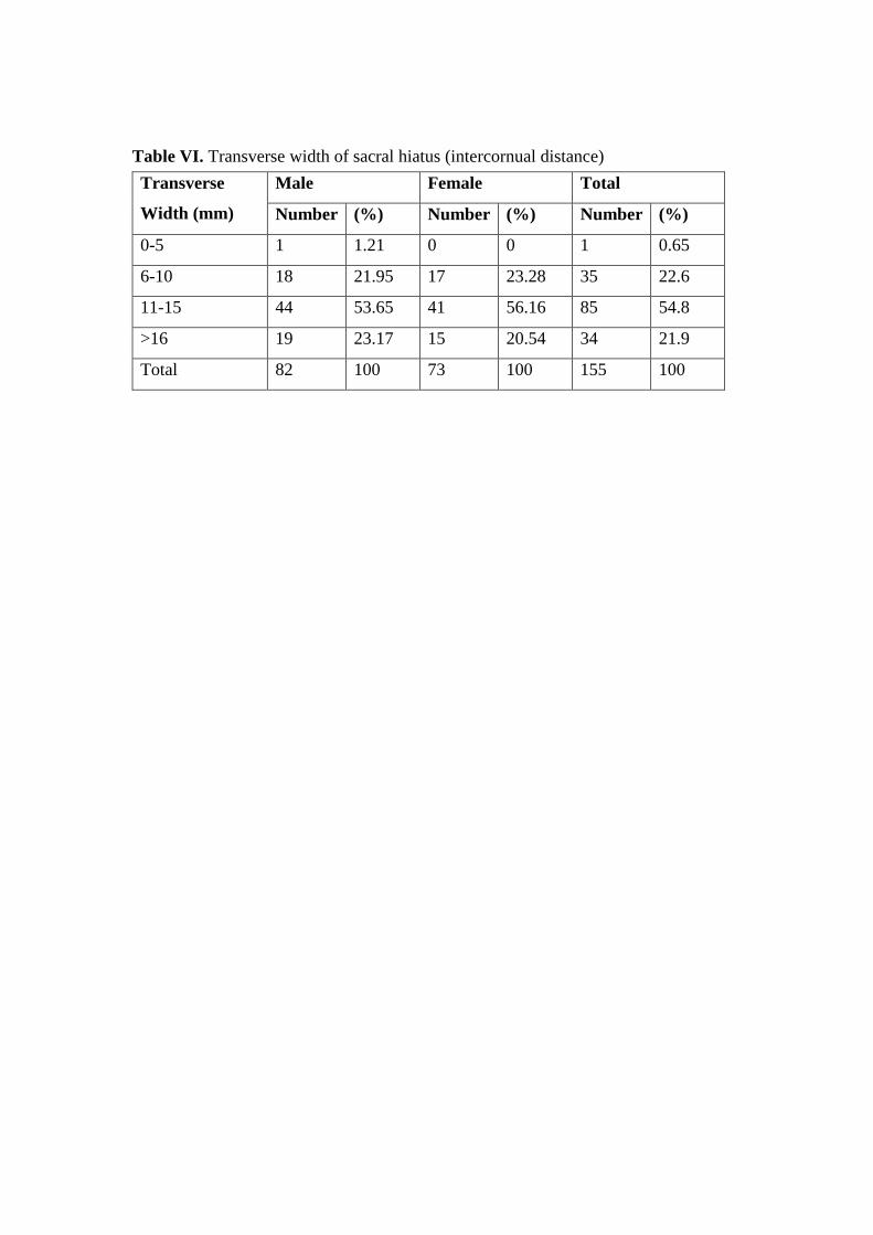
Table VI. Transverse width of sacral hiatus (intercornual distance)
Transverse
Width (mm)
Male Female Total
Number (%) Number (%) Number (%)
0-5 1 1.21 0 0 1 0.65
6-10 18 21.95 17 23.28 35 22.6
11-15 44 53.65 41 56.16 85 54.8
>16 19 23.17 15 20.54 34 21.9
Total 82 100 73 100 155 100

Table VII. Anteroposterior diameter (depth) of sacral hiatus at the level of apex
Anteroposterior
diameter (mm)
Male Female Total
Number (%) Number (%) Number (%)
0-3 4 4.87 11 15.06 15 9.7
4-6 48 58.53 38 52.05 86 55.6
7-9 27 32.92 23 31.50 50 32.2
>9 3 3.65 1 1.36 4 2.5
Total 82 100 73 100 155 100

Table VIII. Sexual Dimorphism
Parameters
(mm)
Sex Mean
(mm)
Median
(mm)
SD
(mm)
Range
(mm)
P value
(K-S
test)
Normality P value*
Height of sacral
hiatus
M 20.01 19.02 9.37 1.57-
58.91
0.2 Yes (t-
test)
0.035
F 17.8 17.14 7.44 5.66-
35.66
Transverse Width
of sacral hiatus
(intercornual
distance)
M 12.87 13.29 3.35 3.32-
20.09
0.2 Yes (t-
test)
0.109
F 11.79 11.8 2.79 5.43-
16.34
Anteroposterior
diameter (depth) of
sacral hiatus at the
level of its apex
M 5.62 5.61 1.75 1.38-
12.12
0.2 Yes (t-
test)
0.067
F 5.07 5.32 1.93 1.22-
8.98
*Differences among groups were considered statistically significant at P values of less
than 0.05.

Table IX. Comparison between different studies regarding shapes of sacral hiatus
Author Ethnicity/
Race
Inverted
“U”
Inverted
“V”
Irregular Dumbbell Bifid
Kumar [21] India 46.53% 29.7%
Nagar [27] India 27% 41.5% 14.1% 13.3% 1.5%
Patel [33] India 49.3% 20% 4%
Njihia [30] Kenya 16.7% 32.1% 19% 31%
Seema [38] India 42.95 % 27.51% 16.10% 13.41%
Suwanlikhid [44] Thailand 54.47% 19.57% 11.06% 2.13% 3.83%
Shewale N [42] India 40.69% 32.35% 9.31% 5.89% 0.98%
Phalgunan V [34] India 35% 35% 28%
Bhattacharya [6] India 65% 23% 12%
Ukoha [45] Nigeria 48.2% 34.9% 4.8% 4.8% 4.8%
Nasr A [28] Egypt 31.33% 38.66% 15.33% 12% 2.66%
Mayuri J [24] India 42.37% 27.11% 16.1% 12.71% 1.69%
Rajeev R [35] India 42.12 % 35.43% 12.99% 4.00% 5.51%
Osunwoke [31] Nigeria 24.1% 33.1% 13% 9.3% 5.6%
Kamal A [18] Bangladesh 38.00% 35.10% 15.20% 5.30% 0.60%
Nadeem G [26] Caucasians
Leaving in
Germany
56% 14% 16% 10% 2%
Malarvani T [23] Nepal 35% 32% 14% 3% 2%
Vasuki [46] India 36% 20% 33% 23% 3%
Laishram D [22] India 39.35% 16.77% 24.51% 7.091%
Saha D [36] India 70.09% 14.53% 12.82% 0.85% 1.71%
Bagheri H [5] Turkey 33.3% 19.45% 19.45% 6.9% 3.45%
Dhuria [13] India 35.22% 29.54% 14.77%
Bagoji IB [4] India 42,02% 26,08% 7,24% 12.31% 5.07%
Abera [2] Ethiopia 37.7% 41% 4.9% 11.5%) 3.3%

Table X. Comparison between different studies regarding the location of hiatal apex and base in relation to the level of sacral/ coccygeal
vertebra
Location of hiatal apex in relation to level of
sacral vertebra
Location of hiatal base in relation to
level of sacral/coccygeal vertebra
Author Ethnicity/ Race S2 S3 S4 S5 S4 S5 C1
Nagar [27] India 3.4% 37.3% 55.9% 3.4% 11.1% 72.6% 16.3%
Patel [33] India 0.66% 26.6% 53.3% 12.67% 10.67% 79.33%
Seema [38] India 4.02% 35.57% 56.36% 4.02 % 13.42% 70.46% 16.10%
Suwanlikhid N [44] Thailand 1.18% 16.2% 58.4% 15.01% 7.50% 71.14% 8.69%
Shewale N [42] India 4% 15% 66% 14.5% 2% 82% 16%
Phalgunan V [34] India 7.1% 46% 46%
Bhattacharya S [6] India 5% 72% 23%
Ukoha [45] Nigeria 2.04% 20.05% 69.9% 4.8% 2,4% 88% 7.2%
Nasr A [28] Egypt 1.33% 14.66% 54.00% 27.33% 12% 70% 18%
Mayuri J [24] India 4.23% 35.39% 56.77% 3.38%
Rajeev R [35] India 5.60% 17.71% 60.23% 16.53% 4.33% 75.19% 21.25%
Osunwoke E [31] Nigeria 5.6% 20.4% 66.6% 7.4% 11.1% 59.3% 29.6%
Kamal A [18] Bangladesh 4.70% 30.40% 60.20% 4.70% 0.60% 91.20% 8.20%
Nadeem G [26] Caucasians living in Germany 2% 62% 34% 2% 62% 24% 14%
Malarvani T [23] Nepal 3% 42% 39% 13% 31% 54% 1%
Vasuki [46] India 3% 43% 48% 5% 16% 64% 20%
Bagheri H [5] Turkey 1.15% 10.34% 71.11% 11.49% 2.3% 82.76% 8.05%

Dhuria [13] India 5.68% 21.59% 57.95% 9.09% 92.04% 7.95%
Bagoji IB [4] India 2.89% 26.81% 58.69% 6.52% 18.11% 70.28% 6.52%
Abera [2] Ethiopia 3.3% 26.2% 60.7 9.8% 78.7% 21.3%
Table XI. Morphometric sacral hiatus measurements from various investigators worldwide
Anteroposterior diameter (depth) of
SH at the level of apex
Height of sacral hiatus Transverse width (intercornual
distance) of SH at the level of base
Author Ethnicit
y/ Race
0-3
mm
4-6
mm
7-9
mm
>9
mm
0 to 10
mm
11 to
20
mm
21 to
30
mm
31 to
40
mm
41 to
50
mm
>51
mm
0 to 5
mm
6 to 10
mm
11 to
15
mm
>15
mm
Nagar [27] India 15.6% 64.2% 19.8% 0.4% 10.3% 35% 30.8% 17.1% 4.9% 1.9%
Senoglou [40] Turkey 4.4% 35.6% 36.7% 20% 3.3%
Seema [38] India 5.36% 71.81% 22.14% 0.67% 11.4% 34.8% 29.5% 16.7% 4.6% 2.6% 9.39% 30.20% 51.67% 8.72%
Shewale [42] India 7.5% 76% 16% 0.67% 5.5% 40% 37% 10.5% 5% 2% 1% 22% 56% 21%
Phalgunan [34] India 57% 39% 3.5%
Nasr [28] Egypt 18.66% 60.66% 20.66% 8% 36.6% 30% 16.6% 5.3% 3.3% 6% 33.33% 53.33% 7.33%
Rajeev [35] India 10.62% 79.13% 9.84% 0.4% 11.02% 31.10% 39.76% 12.16% 5.51% 15.35% 38.20% 6.92% 10.23%

Mayuri [24] India 5.08% 71.18% 22.03% 1.69% 11.01% 34.74% 29.66% 16.94% 4.23% 3.38%
Chhabra [11] 50% 40% 10% 3.33% 30% 33.33% 30% 3.33% 10% 63.33% 26.67%
Nadeem [26] India 5% 60% 30% 5%
Mishra [25] India 23.86% 63.63% 12.51% 9.09% 44.31% 30.70% 14.77% 1.13% 1.14% 20.46% 57.95% 20.45%
Parashuram[32] India 29.9% 68.1% 2.1%
Vasuki [46] India 8% 42% 33% 6% 6% 9% 48% 43%
Dhuria [13] India 5.68% 47.72% 28.4% 10.22% 10.22% 15.9% 37.5% 25% 2.27% 5.68% 4.54% 30.68% 64.76%
Bagoji [4] India 4.34% 54.34% 25.36% 10.14%
Abera [2] Ethiopia 8.2% 60.07% 31.1% 4.9% 45.9% 34.4% 13.1% 1.6% 19.7% 63.9% 14.8%
Figure 1. Metric parameters evaluated: (1) Height of sacral hiatus, (2) transverse width of the sacral hiatus at the base (intercornual distance), (3)
distance from sacral apex to the upper border of S2 foramina (4) Distance from the base of sacral hiatus to the upper border of S2 foramina; (4 =
1 + 3)
Figure 2. Anteroposterior diameter (depth) of the sacral hiatus at the level of its apex.
Figure 3. Observed sacral hiatus shapes: a. inverted U, b. inverted V, c. irregular, d. dumbbell, e. bifid, f. M pattern, g. elongated, h. complete
dorsal wall agenesis and i. absence of sacral hiatus.
Figure 4. Level of sacral hiatus apex at S4.
Figure 5. Level of sacral hiatus base at S5.


Related Documents