Dissertation on MORPHOLOGICAL ANALYSIS AND MORPHOMETRIC STUDY OF THE FORAMEN MAGNUM Submitted in partial fulfillment for M.D. DEGREE EXAMINATION BRANCH- XXIII, ANATOMY Upgraded Institute of Anatomy Madras Medical College & Rajiv Gandhi Government General Hospital, Chennai- 600 003 THE TAMILNADU Dr.M.G.R. MEDICAL UNIVERSITY CHENNAI – 600 032 TAMILNADU APRIL 2015

Welcome message from author
This document is posted to help you gain knowledge. Please leave a comment to let me know what you think about it! Share it to your friends and learn new things together.
Transcript
Dissertation on
MORPHOLOGICAL ANALYSIS AND MORPHOMETRIC
STUDY OF THE FORAMEN MAGNUM
Submitted in partial fulfillment for
M.D. DEGREE EXAMINATION
BRANCH- XXIII, ANATOMY
Upgraded Institute of Anatomy
Madras Medical College & Rajiv Gandhi Government General
Hospital,
Chennai- 600 003
THE TAMILNADU Dr.M.G.R. MEDICAL UNIVERSITY
CHENNAI – 600 032
TAMILNADU
APRIL 2015
CERTIFICATE
This is to certify that this dissertation entitled
“MORPHOLOGICAL ANALYSIS AND MORPHOMETRIC STUDY
OF THE FORAMEN MAGNUM”
is a bonafide record of the research work done by Dr.M.ANURADHA,
Post graduate in the Institute of Anatomy, Madras Medical College and
Research Institute, Rajiv Gandhi Government General Hospital, Chennai-03,
in partial fulfillment of the regulations laid down by The Tamil Nadu
Dr.M.G.R. Medical University for the award of M.D. Degree Branch XXIII-
Anatomy, under my guidance and supervision during the academic year from
2012-2015.
Dean
Madras Medical College and
Rajiv Gandhi Government General
Hospital
Chennai-600 003
Dr.Sudha Seshayyan, M.B.B.S.,M.S.,
Director and Professor,
Institute of Anatomy,
Madras Medical College,
Chennai 600003.


ACKNOWLEDGEMENT
I wish to express exquisite thankfulness and gratitude to my most
respected teacher and guide Dr. Mrs. SudhaSeshayyan, M.S., Director and
Professor, Institute ofAnatomy, Madras Medical College, Chennai – 3, for
their invaluable guidance, persistent support and quest for perfection which
has made this dissertation take its present shape.
I’m thankful to Dr.R.Vimala, M.D., Dean, Madras Medical College,
Chennai – 3 for permitting me to avail the facilities in this college for
performing this study.
My heartfelt thanks to Dr.B.Chezhian, Dr.V.Lokanayaki, Associate
Professors Dr.S.Lakshmi, Dr.T.Anitha, Dr.P.Kanagavalli, Dr.J.Sreevidya,
Dr.Ilamathi Bose and Dr.S.Arrchana Assistant Professors, Institute of
Anatomy, Madras Medical College, Chennai – 3 for their valuable suggestions
and encouragement throughout the study.
I sincerely thank Dr.Deivigam, Mch., Director, Institute Of
Neurosurgery, Rajiv Gandhi Govt.General Hospital, Chennai – 3 who was
instrumental in the selection of the topic for my dissertation and continued to
extend his valuable support throughout my study. I also thank Dr.Atul Goel,
Mch., Professor, Department of Neurosurgery, KEM and Seth G.S. Medical
College, Mumbai who helped me to complete this study with his valuable
guidance.
My gratefulness to Dr.Vanitha, M.D., Director, Barnard Institute of
Radiology, Rajiv Gandhi Govt.General Hospital,Chennai – 3 and Dr.Babu
Peter, M.D., Associate Professor, for their help in the radiological study.
I also thank Dr.Evangeline Mary, Post graduate in Community
Medicine, Dr.Subisen, Post graduate in Neurosurgery and Dr.Arun, Post
graduate in Radiology for their help in the completion of this study.
I earnestly thank my seniors Dr.P.Radhakrishnan, Dr.K.Arumugam,
Assistant Professor, Thirunelveli Medical College, Thirunelveli and my
helpful juniors Dr.Keerthi, Dr.Prefulla, Dr.Ganga and other members of
faculty who have been supportive and encouraging throughout the study.
I extend my heartfelt thanks to my colleagues Dr.S.Elizabeth
Priyadarisini, Dr. B.J.Bhuvaneswari and Dr.E.Srividhya for their constant
encouragement and unstinted co-operation.
I’m especially thankful to Mr.Mathews and Mr.Senthilkumar,
technicians, who extended great support for this study and all other staff
members including Mr.Jagadeesan, Mr.Manish and Mr.Devaraj for
helping me to carry out the study.

I’m grateful to my parents, my sister and my brother who have
helped making this study a reality.
Above all, I thank the ALMIGHTY GOD who has showered His
choicest blessings on me and guided me in every step of the dissertation.

CONTENTS
SL. NO. TITLE PAGE NO.
1. INTRODUCTION 1
2. AIM OF THE STUDY 5
3. REVIEW OF LITERATURE 8
4. EMBRYOLOGY 35
5. MATERIALS AND METHODS 38
6. OBSERVATION 43
7. DISCUSSION 68
8. CONCLUSION 109
9. BIBLIOGRAPHY
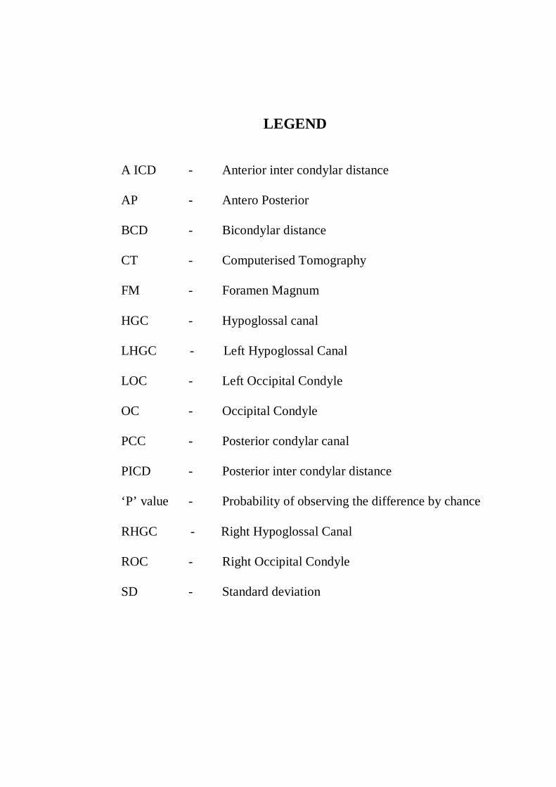
LEGEND
A ICD - Anterior inter condylar distance
AP - Antero Posterior
BCD - Bicondylar distance
CT - Computerised Tomography
FM - Foramen Magnum
HGC - Hypoglossal canal
LHGC - Left Hypoglossal Canal
LOC - Left Occipital Condyle
OC - Occipital Condyle
PCC - Posterior condylar canal
PICD - Posterior inter condylar distance
‘P’ value - Probability of observing the difference by chance
RHGC - Right Hypoglossal Canal
ROC - Right Occipital Condyle
SD - Standard deviation
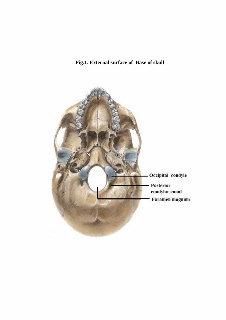
Fig.1. External surface of Base of skull
MORPHOLOGICAL ANALYSIS AND MORPHOMETRIC
STUDY OF THE FORAMEN MAGNUM
ABSTRACT
The foramen magnum is the oval shape opening situated at the base of the
skull. The transcondylar approach is being increasingly used to access lesions of
the Craniovertebral junction. Understanding the anatomy of the foramen
magnum is important for skull base surgery.
The present study was aimed at analysing the foramen magnum
morphologically and morphometrically.100 adult human dry skullsat the
Institute of Anatomy, Madras Medical College and twenty cranial CT scans
obtained from the archives of Barnard Institute of Radiology, Rajiv Gandhi
Government General Hospital, Chennai were used for the study.
In the present study it was found that the foramen magnum was oval in
40% of the skulls studied. The mean AP diameter of the foramen magnum in
dry skulls and cranial computerized tomographicscans were measured as
35.12±2.65mm and 35.03±0.95mm respectively. The mean transverse diameter
of the foramen magnum in dry skulls and cranial CT were measured as
29.03±2.15mm and 28.79±1.17mm respectively. The mean maximum length of
the right and left occipital condyle were measured as 23.85±2.12mm and
23.77±2.29mm respectively. The maximum and minimum width of the right
occipital condyle were measured as 13.2 ±1.36mm and 6.86±1.34mm
respectively. The maximum and minimum width of the left occipital condyle
were measured as 13.44±1.41mm and 7.04±1.26mm respectively. The mean
length of right and left occipital condyle were measured as 23.11±0.73mm and
23.20±0.74mm respectively in cranial CT.The mean width of right and left
occipital condyle were measured as 12.92±0.65mm to 12.88±0.69mm
respectively in cranial CT. The bicondylar distance, anterior intercondylar
distance and the posterior intercondylar distance were measured as
47.23±3.10mm, 20.81±2.40mm and 41.97±1.67mm respectively. The posterior
condylar canal was present in 40 skulls on right side and 49 skulls on left side.
The Hypoglossal canal septum was present in 24%. The mean distance between
intracranial edge of right hypoglossal canal and anterior margin of right
occipital condyle was measured as 11.02±1.29mm and from left hypoglossal
canal and anterior margin of left occipital condyle was measured
as10.93±1.3mm.The mean distance between intracranial edge of right
hypoglossal canal and posterior margin of right occipital condyle was measured
as 12.26±0.59mm and from left hypoglossal canal and posterior margin of left
occipital condyle was measured as12.25±0.59mm.
The data obtained will be useful for neurosurgeons in analyzing the
anatomy of Craniovertebral junction for preoperative planning and management
of skull base surgery. The findings will also be enlightening for Radiologists,
Orthopedicians, Anthropologists, Morphologists and Clinical Anatomists.
KEY WORDS: Foramen magnum, Occipital condyle, Hypoglossal canal,
Transcondylar approach.
1
INTRODUCTION
The foramen magnum is the largest bony foramen in the central basal
region of the occipital bone. Occipital bone with the foramen magnum and
the occipital condyles form the cranial aspect of the craniovertebral junction
(Fig.1). Bony malformations at the craniovertebral junction may lead to
symptoms secondary to compression of vital structures or may manifest as
instability due to malalignment of bones1. Therefore it is of great
importance to study the dimensions of foramen magnum and occipital
condyles.
The posterior part of the cranial base is largely formed by the
occipital bone. The occipital bone is trapezoid, concave internally and
invests the foramen magnum. It consists of four parts namely the basilar or
basioccipital part, squamous part and two lateral or condylar parts. The
basilar part is quadrilateral in shape and lies in front of foramen magnum.
The squamous part is an expanded plate and lies posterosuperior to the
foramen magnum and the two lateral or condylar or exoccipital parts lie on
each side of the foramen magnum.55,14
The occipital bone provides attachment to the muscles of neck and
back. It articulates with the first cervical vertebra at atlanto-occipital joints.
2
Foramen magnum is unpaired, oval and oriented obliquely. The
anteroposterior diameter of the foramen magnum is more than the transverse
diameter. The anterior margin of the foramen magnum is encroached on
each side by the occipital condyles which project down to articulate with the
superior articular facets of the atlas. Anterior and posterior atlanto occipital
membranes are attached to the corresponding margins of the foramen
magnum.
The structures adjacent to the foramen magnum are the bilateral
occipital condyles, jugular foramina, mastoid notches, squamous parts of the
occipital bone, hypoglossal canals (anterior condylar canal) and posterior
condylar canals. The posterior cranial fossa communicates with the
vertebral canal through the foramen magnum.
The following structures traverse through the foramen magnum:
Anteriorly, the upper surface of the basilar part of the foramen
magnum gives attachment to apical ligament of dens and membrana
tectoria which is the upward prolongation of the posterior
longitudinal ligament.
Its wider posterior part transmits the lower end of medulla oblongata
which continues down as the spinal cord.
Cranial meninges
Vertebral arteries
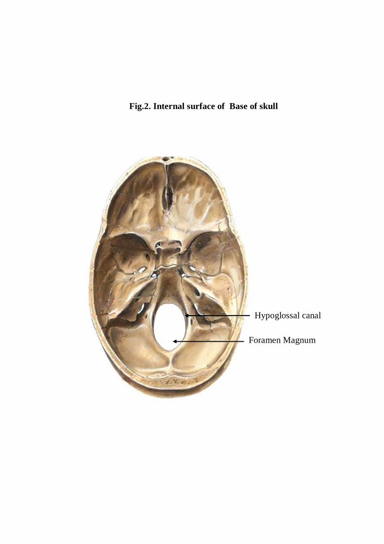
Fig.2. Internal surface of Base of skull
Foramen Magnum
Hypoglossal canal
3
Anterior and posterior spinal arteries
Spinal accessory nerve
Upper three cervical meningeal nerves
OCCIPITAL CONDYLE
The occipital condyles are oval or reniform in shape, with their long
axes converging anteromedially. On the medial aspect of each condyle, a
tubercle for the alar or check ligament is present.25
The anterior one third of each condyle extends forwards on to the
basilar part of the bone. The site of union between the basilar and condylar
parts is marked by the anterior condylar or hypoglossal canal (Fig.2). The
hypoglossal canal is directed laterally and slightly forwards, and transmits
the hypoglossal nerve, a meningeal branch of the ascending pharyngeal
artery and an emissary vein. Behind each condyle there is a condylar fossa.
In some cases there is a posterior condylar canal which transmits the
emissary vein.
The third occipital condyle is an occasional tubercle which projects
from the anterior border of the foramen magnum to articulate with the dens
of the axis.25
4
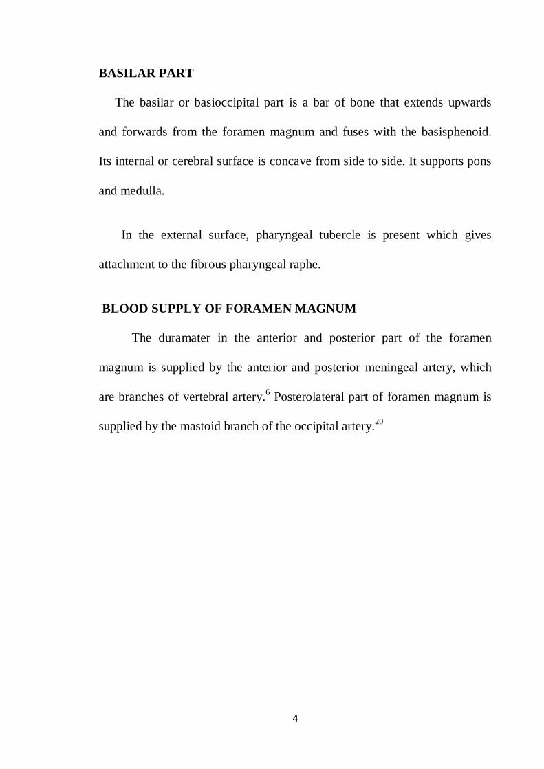
BASILAR PART
The basilar or basioccipital part is a bar of bone that extends upwards
and forwards from the foramen magnum and fuses with the basisphenoid.
Its internal or cerebral surface is concave from side to side. It supports pons
and medulla.
In the external surface, pharyngeal tubercle is present which gives
attachment to the fibrous pharyngeal raphe.
BLOOD SUPPLY OF FORAMEN MAGNUM
The duramater in the anterior and posterior part of the foramen
magnum is supplied by the anterior and posterior meningeal artery, which
are branches of vertebral artery.6 Posterolateral part of foramen magnum is
supplied by the mastoid branch of the occipital artery.20

Aim of the study
5
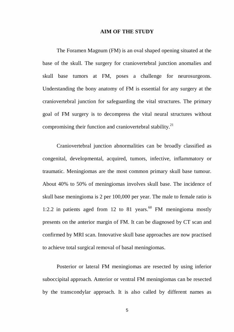
AIM OF THE STUDY
The Foramen Magnum (FM) is an oval shaped opening situated at the
base of the skull. The surgery for craniovertebral junction anomalies and
skull base tumors at FM, poses a challenge for neurosurgeons.
Understanding the bony anatomy of FM is essential for any surgery at the
craniovertebral junction for safeguarding the vital structures. The primary
goal of FM surgery is to decompress the vital neural structures without
compromising their function and craniovertebral stability.21
Craniovertebral junction abnormalities can be broadly classified as
congenital, developmental, acquired, tumors, infective, inflammatory or
traumatic. Meningiomas are the most common primary skull base tumour.
About 40% to 50% of meningiomas involves skull base. The incidence of
skull base meningioma is 2 per 100,000 per year. The male to female ratio is
1:2.2 in patients aged from 12 to 81 years.60 FM meningioma mostly
presents on the anterior margin of FM. It can be diagnosed by CT scan and
confirmed by MRI scan. Innovative skull base approaches are now practised
to achieve total surgical removal of basal meningiomas.
Posterior or lateral FM meningiomas are resected by using inferior
suboccipital approach. Anterior or ventral FM meningiomas can be resected
by the transcondylar approach. It is also called by different names as
6
far-lateral, posterolateral or extreme lateral approach.60 In the far-lateral
approach craniovertebral stability is not affected due to minimal removal of
occipital condyles. It also provides an adequate exposure to ventral
brainstem. Many varieties of lateral approaches have been reported
including transfacetal approach, partial or complete transcondylar approach,
extreme lateral transjugular approach and transtubercular approach.60,28
Hence, neurosurgeons performing posterior or lateral approaches to
Craniovertebral junction surgery should be familiar with the normal
anatomy and possible variations of the foramen magnum, occipital condyle
and hypoglossal canal to reduce the surgical morbidity.
The aim of the present study is to analyse the FM and occipital
condyles morphologically and morphometrically. Hopefully the data will
be beneficial to neurosurgeons, radiologists and orthopaedicians for
preoperative planning and management of Craniovertebral junction
surgeries.
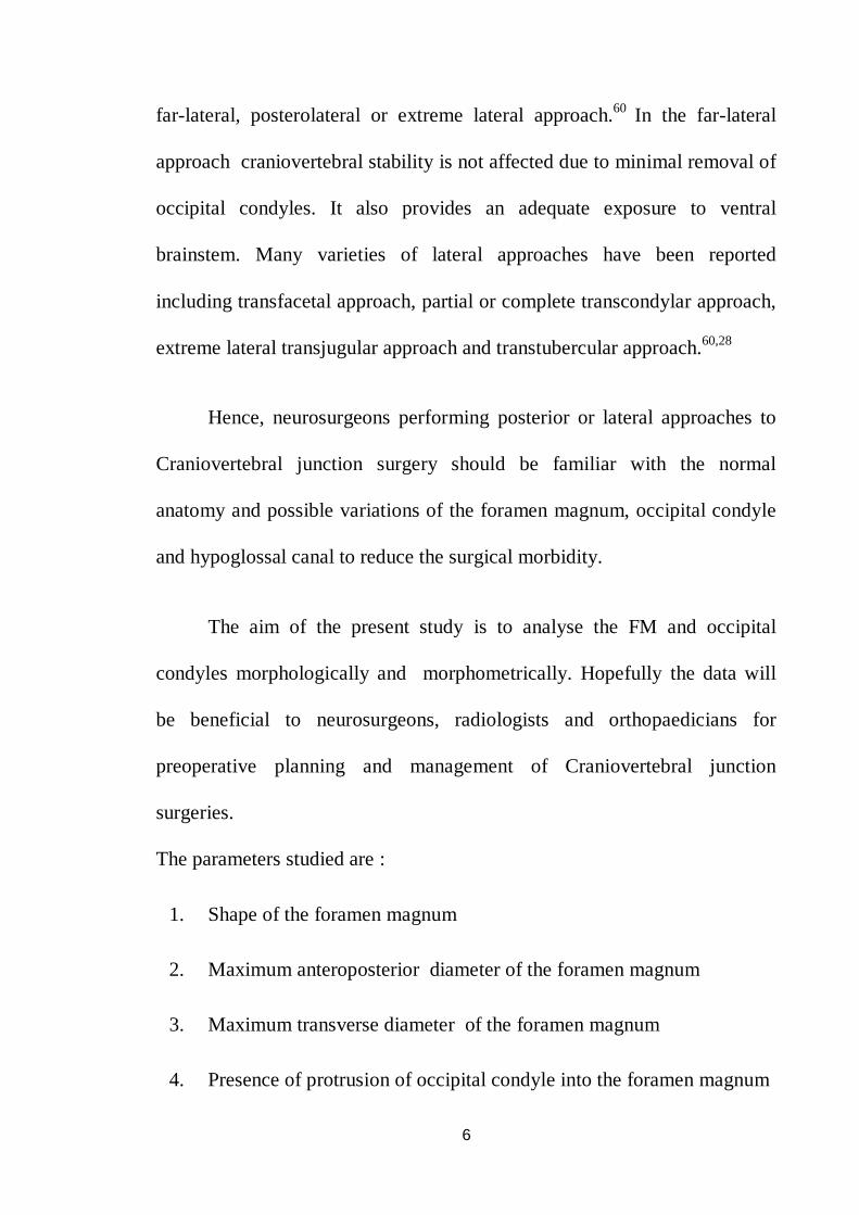
The parameters studied are :
1. Shape of the foramen magnum
2. Maximum anteroposterior diameter of the foramen magnum
3. Maximum transverse diameter of the foramen magnum
4. Presence of protrusion of occipital condyle into the foramen magnum
7
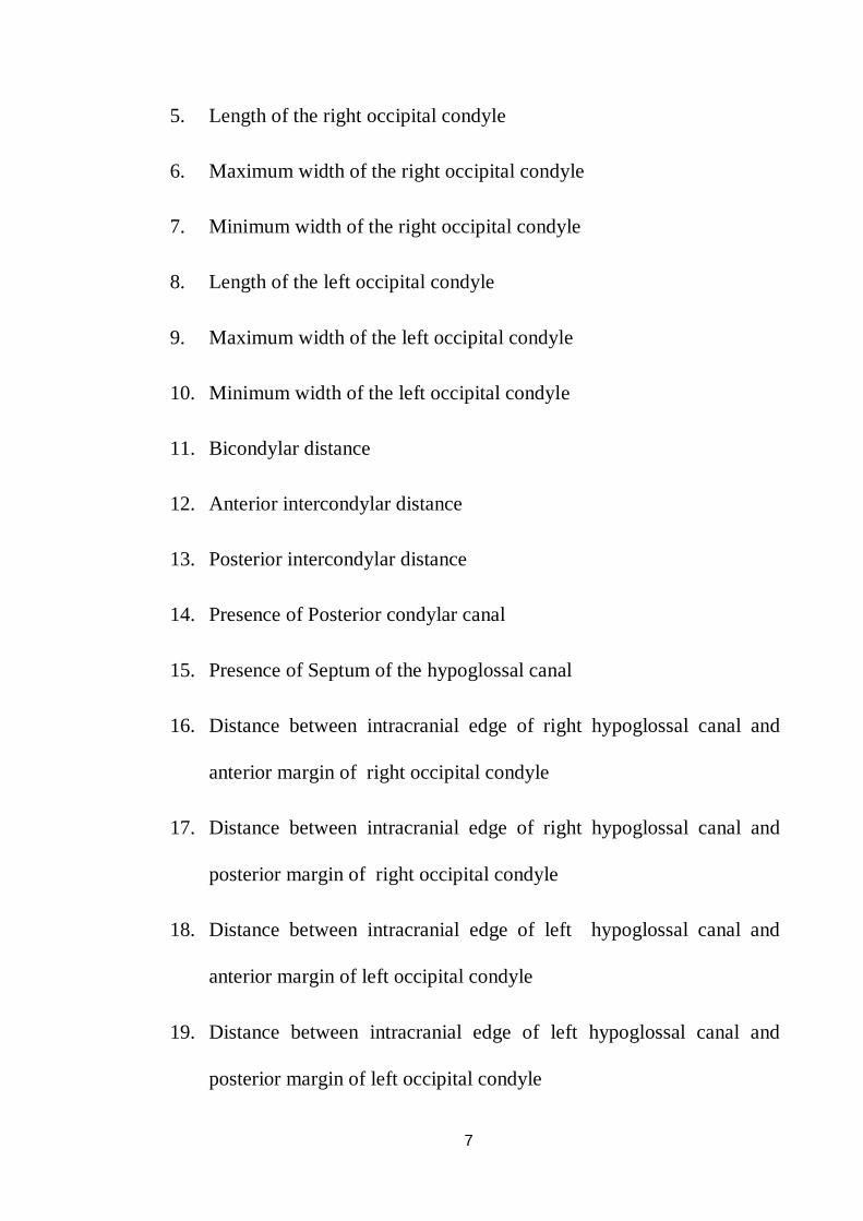
5. Length of the right occipital condyle
6. Maximum width of the right occipital condyle
7. Minimum width of the right occipital condyle
8. Length of the left occipital condyle
9. Maximum width of the left occipital condyle
10. Minimum width of the left occipital condyle
11. Bicondylar distance
12. Anterior intercondylar distance
13. Posterior intercondylar distance
14. Presence of Posterior condylar canal
15. Presence of Septum of the hypoglossal canal
16. Distance between intracranial edge of right hypoglossal canal and
anterior margin of right occipital condyle
17. Distance between intracranial edge of right hypoglossal canal and
posterior margin of right occipital condyle
18. Distance between intracranial edge of left hypoglossal canal and
anterior margin of left occipital condyle
19. Distance between intracranial edge of left hypoglossal canal and
posterior margin of left occipital condyle

Review of literature
8
REVIEW OF LITERATURE
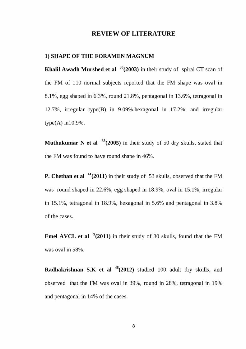
1) SHAPE OF THE FORAMEN MAGNUM
Khalil Awadh Murshed et al 30(2003) in their study of spiral CT scan of
the FM of 110 normal subjects reported that the FM shape was oval in
8.1%, egg shaped in 6.3%, round 21.8%, pentagonal in 13.6%, tetragonal in
12.7%, irregular type(B) in 9.09%.hexagonal in 17.2%, and irregular
type(A) in10.9%.
Muthukumar N et al 35(2005) in their study of 50 dry skulls, stated that
the FM was found to have round shape in 46%.
P. Chethan et al 41(2011) in their study of 53 skulls, observed that the FM
was round shaped in 22.6%, egg shaped in 18.9%, oval in 15.1%, irregular
in 15.1%, tetragonal in 18.9%, hexagonal in 5.6% and pentagonal in 3.8%
of the cases.
Emel AVCL et al 9(2011) in their study of 30 skulls, found that the FM
was oval in 58%.
Radhakrishnan S.K et al 46(2012) studied 100 adult dry skulls, and
observed that the FM was oval in 39%, round in 28%, tetragonal in 19%
and pentagonal in 14% of the cases.
9
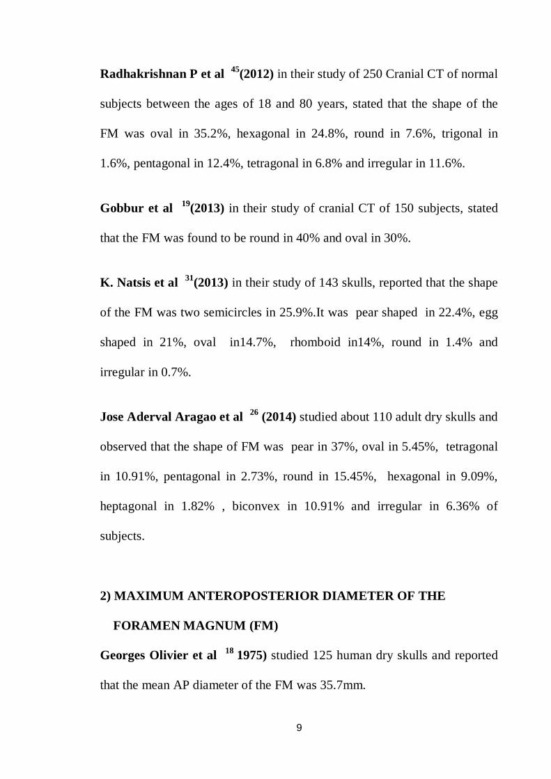
Radhakrishnan P et al 45(2012) in their study of 250 Cranial CT of normal
subjects between the ages of 18 and 80 years, stated that the shape of the
FM was oval in 35.2%, hexagonal in 24.8%, round in 7.6%, trigonal in
1.6%, pentagonal in 12.4%, tetragonal in 6.8% and irregular in 11.6%.
Gobbur et al 19(2013) in their study of cranial CT of 150 subjects, stated
that the FM was found to be round in 40% and oval in 30%.
K. Natsis et al 31(2013) in their study of 143 skulls, reported that the shape
of the FM was two semicircles in 25.9%.It was pear shaped in 22.4%, egg
shaped in 21%, oval in14.7%, rhomboid in14%, round in 1.4% and
irregular in 0.7%.
Jose Aderval Aragao et al 26 (2014) studied about 110 adult dry skulls and
observed that the shape of FM was pear in 37%, oval in 5.45%, tetragonal
in 10.91%, pentagonal in 2.73%, round in 15.45%, hexagonal in 9.09%,
heptagonal in 1.82% , biconvex in 10.91% and irregular in 6.36% of
subjects.
2) MAXIMUM ANTEROPOSTERIOR DIAMETER OF THE
FORAMEN MAGNUM (FM)
Georges Olivier et al 18 1975) studied 125 human dry skulls and reported
that the mean AP diameter of the FM was 35.7mm.
10
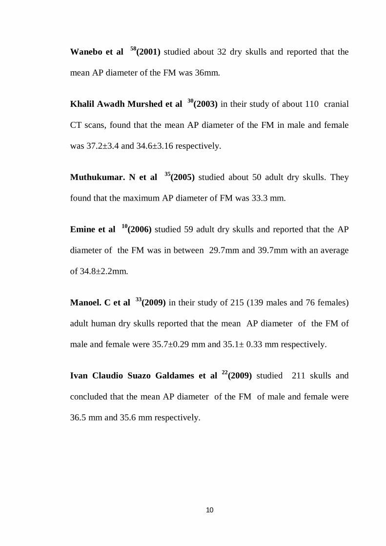
Wanebo et al 58(2001) studied about 32 dry skulls and reported that the
mean AP diameter of the FM was 36mm.
Khalil Awadh Murshed et al 30(2003) in their study of about 110 cranial
CT scans, found that the mean AP diameter of the FM in male and female
was 37.2±3.4 and 34.6±3.16 respectively.
Muthukumar. N et al 35(2005) studied about 50 adult dry skulls. They
found that the maximum AP diameter of FM was 33.3 mm.
Emine et al 10(2006) studied 59 adult dry skulls and reported that the AP
diameter of the FM was in between 29.7mm and 39.7mm with an average
of 34.8±2.2mm.
Manoel. C et al 33(2009) in their study of 215 (139 males and 76 females)
adult human dry skulls reported that the mean AP diameter of the FM of
male and female were 35.7±0.29 mm and 35.1± 0.33 mm respectively.
Ivan Claudio Suazo Galdames et al 22(2009) studied 211 skulls and
concluded that the mean AP diameter of the FM of male and female were
36.5 mm and 35.6 mm respectively.
11
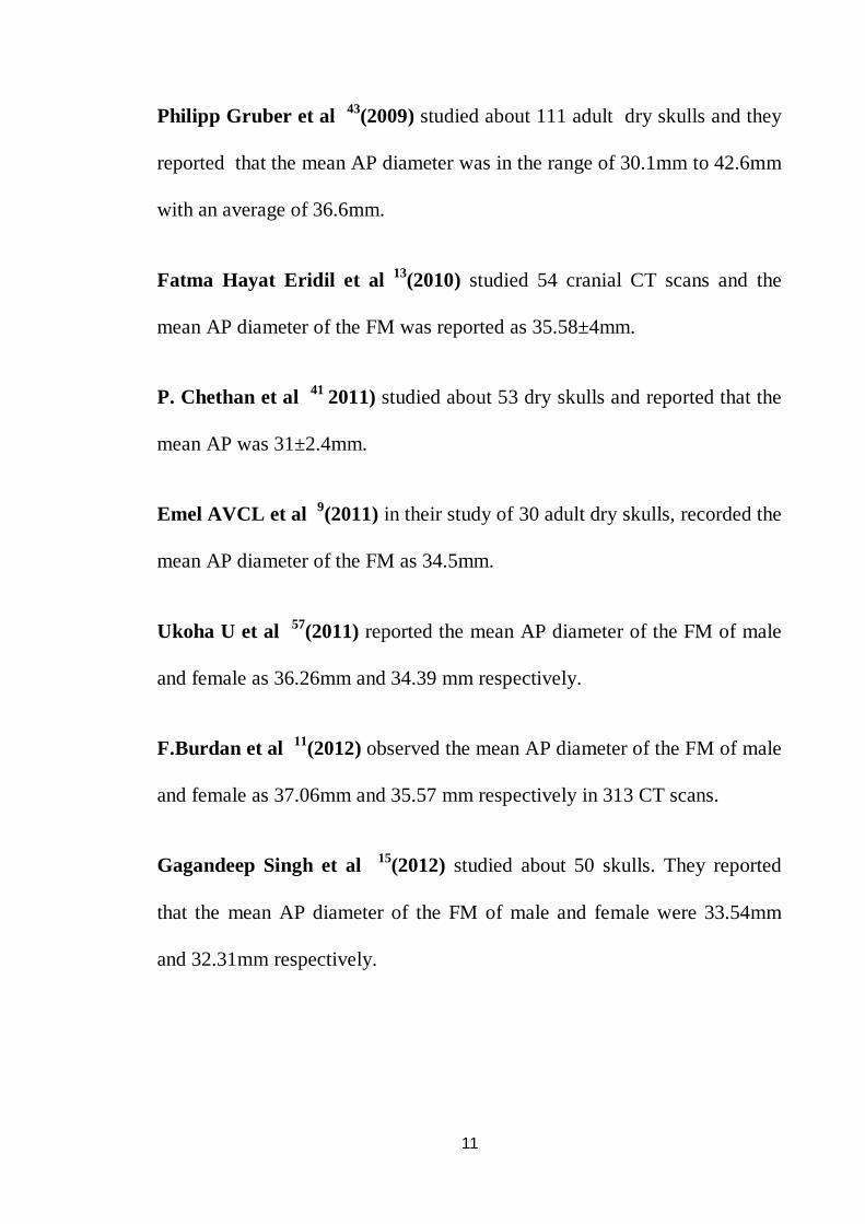
Philipp Gruber et al 43(2009) studied about 111 adult dry skulls and they
reported that the mean AP diameter was in the range of 30.1mm to 42.6mm
with an average of 36.6mm.
Fatma Hayat Eridil et al 13(2010) studied 54 cranial CT scans and the
mean AP diameter of the FM was reported as 35.58±4mm.
P. Chethan et al 41 2011) studied about 53 dry skulls and reported that the
mean AP was 31±2.4mm.
Emel AVCL et al 9(2011) in their study of 30 adult dry skulls, recorded the
mean AP diameter of the FM as 34.5mm.
Ukoha U et al 57(2011) reported the mean AP diameter of the FM of male
and female as 36.26mm and 34.39 mm respectively.
F.Burdan et al 11(2012) observed the mean AP diameter of the FM of male
and female as 37.06mm and 35.57 mm respectively in 313 CT scans.
Gagandeep Singh et al 15(2012) studied about 50 skulls. They reported
that the mean AP diameter of the FM of male and female were 33.54mm
and 32.31mm respectively.
12
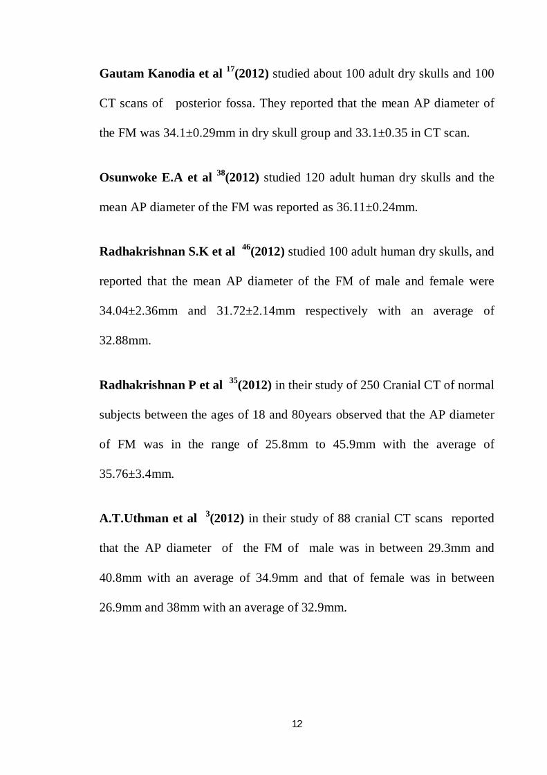
Gautam Kanodia et al 17(2012) studied about 100 adult dry skulls and 100
CT scans of posterior fossa. They reported that the mean AP diameter of
the FM was 34.1±0.29mm in dry skull group and 33.1±0.35 in CT scan.
Osunwoke E.A et al 38(2012) studied 120 adult human dry skulls and the
mean AP diameter of the FM was reported as 36.11±0.24mm.
Radhakrishnan S.K et al 46(2012) studied 100 adult human dry skulls, and
reported that the mean AP diameter of the FM of male and female were
34.04±2.36mm and 31.72±2.14mm respectively with an average of
32.88mm.
Radhakrishnan P et al 35(2012) in their study of 250 Cranial CT of normal
subjects between the ages of 18 and 80years observed that the AP diameter
of FM was in the range of 25.8mm to 45.9mm with the average of
35.76±3.4mm.
A.T.Uthman et al 3(2012) in their study of 88 cranial CT scans reported
that the AP diameter of the FM of male was in between 29.3mm and
40.8mm with an average of 34.9mm and that of female was in between
26.9mm and 38mm with an average of 32.9mm.
13
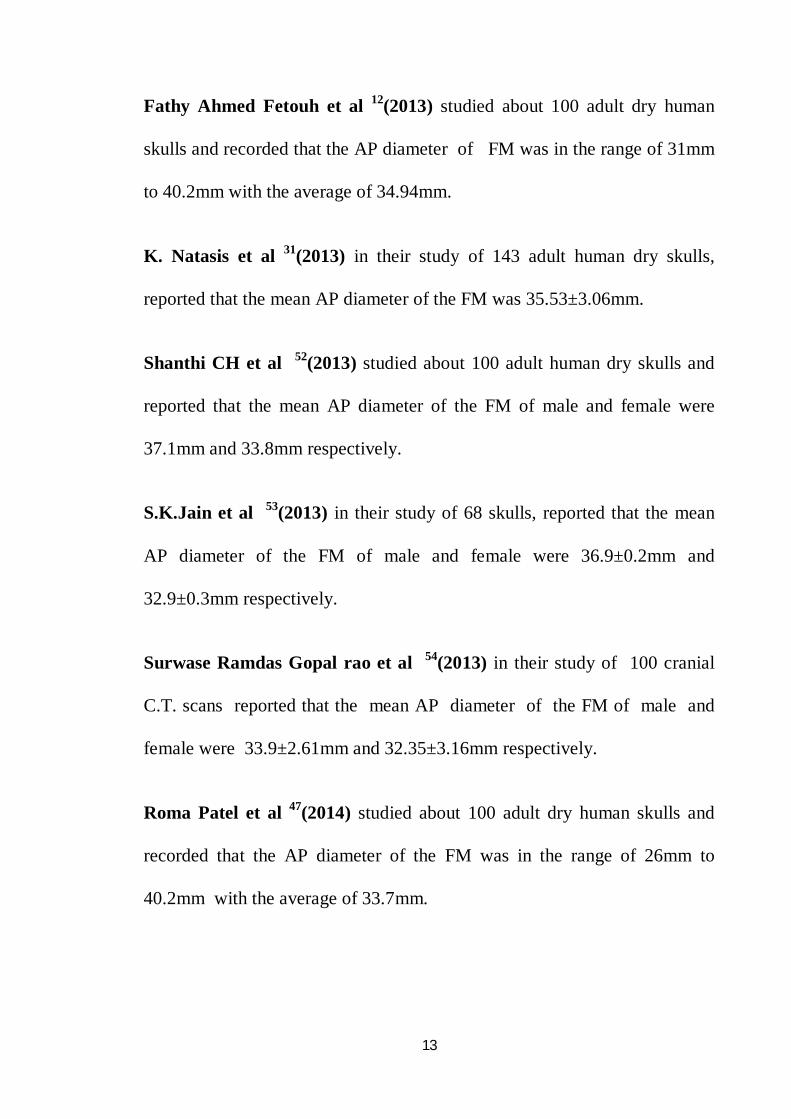
Fathy Ahmed Fetouh et al 12(2013) studied about 100 adult dry human
skulls and recorded that the AP diameter of FM was in the range of 31mm
to 40.2mm with the average of 34.94mm.
K. Natasis et al 31(2013) in their study of 143 adult human dry skulls,
reported that the mean AP diameter of the FM was 35.53±3.06mm.
Shanthi CH et al 52(2013) studied about 100 adult human dry skulls and
reported that the mean AP diameter of the FM of male and female were
37.1mm and 33.8mm respectively.
S.K.Jain et al 53(2013) in their study of 68 skulls, reported that the mean
AP diameter of the FM of male and female were 36.9±0.2mm and
32.9±0.3mm respectively.
Surwase Ramdas Gopal rao et al 54(2013) in their study of 100 cranial
C.T. scans reported that the mean AP diameter of the FM of male and
female were 33.9±2.61mm and 32.35±3.16mm respectively.
Roma Patel et al 47(2014) studied about 100 adult dry human skulls and
recorded that the AP diameter of the FM was in the range of 26mm to
40.2mm with the average of 33.7mm.
14
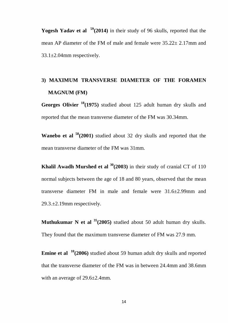
Yogesh Yadav et al 59(2014) in their study of 96 skulls, reported that the
mean AP diameter of the FM of male and female were 35.22± 2.17mm and
33.1±2.04mm respectively.
3) MAXIMUM TRANSVERSE DIAMETER OF THE FORAMEN
MAGNUM (FM)
Georges Olivier 18(1975) studied about 125 adult human dry skulls and
reported that the mean transverse diameter of the FM was 30.34mm.
Wanebo et al 58(2001) studied about 32 dry skulls and reported that the
mean transverse diameter of the FM was 31mm.
Khalil Awadh Murshed et al 30(2003) in their study of cranial CT of 110
normal subjects between the age of 18 and 80 years, observed that the mean
transverse diameter FM in male and female were 31.6±2.99mm and
29.3.±2.19mm respectively.
Muthukumar N et al 35(2005) studied about 50 adult human dry skulls.
They found that the maximum transverse diameter of FM was 27.9 mm.
Emine et al 10(2006) studied about 59 human adult dry skulls and reported
that the transverse diameter of the FM was in between 24.4mm and 38.6mm
with an average of 29.6±2.4mm.
15
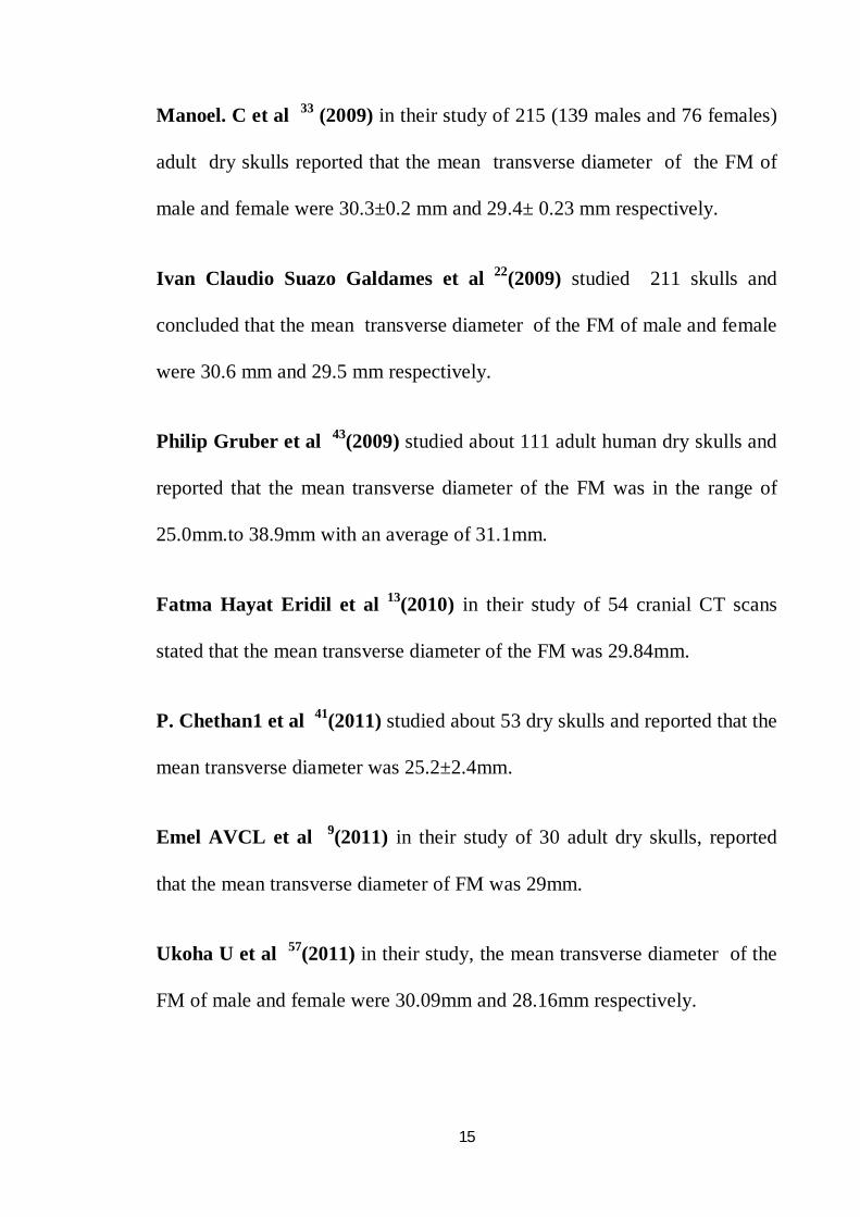
Manoel. C et al 33 (2009) in their study of 215 (139 males and 76 females)
adult dry skulls reported that the mean transverse diameter of the FM of
male and female were 30.3±0.2 mm and 29.4± 0.23 mm respectively.
Ivan Claudio Suazo Galdames et al 22(2009) studied 211 skulls and
concluded that the mean transverse diameter of the FM of male and female
were 30.6 mm and 29.5 mm respectively.
Philip Gruber et al 43(2009) studied about 111 adult human dry skulls and
reported that the mean transverse diameter of the FM was in the range of
25.0mm.to 38.9mm with an average of 31.1mm.
Fatma Hayat Eridil et al 13(2010) in their study of 54 cranial CT scans
stated that the mean transverse diameter of the FM was 29.84mm.
P. Chethan1 et al 41(2011) studied about 53 dry skulls and reported that the
mean transverse diameter was 25.2±2.4mm.
Emel AVCL et al 9(2011) in their study of 30 adult dry skulls, reported
that the mean transverse diameter of FM was 29mm.
Ukoha U et al 57(2011) in their study, the mean transverse diameter of the
FM of male and female were 30.09mm and 28.16mm respectively.
16
Gagandeep Singh et al 15(2012) studied about 50 skulls (26 males and 24
females). They reported that the mean transverse diameter of the FM of
male and female were 27.77mm and 27.21mm respectively.
Gautam Kanodia et al 17(2012) studied about 100 adult dry human skulls
and 100 CT scans of posterior fossa. They reported that the mean transverse
diameter of the FM was 27.5±o.25mm in dry skull group and 27.6±0.31 in
CT scan.
Osunwoke E.A et al 38(2012) in their study of 120 human dry skulls,
reported that the mean transverse diameter of the FM was 29.65±0.24mm.
F.Burdan et al 11(2012) in their study of the mean transverse diameter of
the FM, reported that the values for male and female were 30.95mm and
32.98 mm respectively in 313 CT scans.
Radhakrishnan S.K et al 46(2012) studied 100 adult human dry skulls.
They reported that the mean transverse diameter of the FM of male and
female were 28.63±1.89mm and 25.59±1.64mm respectively.
Radhakrishnan P et al 45(2012) in their study of 250 Cranial CT of
normal subjects between the age of 18 and 80years stated that the mean
transverse diameter of FM was in between 39.1mm and 22mm with the
average of 29.79±2.85mm.
17
A.T.Uthman et al 3(2012)) in their study of 88 cranial CT scans reported
that the mean transverse diameter of the FM of male 24mm.to 34.8mm
with an average of 29.5mm and that of female was in between 22.3mm and
31.8mm with an average of 27.3mm.
K. Natasis et al 31(2013) in their study of 143 adult human dry skulls,
observed that the mean transverse diameter of the FM was 30.31±2.79mm.
Shanthi CH et al 52(2013) studied about 100 adult human dry skulls and
reported that the mean transverse diameter of the FM of male and female
were 32.0mm and 30.4mm respectively.
S.K.Jain et al 5 (2013) in their study of 68 skulls, reported that the mean
transverse diameter of the FM of male and female were 31.5±0.27mm and
29.5±0.28mm respectively.
Surwase Ramdas Gopalrao et al 54(2013) in their study of 100 cranial CT
scans reported that the mean transverse diameter of the FM of male was
28.05±2.22mm and that of female was 26.88±2.96mm.
Roma Patel et al 47(2014) studied about 100 adult dry human skulls and
recorded that the transverse diameter of FM was in the range of 33.5mm to
21.5mm with the average of 28.29mm.
18
Yogesh Yadav et al 59(2014) in their study of 96 skulls, reported that the
mean transverse diameter of the FM of male and female were 27.6±2.26mm
and 26.71±1.76mm respectively.
4) PROTRUSION OF OCCIPITAL CONDYLE (OC) INTO THE
FORAMEN MAGNUM.
Muthukumar N et al 35(2005) in their study of 50 dry skulls, observed that
the OC protrude into the FM in 20% of adult dry skulls.
P. Chethan1 et al 41(2011) studied about 53 skulls and found that the OC
protruded into the FM in 20.7% of skulls.
Emel AVCL et al 9(2011) in their study of 30 adult dry skulls, observed
that the OC protruded into the FM in 57% of skulls.
5) LENGTH OF THE RIGHT OCCIPITAL CONDYLE (ROC)
Georges Olivier 18(1975) studied about 125 adult human dry skulls and
stated that the mean length of the ROC was 23.75mm.
Daniel J et al 7(2001) in their study of 522 adult dry skulls, recorded that
the mean maximum length of ROC of black male and female were 23.2mm
and 22.0mm respectively and white male and female were 24.7mm and
22.8mm respectively.
19
Muthukumar N et al 35(2005) studied about 50 adult human dry skulls.
They found that the mean length of the ROC was 23.6mm.
Sait Naderi et al 49(2005) in their study of 202 adult human dry skulls,
found that the length of the ROC was 23.6mm.
Emine et al 10(2006) studied about 59 human adult dry skulls and reported
that the length of the ROC was in the range of 19.7mm to 30.7mm with an
average of 24.4±2.2mm.
Nehi’r Barut et al 36(2009) studied about 56 dry human skulls. They found
that the mean length of the OC was 23.1mm.
Emel AVCL et al 9(2011) in their study of 30 adult dry skulls, reported
that the mean maximum length of ROC was in the range of 18.2mm to
28.7mm with an average of 23.7±2.6mm.
J.T.Hong et al 27(2011) studied 13 frozen cadaveric specimens and
reported that the mean length of OC was 22.9±2.5mm.
Mehmet Asim Ozer et al 34(2011) studied 144 adult dry skulls and
recorded that the length of ROC was 23.9±3.4mm.
di Vasudha V. Saralayaet al 8(2012) studied about70 adult human dry
skulls. They reported that the mean length of the ROC was 21.9mm.
20
Tien V et al 56(2011) in their study of 170 cranial CT scans reported that
the mean length of ROC was 22.2±2.1mm.
Pereira G.A et al 42(2012) in their study of 111 adult human dry skulls
found that the mean length of the ROC was 24±3.6mm.
Fathy Ahmed Fetouh et al 12(2013)studied about 100 adult dry human
skulls and recorded that the mean maximum length of ROC varied from
18mm to 31mm with an average of 23.5mm.
K. Natasis et al 31(2013) in their study of 143 adult human dry skulls found
that the length of ROC was 25.60±2.91mm.
Pooja Gangrade et al 44(2013) )studied 100 adult dry skulls and recorded
that the mean length of ROC of male and female was 25.55mm and
23.1mm respectively.
Bello S.S et al 4(2013) studied about 240 cranial CT scans and reported that
the mean length of ROC was 23.5±2.7mm.
Parvindokht Bayat et al 39(2014) in their study of 50 adult dry skulls,
reported that the mean maximum length of ROC was in the range of 4mm
to 27mm with an average of 19.43±3.27mm.
21
6) MAXIMUM WIDTH OF THE RIGHT OCCIPITAL CONDYLE
Georges Olivier 18(1975) studied about 125 adult human dry skulls and
reported that mean width of the ROC was 11.5mm.
Daniel J et al 7(2001) in their study of 522 adult dry skulls, reported that
the mean maximum width of ROC of black male and female were 12.8mm
and 12mm respectively and white male and female were 12.3mm and
11.7mm respectively.
Muthukumar N et al 35(2005) studied about 50 adult human dry skulls.
They found that the mean width of the ROC was 14.72mm.
Sait Naderi et al 49(2005) in their study of 202 adult human dry skulls,
reported that the width of the ROC was 10.6mm.
Emine et al 10(2006) studied about 59 human dry skulls and reported that
the width of the ROC varied from 10.3mm to 16.9mm with an average of
13±1.5mm.
Emel AVCL et al 9(2011) in their study of 30 adult dry skulls, reported
that the maximum width of ROC was in the range of 9 mm to 14.5 mm with
an average of 12.2±1.2mm.
J.T.Hong et al 27(2011) studied 13 frozen cadaveric specimens and
reported that the mean width of OC was 14.1±1.8mm.
22
Mehmet Asim Ozer et al 34(2011) studied 144 adult dry skulls and
recorded that the width of ROC was 11.9±2.3mm.
Tien V et al 56(2011) in their study of 170 cranial CTscans reported that the
mean width of ROC was 11.2±1.4mm.
di Vasudha V. Saralaya et al 8(2012)studied about 70 adult human dry
skulls and reported that the mean width of the ROC was 11.26mm.
Pereira G.A et al 42(2012) in their study of 111 adult human dry skulls,
reported that the mean width of the ROC was 13.4±1.4mm.
Fathy Ahmed Fetouh et al 12(2013) studied about 100 adult dry human
skulls and recorded that the mean maximum width of ROC varies from
9.5mm to 18mm with an average of 13.58mm.
Bello S.S et al 44(2013) studied about 240 cranial CT scans and reported
that the mean width of ROC was 12.8±1.7mm.
Parvindokht Bayat et al 39(2014) in their study of 50 adult dry skulls,
reported that the mean maximum width of ROC ranged from 6mm to
13mm with an average of 9.21±1.97mm.
K. Natasis et al 31(2013) in their study of 143 adult human dry skulls,
found that the maximum width of ROC was 13.09±1.99mm.
23
7) MINIMUM WIDTH OF THE RIGHT OCCIPITAL CONDYLE
K. Natasis et al 31(2013) in their study of 143 adult human dry skulls,
found that the minimum width of ROC was 5.71±1.61mm.
8) LENGTH OF THE LEFT OCCIPITAL CONDYLE (LOC)
Sait Naderi et al 49(2005) in their study of 202 human dry skulls, reported
that the length of the LOC was 23.2mm.
Emine et al 10(2006) studied about 59 human dry skulls and stated that the
length of the LOC was in the range of 18.2mm to 31.1mm with an average
of 24.6±2.5mm.
Emel AVCL et al 9(2011) in their study of 30 adult dry skulls reported that
the maximum length of LOC was in the range of 18.8 mm to 30.9mm with
an average of 24.7±2.7mm.
Mehmet Asim Ozer et al 34(2011) studied 144 adult dry skulls and
recorded that the length of LOC was 23.92±3.3mm.
Tien V et al 56(2011) in their study of 170 cranial CT scans reported that
the mean length of LOC was 22.5±2.2mm.
Pereira G.A et al 42(2012) in their study of 111 adult human dry skulls
reported that the mean length of the LOC was 23.3±2.6mm.
24
Bello S.S et al 4(2013) studied about 240 cranial CT scans and reported that
the LOC mean length was 23.7±2.8mm.
Fathy Ahmed Fetouh et al 12(2013) studied about 100 adult dry skulls and
recorded that the mean maximum length of LOC was in the range of
18.3mm to 29.4mm with an average of 23.75mm.
K. Natasis et al 31(2013) in their study of 143 adult human dry skulls,
found that the length of LOC was 25.60±2.70mm.
Pooja Gangrade et al 44(2013) studied 100 adult dry skulls and recorded
that the mean length of LOC of male and female were 26.12mm and
22.18mm respectively.
Parvindokht Bayat et al 39(2014) in their study of 50 adult dry skulls,
reported that the mean maximum length of LOC varied from 10mm to
26mm with an average of 19.28±3.57mm.
9) MAXIMUM WIDTH OF THE LEFT OCCIPITAL CONDYLE
Sait Naderi et al 49(2005) in their study of 202 adult human dry skulls
reported that the width of the LOC was 10.6mm.
25
Emine et al 10(2006) studied about 59 human dry skulls and reported that
the width of the LOC was from 10.1mm to 17.2mm with an average of
13±1.5mm.
Emel AVCL et al 9(2011) in their study of 30 dry skulls reported that the
maximum width of LOC was in the range of 9.3 mm to 15.3 mm with an
average of 12.4±1.5mm.
Mehmet Asim Ozer et al 34(2011) studied 144 adult dry skulls and
recorded that the width of LOC was 10.7±2.3mm.
Tien V et al 56(2011) in their study of 170 cranial CT scans reported that
the mean width of LOC was 11.2±1.5mm.
Pereira G.A et al 42(2012) in their study of 111 adult human dry skulls
reported that the mean width of the LOC was 16.4±1.6mm.
Bello S.S et al 4(2013) studied about 240 cranial CT scans and reported that
the mean width of LOC was 12.7± 1.4mm.
Fathy Ahmed Fetouh et al 12(2013) studied about 100 adult dry human
skulls and recorded that the mean maximum width of LOC varied from
10.2mm to 16.8mm with an average of 13.62mm.
26
K. Natasis et al 31(2013) in their study of 143 adult human dry skulls,
found that the maximum width of LOC was 13.01±1.98mm.
Parvindokht Bayat et al 39(2014) in their study of 50 adult dry skulls,
reported that the mean maximum width of LOC varied from 6mm to
13mm with an average of 9.40±1.8mm.
10) MINIMUM WIDTH OF THE LEFT OCCIPITAL CONDYLE
K. Natsis et al 31(2013) in their study of 143 adult human dry skulls, found
that the minimum width of LOC was 6.25±1.76mm.
11) BICONDYLAR DISTANCE (BCD)
Daniel J et al 7(2001) in their study of 522 adult dry skulls, reported that
the BCD of black male and female were 49.6mm and 47.3mm respectively
and white male and female were 51.9mm and 49.8mm respectively.
Gagandeep Singh et al 15(2012) studied about 50 skulls (26 males and 24
females). They reported that the BCD of the FM of male and female were
46.73mm and 44.29mm respectively.
27
12) ANTERIOR INTERCONDYLAR DISTANCE (AICD)
Daniel J et al 7(2001)in their study of 522 adult dry skulls, reported that
the AICD of black male and female were 20.1mm and 18.6mm respectively
and white male and female were 20.9mm and 19.2mm respectively.
Aynur Emine Cicekcibasi et al 2(2004) studied about 60 skulls (34 male
and 26 female). They reported that the AICD of male and female were
16.09±1.93mm and 14.68±1.80mm respectively.
Sati Naderi et al 49(2005) in their study of 202 adult human dry skulls,
reported that the AICD was 21.0mm.
Emine et al 10(2006) studied about 59 human adult dry skulls and reported
that the AICD was in between 15mm and 32mm with an average of
22.6±3.9mm.
Mehmet AsimOzer et al 34(2011) studied 144 adult dry skulls and reported
that the mean AICD was 20.9 ±3.6mm.
di Vasudha V. Saralaya et al 8(2012) reported that the mean AICD was
18.7mm.
Gagandeep Singh et al 15(2012) studied about 50 skulls (26 male and 24
female). They reported that the AICD of male and female were 14.88mm
and 14.33mm respectively.
28
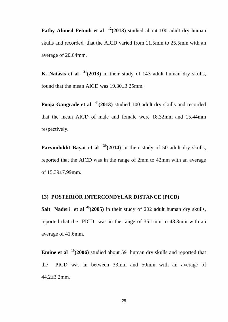
Fathy Ahmed Fetouh et al 12(2013) studied about 100 adult dry human
skulls and recorded that the AICD varied from 11.5mm to 25.5mm with an
average of 20.64mm.
K. Natasis et al 31(2013) in their study of 143 adult human dry skulls,
found that the mean AICD was 19.30±3.25mm.
Pooja Gangrade et al 44(2013) studied 100 adult dry skulls and recorded
that the mean AICD of male and female were 18.32mm and 15.44mm
respectively.
Parvindokht Bayat et al 39(2014) in their study of 50 adult dry skulls,
reported that the AICD was in the range of 2mm to 42mm with an average
of 15.39±7.99mm.
13) POSTERIOR INTERCONDYLAR DISTANCE (PICD)
Sait Naderi et al 49(2005) in their study of 202 adult human dry skulls,
reported that the PICD was in the range of 35.1mm to 48.3mm with an
average of 41.6mm.
Emine et al 10(2006) studied about 59 human dry skulls and reported that
the PICD was in between 33mm and 50mm with an average of
44.2±3.2mm.
29
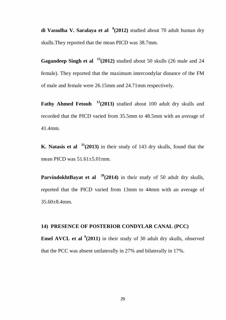
di Vasudha V. Saralaya et al 8(2012) studied about 70 adult human dry
skulls.They reported that the mean PICD was 38.7mm.
Gagandeep Singh et al 15(2012) studied about 50 skulls (26 male and 24
female). They reported that the maximum intercondylar distance of the FM
of male and female were 26.15mm and 24.71mm respectively.
Fathy Ahmed Fetouh 12(2013) studied about 100 adult dry skulls and
recorded that the PICD varied from 35.5mm to 48.5mm with an average of
41.4mm.
K. Natasis et al 31(2013) in their study of 143 dry skulls, found that the
mean PICD was 51.61±5.01mm.
ParvindokhtBayat et al 39(2014) in their study of 50 adult dry skulls,
reported that the PICD varied from 13mm to 44mm with an average of
35.60±8.4mm.
14) PRESENCE OF POSTERIOR CONDYLAR CANAL (PCC)
Emel AVCL et al 9(2011) in their study of 30 adult dry skulls, observed
that the PCC was absent unilaterally in 27% and bilaterally in 17%.
30
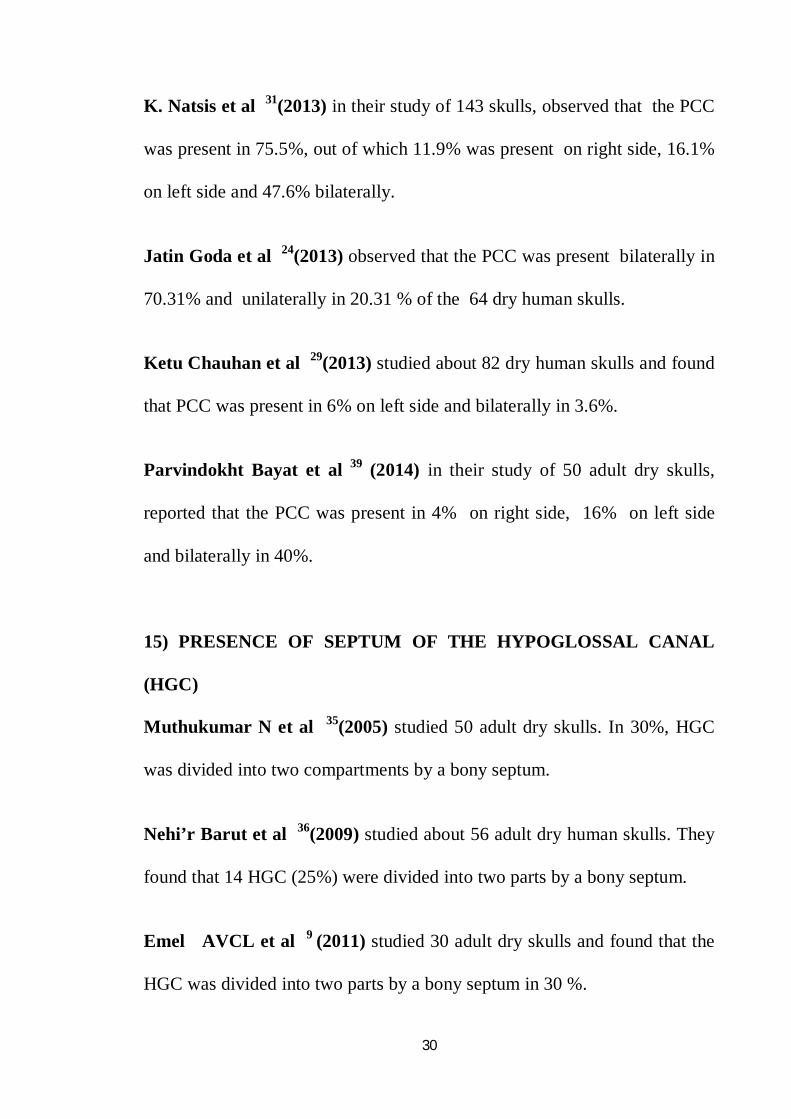
K. Natsis et al 31(2013) in their study of 143 skulls, observed that the PCC
was present in 75.5%, out of which 11.9% was present on right side, 16.1%
on left side and 47.6% bilaterally.
Jatin Goda et al 24(2013) observed that the PCC was present bilaterally in
70.31% and unilaterally in 20.31 % of the 64 dry human skulls.
Ketu Chauhan et al 29(2013) studied about 82 dry human skulls and found
that PCC was present in 6% on left side and bilaterally in 3.6%.
Parvindokht Bayat et al 39 (2014) in their study of 50 adult dry skulls,
reported that the PCC was present in 4% on right side, 16% on left side
and bilaterally in 40%.
15) PRESENCE OF SEPTUM OF THE HYPOGLOSSAL CANAL
(HGC)
Muthukumar N et al 35(2005) studied 50 adult dry skulls. In 30%, HGC
was divided into two compartments by a bony septum.
Nehi’r Barut et al 36(2009) studied about 56 adult dry human skulls. They
found that 14 HGC (25%) were divided into two parts by a bony septum.
Emel AVCL et al 9 (2011) studied 30 adult dry skulls and found that the
HGC was divided into two parts by a bony septum in 30 %.
31
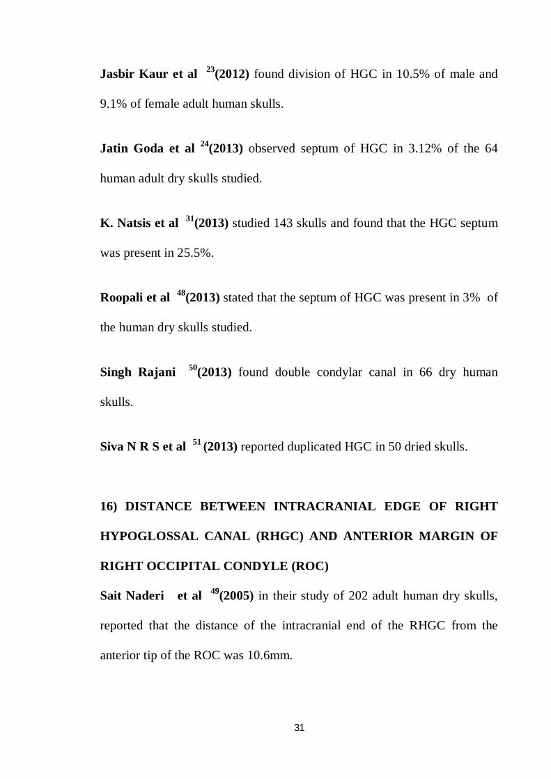
Jasbir Kaur et al 23(2012) found division of HGC in 10.5% of male and
9.1% of female adult human skulls.
Jatin Goda et al 24(2013) observed septum of HGC in 3.12% of the 64
human adult dry skulls studied.
K. Natsis et al 31(2013) studied 143 skulls and found that the HGC septum
was present in 25.5%.
Roopali et al 48(2013) stated that the septum of HGC was present in 3% of
the human dry skulls studied.
Singh Rajani 50(2013) found double condylar canal in 66 dry human
skulls.
Siva N R S et al 51 (2013) reported duplicated HGC in 50 dried skulls.
16) DISTANCE BETWEEN INTRACRANIAL EDGE OF RIGHT
HYPOGLOSSAL CANAL (RHGC) AND ANTERIOR MARGIN OF
RIGHT OCCIPITAL CONDYLE (ROC)
Sait Naderi et al 49(2005) in their study of 202 adult human dry skulls,
reported that the distance of the intracranial end of the RHGC from the
anterior tip of the ROC was 10.6mm.
32
Emine et al 10(2006) studied about 59 human dry skulls and reported that
the distance of the intracranial end of the RHGC from the anterior margin of
the ROC was in the range of 8.1mm to 16.9mm with an average of
11.0±1.6mm.
Pereira G.A et al 42(2012) in their study of 111 adult human dry skulls
reported that the intracranial end of the RHGC from the anterior margin of
the ROC was 11.0±1.8mm.
17) DISTANCE BETWEEN INTRACRANIAL EDGE OF RIGHT
HYPOGLOSSAL CANAL (RHGC) AND POSTERIOR MARGIN
OF RIGHT OCCIPITAL CONDYLE (ROC)
Muthukumar N et al 35(2005) studied about 50 adult human dry skulls.
They found that the distance of the intracranial end of the RHGC from the
posterior margin of the ROC was12.2mm.
Emine et al 10(2006) studied about 59 human dry skulls and reported that
the distance of intracranial end of the RHGC from the posterior margin of
the ROC was in between 8.2mm and 17.4mm with an average of
12.2±2.2mm.
33
Nehi’r Barut et al 36(2009) studied about 56 adult dry human skulls. They
found that the distance between the intracranial edge of RHGC and posterior
margin of ROC was 12.5mm.
Emel AVCL et al 9(2011) in their study of 30 adult dry skulls, reported
that the distance between the RHGC and posterior border of ROC varied
from 7.9 mm to 12.2mm with an average of 9.8±1.1mm.
Pereira G.A et al 42(2012) in their study of 111 adult human dry skulls
reported that the distance of the intracranial end of the RHGC from the
posterior margin of the ROC was 10.3±2.5mm.
18) DISTANCE BETWEEN INTRACRANIAL EDGE OF LEFT
HYPOGLOSSAL (LHGC) CANAL AND ANTERIOR MARGIN OF
LEFT OCCIPITAL CONDYLE (LOC)
Sait Naderi et al 49(2005) in their study of 202 adult human dry skulls,
reported that the distance of the intracranial end of the LHGC from the
anterior tip of the LOC was 9.6mm.
Emine et al 10(2006) studied about 59 human dry skulls and reported that
the distance between the intracranial end of the LHGC and the anterior
margin of the LOC was in the range of 8.2mm to 16.9mm with an average
of 11.3±1.5mm.
34
Pereira G.A et al 42(2012) in their study of 111 adult human dry skulls
reported that the distance of intracranial end of the LHGC from the anterior
margin of the LOC was 10.7±1.8mm.
19) DISTANCE BETWEEN INTRACRANIAL EDGE OF LEFT
HYPOGLOSSAL CANAL (LHGC) AND POSTERIOR MARGIN OF
LEFT OCCIPITAL CONDYLE (LOC)
Emel AVCL et al 9(2011) in their study of 30 adult dry skulls, reported
that the distance between LHGC and Posterior border of LOC varied from
6.6 mm to 12.2 mm with an average of 9.9±1.4mm.
Emine et al 10(2006) studied about 59 human dry skulls and reported that
the intracranial end of the LHGC from the posterior margin of the LOC was
in between 8.4mm and 17.6mm with an average of 12.4±2.3mm.
Nehi’r Barut et al 36(2009) studied about 56 adult dry human skulls. They
found that the distance between the intracranial edge of LHGC and posterior
margin of LOC was 12.6mm.
Pereira G.A et al 42(2012) in their study of 111 adult human dry skulls
reported that the intracranial end of the LHGC from the posterior margin of
the LOC was 11.3±2.1mm.

Embryology
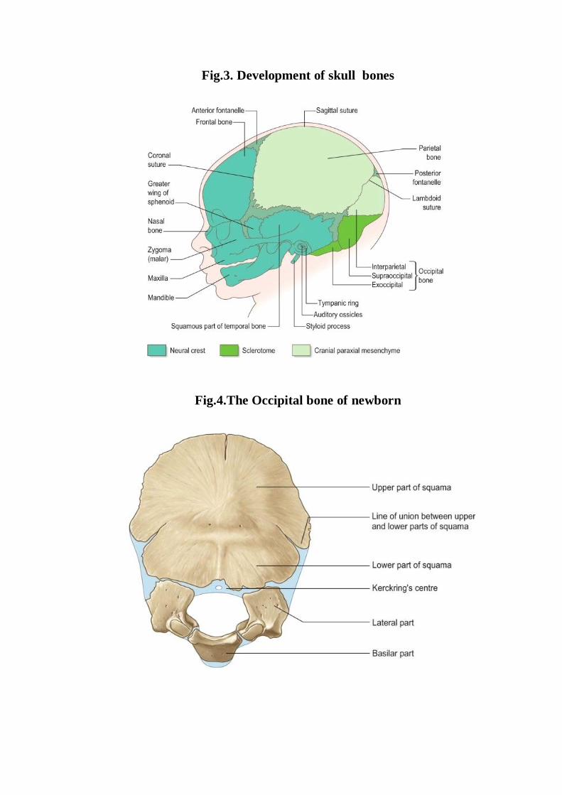
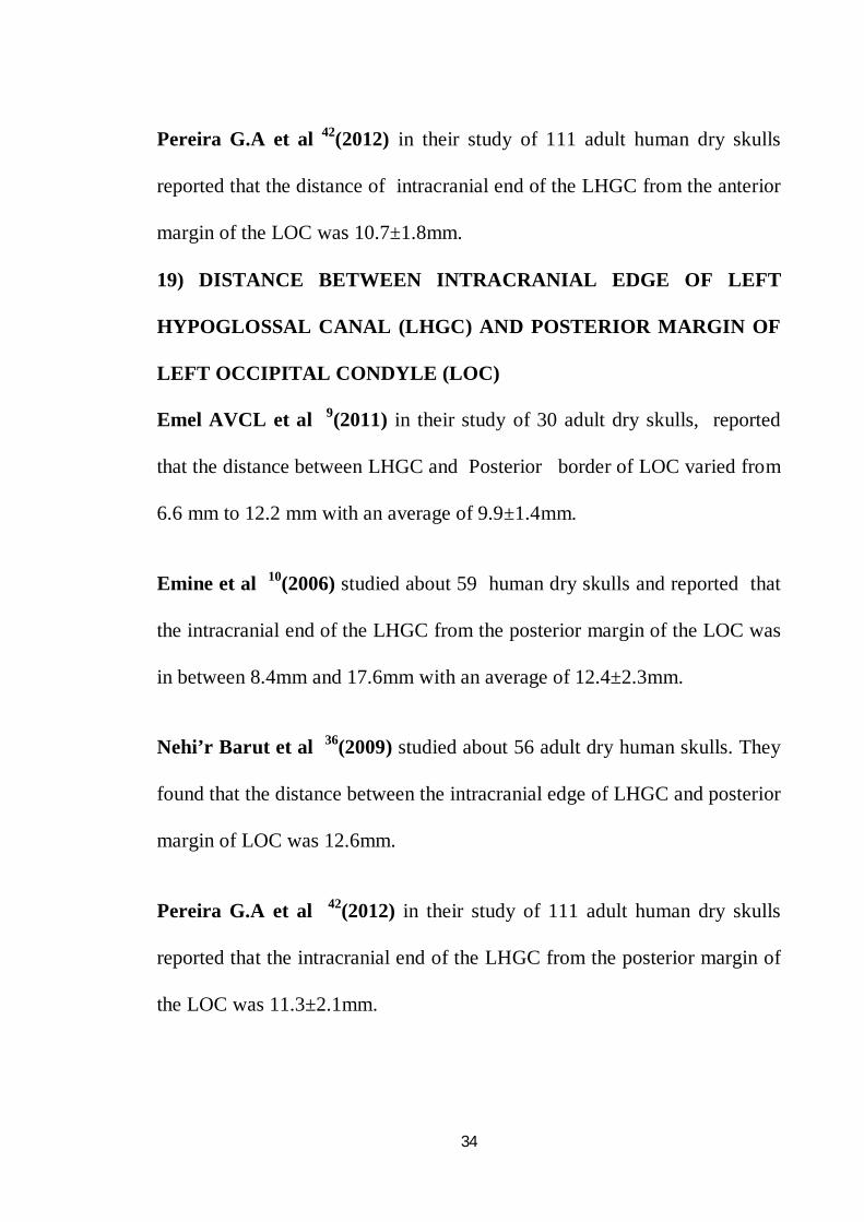
Fig.3. Development of skull bones
Fig.4.The Occipital bone of newborn
35
EMBRYOLOGY
The skull consists of two major anatomical and functional
components: the neurocranium and viscerocranium. The neurocranium
forms a protective covering around the brain and viscerocranium forms the
skeleton of the face.32
The skull develops from neural crest cells, cranial paraxial mesoderm
and sclerotome(Fig.3). The neural crest cells form the whole viscerocranium
and the rostral portion of neurocranium. The skull base is formed by neural
crest rostral to the tip of the notochord and by sclerotome (mesoderm) in the
notochordal region.
Neurocranium: It has two portions- membranous and cartilaginous
portions.
Membranous neurocranium: It is derived from neural crest cells and
paraxial mesoderm. The mesenchyme from both sources covers the brain
and undergoes membranous ossification. It forms the cranial vault.
Cartilaginous neurocranium: It is also called chondrocranium. It is
composed of a number of separate cartilages. The cartilages fuse and ossify
by endochondral ossification and form the base of the skull.
36
The cartilages that are present posterior to the rostral limit of
notochord arise from occipital sclerotomes formed by paraxial mesoderm.
The central region of occipital sclerotomes contribute to the parachordal
cartilage, which enclose the notochord and extends as a flat plate on either
side of it and forms the basioccipital component of the occipital bone.40 The
exoccipital components chondrify and border the foramen magnum.
Roots of the hypoglossal nerve run between the parachordal and
exoccipital cartilages. The fusion of exoccipital and parachordal
components forms the foramina for hypoglossal nerve roots bilaterally.
OSSIFICATION
The occipital bone is a compound structure with respect to its origin
and type of ossification (Fig.4).
The squamous part of occipital bone above the highest nuchal lines
develops in membrane. It ossifies from two centres in the second foetal
month. The squamous part below the highest nuchal lines ossifies from two
centres which make their appearance in about the seventh week and unite
immediately. The line of union of the two components of the squamous part
is identifiable at birth. Kerckring’s centre, which is an occasional centre for
posterior margin of foramen magnum, appears at sixteenth week. The rest of
the cartilage of occipital bone ossifies from five centres. During eighth week
37
of intauterine life, two centres each for the lateral or condylar or exoccipital
parts appear. During sixth week one centre for the basilar part appears and it
unites with the rest of the bone by sixth year.
The occipital bone is made up of four parts at birth – basilar, two
lateral and a squamous part which fuse by cartilage and form a ring around
foramen magnum. The squamous part is present posteriorly, the lateral or
condylar parts are present on each side, and the basilar part or basiocciput is
anterior. These names are retained for the parts of the adult bone also.25,16
The union of the squamous and lateral parts start from the second
year. At 3-4 years, the lateral part unites with the basilar part and is
completed by seventh year. The basilar part and body of the sphenoid unites
by cartilage and is completely ossified by 25years.55

Materials and Methods
38
MATERIALS AND METHODS
STUDY MATERIALS:
Hundred human adult dry skulls of unknown sex.
20 Computerized Tomographic Scan Images.
Digital Vernier Calipers.
Flexible wire.
STUDY METHODS:
1. Dry skull Method
2. Radiological Study
SPECIMEN COLLECTION:
Hundred human adult dry skulls of unknown sex available in the
Institute of Anatomy, Madras Medical College were used for this study.
A. DRY SKULL METHOD:
Inclusion criteria:
1. Adult human dry skull of unknown sex.
2. Third molar tooth erupted.
3. Well defined skull sutures.
Exclusion criteria:
1. Abnormal skulls.
2. Damaged skulls.
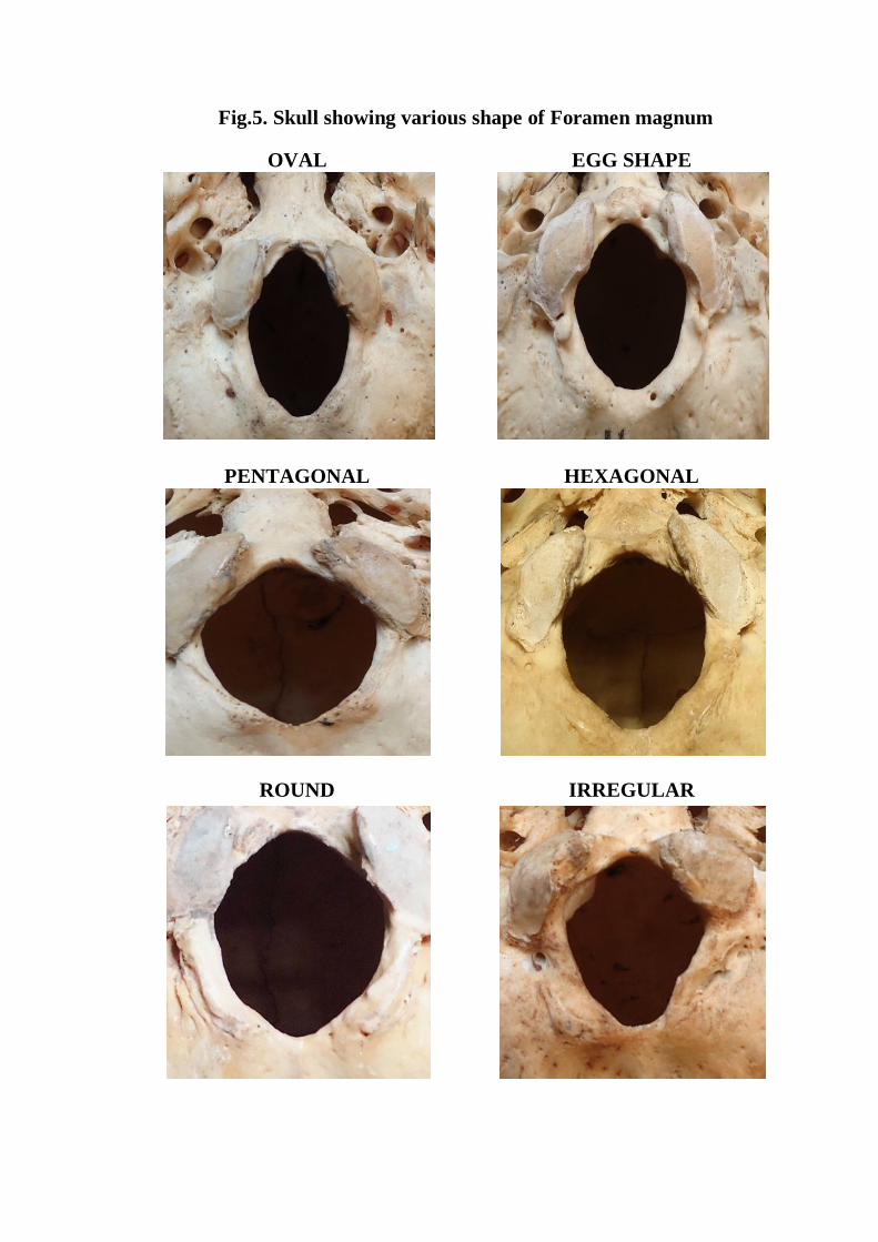
Fig.5. Skull showing various shape of Foramen magnum
OVAL EGG SHAPE
PENTAGONAL HEXAGONAL
ROUND IRREGULAR
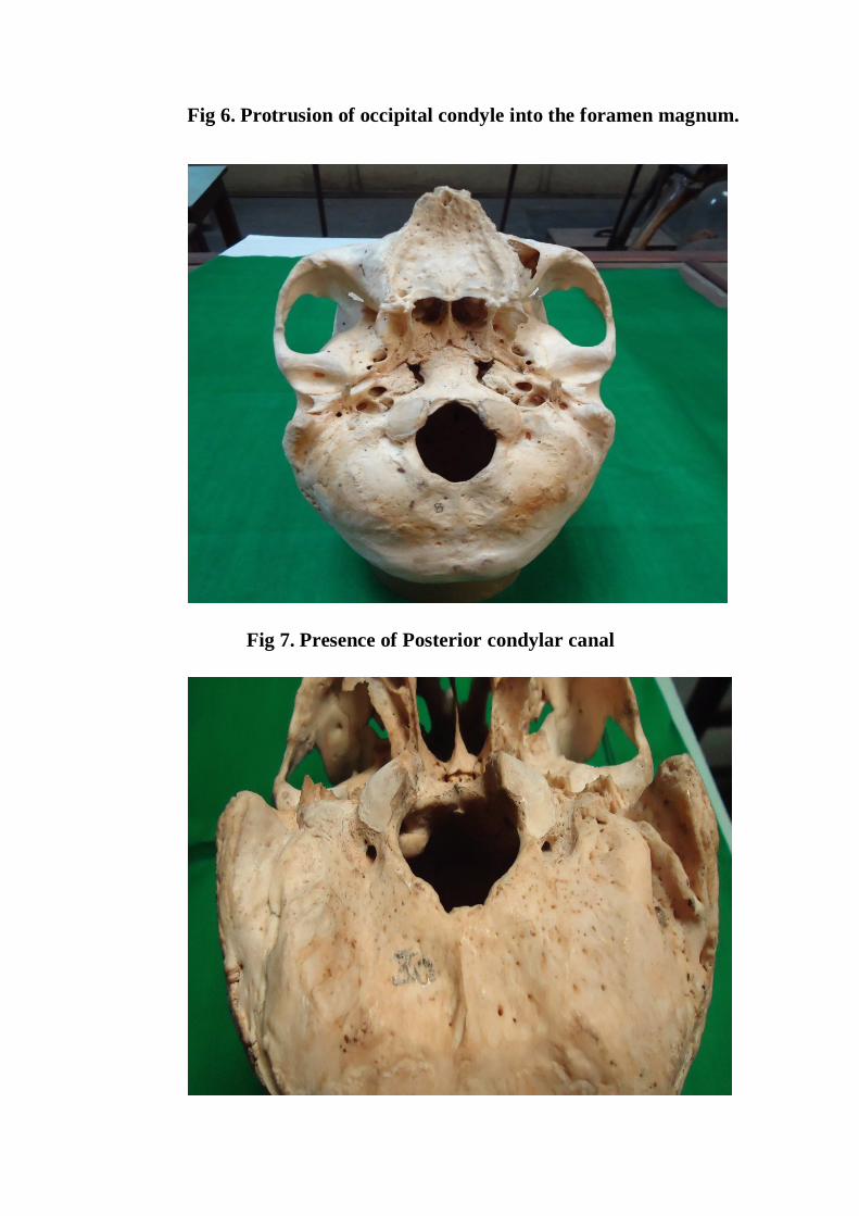
Fig 6. Protrusion of occipital condyle into the foramen magnum.
Fig 7. Presence of Posterior condylar canal
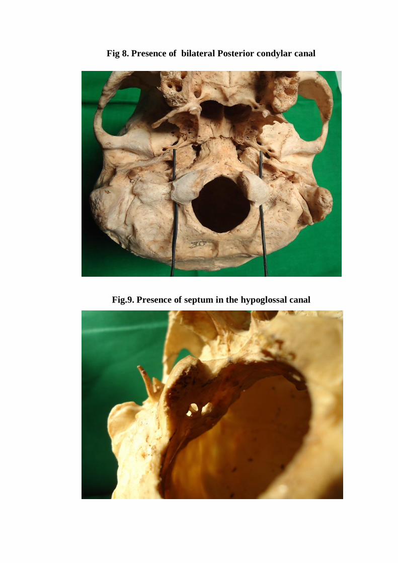
Fig 8. Presence of bilateral Posterior condylar canal
Fig.9. Presence of septum in the hypoglossal canal
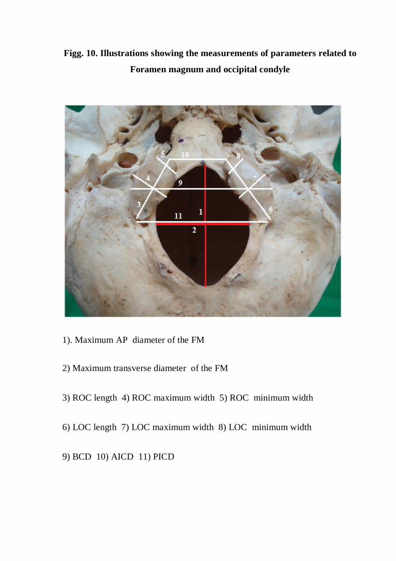
Figg. 10. Illustrations showing the measurements of parameters related to
Foramen magnum and occipital condyle
1). Maximum AP diameter of the FM
2) Maximum transverse diameter of the FM
3) ROC length 4) ROC maximum width 5) ROC minimum width
6) LOC length 7) LOC maximum width 8) LOC minimum width
9) BCD 10) AICD 11) PICD
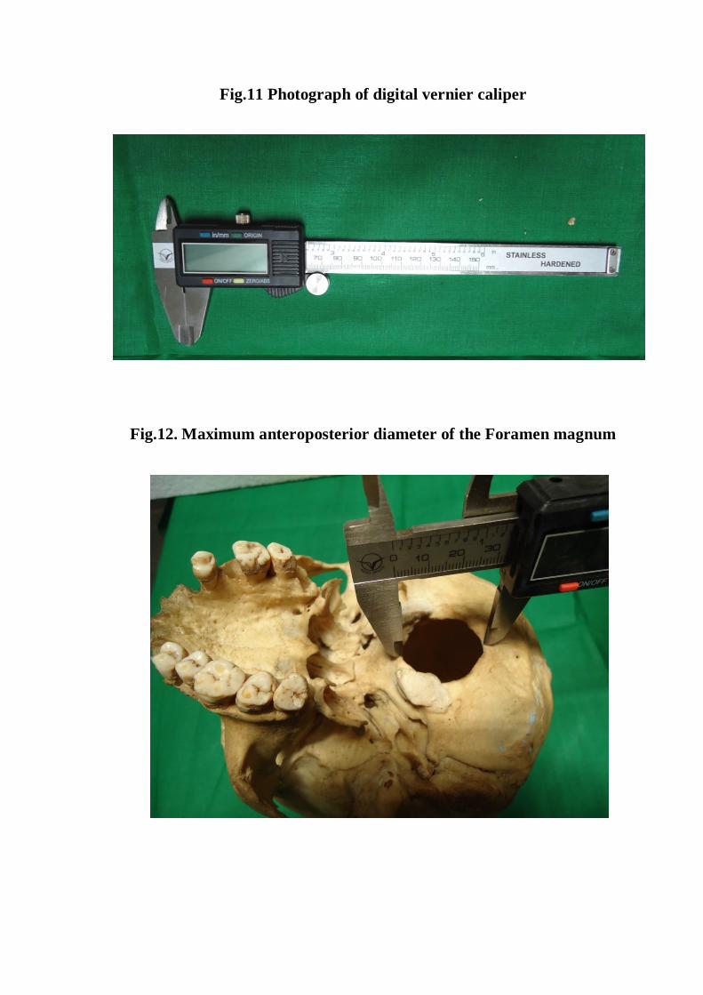
Fig.11 Photograph of digital vernier caliper
Fig.12. Maximum anteroposterior diameter of the Foramen magnum
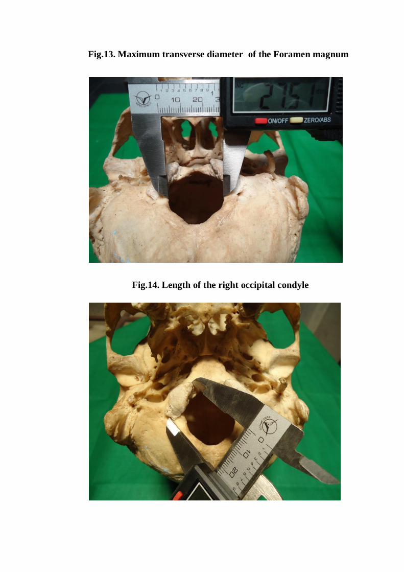
Fig.13. Maximum transverse diameter of the Foramen magnum
Fig.14. Length of the right occipital condyle
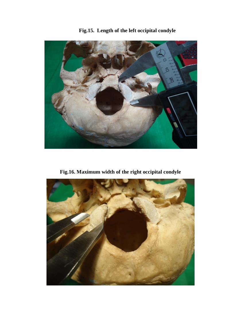
Fig.15. Length of the left occipital condyle
Fig.16. Maximum width of the right occipital condyle
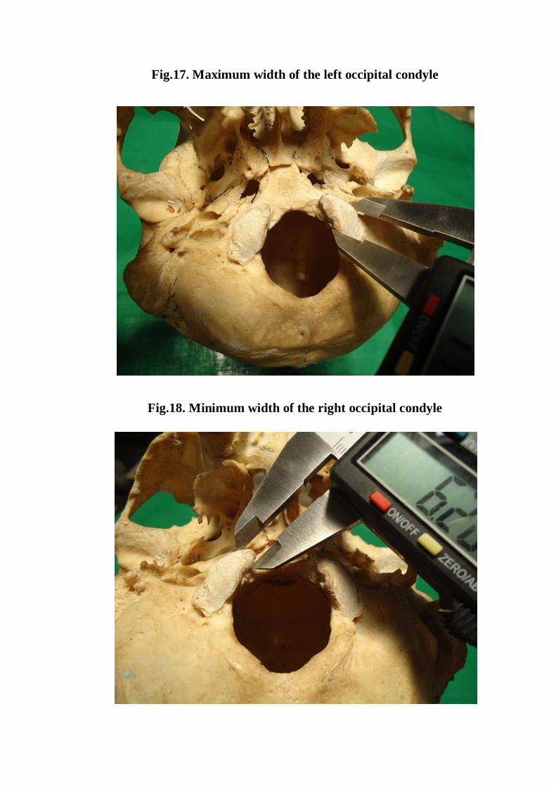
Fig.17. Maximum width of the left occipital condyle
Fig.18. Minimum width of the right occipital condyle
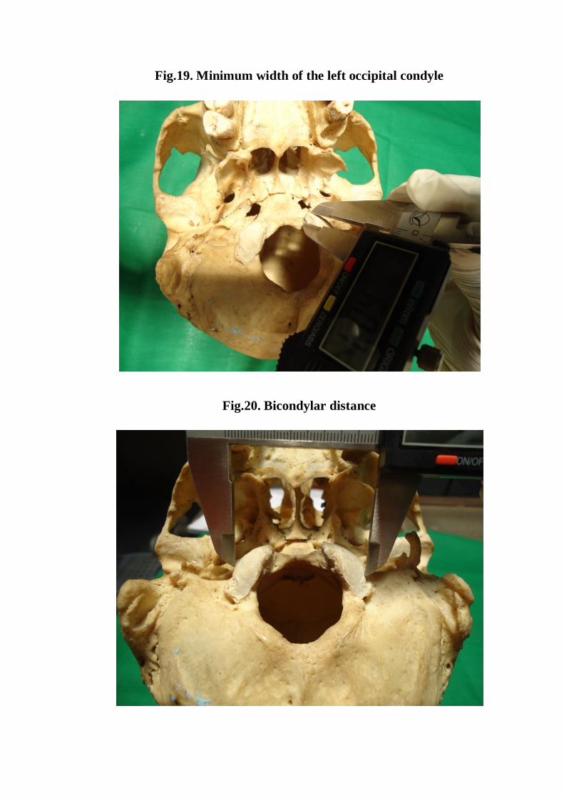
Fig.19. Minimum width of the left occipital condyle
Fig.20. Bicondylar distance
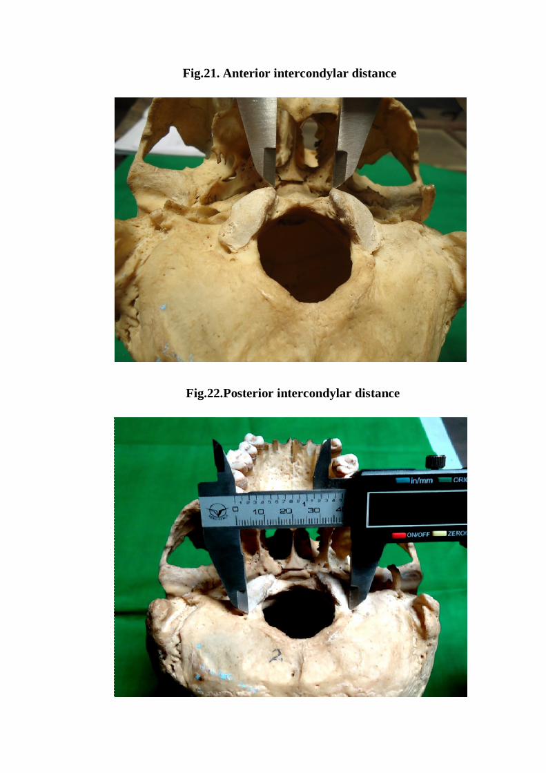
Fig.21. Anterior intercondylar distance
Fig.22.Posterior intercondylar distance
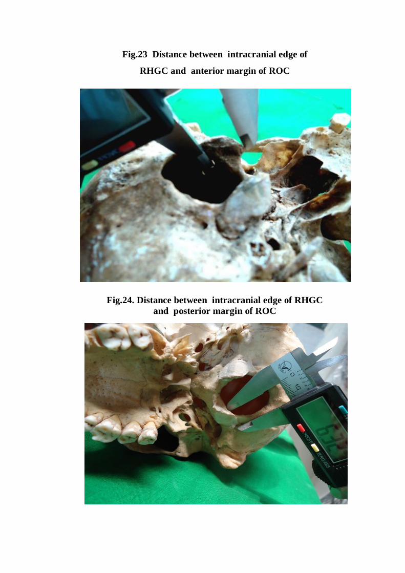
Fig.23 Distance between intracranial edge of
RHGC and anterior margin of ROC
Fig.24. Distance between intracranial edge of RHGC and posterior margin of ROC
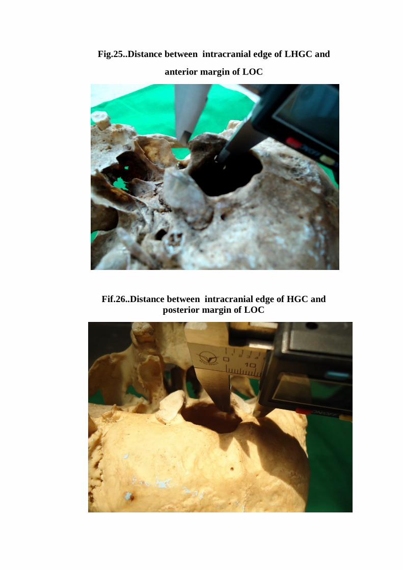
Fig.25..Distance between intracranial edge of LHGC and
anterior margin of LOC
Fif.26..Distance between intracranial edge of HGC and posterior margin of LOC
39
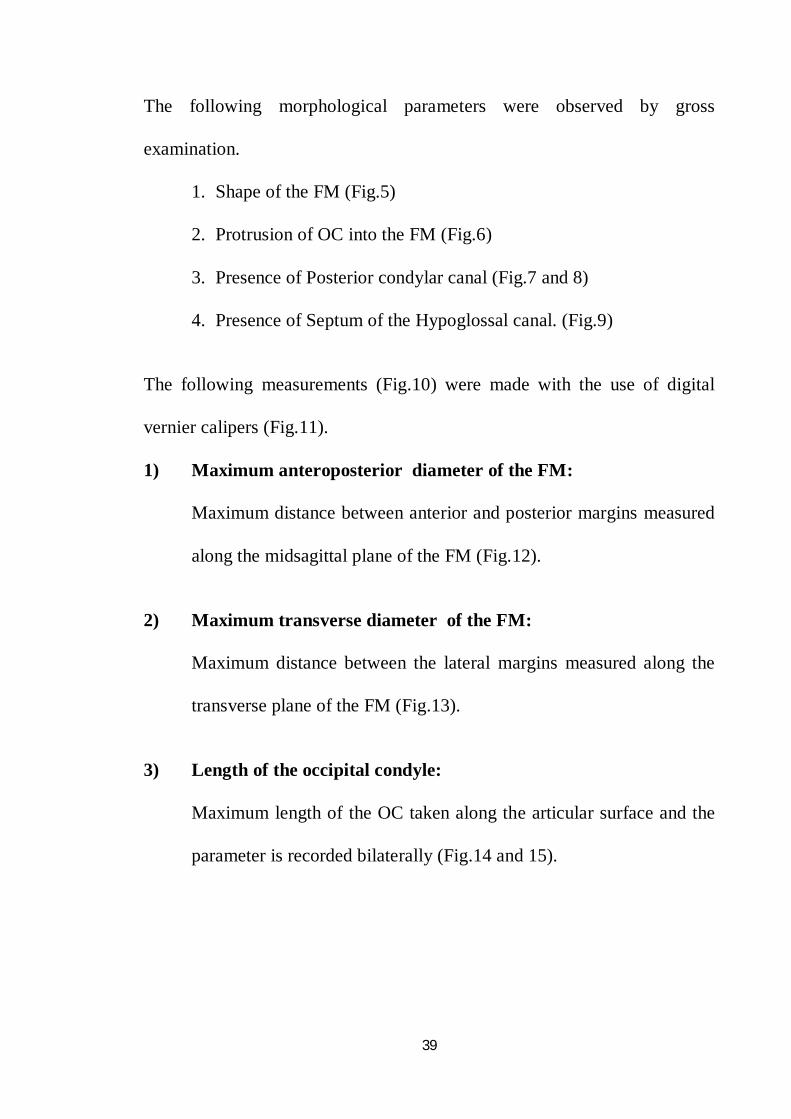
The following morphological parameters were observed by gross
examination.
1. Shape of the FM (Fig.5)
2. Protrusion of OC into the FM (Fig.6)
3. Presence of Posterior condylar canal (Fig.7 and 8)
4. Presence of Septum of the Hypoglossal canal. (Fig.9)
The following measurements (Fig.10) were made with the use of digital
vernier calipers (Fig.11).
1) Maximum anteroposterior diameter of the FM:
Maximum distance between anterior and posterior margins measured
along the midsagittal plane of the FM (Fig.12).
2) Maximum transverse diameter of the FM:
Maximum distance between the lateral margins measured along the
transverse plane of the FM (Fig.13).
3) Length of the occipital condyle:
Maximum length of the OC taken along the articular surface and the
parameter is recorded bilaterally (Fig.14 and 15).
40
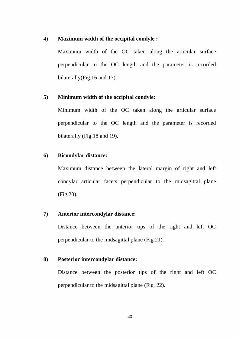
4) Maximum width of the occipital condyle :
Maximum width of the OC taken along the articular surface
perpendicular to the OC length and the parameter is recorded
bilaterally(Fig.16 and 17).
5) Minimum width of the occipital condyle:
Minimum width of the OC taken along the articular surface
perpendicular to the OC length and the parameter is recorded
bilaterally (Fig.18 and 19).
6) Bicondylar distance:
Maximum distance between the lateral margin of right and left
condylar articular facets perpendicular to the midsagittal plane
(Fig.20).
7) Anterior intercondylar distance:
Distance between the anterior tips of the right and left OC
perpendicular to the midsagittal plane (Fig.21).
8) Posterior intercondylar distance:
Distance between the posterior tips of the right and left OC
perpendicular to the midsagittal plane (Fig. 22).
41
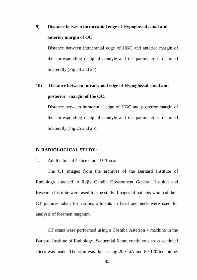
9) Distance between intracranial edge of Hypoglossal canal and
anterior margin of OC:
Distance between intracranial edge of HGC and anterior margin of
the corresponding occipital condyle and the parameter is recorded
bilaterally (Fig.23 and 24).
10) Distance between intracranial edge of Hypoglossal canal and
posterior margin of the OC:
Distance between intracranial edge of HGC and posterior margin of
the corresponding occipital condyle and the parameter is recorded
bilaterally (Fig.25 and 26).
B. RADIOLOGICAL STUDY:
1. Adult Clinical 4 slice cranial CT scan.
The CT images from the archives of the Barnard Institute of
Radiology attached to Rajiv Gandhi Government General Hospital and
Research Institute were used for the study. Images of patients who had their
CT pictures taken for various ailments in head and neck were used for
analysis of foramen magnum.
CT scans were performed using a Toshiba Asterion 4 machine in the
Barnard Institute of Radiology. Sequential 5 mm continuous cross sectional
slices was made. The scan was done using 200 mA and 80-120 technique.
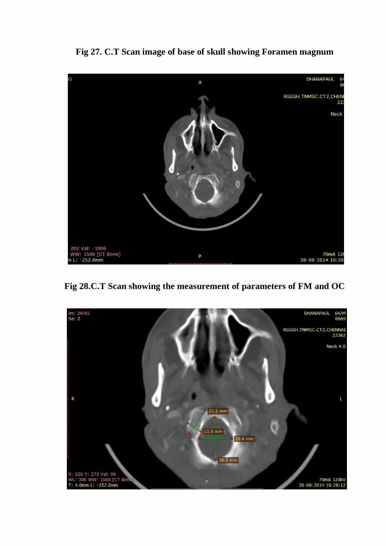
Fig 27. C.T Scan image of base of skull showing Foramen magnum
Fig 28.C.T Scan showing the measurement of parameters of FM and OC
42
The images were digitized and stored on the Picture Archiving
Communication System which was later retrieved for measurement of
parameters. The system was incorporated with image enhancement and
manipulation tools. The software also had a sensitive measuring tool.
From CT images, parameters were measured (Fig 27 and 28). Some
of the parameters were measured bilaterally.

Observation
43
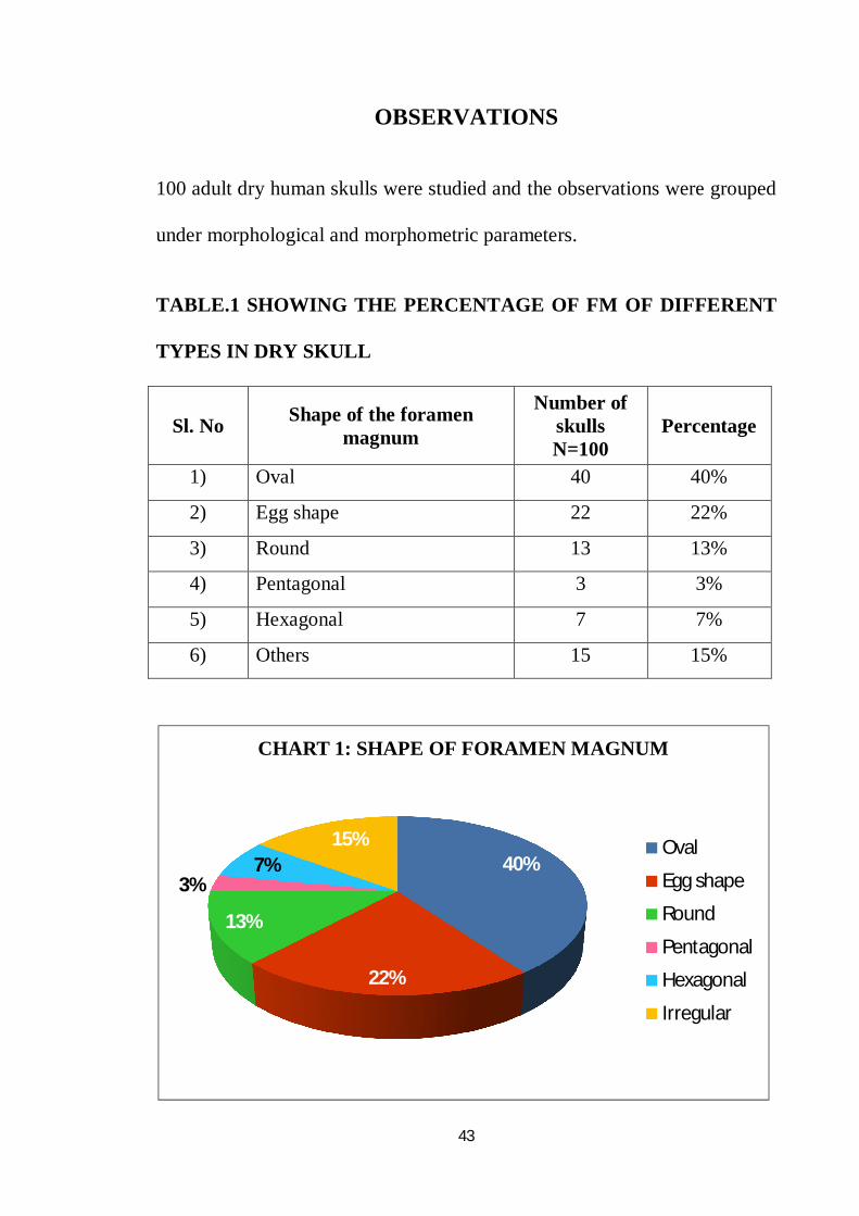
OBSERVATIONS
100 adult dry human skulls were studied and the observations were grouped
under morphological and morphometric parameters.
TABLE.1 SHOWING THE PERCENTAGE OF FM OF DIFFERENT
TYPES IN DRY SKULL
Sl. No Shape of the foramen magnum
Number of skulls N=100
Percentage
1) Oval 40 40%
2) Egg shape 22 22%
3) Round 13 13%
4) Pentagonal 3 3%
5) Hexagonal 7 7%
6) Others 15 15%
40%
22%
13%
3%7%
15%
CHART 1: SHAPE OF FORAMEN MAGNUM
Oval
Egg shape
Round
Pentagonal
Hexagonal
Irregular
44
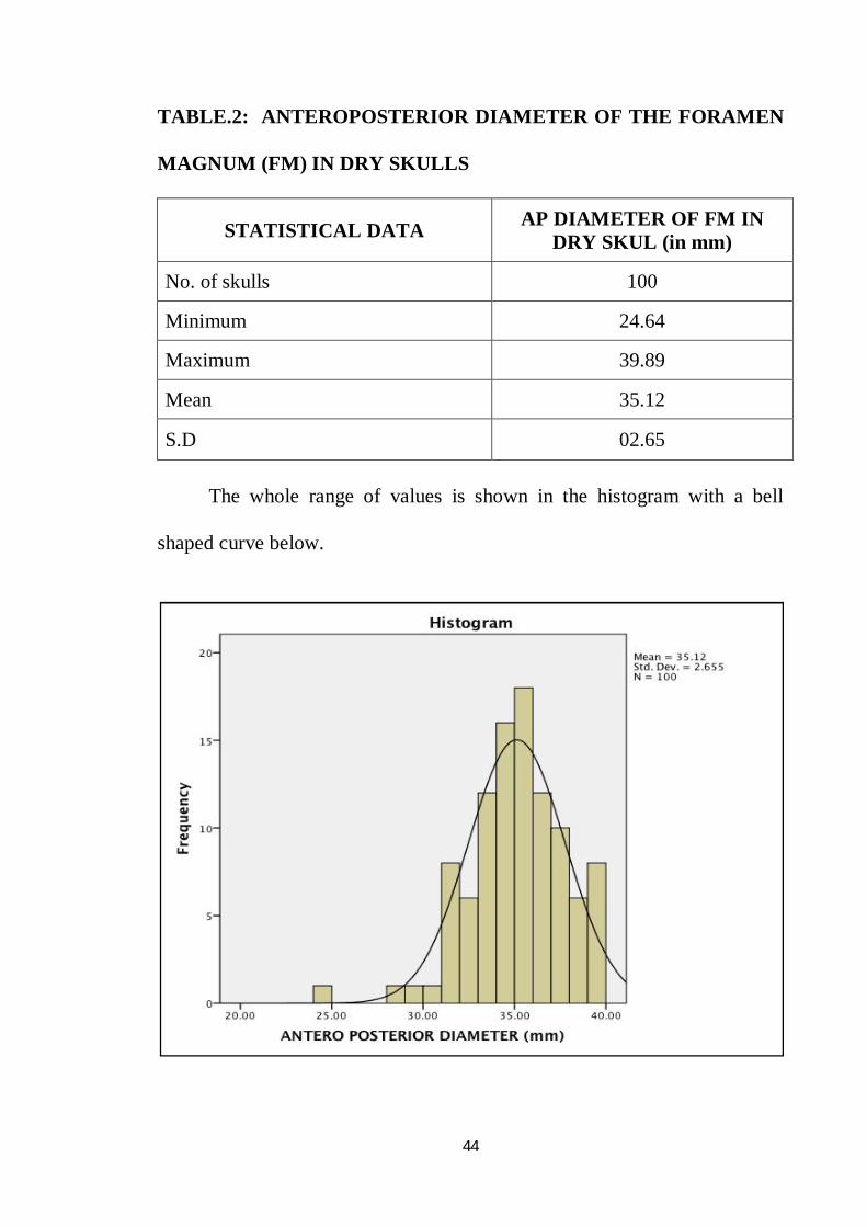
TABLE.2: ANTEROPOSTERIOR DIAMETER OF THE FORAMEN
MAGNUM (FM) IN DRY SKULLS
STATISTICAL DATA AP DIAMETER OF FM IN DRY SKUL (in mm)
No. of skulls 100
Minimum 24.64
Maximum 39.89
Mean 35.12
S.D 02.65 The whole range of values is shown in the histogram with a bell
shaped curve below.
45
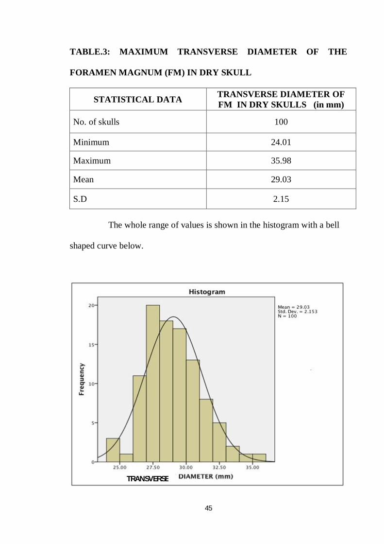
TABLE.3: MAXIMUM TRANSVERSE DIAMETER OF THE
FORAMEN MAGNUM (FM) IN DRY SKULL
STATISTICAL DATA TRANSVERSE DIAMETER OF FM IN DRY SKULLS (in mm)
No. of skulls 100
Minimum 24.01
Maximum 35.98
Mean 29.03
S.D 2.15
The whole range of values is shown in the histogram with a bell
shaped curve below.
TRANSVERSE
46
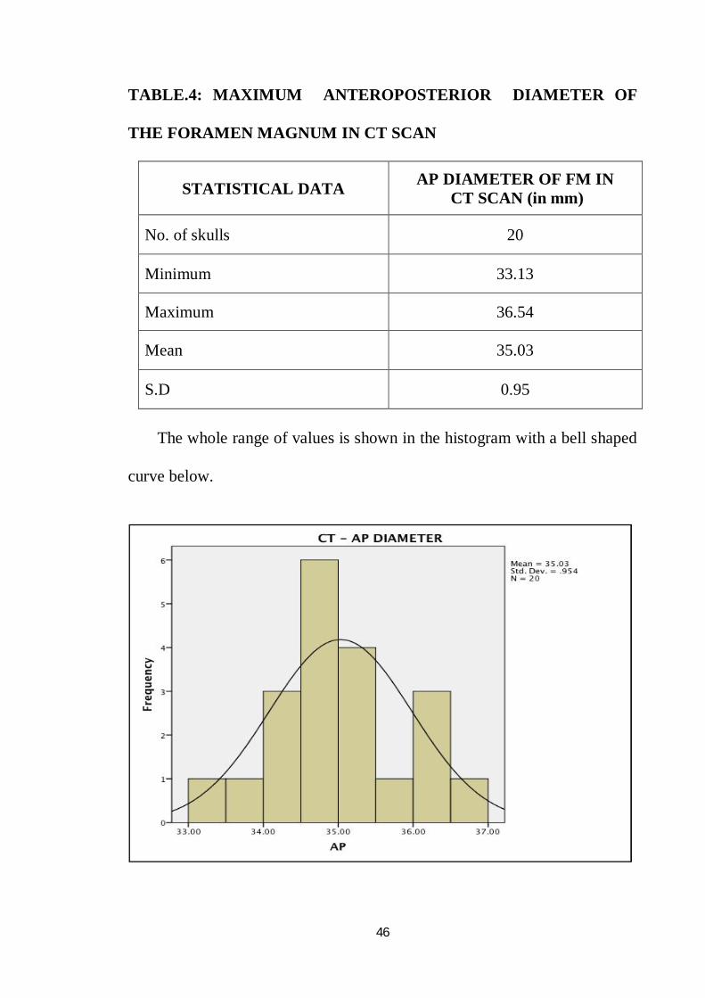
TABLE.4: MAXIMUM ANTEROPOSTERIOR DIAMETER OF
THE FORAMEN MAGNUM IN CT SCAN
STATISTICAL DATA AP DIAMETER OF FM IN CT SCAN (in mm)
No. of skulls 20
Minimum 33.13
Maximum 36.54
Mean 35.03
S.D 0.95
The whole range of values is shown in the histogram with a bell shaped
curve below.
47
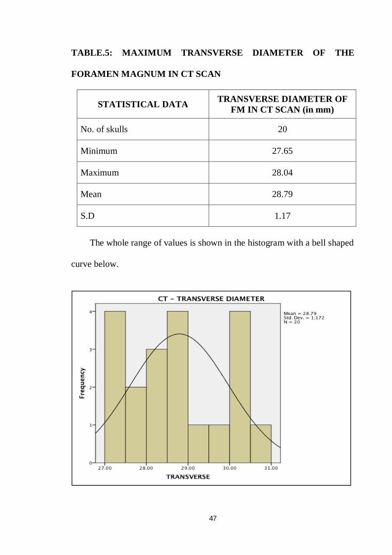
TABLE.5: MAXIMUM TRANSVERSE DIAMETER OF THE
FORAMEN MAGNUM IN CT SCAN
STATISTICAL DATA TRANSVERSE DIAMETER OF FM IN CT SCAN (in mm)
No. of skulls 20
Minimum 27.65
Maximum 28.04
Mean 28.79
S.D 1.17
The whole range of values is shown in the histogram with a bell shaped
curve below.
48
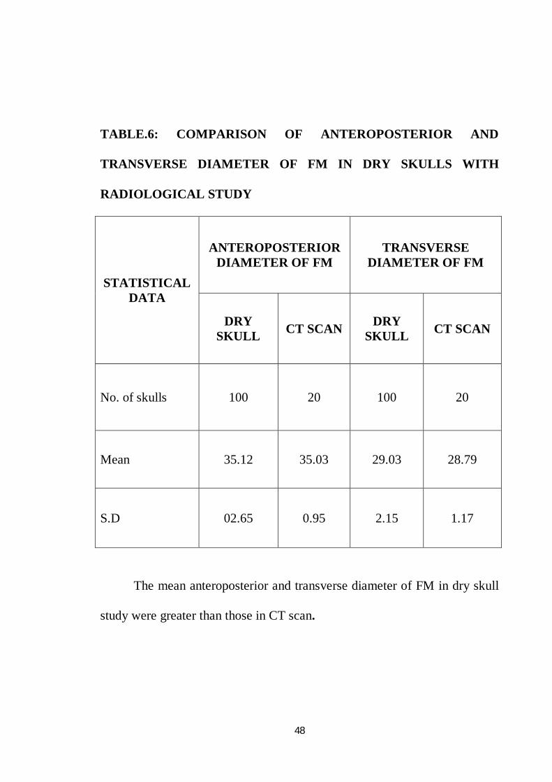
TABLE.6: COMPARISON OF ANTEROPOSTERIOR AND
TRANSVERSE DIAMETER OF FM IN DRY SKULLS WITH
RADIOLOGICAL STUDY
STATISTICAL DATA
ANTEROPOSTERIOR DIAMETER OF FM
TRANSVERSE DIAMETER OF FM
DRY SKULL CT SCAN DRY
SKULL CT SCAN
No. of skulls 100 20 100 20
Mean 35.12 35.03 29.03 28.79
S.D 02.65 0.95 2.15 1.17
The mean anteroposterior and transverse diameter of FM in dry skull
study were greater than those in CT scan.
49
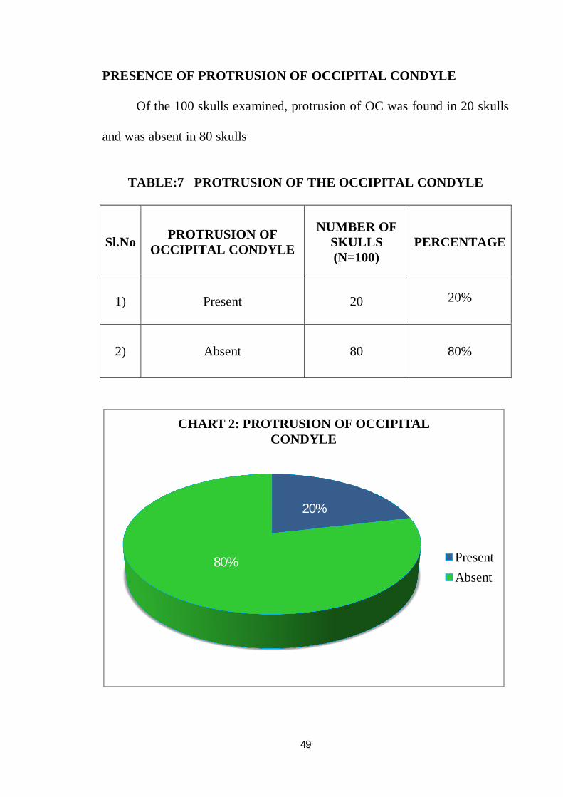
PRESENCE OF PROTRUSION OF OCCIPITAL CONDYLE
Of the 100 skulls examined, protrusion of OC was found in 20 skulls
and was absent in 80 skulls
TABLE:7 PROTRUSION OF THE OCCIPITAL CONDYLE
20%
80%
CHART 2: PROTRUSION OF OCCIPITAL CONDYLE
PresentAbsent
Sl.No PROTRUSION OF OCCIPITAL CONDYLE
NUMBER OF SKULLS (N=100)
PERCENTAGE
1) Present 20 20%
2) Absent 80 80%
50
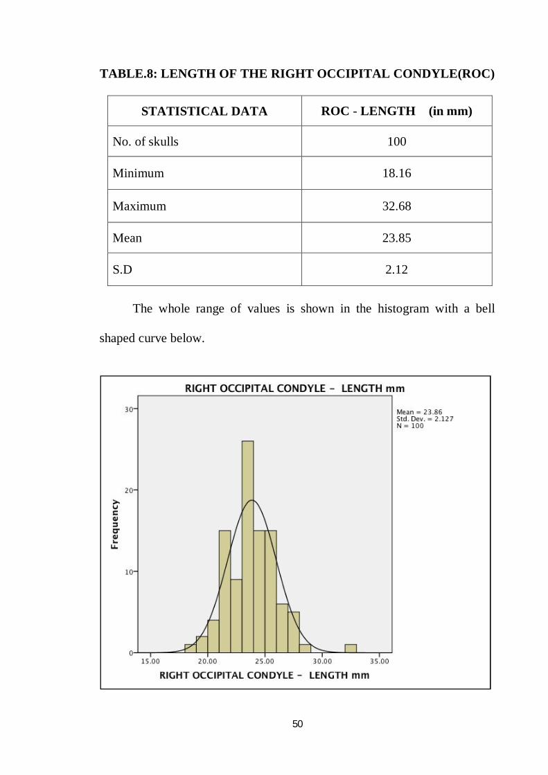
TABLE.8: LENGTH OF THE RIGHT OCCIPITAL CONDYLE(ROC)
STATISTICAL DATA ROC - LENGTH (in mm)
No. of skulls 100
Minimum 18.16
Maximum 32.68
Mean 23.85
S.D 2.12
The whole range of values is shown in the histogram with a bell
shaped curve below.
51
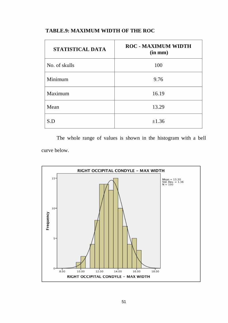
TABLE.9: MAXIMUM WIDTH OF THE ROC
STATISTICAL DATA ROC - MAXIMUM WIDTH (in mm)
No. of skulls 100
Minimum 9.76
Maximum 16.19
Mean 13.29
S.D ±1.36
The whole range of values is shown in the histogram with a bell
curve below.
52
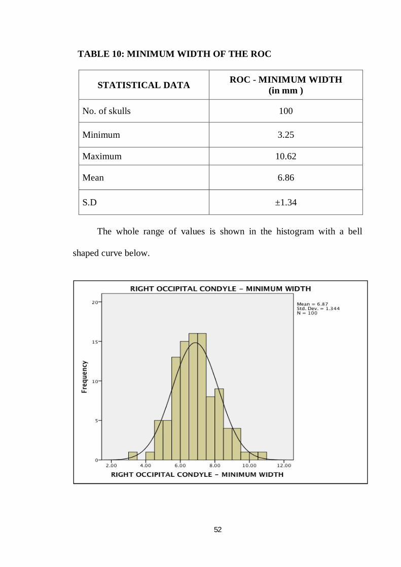
TABLE 10: MINIMUM WIDTH OF THE ROC
STATISTICAL DATA ROC - MINIMUM WIDTH (in mm )
No. of skulls 100
Minimum 3.25
Maximum 10.62
Mean 6.86
S.D ±1.34
The whole range of values is shown in the histogram with a bell
shaped curve below.
53
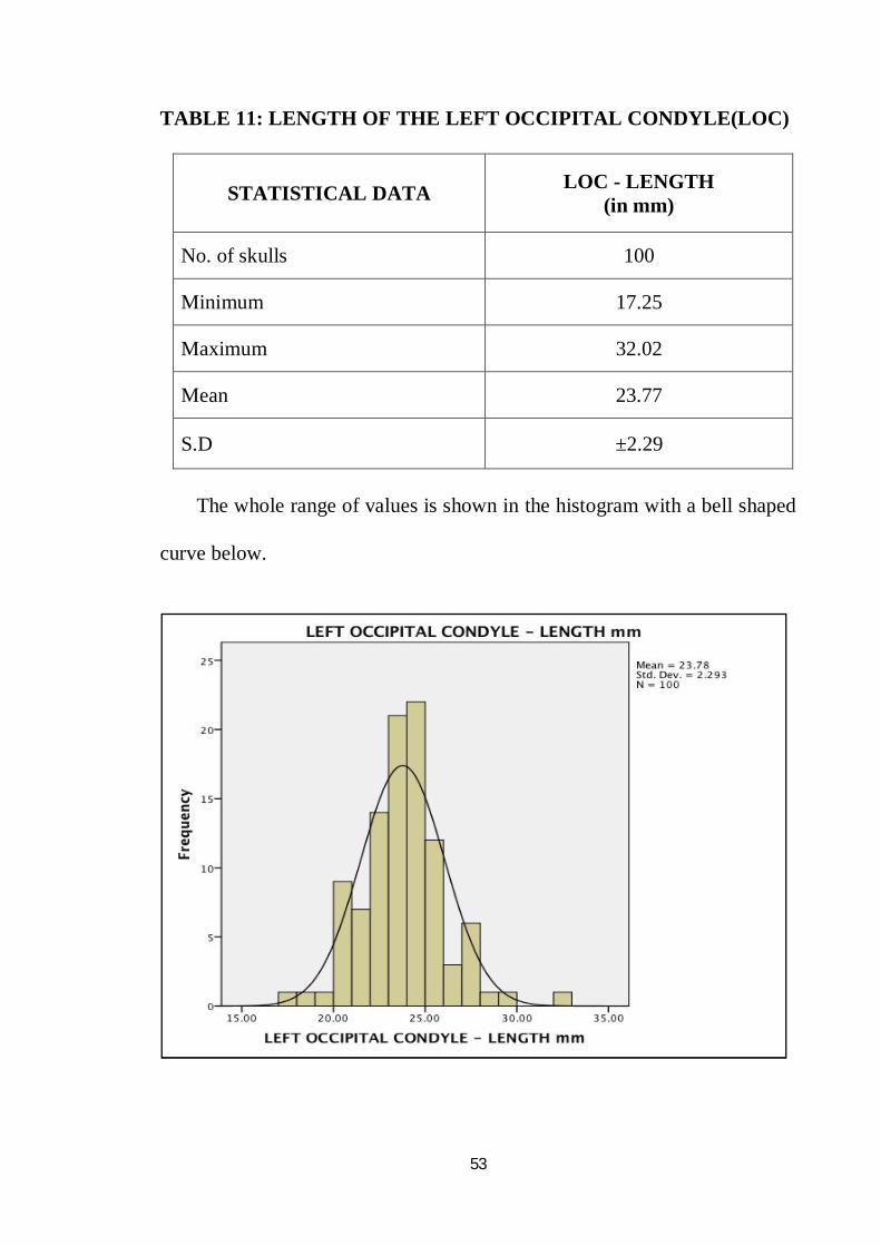
TABLE 11: LENGTH OF THE LEFT OCCIPITAL CONDYLE(LOC)
STATISTICAL DATA LOC - LENGTH (in mm)
No. of skulls 100
Minimum 17.25
Maximum 32.02
Mean 23.77
S.D ±2.29
The whole range of values is shown in the histogram with a bell shaped
curve below.
54
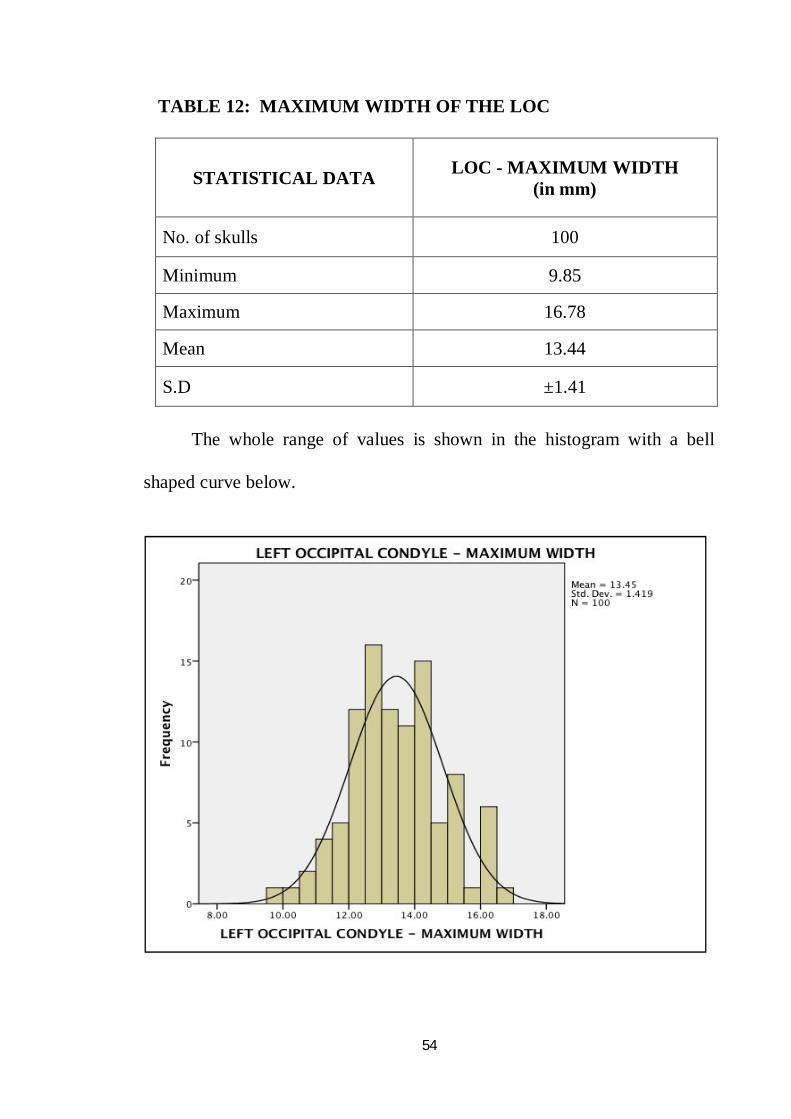
TABLE 12: MAXIMUM WIDTH OF THE LOC
STATISTICAL DATA LOC - MAXIMUM WIDTH (in mm)
No. of skulls 100
Minimum 9.85
Maximum 16.78
Mean 13.44
S.D ±1.41
The whole range of values is shown in the histogram with a bell
shaped curve below.
55
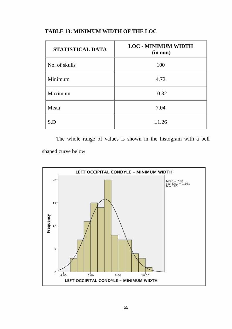
TABLE 13: MINIMUM WIDTH OF THE LOC
STATISTICAL DATA LOC - MINIMUM WIDTH (in mm)
No. of skulls 100
Minimum 4.72
Maximum 10.32
Mean 7.04
S.D ±1.26
The whole range of values is shown in the histogram with a bell
shaped curve below.
56
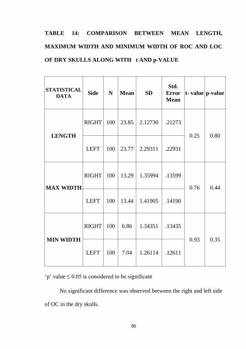
TABLE 14: COMPARISON BETWEEN MEAN LENGTH,
MAXIMUM WIDTH AND MINIMUM WIDTH OF ROC AND LOC
OF DRY SKULLS ALONG WITH t AND p-VALUE
STATISTICAL DATA Side N Mean SD
Std. Error Mean
t- value p-value
LENGTH
RIGHT 100 23.85 2.12730 .21273
0.25 0.80
LEFT 100 23.77 2.29311 .22931
MAX WIDTH
RIGHT 100 13.29 1.35994 .13599
0.76 0.44
LEFT 100 13.44 1.41905 .14190
MIN WIDTH
RIGHT 100 6.86 1.34351 .13435
0.93 0.35
LEFT 100 7.04 1.26114 .12611
‘p’ value ≤ 0.05 is considered to be significant No significant difference was observed between the right and left side
of OC in the dry skulls.
57
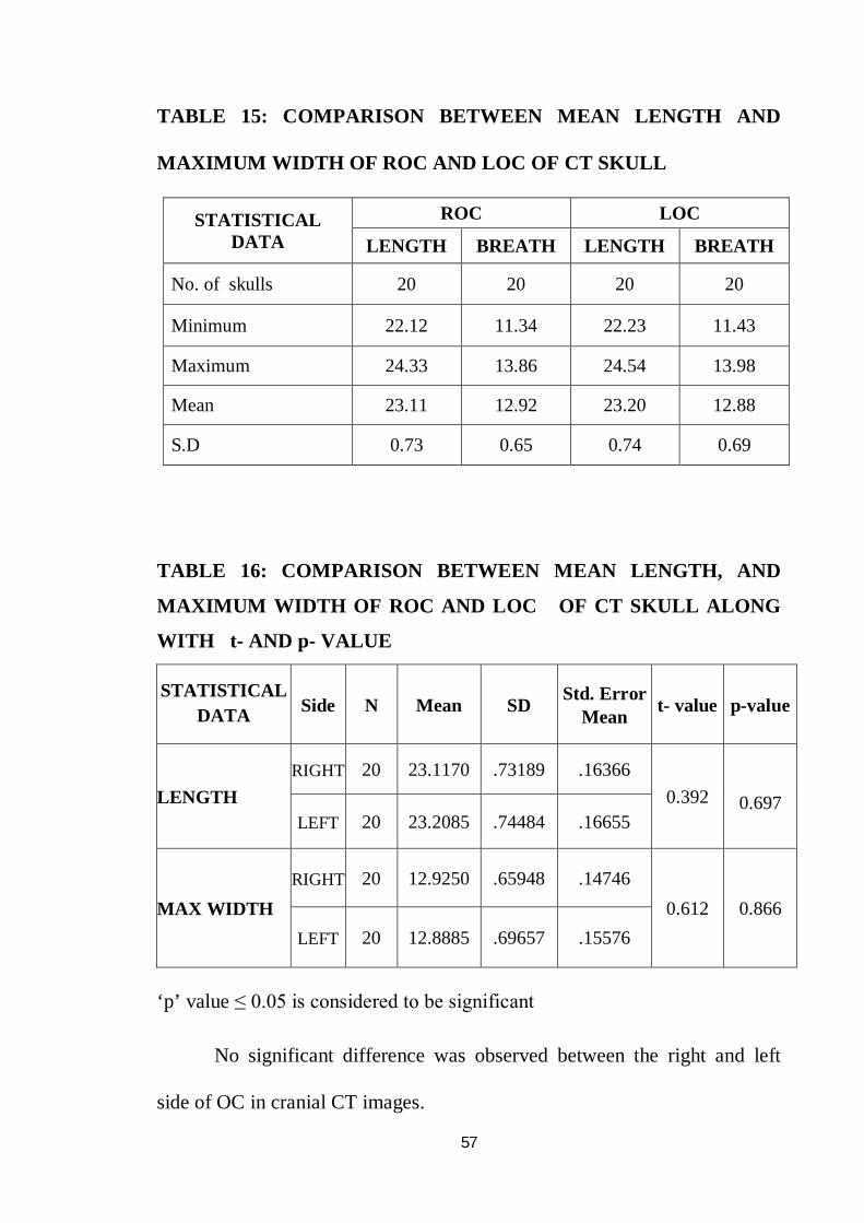
TABLE 15: COMPARISON BETWEEN MEAN LENGTH AND
MAXIMUM WIDTH OF ROC AND LOC OF CT SKULL
STATISTICAL DATA
ROC LOC
LENGTH BREATH LENGTH BREATH
No. of skulls 20 20 20 20
Minimum 22.12 11.34 22.23 11.43
Maximum 24.33 13.86 24.54 13.98
Mean 23.11 12.92 23.20 12.88
S.D 0.73 0.65 0.74 0.69
TABLE 16: COMPARISON BETWEEN MEAN LENGTH, AND
MAXIMUM WIDTH OF ROC AND LOC OF CT SKULL ALONG
WITH t- AND p- VALUE
STATISTICAL DATA Side N Mean SD Std. Error
Mean t- value p-value
LENGTH RIGHT 20 23.1170 .73189 .16366
0.392
0.697 LEFT 20 23.2085 .74484 .16655
MAX WIDTH RIGHT 20 12.9250 .65948 .14746
0.612 0.866 LEFT 20 12.8885 .69657 .15576
‘p’ value ≤ 0.05 is considered to be significant No significant difference was observed between the right and left
side of OC in cranial CT images.
58
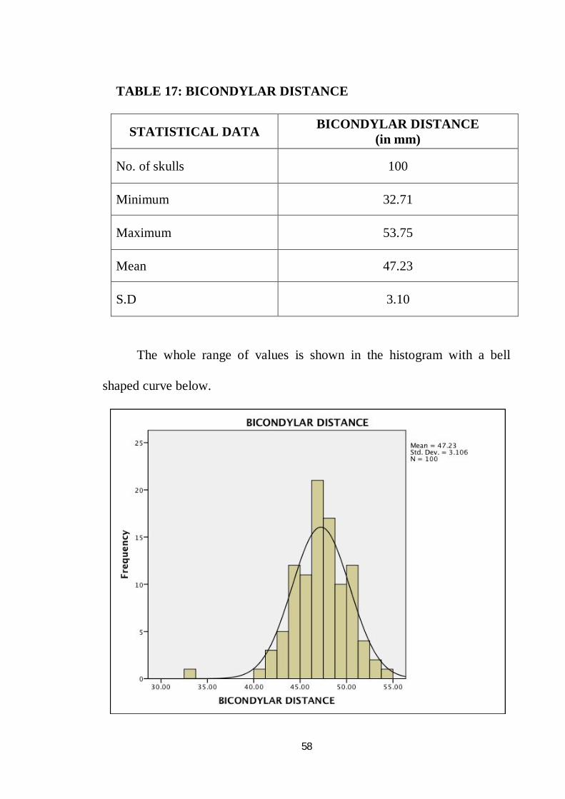
TABLE 17: BICONDYLAR DISTANCE
STATISTICAL DATA BICONDYLAR DISTANCE (in mm)
No. of skulls 100
Minimum 32.71
Maximum 53.75
Mean 47.23
S.D 3.10
The whole range of values is shown in the histogram with a bell
shaped curve below.
59
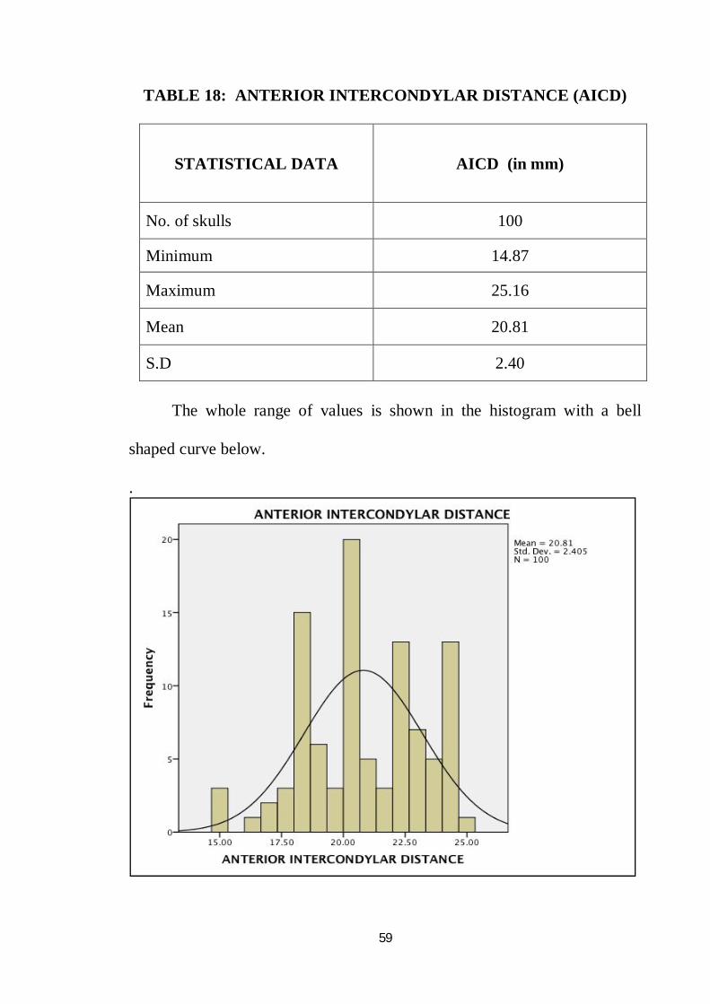
TABLE 18: ANTERIOR INTERCONDYLAR DISTANCE (AICD)
STATISTICAL DATA AICD (in mm)
No. of skulls 100
Minimum 14.87
Maximum 25.16
Mean 20.81
S.D 2.40
The whole range of values is shown in the histogram with a bell
shaped curve below.
.
60
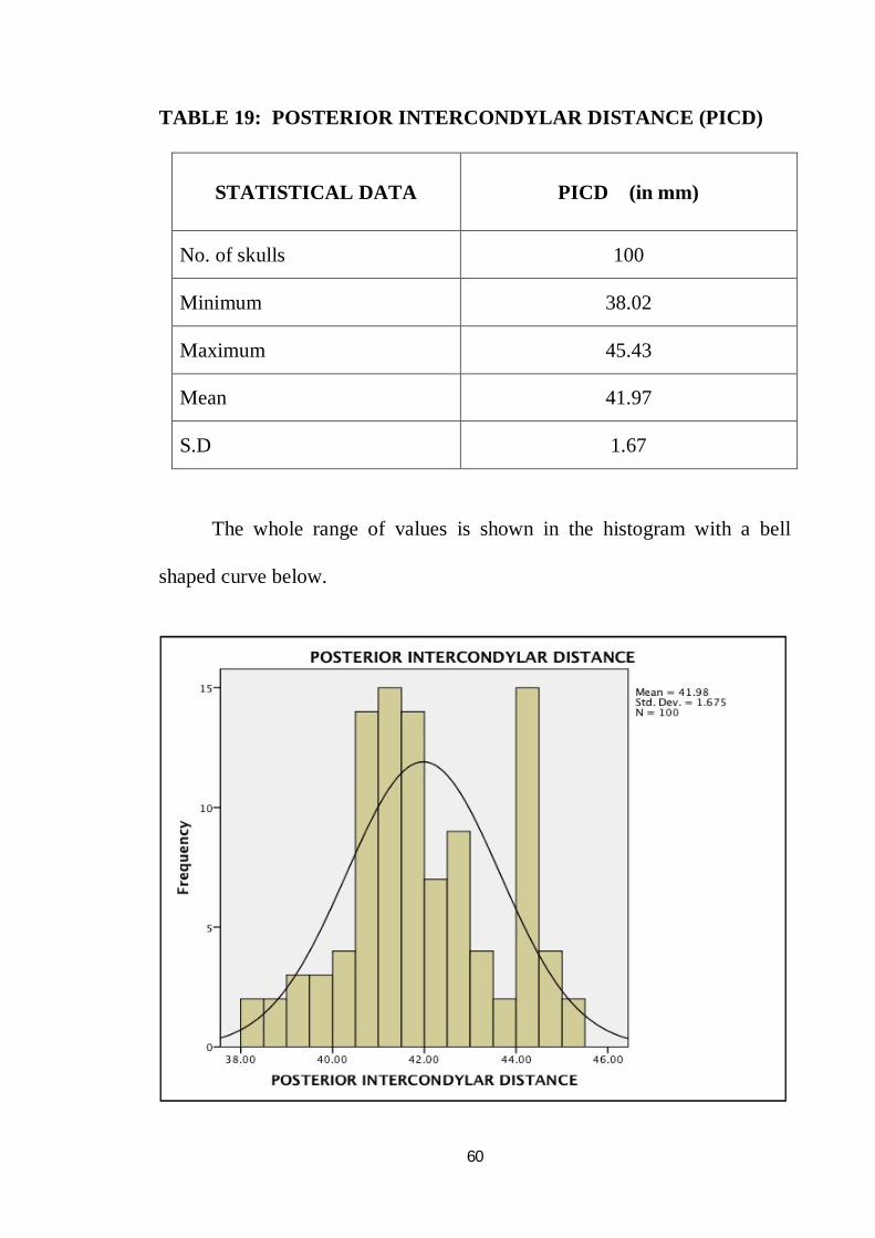
TABLE 19: POSTERIOR INTERCONDYLAR DISTANCE (PICD)
STATISTICAL DATA PICD (in mm)
No. of skulls 100
Minimum 38.02
Maximum 45.43
Mean 41.97
S.D 1.67
The whole range of values is shown in the histogram with a bell
shaped curve below.
61
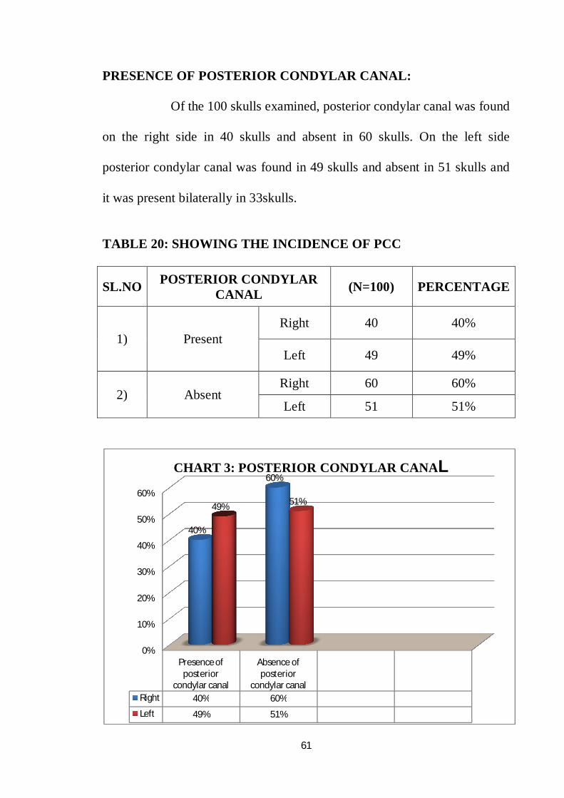
PRESENCE OF POSTERIOR CONDYLAR CANAL:
Of the 100 skulls examined, posterior condylar canal was found
on the right side in 40 skulls and absent in 60 skulls. On the left side
posterior condylar canal was found in 49 skulls and absent in 51 skulls and
it was present bilaterally in 33skulls.
TABLE 20: SHOWING THE INCIDENCE OF PCC
SL.NO POSTERIOR CONDYLAR CANAL (N=100) PERCENTAGE
1) Present Right 40 40%
Left 49 49%
2) Absent Right 60 60%
Left 51 51%
0%
10%
20%
30%
40%
50%
60%
Presence of posterior
condylar canal
Absence of posterior
condylar canalRight 40% 60%
Left 49% 51%
40%
60%
49% 51%
CHART 3: POSTERIOR CONDYLAR CANAL
62
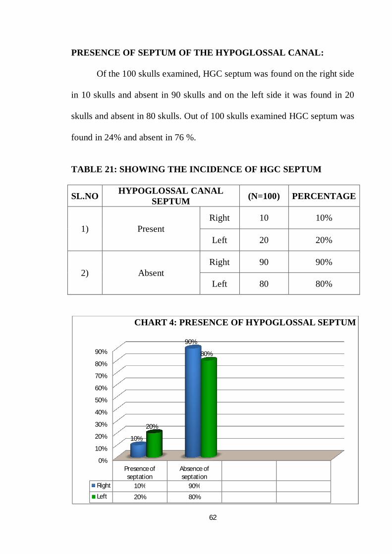
PRESENCE OF SEPTUM OF THE HYPOGLOSSAL CANAL:
Of the 100 skulls examined, HGC septum was found on the right side
in 10 skulls and absent in 90 skulls and on the left side it was found in 20
skulls and absent in 80 skulls. Out of 100 skulls examined HGC septum was
found in 24% and absent in 76 %.
TABLE 21: SHOWING THE INCIDENCE OF HGC SEPTUM
SL.NO HYPOGLOSSAL CANAL SEPTUM (N=100) PERCENTAGE
1) Present Right 10 10%
Left 20 20%
2) Absent Right 90 90%
Left 80 80%
0%
10%
20%
30%
40%
50%
60%
70%
80%
90%
Presence of septation
Absence of septation
Right 10% 90%
Left 20% 80%
10%
90%
20%
80%
CHART 4: PRESENCE OF HYPOGLOSSAL SEPTUM
63
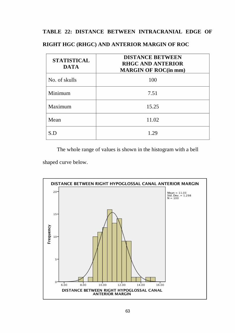
TABLE 22: DISTANCE BETWEEN INTRACRANIAL EDGE OF
RIGHT HGC (RHGC) AND ANTERIOR MARGIN OF ROC
STATISTICAL DATA
DISTANCE BETWEEN RHGC AND ANTERIOR
MARGIN OF ROC(in mm)
No. of skulls 100
Minimum 7.51
Maximum 15.25
Mean 11.02
S.D 1.29
The whole range of values is shown in the histogram with a bell
shaped curve below.

64
TABLE 23: DISTANCE BETWEEN INTRACRANIAL EDGE OF
RIGHT HGC (RHGC) AND POSTERIOR MARGIN OF ROC.
STATISTICAL DATA DISTANCE BETWEEN
RHGC AND POSTERIOR MARGIN OF ROC (in mm)
No. of skulls 100
Minimum 9.8
Maximum 14.90
Mean 12.27
S.D 0.6
The whole range of values is shown in the histogram with a bell
shaped curve below.
65
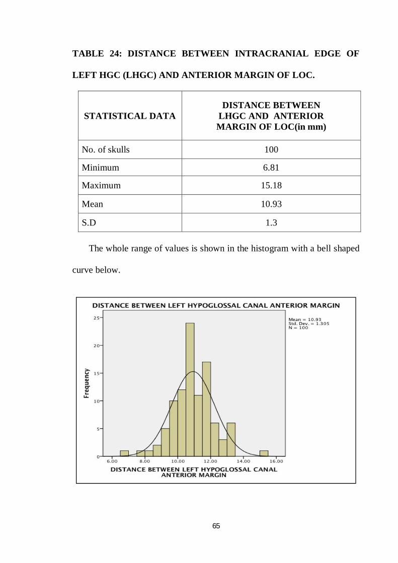
TABLE 24: DISTANCE BETWEEN INTRACRANIAL EDGE OF
LEFT HGC (LHGC) AND ANTERIOR MARGIN OF LOC.
STATISTICAL DATA DISTANCE BETWEEN
LHGC AND ANTERIOR MARGIN OF LOC(in mm)
No. of skulls 100
Minimum 6.81
Maximum 15.18
Mean 10.93
S.D 1.3 The whole range of values is shown in the histogram with a bell shaped
curve below.
66
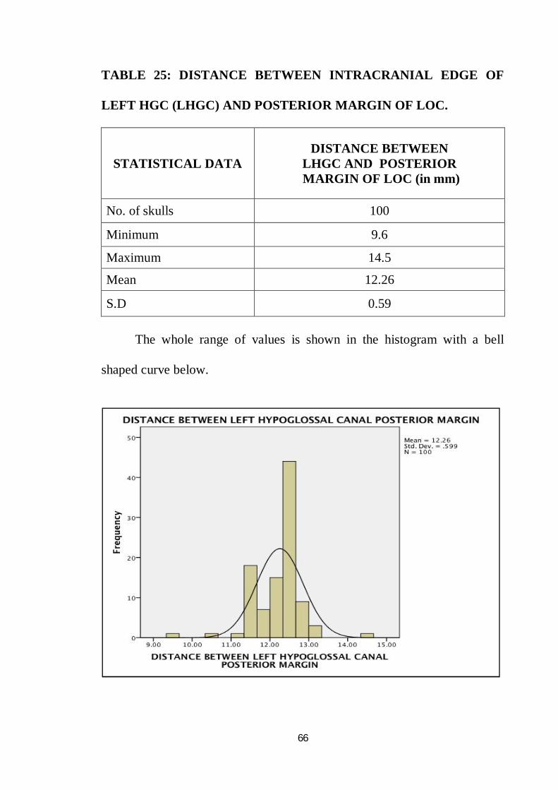
TABLE 25: DISTANCE BETWEEN INTRACRANIAL EDGE OF
LEFT HGC (LHGC) AND POSTERIOR MARGIN OF LOC.
STATISTICAL DATA DISTANCE BETWEEN
LHGC AND POSTERIOR MARGIN OF LOC (in mm)
No. of skulls 100
Minimum 9.6
Maximum 14.5
Mean 12.26
S.D 0.59 The whole range of values is shown in the histogram with a bell
shaped curve below.
67
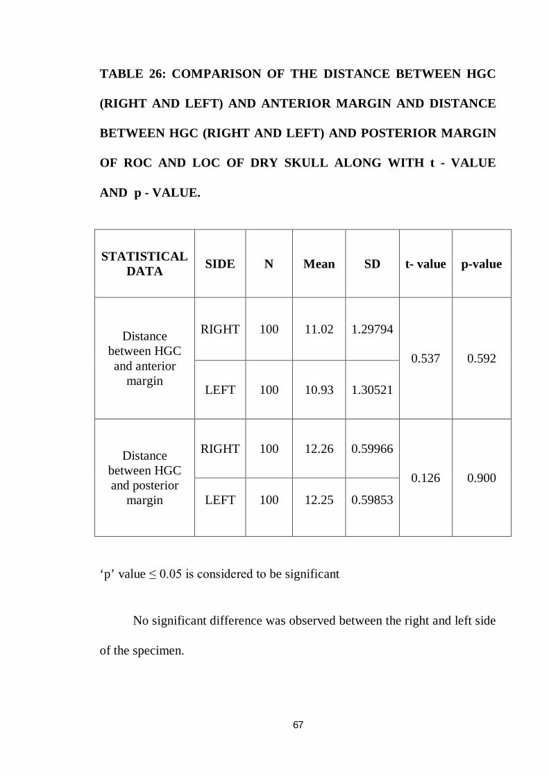
TABLE 26: COMPARISON OF THE DISTANCE BETWEEN HGC
(RIGHT AND LEFT) AND ANTERIOR MARGIN AND DISTANCE
BETWEEN HGC (RIGHT AND LEFT) AND POSTERIOR MARGIN
OF ROC AND LOC OF DRY SKULL ALONG WITH t - VALUE
AND p - VALUE.
STATISTICAL DATA SIDE N Mean SD t- value p-value
Distance between HGC and anterior
margin
RIGHT 100 11.02 1.29794
0.537 0.592
LEFT 100 10.93 1.30521
Distance between HGC and posterior
margin
RIGHT 100 12.26 0.59966
0.126 0.900
LEFT 100 12.25 0.59853
‘p’ value ≤ 0.05 is considered to be significant
No significant difference was observed between the right and left side
of the specimen.

Discussion
68
DISCUSSION
The findings of the present study were correlated with the findings of
other similar studies conducted in different parts of India and in other
countries.
1) SHAPE OF THE FORAMEN MAGNUM
Muthukumar N et al 35(2005) reported that the FM was ovoid in 46%.
P. Chethan et al 41(2011) recorded that the FM was observed to be round in
22.6%, tetragonal in 18.9%, oval in 15.1%, egg shaped in 18.9%,
pentagonal in 3.8% , irregular in 15.1%, and hexagonal in 5.6% of the cases.
Emel AVCL et al 9(2011) stated that the FM was oval in 58%.
Radhakrishnan S.K et al 46(2012) reported that the FM was oval in 39%,
round in 28%, pentagonal in 14% and tetragonal in 19%of the cases.
Radhakrishnan P et al 45(2012) reported that the FM was oval in 35.2%,
hexagonal in 24.8%, pentagonal in 12.4%,round in 7.6%,irregular in 11.6%
,trigonal in 1.6%, pentagonal in 12.4%and tetragonal in 6.8% in cranial CT.
K. Natsis et al 31(2013) found that the FM was two semicircles in 25.9%. It
was pear shaped in 22.4 %, oval 14.7 %, egg shaped in 21 %, rhomboid in
14%,round in1.4 % and irregular in 0.7%.
69
Khalil Awadh Murshed et al 30 (2003)studied CT images of the FM and
recorded that the FM was oval in 8.1%, egg shaped in 6.3%, round in
21.8%, pentagonal in 13.6%, irregular (type A) in 10.9%,hexagonal
in17.2%, tetragonal in 12.7%, and irregular (type B) in 9.09%.
Gobbur et al 19 (2013) reported that the FM was round in 40% and oval
in30% in CT images.
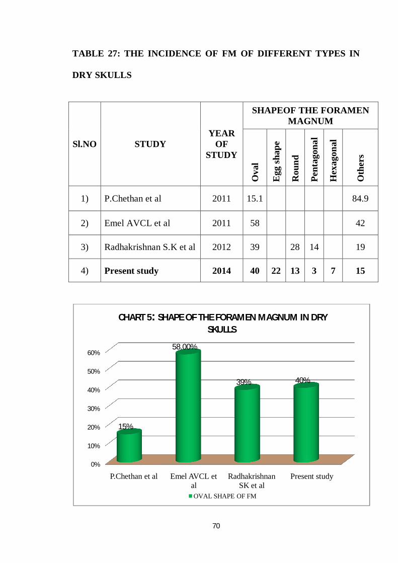
Comparison was done with various studies showing the shape of the
FM and was tabulated.The most common shape of the FM was oval. The
present study also showed that the FM was oval in 40% and egg shaped in
22%.
The variation in the shape of FM should be taken into consideration
during neuroimaging techniques and surgical approaches. In the oval shaped
FM, exposing the anterior portion might be difficult during surgeries.
70
TABLE 27: THE INCIDENCE OF FM OF DIFFERENT TYPES IN
DRY SKULLS
Sl.NO STUDY YEAR
OF STUDY
SHAPEOF THE FORAMEN MAGNUM
Ova
l
Egg
shap
e
Rou
nd
Pent
agon
al
Hex
agon
al
Oth
ers
1) P.Chethan et al 2011 15.1 84.9
2) Emel AVCL et al 2011 58 42
3) Radhakrishnan S.K et al 2012 39 28 14 19
4) Present study 2014 40 22 13 3 7 15
0%
10%
20%
30%
40%
50%
60%
P.Chethan et al Emel AVCL et al
Radhakrishnan SK et al
Present study
15%
58.00%
39% 40%
CHART 5: SHAPE OF THE FORAMEN MAGNUM IN DRY SKULLS
OVAL SHAPE OF FM
71
2) MAXIMUM ANTEROPOSTERIOR DIAMETER OF THE
FORAMEN MAGNUM (FM)
Georges Olivier et al 18(1975) reported that the mean AP diameter of the
FMwas 35.7mm.
Manoel. C et al 33(2009) stated that the mean AP diameter of the FM of
male and female were 35.7±0.29 mm and 35.1± 0.33 mm respectively.
Philipp Gruber et al 43(2009) recorded that the mean AP diameter ranged
from 30.1mm.to 42.6mm with an average of 36.6mm.
Fatma Hayat Eridil et al 13(2010) stated that the mean AP diameter of the
FM was 35±5.8mm in CT scans.
Emel AVCL et al 9(2011) found that the mean AP diameter of the FM was
34.5mm.
F.Burdan et al 11(2012) recorded the mean AP diameter of the FM in male
and female were 37.06mm and 35.57 mm respectively in CT scans.
Gautam Kanodia et al 17(2012) concluded that the mean AP diameter of
the FM was 34.1±0.29mm in dry skull group and 33.1±0.35mm in CT scan.
Osunwoke E.A et al 38(2012) reported that the mean AP diameter of the
FM was 36.11±0.24mm.
72
Radhakrishnan P et al 45(2012) concluded that the AP diameter of FM
varied from 25.8mm to 45.9mm with the average of 35.76±3.4mm in cranial
CT scans.
Fathy Ahmed Fetouh et al 12(2013) recorded that the AP diameter of FM
varied from 31mm to 40.2mm with the average of 34.94mm.
K. Natasis et al 31 (2013) recorded that the mean AP diameter of the FM
was 35.53±3.06mm.
Yogesh Yadav et al 59(2014) reported that the mean AP diameter of the
FM of male and female were 35.22± 2.17mm and 33.1±2.04mm
respectively.
In the Present study, the AP diameter of FM ranged from 24.64mm
to 39.89mm with the average of 35.12±2.65mm. The mean AP diameter of
the FM was compared with that found in various other studies and tabulated.
Longer anteroposterior dimension of the FM permits greater surgical
exposure for occipital condyle resection.
73
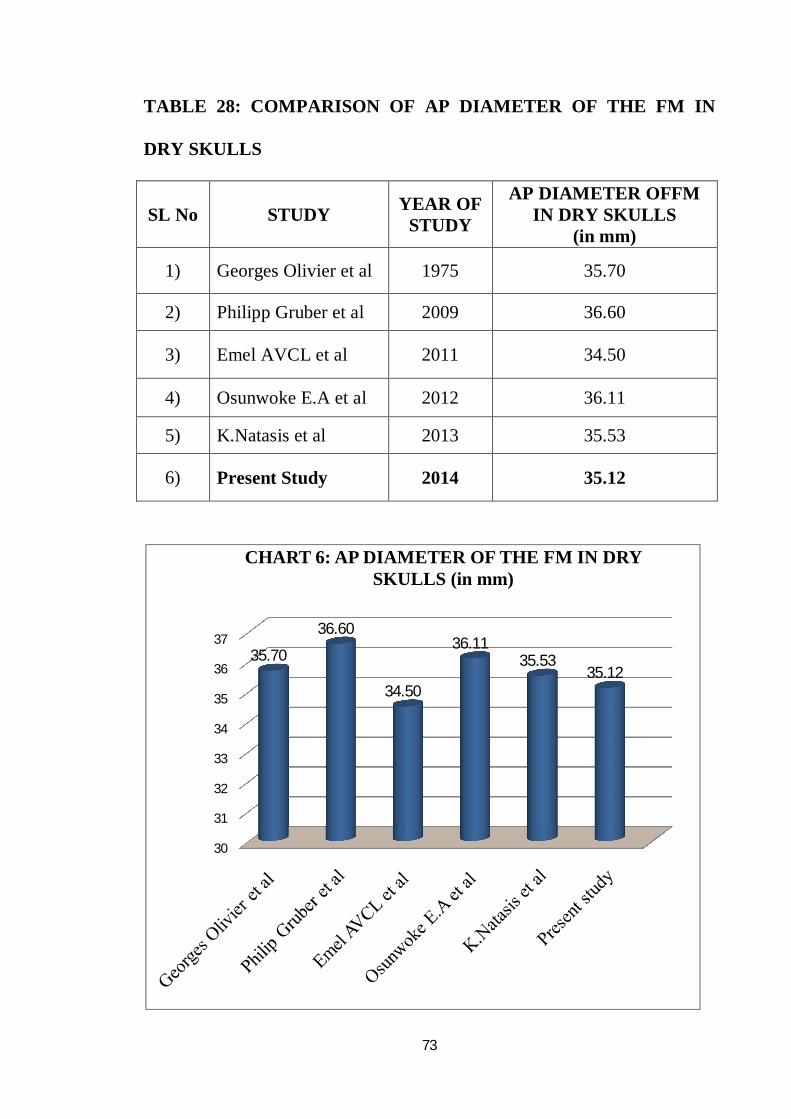
TABLE 28: COMPARISON OF AP DIAMETER OF THE FM IN
DRY SKULLS
SL No STUDY YEAR OF STUDY
AP DIAMETER OFFM IN DRY SKULLS
(in mm)
1) Georges Olivier et al 1975 35.70
2) Philipp Gruber et al 2009 36.60
3) Emel AVCL et al 2011 34.50
4) Osunwoke E.A et al 2012 36.11
5) K.Natasis et al 2013 35.53
6) Present Study 2014 35.12
30
31
32
33
34
35
36
3735.70
36.60
34.50
36.1135.53
35.12
CHART 6: AP DIAMETER OF THE FM IN DRY SKULLS (in mm)
74
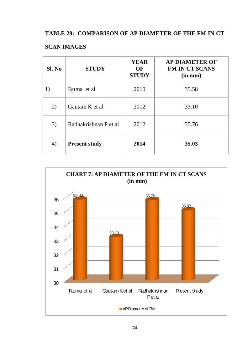
TABLE 29: COMPARISON OF AP DIAMETER OF THE FM IN CT
SCAN IMAGES
Sl. No STUDY YEAR
OF STUDY
AP DIAMETER OF FM IN CT SCANS
(in mm)
1) Fatma et al 2010 35.58
2) Gautam K et al 2012 33.10
3) Radhakrishnan P et al 2012 35.76
4) Present study 2014 35.03
30
31
32
33
34
35
36
Fatma et al Gautam K et al Radhakrishnan P et al
Present study
35.80
33.10
35.76
35.03
CHART 7: AP DIAMETER OF THE FM IN CT SCANS(in mm)
AP Diameter of FM
75
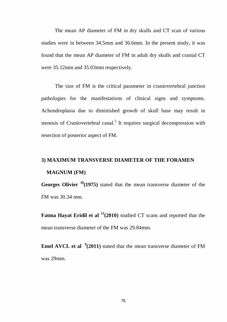
The mean AP diameter of FM in dry skulls and CT scan of various
studies were in between 34.5mm and 36.6mm. In the present study, it was
found that the mean AP diameter of FM in adult dry skulls and cranial CT
were 35.12mm and 35.03mm respectively.
The size of FM is the critical parameter in craniovertebral junction
pathologies for the manifestations of clinical signs and symptoms.
Achondroplasia due to diminished growth of skull base may result in
stenosis of Craniovertebral canal.5 It requires surgical decompression with
resection of posterior aspect of FM.
3) MAXIMUM TRANSVERSE DIAMETER OF THE FORAMEN
MAGNUM (FM)
Georges Olivier 18(1975) stated that the mean transverse diameter of the
FM was 30.34 mm.
Fatma Hayat Eridil et al 13(2010) studied CT scans and reported that the
mean transverse diameter of the FM was 29.84mm.
Emel AVCL et al 9(2011) stated that the mean transverse diameter of FM
was 29mm.
76
Gautam Kanodia et al 17(2012) reported that the mean transverse diameter
of the FM was 27.5±0.25mm in dry skull group and 27.6±0.31mm in cranial
CT scan.
Osunwoke E.A et al 38(2012) reported that the mean transverse diameter of
the FM was29.65±0.24mm.
Radhakrishnan P et al 45(2012) reported that the mean transverse diameter
of FM ranged from 22mm to 39.1mm with the average of 29.79±2.85mm
in cranial CT scans.
K. Natasis et al 31(2013) reported the mean transverse diameter of the
FMwas 30.31±2.79mm.
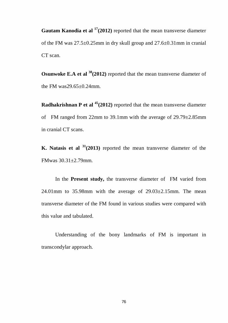
In the Present study, the transverse diameter of FM varied from
24.01mm to 35.98mm with the average of 29.03±2.15mm. The mean
transverse diameter of the FM found in various studies were compared with
this value and tabulated.
Understanding of the bony landmarks of FM is important in
transcondylar approach.
77
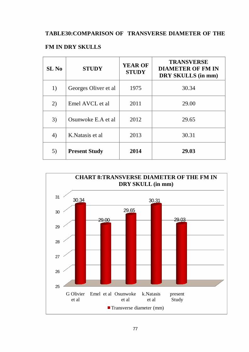
TABLE30:COMPARISON OF TRANSVERSE DIAMETER OF THE
FM IN DRY SKULLS
SL No STUDY YEAR OF STUDY
TRANSVERSE DIAMETER OF FM IN DRY SKULLS (in mm)
1) Georges Oliver et al 1975 30.34
2) Emel AVCL et al 2011 29.00
3) Osunwoke E.A et al 2012 29.65
4) K.Natasis et al 2013 30.31
5) Present Study 2014 29.03
25
26
27
28
29
30
31
G Olivier et al
Emel et al Osunwoke et al
k.Natasis et al
present Study
30.34
29.00
29.65
30.31
29.03
CHART 8:TRANSVERSE DIAMETER OF THE FM IN DRY SKULL (in mm)
Transverse diameter (mm)
78
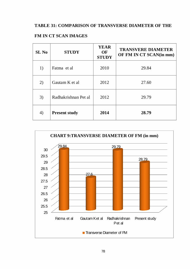
TABLE 31: COMPARISON OF TRANSVERSE DIAMETER OF THE
FM IN CT SCAN IMAGES
SL No STUDY YEAR
OF STUDY
TRANSVERE DIAMETER OF FM IN CT SCAN(in mm)
1) Fatma et al 2010 29.84
2) Gautam K et al 2012 27.60
3) Radhakrishnan Pet al 2012 29.79
4) Present study 2014 28.79
25
25.5
26
26.5
27
27.5
28
28.5
29
29.5
30
Fatma et al Gautam K et al Radhakrishnan P et al
Present study
29.84
27.6
29.79
28.79
CHART 9:TRANSVERSE DIAMETER OF FM (in mm)
Transverse Diameter of FM
79
The mean transverse diameter of FM in dry skulls and CT scans of
various studies gave values in between 34.5mm and36.6mm. In the present
study, the mean transverse diameter of FM in adult human dry skulls and
cranial CT were measured as 29mm and 28.79mm respectively. Minor
controversies were seen in some studies.
The diminished size of FM is seen in craniometaphyseal dysplasia
and Marchesani’s syndrome which cause stenos is of Craniovertebral
junction.
4) PROTRUSION OF OCCIPITAL CONDYLE INTO THE
FORAMEN MAGNUM.
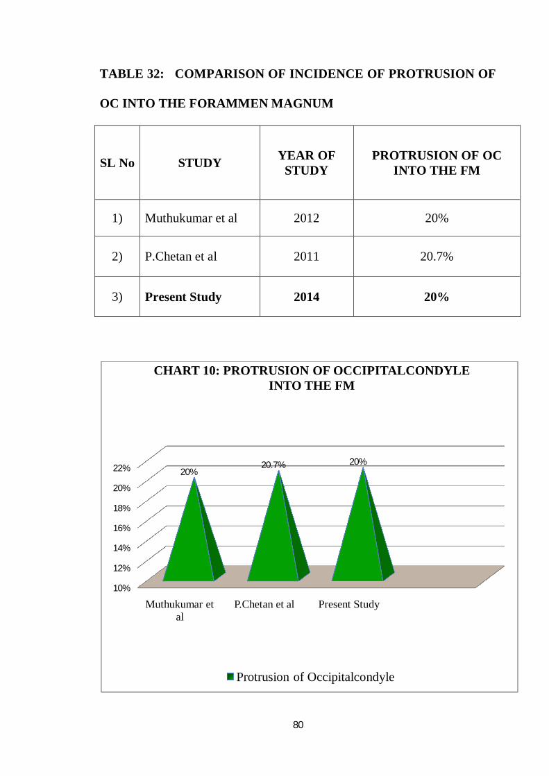
Muthukumar N et al 35(2005) reported that the OCs protrude into the FM
in 20% of adult dry skulls.
P. Chethan et al 41(2011)and Emel AVCL et al 9 (2011)found that the OCs
protruded into the FM in 20.7% and57% of skulls respectively.
In the Present study it was found that the occipital condyles
protruded into the FM in 20% of adult dry skulls.
In other studies, protrusion of OC into the FM was mostly seen in
oval or egg shaped FM. In case of protrusion of OC, more extensive
removal of OC may be indicated during surgeries involving skull base,
which may cause greater craniovertebral instability.
80
TABLE 32: COMPARISON OF INCIDENCE OF PROTRUSION OF
OC INTO THE FORAMMEN MAGNUM
SL No STUDY YEAR OF STUDY
PROTRUSION OF OC INTO THE FM
1) Muthukumar et al 2012 20%
2) P.Chetan et al 2011 20.7%
3) Present Study 2014 20%
10%
12%
14%
16%
18%
20%
22%
Muthukumar et al
P.Chetan et al Present Study
20%20.7% 20%
CHART 10: PROTRUSION OF OCCIPITALCONDYLE INTO THE FM
Protrusion of Occipitalcondyle
81
5) LENGTH OF THE RIGHT OCCIPITAL CONDYLE
Georges Olivier 18(1975) reported that the mean length of the ROC was
23.75mm.
Muthukumar N et al 35(2005) and Sait Naderi et al 49 (2005) reported
that the length of the ROC was 23.6mm.
Emel AVCL et al 9(2011) reported that the mean maximum length of ROC
varied from 18.2mm to 28.7mm with an average of 23.7±2.6 mm.
Mehmet Asim Ozer et al 34 (2011) recorded that the mean length ofROC
was 23.9±3.4mm.
Tien V et al 56 (2011) stated that the mean length of ROC was 22.2±2.1mm
in cranial CT scans.
Fathy Ahmed Fetouh et al 12 (2013) recorded that the mean maximum
length of ROC ranged from 18mm to 31mm with an average of 23.5mm.
Bello S.S et al 4(2013) reported in cranial CT that the mean length of ROC
was 23.5±2.7mm.
In the Present study the mean maximum length of ROC ranged from
18.16mm to 32.68mm with an average of 23.85±2.12mm.
The mean maximum lengths of ROC were compared and results
shown in the table where the values were in between 23.5mm and 23.85mm.
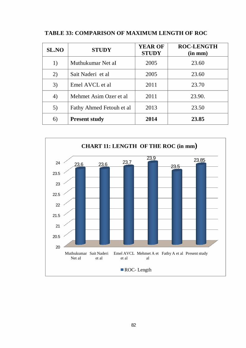
82
TABLE 33: COMPARISON OF MAXIMUM LENGTH OF ROC
SL.NO STUDY YEAR OF STUDY
ROC-LENGTH (in mm)
1) Muthukumar Net aI 2005 23.60
2) Sait Naderi et al 2005 23.60
3) Emel AVCL et al 2011 23.70
4) Mehmet Asim Ozer et al 2011 23.90.
5) Fathy Ahmed Fetouh et al 2013 23.50
6) Present study 2014 23.85
20
20.5
21
21.5
22
22.5
23
23.5
24
Muthukumar Net aI
Sait Naderi et al
Emel AVCL et al
Mehmet A et al
Fathy A et al Present study
23.6 23.6 23.723.9
23.523.85
CHART 11: LENGTH OF THE ROC (in mm)
ROC- Length
83
6) MAXIMUM WIDTH OF THE RIGHT OCCIPITAL CONDYLE
Muthukumar N et al 35(2005) found that the mean ROC width was
14.72mm.
Sait Naderi et al 49(2005) reported that the ROC width was 10.6mm.
Emel AVCL et al 9(2011) recorded that the maximum width of ROC
varied from 9 mm to 14.5 mm with an average of 12.2±1.2mm .
Pereira G.A et al 42(2012) reported that the ROC mean width was
13.4±1.4mm.
Bello S.S et al 4(2013) stated that the mean width of ROC in cranial CT was
12.8±1.7mm.
Fathy Ahmed Fetouh et al 12(2013) recorded that the mean maximum
width of ROC ranged from 9.5mm to 18mm with an average of 13.58mm.
K. Natasis et al 31(2013) found that the ROC maximum width was
13.09±1.99mm.
In the Present study the maximum width of ROC was in the range of
9.76mm to 16.19mm with an average of 13.29 ±1.36mm.
The maximum length of ROC was compared and results shown in the
table where the values were in between 12.2mm and 14.72mm.
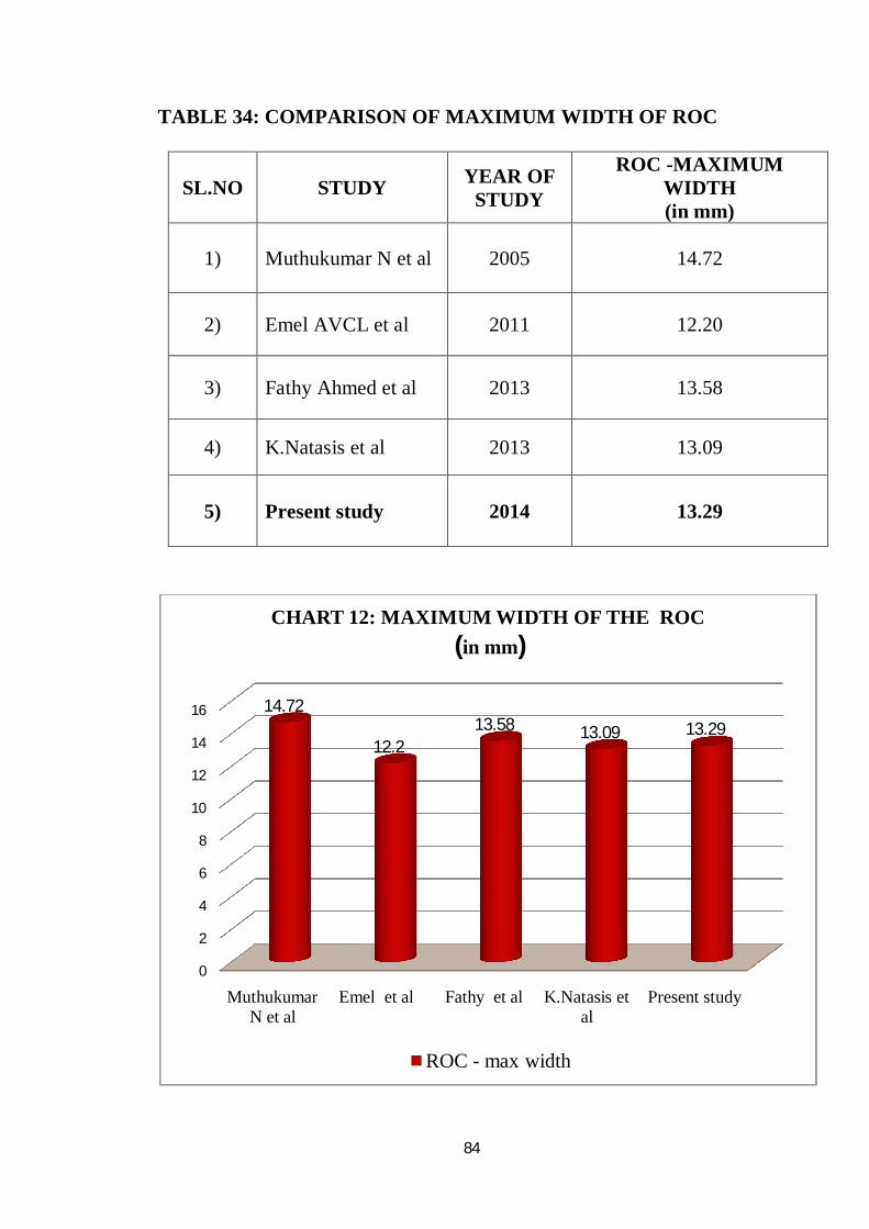
84
TABLE 34: COMPARISON OF MAXIMUM WIDTH OF ROC
SL.NO STUDY YEAR OF STUDY
ROC -MAXIMUM WIDTH (in mm)
1) Muthukumar N et al 2005 14.72
2) Emel AVCL et al 2011 12.20
3) Fathy Ahmed et al 2013 13.58
4) K.Natasis et al 2013 13.09
5) Present study 2014 13.29
0
2
4
6
8
10
12
14
16
Muthukumar N et al
Emel et al Fathy et al K.Natasis et al
Present study
14.72
12.213.58 13.09 13.29
CHART 12: MAXIMUM WIDTH OF THE ROC(in mm)
ROC - max width
85
7) MINIMUM WIDTH OF THE RIGHT OCCIPITAL CONDYLE
K. Natasis et al 31(2013) found that the minimum width of ROC was
5.71±1.61mm.
In thePresent study the minimum width of ROC was in the range of
3.25mm to 10.62mm with an average of 6.86mm.
8) LENGTH OF THE LEFT OCCIPITAL CONDYLE
Sait Naderi et al 49(2005) reported that the length of the LOC was 23.2mm.
Emine et al 10(2006) reported that the LOC length varied from 18.2mm to
31.1mm with an average of 24.6±2.5mm
Emel AVCL et al 9(2011) reported that the maximum length of LOC was
in the range of 18.8 mm to 30.9mm with an average of 24.7±2.7mm.
Mehmet Asim Ozer et al 34(2011) recorded that the length of LOC was
23.92±3.3mm.
Tien V et al 56(2011) stated that the mean length of LOC was 22.5±2.2mm
in cranial CT scans.
Pereira G.A et al 42(2012) reported that the LOC mean length was
23.3±2.6mm.
86
Bello S.S et al 4(2013) reported that the mean length of LOC was
23.7±2.8mm in CT scans.
Fathy Ahmed Fetouh et al 12(2013)recorded that the mean length of LOC
varied from 18.3mm to 29.4mm with an average of 23.75mm.
K. Natasis et al 31(2013) found that the LOC length was 25.60±2.70mm.
In the Present study the maximum length of LOC was in the range of
17.25 mm to 32.02mm with an average of 23.77±2.29mm.
The maximum length of OC on right and left side was compared and
results shown in the table, where the values were in between 23.50mm and
23.85mm.
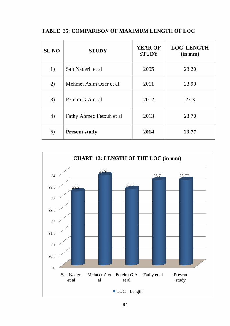
87
TABLE 35: COMPARISON OF MAXIMUM LENGTH OF LOC
SL.NO STUDY YEAR OF STUDY
LOC LENGTH (in mm)
1) Sait Naderi et al 2005 23.20
2) Mehmet Asim Ozer et al 2011 23.90
3) Pereira G.A et al 2012 23.3
4) Fathy Ahmed Fetouh et al 2013 23.70
5) Present study 2014 23.77
20
20.5
21
21.5
22
22.5
23
23.5
24
Sait Naderi et al
Mehmet A et al
Pereira G.A et al
Fathy et al Present study
23.2
23.9
23.3
23.7 23.77
CHART 13: LENGTH OF THE LOC (in mm)
LOC - Length
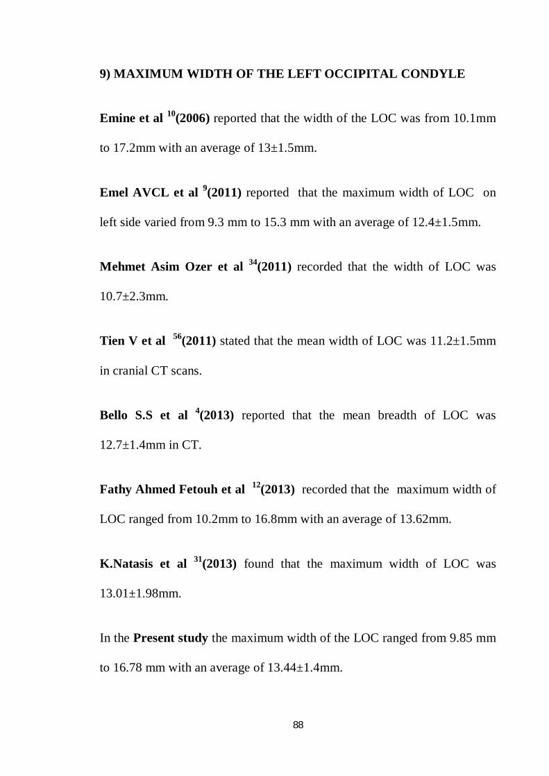
88
9) MAXIMUM WIDTH OF THE LEFT OCCIPITAL CONDYLE
Emine et al 10(2006) reported that the width of the LOC was from 10.1mm
to 17.2mm with an average of 13±1.5mm.
Emel AVCL et al 9(2011) reported that the maximum width of LOC on
left side varied from 9.3 mm to 15.3 mm with an average of 12.4±1.5mm.
Mehmet Asim Ozer et al 34(2011) recorded that the width of LOC was
10.7±2.3mm.
Tien V et al 56(2011) stated that the mean width of LOC was 11.2±1.5mm
in cranial CT scans.
Bello S.S et al 4(2013) reported that the mean breadth of LOC was
12.7±1.4mm in CT.
Fathy Ahmed Fetouh et al 12(2013) recorded that the maximum width of
LOC ranged from 10.2mm to 16.8mm with an average of 13.62mm.
K.Natasis et al 31(2013) found that the maximum width of LOC was
13.01±1.98mm.
In the Present study the maximum width of the LOC ranged from 9.85 mm
to 16.78 mm with an average of 13.44±1.4mm.
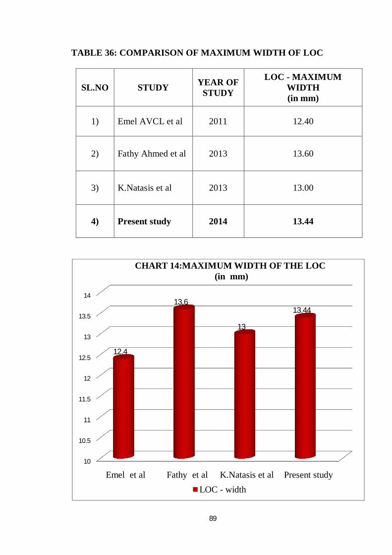
89
TABLE 36: COMPARISON OF MAXIMUM WIDTH OF LOC
SL.NO STUDY YEAR OF STUDY
LOC - MAXIMUM WIDTH (in mm)
1) Emel AVCL et al 2011 12.40
2) Fathy Ahmed et al 2013 13.60
3) K.Natasis et al 2013 13.00
4) Present study 2014 13.44
10
10.5
11
11.5
12
12.5
13
13.5
14
Emel et al Fathy et al K.Natasis et al Present study
12.4
13.6
13
13.44
CHART 14:MAXIMUM WIDTH OF THE LOC(in mm)
LOC - width
90
10) MINIMUM WIDTH OF THE LEFT OCCIPITAL CONDYLE
K. Natasis et al 31(2013) found that the minimum width of LOC was
6.25±1.76mm.
In the Present study the minimum width of LOC ranged from 4.7
mm to 10.32 mm with an average of 7.04±1.26mm.
TABLE 37: COMPARISON OF OC DIMENSION OF THE PRESENT
STUDY WITH OTHER RADIOLOGICAL STUDIES.
SL.NO STUDY YEAR
OF STUDY
ROC LOC
LENGTH(in mm)
WIDTH (in mm)
LENGTH (in mm)
WIDTH (in mm)
1) Tein et al 2011 22.20 11.20 22.50 11.20
2) Bello et al 2013 23.50 12.80 23.70 12.70
3) Present study 2014 23.11 12.92 23.20 12.88
In the Present study it was found that the mean length of ROC and
LOC were 23.85mm and 23.77mm respectively in dry skulls. The mean
length of ROC and LOC were 23.11mm and 23.20mm respectively in
cranial CT.
91
The mean width of ROC and LOC were 13.29mm and 13.44mm
respectively in dry skulls. The mean width of ROC and LOC were
12.92mmand 12.88mm respectively in cranial CTs. The measurements of
OC in dry skulls were greater than those in cranial CT scans.
The OC forms the lateral boundary of the FM. The morphology and
metric measurements of OC may affect the skull base surgery. The
transcondylar approach is the optimal approach to resect lesions present
ventral to FM.37Many studies agreed that the craniovertebral stability was
not affected by one third removal of OC, however, in short OCs the same
amount of condylectomy may cause Craniovertebral instability. Long OC
may require more extensive resection for better surgical exposure.
11) BICONDYLAR DISTANCE (BCD)
Daniel J et al 7(2001) reported that the BCD of black male and female
were 49.6mm and 47.3mm respectively and white male and female were
51.9mm and 49.8mm respectively
Gagandeep Singh et al 15(2012) reported that the BCD of male and female
were 46.73mm and 44.29mm respectively.
In the Present study, the maximum bicondylar distance of the FM
ranged from 32.71mmto 53.75mm with an average of 47.23±3.10 mm.
92
The BCD may show differences between skulls of female and male,
which is greater in male.
12) ANTERIOR INTERCONDYLAR DISTANCE (AICD)
Daniel J et al 7(2001) reported that the AICD of black male and female
were 20.1mm and 18.6mm respectively and white male and female were
20.9mm and 19.2mm respectively
Sait Naderi et al 49(2005) stated that the AICD was 21.0mm.
Emine et al 10(2006) reported that the AICD varied from 15mm to 32mm
with an average of 22.6±3.9mm.
Mehmet AsimOzer et al 34(2011) recorded that the mean AICD was
20.9±3.6mm.
di Vasudha V. Saralaya et al 8(2012) reported that the mean AICD was
18.7mm.
Gagandeep Singh et al15(2012) reported that the minimum intercondylar
distance of the FM of male and female were 14.88mm and 14.33mm
respectively.
Fathy Ahmed Fetouh et al 12(2013) recorded that the AICD varied from
11.5mm to 25.5mm with an average of 20.64mm.
93
K. Natasis et al 31(2013) found that the mean AICD was 19.30±3.25mm.
Pooja Gangrade et al 44(2013) recorded that the mean AICD of male and
female were 18.32mm and 15.44mm respectively.
Parvindokht Bayat et al 39(2014) found that the AICD ranged from 2mm
to 42mm with an average of 15.39±7.99mm.
In the present study the AICD was in the range of 14.87mm to
25.16mm with an average of 20.81±2.40mm.
The values of present study coincided with various other studies.
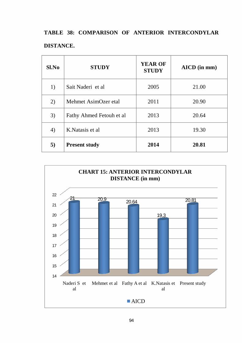
94
TABLE 38: COMPARISON OF ANTERIOR INTERCONDYLAR
DISTANCE.
Sl.No STUDY YEAR OF STUDY AICD (in mm)
1) Sait Naderi et al 2005 21.00
2) Mehmet AsimOzer etal 2011 20.90
3) Fathy Ahmed Fetouh et al 2013 20.64
4) K.Natasis et al 2013 19.30
5) Present study 2014 20.81
14
15
16
17
18
19
20
21
22
Naderi S et al
Mehmet et al Fathy A et al K.Natasis et al
Present study
21 20.9 20.64
19.3
20.81
CHART 15: ANTERIOR INTERCONDYLAR DISTANCE (in mm)
AICD
95
13) POSTERIOR INTERCONDYLAR DISTANCE (PICD)
di Vasudha V. Saralaya et al 8(2012) reported that the mean PICD was
38.7mm.
Saiet Naderi et al 49(2005) stated that the PICD ranged from 35.1mm to
48.3mm with an average of 41.6mm.
Emine et al 10(2006) reported that the PICD varied from 33mm to 50mm
with an average of 44.2±3.2mm.
Gagandeep Singh et al 15(2012) reported that the maximum intercondylar
distance of male and female were 26.15mm and 24.71mm respectively.
Fathy Ahmed Fetouh et al 12(2013) recorded that the PICD ranged from
35.5mm to 48.5mm with an average of 41.4mm.
K. Natasis et al 31(2013) found that the mean anterior inter condylar
distance was 51.61±5.01mm.
Parvindokht Bayat et al 39(2014) reported that the PICD varied from
13mm to 44mm with an average of 35.60±8.4mm.
In the Present study the PICD was in the range of 38.02 mm to 45.43
mm with an average of 41.97±1.67mm. The values of present study
coincided with other studies.
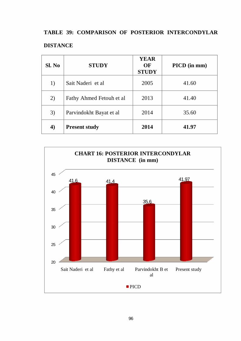
96
TABLE 39: COMPARISON OF POSTERIOR INTERCONDYLAR
DISTANCE
Sl. No STUDY YEAR
OF STUDY
PICD (in mm)
1) Sait Naderi et al 2005 41.60
2) Fathy Ahmed Fetouh et al 2013 41.40
3) Parvindokht Bayat et al 2014 35.60
4) Present study 2014 41.97
20
25
30
35
40
45
Sait Naderi et al Fathy et al Parvindokht B et al
Present study
41.6 41.4
35.6
41.97
CHART 16: POSTERIOR INTERCONDYLAR DISTANCE (in mm)
PICD
97
As occipital condyles bound the FM laterally and converge ventrally, the
OCs have different ventral and dorsal dimensions. Hence, AICD and PICD
showed different values. Transcondylar approach required condylectomy for
optimum visualization to resect any lesion in the ventral or ventrolateral
aspect of FM.
14) PRESENCE OF POSTERIOR CONDYLAR CANAL
Emel AVCL et al 9(2011) reported that the posterior condylar canal was
absent unilaterally in 27% of skulls and bilaterally in 17% of the skulls.
K. Natsis et al 31(2013) found that the PCC was present in 75.5% , out of
this on right side in 11.9% and on left side in 16.1% and bilaterally in
47.6%.
Jatin Goda et al 24(2013) observed that PCC was present in 70.31%
bilaterally and in 20.31% unilaterally.
Ketu Chauhan et al29 (2013) found that PCC was present in 6% on left side
and bilaterally in 3.6%.
Parvindokht Bayat et al 39(2014) found that the PCC was present in 4% on
right side, 16% on left side and bilaterally in 40%.
98
In the Present study, it was observed that the PCC was found in 40
skulls and absent in 60 skulls on right side.
On the left side, it was found in 49 skulls and absent in 51 skulls.
In around 33 skulls it was present bilaterally.
Through the PCC, an emissary vein and a nerve to the duramater of the
posterior cranial fossa pass. On full extension of neck, compression of the
structures passing through the PCC can occur, because the posterior margin
of the atlas impinges itself into the condylar fossa.
The percentage of PCC in various studies was tabulated. This study
showed greater values, which is significant.
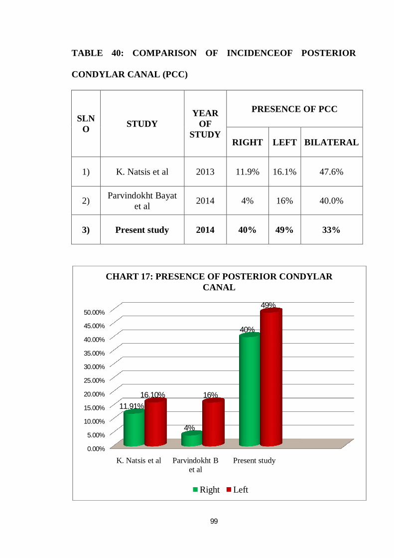
99
TABLE 40: COMPARISON OF INCIDENCEOF POSTERIOR
CONDYLAR CANAL (PCC)
SLNO STUDY
YEAR OF
STUDY
PRESENCE OF PCC
RIGHT LEFT BILATERAL
1) K. Natsis et al 2013 11.9% 16.1% 47.6%
2) Parvindokht Bayat et al 2014 4% 16% 40.0%
3) Present study 2014 40% 49% 33%
0.00%
5.00%
10.00%
15.00%
20.00%
25.00%
30.00%
35.00%
40.00%
45.00%
50.00%
K. Natsis et al Parvindokht B et al
Present study
11.91%
4%
40%
16.10% 16%
49%
CHART 17: PRESENCE OF POSTERIOR CONDYLAR CANAL
Right Left
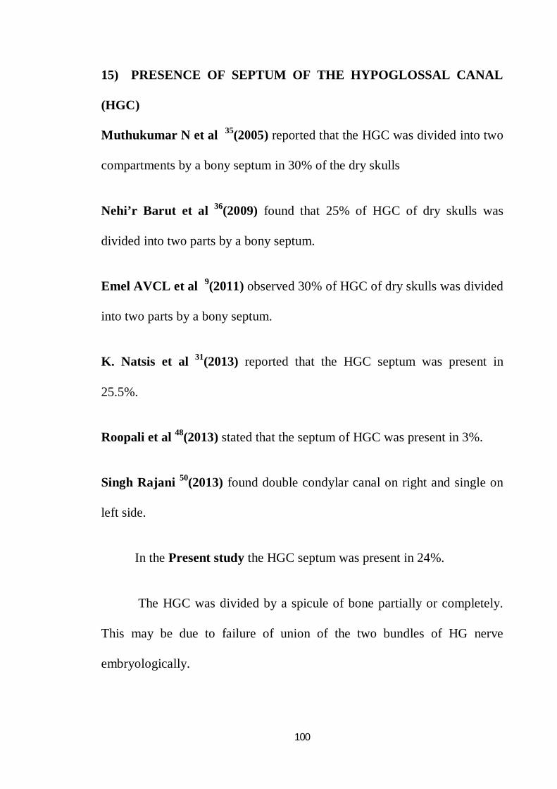
100
15) PRESENCE OF SEPTUM OF THE HYPOGLOSSAL CANAL
(HGC)
Muthukumar N et al 35(2005) reported that the HGC was divided into two
compartments by a bony septum in 30% of the dry skulls
Nehi’r Barut et al 36(2009) found that 25% of HGC of dry skulls was
divided into two parts by a bony septum.
Emel AVCL et al 9(2011) observed 30% of HGC of dry skulls was divided
into two parts by a bony septum.
K. Natsis et al 31(2013) reported that the HGC septum was present in
25.5%.
Roopali et al 48(2013) stated that the septum of HGC was present in 3%.
Singh Rajani 50(2013) found double condylar canal on right and single on
left side.
In the Present study the HGC septum was present in 24%.
The HGC was divided by a spicule of bone partially or completely.
This may be due to failure of union of the two bundles of HG nerve
embryologically.
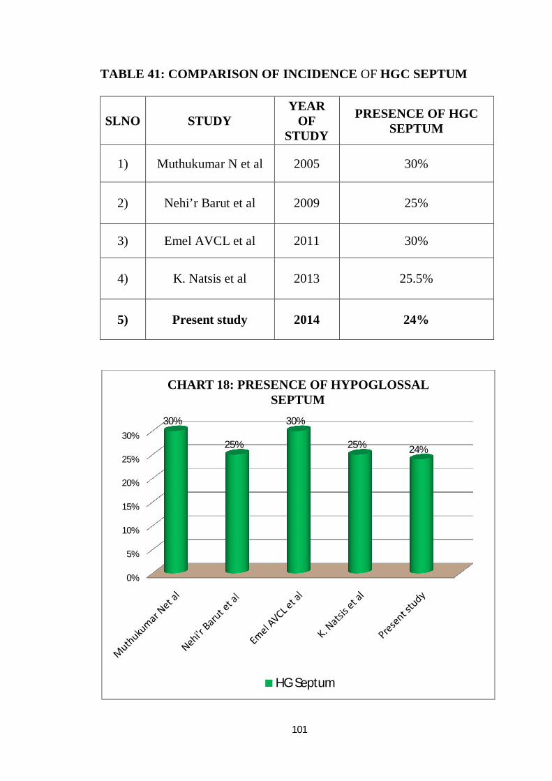
101
TABLE 41: COMPARISON OF INCIDENCE OF HGC SEPTUM
SLNO STUDY YEAR
OF STUDY
PRESENCE OF HGC SEPTUM
1) Muthukumar N et al 2005 30%
2) Nehi’r Barut et al 2009 25%
3) Emel AVCL et al 2011 30%
4) K. Natsis et al 2013 25.5%
5) Present study 2014 24%
0%
5%
10%
15%
20%
25%
30%
30%
25%
30%
25% 24%
CHART 18: PRESENCE OF HYPOGLOSSAL SEPTUM
HG Septum
102
16) DISTANCE BETWEEN INTRACRANIAL EDGE OF RIGHT
HYPOGLOSSAL CANAL (RHGC) AND ANTERIOR MARGINOF ROC
Sait Naderi et al 49(2005) reported that the distance of the intracranial end
of the RHGC from the anterior tip of the ROC was10.6mm.
Emine et al 10(2006) reported that the distance of the intracranial end of the
RHGC from the anterior margin of the ROC varied from 8.1mm to 16.9mm
with an average of 11.0±1.6mm.
Pereira G.A et al 42(2012) reported that the distance of the intracranial end
of the RHGC from the anterior margin of the ROC was 11.0±1.8mm.
In the Present study the distance of the intracranial edge of the
RHGC from the anterior margin of the ROC ranged from 7.51mm to
15.25mm with an average of 11.02±1.29mm.
TABLE 42: COMPARISON OF DISTANCE BETWEEN INTRACRANIAL
EDGE OF RHGC AND ANTERIOR MARGIN OF ROC.
SLNO STUDY YEAR OF STUDY
DISTANCE BETWEEN INTRACRANIAL EDGEOF
RHGCAND ANTERIOR MARGIN OF ROC (in mm)
1) Sait Naderi et al 2005 10.60
2) Emine et al 2006 11.00
3) Pereira G.A et al 2012 11.00
4) Present study 2014 11.02
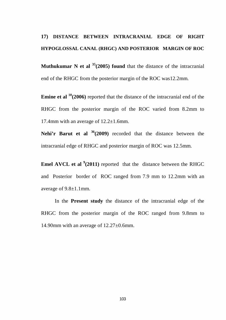
103
17) DISTANCE BETWEEN INTRACRANIAL EDGE OF RIGHT
HYPOGLOSSAL CANAL (RHGC) AND POSTERIOR MARGIN OF ROC
Muthukumar N et al 35(2005) found that the distance of the intracranial
end of the RHGC from the posterior margin of the ROC was12.2mm.
Emine et al 10(2006) reported that the distance of the intracranial end of the
RHGC from the posterior margin of the ROC varied from 8.2mm to
17.4mm with an average of 12.2±1.6mm.
Nehi’r Barut et al 36(2009) recorded that the distance between the
intracranial edge of RHGC and posterior margin of ROC was 12.5mm.
Emel AVCL et al 9(2011) reported that the distance between the RHGC
and Posterior border of ROC ranged from 7.9 mm to 12.2mm with an
average of 9.8±1.1mm.
In the Present study the distance of the intracranial edge of the
RHGC from the posterior margin of the ROC ranged from 9.8mm to
14.90mm with an average of 12.27±0.6mm.
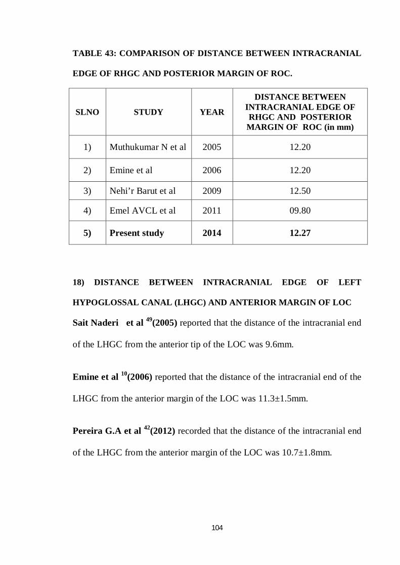
104
TABLE 43: COMPARISON OF DISTANCE BETWEEN INTRACRANIAL
EDGE OF RHGC AND POSTERIOR MARGIN OF ROC.
SLNO STUDY YEAR
DISTANCE BETWEEN INTRACRANIAL EDGE OF RHGC AND POSTERIOR
MARGIN OF ROC (in mm)
1) Muthukumar N et al 2005 12.20
2) Emine et al 2006 12.20
3) Nehi’r Barut et al 2009 12.50
4) Emel AVCL et al 2011 09.80
5) Present study 2014 12.27
18) DISTANCE BETWEEN INTRACRANIAL EDGE OF LEFT
HYPOGLOSSAL CANAL (LHGC) AND ANTERIOR MARGIN OF LOC
Sait Naderi et al 49(2005) reported that the distance of the intracranial end
of the LHGC from the anterior tip of the LOC was 9.6mm.
Emine et al 10(2006) reported that the distance of the intracranial end of the
LHGC from the anterior margin of the LOC was 11.3±1.5mm.
Pereira G.A et al 42(2012) recorded that the distance of the intracranial end
of the LHGC from the anterior margin of the LOC was 10.7±1.8mm.
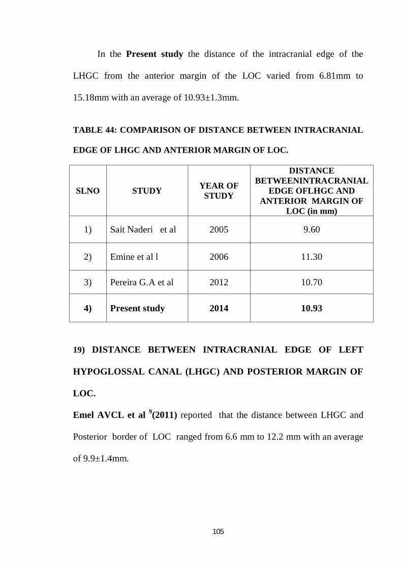
105
In the Present study the distance of the intracranial edge of the
LHGC from the anterior margin of the LOC varied from 6.81mm to
15.18mm with an average of 10.93±1.3mm.
TABLE 44: COMPARISON OF DISTANCE BETWEEN INTRACRANIAL
EDGE OF LHGC AND ANTERIOR MARGIN OF LOC.
SLNO STUDY YEAR OF STUDY
DISTANCE BETWEENINTRACRANIAL
EDGE OFLHGC AND ANTERIOR MARGIN OF
LOC (in mm)
1) Sait Naderi et al 2005 9.60
2) Emine et al l 2006 11.30
3) Pereira G.A et al 2012 10.70
4) Present study 2014 10.93
19) DISTANCE BETWEEN INTRACRANIAL EDGE OF LEFT
HYPOGLOSSAL CANAL (LHGC) AND POSTERIOR MARGIN OF
LOC.
Emel AVCL et al 9(2011) reported that the distance between LHGC and
Posterior border of LOC ranged from 6.6 mm to 12.2 mm with an average
of 9.9±1.4mm.
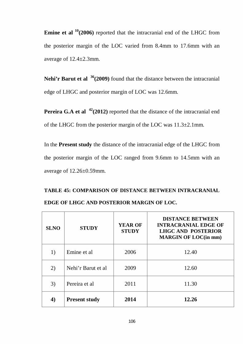
106
Emine et al 10(2006) reported that the intracranial end of the LHGC from
the posterior margin of the LOC varied from 8.4mm to 17.6mm with an
average of 12.4±2.3mm.
Nehi’r Barut et al 36(2009) found that the distance between the intracranial
edge of LHGC and posterior margin of LOC was 12.6mm.
Pereira G.A et al 42(2012) reported that the distance of the intracranial end
of the LHGC from the posterior margin of the LOC was 11.3±2.1mm.
In the Present study the distance of the intracranial edge of the LHGC from
the posterior margin of the LOC ranged from 9.6mm to 14.5mm with an
average of 12.26±0.59mm.
TABLE 45: COMPARISON OF DISTANCE BETWEEN INTRACRANIAL
EDGE OF LHGC AND POSTERIOR MARGIN OF LOC.
SLNO STUDY YEAR OF STUDY
DISTANCE BETWEEN INTRACRANIAL EDGE OF LHGC AND POSTERIOR MARGIN OF LOC(in mm)
1) Emine et al 2006 12.40
2) Nehi’r Barut et al 2009 12.60
3) Pereira et al 2011 11.30
4) Present study 2014 12.26
107
The mean distance between intracranial edge of HGC (Right and
Left) and anterior margin of ROC and LOC were measured as
11.02±1.29mm and 10.93±1.30mm respectively. The mean distance
between intracranial edge of HGC (Right and Left) and posterior margin of
ROC and LOC were measured as 12.27±0.6mm and12.26±0.59mm
respectively.
The measured value was compared with various studies and
tabulated. The values of present study coincided with other studies.
This is clinically significant for a safe occipital condyle resection. The
OC can be safely drilled for a distance of 12mm from posterior margin
before encountering the HGC and it is observed to be 12.27 ±0.59mm in our
study.
HGC distance from anterior and posterior ROC and LOC of present
study was compared with various studies and tabulated.
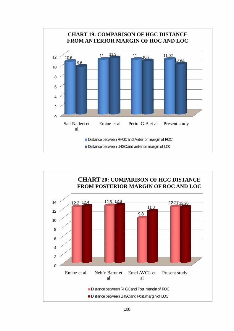
108
0
2
4
6
8
10
12
Sait Naderi et al
Emine et al Perira G.A et al Present study
10.6 11 11 11.02
9.6
11.310.7
10.93
CHART 19: COMPARISON OF HGC DISTANCE FROM ANTERIOR MARGIN OF ROC AND LOC
Distance between RHGC and Anterior margin of ROC
Distance between LHGC and anterior margin of LOC
0
2
4
6
8
10
12
14
Emine et al Nehi'r Barut et al
Emel AVCL et al
Present study
12.2 12.5
9.8
12.2712.4 12.611.3
12.26
CHART 20: COMPARISON OF HGC DISTANCE FROM POSTERIOR MARGIN OF ROC AND LOC
Distance between RHGC and Post.margin of ROC
Distance between LHGC and Post.margin of LOC

Conclusion
109
CONCLUSION
An effort was made to assess the foramen magnum, occipital
condyles and hypoglossal canals morphometrically through this study.
Advances in skull base approaches have improved the surgical exposure in
skull base surgeries. These manoeuvres may be complicated by injury to the
neurovascular structures or craniocervical instability due to removal of
occipital condyles. Hence, neurosurgeons performing this surgery should be
familiar with the normal anatomy as well as variations of the foramen
magnum region.
The parameters were measured and compared with other studies. The
following were the conclusions derived from the present study:
In the present study, it was found that the foramen magnum was oval
in 40%. This variation in configuration should be taken into consideration
during neuroimaging techniques and skull base surgery.
The mean Anteroposterior diameter of the foramen magnum in adult
dry skulls and cranial CT were measured as35.12±2.65mm and
35.03±0.95mm respectively. The size of the foramen magnum is the
critical parameter in craniovertebral junction pathology for the
manifestation of clinical signs and symptoms.
110
The mean transverse diameter of the foramen magnum in adult dry
skulls and cranial CT were measured as29.03±2.15mm and 28.79±1.17mm
respectively. It is significant in transcondylar approach.
Protrusion of occipital condyle was present in 20%
The maximum length of the right and left occipital condyle were
measured as 23.85±2.12mm and 23.77±2.29mm respectively.
The maximum and minimum width of the right occipital condyle
were measured as13.29 ±1.36mm and 6.86±1.34mm respectively.
The maximum and minimum width of the left occipital condyle were
measured as 13.44±1.41mm and 7.04±1.26mm respectively.
The maximum length of right and left occipital condyle were
measured as 23.11±0.73mm and 23.20±0.74mm respectively in cranial CT.
The maximum width of right and left occipital condyle were
measured as12.92±0.65mm to 12.88±0.69mm respectively in cranial CT.
In the present study the measurements of dry skull were greater than those
of cranial CT scans.
The radiological analysis of foramen magnum and occipital condyle
is required prior to craniovertebral junction surgery to prevent complications
like craniovertebral instability and haemorrhage.
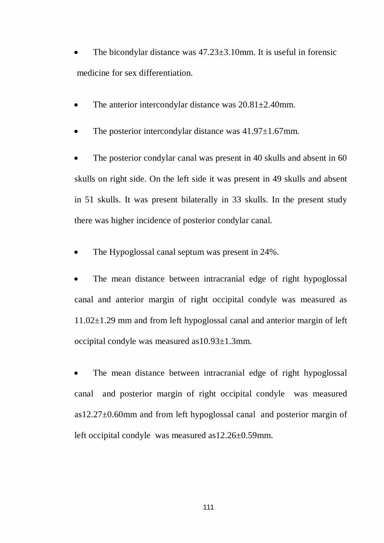
111
The bicondylar distance was 47.23±3.10mm. It is useful in forensic
medicine for sex differentiation.
The anterior intercondylar distance was 20.81±2.40mm.
The posterior intercondylar distance was 41.97±1.67mm.
The posterior condylar canal was present in 40 skulls and absent in 60
skulls on right side. On the left side it was present in 49 skulls and absent
in 51 skulls. It was present bilaterally in 33 skulls. In the present study
there was higher incidence of posterior condylar canal.
The Hypoglossal canal septum was present in 24%.
The mean distance between intracranial edge of right hypoglossal
canal and anterior margin of right occipital condyle was measured as
11.02±1.29 mm and from left hypoglossal canal and anterior margin of left
occipital condyle was measured as10.93±1.3mm.
The mean distance between intracranial edge of right hypoglossal
canal and posterior margin of right occipital condyle was measured
as12.27±0.60mm and from left hypoglossal canal and posterior margin of
left occipital condyle was measured as12.26±0.59mm.
112
This distance is clinically significant for a safe occipital condyle
resection during transcondylar approach and it was observed to be
12.27±0.59mm in this study.
The present study showed that the difference between radiological
and anatomical measurements were insignificant, which strengthen the
importance of preoperative radiological evaluation for achieving better
surgical success.
The present study also revealed the various shapes of FM and its
morphometry. The data obtained may be useful to neurosurgeons in
analyzing the anatomy of craniovertebral junction for preoperative planning
and management of skull base surgery. The findings will also be
enlightening for Radiologists, Orthopedicians, Anthropologists,
Morphologists and Clinical Anatomists.

Bibliography
BIBLIOGRAPHY
1) Atul Goel., Craniovertebral Anomalies : role for Craniovertebral
Realignment, Neurology India (2004), December; Volume 52 Issue 4.
2) Aynur Emine Cicekcibasi, Khalil Awadh Murshed, Taner Ziylan,
Muzaffer Seker, Isik Tuncer., A Morphometric Evaluation of Some
Important Bony Landmarks on the Skull Base Related to Sexes, Turk J
Med Sci (2004) 37-42.
3) AT Uthman, NH Al-Rawi and JF Al-Timimi., Evaluation of
foramen magnum in gender determination using helical CT
scanning, Dentomaxillofacial Radiology (2012) 41, 197–202.
4) Bello S.S, Zagga A.D, Kalale S.B, Usman J.D, Bello A,
Abdulhameed A, Bello S.M. and Tadros., Measurements of
Occipital Condyles using Computerised Tomography from Sokoto
State, Nigeria, International Journal of Health and Medical
Information (2013) December ;Volume 2, Number 3.
5) Carlos A. Bagley, Jonathan A. Pindrik, Markus J. Bookland,
Joaquin Q. Camara-quintana and Benjamin S. Carson.,
Cervicomedullary decompression for Foramen magnum stenosis in
Achondroplasia, Journal of Neurosurgery: Pediatrics (2006), Volume
104:166–172.
6) Chummy S.Sinnathamby., LAST’S Anatomy Regional and Applied,
Twelfth Edition (2011) Pg 506 & 507.
7) Daniel J. Wescott, M.A. Peer H. Moore-Jansen., Metric Variation
in the Human Occipital Bone: Forensic Anthropological
Applications, Journal of Forensic Science (2001),46(5):1159–1163.
8) di Vasudha V.Saralaya, B.V.Murlimanju, Rajesh Vaderav,
Mamatha Tonse, M.D.Prameela, P.J.Jiji., Occipital condyle
morphometry and incidence of condylus tertius: phylogenetic and
clinical significance, La clinica Terapeutica journal (2012)
Volume 163 Num 6.
9) Emel Avci, Ahmet Dagtekin, A. Hakan Ozturk, Engin Kara, Nail
Can Ozturk, Kutluay Uluc, Erinc Akture, Mustafa K. Baskaya.,
Anatomical Variations of the Foramen Magnum, Occipital Condyle
and Jugular Tubercle, Turkish Neurosurgery (2011),Volume: 21,
No:2,181-190
10) Emine Dondu Kizilkanat, Neslihan Boyan, Roger Soames and
Ozkan Oguz., Morphometry of the Hypoglossal Canal, Occipital
Condyle, and Foramen Magnum Neurosurg Q (2006) Volume 16,
Number 3.
11) F.Burdan, J.Szumiło, J.Walocha, L.Klepacz, B.Madej,
W.Dworzański, R.Klepacz, A.Dworzańska1, E.Czekajska-
Chehab, A.Drop., Morphology of the foramen magnum in young
Eastern European adults, Folia Morphol.(2012),Vol.71,No. 4, pp.
205–216.
12) Fathy Ahmed Fetouh, Akram M. Awadalla., Morphometric
analysis of the occipital condyle and its surgical implications in
transcondylar approach, Pan Arab Journal of Neurosurgery 2013 Jan.
13) Fatma Hayat Erdil, Vedat Sabanciogullari, Mehmet Cimen,
Oktay Isik., Morphometric Analysis of the Foramen Magnum by
Computed Tomography, (2010), Erciyes Medical Journal, 32(3):
167-170.
14) Frazer’s Anatomy of the Human Skeleton, Sixth edition (1965) Page
no:163 -167.
15) Gagandeep Singh and Indu Talwar., Morphometric analysis of
foramen magnum in human skull for sex determination, Human
Biology Review (2013), 2(1), pg: 29-41.
16) Gary D. Richards, and Rebecca S. Jabbour., Foramen Magnum
Ontogeny in Homo sapiens: A Functional Matrix Perspective, The
anatomical record, (2011)294:199–216.
17) Gautam Kanodia, Vijay Parihar, Yad R Yadav, Pushp R Bhatele
and Dhananjay Sharma., Morphometric analysis of posterior fossa
and foramen magnum, Journal of Neurosciences in Rural
Practice.( 2012) Sep-Dec; 3(3): 261–266.
18) Georges Olivier, Biometry of the human occipital bone, J. Anat.
(1975), 120, 3, pp. 507-518 507
19) Gobbur, Arjun R. Rai, Santosh P.V. Nagesh, K.R. Kharoshah,
Magdy A Madadin, Mohammed Luis, Neil A. Barai, Prakash
Arathisenthil, S. V. Choudhary, Nishant, Morphometric evaluation
of the foramen magnum: A study on computerized tomographic
images of South Indian adults, International Journal of the A.J.
Institute of Medical Sciences, (2013),Vol. 2 Issue 1, Pg 65.
20) Henry H Schmidek and Sweet., Operative Neurosurgical
Techniques-Indications,Methods and Results, Fifth Edition (2009)
Elsevier, Pg1755-1764
21) Hisham Aboul-Enein., Microsurgical excision of anterior and
anterolateral intradural lesions of the foramen magnum, Pan Arab
journal of neurosurgery (2010), volume 14, no. 2.
22) Ivan Claudio Suazo Galdames, Priscilla Perez Russo, Daniela
Alejandra Zavando Matamala & Ricardo Luiz Smith., Sexual
Dimorphism in the Foramen Magnum Dimensions, Int. J. Morphol.,
(2009)27(1):21-23.
23) Jasbir Kaur, Dhirendra Srivastava, Davinder Singh, Shashi
Raheja., The study of hyperostosic variants: significance of
hyperostotic variants of human skulls in anthropology, Anatomy Cell
Biology (2012),45:268-273.
24) Jatin Goda, Shailesh Patel, Laxmi Chandravadiya, Srushti
Rupareliya, Shamin Patel, Sanjay Chavda., Variations of the
Posterior Condylar Canals, Int J Res Med. 2013; 2(1);118-120.
25) J.C.Boileau Grant, A Method of Anatomy, Descriptive and
deductive, Sixth Edition, Baltimore (1958), Page 641-643,808 & 809.
26) Jose Aderval Aragao, Raisa de Oliveira Pereira, Rebeca Zelice da
Cruz de Moraes and Francisco Prado Reis., Morphological Types
of Foramen Magnum, Annual Research & Review in Biology (2014),
4(9): Pg-1372-1378.
27) J.T.Hong, Tomoyuki Takigawa, Keizo Sugisaki, Alejandro A.
Espinoza Orias, Nozomu Inouse and Howarad .S., Biomechanical
and Morphometric evaluation of Occipital Condyle for
Occipitocervical Segmental Fixation, Neurol Med Chir (Tokyo)
(2011) 51,701-706.
28) Kenan I. Arnautovic, and Ossama AI-Mefty., Foramen Magnum
Meningiomas Part II: Surgical Approaches, Surgical Anatomy,
Complications of Treatment and Results.
29) Ketu Chauhan, Archana Sharma., Osteotic variations in
Paracondylar region of Adult Human skulls – Incidence and Clinical
significance, International Journal of Anatomy and Research,
International Journal of Anatomy and Research 2013, Volume 1(3):
158-60.
30) Khalil Awadh Murshed, Aynur Emine Cicekci basi and Isik
Tuner., Morphometric Evaluation of the Foramen Magnum and
Variations in its Shape: A Study on Computerized Tomographic
Images of Normal Adults, Turkey Journal of Medical Science (2003)
33 Pg 301-306.
31) K.Natsis, M.Piagkou, G.Skotsimara, G.Piagkos, P.Skandalakis.,
A morphometric anatomical and comparative study of the foramen
magnum region in a Greek population, Surg Radiol Anat (2013),
April 10.1007/s00276-013-1119.
32) LANGMAN’S Medical Embryology T.W,Sadler, Eleventh Edition,
Pg127-133.
33) Manoel C, Prado FB, Caria, PHF and Groppo FC., Morphometric
analysis of the foramen magnum in human skulls of brazilian
individuals: its relation to gender, Braz. J. Morphol. Sci., (2009) vol.
26, no. 2, p. 104-108.
34) Mehmet Asim Ozer, Servet Celik, Figen Govsa, Mahmut Oguz
Ulusoy., Anatomical determination of a safe entry point for occipital
condyle screw using three-dimensional landmarks, Eur Spine J
(2011) 20:1510–1517.
35) Muthukumar N, Swaminathan R, Venkatesh G, Bhanumathy
SP., A morphometric analysis of the foramen magnum region as it
relates to the transcondylar approach, Acta Neurochir (Wien).
(2005), Aug;147(8):889-95. Epub 2005 Jun 9.
36) Nehi R Barut, Aysi N Kale, Hi kmet Turan, Adnan Ozturk,
Mustafa Bozbuga and Kayihan Sahinoglu., Evaluation of the bony
landmarks in transcondylar approach, British Journal of
Neurosurgery (2009);Vol. 23,No.3, Pages 276-281.
37) Ossama Al-Mefty, Luis A. B. Borba, Nobuo Aoki, Edgardo
Angtuaco, and T. Glenn Pait., The transcondylar approach to
extradural nonneoplastic lesions of the craniovertebral junction,
Journal of Neurosurgery (1996 ) January Vol.84- No. 1 Pages 1-6.
38) Osunwoke E.A, Oladipo G.S, Gwunireama I.U, Ngaokere J.O.,
Morphometric analysis of the Foramen magnum and Jugular foramen
in Adult skulls in southern Nigerian population, American journal of
scientific and Industrial research ( 2012), 3(6):446-448.
39) Parvindokht Bayat, Mahdie Bagheri, Ali Ghanbari & Amir
Raoofi., Characterization of Occipital Condyle and Comparison of
its Dimensions with Head and Foramen Magnum Circumferences in
Dry Skulls of Iran, Int. J. Morphol.,(2014),32(2):444-448.
40) Patricia Collins, Embryology and Development in: Gray's Anatomy,
The Anatomical Basis of Medicine and Surgery,38th edition (1995)
Edinburgh: Elsevier Churchill Livingstone. Pg- 271-274.
41) P.Chethan, K.G.Prakash, B.V.Murlımanju, K.U.Prashanth,
Latha V.Prabhu, Vasudha V.Saralaya, Ashwin Krıshnamurthy,
M.S.Somesh, C.Ganesh Kumar., Morphological Analysis and
Morphometry of the Foramen Magnum: An Anatomical Investigation,
Turkish Neurosurgery (2012) Vol: 22, No: 4, 416-419.
42) Pereira.G.A.M, Lopes.P.T.C, Santos.A.M.P.V, Duarte.R.D,
Piva.L & Pozzobon.A., Morphometric Analysis Related to the
Transcondylar Approach in Dry Skulls and Computed Tomography,
International Journal of Morphology (2012) vol. 30(2):399-404.
43) Philipp gruber, Maciej Henneberg, Thomas Boni, and Frank J.
Ruhli., Variability of Human Foramen Magnum Size, The
Anatomical Record (2009)292:1713–1719.
44) Pooja Gangrade, Ramavtar Saini, Rajeev Yadav, and Anamika
Vyas., Evaluation of New Morphometric Parameters for Sex
Determination of Human Skull, Research and Reviews: Journal of
Medical and Health Sciences. (July – September, 2013), Volume 2 ,
Issue 3.
45) Radhakrishnan P, Chandni Gupta, Sandeep Kumar, Antony
Sylvan D’souza1., A Morphometric Analysis of the Foramen
Magnum and Variations in its Shape: A Computerized Tomographic
Study, Novel Science International Journal of Medical Science
(2012), 1(9-10): 281-285.
46) Radhakrishna S.K., Shivarama C.H., Ramakrishna A.,
Bhagya.B., Morphometric analysis of Foramen magnum for sex
determination in South Indian population, Nitte University Journal of
Health Science (2012) March Vol. 2, No.1, ISSN 2249-7110.
47) Roma Patel, C.D.Mehta., Morphometric study of Foramen Magnum
at the base of human skull in South Gujarat, IOSR Journal of Dental
and Medical Sciences (2014) June, Vol 13, Issue 6 Ver. IV, PP 23-25.
48) Roopali D.Nikumbh, Dhiraj B.Nikumbh, Rohini R.Karambelkar,
Avinash D. Shewale, Morphological Study of Hypoglossal Canal
and Its Anatomical Variation, International Journal of Health
Sciences & Research (2013) June, 54 Vol.3; Issue: 6.
49) Sait Naderi, Esin Korman, Guven Citak, Mustafa Guvencer,
Candan Arman, Mehmet Senoglu, SuleymanTetik, Nuri Arda.,
Morphometric analysis of human occipital condyle, Clinical
Neurology and Neurosurgery (2005)April;107(3): 191–199.
50) Singh Rajani., Duplicate Condylar Canal in Indian Dry Skull, EJBS
(2013) July, 7 (1).
51) Siva nageswara Rao, Sundara Setty, Raja Sekhar Katikireddi.,
Study of Duplicated Hypoglossal canal in South Indian human skulls-
Original article, Int J Cur Res Rev, (2013) Vol 05 (14) Page 103-105
52) Shanthi CH, S.Lokanadham., Morphometric Study on Foramen
Magnum of Human Skulls, Jou Medicine Science 2013;2(4):792-8
53) S.K.Jain, Alok Kumar Choudhary, Pankaj Mishra.,
Morphometric evaluation of Foramen magnum for sex determination
in a documented North Indian sample, Journal of Evolution of
Medical and Dental Sciences,(2013) Volume 2/ Issue 42- Page 8094.
54) Surwase Ramdas Gopalrao1, Prity Solanke, Mahesh Ugale1,
Smita Balsurkar., Computed Tomographic scan study of
Morphometry of Foramen magnum, Int J Cur Res Rev, Oct 2013 /
Vol 05 (19) Page 41-48.
55) Susan Standring, Gray's Anatomy, The Anatomical basis of clinical
practice, 40th Edition (2008) Page no- 415,710-711.
56) Tien V. Le, Elias Dakwar, Shannon Hann, Euclides Effio, Ali A.
Baaj, Carlos Martinez, Fernando L.Vale, and Juan S.Uribe.,
Computed tomography–based morphometric analysis of the human
occipital condyle for occipital condyle–cervical fusion, J Neurosurg:
Spine / Volume 15 /pg 328–331September 2011.
57) Ukoha U, Egwu OA, Okafor IJ, Anyabolu AE, Ndukwe GU,
Okpala I., Sexual Dimorphism in the Foramen Magnum of Nigerian
Adult, International Journal of Biological & Medical Research
(2011); 2(4): 878 – 881.
58) Wanebo, John E, Chicoine, Michael R., Quantitative Analysis of
the Transcondylar Approach to the Foramen Magnum, Journal of
Neurosurgery (2001) Volume 49 - Issue 4 - Pg 934-943.
59) Yogesh Yadav, Preeti Goswami., A Study of Length and Width of
Foramen Magnum in North India., International Journal of Science
and Research (2014) June ;Volume 3 Issue 6.
60) Youmans Neurological surgery H. Richard Winn, sixth edition
(2011), Pg 1569-1586, 1918-1927 and 2333-2244.
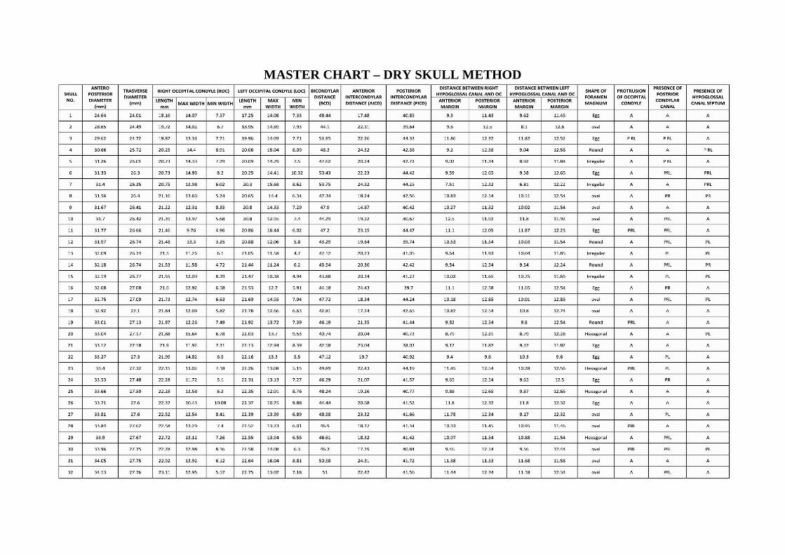
MASTER CHART – DRY SKULL METHOD
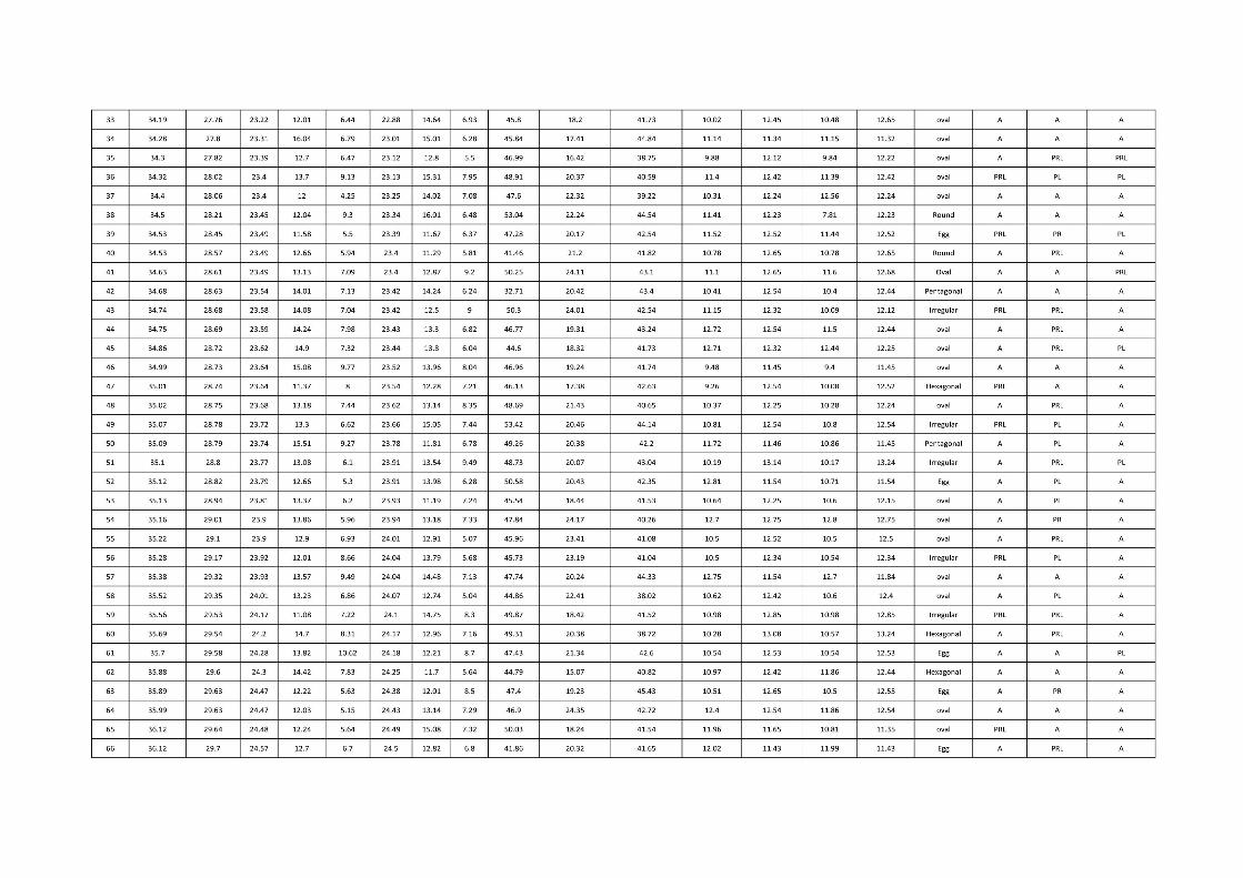
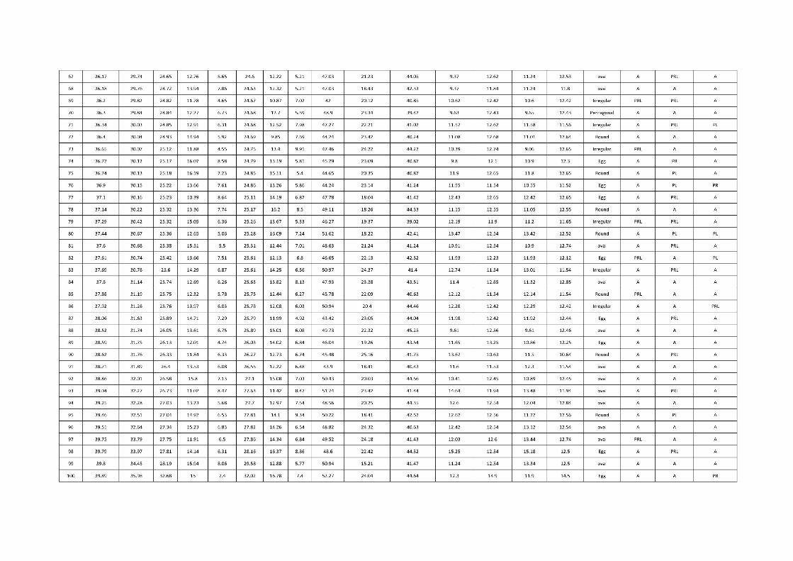
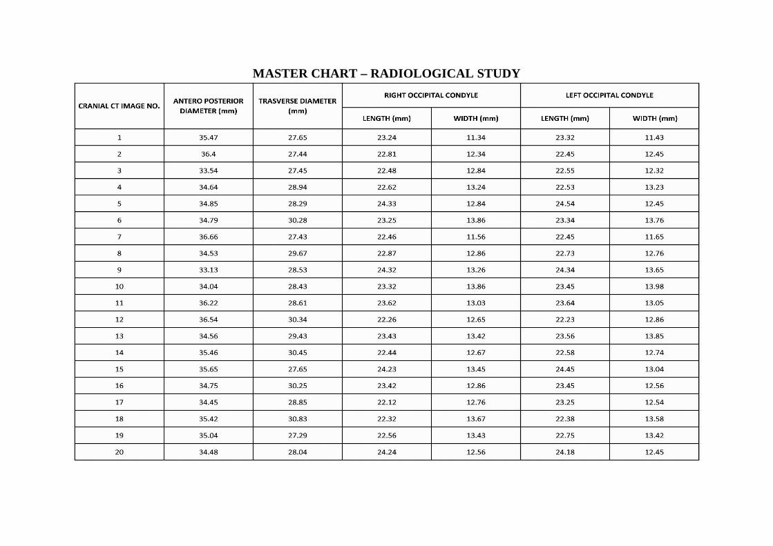
MASTER CHART – RADIOLOGICAL STUDY
Related Documents