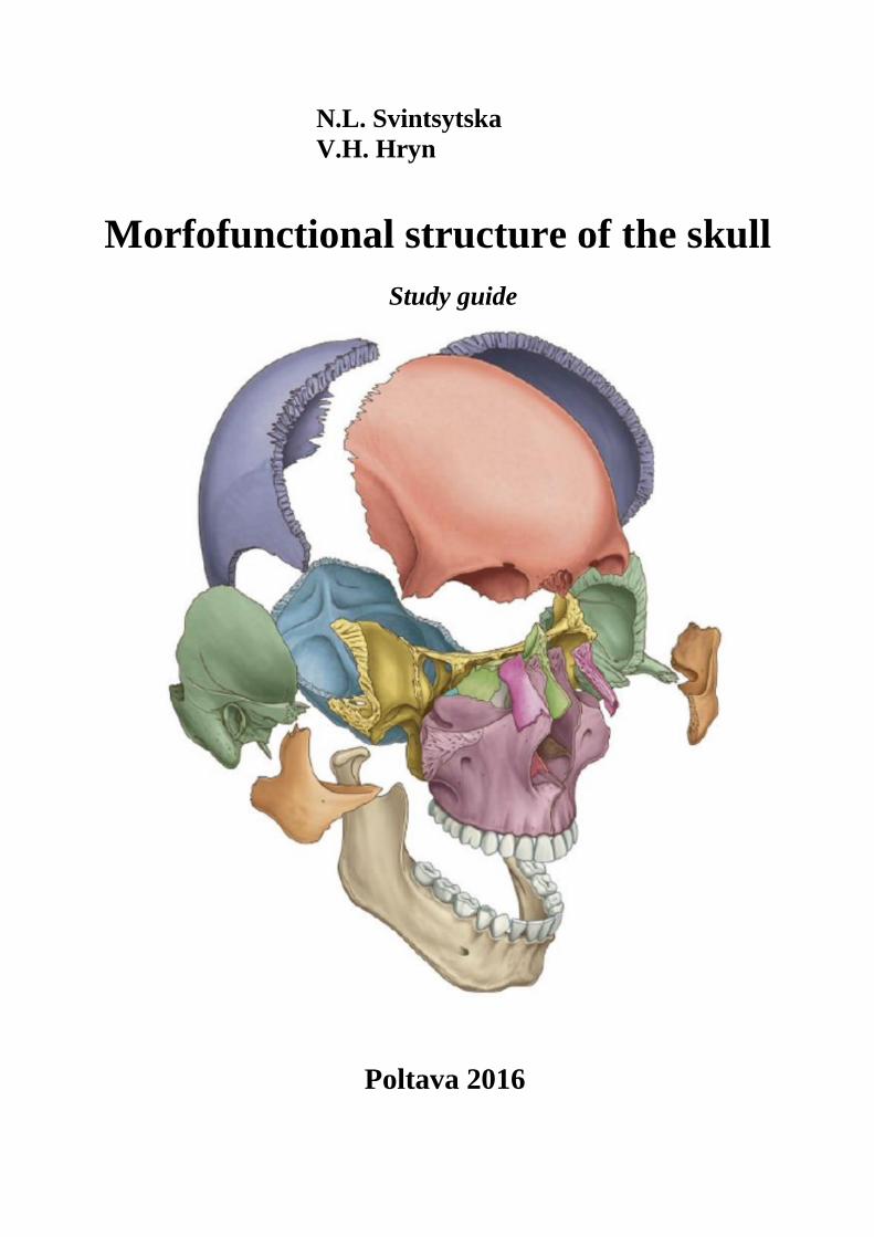
N.L. Svintsytska V.H. Hryn Morfofunctional structure of the skull Study guide Poltava 2016

Welcome message from author
This document is posted to help you gain knowledge. Please leave a comment to let me know what you think about it! Share it to your friends and learn new things together.
Transcript
N.L. Svintsytska
V.H. Hryn
Morfofunctional structure of the skull
Study guide
Poltava 2016
2
Ministry of Public Health of Ukraine
Public Institution «Central Methodological Office for
Higher Medical Education of MPH of Ukraine»
Higher State Educational Establishment of Ukraine
«Ukranian Medical Stomatological Academy»
N.L. Svintsytska, V.H. Hryn
Morfofunctional
structure of the skull
Study guide
Poltava 2016
3
LBC 28.706
UDC 611.714/716
S 24
«Recommended by the Ministry of Health of Ukraine as textbook for English-
speaking students of higher educational institutions of the MPH of Ukraine» (minutes of
the meeting of the Commission for the organization of training and methodical
literature for the persons enrolled in higher medical (pharmaceutical) educational
establishments of postgraduate education MPH of Ukraine, from 02.06.2016 №2).
Letter of the MPH of Ukraine of 11.07.2016 № 08.01-30/17321
Composed by:
N.L. Svintsytska, Associate Professor at the Department of Human Anatomy of Higher State
Educational Establishment of Ukraine «Ukrainian Medical Stomatological Academy», PhD in
Medicine, Associate Professor
V.H. Hryn, Associate Professor at the Department of Human Anatomy of Higher State
Educational Establishment of Ukraine «Ukrainian Medical Stomatological Academy», PhD in
Medicine, Associate Professor
This textbook is intended for undergraduate, postgraduate students and continuing
education of health care professionals in a variety of clinical disciplines (medicine, pediatrics,
dentistry) as it includes the basic concepts of human anatomy of the skull in adults and
newborns.
Rewiewed by:
O.M. Slobodian, Head of the Department of Anatomy, Topographic Anatomy and Operative
Surgery of Higher State Educational Establishment of Ukraine «Bukovinian State Medical
University», Doctor of Medical Sciences, Professor
M.V. Hubin, Associate Professor of the Department of Forensic Medicine, Medical Low of
Kharkiv National Medical University, PhD in Medicine, Associate Professor
Yu. V. Lysanets, Senior Lecturer of the Department of Foreign Languages with Latin
Language and Medical Terminology of Higher State Educational Establishment of Ukraine
«Ukrainian Medical Stomatological Academy» Candidate of Philological Sciences
4
CONTENTS
Preface...................................................................................................................6
Bones of the neurocranium...................................................................................7
The occipital bone..............................................................................................7
The parietal bone..............................................................................................12
The frontal bone...............................................................................................14
The sphenoid bone............................................................................................18
The ethmoid bone.............................................................................................26
The temporal bone............................................................................................31
Bones of visceral skeleton...................................................................................44
The mandible....................................................................................................44
The maxilla.......................................................................................................52
The palatine bone.............................................................................................62
The nasal bone..................................................................................................64
The lacrimal bone.............................................................................................65
The zygomatic bone.........................................................................................65
The hyoid bone.................................................................................................67
The inferior nasal concha, vomer.....................................................................69
The orbit...........................................................................................................70
The nasal cavity...................................................................................................74
Skull joints...........................................................................................................84
Norma verticalis...............................................................................................86
Norma occipitalis..............................................................................................88
Norma frontalis.................................................................................................92
Norma lateralis.................................................................................................99
Norma basalis.................................................................................................107
Structure passing through foramina...................................................................114
Interior of the skull............................................................................................118
Anterior cranial fossa.....................................................................................121
5
Middle cranial fossa.......................................................................................123
Posterior cranial fossa.....................................................................................126
Foetal skull........................................................................................................133
Postnatal growth of skull...................................................................................137
Sex differences in the skull................................................................................139
Development of the skull...................................................................................142
Control questions...............................................................................................148
Situation problems.............................................................................................150
Literature...........................................................................................................159
For self-preparing..............................................................................................163
6
PREFACE
Modern craniology successfully solves the fundamental problem of
medicine, namely, the study of regularitis of structure of a human skull and
clarifying the morphological and functional features of various forms. Due to the
complexity of its structure, the skull is the placesora large number of anatomic
variations, anomalies and dysplasias.
Facial bones are an important architectural part of the face, ensuring the
perfection of its design fundamentals and uniqua individuality. Besides, they
provide mechanical strength of the skull. By participating in the formation of
cavities (orbit, nasal and oral), they are used for protection and normal
functioning of the primary divisions of digestive, respiratory systems and
sensory organs (sight, smell, taste). Fissures, foramens and channels which the
cranial cavities have, allow the passage of the neurovascular bundles for the
innervation of these organs. The structure of the maxilla and mandible and
palatine bones represents the most pronounced signs of the evolution of the
skull, characteristic of the skull of Homo sapiens and associated with the
development of articulate speech, the development of the brain, the consumption
of processed food.
The study of the skull bones, their connections and relationships with
blood vessels and nerves is necessary to understand the features of rendering
proper medical care in case of disruption of their normal functioning.
Skull lodges the brain, teeth and special senses like cochlear and
vestibular apparatus, retina, olfactory mucous membrane, and taste buds. The
weight of the brain is not felt as it is floating in the cerebrospinal fluid. Our
personality, power of speech, attention, concentration, judgement, and intellect
are because of the brain that we possess and its proper use, for our own good
and for the good of the society as well.
The skeleton of the head is called the skull. It consists of several bones
that are joined together to form the cranium. The term skull also includes the
7
mandible or lower jaw, which is a separate bone. However, the two terms skull
and cranium, are often used synonymously. The skullcap is formed by frontal,
parietal, squamous temporal and a part of occipital bones, develop by
intramembranous ossification, being a quicker one-stage process. The base of
the skull in contrast ossifies by intracartilaginous ossification, which is a two-
stage process (membrane-cartilage-bone).
The skull can be divided into two main parts:
1. The neurocranium, consists of the frontal, parietal, occipital, temporal,
sphenoid and ethmoid bones. The calvaria or skullcap (brain box) is the upper
convex part of the cranium, which encloses the brain.
2. The facial skeleton, viscerocranium, constitutes the rest of the skull and
includes the mandible.
Bones of the neurocranium
The occipital bone.
The occipital bone (os occipitale), situated at the back and lower part of
the cranium, is trapezoid in shape and curved on itself. It is pierced by a large
oval aperture, the foramen magnum, through which the cranial cavity
communicates with the vertebral canal.
The curved, expanded plate behind the foramen magnum is named the
squama; the thick, somewhat quadrilateral piece in front of the foramen is
called the basilar part, whilst on either side of the foramen is the lateral
portion.
The Squama (squama occipitalis). The squama, situated above and
behind the foramen magnum, is curved from above downward and from side to
side.
Surfaces. The external surface is convex and presents midway between
the summit of the bone and the foramen magnum; a prominence, the external
occipital protuberance. Extending lateralward from this on either side are two
8
curved lines, one a little above the other. The upper, often faintly marked, is
named the highest nuchal line, and to it, the galea aponeurotica is attached. The
lower is termed the superior nuchal line. That part of the squama, which lies
above the highest nuchal lines, is named the planum occipitale, and is covered
by the occipital muscle; that below, termed the planum nuchale is rough and
irregular for the attachment of several muscles. From the external occipital
protuberance a ridge or crest, the external occipital crest, often faintly marked,
descends to the foramen magnum, and affords attachment to the ligamentum
nuchae; running from the middle of this line across either half of the nuchal
plane is the inferior nuchal line.
Several muscles are attached to the outer surface of the squama, thus: the
superior nuchal line gives origin to the Occipitalis and Trapezius, and insertion
to the Sternocleidomastoideus and Splenius capitis: into the surface between the
superior and inferior nuchal lines the Semispinalis capitis and the Obliquus
capitis superior are inserted, while the inferior nuchal line and the area below it
receive the insertions of the Recti capitis posteriores major and minor. The
posterior atlantoccipital membrane is attached around the postero-lateral part of
the foramen magnum, just outside the margin of the foramen.
The internal surface is deeply concave and divided into four fossae by a
cruciate eminence. The upper two fossae are triangular and lodge the occipital
lobes of the cerebrum; the lower two are quadrilateral and accommodate the
hemispheres of the cerebellum. At the point of intersection of the four divisions
of the cruciate eminence is the internal occipital protuberance. From this
protuberance, the upper division of the cruciate eminence runs to the superior
angle of the bone, and on one side of it (generally the right) is a deep groove, the
sagittal sulcus, which lodges the hinder part of the superior sagittal sinus; to the
margins of this sulcus, the falx cerebri is attached. The lower division of the
cruciate eminence is prominent, and is named the internal occipital crest; it
bifurcates near the foramen magnum and gives the falx cerebelli;
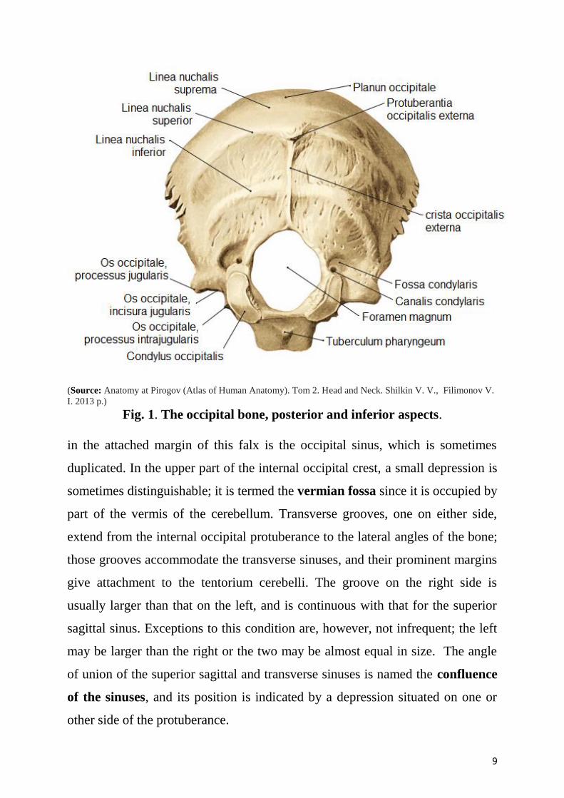
9
(Source: Anatomy at Pirogov (Atlas of Human Anatomy). Tom 2. Head and Neck. Shilkin V. V., Filimonov V.
I. 2013 p.)
Fig. 1. The occipital bone, posterior and inferior aspects.
in the attached margin of this falx is the occipital sinus, which is sometimes
duplicated. In the upper part of the internal occipital crest, a small depression is
sometimes distinguishable; it is termed the vermian fossa since it is occupied by
part of the vermis of the cerebellum. Transverse grooves, one on either side,
extend from the internal occipital protuberance to the lateral angles of the bone;
those grooves accommodate the transverse sinuses, and their prominent margins
give attachment to the tentorium cerebelli. The groove on the right side is
usually larger than that on the left, and is continuous with that for the superior
sagittal sinus. Exceptions to this condition are, however, not infrequent; the left
may be larger than the right or the two may be almost equal in size. The angle
of union of the superior sagittal and transverse sinuses is named the confluence
of the sinuses, and its position is indicated by a depression situated on one or
other side of the protuberance.
10
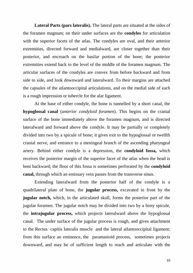
Lateral Parts (pars lateralis). The lateral parts are situated at the sides of
the foramen magnum; on their under surfaces are the condyles for articulation
with the superior facets of the atlas. The condyles are oval, and their anterior
extremities, directed forward and medialward, are closer together than their
posterior, and encroach on the basilar portion of the bone; the posterior
extremities extend back to the level of the middle of the foramen magnum. The
articular surfaces of the condyles are convex from before backward and from
side to side, and look downward and lateralward. To their margins are attached
the capsules of the atlantooccipital articulations, and on the medial side of each
is a rough impression or tubercle for the alar ligament.
At the base of either condyle, the bone is tunnelled by a short canal, the
hypoglossal canal (anterior condyloid foramen). This begins on the cranial
surface of the bone immediately above the foramen magnum, and is directed
lateralward and forward above the condyle. It may be partially or completely
divided into two by a spicule of bone; it gives exit to the hypoglossal or twelfth
cranial nerve, and entrance to a meningeal branch of the ascending pharyngeal
artery. Behind either condyle is a depression, the condyloid fossa, which
receives the posterior margin of the superior facet of the atlas when the head is
bent backward; the floor of this fossa is sometimes perforated by the condyloid
canal, through which an emissary vein passes from the transverse sinus.
Extending lateralward from the posterior half of the condyle is a
quadrilateral plate of bone, the jugular process, excavated in front by the
jugular notch, which, in the articulated skull, forms the posterior part of the
jugular foramen. The jugular notch may be divided into two by a bony spicule,
the intrajugular process, which projects lateralward above the hypoglossal
canal. The under surface of the jugular process is rough, and gives attachment
to the Rectus capitis lateralis muscle and the lateral atlantooccipital ligament;
from this surface an eminence, the paramastoid process, sometimes projects
downward, and may be of sufficient length to reach and articulate with the
11
transverse process of the atlas. Laterally the jugular process presents a rough
quadrilateral or triangular area, which is joined to the jugular surface of the
temporal bone by a plate of cartilage; after the age of twenty-five, this plate
tends to ossify.
(Source: Atlas of Human Anatomy / Frank H. Netter, M.D. Arthur F. Dalley; 2nd ed. // ILS, Medimedia USA
Company, 1997.)
Fig 2. The occipital bone, anterior and superior aspects.
The upper surface of the lateral part presents an oval eminence, the
jugular tubercle, which overlies the hypoglossal canal and is sometimes
crossed by an oblique groove for the glossopharyngeal, vagus, and accessory
nerves. On the upper surface of the jugular process is a deep groove, which
curves medialward and forward and is continuous with the jugular notch. This
groove lodges the terminal part of the transverse sinus, and opening into it, close
to its medial margin, is the orifice of the condyloid canal.
12
Basilar Part (pars basilaris). The basilar part extends forward and
upward from the foramen magnum, and presents in front an area more or less
quadrilateral in outline. In the young skull, this area is rough and uneven, and is
joined to the body of the sphenoid by a plate of cartilage. By the twenty-fifth
year, this cartilaginous plate is ossified, and the occipital and sphenoid form a
continuous bone.
The parietal bone
The parietal bones (os parietale) form, by their union, the sides and roof
of the cranium. Each bone is irregularly quadrilateral in form, and has two
surfaces, four borders, and four angles.
Surfaces. The external surface is convex, smooth, and marked near the
center by an eminence, the parietal eminence (tuber parietale), which indicates
the point where ossification commenced. Crossing the middle of the bone in an
arched direction are two curved lines, the superior and inferior temporal lines;
the former gives attachment to the temporal fascia, and the latter indicates the
upper limit of the muscular origin of the Temporalis. Above these lines the bone
is covered by the galea aponeurotica; below them it forms part of the temporal
fossa, and affords attachment to the Temporalis muscle. At the back part and
close to the upper or sagittal border is the parietal foramen, which transmits a
vein to the superior sagittal sinus, and sometimes a small branch of the occipital
artery; it is not constantly present, and its size varies considerably.
The internal surface is concave; it presents depressions corresponding to
the cerebral convolutions, and numerous furrows for the ramifications of the
middle meningeal vessel; the latter run upward and backward from the
sphenoidal angle, and from the central and posterior part of the squamous
border. Along the upper margin is a shallow groove, which, together with that
on the opposite parietal, forms a channel, the sagittal sulcus, for the superior
sagittal sinus; the edges of the sulcus afford attachment to the falx cerebri. Near
the groove are several depressions, best marked in the skulls of old persons, for
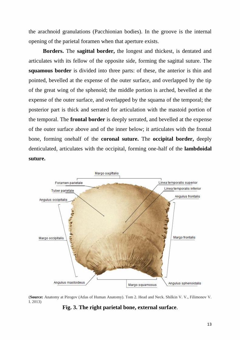
13
the arachnoid granulations (Pacchionian bodies). In the groove is the internal
opening of the parietal foramen when that aperture exists.
Borders. The sagittal border, the longest and thickest, is dentated and
articulates with its fellow of the opposite side, forming the sagittal suture. The
squamous border is divided into three parts: of these, the anterior is thin and
pointed, bevelled at the expense of the outer surface, and overlapped by the tip
of the great wing of the sphenoid; the middle portion is arched, bevelled at the
expense of the outer surface, and overlapped by the squama of the temporal; the
posterior part is thick and serrated for articulation with the mastoid portion of
the temporal. The frontal border is deeply serrated, and bevelled at the expense
of the outer surface above and of the inner below; it articulates with the frontal
bone, forming onehalf of the coronal suture. The occipital border, deeply
denticulated, articulates with the occipital, forming one-half of the lambdoidal
suture.
(Source: Anatomy at Pirogov (Atlas of Human Anatomy). Tom 2. Head and Neck. Shilkin V. V., Filimonov V.
I. 2013)
Fig. 3. The right parietal bone, external surface.
14
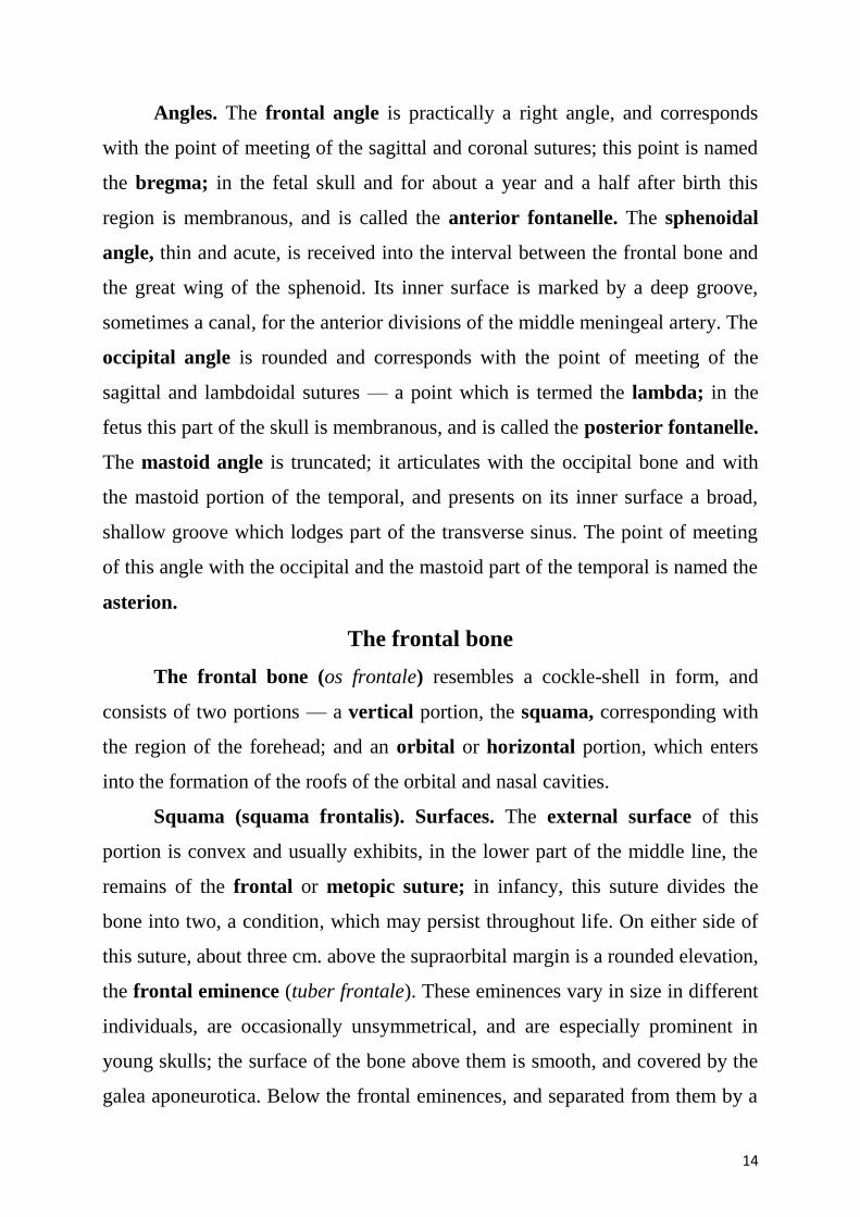
Angles. The frontal angle is practically a right angle, and corresponds
with the point of meeting of the sagittal and coronal sutures; this point is named
the bregma; in the fetal skull and for about a year and a half after birth this
region is membranous, and is called the anterior fontanelle. The sphenoidal
angle, thin and acute, is received into the interval between the frontal bone and
the great wing of the sphenoid. Its inner surface is marked by a deep groove,
sometimes a canal, for the anterior divisions of the middle meningeal artery. The
occipital angle is rounded and corresponds with the point of meeting of the
sagittal and lambdoidal sutures — a point which is termed the lambda; in the
fetus this part of the skull is membranous, and is called the posterior fontanelle.
The mastoid angle is truncated; it articulates with the occipital bone and with
the mastoid portion of the temporal, and presents on its inner surface a broad,
shallow groove which lodges part of the transverse sinus. The point of meeting
of this angle with the occipital and the mastoid part of the temporal is named the
asterion.
The frontal bone
The frontal bone (os frontale) resembles a cockle-shell in form, and
consists of two portions — a vertical portion, the squama, corresponding with
the region of the forehead; and an orbital or horizontal portion, which enters
into the formation of the roofs of the orbital and nasal cavities.
Squama (squama frontalis). Surfaces. The external surface of this
portion is convex and usually exhibits, in the lower part of the middle line, the
remains of the frontal or metopic suture; in infancy, this suture divides the
bone into two, a condition, which may persist throughout life. On either side of
this suture, about three cm. above the supraorbital margin is a rounded elevation,
the frontal eminence (tuber frontale). These eminences vary in size in different
individuals, are occasionally unsymmetrical, and are especially prominent in
young skulls; the surface of the bone above them is smooth, and covered by the
galea aponeurotica. Below the frontal eminences, and separated from them by a
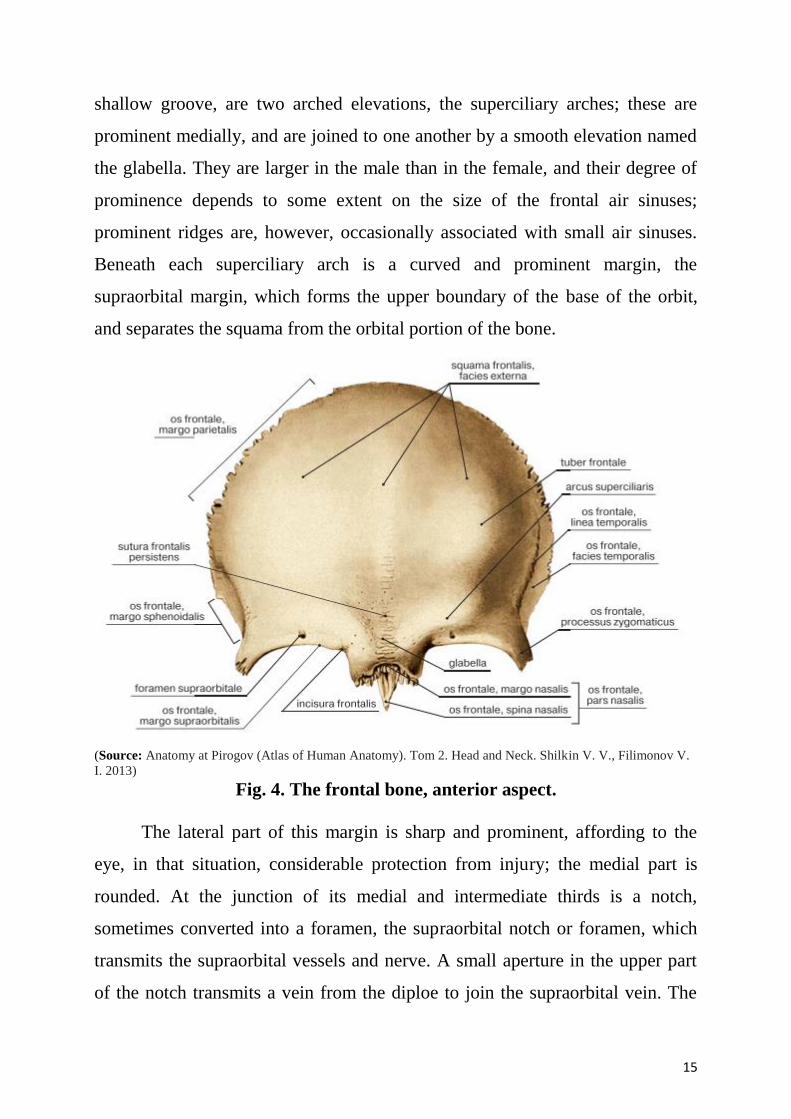
15
shallow groove, are two arched elevations, the superciliary arches; these are
prominent medially, and are joined to one another by a smooth elevation named
the glabella. They are larger in the male than in the female, and their degree of
prominence depends to some extent on the size of the frontal air sinuses;
prominent ridges are, however, occasionally associated with small air sinuses.
Beneath each superciliary arch is a curved and prominent margin, the
supraorbital margin, which forms the upper boundary of the base of the orbit,
and separates the squama from the orbital portion of the bone.
(Source: Anatomy at Pirogov (Atlas of Human Anatomy). Tom 2. Head and Neck. Shilkin V. V., Filimonov V.
I. 2013)
Fig. 4. The frontal bone, anterior aspect.
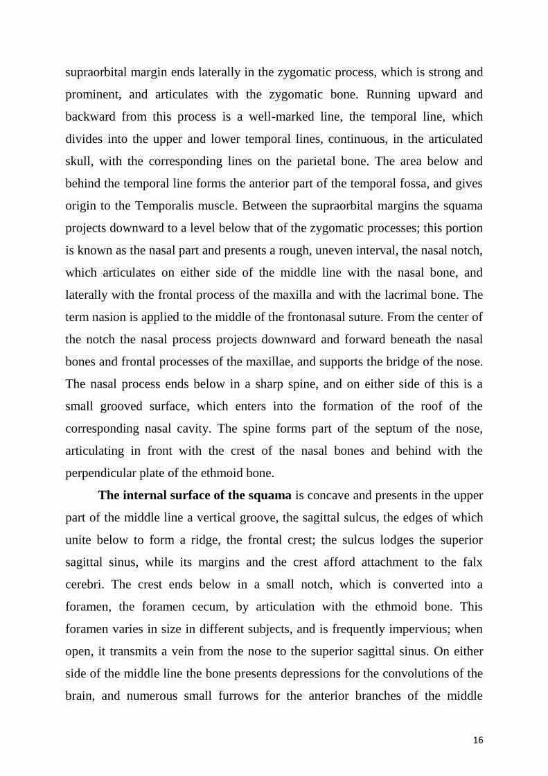
The lateral part of this margin is sharp and prominent, affording to the
eye, in that situation, considerable protection from injury; the medial part is
rounded. At the junction of its medial and intermediate thirds is a notch,
sometimes converted into a foramen, the supraorbital notch or foramen, which
transmits the supraorbital vessels and nerve. A small aperture in the upper part
of the notch transmits a vein from the diploe to join the supraorbital vein. The
16
supraorbital margin ends laterally in the zygomatic process, which is strong and
prominent, and articulates with the zygomatic bone. Running upward and
backward from this process is a well-marked line, the temporal line, which
divides into the upper and lower temporal lines, continuous, in the articulated
skull, with the corresponding lines on the parietal bone. The area below and
behind the temporal line forms the anterior part of the temporal fossa, and gives
origin to the Temporalis muscle. Between the supraorbital margins the squama
projects downward to a level below that of the zygomatic processes; this portion
is known as the nasal part and presents a rough, uneven interval, the nasal notch,
which articulates on either side of the middle line with the nasal bone, and
laterally with the frontal process of the maxilla and with the lacrimal bone. The
term nasion is applied to the middle of the frontonasal suture. From the center of
the notch the nasal process projects downward and forward beneath the nasal
bones and frontal processes of the maxillae, and supports the bridge of the nose.
The nasal process ends below in a sharp spine, and on either side of this is a
small grooved surface, which enters into the formation of the roof of the
corresponding nasal cavity. The spine forms part of the septum of the nose,
articulating in front with the crest of the nasal bones and behind with the
perpendicular plate of the ethmoid bone.
The internal surface of the squama is concave and presents in the upper
part of the middle line a vertical groove, the sagittal sulcus, the edges of which
unite below to form a ridge, the frontal crest; the sulcus lodges the superior
sagittal sinus, while its margins and the crest afford attachment to the falx
cerebri. The crest ends below in a small notch, which is converted into a
foramen, the foramen cecum, by articulation with the ethmoid bone. This
foramen varies in size in different subjects, and is frequently impervious; when
open, it transmits a vein from the nose to the superior sagittal sinus. On either
side of the middle line the bone presents depressions for the convolutions of the
brain, and numerous small furrows for the anterior branches of the middle
17
meningeal vessels. Several small, irregular fossae may also be seen on either
side of the sagittal sulcus, for the reception of the arachnoid granulations.
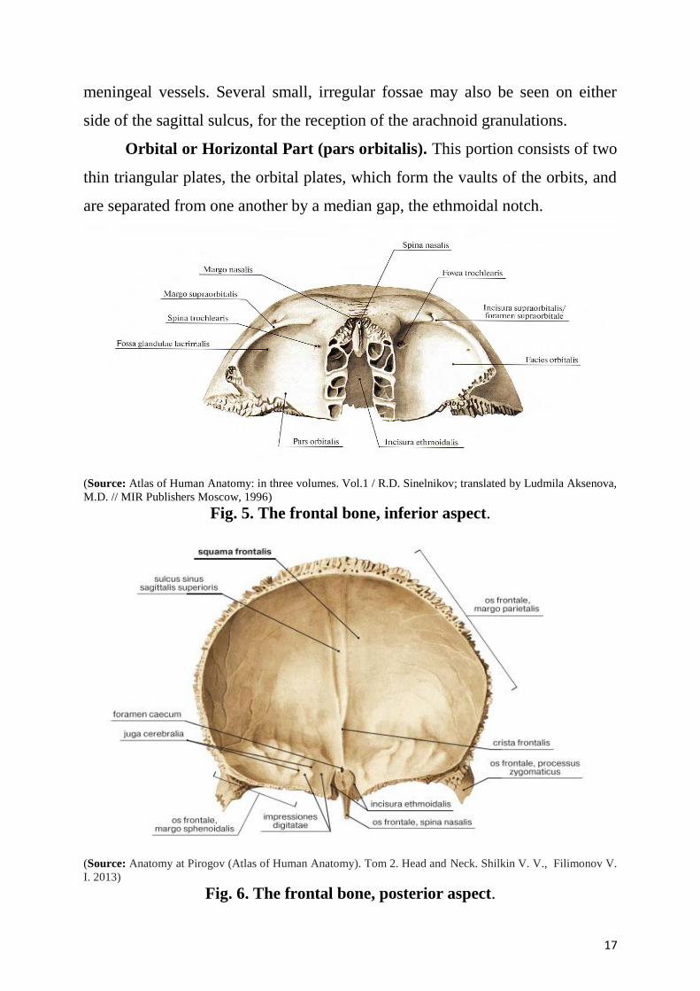
Orbital or Horizontal Part (pars orbitalis). This portion consists of two
thin triangular plates, the orbital plates, which form the vaults of the orbits, and
are separated from one another by a median gap, the ethmoidal notch.
(Source: Atlas of Human Anatomy: in three volumes. Vol.1 / R.D. Sinelnikov; translated by Ludmila Aksenova,
M.D. // MIR Publishers Moscow, 1996)
Fig. 5. The frontal bone, inferior aspect.
(Source: Anatomy at Pirogov (Atlas of Human Anatomy). Tom 2. Head and Neck. Shilkin V. V., Filimonov V.
I. 2013)
Fig. 6. The frontal bone, posterior aspect.
18
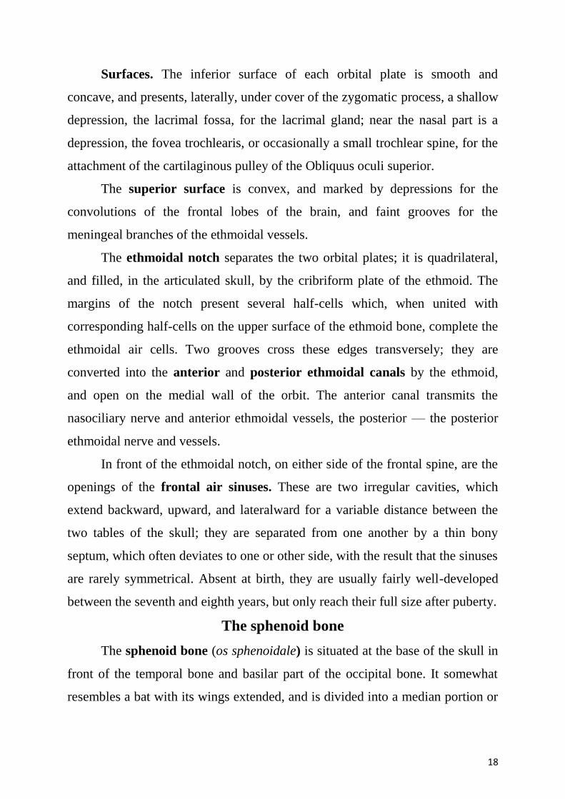
Surfaces. The inferior surface of each orbital plate is smooth and
concave, and presents, laterally, under cover of the zygomatic process, a shallow
depression, the lacrimal fossa, for the lacrimal gland; near the nasal part is a
depression, the fovea trochlearis, or occasionally a small trochlear spine, for the
attachment of the cartilaginous pulley of the Obliquus oculi superior.
The superior surface is convex, and marked by depressions for the
convolutions of the frontal lobes of the brain, and faint grooves for the
meningeal branches of the ethmoidal vessels.
The ethmoidal notch separates the two orbital plates; it is quadrilateral,
and filled, in the articulated skull, by the cribriform plate of the ethmoid. The
margins of the notch present several half-cells which, when united with
corresponding half-cells on the upper surface of the ethmoid bone, complete the
ethmoidal air cells. Two grooves cross these edges transversely; they are
converted into the anterior and posterior ethmoidal canals by the ethmoid,
and open on the medial wall of the orbit. The anterior canal transmits the
nasociliary nerve and anterior ethmoidal vessels, the posterior — the posterior
ethmoidal nerve and vessels.
In front of the ethmoidal notch, on either side of the frontal spine, are the
openings of the frontal air sinuses. These are two irregular cavities, which
extend backward, upward, and lateralward for a variable distance between the
two tables of the skull; they are separated from one another by a thin bony
septum, which often deviates to one or other side, with the result that the sinuses
are rarely symmetrical. Absent at birth, they are usually fairly well-developed
between the seventh and eighth years, but only reach their full size after puberty.
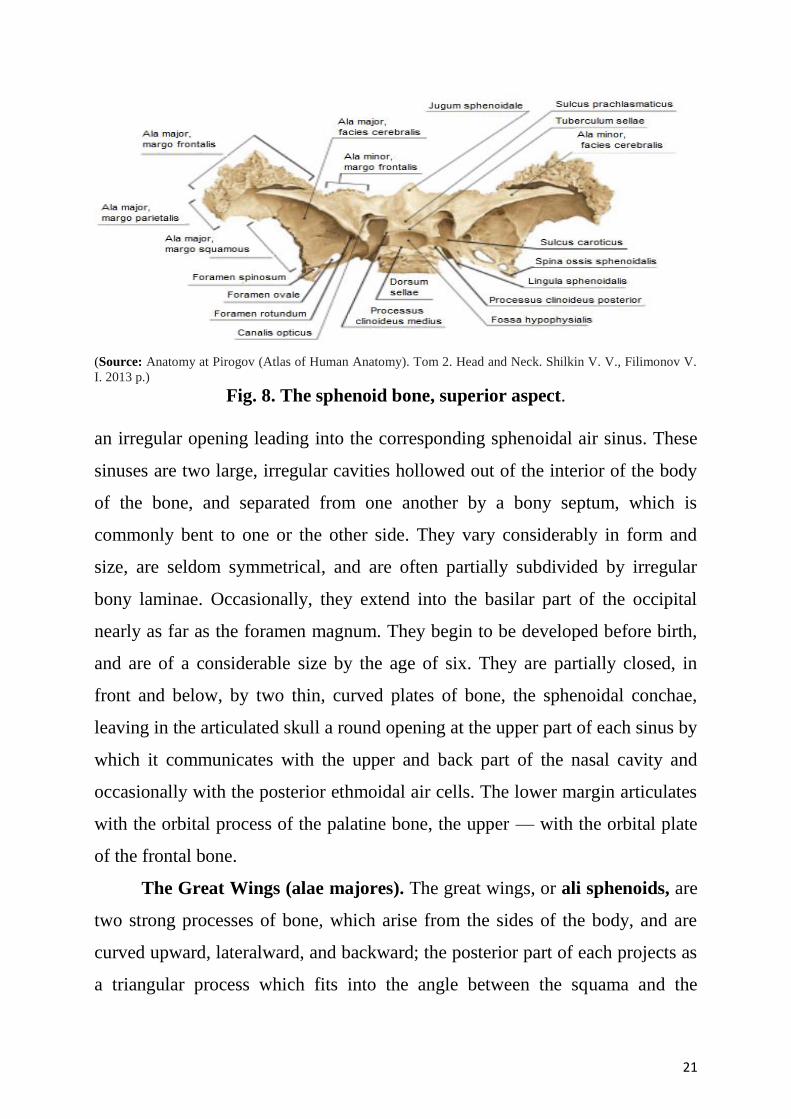
The sphenoid bone
The sphenoid bone (os sphenoidale) is situated at the base of the skull in
front of the temporal bone and basilar part of the occipital bone. It somewhat
resembles a bat with its wings extended, and is divided into a median portion or
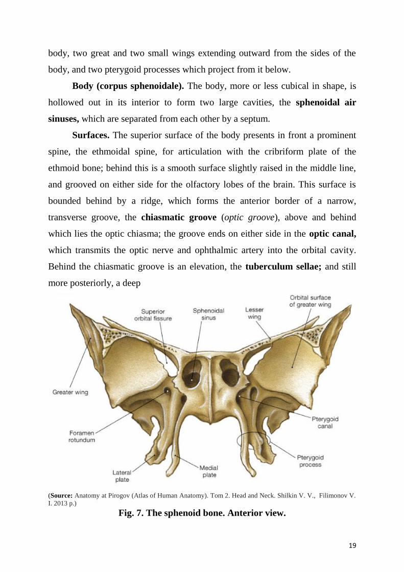
19
body, two great and two small wings extending outward from the sides of the
body, and two pterygoid processes which project from it below.
Body (corpus sphenoidale). The body, more or less cubical in shape, is
hollowed out in its interior to form two large cavities, the sphenoidal air
sinuses, which are separated from each other by a septum.
Surfaces. The superior surface of the body presents in front a prominent
spine, the ethmoidal spine, for articulation with the cribriform plate of the
ethmoid bone; behind this is a smooth surface slightly raised in the middle line,
and grooved on either side for the olfactory lobes of the brain. This surface is
bounded behind by a ridge, which forms the anterior border of a narrow,
transverse groove, the chiasmatic groove (optic groove), above and behind
which lies the optic chiasma; the groove ends on either side in the optic canal,
which transmits the optic nerve and ophthalmic artery into the orbital cavity.
Behind the chiasmatic groove is an elevation, the tuberculum sellae; and still
more posteriorly, a deep
(Source: Anatomy at Pirogov (Atlas of Human Anatomy). Tom 2. Head and Neck. Shilkin V. V., Filimonov V.
I. 2013 p.)
Fig. 7. The sphenoid bone. Anterior view.
20
depression, the sella turcica, the deepest part of which lodges the hypophysis
cerebri and is known as the fossa hypophysealis. The anterior boundary of the
sella turcica is completed by two small eminences, one on either side, called the
middle clinoid processes, while the posterior boundary is formed by a square-
shaped plate of bone, the dorsum sellae, ending at its superior angles in two
tubercles, the posterior clinoid processes, the size and form of which vary
considerably in different individuals. The posterior clinoid processes deepen the
sella turcica, and give attachment to the tentorium cerebelli.
On either side of the dorsum sellae is a notch for the passage of the
abducent nerve, and below the notch a sharp process, the petrosal process, which
articulates with the apex of the petrous portion of the temporal bone, and forms
the medial boundary of the foramen lacerum. Behind the dorsum sellae is a
shallow depression, the clivus, which slopes obliquely backward, and is
continuous with the groove on the basilar portion of the occipital bone; it
supports the upper part of the pons.
The lateral surfaces of the body are united with the great wings and the
medial pterygoid plates. Above the attachment of each great wing is a broad
groove, curved something like the italic letter f; it lodges the internal carotid
artery and the cavernous sinus, and is named the carotid groove. Along the
posterior part of the lateral margin of this groove, in the angle between the body
and great wing, is a ridge of bone, called the lingula.
The posterior surface, quadrilateral in form, is joined, during infancy and
adolescence, to the basilar part of the occipital bone by a plate of cartilage.
Between the eighteenth and twenty-fifth years this becomes ossified, ossification
commencing above and extending downward.
The anterior surface of the body presents, in the middle line, a vertical
crest, the sphenoidal crest, which articulates with the perpendicular plate of the
ethmoid, and forms part of the septum of the nose. On either side of the crest is
21
(Source: Anatomy at Pirogov (Atlas of Human Anatomy). Tom 2. Head and Neck. Shilkin V. V., Filimonov V.
I. 2013 p.)
Fig. 8. The sphenoid bone, superior aspect.
an irregular opening leading into the corresponding sphenoidal air sinus. These
sinuses are two large, irregular cavities hollowed out of the interior of the body
of the bone, and separated from one another by a bony septum, which is
commonly bent to one or the other side. They vary considerably in form and
size, are seldom symmetrical, and are often partially subdivided by irregular
bony laminae. Occasionally, they extend into the basilar part of the occipital
nearly as far as the foramen magnum. They begin to be developed before birth,
and are of a considerable size by the age of six. They are partially closed, in
front and below, by two thin, curved plates of bone, the sphenoidal conchae,
leaving in the articulated skull a round opening at the upper part of each sinus by
which it communicates with the upper and back part of the nasal cavity and
occasionally with the posterior ethmoidal air cells. The lower margin articulates
with the orbital process of the palatine bone, the upper — with the orbital plate
of the frontal bone.
The Great Wings (alae majores). The great wings, or ali sphenoids, are
two strong processes of bone, which arise from the sides of the body, and are
curved upward, lateralward, and backward; the posterior part of each projects as
a triangular process which fits into the angle between the squama and the
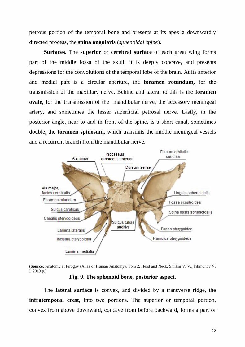
22
petrous portion of the temporal bone and presents at its apex a downwardly
directed process, the spina angularis (sphenoidal spine).
Surfaces. The superior or cerebral surface of each great wing forms
part of the middle fossa of the skull; it is deeply concave, and presents
depressions for the convolutions of the temporal lobe of the brain. At its anterior
and medial part is a circular aperture, the foramen rotundum, for the
transmission of the maxillary nerve. Behind and lateral to this is the foramen
ovale, for the transmission of the mandibular nerve, the accessory meningeal
artery, and sometimes the lesser superficial petrosal nerve. Lastly, in the
posterior angle, near to and in front of the spine, is a short canal, sometimes
double, the foramen spinosum, which transmits the middle meningeal vessels
and a recurrent branch from the mandibular nerve.
(Source: Anatomy at Pirogov (Atlas of Human Anatomy). Tom 2. Head and Neck. Shilkin V. V., Filimonov V.
I. 2013 p.)
Fig. 9. The sphenoid bone, posterior aspect.
The lateral surface is convex, and divided by a transverse ridge, the
infratemporal crest, into two portions. The superior or temporal portion,
convex from above downward, concave from before backward, forms a part of
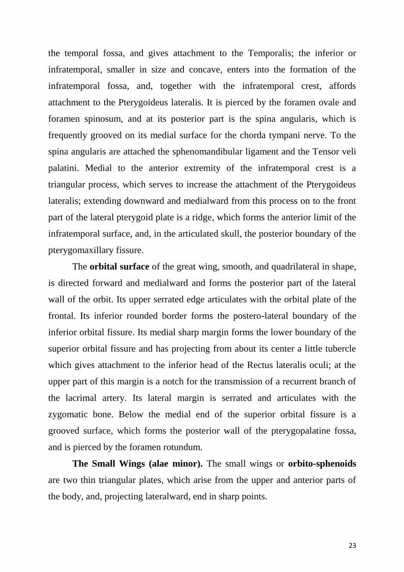
23
the temporal fossa, and gives attachment to the Temporalis; the inferior or
infratemporal, smaller in size and concave, enters into the formation of the
infratemporal fossa, and, together with the infratemporal crest, affords
attachment to the Pterygoideus lateralis. It is pierced by the foramen ovale and
foramen spinosum, and at its posterior part is the spina angularis, which is
frequently grooved on its medial surface for the chorda tympani nerve. To the
spina angularis are attached the sphenomandibular ligament and the Tensor veli
palatini. Medial to the anterior extremity of the infratemporal crest is a
triangular process, which serves to increase the attachment of the Pterygoideus
lateralis; extending downward and medialward from this process on to the front
part of the lateral pterygoid plate is a ridge, which forms the anterior limit of the
infratemporal surface, and, in the articulated skull, the posterior boundary of the
pterygomaxillary fissure.
The orbital surface of the great wing, smooth, and quadrilateral in shape,
is directed forward and medialward and forms the posterior part of the lateral
wall of the orbit. Its upper serrated edge articulates with the orbital plate of the
frontal. Its inferior rounded border forms the postero-lateral boundary of the
inferior orbital fissure. Its medial sharp margin forms the lower boundary of the
superior orbital fissure and has projecting from about its center a little tubercle
which gives attachment to the inferior head of the Rectus lateralis oculi; at the
upper part of this margin is a notch for the transmission of a recurrent branch of
the lacrimal artery. Its lateral margin is serrated and articulates with the
zygomatic bone. Below the medial end of the superior orbital fissure is a
grooved surface, which forms the posterior wall of the pterygopalatine fossa,
and is pierced by the foramen rotundum.
The Small Wings (alae minor). The small wings or orbito-sphenoids
are two thin triangular plates, which arise from the upper and anterior parts of
the body, and, projecting lateralward, end in sharp points.
24
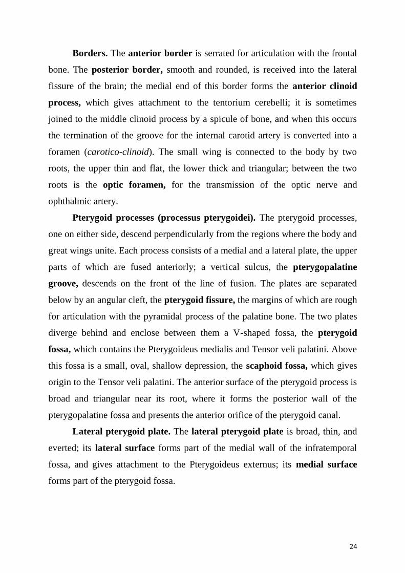
Borders. The anterior border is serrated for articulation with the frontal
bone. The posterior border, smooth and rounded, is received into the lateral
fissure of the brain; the medial end of this border forms the anterior clinoid
process, which gives attachment to the tentorium cerebelli; it is sometimes
joined to the middle clinoid process by a spicule of bone, and when this occurs
the termination of the groove for the internal carotid artery is converted into a
foramen (carotico-clinoid). The small wing is connected to the body by two
roots, the upper thin and flat, the lower thick and triangular; between the two
roots is the optic foramen, for the transmission of the optic nerve and
ophthalmic artery.
Pterygoid processes (processus pterygoidei). The pterygoid processes,
one on either side, descend perpendicularly from the regions where the body and
great wings unite. Each process consists of a medial and a lateral plate, the upper
parts of which are fused anteriorly; a vertical sulcus, the pterygopalatine
groove, descends on the front of the line of fusion. The plates are separated
below by an angular cleft, the pterygoid fissure, the margins of which are rough
for articulation with the pyramidal process of the palatine bone. The two plates
diverge behind and enclose between them a V-shaped fossa, the pterygoid
fossa, which contains the Pterygoideus medialis and Tensor veli palatini. Above
this fossa is a small, oval, shallow depression, the scaphoid fossa, which gives
origin to the Tensor veli palatini. The anterior surface of the pterygoid process is
broad and triangular near its root, where it forms the posterior wall of the
pterygopalatine fossa and presents the anterior orifice of the pterygoid canal.
Lateral pterygoid plate. The lateral pterygoid plate is broad, thin, and
everted; its lateral surface forms part of the medial wall of the infratemporal
fossa, and gives attachment to the Pterygoideus externus; its medial surface
forms part of the pterygoid fossa.
25
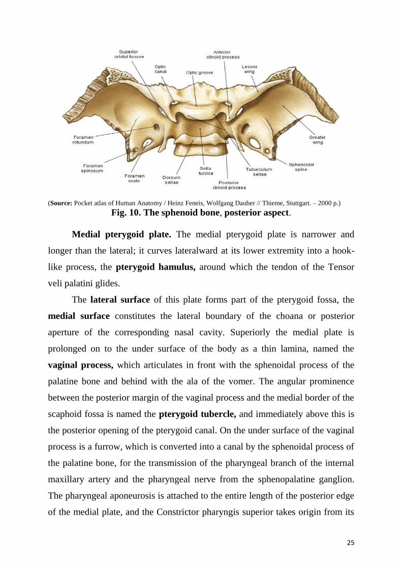
(Source: Pocket atlas of Human Anatomy / Heinz Feneis, Wolfgang Dauber // Thieme, Stuttgart. – 2000 p.)
Fig. 10. The sphenoid bone, posterior aspect.
Medial pterygoid plate. The medial pterygoid plate is narrower and
longer than the lateral; it curves lateralward at its lower extremity into a hook-
like process, the pterygoid hamulus, around which the tendon of the Tensor
veli palatini glides.
The lateral surface of this plate forms part of the pterygoid fossa, the
medial surface constitutes the lateral boundary of the choana or posterior
aperture of the corresponding nasal cavity. Superiorly the medial plate is
prolonged on to the under surface of the body as a thin lamina, named the
vaginal process, which articulates in front with the sphenoidal process of the
palatine bone and behind with the ala of the vomer. The angular prominence
between the posterior margin of the vaginal process and the medial border of the
scaphoid fossa is named the pterygoid tubercle, and immediately above this is
the posterior opening of the pterygoid canal. On the under surface of the vaginal
process is a furrow, which is converted into a canal by the sphenoidal process of
the palatine bone, for the transmission of the pharyngeal branch of the internal
maxillary artery and the pharyngeal nerve from the sphenopalatine ganglion.
The pharyngeal aponeurosis is attached to the entire length of the posterior edge
of the medial plate, and the Constrictor pharyngis superior takes origin from its
26
lower third. Projecting backward from near the middle of the posterior edge of
this plate is an angular process, the processus tubarius, which supports the
pharyngeal end of the auditory tube. The anterior margin of the plate articulates
with the posterior border of the vertical part of the palatine bone.
The ethmoid bone
The ethmoid bone (os ethmoidale) is exceedingly light and spongy, and
cubical in shape; it is situated at the anterior part of the base of the cranium,
between the two orbits, at the roof of the nose, and contributes to each of these
cavities. It consists of four parts: a horizontal or cribriform plate, forming part
of the base of the cranium; a perpendicular plate, constituting part of the nasal
septum; and two lateral masses or labyrinths.
Cribiform plate (lamina cribrosa; horizontal lamina). The cribriform
plate is received into the ethmoidal notch of the frontal bone and roofs in the
nasal cavities. Projecting upward from the middle line of this plate is a thick,
smooth, triangular process, the crista galli, so called from its resemblance to a
cock’s comb. The long thin posterior border of the crista galli serves for the
attachment of the falx cerebri. Its anterior border, short and thick, articulates
with the frontal bone, and presents two small projecting alae, which are received
into corresponding depressions in the frontal bone and complete the foramen
cecum. Its sides are smooth, and sometimes bulging from the presence of a
small air sinus in the interior. On either side of the crista galli, the cribriform
plate is narrow and deeply grooved; it supports the olfactory bulb and is
perforated by foramina for the passage of the olfactory nerves. The foramina in
the middle of the groove are small and transmit the nerves to the roof of the
nasal cavity; those at the medial and lateral parts of the groove are larger — the
former transmit the nerves to the upper part of the nasal septum, the latter those
to the superior nasal concha. At the front part of the cribriform plate, on either
side of the crista galli, is a small fissure which is occupied by a process of dura
27
mater. Lateral to this fissure is a notch or foramen, which transmits the
nasociliary nerve; from this notch a groove extends backward to the anterior
ethmoidal foramen.
Perpendicular plate (lamina perpendicularis; vertical plate). The
perpendicular plate is a thin, flattened lamina, polygonal in form, which
descends from the under surface of the cribriform plate, and assists in forming
the septum of the nose; it is generally deflected a little to one or other side. The
anterior border articulates with the spine of the frontal bone and the crest of the
nasal bones. The posterior border articulates by its upper half with the
sphenoidal crest, by its lower with the vomer. The inferior border is thicker than
the posterior, and serves for the attachment of the septal cartilage of the nose.
The surfaces of the plate are smooth, except above, where numerous grooves
and canals are seen; these lead from the medial foramina on the cribriform plate
and lodge filaments of the olfactory nerves.
The Labyrinth or Lateral Mass (labyrinthus ethmoidalis) consists of a
number of thin-walled cellular cavities, the ethmoidal cells, arranged in three
groups, anterior, middle, and posterior, and interposed between two vertical
plates of bone; the lateral plate forms part of the orbit, the medial, part of the
corresponding nasal cavity. In the disarticulated bone, many of these cells are
opened into, but when the bones are articulated, they are closed in at every part,
except where they open into the nasal cavity.
28
(Source: Gray′s anatomy for students / Richard L. Drake, A. Wayne Vogl, and Adam W. M. Mitchell;
illustrations by Richard M. Tibbitts and Paul E. Richardson; photographs by Ansell Horn; 2nd ed. // Churchill
Livingstone, 2012.)
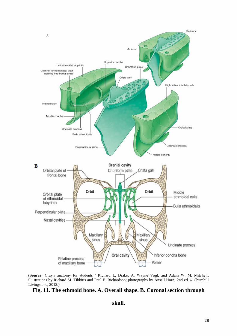
Fig. 11. The ethmoid bone. A. Overall shape. B. Coronal section through
skull.
29
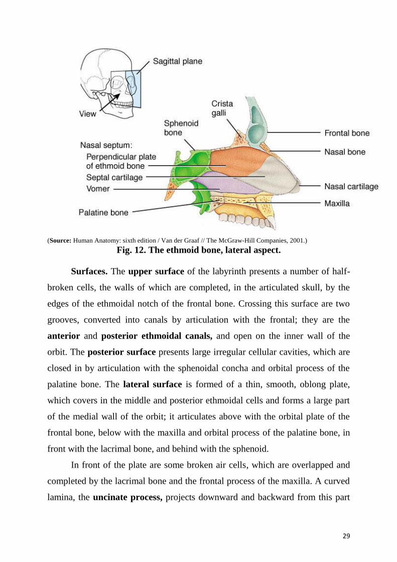
(Source: Human Anatomy: sixth edition / Van der Graaf // The McGraw-Hill Companies, 2001.)
Fig. 12. The ethmoid bone, lateral aspect.
Surfaces. The upper surface of the labyrinth presents a number of half-
broken cells, the walls of which are completed, in the articulated skull, by the
edges of the ethmoidal notch of the frontal bone. Crossing this surface are two
grooves, converted into canals by articulation with the frontal; they are the
anterior and posterior ethmoidal canals, and open on the inner wall of the
orbit. The posterior surface presents large irregular cellular cavities, which are
closed in by articulation with the sphenoidal concha and orbital process of the
palatine bone. The lateral surface is formed of a thin, smooth, oblong plate,
which covers in the middle and posterior ethmoidal cells and forms a large part
of the medial wall of the orbit; it articulates above with the orbital plate of the
frontal bone, below with the maxilla and orbital process of the palatine bone, in
front with the lacrimal bone, and behind with the sphenoid.
In front of the plate are some broken air cells, which are overlapped and
completed by the lacrimal bone and the frontal process of the maxilla. A curved
lamina, the uncinate process, projects downward and backward from this part
30
of the labyrinth; it forms a small part of the medial wall of the maxillary sinus,
and articulates with the ethmoidal process of the inferior nasal concha.
(Source: Anatomy at Pirogov (Atlas of Human Anatomy). Tom 2. Head and Neck. Shilkin V. V., Filimonov V.
I. 2013 p.)
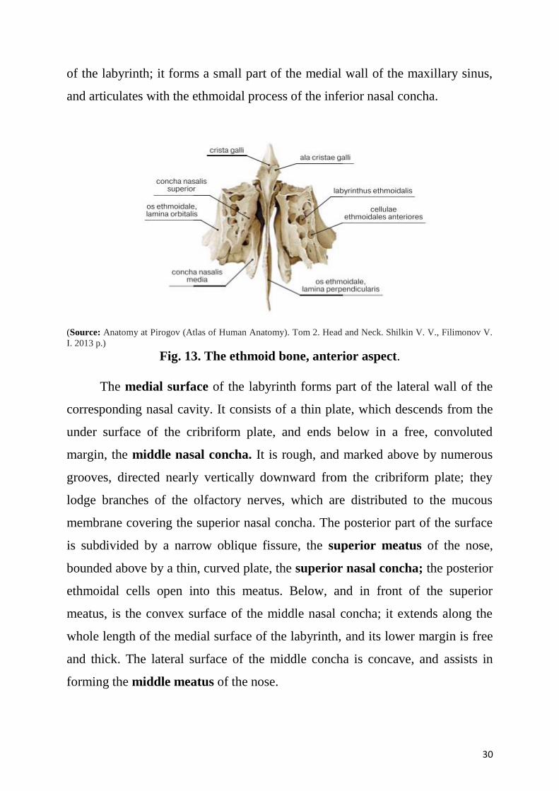
Fig. 13. The ethmoid bone, anterior aspect.
The medial surface of the labyrinth forms part of the lateral wall of the
corresponding nasal cavity. It consists of a thin plate, which descends from the
under surface of the cribriform plate, and ends below in a free, convoluted
margin, the middle nasal concha. It is rough, and marked above by numerous
grooves, directed nearly vertically downward from the cribriform plate; they
lodge branches of the olfactory nerves, which are distributed to the mucous
membrane covering the superior nasal concha. The posterior part of the surface
is subdivided by a narrow oblique fissure, the superior meatus of the nose,
bounded above by a thin, curved plate, the superior nasal concha; the posterior
ethmoidal cells open into this meatus. Below, and in front of the superior
meatus, is the convex surface of the middle nasal concha; it extends along the
whole length of the medial surface of the labyrinth, and its lower margin is free
and thick. The lateral surface of the middle concha is concave, and assists in
forming the middle meatus of the nose.
31
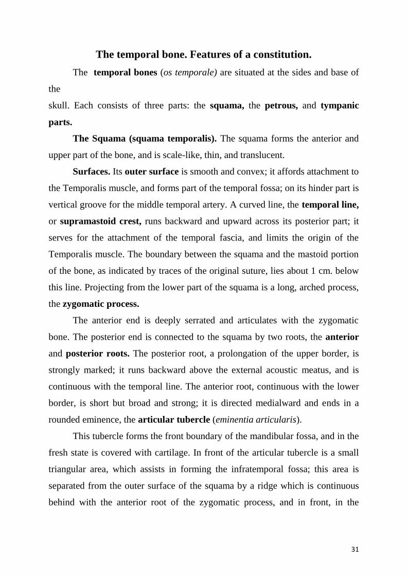
The temporal bone. Features of a constitution.
The temporal bones (os temporale) are situated at the sides and base of
the
skull. Each consists of three parts: the squama, the petrous, and tympanic
parts.
The Squama (squama temporalis). The squama forms the anterior and
upper part of the bone, and is scale-like, thin, and translucent.
Surfaces. Its outer surface is smooth and convex; it affords attachment to
the Temporalis muscle, and forms part of the temporal fossa; on its hinder part is
vertical groove for the middle temporal artery. A curved line, the temporal line,
or supramastoid crest, runs backward and upward across its posterior part; it
serves for the attachment of the temporal fascia, and limits the origin of the
Temporalis muscle. The boundary between the squama and the mastoid portion
of the bone, as indicated by traces of the original suture, lies about 1 cm. below
this line. Projecting from the lower part of the squama is a long, arched process,
the zygomatic process.
The anterior end is deeply serrated and articulates with the zygomatic
bone. The posterior end is connected to the squama by two roots, the anterior
and posterior roots. The posterior root, a prolongation of the upper border, is
strongly marked; it runs backward above the external acoustic meatus, and is
continuous with the temporal line. The anterior root, continuous with the lower
border, is short but broad and strong; it is directed medialward and ends in a
rounded eminence, the articular tubercle (eminentia articularis).
This tubercle forms the front boundary of the mandibular fossa, and in the
fresh state is covered with cartilage. In front of the articular tubercle is a small
triangular area, which assists in forming the infratemporal fossa; this area is
separated from the outer surface of the squama by a ridge which is continuous
behind with the anterior root of the zygomatic process, and in front, in the
32
articulated skull, with the infratemporal crest on the great wing of the
sphenoid bone.
(Source: Anatomy at Pirogov (Atlas of Human Anatomy). Tom 2. Head and Neck. Shilkin V. V., Filimonov V.
I. 2013 p.)
Fig. 14. The temporal bone, external aspect.
Between the posterior wall of the external acoustic meatus and the posterior root
of the zygomatic process is the area called the suprameatal triangle
(Macewen), or mastoid fossa, through which an instrument may be pushed
into the tympanic antrum. At the junction of the anterior root with the zygomatic
process is a projection for the attachment of the temporomandibular ligament;
and behind the anterior root is an oval depression, forming part of the
mandibular fossa, for the reception of the condyle of the mandible. The
mandibular fossa (glenoid fossa) is bounded, in front, by the articular tubercle;
behind, by the tympanic part of the bone, which separates it from the external
acoustic meatus; it is divided into two parts by a narrow slit, the petrotympanic
fissure (Glaserian fissure). The anterior part, formed by the squama, is smooth,
covered in the fresh state with cartilage, and articulates with the condyle of the
mandible. Behind this part of the fossa is a small conical eminence; this is the
33
representative of a prominent tubercle which, in some mammals, descends
behind the condyle of the mandible, and prevents its backward displacement.
The posterior part of the mandibular fossa, formed by the tympanic part of the
bone, is non-articular, and sometimes lodges a portion of the parotid gland. The
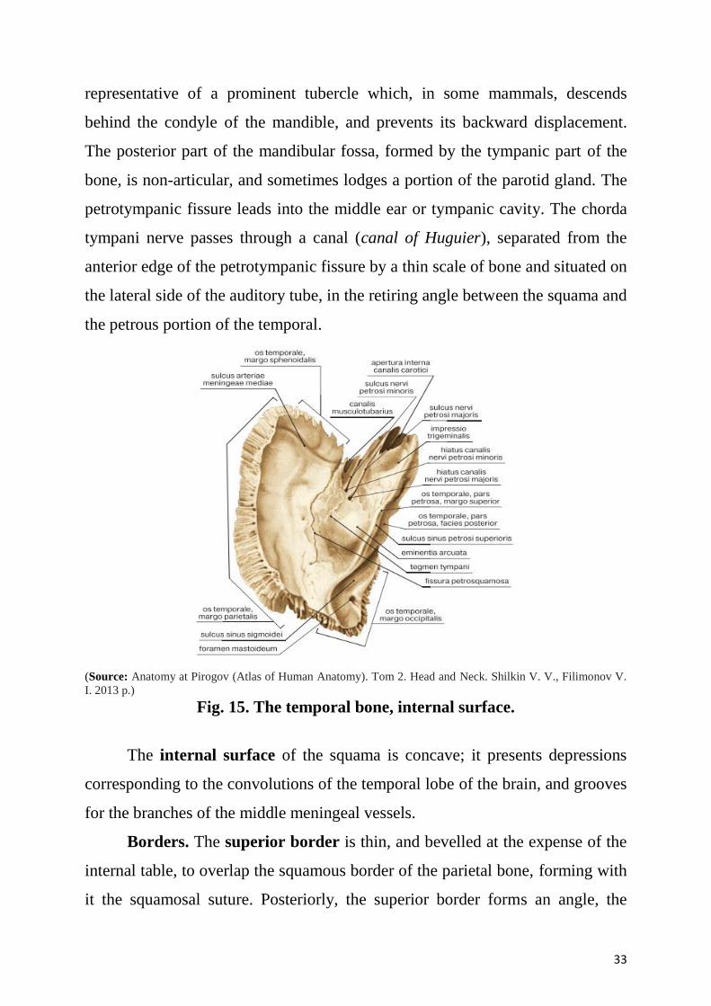
petrotympanic fissure leads into the middle ear or tympanic cavity. The chorda
tympani nerve passes through a canal (canal of Huguier), separated from the
anterior edge of the petrotympanic fissure by a thin scale of bone and situated on
the lateral side of the auditory tube, in the retiring angle between the squama and
the petrous portion of the temporal.
(Source: Anatomy at Pirogov (Atlas of Human Anatomy). Tom 2. Head and Neck. Shilkin V. V., Filimonov V.
I. 2013 p.)
Fig. 15. The temporal bone, internal surface.
The internal surface of the squama is concave; it presents depressions
corresponding to the convolutions of the temporal lobe of the brain, and grooves
for the branches of the middle meningeal vessels.
Borders. The superior border is thin, and bevelled at the expense of the
internal table, to overlap the squamous border of the parietal bone, forming with
it the squamosal suture. Posteriorly, the superior border forms an angle, the
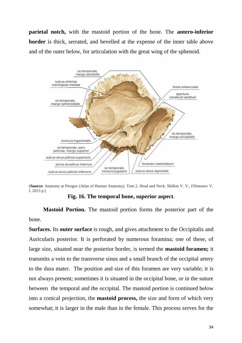
34
parietal notch, with the mastoid portion of the bone. The antero-inferior
border is thick, serrated, and bevelled at the expense of the inner table above
and of the outer below, for articulation with the great wing of the sphenoid.
(Source: Anatomy at Pirogov (Atlas of Human Anatomy). Tom 2. Head and Neck. Shilkin V. V., Filimonov V.
I. 2013 p.)
Fig. 16. The temporal bone, superior aspect.
Mastoid Portion. The mastoid portion forms the posterior part of the
bone.
Surfaces. Its outer surface is rough, and gives attachment to the Occipitalis and
Auricularis posterior. It is perforated by numerous foramina; one of these, of
large size, situated near the posterior border, is termed the mastoid foramen; it
transmits a vein to the transverse sinus and a small branch of the occipital artery
to the dura mater. The position and size of this foramen are very variable; it is
not always present; sometimes it is situated in the occipital bone, or in the suture
between the temporal and the occipital. The mastoid portion is continued below
into a conical projection, the mastoid process, the size and form of which very
somewhat; it is larger in the male than in the female. This process serves for the
35
attachment of the Sternocleidomastoideus, Splenius capitis, and Longissimus
capitis. On the medial side of the process is a deep groove, the mastoid notch
(digastric fossa), for the attachment of the Digastricus; medial to this is a
shallow furrow, the occipital groove, which lodges the occipital artery.
The inner surface of the mastoid portion presents a deep, curved groove,
the sigmoid sulcus, which lodges part of the transverse sinus; in it may be seen
the opening of the mastoid foramen. The groove for the transverse sinus is
separated from the innermost of the mastoid air cells by a very thin lamina of
bone, and even this may be partly deficient.
A section of the mastoid process shows it to be hollowed out into a
number of spaces, the mastoid cells, which exhibit the greatest possible variety
as to their size and number. At the upper and front part of the process they are
large and irregular and contain air, but toward the lower part they diminish in
size, while those at the apex of the process are frequently quite small and
contain marrow; occasionally they are entirely absent, and the mastoid is then
solid throughout. In addition to these a large irregular cavity is situated at the
upper and front part of the bone. It is called the tympanic antrum, and must be
distinguished from the mastoid cells, though it communicates with them. Like
the mastoid cells, it is filled with air and lined by a prolongation of the mucous
membrane of the tympanic cavity, with which it communicates. The tympanic
antrum is bounded above by a thin plate of bone, the tegmen tympani, which
separates it from the middle fossa of the base of the skull; below by the mastoid
process; laterally by the squama just below the temporal line, and medially by
the lateral semicircular canal of the internal ear which projects into its cavity. It
opens in front into that portion of the tympanic cavity, which is known as
epitympanic recess. The tympanic antrum is a cavity of some considerable size
at the time of birth; the mastoid air cells may be regarded as diverticula from the
antrum, and begin to appear at or before birth; by the fifth year they are well-
marked, but their development is not completed until toward puberty.
36
In the clinic. Mastoiditis. Infection within the mastoid antrum and
mastoid cells is usually secondary to infection in the middle ear. The mastoid
cells provide an excellent culture medium for infection. Infection of the bone
(osteomyelitis) may also develop, spreading into the middle cranial fossa.
Drainage of the pus within the mastoid air cells is necessary and there are
numerous approaches for doing this. When undertaking this type of surgery, it is
extremely important that care is taken not to damage the mastoid wall of the
middle ear to prevent injury to the facial nerve [VII]. Any breach of the inner
table of the cranial vault may allow bacteria to enter the cranial cavity and
meningitis will ensue.
Petrous Portion (pars petrosa [pyramis]). The petrous portion or
pyramid is pyramidal and is wedged in at the base of the skull between the
sphenoid and occipital. Directed medialward, forward, and a little upward, it
presents for examination a base, an apex, three surfaces, and three angles, and
contains, in its interior, the essential parts of the organ of hearing.
Base. The base is fused with the internal surfaces of the squama and
mastoid portion.
Apex. The apex, rough and uneven, is received into the angular interval
between the posterior border of the great wing of the sphenoid and the basilar
part of the occipital; it presents the anterior or internal orifice of the carotid
canal, and forms the postero-lateral boundary of the foramen lacerum.
Surfaces. The anterior surface forms the posterior part of the middle
fossa of the base of the skull, and is continuous with the inner surface of the
squamous portion, to which it is united by the petrosquamous suture, remains
of which are distinct even at a late period of life. It is marked by depressions for
the convolutions of the brain, and presents six points for examination:
1) near the center, an eminence (eminentia arcuata) which indicates the
situation of the superior semicircular canal; 2) in front of and a little lateral to
this eminence, a depression indicating the position of the tympanic cavity: here
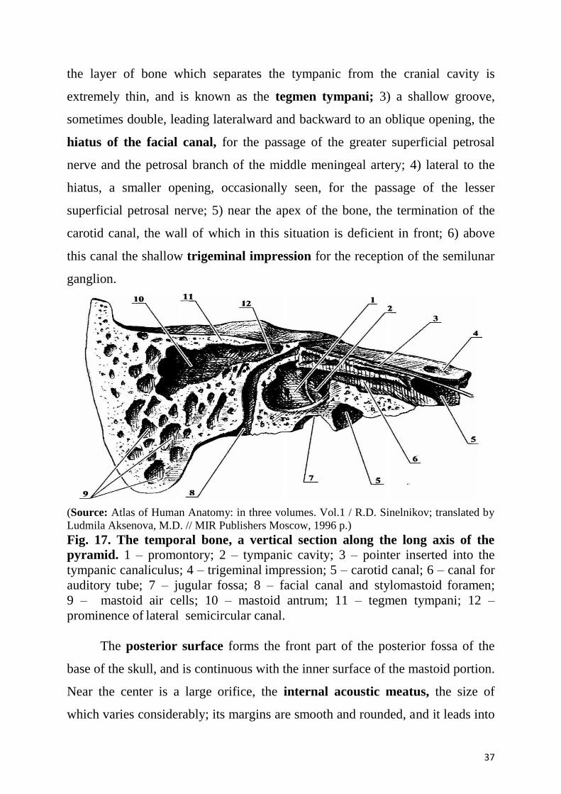
37
the layer of bone which separates the tympanic from the cranial cavity is
extremely thin, and is known as the tegmen tympani; 3) a shallow groove,
sometimes double, leading lateralward and backward to an oblique opening, the
hiatus of the facial canal, for the passage of the greater superficial petrosal
nerve and the petrosal branch of the middle meningeal artery; 4) lateral to the
hiatus, a smaller opening, occasionally seen, for the passage of the lesser
superficial petrosal nerve; 5) near the apex of the bone, the termination of the
carotid canal, the wall of which in this situation is deficient in front; 6) above
this canal the shallow trigeminal impression for the reception of the semilunar
ganglion.
(Source: Atlas of Human Anatomy: in three volumes. Vol.1 / R.D. Sinelnikov; translated by
Ludmila Aksenova, M.D. // MIR Publishers Moscow, 1996 p.)
Fig. 17. The temporal bone, a vertical section along the long axis of the
pyramid. 1 – promontory; 2 – tympanic cavity; 3 – pointer inserted into the
tympanic canaliculus; 4 – trigeminal impression; 5 – carotid canal; 6 – canal for
auditory tube; 7 – jugular fossa; 8 – facial canal and stylomastoid foramen;
9 – mastoid air cells; 10 – mastoid antrum; 11 – tegmen tympani; 12 –
prominence of lateral semicircular canal.
The posterior surface forms the front part of the posterior fossa of the
base of the skull, and is continuous with the inner surface of the mastoid portion.
Near the center is a large orifice, the internal acoustic meatus, the size of
which varies considerably; its margins are smooth and rounded, and it leads into
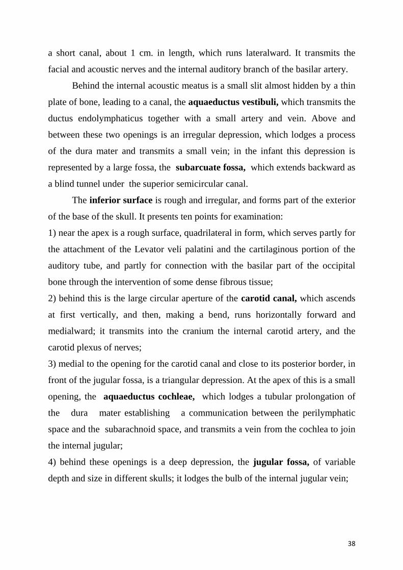
38
a short canal, about 1 cm. in length, which runs lateralward. It transmits the
facial and acoustic nerves and the internal auditory branch of the basilar artery.
Behind the internal acoustic meatus is a small slit almost hidden by a thin
plate of bone, leading to a canal, the aquaeductus vestibuli, which transmits the
ductus endolymphaticus together with a small artery and vein. Above and
between these two openings is an irregular depression, which lodges a process
of the dura mater and transmits a small vein; in the infant this depression is
represented by a large fossa, the subarcuate fossa, which extends backward as
a blind tunnel under the superior semicircular canal.
The inferior surface is rough and irregular, and forms part of the exterior
of the base of the skull. It presents ten points for examination:
1) near the apex is a rough surface, quadrilateral in form, which serves partly for
the attachment of the Levator veli palatini and the cartilaginous portion of the
auditory tube, and partly for connection with the basilar part of the occipital
bone through the intervention of some dense fibrous tissue;
2) behind this is the large circular aperture of the carotid canal, which ascends
at first vertically, and then, making a bend, runs horizontally forward and
medialward; it transmits into the cranium the internal carotid artery, and the
carotid plexus of nerves;
3) medial to the opening for the carotid canal and close to its posterior border, in
front of the jugular fossa, is a triangular depression. At the apex of this is a small
opening, the aquaeductus cochleae, which lodges a tubular prolongation of
the dura mater establishing a communication between the perilymphatic
space and the subarachnoid space, and transmits a vein from the cochlea to join
the internal jugular;
4) behind these openings is a deep depression, the jugular fossa, of variable
depth and size in different skulls; it lodges the bulb of the internal jugular vein;
39
5) in the bony ridge dividing the carotid canal from the jugular fossa is the small
inferior tympanic canaliculus for the passage of the tympanic branch of the
glossopharyngeal nerve;
6) in the lateral part of the jugular fossa is the mastoid canaliculus for the
entrance of the auricular branch of the vagus nerve;
7) behind the jugular fossa is a quadrilateral area, the jugular surface, covered
with cartilage in the fresh state, and articulating with the jugular process of the
occipital bone;
8) extending backward from the carotid canal is the vaginal process, a sheath-
like plate of bone, which divides behind into two laminae; the lateral lamina is
continuous with the tympanic part of the bone, the medial with the lateral
margin of the jugular surface;
9) between these laminae is the styloid process, a sharp spine, about 2.5 cm. in
length;
10) between the styloid and mastoid processes is the stylomastoid foramen; it
is the termination of the facial canal, and transmits the facial nerve and
stylomastoid artery;
Angles. The superior angle, the longest, is grooved for the superior
petrosal sinus, and gives attachment to the tentorium cerebelli; at its medial
extremity is a notch, in which the trigeminal nerve lies. The posterior angle is
intermediate in length between the superior and the anterior. Its medial half is
marked by a sulcus, which forms, with a corresponding sulcus on the occipital
bone, the channel for the inferior petrosal sinus. Its lateral half presents an
excavation — the jugular fossa — which, with the jugular notch on the
occipital, forms the jugular foramen; an eminence occasionally projects from
the center of the fossa, and divides the foramen into two. The anterior angle is
divided into two parts — a lateral joined to the squama by a suture
(petrosquamous), the remains of which are more or less distinct; a medial, free,
which articulates with the spinous process of the sphenoid.
40
At the angle of junction of the petrous part and the squama are two canals, one
above the other, and separated by a thin plate of bone, the septum canalis
musculotubarii (processus cochleariformis); both canals lead into the tympanic
cavity. The upper one (semicanalis m. tensoris tympani) transmits the Tensor
tympani, the lower one (semicanalis tubae auditivae) forms the bony part of the
auditory tube.
Tympanic Part (pars tympanica). The tympanic part is a curved plate of
bone lying below the squama and in front of the mastoid process.
Surfaces. Its postero-superior surface is concave, and forms the anterior
wall, the floor, and part of the posterior wall of the bony external acoustic
meatus. Medially, it presents a narrow furrow, the tympanic sulcus, for the
attachment of the tympanic membrane. Its antero-inferior surface is
quadrilateral and slightly concave; it constitutes the posterior boundary of the
mandibular fossa, and is in contact with the retromandibular part of the parotid
gland.
The external acoustic meatus is nearly 2 cm. long and is directed inward
and slightly forward: at the same time it forms a slight curve, so that the floor of
the canal is convex upward. In sagittal section it presents an oval or elliptical
shape with the long axis directed downward and slightly backward. Its anterior
wall and floor and the lower part of its posterior wall are formed by the
tympanic part; the roof and upper part of the posterior wall by the squama. Its
inner end is closed, in the recent state, by the tympanic membrane; the upper
limit of its outer orifice is formed by the posterior root of the zygomatic process,
immediately below which there is sometimes seen a small spine, the
suprameatal spine, situated at the upper and posterior part of the orifice.
Styloid Processus (processus styloideus). The styloid process is slender,
pointed, and of varying length; it projects downward and forward, from the
under surface of the temporal bone. Its proximal part (tympanohyal) is
unsheathed by the vaginal process of the tympanic portion, while its distal part
41
(stylohyal) gives attachment to the stylohyoid and stylomandibular ligaments,
and to the Styloglossus,
Stylohyoideus, and Stylopharyngeus muscles. The stylohyoid ligament extends
from the apex of the process to the lesser horn of the hyoid bone, and in some
instances is partially, in others completely, ossified.
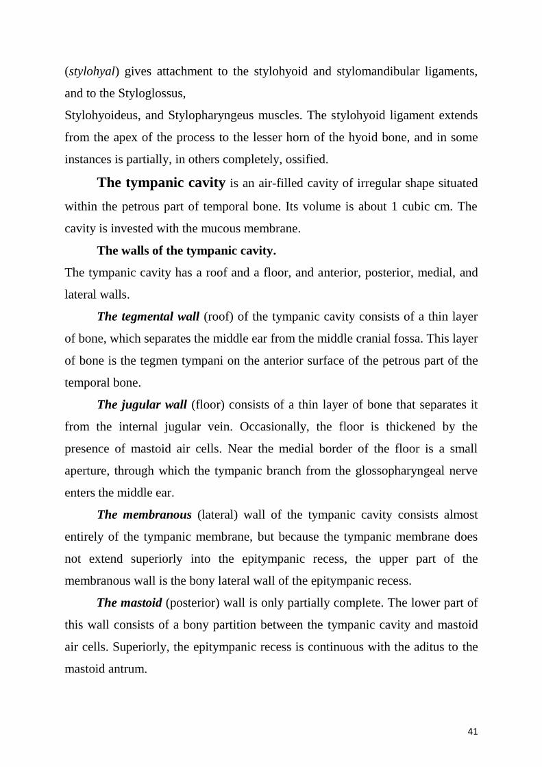
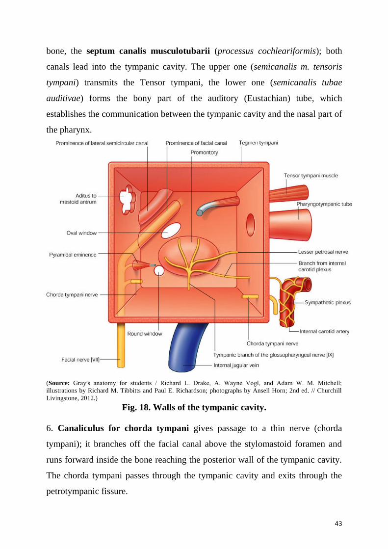
The tympanic cavity is an air-filled cavity of irregular shape situated
within the petrous part of temporal bone. Its volume is about 1 cubic cm. The
cavity is invested with the mucous membrane.
The walls of the tympanic cavity.
The tympanic cavity has a roof and a floor, and anterior, posterior, medial, and
lateral walls.
The tegmental wall (roof) of the tympanic cavity consists of a thin layer
of bone, which separates the middle ear from the middle cranial fossa. This layer
of bone is the tegmen tympani on the anterior surface of the petrous part of the
temporal bone.
The jugular wall (floor) consists of a thin layer of bone that separates it
from the internal jugular vein. Occasionally, the floor is thickened by the
presence of mastoid air cells. Near the medial border of the floor is a small
aperture, through which the tympanic branch from the glossopharyngeal nerve
enters the middle ear.
The membranous (lateral) wall of the tympanic cavity consists almost
entirely of the tympanic membrane, but because the tympanic membrane does
not extend superiorly into the epitympanic recess, the upper part of the
membranous wall is the bony lateral wall of the epitympanic recess.
The mastoid (posterior) wall is only partially complete. The lower part of
this wall consists of a bony partition between the tympanic cavity and mastoid
air cells. Superiorly, the epitympanic recess is continuous with the aditus to the
mastoid antrum.
42
The anterior wall of the tympanic cavity is only partially complete. The
lower part consists of a thin layer of bone that separates the tympanic cavity
from the internal carotid artery. Superiorly, the wall is deficient because of the
presence of:
- a large opening for the entrance of the pharyngotympanic tube into the
middle ear;
- a smaller opening for the canal containing the tensor tympani muscle.
The foramen for the exit of the chorda tympani nerve from the middle ear is also
associated with this wall.
The labyrinthine (medial) wall of the middle ear is also the lateral wall of
the internal ear. A prominent structure on this wall is a rounded bulge (the
promontory) produced by the basal coil of the cochlea, which is an internal ear
structure involved with hearing.
Channels of a temporal bone.
1. Carotid canal ascends at first vertically, and then, making a bend, runs
horizontally forward and medialward; it transmits into the cranium the internal
carotid artery, and the carotid plexus of nerves.
2. Mastoid canaliculus begins in the lateral part of the jugular fossa and ends
in tympanomastoidum fissure.
3. Facial canal begins in the porus acusticus internus. Then it goes frontward
and lateralward till the hiatus of the facial canal on the anterior surface of the
pyramid. Here the canal turns at right angle lateralward and backward and forms
geniculum canalis facialis, then it goes downward and ends by foramen
stylomastoideum.
4. In the bony ridge dividing the carotid canal from the jugular fossa is the small
inferior tympanic canaliculus for the passage of the tympanic branch of the
glossopharyngeal nerve. This canal leads to the tympanic cavity.
5. Canalis musculotubarius. At the angle of junction of the petrous part and
the squama are two canals, one above the other, and separated by a thin plate of
43
bone, the septum canalis musculotubarii (processus cochleariformis); both
canals lead into the tympanic cavity. The upper one (semicanalis m. tensoris
tympani) transmits the Tensor tympani, the lower one (semicanalis tubae
auditivae) forms the bony part of the auditory (Eustachian) tube, which
establishes the communication between the tympanic cavity and the nasal part of
the pharynx.
(Source: Gray′s anatomy for students / Richard L. Drake, A. Wayne Vogl, and Adam W. M. Mitchell;
illustrations by Richard M. Tibbitts and Paul E. Richardson; photographs by Ansell Horn; 2nd ed. // Churchill
Livingstone, 2012.)
Fig. 18. Walls of the tympanic cavity.
6. Canaliculus for chorda tympani gives passage to a thin nerve (chorda
tympani); it branches off the facial canal above the stylomastoid foramen and
runs forward inside the bone reaching the posterior wall of the tympanic cavity.
The chorda tympani passes through the tympanic cavity and exits through the
petrotympanic fissure.
44
7. Caroticotympanic canaliculi begin on the posterior wall of the carotid canal
and enter the tympanic cavity penetrating its anterior wall.
8. Tympanic canaliculus begins on the inferior surface of the pyramid in the
petrous fossula and runs vertically upward penetrating the inferior wall of the
tympanic cavity.
Bones of visceral skeleton.
The skull is supported on the summit of the vertebral column, and is of an
oval shape, wider behind than in front. It is composed of a series of flattened or
irregular bones which, with one exception (the mandible), are immovably
jointed together.
The facial skeleton is composed of 14 bones.
Paired Unpaired
1. Maxilla 1. Mandible
2. Zygomatic 2. Vomer
3. Nasal
4. Lacrimal
5. Palatine
6. Inferior nasal concha.
The mandible
The mandible, or the lower jaw, is the largest and the strongest bone of
the face. It develops from the first pharyngeal arch. It has a horseshoe-shaped
body, which lodges the teeth, and a pair of rami, which project upwards from
the posterior ends of the body. The rami provide attachment to the muscles of
mastication.
BODY. Each half of the body has outer and inner surfaces, and upper and
lower borders.
The outer surface presents the following features.
1. The symphysis menti is the line at which the right and left halves of the bone
meet each other. It is marked by a faint ridge.
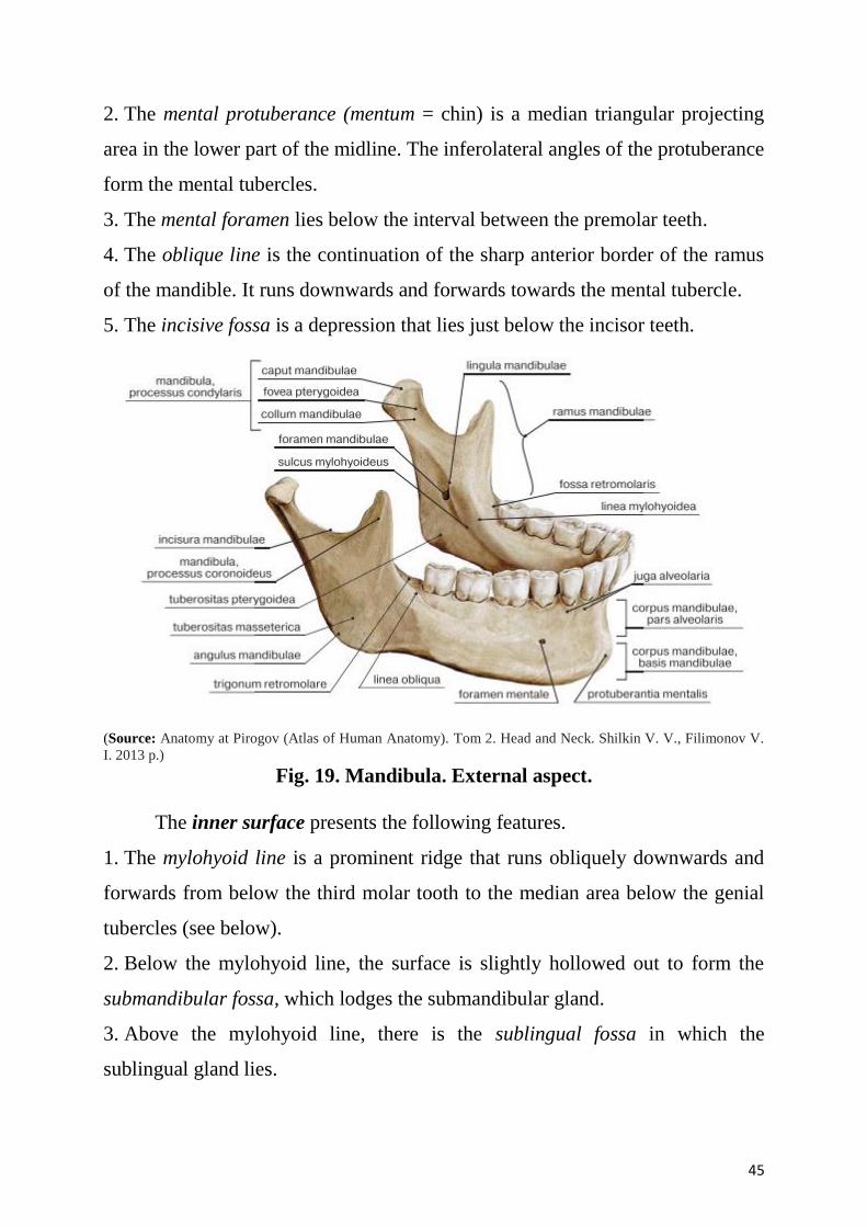
45
2. The mental protuberance (mentum = chin) is a median triangular projecting
area in the lower part of the midline. The inferolateral angles of the protuberance
form the mental tubercles.
3. The mental foramen lies below the interval between the premolar teeth.
4. The oblique line is the continuation of the sharp anterior border of the ramus
of the mandible. It runs downwards and forwards towards the mental tubercle.
5. The incisive fossa is a depression that lies just below the incisor teeth.
(Source: Anatomy at Pirogov (Atlas of Human Anatomy). Tom 2. Head and Neck. Shilkin V. V., Filimonov V.
I. 2013 p.)
Fig. 19. Mandibula. External aspect.
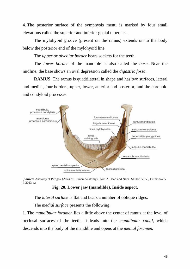
The inner surface presents the following features.
1. The mylohyoid line is a prominent ridge that runs obliquely downwards and
forwards from below the third molar tooth to the median area below the genial
tubercles (see below).
2. Below the mylohyoid line, the surface is slightly hollowed out to form the
submandibular fossa, which lodges the submandibular gland.
3. Above the mylohyoid line, there is the sublingual fossa in which the
sublingual gland lies.
46
4. The posterior surface of the symphysis menti is marked by four small
elevations called the superior and inferior genial tubercles.
The mylohyoid groove (present on the ramus) extends on to the body
below the posterior end of the mylohyoid line
The upper or alveolar border bears sockets for the teeth.
The lower border of the mandible is also called the base. Near the
midline, the base shows an oval depression called the digastric fossa.
RAMUS. The ramus is quadrilateral in shape and has two surfaces, lateral
and medial, four borders, upper, lower, anterior and posterior, and the coronoid
and condyloid processes.
(Source: Anatomy at Pirogov (Atlas of Human Anatomy). Tom 2. Head and Neck. Shilkin V. V., Filimonov V.
I. 2013 p.)
Fig. 20. Lower jaw (mandible). Inside aspect.
The lateral surface is flat and bears a number of oblique ridges.
The medial surface presents the following:
1. The mandibular foramen lies a little above the center of ramus at the level of
occlusal surfaces of the teeth. It leads into the mandibular canal, which
descends into the body of the mandible and opens at the mental foramen.
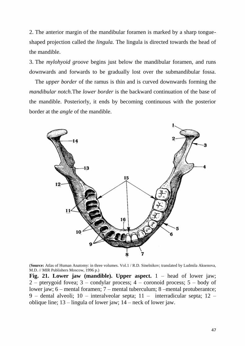
47
2. The anterior margin of the mandibular foramen is marked by a sharp tongue-
shaped projection called the lingula. The lingula is directed towards the head of
the mandible.
3. The mylohyoid groove begins just below the mandibular foramen, and runs
downwards and forwards to be gradually lost over the submandibular fossa.
The upper border of the ramus is thin and is curved downwards forming the
mandibular notch.The lower border is the backward continuation of the base of
the mandible. Posteriorly, it ends by becoming continuous with the posterior
border at the angle of the mandible.
(Source: Atlas of Human Anatomy: in three volumes. Vol.1 / R.D. Sinelnikov; translated by Ludmila Aksenova,
M.D. // MIR Publishers Moscow, 1996 p.)
Fig. 21. Lower jaw (mandible). Upper aspect. 1 – head of lower jaw;
2 – pterygoid fovea; 3 – condylar process; 4 – coronoid process; 5 – body of
lower jaw; 6 – mental foramen; 7 – mental tuberculum; 8 –mental protuberantce;
9 – dental alveoli; 10 – interalveolar septa; 11 – interradicular septa; 12 –
oblique line; 13 – lingula of lower jaw; 14 – neck of lower jaw.
48
The anterior border is thin, while the posterior border is thick.
The coronoid process is a flattened triangular upward projection from the
anterosuperior part of the ramus. Its anterior border is continuous with the
anterior border of the ramus. The posterior border bounds the mandibular notch.
The condyloid process is a strong upward projection from the
posterosuperior part of the ramus. Its upper end is expanded from side to side to
form the head. The head is covered with fibrocartilage and articulates with the
temporal bone to form the temporomandibular joint. The constriction below the
head is the neck. Its anterior surface presents a depression called the pterygoid
fovea.
Attachments and Relations of the Mandible
1. The oblique line on the lateral side of the body gives origin to the buccinator
as far forwards as the anterior border of the first molar tooth. In front of this
origin, the depressor labii inferioris and the depressor anguli oris arise from the
oblique line below the mental foramen.
2. The parts of both the inner and outer surfaces just below the alveolar margin
are covered by the mucous membrane of the mouth.
3. Mylohyoid line gives origin to the mylohyoid muscle.
4. Superior constrictor muscle of the pharynx arises from an area above the
posterior end of the mylohyoid line.
5. Pterygomandibular raphe is attached immediately behind the third molar
tooth in continuation with the origin of superior constrictor.
6. Upper genial tubercle gives origin to the genioglossus, and the lower tubercle
to geniohyoid.
7. Anterior belly of the digastric muscle arises from the digastric fossa.
8. Deep cervical fascia (investing layer) is attached to the whole length of lower
border.
9. The platysma is inserted into the lower border.
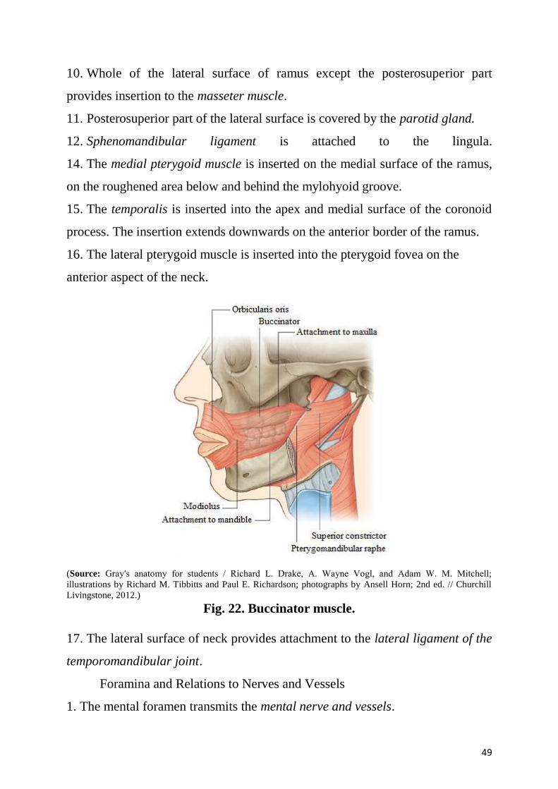
49
10. Whole of the lateral surface of ramus except the posterosuperior part
provides insertion to the masseter muscle.
11. Posterosuperior part of the lateral surface is covered by the parotid gland.
12. Sphenomandibular ligament is attached to the lingula.
14. The medial pterygoid muscle is inserted on the medial surface of the ramus,
on the roughened area below and behind the mylohyoid groove.
15. The temporalis is inserted into the apex and medial surface of the coronoid
process. The insertion extends downwards on the anterior border of the ramus.
16. The lateral pterygoid muscle is inserted into the pterygoid fovea on the
anterior aspect of the neck.
(Source: Gray′s anatomy for students / Richard L. Drake, A. Wayne Vogl, and Adam W. M. Mitchell;
illustrations by Richard M. Tibbitts and Paul E. Richardson; photographs by Ansell Horn; 2nd ed. // Churchill
Livingstone, 2012.)
Fig. 22. Buccinator muscle.
17. The lateral surface of neck provides attachment to the lateral ligament of the
temporomandibular joint.
Foramina and Relations to Nerves and Vessels
1. The mental foramen transmits the mental nerve and vessels.
50
2. The inferior alveolar nerve and vessels enter – the mandibular canal through
the mandibular foramen, and run forwards within the canal.
3. The mylohyoid nerve and vessels lie in the mylohyoid groove.
4. The lingual nerve is related to the medial surface of the ramus in front of the
mylohyoid groove.
5. The area above and behind the mandibular foramen is related to the inferior
alveolar nerve and vessels and to the maxillary artery.
6. The masseteric nerve and vessels pass through the mandibular notch.
7. The auriculotemporal nerve and superficial temporal artery are related to the
medial side of the neck of mandible.
8. Facial artery is palpable on the lower border of mandible at anteroinferior
angle of masseter.
Ossification: The mandible is the second bone, next to the clavicle, to
ossify in the body. Its greater part ossifies in membrane. The parts ossifying in
cartilage include the incisive part below the incisor teeth, the coronoid and
condyloid processes, and the upper half of the ramus above the level of the
mandibular foramen.
Each half of the mandible ossifies from only one center, which appears at
about the 6th week of intrauterine life in the mesenchymal sheath of Meckel's
cartilage near the future mental foramen. Meckel's cartilage is the skeletal
element of first pharyngeal arch.
At birth, the mandible consists of two halves connected at the symphysis
menti by fibrous tissue. Bony union takes place during the first year of life.
Age Changes in the Mandible
In Infants and Children
1. The two halves of the mandible fuse during the first year of life.
2. At birth, the mental foramen opens below the sockets for the two deciduous
molar teeth near the lower border. This is so because the bone is made up only
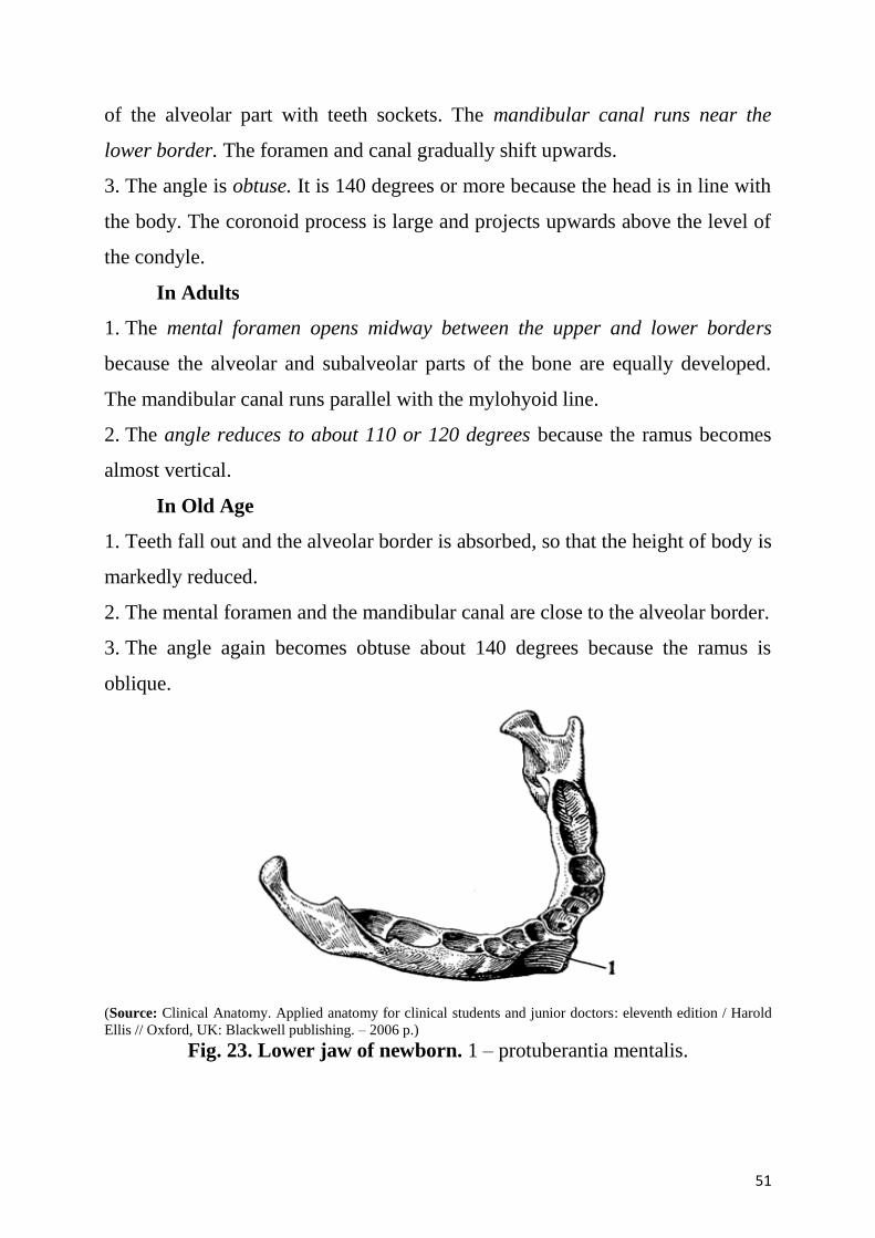
51
of the alveolar part with teeth sockets. The mandibular canal runs near the
lower border. The foramen and canal gradually shift upwards.
3. The angle is obtuse. It is 140 degrees or more because the head is in line with
the body. The coronoid process is large and projects upwards above the level of
the condyle.
In Adults
1. The mental foramen opens midway between the upper and lower borders
because the alveolar and subalveolar parts of the bone are equally developed.
The mandibular canal runs parallel with the mylohyoid line.
2. The angle reduces to about 110 or 120 degrees because the ramus becomes
almost vertical.
In Old Age
1. Teeth fall out and the alveolar border is absorbed, so that the height of body is
markedly reduced.
2. The mental foramen and the mandibular canal are close to the alveolar border.
3. The angle again becomes obtuse about 140 degrees because the ramus is
oblique.
(Source: Clinical Anatomy. Applied anatomy for clinical students and junior doctors: eleventh edition / Harold
Ellis // Oxford, UK: Blackwell publishing. – 2006 p.)
Fig. 23. Lower jaw of newborn. 1 – protuberantia mentalis.
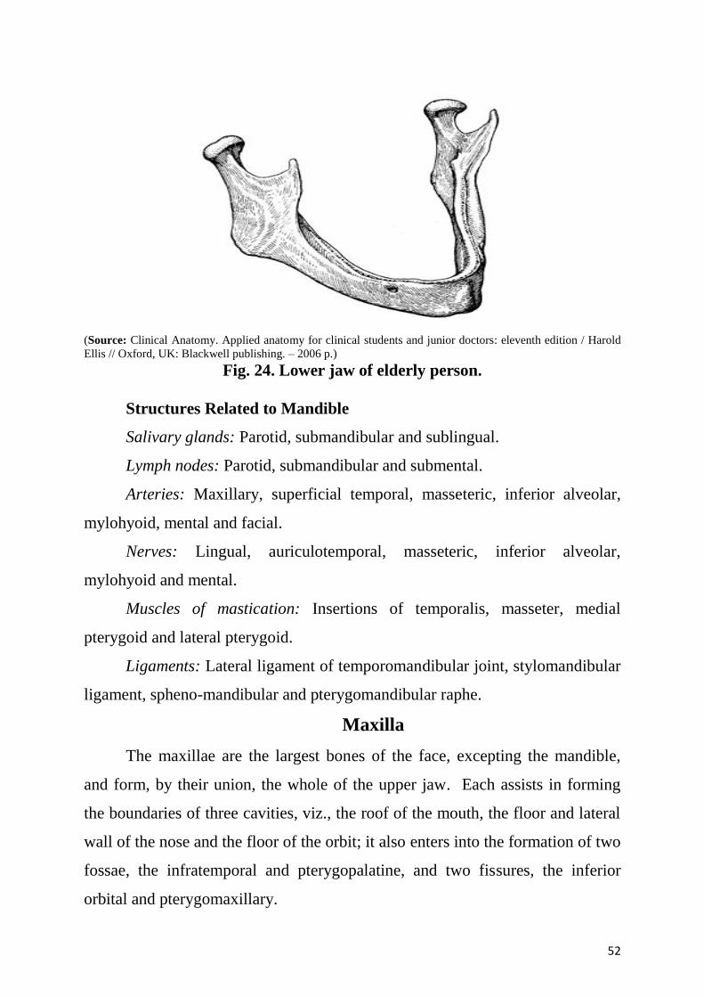
52
(Source: Clinical Anatomy. Applied anatomy for clinical students and junior doctors: eleventh edition / Harold
Ellis // Oxford, UK: Blackwell publishing. – 2006 p.)
Fig. 24. Lower jaw of elderly person.
Structures Related to Mandible
Salivary glands: Parotid, submandibular and sublingual.
Lymph nodes: Parotid, submandibular and submental.
Arteries: Maxillary, superficial temporal, masseteric, inferior alveolar,
mylohyoid, mental and facial.
Nerves: Lingual, auriculotemporal, masseteric, inferior alveolar,
mylohyoid and mental.
Muscles of mastication: Insertions of temporalis, masseter, medial
pterygoid and lateral pterygoid.
Ligaments: Lateral ligament of temporomandibular joint, stylomandibular
ligament, spheno-mandibular and pterygomandibular raphe.
Maxilla
The maxillae are the largest bones of the face, excepting the mandible,
and form, by their union, the whole of the upper jaw. Each assists in forming
the boundaries of three cavities, viz., the roof of the mouth, the floor and lateral
wall of the nose and the floor of the orbit; it also enters into the formation of two
fossae, the infratemporal and pterygopalatine, and two fissures, the inferior
orbital and pterygomaxillary.
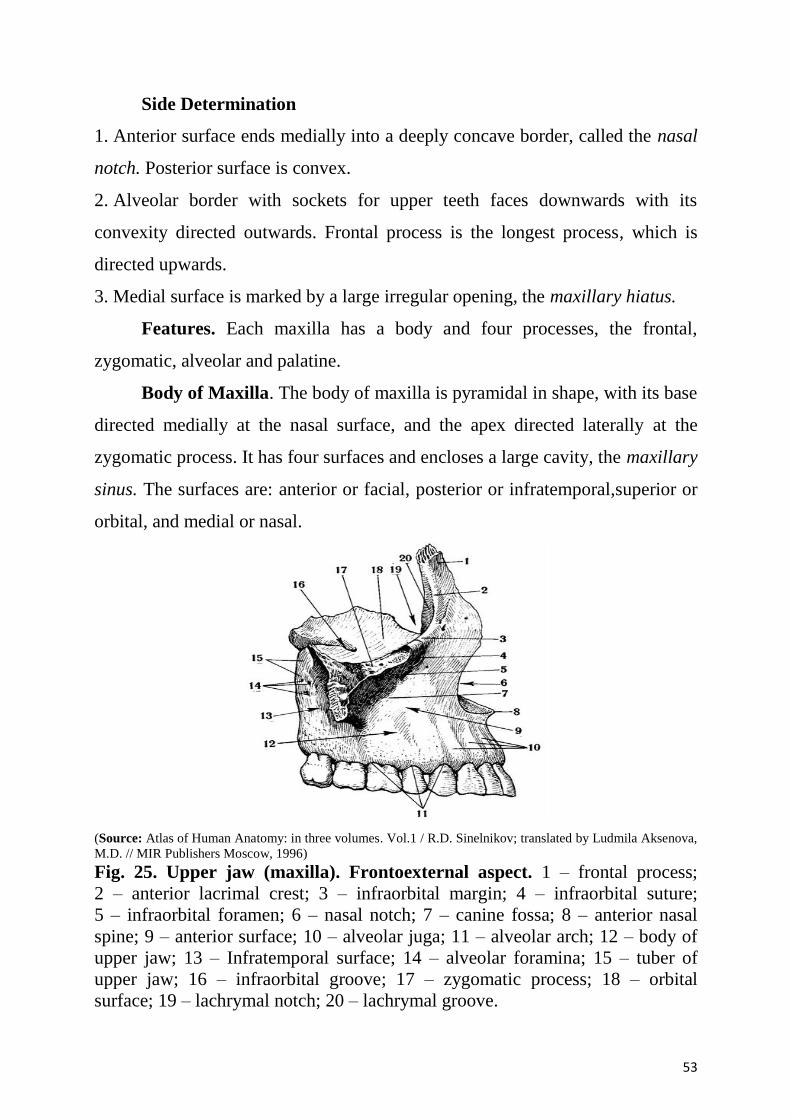
53
Side Determination
1. Anterior surface ends medially into a deeply concave border, called the nasal
notch. Posterior surface is convex.
2. Alveolar border with sockets for upper teeth faces downwards with its
convexity directed outwards. Frontal process is the longest process, which is
directed upwards.
3. Medial surface is marked by a large irregular opening, the maxillary hiatus.
Features. Each maxilla has a body and four processes, the frontal,
zygomatic, alveolar and palatine.
Body of Maxilla. The body of maxilla is pyramidal in shape, with its base
directed medially at the nasal surface, and the apex directed laterally at the
zygomatic process. It has four surfaces and encloses a large cavity, the maxillary
sinus. The surfaces are: anterior or facial, posterior or infratemporal,superior or
orbital, and medial or nasal.
(Source: Atlas of Human Anatomy: in three volumes. Vol.1 / R.D. Sinelnikov; translated by Ludmila Aksenova,
M.D. // MIR Publishers Moscow, 1996)
Fig. 25. Upper jaw (maxilla). Frontoexternal aspect. 1 – frontal process;
2 – anterior lacrimal crest; 3 – infraorbital margin; 4 – infraorbital suture;
5 – infraorbital foramen; 6 – nasal notch; 7 – canine fossa; 8 – anterior nasal
spine; 9 – anterior surface; 10 – alveolar juga; 11 – alveolar arch; 12 – body of
upper jaw; 13 – Infratemporal surface; 14 – alveolar foramina; 15 – tuber of
upper jaw; 16 – infraorbital groove; 17 – zygomatic process; 18 – orbital
surface; 19 – lachrymal notch; 20 – lachrymal groove.
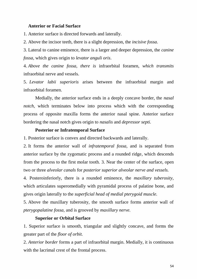
54
Anterior or Facial Surface
1. Anterior surface is directed forwards and laterally.
2. Above the incisor teeth, there is a slight depression, the incisive fossa.
3. Lateral to canine eminence, there is a larger and deeper depression, the canine
fossa, which gives origin to levator anguli oris.
4. Above the canine fossa, there is infraorbital foramen, which transmits
infraorbital nerve and vessels.
5. Levator labii superioris arises between the infraorbital margin and
infraorbital foramen.
Medially, the anterior surface ends in a deeply concave border, the nasal
notch, which terminates below into process which with the corresponding
process of opposite maxilla forms the anterior nasal spine. Anterior surface
bordering the nasal notch gives origin to nasalis and depressor septi.
Posterior or Infratemporal Surface
1. Posterior surface is convex and directed backwards and laterally.
2. It forms the anterior wall of infratemporal fossa, and is separated from
anterior surface by the zygomatic process and a rounded ridge, which descends
from the process to the first molar tooth. 3. Near the center of the surface, open
two or three alveolar canals for posterior superior alveolar nerve and vessels.
4. Posteroinferiorly, there is a rounded eminence, the maxillary tuberosity,
which articulates superomedially with pyramidal process of palatine bone, and
gives origin laterally to the superficial head of medial pterygoid muscle.
5. Above the maxillary tuberosity, the smooth surface forms anterior wall of
pterygopalatine fossa, and is grooved by maxillary nerve.
Superior or Orbital Surface
1. Superior surface is smooth, triangular and slightly concave, and forms the
greater part of the floor of orbit.
2. Anterior border forms a part of infraorbital margin. Medially, it is continuous
with the lacrimal crest of the frontal process.
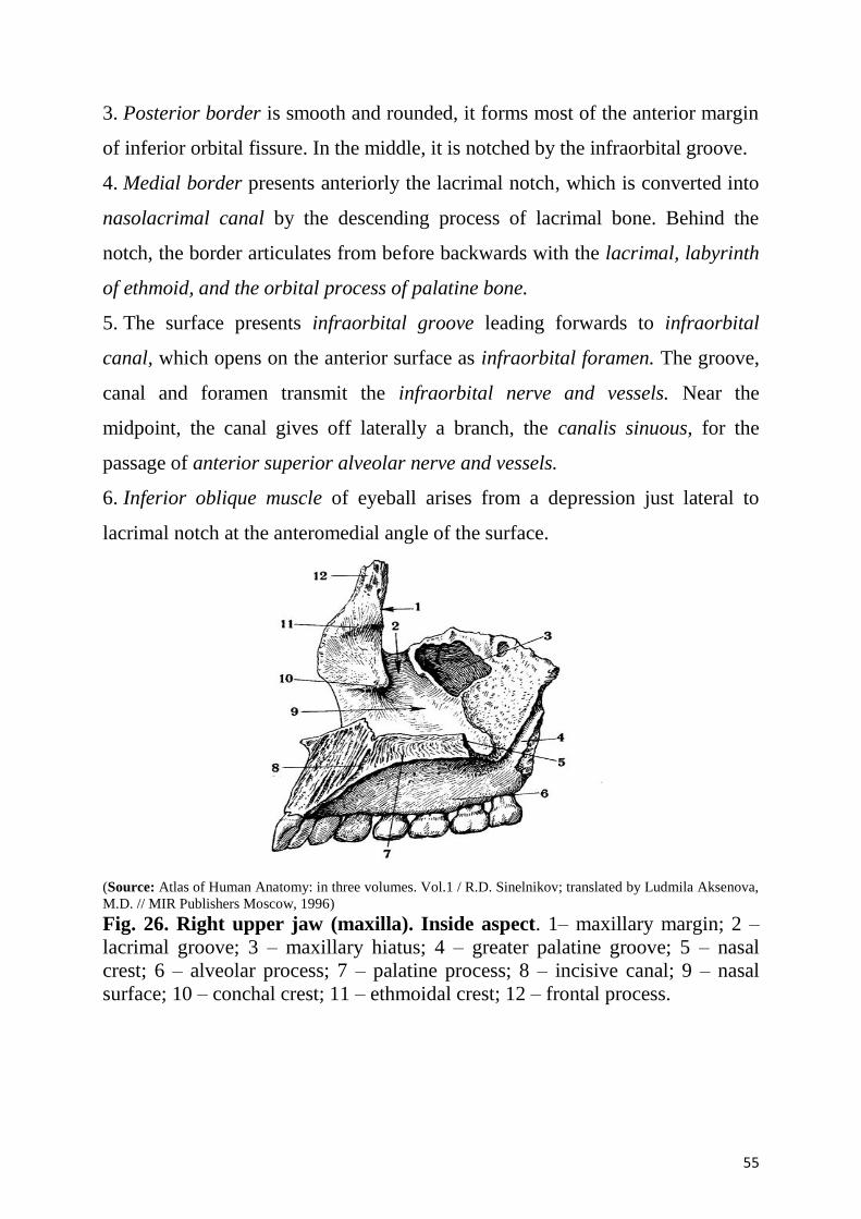
55
3. Posterior border is smooth and rounded, it forms most of the anterior margin
of inferior orbital fissure. In the middle, it is notched by the infraorbital groove.
4. Medial border presents anteriorly the lacrimal notch, which is converted into
nasolacrimal canal by the descending process of lacrimal bone. Behind the
notch, the border articulates from before backwards with the lacrimal, labyrinth
of ethmoid, and the orbital process of palatine bone.
5. The surface presents infraorbital groove leading forwards to infraorbital
canal, which opens on the anterior surface as infraorbital foramen. The groove,
canal and foramen transmit the infraorbital nerve and vessels. Near the
midpoint, the canal gives off laterally a branch, the canalis sinuous, for the
passage of anterior superior alveolar nerve and vessels.
6. Inferior oblique muscle of eyeball arises from a depression just lateral to
lacrimal notch at the anteromedial angle of the surface.
(Source: Atlas of Human Anatomy: in three volumes. Vol.1 / R.D. Sinelnikov; translated by Ludmila Aksenova,
M.D. // MIR Publishers Moscow, 1996)
Fig. 26. Right upper jaw (maxilla). Inside aspect. 1– maxillary margin; 2 –
lacrimal groove; 3 – maxillary hiatus; 4 – greater palatine groove; 5 – nasal
crest; 6 – alveolar process; 7 – palatine process; 8 – incisive canal; 9 – nasal
surface; 10 – conchal crest; 11 – ethmoidal crest; 12 – frontal process.

56
Medial or Nasal Surface
1. Medial surface forms a part of the lateral wall of nose.
2. Posterosuperiorly, it displays a large irregular opening of the maxillary sinus,
the maxillary hiatus.
3. Above the hiatus, there are parts of air sinuses, which are completed by the
ethmoid and lacrimal bones.
4. Below the hiatus, the smooth concave surface forms a part of inferior meatus
of nose.
5. Behind the hiatus, the surface articulates with perpendicular plate of palatine
bone, enclosing the greater palatine canal, which runs downwards and
forwards, and transmits greater palatine vessels and the anterior, middle and
posterior palatine nerves.
6. In front of the hiatus, there is nasolacrimal groove, which is converted into
the nasolacrimal canal by articulation with the descending process of lacrimal
bone and the lacrimal process of inferior nasal concha. The canal transmits
nasolacrimal duct to the inferior meatus of nose.
7. More anteriorly, an oblique ridge forms the conchal crest for articulation with
the inferior nasal concha.
8. Above the conchal crest, the shallow depression forms a part of the atrium of
middle meatus of nose.
The Maxillary Sinus or Antrum of Highmore (sinus maxillaris)
1. The maxillary sinus is a large cavity in the body of maxilla. It is pyramidal in
shape, with its base directed medially towards the lateral wall of nose, and the
apex directed laterally into the zygomatic process of maxilla.
2. The sinus opens into the middle meatus of nose usually by two openings one
of which is closed by mucous membrane. The large bony hiatus of the sinus is
reduced in the articulated skull by following bones: a) from above, by uncinate
process of ethmoid and descending part of lacrimal bone; в) from below, by
inferior nasal concha; с) from behind, by perpendicular plate of palatine bone.
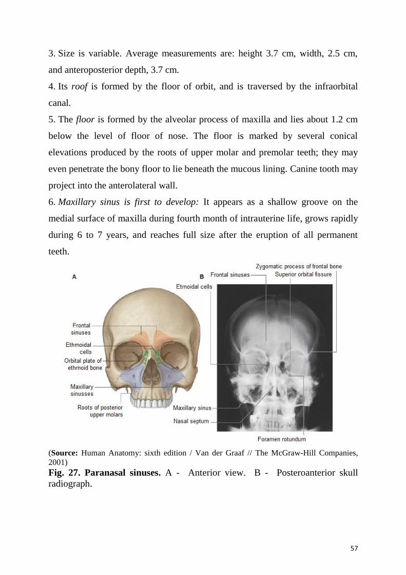
57
3. Size is variable. Average measurements are: height 3.7 cm, width, 2.5 cm,
and anteroposterior depth, 3.7 cm.
4. Its roof is formed by the floor of orbit, and is traversed by the infraorbital
canal.
5. The floor is formed by the alveolar process of maxilla and lies about 1.2 cm
below the level of floor of nose. The floor is marked by several conical
elevations produced by the roots of upper molar and premolar teeth; they may
even penetrate the bony floor to lie beneath the mucous lining. Canine tooth may
project into the anterolateral wall.
6. Maxillary sinus is first to develop: It appears as a shallow groove on the
medial surface of maxilla during fourth month of intrauterine life, grows rapidly
during 6 to 7 years, and reaches full size after the eruption of all permanent
teeth.
(Source: Human Anatomy: sixth edition / Van der Graaf // The McGraw-Hill Companies,
2001)
Fig. 27. Paranasal sinuses. A - Anterior view. B - Posteroanterior skull
radiograph.
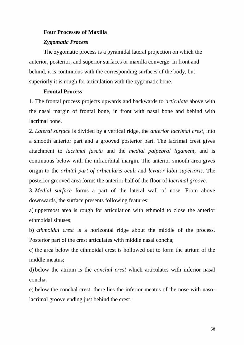
58
Four Processes of Maxilla
Zygomatic Process
The zygomatic process is a pyramidal lateral projection on which the
anterior, posterior, and superior surfaces or maxilla converge. In front and
behind, it is continuous with the corresponding surfaces of the body, but
superiorly it is rough for articulation with the zygomatic bone.
Frontal Process
1. The frontal process projects upwards and backwards to articulate above with
the nasal margin of frontal bone, in front with nasal bone and behind with
lacrimal bone.
2. Lateral surface is divided by a vertical ridge, the anterior lacrimal crest, into
a smooth anterior part and a grooved posterior part. The lacrimal crest gives
attachment to lacrimal fascia and the medial palpebral ligament, and is
continuous below with the infraorbital margin. The anterior smooth area gives
origin to the orbital part of orbicularis oculi and levator labii superioris. The
posterior grooved area forms the anterior half of the floor of lacrimal groove.
3. Medial surface forms a part of the lateral wall of nose. From above
downwards, the surface presents following features:
a) uppermost area is rough for articulation with ethmoid to close the anterior
ethmoidal sinuses;
b) еthmoidal crest is a horizontal ridge about the middle of the process.
Posterior part of the crest articulates with middle nasal concha;
c) the area below the ethmoidal crest is hollowed out to form the atrium of the
middle meatus;
d) below the atrium is the conchal crest which articulates with inferior nasal
concha.
e) below the conchal crest, there lies the inferior meatus of the nose with naso-
lacrimal groove ending just behind the crest.
59
Alveolar Process
1. The alveolar process forms half of the alveolar arch, and bears sockets for the
roots of upper teeth. In adults, there are eight sockets: canine socket is deepest;
molar sockets are widest and divided into three minor sockets by septa; the
incisor and second premolar sockets are single; and the first premolar socket is
sometimes divided into two.
2. Buccinator arises from the posterior part of its outer surface up to the first
molar tooth.
3. A rough ridge, the maxillary torus, is sometimes present on the inner surface
opposite the molar sockets.
Palatine Process
1. Palatine process is a thick horizontal plate projecting medially from the
lowest part of the nasal surface. It forms a large part of the roof of mouth and the
floor of nasal cavity.
2. Inferior surface is concave, and the two palatine processes form anterior
three-fourths of the bony palate. It presents numerous vascular foramina and pits
for palatine glands. Posterolaterally, it is marked by two anteroposterior grooves
for the greater palatine vessels and anterior palatine nerves.
3. Superior surface is concave from side to side, and forms greater part of the
floor of nasal cavity.
4. Medial border is thicker in front than behind. It is raised superiorly into the
nasal crest. Groove between the nasal crests of two maxillae receives lower
border of vomer; anterior part of the ridge is high and is known as incisor crest,
which terminates anteriorly into the anterior nasal spine. Incisive canal traverses
near the anterior part of the medial border.
5. Posterior border articulates with horizontal plate of palatine bone.
6. Lateral border is continuous with the alveolar process.
Articulations of Maxilla
1. Superiorly, it articulates with three bones, the nasal, frontal and lacrimal.
60
2. Medially, it articulates with five bones, the ethmoid, inferior nasal concha,
vomer, palatine and opposite maxilla.
3. Laterally, it articulates with one bone, the zygomatic.
Ossification. Maxilla ossifies in membrane from three centers, one for the
maxilla proper, and two for os incisivum or premaxilla. The center for maxilla
proper appears above the canine fossa during sixth week of intrauterine life.
Of the two premaxillary centres, the main center appears above the
incisive fossa during seventh week of intrauterine life. The second center
(paraseptal or prevomerine) appears at the ventral margin of nasal septum during
tenth week and soon fuses with the palatal process of maxilla. Though
premaxilla begins to fuse with alveolar process almost immediately after the
ossification begins, the evidence for premaxilla as a separate bone may persist
until the middle decades.
AGE CHANGES
At birth:
1. The transverse and anteroposterior diameters are each more than the vertical
diameter. Frontal process is well marked.
2. Body consists of a little more than the alveolar process, the tooth sockets
reaching to the floor of orbit.
3. Maxillary sinus is a mere furrow on the lateral wall of the nose.
In the adult: vertical diameter is greatest due to development of the alveolar
process and increase in the size of the sinus.
In the old: the bone reverts to infantile condition. Its height is reduced because
of absorption of the alveolar process.
DEVELOPMENTAL DISORDERS OF THE JAWS
The cleft palate is failure of fusion of two palatine processes during early
prenatal development (about 12 weeks). Cleft palate may be accompanied by a
cleft lip. These conditions require surgical treatment. A baby with a cleft palate
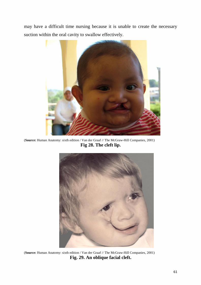
61
may have a difficult time nursing because it is unable to create the necessary
suction within the oral cavity to swallow effectively.
(Source: Human Anatomy: sixth edition / Van der Graaf // The McGraw-Hill Companies, 2001)
Fig 28. The cleft lip.
(Source: Human Anatomy: sixth edition / Van der Graaf // The McGraw-Hill Companies, 2001)
Fig. 29. An oblique facial cleft.
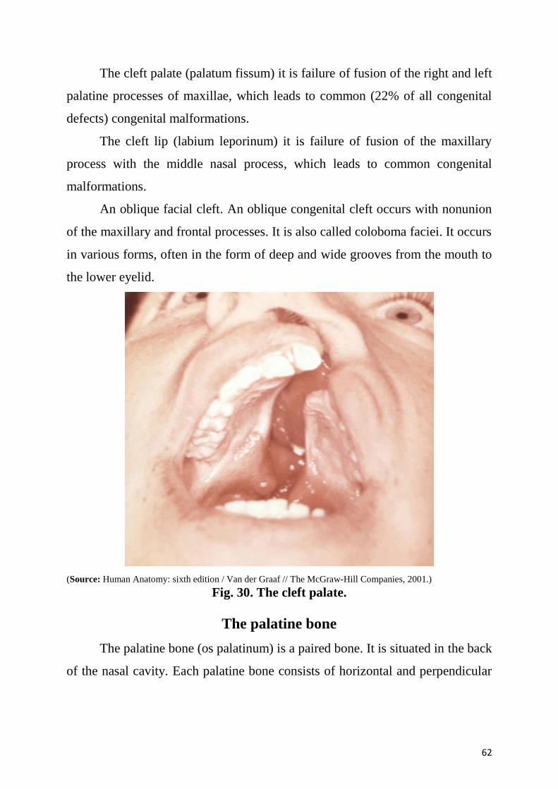
62
The cleft palate (palatum fissum) it is failure of fusion of the right and left
palatine processes of maxillae, which leads to common (22% of all congenital
defects) congenital malformations.
The cleft lip (labium leporinum) it is failure of fusion of the maxillary
process with the middle nasal process, which leads to common congenital
malformations.
An oblique facial cleft. An oblique congenital cleft occurs with nonunion
of the maxillary and frontal processes. It is also called coloboma faciei. It occurs
in various forms, often in the form of deep and wide grooves from the mouth to
the lower eyelid.
(Source: Human Anatomy: sixth edition / Van der Graaf // The McGraw-Hill Companies, 2001.)
Fig. 30. The cleft palate.
The palatine bone
The palatine bone (os palatinum) is a paired bone. It is situated in the back
of the nasal cavity. Each palatine bone consists of horizontal and perpendicular
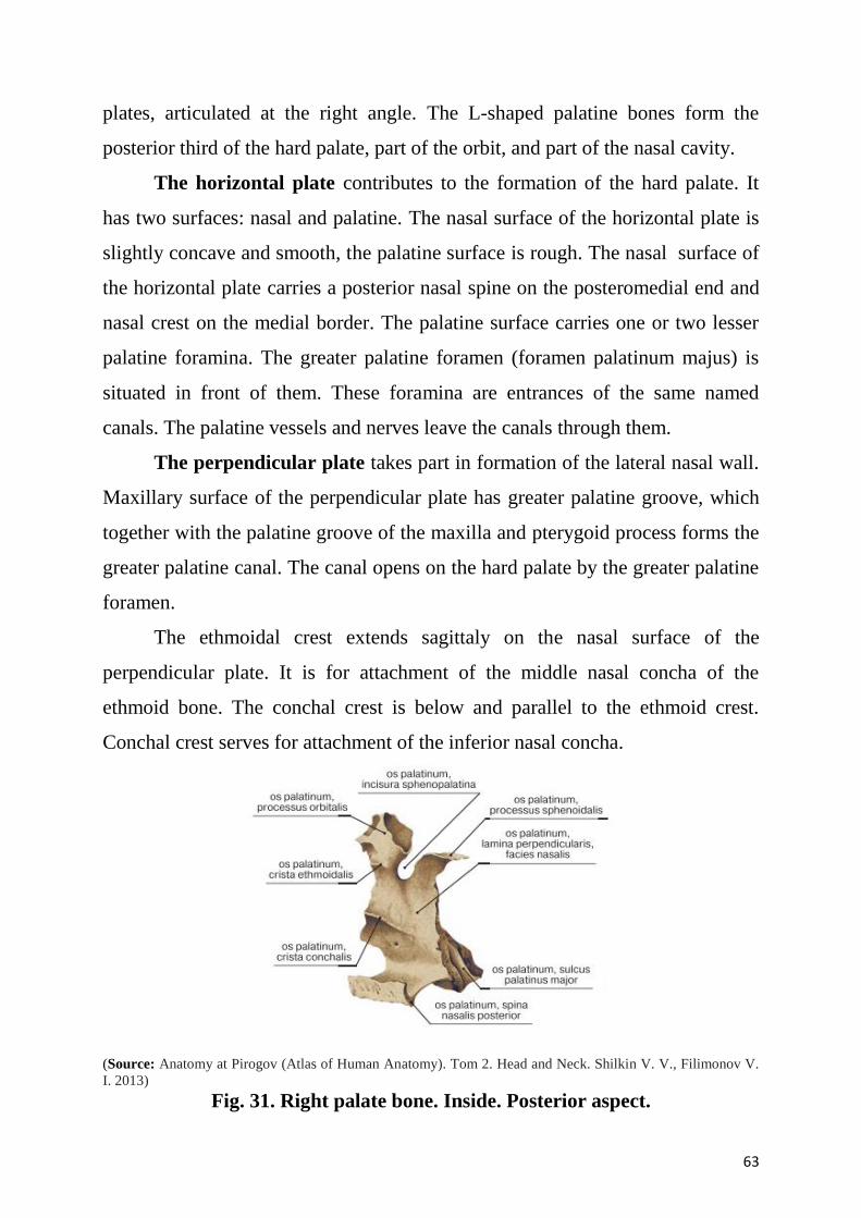
63
plates, articulated at the right angle. The L-shaped palatine bones form the
posterior third of the hard palate, part of the orbit, and part of the nasal cavity.
The horizontal plate contributes to the formation of the hard palate. It
has two surfaces: nasal and palatine. The nasal surface of the horizontal plate is
slightly concave and smooth, the palatine surface is rough. The nasal surface of
the horizontal plate carries a posterior nasal spine on the posteromedial end and
nasal crest on the medial border. The palatine surface carries one or two lesser
palatine foramina. The greater palatine foramen (foramen palatinum majus) is
situated in front of them. These foramina are entrances of the same named
canals. The palatine vessels and nerves leave the canals through them.
The perpendicular plate takes part in formation of the lateral nasal wall.
Maxillary surface of the perpendicular plate has greater palatine groove, which
together with the palatine groove of the maxilla and pterygoid process forms the
greater palatine canal. The canal opens on the hard palate by the greater palatine
foramen.
The ethmoidal crest extends sagittaly on the nasal surface of the
perpendicular plate. It is for attachment of the middle nasal concha of the
ethmoid bone. The conchal crest is below and parallel to the ethmoid crest.
Conchal crest serves for attachment of the inferior nasal concha.
(Source: Anatomy at Pirogov (Atlas of Human Anatomy). Tom 2. Head and Neck. Shilkin V. V., Filimonov V.
I. 2013)
Fig. 31. Right palate bone. Inside. Posterior aspect.
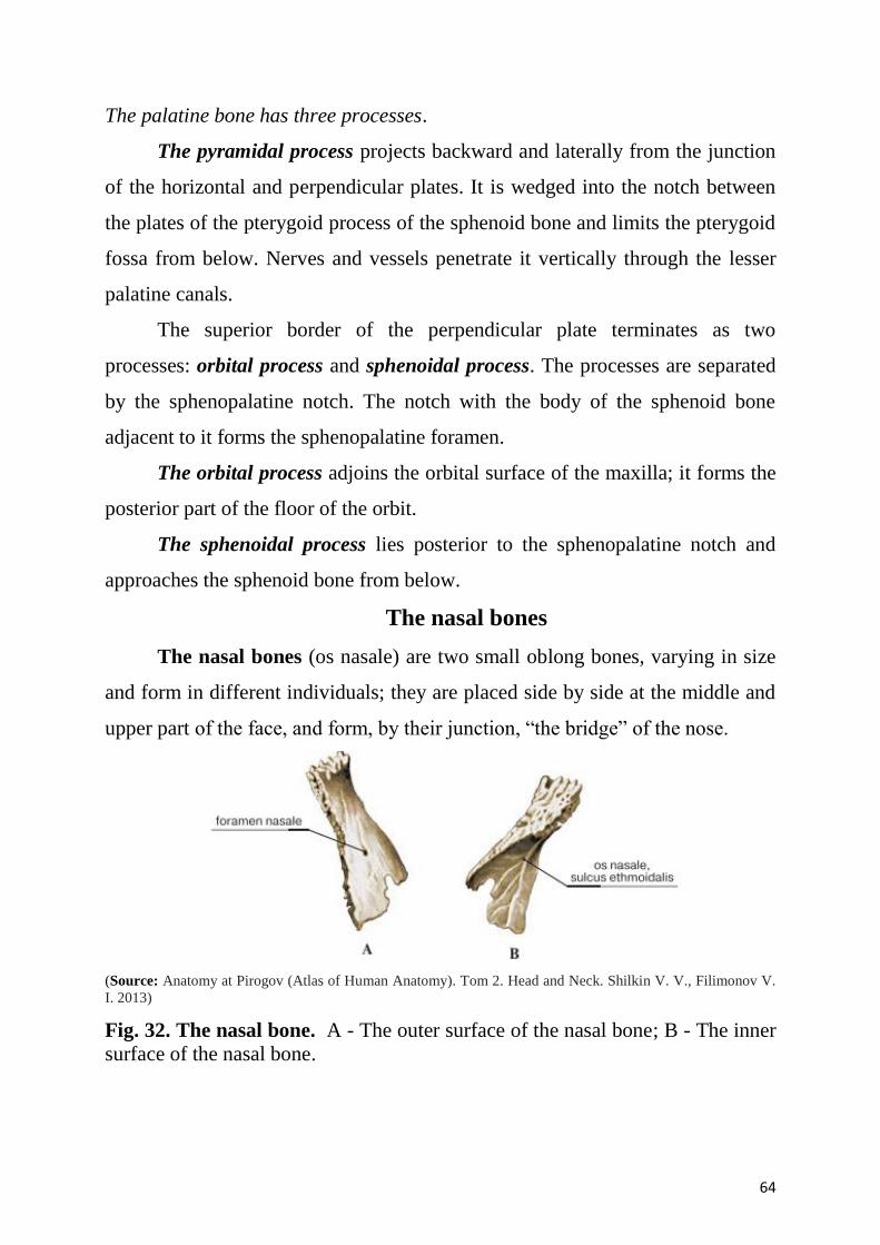
64
The palatine bone has three processes.
The pyramidal process projects backward and laterally from the junction
of the horizontal and perpendicular plates. It is wedged into the notch between
the plates of the pterygoid process of the sphenoid bone and limits the pterygoid
fossa from below. Nerves and vessels penetrate it vertically through the lesser
palatine canals.
The superior border of the perpendicular plate terminates as two
processes: orbital process and sphenoidal process. The processes are separated
by the sphenopalatine notch. The notch with the body of the sphenoid bone
adjacent to it forms the sphenopalatine foramen.
The orbital process adjoins the orbital surface of the maxilla; it forms the
posterior part of the floor of the orbit.
The sphenoidal process lies posterior to the sphenopalatine notch and
approaches the sphenoid bone from below.
The nasal bones
The nasal bones (os nasale) are two small oblong bones, varying in size
and form in different individuals; they are placed side by side at the middle and
upper part of the face, and form, by their junction, “the bridge” of the nose.
(Source: Anatomy at Pirogov (Atlas of Human Anatomy). Tom 2. Head and Neck. Shilkin V. V., Filimonov V.
I. 2013)
Fig. 32. The nasal bone. A - The outer surface of the nasal bone; B - The inner
surface of the nasal bone.
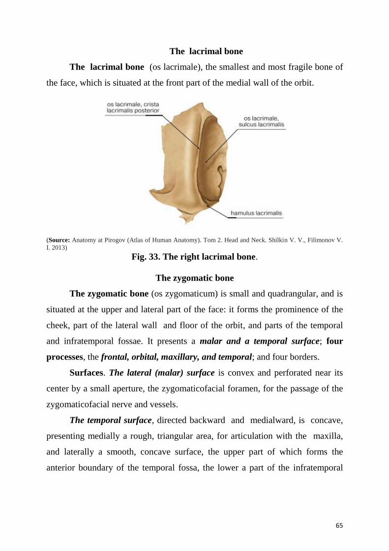
65
The lacrimal bone
The lacrimal bone (os lacrimale), the smallest and most fragile bone of
the face, which is situated at the front part of the medial wall of the orbit.
(Source: Anatomy at Pirogov (Atlas of Human Anatomy). Tom 2. Head and Neck. Shilkin V. V., Filimonov V.
I. 2013)
Fig. 33. The right lacrimal bone.
The zygomatic bone
The zygomatic bone (os zygomaticum) is small and quadrangular, and is
situated at the upper and lateral part of the face: it forms the prominence of the
cheek, part of the lateral wall and floor of the orbit, and parts of the temporal
and infratemporal fossae. It presents a malar and a temporal surface; four
processes, the frontal, orbital, maxillary, and temporal; and four borders.
Surfaces. The lateral (malar) surface is convex and perforated near its
center by a small aperture, the zygomaticofacial foramen, for the passage of the
zygomaticofacial nerve and vessels.
The temporal surface, directed backward and medialward, is concave,
presenting medially a rough, triangular area, for articulation with the maxilla,
and laterally a smooth, concave surface, the upper part of which forms the
anterior boundary of the temporal fossa, the lower a part of the infratemporal
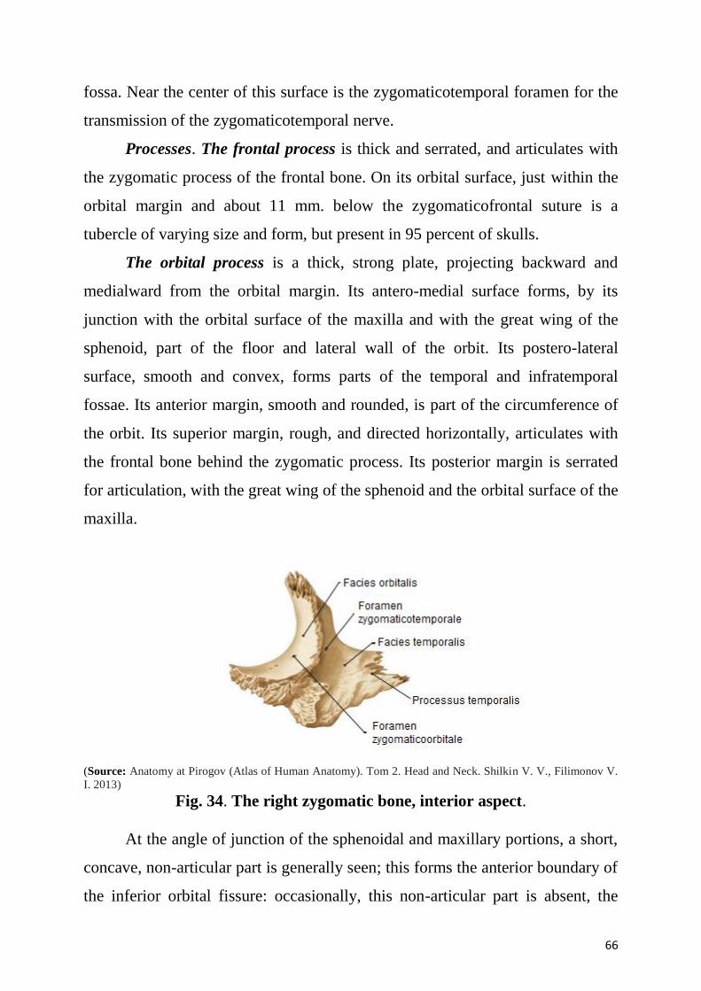
66
fossa. Near the center of this surface is the zygomaticotemporal foramen for the
transmission of the zygomaticotemporal nerve.
Processes. The frontal process is thick and serrated, and articulates with
the zygomatic process of the frontal bone. On its orbital surface, just within the
orbital margin and about 11 mm. below the zygomaticofrontal suture is a
tubercle of varying size and form, but present in 95 percent of skulls.
The orbital process is a thick, strong plate, projecting backward and
medialward from the orbital margin. Its antero-medial surface forms, by its
junction with the orbital surface of the maxilla and with the great wing of the
sphenoid, part of the floor and lateral wall of the orbit. Its postero-lateral
surface, smooth and convex, forms parts of the temporal and infratemporal
fossae. Its anterior margin, smooth and rounded, is part of the circumference of
the orbit. Its superior margin, rough, and directed horizontally, articulates with
the frontal bone behind the zygomatic process. Its posterior margin is serrated
for articulation, with the great wing of the sphenoid and the orbital surface of the
maxilla.
(Source: Anatomy at Pirogov (Atlas of Human Anatomy). Tom 2. Head and Neck. Shilkin V. V., Filimonov V.
I. 2013)
Fig. 34. The right zygomatic bone, interior aspect.
At the angle of junction of the sphenoidal and maxillary portions, a short,
concave, non-articular part is generally seen; this forms the anterior boundary of
the inferior orbital fissure: occasionally, this non-articular part is absent, the
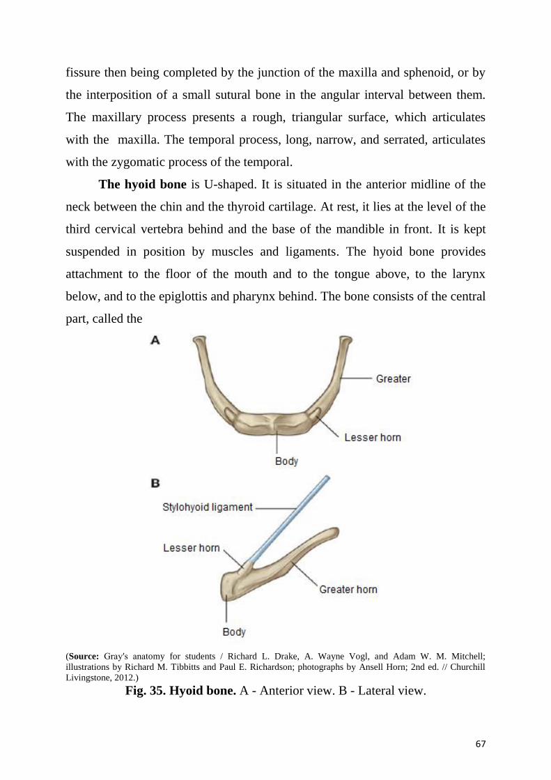
67
fissure then being completed by the junction of the maxilla and sphenoid, or by
the interposition of a small sutural bone in the angular interval between them.
The maxillary process presents a rough, triangular surface, which articulates
with the maxilla. The temporal process, long, narrow, and serrated, articulates
with the zygomatic process of the temporal.
The hyoid bone is U-shaped. It is situated in the anterior midline of the
neck between the chin and the thyroid cartilage. At rest, it lies at the level of the
third cervical vertebra behind and the base of the mandible in front. It is kept
suspended in position by muscles and ligaments. The hyoid bone provides
attachment to the floor of the mouth and to the tongue above, to the larynx
below, and to the epiglottis and pharynx behind. The bone consists of the central
part, called the
(Source: Gray′s anatomy for students / Richard L. Drake, A. Wayne Vogl, and Adam W. M. Mitchell;
illustrations by Richard M. Tibbitts and Paul E. Richardson; photographs by Ansell Horn; 2nd ed. // Churchill
Livingstone, 2012.)
Fig. 35. Hyoid bone. A - Anterior view. B - Lateral view.
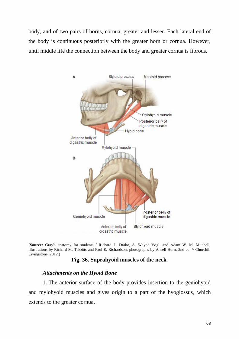
68
body, and of two pairs of horns, cornua, greater and lesser. Each lateral end of
the body is continuous posteriorly with the greater horn or cornua. However,
until middle life the connection between the body and greater cornua is fibrous.
(Source: Gray′s anatomy for students / Richard L. Drake, A. Wayne Vogl, and Adam W. M. Mitchell;
illustrations by Richard M. Tibbitts and Paul E. Richardson; photographs by Ansell Horn; 2nd ed. // Churchill
Livingstone, 2012.)
Fig. 36. Suprahyoid muscles of the neck.
Attachments on the Hyoid Bone
1. The anterior surface of the body provides insertion to the geniohyoid
and mylohyoid muscles and gives origin to a part of the hyoglossus, which
extends to the greater cornua.
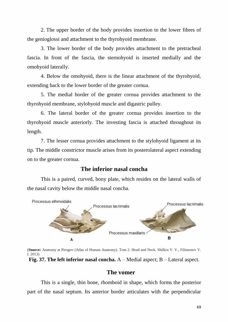
69
2. The upper border of the body provides insertion to the lower fibres of
the genioglossi and attachment to the thyrohyoid membrane.
3. The lower border of the body provides attachment to the pretracheal
fascia. In front of the fascia, the sternohyoid is inserted medially and the
omohyoid laterally.
4. Below the omohyoid, there is the linear attachment of the thyrohyoid,
extending back to the lower border of the greater cornua.
5. The medial border of the greater cornua provides attachment to the
thyrohyoid membrane, stylohyoid muscle and digastric pulley.
6. The lateral border of the greater cornua provides insertion to the
thyrohyoid muscle anteriorly. The investing fascia is attached throughout its
length.
7. The lesser cornua provides attachment to the stylohyoid ligament at its
tip. The middle constrictor muscle arises from its posterolateral aspect extending
on to the greater cornua.
The inferior nasal concha
This is a paired, curved, bony plate, which resides on the lateral walls of
the nasal cavity below the middle nasal concha.
(Source: Anatomy at Pirogov (Atlas of Human Anatomy). Tom 2. Head and Neck. Shilkin V. V., Filimonov V.
I. 2013)
Fig. 37. The left inferior nasal concha. A – Medial aspect; B – Lateral aspect.
The vomer
This is a single, thin bone, rhomboid in shape, which forms the posterior
part of the nasal septum. Its anterior border articulates with the perpendicular
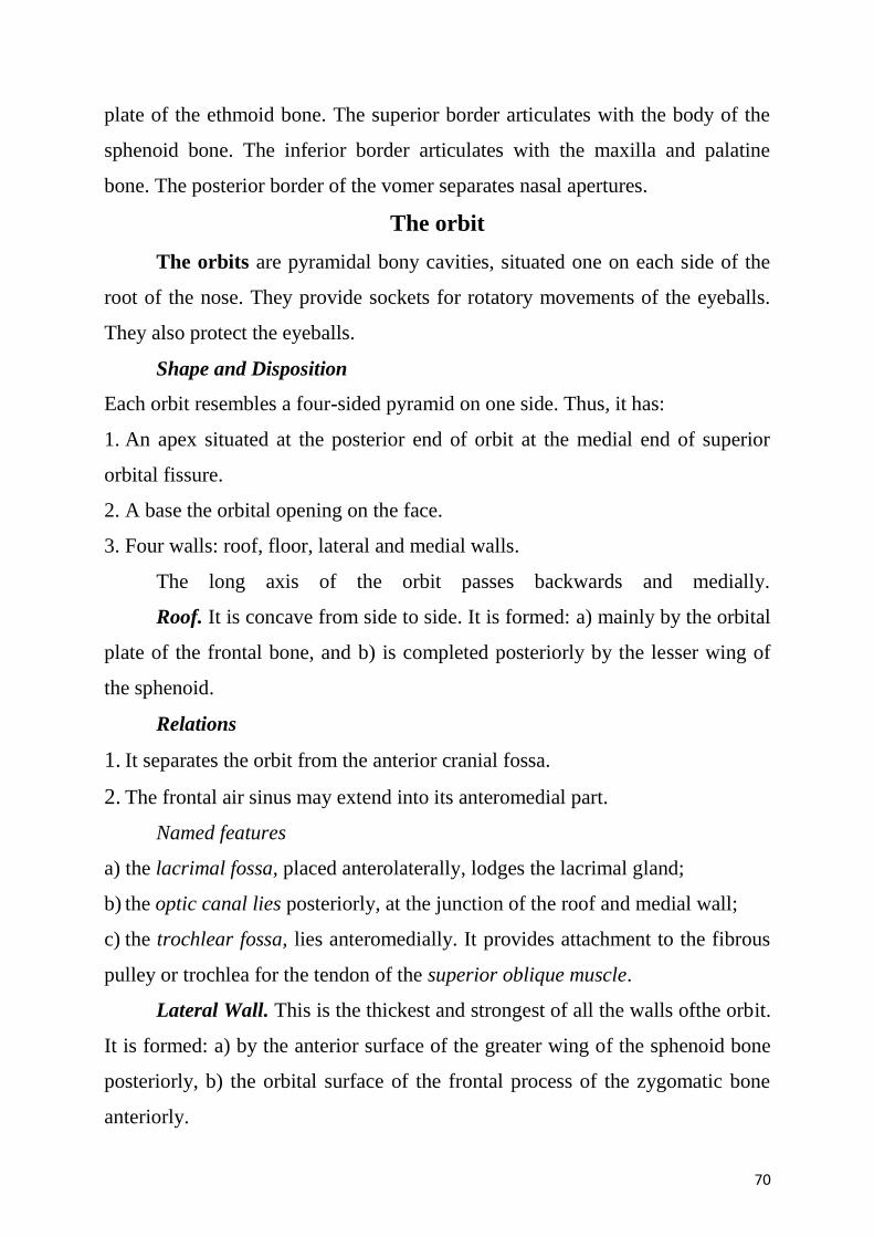
70
plate of the ethmoid bone. The superior border articulates with the body of the
sphenoid bone. The inferior border articulates with the maxilla and palatine
bone. The posterior border of the vomer separates nasal apertures.
The orbit
The orbits are pyramidal bony cavities, situated one on each side of the
root of the nose. They provide sockets for rotatory movements of the eyeballs.
They also protect the eyeballs.
Shape and Disposition
Each orbit resembles a four-sided pyramid on one side. Thus, it has:
1. An apex situated at the posterior end of orbit at the medial end of superior
orbital fissure.
2. A base the orbital opening on the face.
3. Four walls: roof, floor, lateral and medial walls.
The long axis of the orbit passes backwards and medially.
Roof. It is concave from side to side. It is formed: a) mainly by the orbital
plate of the frontal bone, and b) is completed posteriorly by the lesser wing of
the sphenoid.
Relations
1. It separates the orbit from the anterior cranial fossa.
2. The frontal air sinus may extend into its anteromedial part.
Named features
a) the lacrimal fossa, placed anterolaterally, lodges the lacrimal gland;
b) the optic canal lies posteriorly, at the junction of the roof and medial wall;
c) the trochlear fossa, lies anteromedially. It provides attachment to the fibrous
pulley or trochlea for the tendon of the superior oblique muscle.
Lateral Wall. This is the thickest and strongest of all the walls ofthe orbit.
It is formed: a) by the anterior surface of the greater wing of the sphenoid bone
posteriorly, b) the orbital surface of the frontal process of the zygomatic bone
anteriorly.
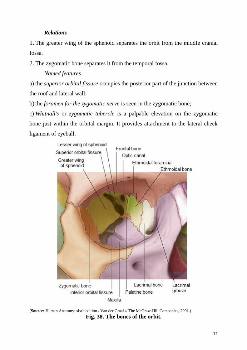
71
Relations
1. The greater wing of the sphenoid separates the orbit from the middle cranial
fossa.
2. The zygomatic bone separates it from the temporal fossa.
Named features
a) the superior orbital fissure occupies the posterior part of the junction between
the roof and lateral wall;
b) the foramen for the zygomatic nerve is seen in the zygomatic bone;
c) Whitnall's or zygomatic tubercle is a palpable elevation on the zygomatic
bone just within the orbital margin. It provides attachment to the lateral check
ligament of eyeball.
(Source: Human Anatomy: sixth edition / Van der Graaf // The McGraw-Hill Companies, 2001.)
Fig. 38. The bones of the orbit.
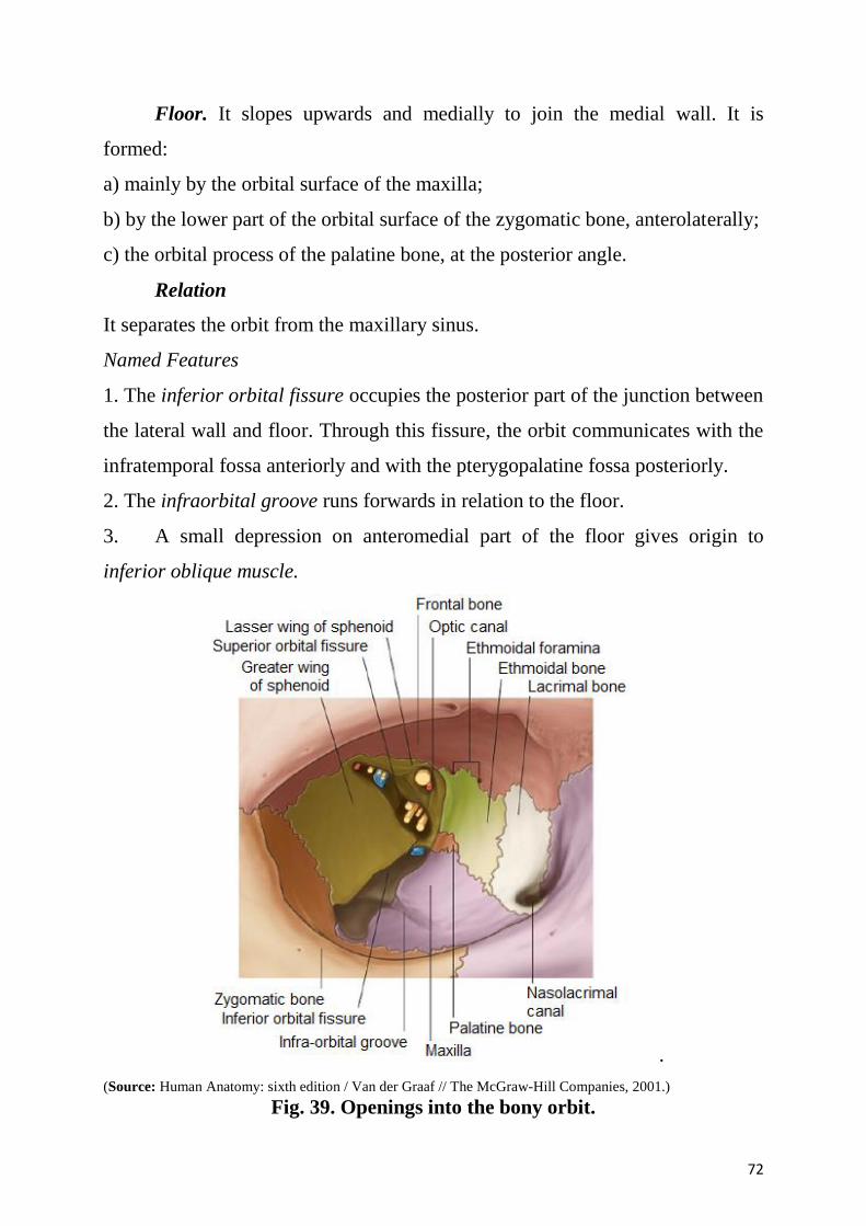
72
Floor. It slopes upwards and medially to join the medial wall. It is
formed:
a) mainly by the orbital surface of the maxilla;
b) by the lower part of the orbital surface of the zygomatic bone, anterolaterally;
c) the orbital process of the palatine bone, at the posterior angle.
Relation
It separates the orbit from the maxillary sinus.
Named Features
1. The inferior orbital fissure occupies the posterior part of the junction between
the lateral wall and floor. Through this fissure, the orbit communicates with the
infratemporal fossa anteriorly and with the pterygopalatine fossa posteriorly.
2. The infraorbital groove runs forwards in relation to the floor.
3. A small depression on anteromedial part of the floor gives origin to
inferior oblique muscle.
.
(Source: Human Anatomy: sixth edition / Van der Graaf // The McGraw-Hill Companies, 2001.)
Fig. 39. Openings into the bony orbit.
73
Medial Wall. It is very thin. From before backwards it is formed by:
1. The frontal process of the maxilla.
2. The lacrimal bone.
3. The orbital plate of the ethmoid.
4. The body of the sphenoid bone.
Relations
1. The lacrimal groove, formed by the maxilla and the lacrimal bone, separates
the orbit from the nasal cavity.
2. The orbital plate of the ethmoid separates the orbit from the ethmoidal air
sinuses.
3. The sphenoidal sinuses, are separated from the orbit only by a thin layer of
bone.
Named Features
A. The lacrimal groove lies anteriorly on the medial wall. It is bounded
anteriorly by the lacrimal crest of the frontal process, of the maxilla, and
posteriorly by the crest of the lacrimal bone. The floor of the groove is formed
by the maxilla in front and by the lacrimal bone behind. The groove lodges the
lacrimal sac, which lies deep to the lacrimal fascia bridging the lacrimal groove.
The groove leads inferiorly, through the nasolacrimal duct, to the inferior
meatus of the nose.
B. The anterior and posterior ethmoidal foramina lie on the
frontoethmoidal suture, at the junction of the roof and medial wall.
Foramina in Relation to the Orbit
1.The structures passing through the superior orbital fissure: the oculomotor
nerve; the trochlear nerve; the lacrimal, frontal and nasociliary branches of the
ophthalmic nerve; the abducent nerve; the superior ophthalmic vein.
2. The optic canal transmits the optic nerve, ophthalmic artery.
3. The inferior orbital fissure transmits the zygomatic nerve, the orbital branches
of the pterygopalatine ganglion, the infraorbital nerve and vessels, and the
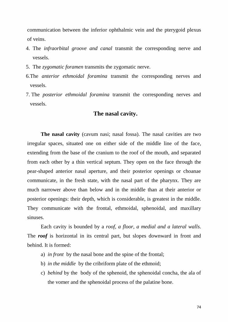
74
communication between the inferior ophthalmic vein and the pterygoid plexus
of veins.
4. The infraorbital groove and canal transmit the corresponding nerve and
vessels.
5. The zygomatic foramen transmits the zygomatic nerve.
6.The anterior ethmoidal foramina transmit the corresponding nerves and
vessels.
7. The posterior ethmoidal foramina transmit the corresponding nerves and
vessels.
The nasal cavity.
The nasal cavity (cavum nasi; nasal fossa). The nasal cavities are two
irregular spaces, situated one on either side of the middle line of the face,
extending from the base of the cranium to the roof of the mouth, and separated
from each other by a thin vertical septum. They open on the face through the
pear-shaped anterior nasal aperture, and their posterior openings or choanae
communicate, in the fresh state, with the nasal part of the pharynx. They are
much narrower above than below and in the middle than at their anterior or
posterior openings: their depth, which is considerable, is greatest in the middle.
They communicate with the frontal, ethmoidal, sphenoidal, and maxillary
sinuses.
Each cavity is bounded by a roof, a floor, a medial and a lateral walls.
The roof is horizontal in its central part, but slopes downward in front and
behind. It is formed:
a) in front by the nasal bone and the spine of the frontal;
b) in the middle by the cribriform plate of the ethmoid;
c) behind by the body of the sphenoid, the sphenoidal concha, the ala of
the vomer and the sphenoidal process of the palatine bone.
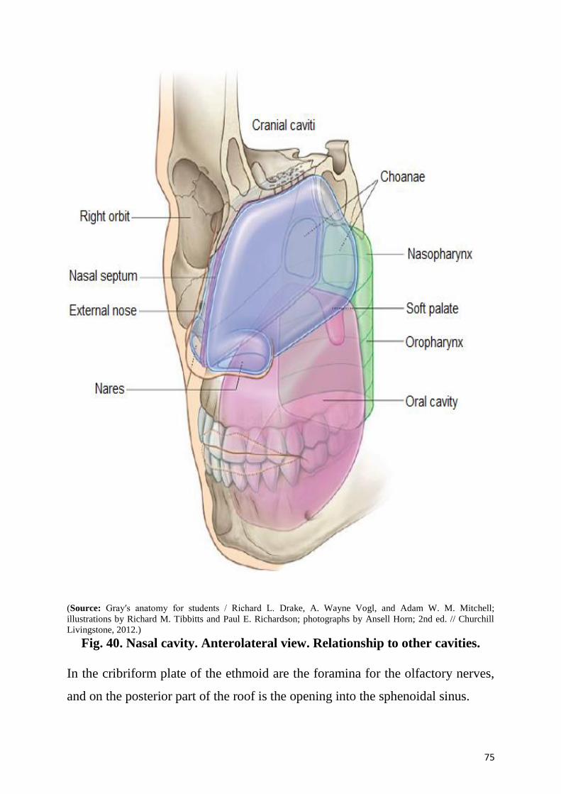
75
(Source: Gray′s anatomy for students / Richard L. Drake, A. Wayne Vogl, and Adam W. M. Mitchell;
illustrations by Richard M. Tibbitts and Paul E. Richardson; photographs by Ansell Horn; 2nd ed. // Churchill
Livingstone, 2012.)
Fig. 40. Nasal cavity. Anterolateral view. Relationship to other cavities.
In the cribriform plate of the ethmoid are the foramina for the olfactory nerves,
and on the posterior part of the roof is the opening into the sphenoidal sinus.
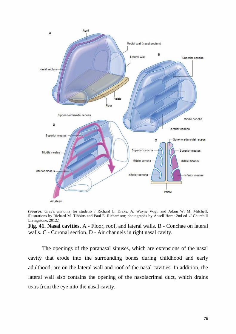
76
(Source: Gray′s anatomy for students / Richard L. Drake, A. Wayne Vogl, and Adam W. M. Mitchell;
illustrations by Richard M. Tibbitts and Paul E. Richardson; photographs by Ansell Horn; 2nd ed. // Churchill
Livingstone, 2012.)
Fig. 41. Nasal cavities. A - Floor, roof, and lateral walls. B - Conchae on lateral
walls. C - Coronal section. D - Air channels in right nasal cavity.
The openings of the paranasal sinuses, which are extensions of the nasal
cavity that erode into the surrounding bones during childhood and early
adulthood, are on the lateral wall and roof of the nasal cavities. In addition, the
lateral wall also contains the opening of the nasolacrimal duct, which drains
tears from the eye into the nasal cavity.
77
The floor is flattened from before backward and concave from side to
side. It is formed by the palatine process of the maxilla and the horizontal part
of the palatine bone; near its anterior end is the opening of the incisive canal.
(Source: Human Anatomy: sixth edition / Van der Graaf // The McGraw-Hill Companies, 2001.)
Fig. 42. Choanae (posterior view). A - Overview. B - Magnified view.
The medial wall (septum nasi), is frequently deflected to one or other
side, more often to the left than to the right. It is formed:
а) in front, by the crest of the nasal bones and frontal spine;
b) in the middle, by the perpendicular plate of the ethmoid;
c) behind, by the vomer and the rostrum of the sphenoid;
d) below, by the crest of the maxillae and palatine bones.
It presents, in front, a large, triangular notch, which receives the cartilage
of the septum; and behind, the free edge of the vomer. Its surface is marked by
numerous furrows for vessels and nerves and by the grooves for the nasopalatine
nerve, and is traversed by sutures connecting the bones of which it is formed.
The superior meatus, the smallest of the three, occupies the middle third of the
lateral wall. It lies between the superior and middle nasal conchae; the
sphenopalatine foramen opens into it behind, and the posterior ethmoidal cells
in front.
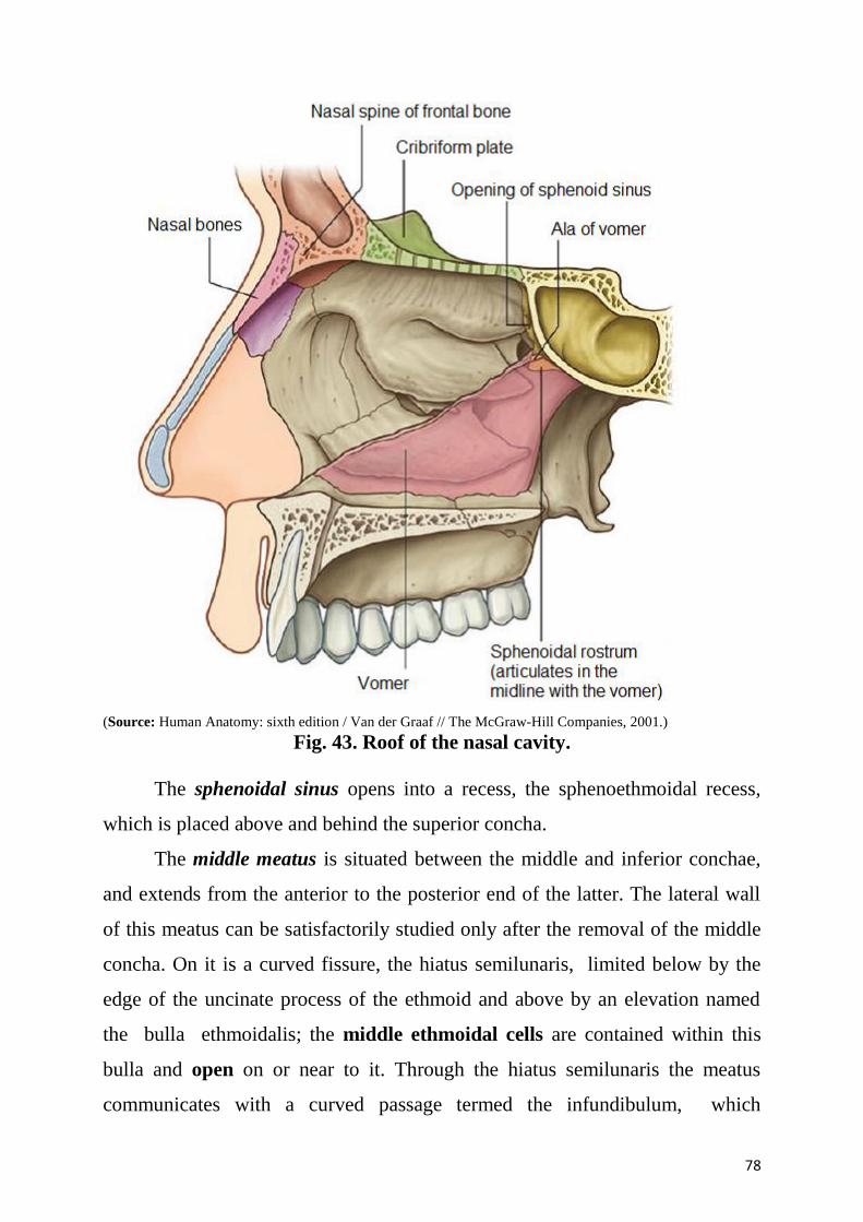
78
(Source: Human Anatomy: sixth edition / Van der Graaf // The McGraw-Hill Companies, 2001.)
Fig. 43. Roof of the nasal cavity.
The sphenoidal sinus opens into a recess, the sphenoethmoidal recess,
which is placed above and behind the superior concha.
The middle meatus is situated between the middle and inferior conchae,
and extends from the anterior to the posterior end of the latter. The lateral wall
of this meatus can be satisfactorily studied only after the removal of the middle
concha. On it is a curved fissure, the hiatus semilunaris, limited below by the
edge of the uncinate process of the ethmoid and above by an elevation named
the bulla ethmoidalis; the middle ethmoidal cells are contained within this
bulla and open on or near to it. Through the hiatus semilunaris the meatus
communicates with a curved passage termed the infundibulum, which
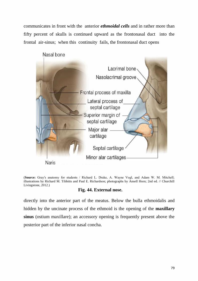
79
communicates in front with the anterior ethmoidal cells and in rather more than
fifty percent of skulls is continued upward as the frontonasal duct into the
frontal air-sinus; when this continuity fails, the frontonasal duct opens
(Source: Gray′s anatomy for students / Richard L. Drake, A. Wayne Vogl, and Adam W. M. Mitchell;
illustrations by Richard M. Tibbitts and Paul E. Richardson; photographs by Ansell Horn; 2nd ed. // Churchill
Livingstone, 2012.)
Fig. 44. External nose.
directly into the anterior part of the meatus. Below the bulla ethmoidalis and
hidden by the uncinate process of the ethmoid is the opening of the maxillary
sinus (ostium maxillare); an accessory opening is frequently present above the
posterior part of the inferior nasal concha.
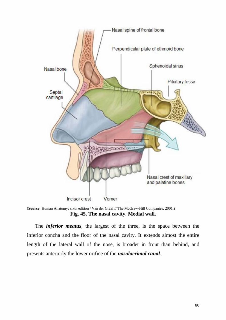
80
(Source: Human Anatomy: sixth edition / Van der Graaf // The McGraw-Hill Companies, 2001.)
Fig. 45. The nasal cavity. Medial wall.
The inferior meatus, the largest of the three, is the space between the
inferior concha and the floor of the nasal cavity. It extends almost the entire
length of the lateral wall of the nose, is broader in front than behind, and
presents anteriorly the lower orifice of the nasolacrimal canal.
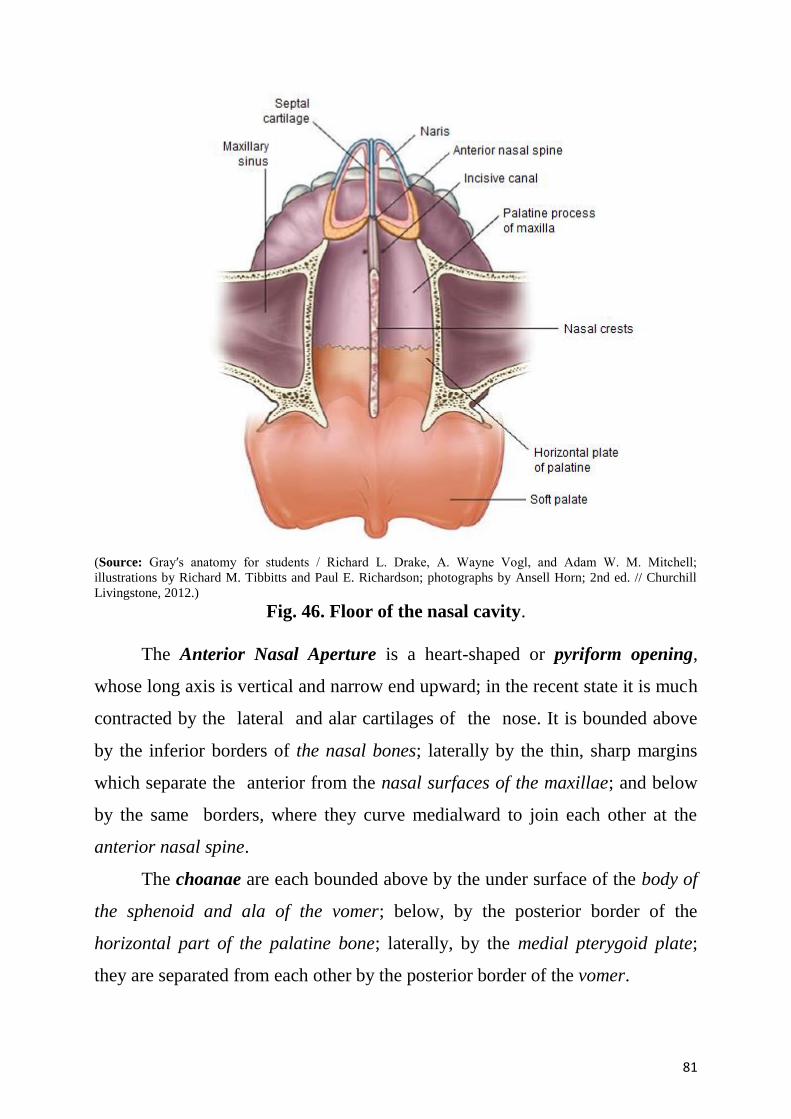
81
(Source: Gray′s anatomy for students / Richard L. Drake, A. Wayne Vogl, and Adam W. M. Mitchell;
illustrations by Richard M. Tibbitts and Paul E. Richardson; photographs by Ansell Horn; 2nd ed. // Churchill
Livingstone, 2012.)
Fig. 46. Floor of the nasal cavity.
The Anterior Nasal Aperture is a heart-shaped or pyriform opening,
whose long axis is vertical and narrow end upward; in the recent state it is much
contracted by the lateral and alar cartilages of the nose. It is bounded above
by the inferior borders of the nasal bones; laterally by the thin, sharp margins
which separate the anterior from the nasal surfaces of the maxillae; and below
by the same borders, where they curve medialward to join each other at the
anterior nasal spine.
The choanae are each bounded above by the under surface of the body of
the sphenoid and ala of the vomer; below, by the posterior border of the
horizontal part of the palatine bone; laterally, by the medial pterygoid plate;
they are separated from each other by the posterior border of the vomer.
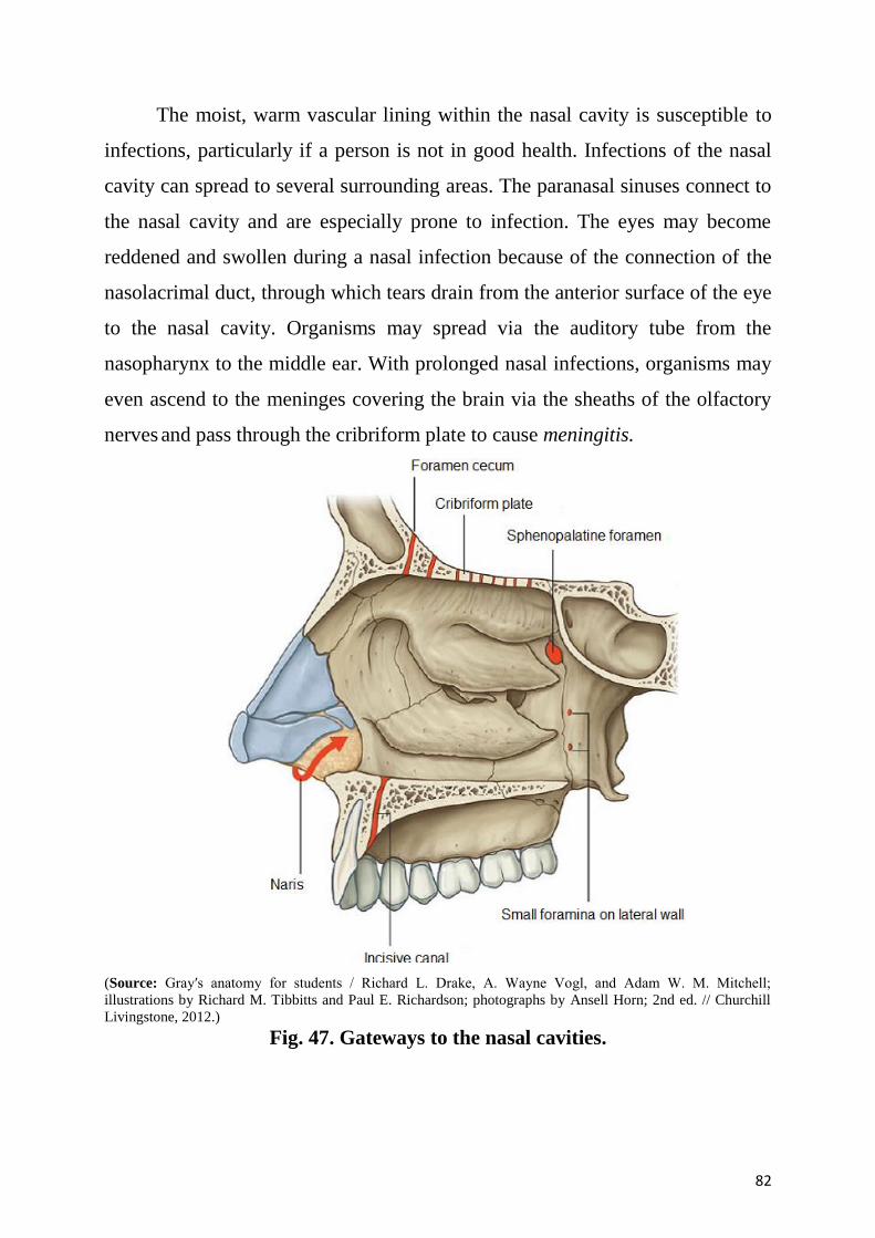
82
The moist, warm vascular lining within the nasal cavity is susceptible to
infections, particularly if a person is not in good health. Infections of the nasal
cavity can spread to several surrounding areas. The paranasal sinuses connect to
the nasal cavity and are especially prone to infection. The eyes may become
reddened and swollen during a nasal infection because of the connection of the
nasolacrimal duct, through which tears drain from the anterior surface of the eye
to the nasal cavity. Organisms may spread via the auditory tube from the
nasopharynx to the middle ear. With prolonged nasal infections, organisms may
even ascend to the meninges covering the brain via the sheaths of the olfactory
nerves and pass through the cribriform plate to cause meningitis.
(Source: Gray′s anatomy for students / Richard L. Drake, A. Wayne Vogl, and Adam W. M. Mitchell;
illustrations by Richard M. Tibbitts and Paul E. Richardson; photographs by Ansell Horn; 2nd ed. // Churchill
Livingstone, 2012.)
Fig. 47. Gateways to the nasal cavities.
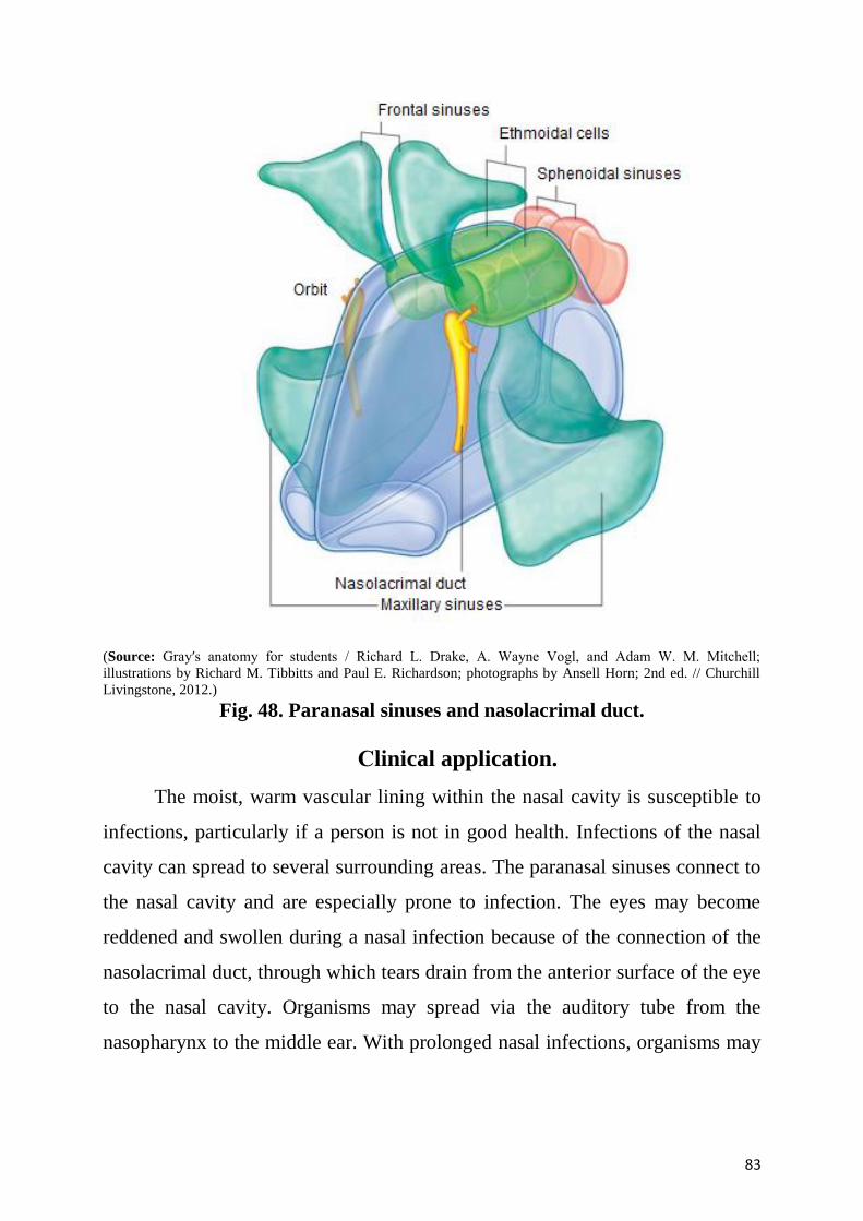
83
(Source: Gray′s anatomy for students / Richard L. Drake, A. Wayne Vogl, and Adam W. M. Mitchell;
illustrations by Richard M. Tibbitts and Paul E. Richardson; photographs by Ansell Horn; 2nd ed. // Churchill
Livingstone, 2012.)
Fig. 48. Paranasal sinuses and nasolacrimal duct.
Сlinical application.
The moist, warm vascular lining within the nasal cavity is susceptible to
infections, particularly if a person is not in good health. Infections of the nasal
cavity can spread to several surrounding areas. The paranasal sinuses connect to
the nasal cavity and are especially prone to infection. The eyes may become
reddened and swollen during a nasal infection because of the connection of the
nasolacrimal duct, through which tears drain from the anterior surface of the eye
to the nasal cavity. Organisms may spread via the auditory tube from the
nasopharynx to the middle ear. With prolonged nasal infections, organisms may
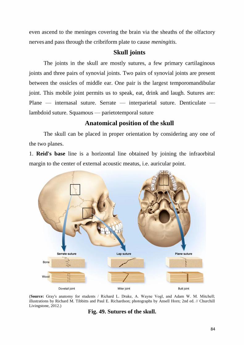
84
even ascend to the meninges covering the brain via the sheaths of the olfactory
nerves and pass through the cribriform plate to cause meningitis.
Skull joints
The joints in the skull are mostly sutures, a few primary cartilaginous
joints and three pairs of synovial joints. Two pairs of synovial joints are present
between the ossicles of middle ear. One pair is the largest temporomandibular
joint. This mobile joint permits us to speak, eat, drink and laugh. Sutures are:
Plane — internasal suture. Serrate — interparietal suture. Denticulate —
lambdoid suture. Squamous — parietotemporal suture
Anatomical position of the skull
The skull can be placed in proper orientation by considering any one of
the two planes.
1. Reid's base line is a horizontal line obtained by joining the infraorbital
margin to the center of external acoustic meatus, i.e. auricular point.
(Source: Gray′s anatomy for students / Richard L. Drake, A. Wayne Vogl, and Adam W. M. Mitchell;
illustrations by Richard M. Tibbitts and Paul E. Richardson; photographs by Ansell Horn; 2nd ed. // Churchill
Livingstone, 2012.)
Fig. 49. Sutures of the skull.
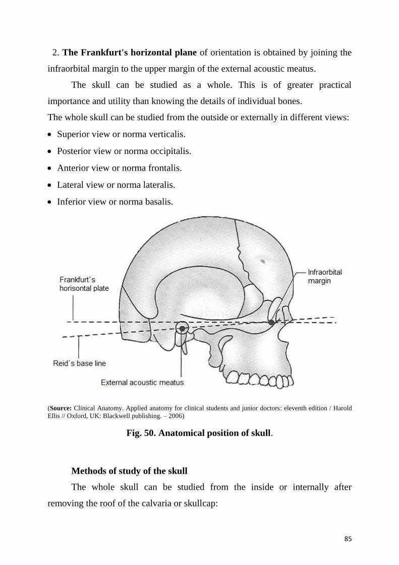
85
2. The Frankfurt's horizontal plane of orientation is obtained by joining the
infraorbital margin to the upper margin of the external acoustic meatus.
The skull can be studied as a whole. This is of greater practical
importance and utility than knowing the details of individual bones.
The whole skull can be studied from the outside or externally in different views:
Superior view or norma verticalis.
Posterior view or norma occipitalis.
Anterior view or norma frontalis.
Lateral view or norma lateralis.
Inferior view or norma basalis.
(Source: Clinical Anatomy. Applied anatomy for clinical students and junior doctors: eleventh edition / Harold
Ellis // Oxford, UK: Blackwell publishing. – 2006)
Fig. 50. Anatomical position of skull.
Methods of study of the skull
The whole skull can be studied from the inside or internally after
removing the roof of the calvaria or skullcap:
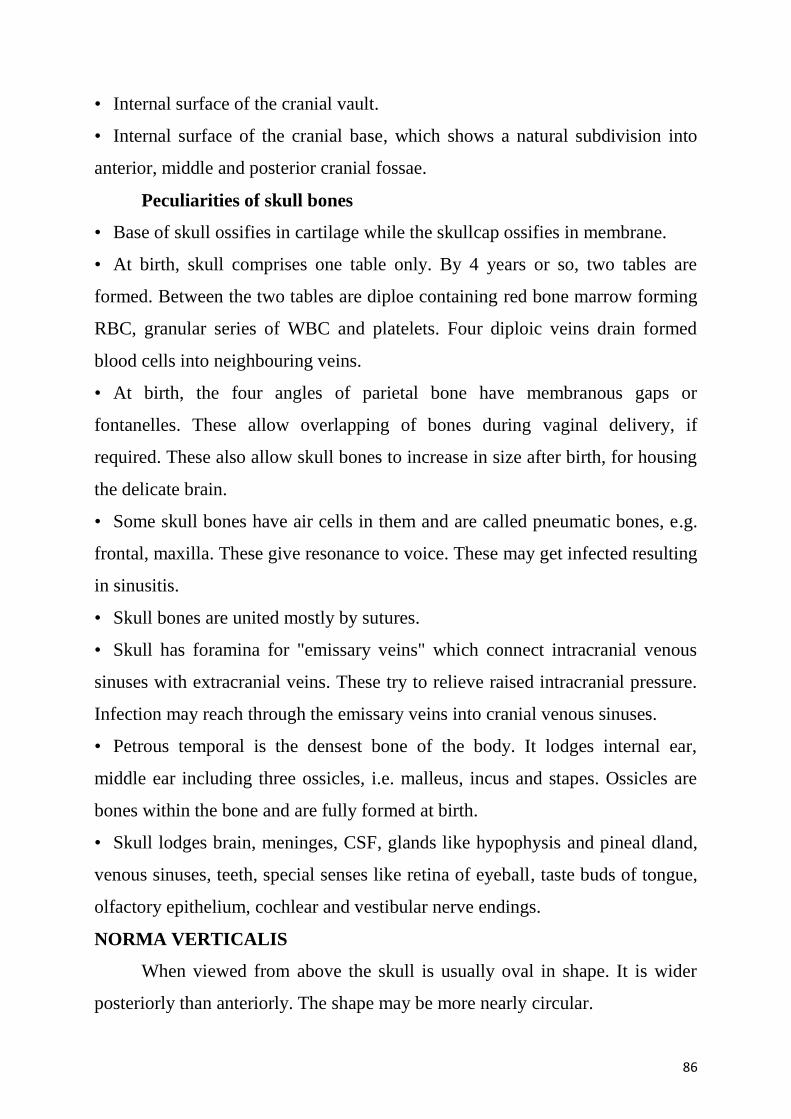
86
• Internal surface of the cranial vault.
• Internal surface of the cranial base, which shows a natural subdivision into
anterior, middle and posterior cranial fossae.
Peculiarities of skull bones
• Base of skull ossifies in cartilage while the skullcap ossifies in membrane.
• At birth, skull comprises one table only. By 4 years or so, two tables are
formed. Between the two tables are diploe containing red bone marrow forming
RBC, granular series of WBC and platelets. Four diploic veins drain formed
blood cells into neighbouring veins.
• At birth, the four angles of parietal bone have membranous gaps or
fontanelles. These allow overlapping of bones during vaginal delivery, if
required. These also allow skull bones to increase in size after birth, for housing
the delicate brain.
• Some skull bones have air cells in them and are called pneumatic bones, e.g.
frontal, maxilla. These give resonance to voice. These may get infected resulting
in sinusitis.
• Skull bones are united mostly by sutures.
• Skull has foramina for "emissary veins" which connect intracranial venous
sinuses with extracranial veins. These try to relieve raised intracranial pressure.
Infection may reach through the emissary veins into cranial venous sinuses.
• Petrous temporal is the densest bone of the body. It lodges internal ear,
middle ear including three ossicles, i.e. malleus, incus and stapes. Ossicles are
bones within the bone and are fully formed at birth.
• Skull lodges brain, meninges, CSF, glands like hypophysis and pineal dland,
venous sinuses, teeth, special senses like retina of eyeball, taste buds of tongue,
olfactory epithelium, cochlear and vestibular nerve endings.
NORMA VERTICALIS
When viewed from above the skull is usually oval in shape. It is wider
posteriorly than anteriorly. The shape may be more nearly circular.
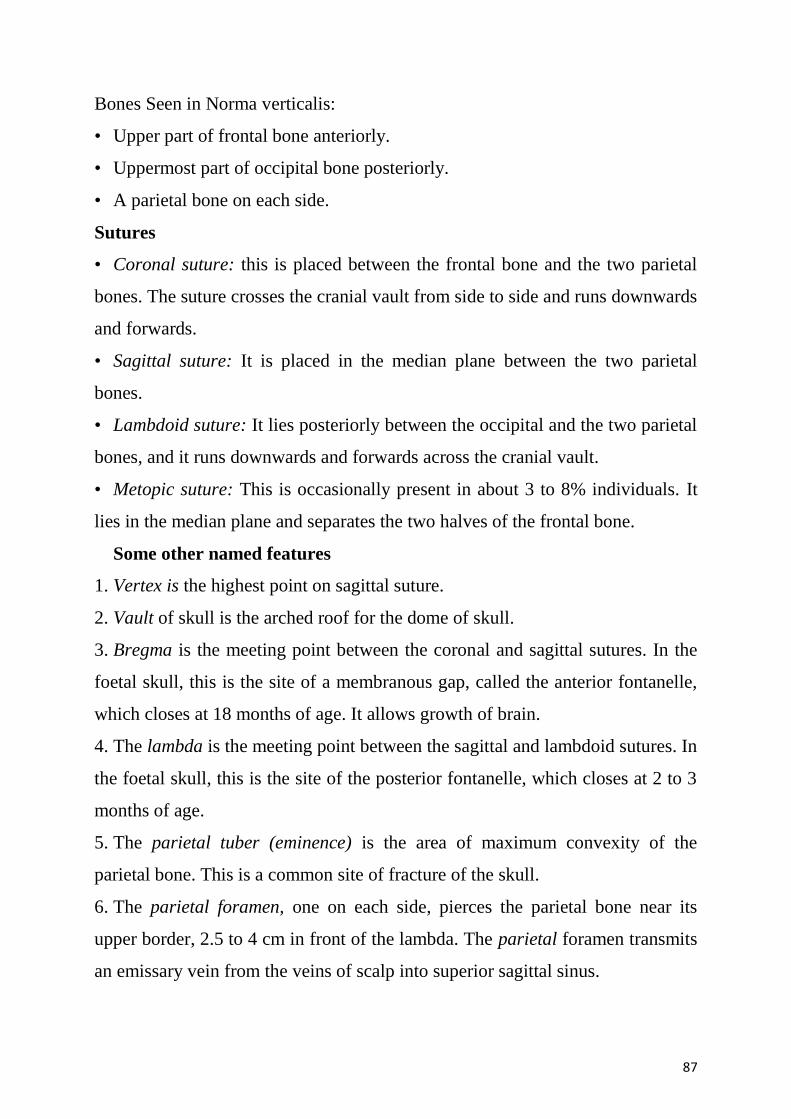
87
Bones Seen in Norma verticalis:
• Upper part of frontal bone anteriorly.
• Uppermost part of occipital bone posteriorly.
• A parietal bone on each side.
Sutures
• Coronal suture: this is placed between the frontal bone and the two parietal
bones. The suture crosses the cranial vault from side to side and runs downwards
and forwards.
• Sagittal suture: It is placed in the median plane between the two parietal
bones.
• Lambdoid suture: It lies posteriorly between the occipital and the two parietal
bones, and it runs downwards and forwards across the cranial vault.
• Metopic suture: This is occasionally present in about 3 to 8% individuals. It
lies in the median plane and separates the two halves of the frontal bone.
Some other named features
1. Vertex is the highest point on sagittal suture.
2. Vault of skull is the arched roof for the dome of skull.
3. Bregma is the meeting point between the coronal and sagittal sutures. In the
foetal skull, this is the site of a membranous gap, called the anterior fontanelle,
which closes at 18 months of age. It allows growth of brain.
4. The lambda is the meeting point between the sagittal and lambdoid sutures. In
the foetal skull, this is the site of the posterior fontanelle, which closes at 2 to 3
months of age.
5. The parietal tuber (eminence) is the area of maximum convexity of the
parietal bone. This is a common site of fracture of the skull.
6. The parietal foramen, one on each side, pierces the parietal bone near its
upper border, 2.5 to 4 cm in front of the lambda. The parietal foramen transmits
an emissary vein from the veins of scalp into superior sagittal sinus.
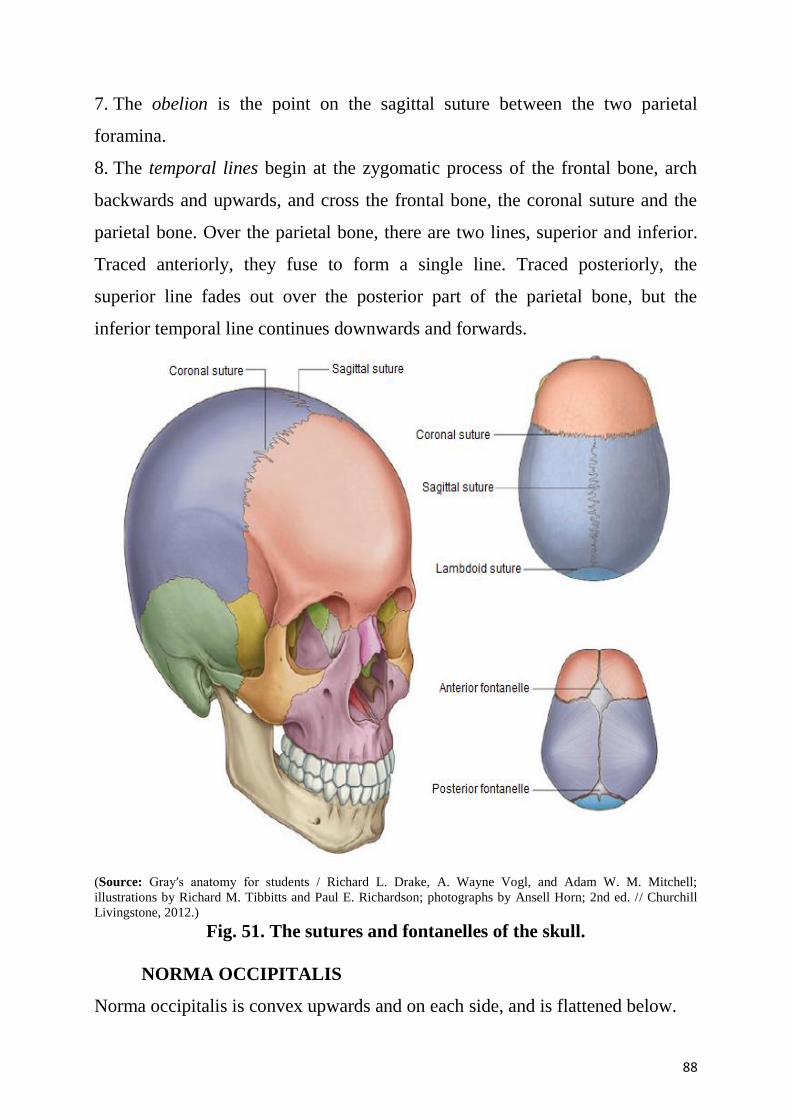
88
7. The obelion is the point on the sagittal suture between the two parietal
foramina.
8. The temporal lines begin at the zygomatic process of the frontal bone, arch
backwards and upwards, and cross the frontal bone, the coronal suture and the
parietal bone. Over the parietal bone, there are two lines, superior and inferior.
Traced anteriorly, they fuse to form a single line. Traced posteriorly, the
superior line fades out over the posterior part of the parietal bone, but the
inferior temporal line continues downwards and forwards.
(Source: Gray′s anatomy for students / Richard L. Drake, A. Wayne Vogl, and Adam W. M. Mitchell;
illustrations by Richard M. Tibbitts and Paul E. Richardson; photographs by Ansell Horn; 2nd ed. // Churchill
Livingstone, 2012.)
Fig. 51. The sutures and fontanelles of the skull.
NORMA OCCIPITALIS
Norma occipitalis is convex upwards and on each side, and is flattened below.
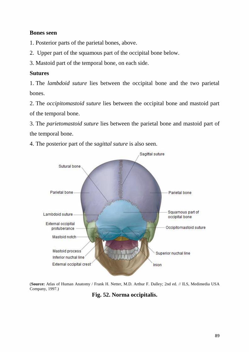
89
Bones seen
1. Posterior parts of the parietal bones, above.
2. Upper part of the squamous part of the occipital bone below.
3. Mastoid part of the temporal bone, on each side.
Sutures
1. The lambdoid suture lies between the occipital bone and the two parietal
bones.
2. The occipitomastoid suture lies between the occipital bone and mastoid part
of the temporal bone.
3. The parietomastoid suture lies between the parietal bone and mastoid part of
the temporal bone.
4. The posterior part of the sagittal suture is also seen.
(Source: Atlas of Human Anatomy / Frank H. Netter, M.D. Arthur F. Dalley; 2nd ed. // ILS, Medimedia USA
Company, 1997.)
Fig. 52. Norma occipitalis.
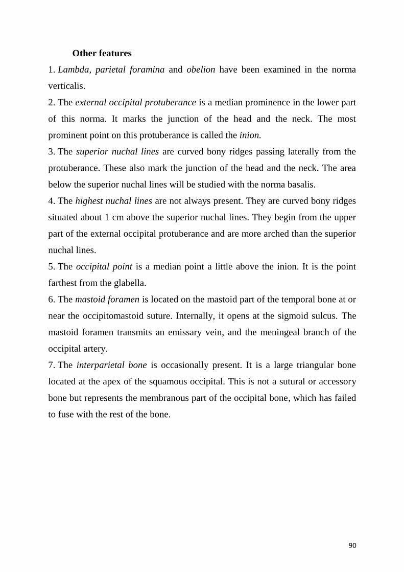
90
Other features
1. Lambda, parietal foramina and obelion have been examined in the norma
verticalis.
2. The external occipital protuberance is a median prominence in the lower part
of this norma. It marks the junction of the head and the neck. The most
prominent point on this protuberance is called the inion.
3. The superior nuchal lines are curved bony ridges passing laterally from the
protuberance. These also mark the junction of the head and the neck. The area
below the superior nuchal lines will be studied with the norma basalis.
4. The highest nuchal lines are not always present. They are curved bony ridges
situated about 1 cm above the superior nuchal lines. They begin from the upper
part of the external occipital protuberance and are more arched than the superior
nuchal lines.
5. The occipital point is a median point a little above the inion. It is the point
farthest from the glabella.
6. The mastoid foramen is located on the mastoid part of the temporal bone at or
near the occipitomastoid suture. Internally, it opens at the sigmoid sulcus. The
mastoid foramen transmits an emissary vein, and the meningeal branch of the
occipital artery.
7. The interparietal bone is occasionally present. It is a large triangular bone
located at the apex of the squamous occipital. This is not a sutural or accessory
bone but represents the membranous part of the occipital bone, which has failed
to fuse with the rest of the bone.
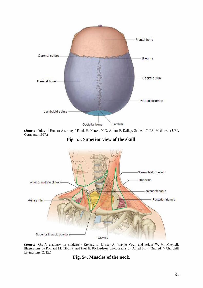
91
(Source: Atlas of Human Anatomy / Frank H. Netter, M.D. Arthur F. Dalley; 2nd ed. // ILS, Medimedia USA
Company, 1997.)
Fig. 53. Superior view of the skull.
(Source: Gray′s anatomy for students / Richard L. Drake, A. Wayne Vogl, and Adam W. M. Mitchell;
illustrations by Richard M. Tibbitts and Paul E. Richardson; photographs by Ansell Horn; 2nd ed. // Churchill
Livingstone, 2012.)
Fig. 54. Muscles of the neck.
92
Attachments
The upper part of the external occipital protuberance gives origin to the
trapezius, and the lower part gives attachment to the upper end of the
ligamentum nuchae.
1. The medial one-third of the superior nuchal line gives origin to the trapezius,
and the lateral part provides insertion to the sternocleidomastoid above and to
the splenius capitis below.
2. The highest nuchal lines if present provide attachment to the epicranial
aponeurosis medially, and give origin to the occipitalis or occipital belly of
occipitofrontalis muscle laterally. In case of absence of highest nuchal lines,
these structures are attached to superior nuchal lines.
NORMA FRONTALIS
The norma frontalis is roughly oval in outline, being wider above than
below.
Bones
1. Frontal bone forms the forehead. Its upper part is smooth and convex, but the
lower part is irregular and is interrupted by the orbits and by the anterior bony
aperture of nose.
2. The right and left maxillae form the upper jaw.
3. The right and left nasal bones form the bridge of the nose.
4. The zygomatic bones form the bony prominence of the superolateral part of
the cheeks.
5. The mandible forms the lower jaw.
The norma frontalis will be studied under the following heads:
Frontal region.
Orbital opening.
Anterior piriform-shaped bony aperture of the nose.
93
Lower part of the face.
Frontal region
The frontal region presents the following features:
1. The superciliary arch is a rounded, curved elevation situated just above the
medial part of each orbit. It overlies the frontal sinus and is better marked in
males than in females.
2. The glabella is a median elevation connecting the two superciliary arches.
Below the glabella, the skull to frontonasal suture at root of the nose.
3. The nasion is a median point at the root of the nose where the internasal
suture meets with the frontonasal suture.
4. The frontal tuber or eminence is a low rounded elevation above the
superciliary arch, one on each side.
Orbital openings
Each orbital opening is quadrangular in shape and is bounded by the
following four margins.
1. The supraorbital margin is formed by the frontal bone. At the junction of its
lateral two-thirds and its medial one-third, it presents the supraorbital notch or
foramen.
2. The infraorbital margin is formed by the zygomatic bone laterally, and
maxilla medially.
3. The medial orbital margin is ill-defined. It is formed by the frontal bone
above, and by the lacrimal crest of the frontal process of the maxilla below.
4. The lateral orbital margin is formed mostly by the frontal process of
zygomatic bone but is completed above by the zygomatic process of frontal
bone. Frontozygomatic suture lies at their union.
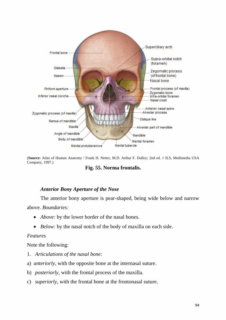
94
(Source: Atlas of Human Anatomy / Frank H. Netter, M.D. Arthur F. Dalley; 2nd ed. // ILS, Medimedia USA
Company, 1997.)
Fig. 55. Norma frontalis.
Anterior Bony Aperture of the Nose
The anterior bony aperture is pear-shaped, being wide below and narrow
above. Boundaries:
Above: by the lower border of the nasal bones.
Below: by the nasal notch of the body of maxilla on each side.
Features
Note the following:
1. Articulations of the nasal bone:
a) anteriorly, with the opposite bone at the internasal suture.
b) posteriorly, with the frontal process of the maxilla.
c) superiorly, with the frontal bone at the frontonasal suture.
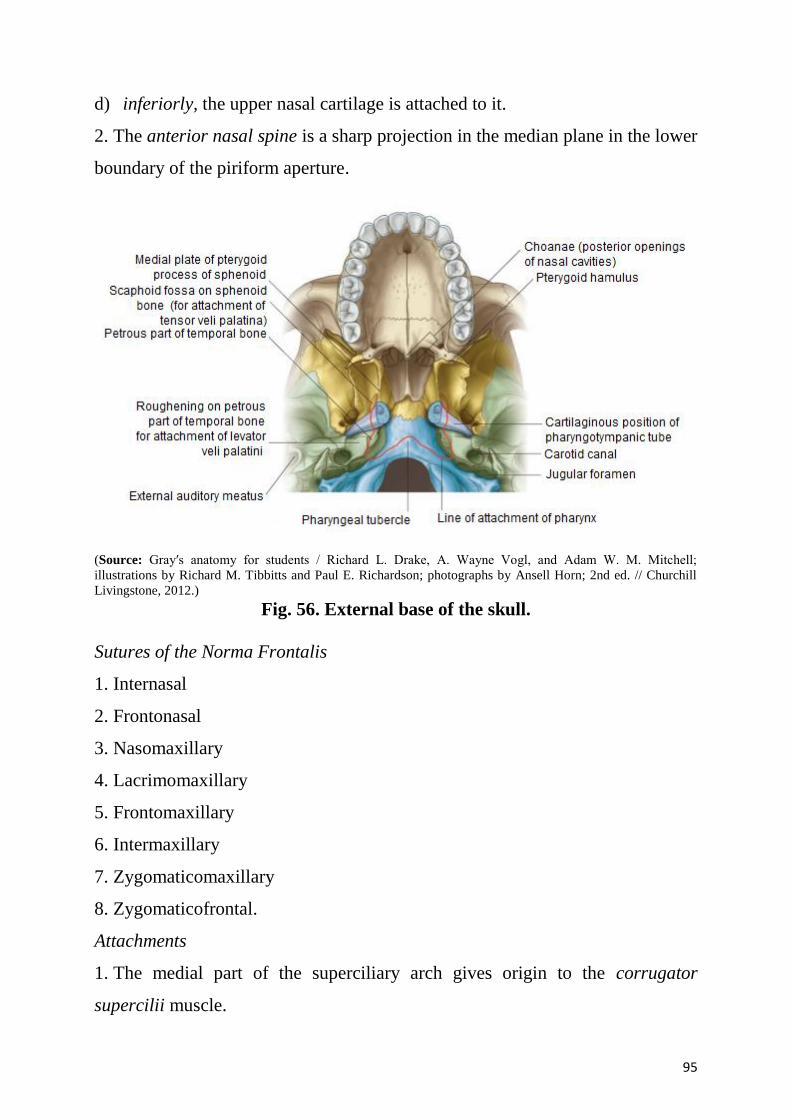
95
d) inferiorly, the upper nasal cartilage is attached to it.
2. The anterior nasal spine is a sharp projection in the median plane in the lower
boundary of the piriform aperture.
(Source: Gray′s anatomy for students / Richard L. Drake, A. Wayne Vogl, and Adam W. M. Mitchell;
illustrations by Richard M. Tibbitts and Paul E. Richardson; photographs by Ansell Horn; 2nd ed. // Churchill
Livingstone, 2012.)
Fig. 56. External base of the skull.
Sutures of the Norma Frontalis
1. Internasal
2. Frontonasal
3. Nasomaxillary
4. Lacrimomaxillary
5. Frontomaxillary
6. Intermaxillary
7. Zygomaticomaxillary
8. Zygomaticofrontal.
Attachments
1. The medial part of the superciliary arch gives origin to the corrugator
supercilii muscle.
96
2. The procerus muscle arises from the nasal bone near the median plane.
3. The orbital part of the orbicularis oculi arises from the frontal process of the
maxilla and from the nasal part of the frontal bone.
4. The medial palpebral ligament is attached to the frontal process of the
maxilla between the frontal and maxillary origins of the orbicularis oculi.
5. The levator labii superioris alaeque nasi arises from the frontal process of the
maxilla in front of the orbicularis oculi.
5. The levator labii superioris arises from the maxilla between the infraorbital
margin and the infraorbital foramen.
6. The levator anguli oris arises from the canine fossa.
7. The nasalis and the depressor septi arise from the surface of the maxilla
bordering the nasal notch.
8. The incisivus muscle arises from an area just below the depressor septi. It
forms part of orbicularis oris.
9. The zygomaticus major and minor arise from the surface of the zygomatic
bone. The zygomaticus minor muscle arises below the zygomaticofacial
foramen. The zygomaticus major arises lateral to the minor muscle.
10. Buccinator arises from maxilla and mandible opposite molar teeth and from
pterygomandibular raphe. It also forms part of orbicularis oris.
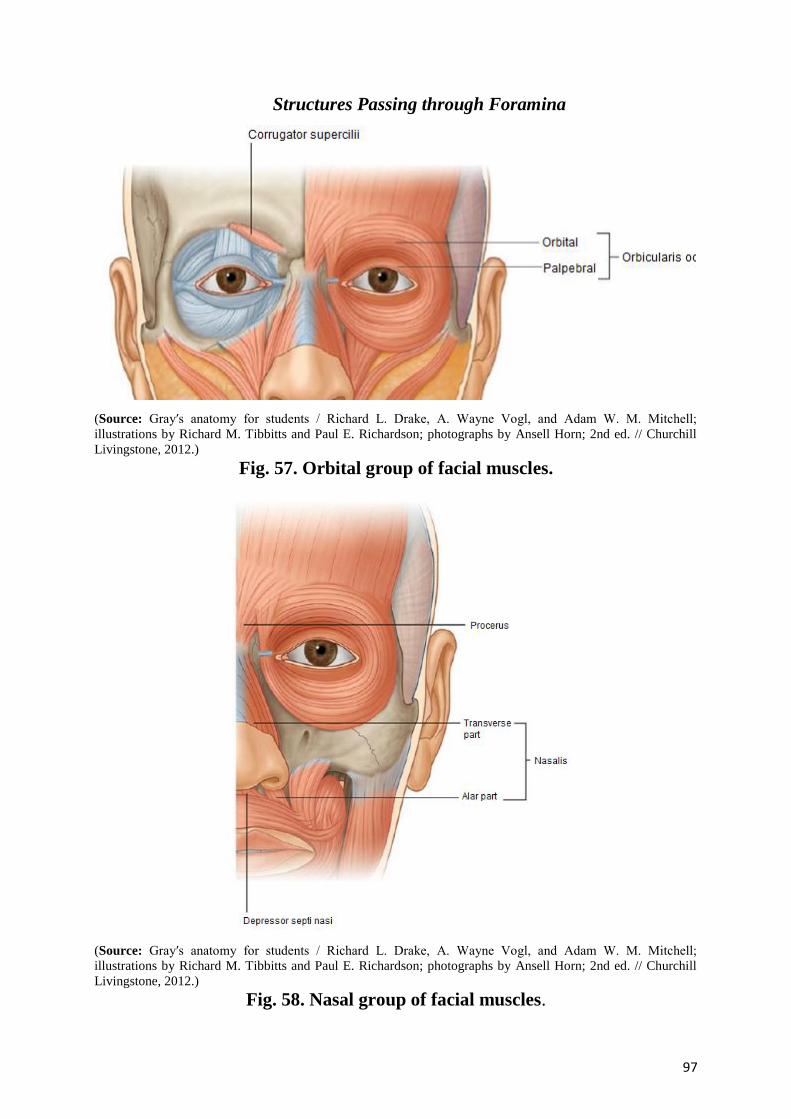
97
Structures Passing through Foramina
(Source: Gray′s anatomy for students / Richard L. Drake, A. Wayne Vogl, and Adam W. M. Mitchell;
illustrations by Richard M. Tibbitts and Paul E. Richardson; photographs by Ansell Horn; 2nd ed. // Churchill
Livingstone, 2012.)
Fig. 57. Orbital group of facial muscles.
(Source: Gray′s anatomy for students / Richard L. Drake, A. Wayne Vogl, and Adam W. M. Mitchell;
illustrations by Richard M. Tibbitts and Paul E. Richardson; photographs by Ansell Horn; 2nd ed. // Churchill
Livingstone, 2012.)
Fig. 58. Nasal group of facial muscles.
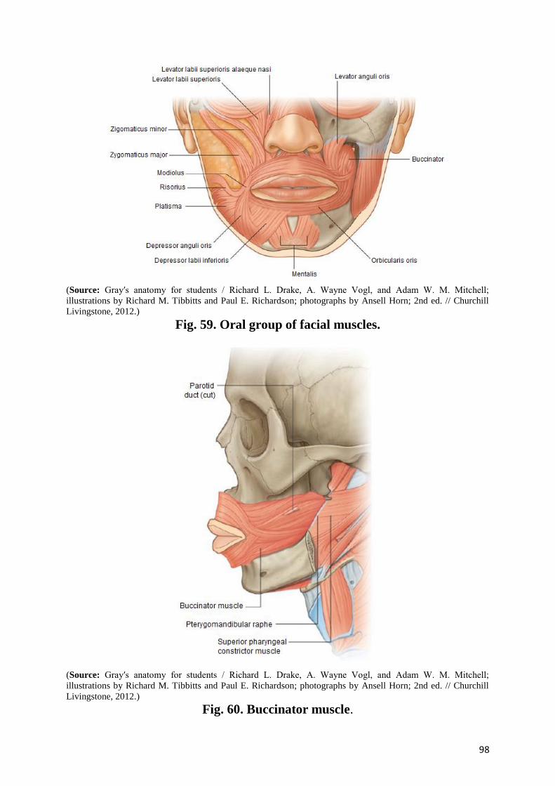
98
(Source: Gray′s anatomy for students / Richard L. Drake, A. Wayne Vogl, and Adam W. M. Mitchell;
illustrations by Richard M. Tibbitts and Paul E. Richardson; photographs by Ansell Horn; 2nd ed. // Churchill
Livingstone, 2012.)
Fig. 59. Oral group of facial muscles.
(Source: Gray′s anatomy for students / Richard L. Drake, A. Wayne Vogl, and Adam W. M. Mitchell;
illustrations by Richard M. Tibbitts and Paul E. Richardson; photographs by Ansell Horn; 2nd ed. // Churchill
Livingstone, 2012.)
Fig. 60. Buccinator muscle.
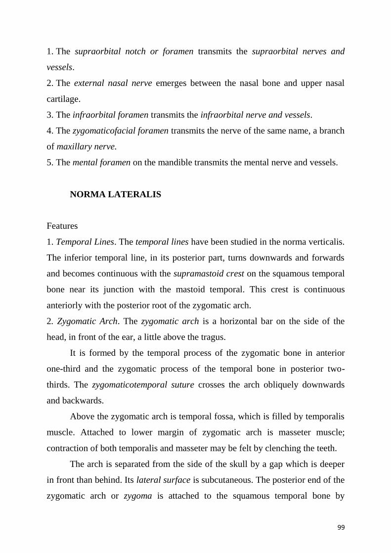
99
1. The supraorbital notch or foramen transmits the supraorbital nerves and
vessels.
2. The external nasal nerve emerges between the nasal bone and upper nasal
cartilage.
3. The infraorbital foramen transmits the infraorbital nerve and vessels.
4. The zygomaticofacial foramen transmits the nerve of the same name, a branch
of maxillary nerve.
5. The mental foramen on the mandible transmits the mental nerve and vessels.
NORMA LATERALIS
Features
1. Temporal Lines. The temporal lines have been studied in the norma verticalis.
The inferior temporal line, in its posterior part, turns downwards and forwards
and becomes continuous with the supramastoid crest on the squamous temporal
bone near its junction with the mastoid temporal. This crest is continuous
anteriorly with the posterior root of the zygomatic arch.
2. Zygomatic Arch. The zygomatic arch is a horizontal bar on the side of the
head, in front of the ear, a little above the tragus.
It is formed by the temporal process of the zygomatic bone in anterior
one-third and the zygomatic process of the temporal bone in posterior two-
thirds. The zygomaticotemporal suture crosses the arch obliquely downwards
and backwards.
Above the zygomatic arch is temporal fossa, which is filled by temporalis
muscle. Attached to lower margin of zygomatic arch is masseter muscle;
contraction of both temporalis and masseter may be felt by clenching the teeth.
The arch is separated from the side of the skull by a gap which is deeper
in front than behind. Its lateral surface is subcutaneous. The posterior end of the
zygomatic arch or zygoma is attached to the squamous temporal bone by
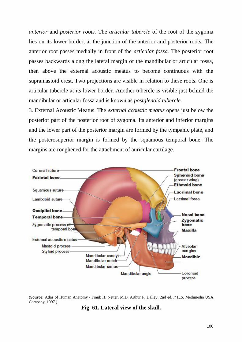
100
anterior and posterior roots. The articular tubercle of the root of the zygoma
lies on its lower border, at the junction of the anterior and posterior roots. The
anterior root passes medially in front of the articular fossa. The posterior root
passes backwards along the lateral margin of the mandibular or articular fossa,
then above the external acoustic meatus to become continuous with the
supramastoid crest. Two projections are visible in relation to these roots. One is
articular tubercle at its lower border. Another tubercle is visible just behind the
mandibular or articular fossa and is known as postglenoid tubercle.
3. External Acoustic Meatus. The external acoustic meatus opens just below the
posterior part of the posterior root of zygoma. Its anterior and inferior margins
and the lower part of the posterior margin are formed by the tympanic plate, and
the posterosuperior margin is formed by the squamous temporal bone. The
margins are roughened for the attachment of auricular cartilage.
(Source: Atlas of Human Anatomy / Frank H. Netter, M.D. Arthur F. Dalley; 2nd ed. // ILS, Medimedia USA
Company, 1997.)
Fig. 61. Lateral view of the skull.
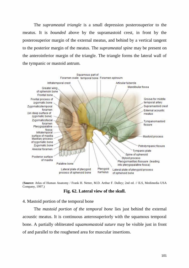
101
The suprameatal triangle is a small depression posterosuperior to the
meatus. It is bounded above by the supramastoid crest, in front by the
posterosuperior margin of the external meatus, and behind by a vertical tangent
to the posterior margin of the meatus. The suprameatal spine may be present on
the anteroinferior margin of the triangle. The triangle forms the lateral wall of
the tympanic or mastoid antrum.
(Source: Atlas of Human Anatomy / Frank H. Netter, M.D. Arthur F. Dalley; 2nd ed. // ILS, Medimedia USA
Company, 1997.)
Fig. 62. Lateral view of the skull.
4. Mastoid portion of the temporal bone
The mastoid portion of the temporal bone lies just behind the external
acoustic meatus. It is continuous anterosuperiorly with the squamous temporal
bone. A partially obliterated squamomastoid suture may be visible just in front
of and parallel to the roughened area for muscular insertions.
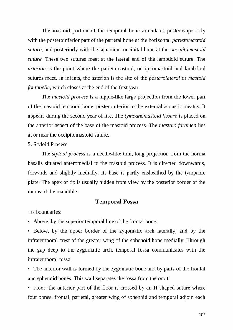
102
The mastoid portion of the temporal bone articulates posterosuperiorly
with the posteroinferior part of the parietal bone at the horizontal parietomastoid
suture, and posteriorly with the squamous occipital bone at the occipitomastoid
suture. These two sutures meet at the lateral end of the lambdoid suture. The
asterion is the point where the parietomastoid, occipitomastoid and lambdoid
sutures meet. In infants, the asterion is the site of the posterolateral or mastoid
fontanelle, which closes at the end of the first year.
The mastoid process is a nipple-like large projection from the lower part
of the mastoid temporal bone, posteroinferior to the external acoustic meatus. It
appears during the second year of life. The tympanomastoid fissure is placed on
the anterior aspect of the base of the mastoid process. The mastoid foramen lies
at or near the occipitomastoid suture.
5. Styloid Process
The styloid process is a needle-like thin, long projection from the norma
basalis situated anteromedial to the mastoid process. It is directed downwards,
forwards and slightly medially. Its base is partly ensheathed by the tympanic
plate. The apex or tip is usually hidden from view by the posterior border of the
ramus of the mandible.
Temporal Fossa
Its boundaries:
• Above, by the superior temporal line of the frontal bone.
• Below, by the upper border of the zygomatic arch laterally, and by the
infratemporal crest of the greater wing of the sphenoid hone medially. Through
the gap deep to the zygomatic arch, temporal fossa communicates with the
infratemporal fossa.
• The anterior wall is formed by the zygomatic bone and by parts of the frontal
and sphenoid bones. This wall separates the fossa from the orbit.
• Floor: the anterior part of the floor is crossed by an H-shaped suture where
four bones, frontal, parietal, greater wing of sphenoid and temporal adjoin each
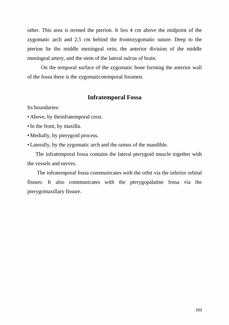
103
other. This area is termed the pterion. It lies 4 cm above the midpoint of the
zygomatic arch and 2.5 cm behind the frontozygomatic suture. Deep to the
pterion lie the middle meningeal vein, the anterior division of the middle
meningeal artery, and the stem of the lateral sulcus of brain.
On the temporal surface of the zygomatic bone forming the anterior wall
of the fossa there is the zygomaticotemporal foramen.
Infratemporal Fossa
Its boundaries:
• Above, by theinfratemporal crest.
• In the front, by maxilla.
• Medially, by pterygoid process.
• Laterally, by the zygomatic arch and the ramus of the mandible.
The infratemporal fossa contains the lateral pterygoid muscle together with
the vessels and nerves.
The infratemporal fossa communicates with the orbit via the inferior orbital
fissure. It also communicates with the pterygopalatine fossa via the
pterygomaxillary fissure.
104
(Source: Gray′s anatomy for students / Richard L. Drake, A. Wayne Vogl, and Adam W. M. Mitchell;
illustrations by Richard M. Tibbitts and Paul E. Richardson; photographs by Ansell Horn; 2nd ed. // Churchill
Livingstone, 2012.)
Fig. 63. Temporal and infratemporal fossae.
(Source: Gray′s anatomy for students / Richard L. Drake, A. Wayne Vogl, and Adam W. M. Mitchell;
illustrations by Richard M. Tibbitts and Paul E. Richardson; photographs by Ansell Horn; 2nd ed. // Churchill
Livingstone, 2012.)
Fig. 64. Infratemporal fossa.
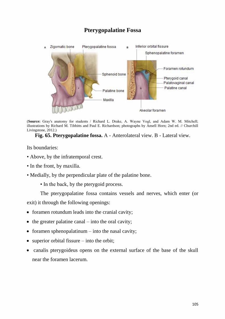
105
Pterygopalatine Fossa
(Source: Gray′s anatomy for students / Richard L. Drake, A. Wayne Vogl, and Adam W. M. Mitchell;
illustrations by Richard M. Tibbitts and Paul E. Richardson; photographs by Ansell Horn; 2nd ed. // Churchill
Livingstone, 2012.)
Fig. 65. Pterygopalatine fossa. A - Anterolateral view. B - Lateral view.
Its boundaries:
• Above, by the infratemporal crest.
• In the front, by maxilla.
• Medially, by the perpendicular plate of the palatine bone.
• In the back, by the pterygoid process.
The pterygopalatine fossa contains vessels and nerves, which enter (or
exit) it through the following openings:
foramen rotundum leads into the cranial cavity;
the greater palatine canal – into the oral cavity;
foramen sphenopalatinum – into the nasal cavity;
superior orbital fissure – into the orbit;
canalis pterygoideus opens on the external surface of the base of the skull
near the foramen lacerum.
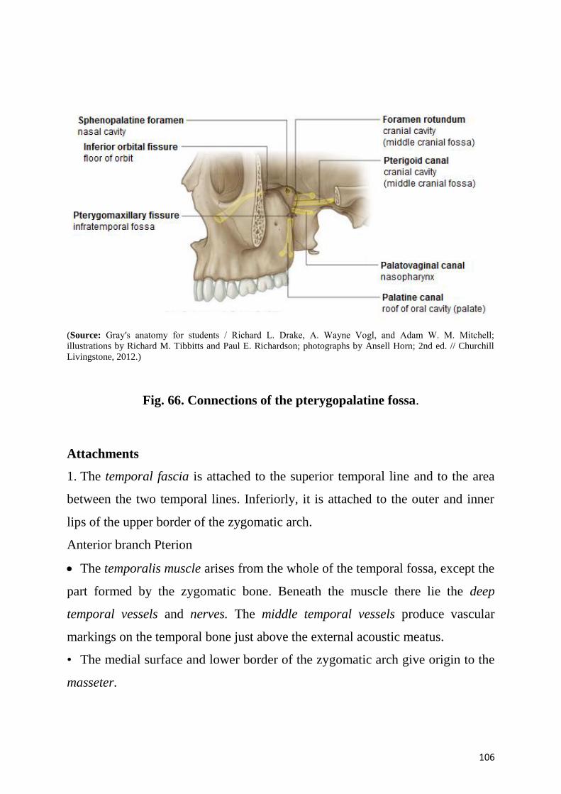
106
(Source: Gray′s anatomy for students / Richard L. Drake, A. Wayne Vogl, and Adam W. M. Mitchell;
illustrations by Richard M. Tibbitts and Paul E. Richardson; photographs by Ansell Horn; 2nd ed. // Churchill
Livingstone, 2012.)
Fig. 66. Connections of the pterygopalatine fossa.
Attachments
1. The temporal fascia is attached to the superior temporal line and to the area
between the two temporal lines. Inferiorly, it is attached to the outer and inner
lips of the upper border of the zygomatic arch.
Anterior branch Pterion
The temporalis muscle arises from the whole of the temporal fossa, except the
part formed by the zygomatic bone. Beneath the muscle there lie the deep
temporal vessels and nerves. The middle temporal vessels produce vascular
markings on the temporal bone just above the external acoustic meatus.
• The medial surface and lower border of the zygomatic arch give origin to the
masseter.
107
• The lateral ligament of the temporomandibular joint is attached to the
tubercle of the root of the zygoma.
• The sternocleidomastoid, splenius capitis and longissimus capitis are inserted
in that order from before backwards on the posterior part of the lateral surface of
the mastoid process. Posterior belly of digastric arises from mastoid notch. The
groove obliquely placed is due to occipital artery.
The gap between the zygomatic arch and the side of the skull transmits:
• Tendon of the temporalis muscle.
• Deep temporal vessels.
• Deep temporal nerves.
Structures Passing Through Foramina
1. The tympanomastoid fissure on the anterior aspect of the base of the mastoid
process transmits the auricular branch of vagus nerve.
2. The mastoid foramen transmits:
• An emissary vein connecting the sigmoid sinus with the posterior auricular
vein.
• A meningeal branch of the occipital artery
3. The zygomaticotemporal foramen transmits the nerve of the same name and a
minute artery
NORMA BASALIS
For convenience of study, the norma basalis is divided arbitrarily into
anterior, middle and posterior parts. The anterior part is formed by the hard
palate and the alveolar arches. The middle and posterior parts are separated by
an imaginary transverse line passing through the anterior margin of the foramen
magnum.
Anterior Part of Norma Basalis: Alveolar Arch
Alveolar arch bears sockets for the roots of the upper teeth.
108
Hard Palate.
Formation:
• Anterior two-thirds, by the palatine processes of the maxillae.
• Posterior one-third by the horizontal plates of the palatine bones.
• Sutures: The palate is crossed by a cruciform suture made up of
intermaxillary, interpalatine and palatomaxillary sutures.
• Shows pits for the palatine glands.
• The incisive fossa is a deep fossa situated anteriorly in the median plane. Two
incisive canals, right and left, pierce the walls of the incisive fossa, usually one
on each side, but occasionally in the median plane, the left being anterior and the
right, posterior.
• The greater palatine foramen, one on each side, is situated just behind the
lateral part of the palatomaxillary suture. A groove leads from the foramen
towards the incisive fossa.
• The lesser palatine foramina, two or three in number on each side, lie behind
the greater palatine foramen, and perforate the pyramidal process of the palatine
bone.
• The posterior border of the hard palate is free and presents the posterior nasal
spine in the median plane.
• The palatine crest is a curved ridge near the posterior border. It begins behind
the greater palatine foramen and runs medially.
Middle part of norma basalis
The middle part extends from the posterior border of the hard palate to the
arbitrary transverse line passing through the anterior margin of the foramen
magnum.
Median area shows:
• The posterior border of the vomer.
• A broad bar of bone formed by fusion of the posterior part of the body of
sphenoid and the basilar part of occipital bone.
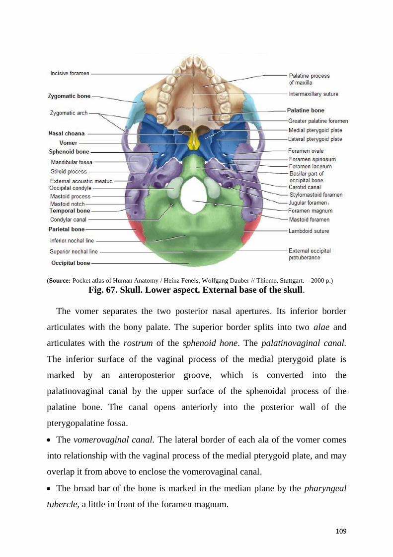
109
(Source: Pocket atlas of Human Anatomy / Heinz Feneis, Wolfgang Dauber // Thieme, Stuttgart. – 2000 p.)
Fig. 67. Skull. Lower aspect. External base of the skull.
The vomer separates the two posterior nasal apertures. Its inferior border
articulates with the bony palate. The superior border splits into two alae and
articulates with the rostrum of the sphenoid hone. The palatinovaginal canal.
The inferior surface of the vaginal process of the medial pterygoid plate is
marked by an anteroposterior groove, which is converted into the
palatinovaginal canal by the upper surface of the sphenoidal process of the
palatine bone. The canal opens anteriorly into the posterior wall of the
pterygopalatine fossa.
The vomerovaginal canal. The lateral border of each ala of the vomer comes
into relationship with the vaginal process of the medial pterygoid plate, and may
overlap it from above to enclose the vomerovaginal canal.
The broad bar of the bone is marked in the median plane by the pharyngeal
tubercle, a little in front of the foramen magnum.
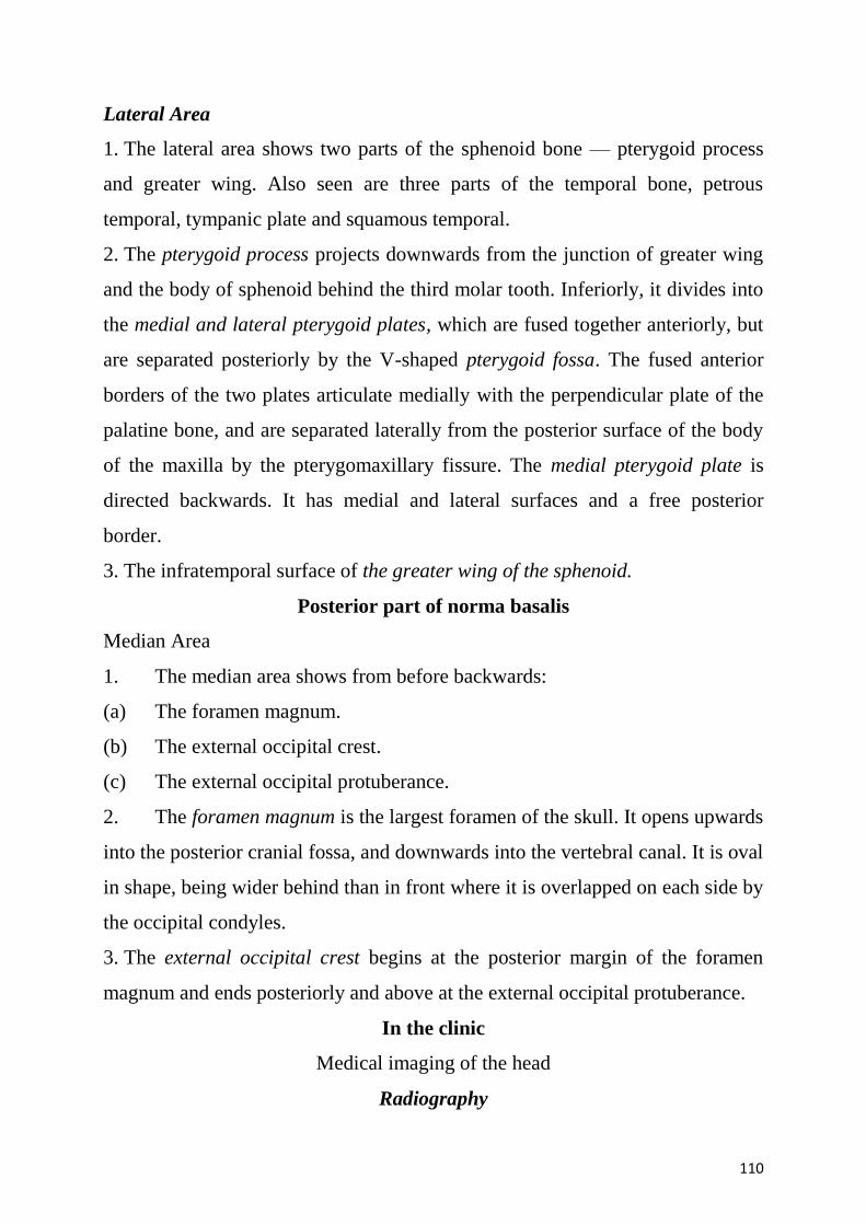
110
Lateral Area
1. The lateral area shows two parts of the sphenoid bone — pterygoid process
and greater wing. Also seen are three parts of the temporal bone, petrous
temporal, tympanic plate and squamous temporal.
2. The pterygoid process projects downwards from the junction of greater wing
and the body of sphenoid behind the third molar tooth. Inferiorly, it divides into
the medial and lateral pterygoid plates, which are fused together anteriorly, but
are separated posteriorly by the V-shaped pterygoid fossa. The fused anterior
borders of the two plates articulate medially with the perpendicular plate of the
palatine bone, and are separated laterally from the posterior surface of the body
of the maxilla by the pterygomaxillary fissure. The medial pterygoid plate is
directed backwards. It has medial and lateral surfaces and a free posterior
border.
3. The infratemporal surface of the greater wing of the sphenoid.
Posterior part of norma basalis
Median Area
1. The median area shows from before backwards:
(a) The foramen magnum.
(b) The external occipital crest.
(c) The external occipital protuberance.
2. The foramen magnum is the largest foramen of the skull. It opens upwards
into the posterior cranial fossa, and downwards into the vertebral canal. It is oval
in shape, being wider behind than in front where it is overlapped on each side by
the occipital condyles.
3. The external occipital crest begins at the posterior margin of the foramen
magnum and ends posteriorly and above at the external occipital protuberance.
In the clinic
Medical imaging of the head
Radiography
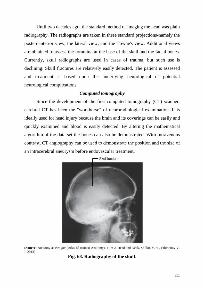
111
Until two decades ago, the standard method of imaging the head was plain
radiography. The radiographs are taken in three standard projections-namely the
posteroanterior view, the lateral view, and the Towne's view. Additional views
are obtained to assess the foramina at the base of the skull and the facial bones.
Currently, skull radiographs are used in cases of trauma, but such use is
declining. Skull fractures are relatively easily detected. The patient is assessed
and treatment is based upon the underlying neurological or potential
neurological complications.
Computed tomography
Since the development of the first computed tomography (CT) scanner,
cerebral CT has been the "workhorse" of neuroradiological examination. It is
ideally used for head injury because the brain and its coverings can be easily and
quickly examined and blood is easily detected. By altering the mathematical
algorithm of the data set the bones can also be demonstrated. With intravenous
contrast, CT angiography can be used to demonstrate the position and the size of
an intracerebral aneurysm before endovascular treatment.
(Source: Anatomy at Pirogov (Atlas of Human Anatomy). Tom 2. Head and Neck. Shilkin V. V., Filimonov V.
I. 2013)
Fig. 68. Radiography of the skull.
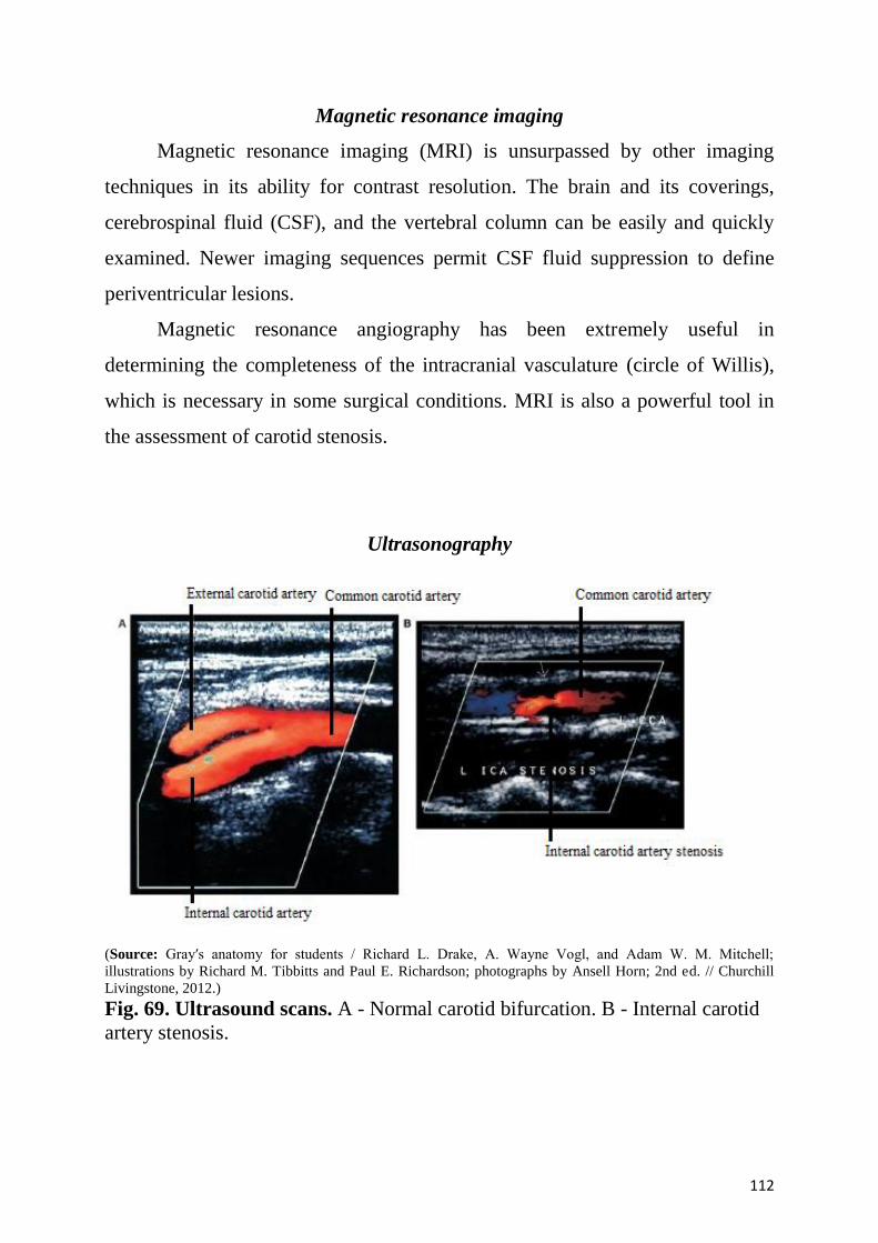
112
Magnetic resonance imaging
Magnetic resonance imaging (MRI) is unsurpassed by other imaging
techniques in its ability for contrast resolution. The brain and its coverings,
cerebrospinal fluid (CSF), and the vertebral column can be easily and quickly
examined. Newer imaging sequences permit CSF fluid suppression to define
periventricular lesions.
Magnetic resonance angiography has been extremely useful in
determining the completeness of the intracranial vasculature (circle of Willis),
which is necessary in some surgical conditions. MRI is also a powerful tool in
the assessment of carotid stenosis.
Ultrasonography
(Source: Gray′s anatomy for students / Richard L. Drake, A. Wayne Vogl, and Adam W. M. Mitchell;
illustrations by Richard M. Tibbitts and Paul E. Richardson; photographs by Ansell Horn; 2nd ed. // Churchill
Livingstone, 2012.)
Fig. 69. Ultrasound scans. A - Normal carotid bifurcation. B - Internal carotid
artery stenosis.
113
Initial work using ultrasound to assess the brain appeared fruitless, but
with increasing probe technology, it is now possible to carry out intracranial
Doppler studies, which enable a surgeon to detect whether a patient is
experiencing cerebral embolization from a carotid plaque. Extracranial
ultrasound is extremely important in tumor staging and in assessing neck masses
and the carotid bifurcation. Ultrasound is useful in children because they have
an acoustic window through the fontanelles.
Clinical application
Fractures of the skull vault and extradural hematoma
The skull vault is a remarkably strong structure-and quite rightly, because
it protects our most vital organ, the brain. The shape of the skull vault is of
critical importance and its biomechanics prevent fracture. From a clinical
standpoint skull fractures alert clinicians to the nature and force of an injury and
potential complications. The fracture itself is usually of little consequence
(unlike, say, a fracture of the tibia). Of key importance is the need to minimize
the extent of primary brain injury and to treat potential secondary complications,
rather than focusing on the skull fracture. Skull fractures that have particular
significance include depressed skull fractures, compound fractures, and pterion
fractures.
Depressed skull fractures
In a depressed skull fracture, a bony fragment is depressed below the
normal skull convexity. This may lead to secondary arterial and venous damage
with hematoma formation. Furthermore, a primary brain injury can also result
from this type of fracture.
Compound fractures
In a compound fracture, there is a fracture of the bone together with a
breach of the skin, which may allow an infection to enter. Typically, these
fractures are associated with scalp lacerations and can usually be treated with
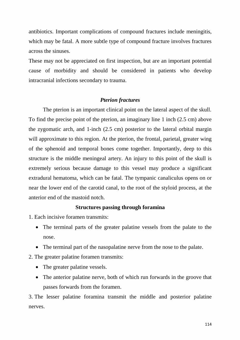
114
antibiotics. Important complications of compound fractures include meningitis,
which may be fatal. A more subtle type of compound fracture involves fractures
across the sinuses.
These may not be appreciated on first inspection, but are an important potential
cause of morbidity and should be considered in patients who develop
intracranial infections secondary to trauma.
Pterion fractures
The pterion is an important clinical point on the lateral aspect of the skull.
To find the precise point of the pterion, an imaginary line 1 inch (2.5 cm) above
the zygomatic arch, and 1-inch (2.5 cm) posterior to the lateral orbital margin
will approximate to this region. At the pterion, the frontal, parietal, greater wing
of the sphenoid and temporal bones come together. Importantly, deep to this
structure is the middle meningeal artery. An injury to this point of the skull is
extremely serious because damage to this vessel may produce a significant
extradural hematoma, which can be fatal. The tympanic canaliculus opens on or
near the lower end of the carotid canal, to the root of the styloid process, at the
anterior end of the mastoid notch.
Structures passing through foramina
1. Each incisive foramen transmits:
The terminal parts of the greater palatine vessels from the palate to the
nose.
The terminal part of the nasopalatine nerve from the nose to the palate.
2. The greater palatine foramen transmits:
The greater palatine vessels.
The anterior palatine nerve, both of which run forwards in the groove that
passes forwards from the foramen.
3. The lesser palatine foramina transmit the middle and posterior palatine
nerves.
115
4. The palatinovaginal canal transmits:
A pharyngeal branch from the pterygopalatine ganglion.
A small pharyngeal branch of the maxillary artery.
5. The vomerovaginal canal (if patent) transmits branches of the pharyngeal
nerve from pterygopalatine ganglion and vessels.
6. The foramen ovale transmits:
• The mandibular nerve.
• The accessory meningeal artery.
• An emissary vein connecting the cavernous sinus with the pterygoid plexus of
veins.
7. The foramen spinosum transmits the middle meningeal artery, the meningeal
branch of the mandibular nerve or nervus spinosus, and the posterior trunk of the
middle meningeal vein.
8. The emissary sphenoidal foramen transmits an emissary vein connecting the
cavernous sinus with the pterygoid plexus of veins.
9. The carotid canal transmits the internal carotid artery, and the venous and
sympathetic plexuses around the artery.
10. The structures passing through the foramen lacerum are: during life the
lower part of the foramen is filled with cartilage, and no significant structure
passes through the whole length of the canal, except for the meningeal branch of
the ascending pharyngeal artery and an emissary vein from the cavernous sinus.
However, the upper part of the foramen is traversed by the internal carotid artery
with venous and sympathetic plexuses around it. In the anterior part of the
foramen, the greater petrosal nerve unites with the deep petrosal nerve to form
the nerve of the pterygoid canal, which leaves the foramen by entering the
pterygoid canal in the anterior wall of the foramen lacerum.
11. The medial end of the petrotympanic fissure transmits the chorda tympani
nerve, anterior ligament of malleus and the anterior tympanic artery.
13. The foramen magnum transmits the following.
116
Through the narrow anterior part – 3 structures:
Apical ligament of dens.
Vertical band of cruciate ligament.
Membrana tectoria.
Through wider posterior part – 2 structures:
Lowest part of medulla oblongata
Three meninges.
Through the subarachnoid space – 9 structures pass:
Spinal accessory nerves – 2.
Vertebral arteries – 2.
Sympathetic plexus around the vertebral arteries – 2.
Posterior spinal arteries – 2.
Anterior spinal artery – 1.
14. The hypoglossal or anterior condylar canal transmits the hypoglossal nerve,
the meningeal branch of the hypoglossal nerve, the meningeal branch of the
ascending pharyngeal artery, and an emissary vein connecting the sigmoid sinus
with the internal jugular vein.
15. The posterior condylar canal transmits an emissary vein connecting the
sigmoid sinus with suboccipital venous plexus.
16. The jugular foramen transmits the following structures.
Through the anterior part:
Inferior petrosal sinus.
Meningeal branch of the ascending pharyngeal artery.
Through the middle part: IX, X and XI cranial nerves.
Through the posterior part:
Internal jugular vein.
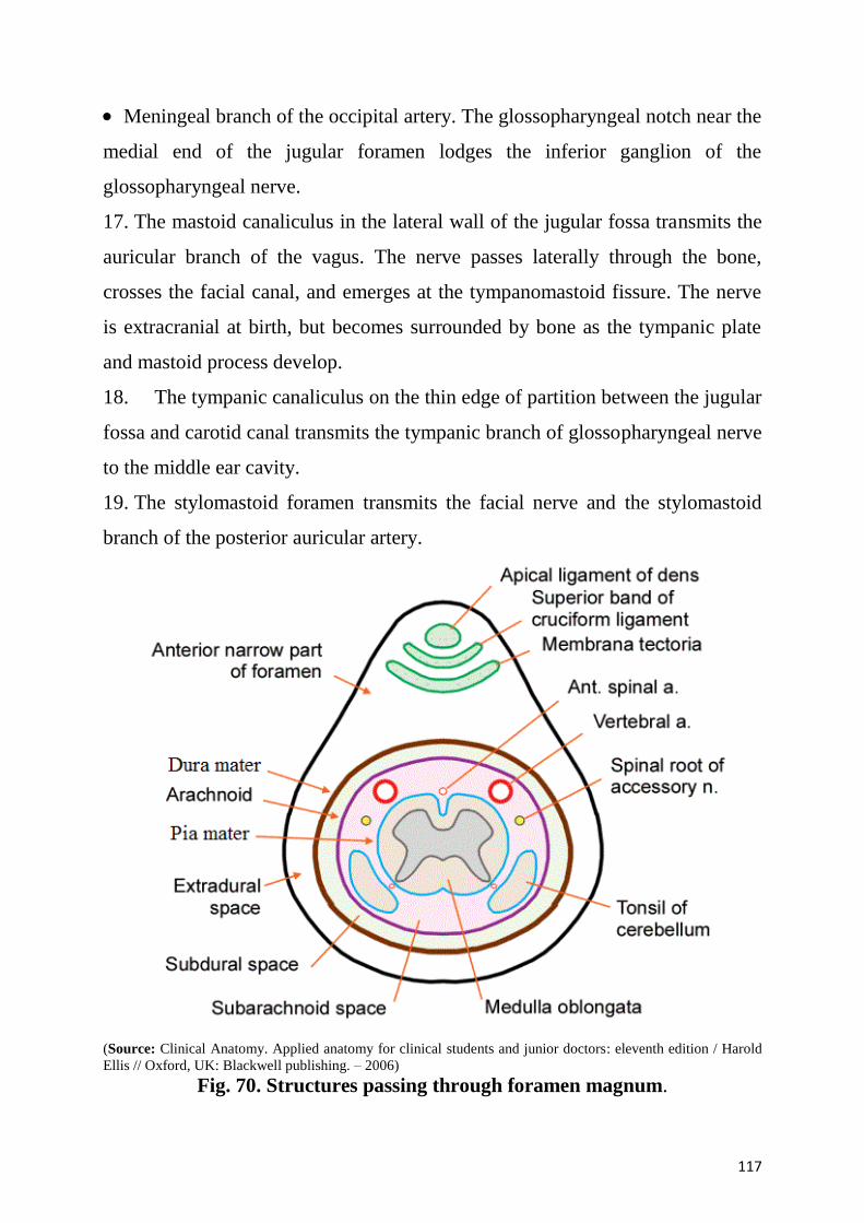
117
Meningeal branch of the occipital artery. The glossopharyngeal notch near the
medial end of the jugular foramen lodges the inferior ganglion of the
glossopharyngeal nerve.
17. The mastoid canaliculus in the lateral wall of the jugular fossa transmits the
auricular branch of the vagus. The nerve passes laterally through the bone,
crosses the facial canal, and emerges at the tympanomastoid fissure. The nerve
is extracranial at birth, but becomes surrounded by bone as the tympanic plate
and mastoid process develop.
18. The tympanic canaliculus on the thin edge of partition between the jugular
fossa and carotid canal transmits the tympanic branch of glossopharyngeal nerve
to the middle ear cavity.
19. The stylomastoid foramen transmits the facial nerve and the stylomastoid
branch of the posterior auricular artery.
(Source: Clinical Anatomy. Applied anatomy for clinical students and junior doctors: eleventh edition / Harold
Ellis // Oxford, UK: Blackwell publishing. – 2006)
Fig. 70. Structures passing through foramen magnum.
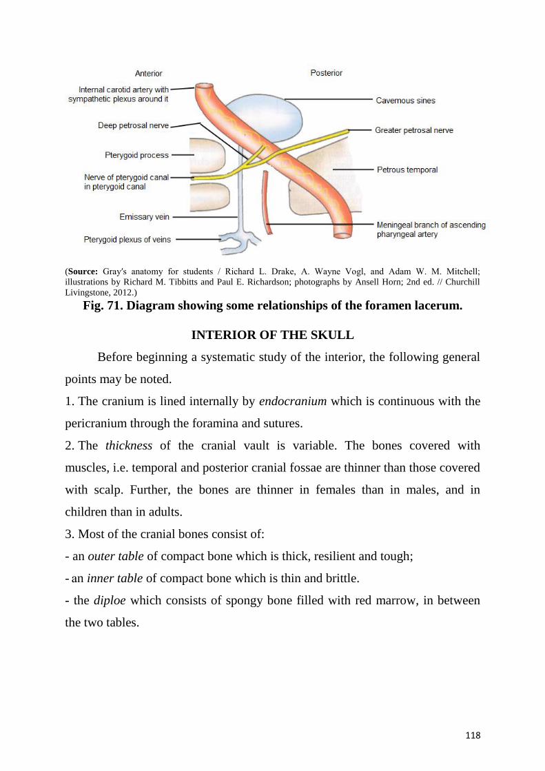
118
(Source: Gray′s anatomy for students / Richard L. Drake, A. Wayne Vogl, and Adam W. M. Mitchell;
illustrations by Richard M. Tibbitts and Paul E. Richardson; photographs by Ansell Horn; 2nd ed. // Churchill
Livingstone, 2012.)
Fig. 71. Diagram showing some relationships of the foramen lacerum.
INTERIOR OF THE SKULL
Before beginning a systematic study of the interior, the following general
points may be noted.
1. The cranium is lined internally by endocranium which is continuous with the
pericranium through the foramina and sutures.
2. The thickness of the cranial vault is variable. The bones covered with
muscles, i.e. temporal and posterior cranial fossae are thinner than those covered
with scalp. Further, the bones are thinner in females than in males, and in
children than in adults.
3. Most of the cranial bones consist of:
- an outer table of compact bone which is thick, resilient and tough;
- аn inner table of compact bone which is thin and brittle.
- the diploe which consists of spongy bone filled with red marrow, in between
the two tables.
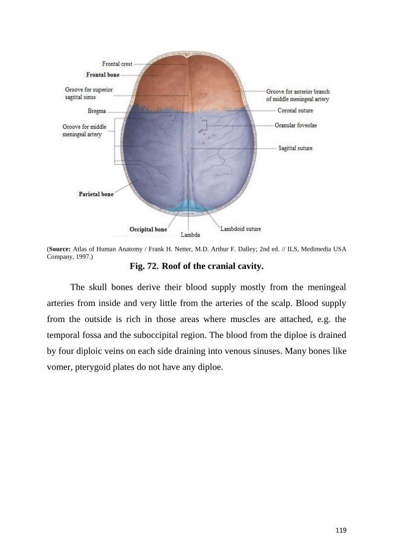
119
(Source: Atlas of Human Anatomy / Frank H. Netter, M.D. Arthur F. Dalley; 2nd ed. // ILS, Medimedia USA
Company, 1997.)
Fig. 72. Roof of the cranial cavity.
The skull bones derive their blood supply mostly from the meningeal
arteries from inside and very little from the arteries of the scalp. Blood supply
from the outside is rich in those areas where muscles are attached, e.g. the
temporal fossa and the suboccipital region. The blood from the diploe is drained
by four diploic veins on each side draining into venous sinuses. Many bones like
vomer, pterygoid plates do not have any diploe.
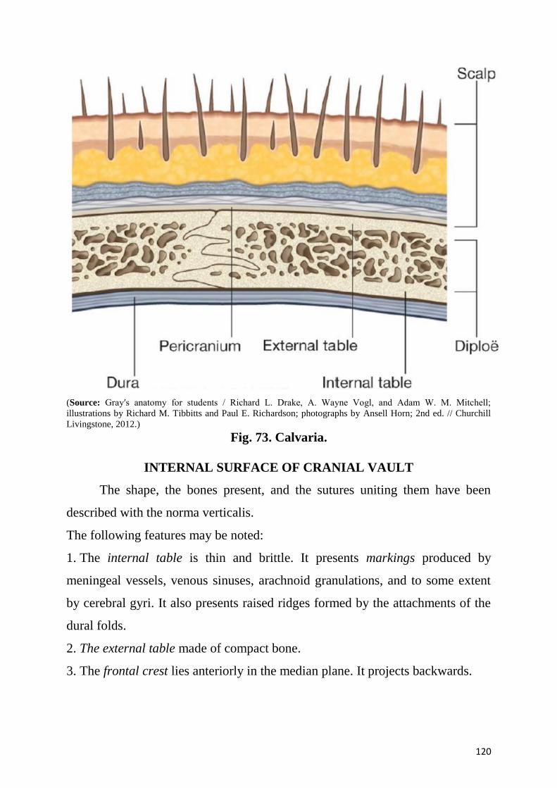
120
(Source: Gray′s anatomy for students / Richard L. Drake, A. Wayne Vogl, and Adam W. M. Mitchell;
illustrations by Richard M. Tibbitts and Paul E. Richardson; photographs by Ansell Horn; 2nd ed. // Churchill
Livingstone, 2012.)
Fig. 73. Calvaria.
INTERNAL SURFACE OF CRANIAL VAULT
The shape, the bones present, and the sutures uniting them have been
described with the norma verticalis.
The following features may be noted:
1. The internal table is thin and brittle. It presents markings produced by
meningeal vessels, venous sinuses, arachnoid granulations, and to some extent
by cerebral gyri. It also presents raised ridges formed by the attachments of the
dural folds.
2. The external table made of compact bone.
3. The frontal crest lies anteriorly in the median plane. It projects backwards.
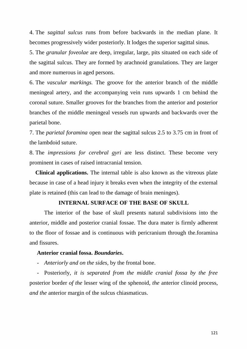
121
4. The sagittal sulcus runs from before backwards in the median plane. It
becomes progressively wider posteriorly. It lodges the superior sagittal sinus.
5. The granular foveolae are deep, irregular, large, pits situated on each side of
the sagittal sulcus. They are formed by arachnoid granulations. They are larger
and more numerous in aged persons.
6. The vascular markings. The groove for the anterior branch of the middle
meningeal artery, and the accompanying vein runs upwards 1 cm behind the
coronal suture. Smaller grooves for the branches from the anterior and posterior
branches of the middle meningeal vessels run upwards and backwards over the
parietal bone.
7. The parietal foramina open near the sagittal sulcus 2.5 to 3.75 cm in front of
the lambdoid suture.
8. The impressions for cerebral gyri are less distinct. These become very
prominent in cases of raised intracranial tension.
Clinical applications. The internal table is also known as the vitreous plate
because in case of a head injury it breaks even when the integrity of the external
plate is retained (this can lead to the damage of brain meninges).
INTERNAL SURFACE OF THE BASE OF SKULL
The interior of the base of skull presents natural subdivisions into the
anterior, middle and posterior cranial fossae. The dura mater is firmly adherent
to the floor of fossae and is continuous with pericranium through the.foramina
and fissures.
Anterior cranial fossa. Boundaries.
- Anteriorly and on the sides, by the frontal bone.
- Posteriorly, it is separated from the middle cranial fossa by the free
posterior border of the lesser wing of the sphenoid, the anterior clinoid process,
and the anterior margin of the sulcus chiasmaticus.
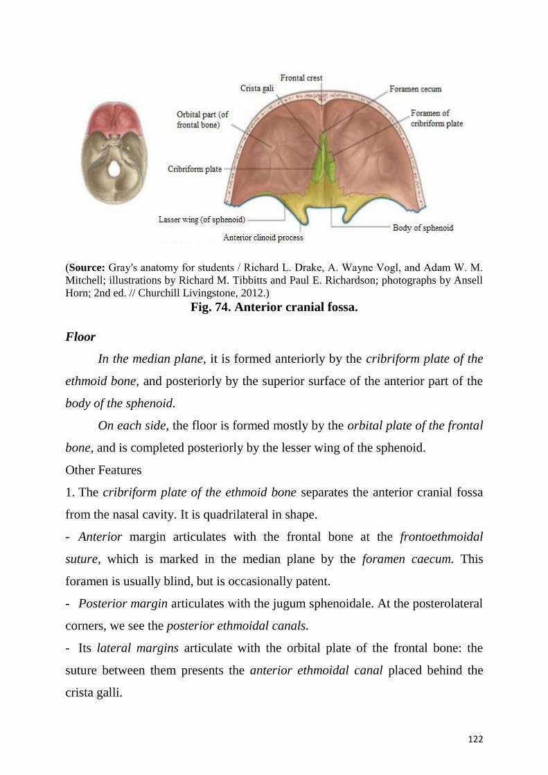
122
(Source: Gray′s anatomy for students / Richard L. Drake, A. Wayne Vogl, and Adam W. M.
Mitchell; illustrations by Richard M. Tibbitts and Paul E. Richardson; photographs by Ansell
Horn; 2nd ed. // Churchill Livingstone, 2012.)
Fig. 74. Anterior cranial fossa.
Floor
In the median plane, it is formed anteriorly by the cribriform plate of the
ethmoid bone, and posteriorly by the superior surface of the anterior part of the
body of the sphenoid.
On each side, the floor is formed mostly by the orbital plate of the frontal
bone, and is completed posteriorly by the lesser wing of the sphenoid.
Other Features
1. The cribriform plate of the ethmoid bone separates the anterior cranial fossa
from the nasal cavity. It is quadrilateral in shape.
- Anterior margin articulates with the frontal bone at the frontoethmoidal
suture, which is marked in the median plane by the foramen caecum. This
foramen is usually blind, but is occasionally patent.
- Posterior margin articulates with the jugum sphenoidale. At the posterolateral
corners, we see the posterior ethmoidal canals.
- Its lateral margins articulate with the orbital plate of the frontal bone: the
suture between them presents the anterior ethmoidal canal placed behind the
crista galli.
123
Anteriorly, the cribriform plate has a midline projection called the crista galli.
On each side of the crista galli there are foramina through which the anterior
ethmoidal nerve and vessels pass to the nasal cavity. The plate is also perforated
by numerous foramina for the passage of olfactory nerve rootlets.
2. The orbital plate of the frontal bone separates the anterior cranial fossa from
the orbit. It supports the orbital surface of the frontal lobe of the brain, and
presents reciprocal impressions. The frontal air sinus may extend into its
anteromedial part. The medial margin of the plate covers the labyrinth of the
ethmoid; and the posterior margin articulates with the lesser wing of the
sphenoid.
3. The lesser wing of the sphenoid is broad medially where it is continuous with
the jugum sphenoidale and tapers laterally. The free posterior border fits into
the stem of the lateral sulcus of the brain. It ends medially as a prominent
projection, the anterior clinoid process. Inferiorly, the posterior border forms
the upper boundary of the superior orbital fissure. Medially, the lesser wing is
connected to the body of the sphenoid by anterior and posterior roots, which
enclose the optic canal.
Middle cranial fossa
It is deeper than the anterior cranial fossa, and is shaped like a butterfly,
being narrow and shallow in the middle; and wide and deep on each side.
Boundaries. Anterior
- Posterior border of the lesser wing of the sphenoid.
- Anterior clinoid process.
- Anterior margin of the sulcus chiasmaticus.
Posterior
- Superior border of the petrous temporal bone.
- The dorsum sellae of the sphenoid. Lateral
- Greater wing of the sphenoid.
- Anteroinferior angle of the parietal bone.
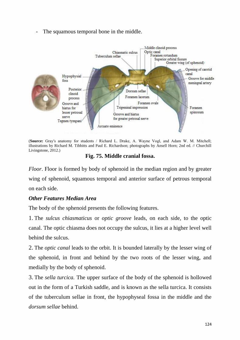
124
- The squamous temporal bone in the middle.
(Source: Gray′s anatomy for students / Richard L. Drake, A. Wayne Vogl, and Adam W. M. Mitchell;
illustrations by Richard M. Tibbitts and Paul E. Richardson; photographs by Ansell Horn; 2nd ed. // Churchill
Livingstone, 2012.)
Fig. 75. Middle cranial fossa.
Floor. Floor is formed by body of sphenoid in the median region and by greater
wing of sphenoid, squamous temporal and anterior surface of petrous temporal
on each side.
Other Features Median Area
The body of the sphenoid presents the following features.
1. The sulcus chiasmaticus or optic groove leads, on each side, to the optic
canal. The optic chiasma does not occupy the sulcus, it lies at a higher level well
behind the sulcus.
2. The optic canal leads to the orbit. It is bounded laterally by the lesser wing of
the sphenoid, in front and behind by the two roots of the lesser wing, and
medially by the body of sphenoid.
3. The sella turcica. The upper surface of the body of the sphenoid is hollowed
out in the form of a Turkish saddle, and is known as the sella turcica. It consists
of the tuberculum sellae in front, the hypophyseal fossa in the middle and the
dorsum sellae behind.
125
4. The tuberculum sellae separates the optic groove from the hypophyseal fossa.
Its lateral ends form the middle clinoid process, which may join the anterior
clinoid process.
5. The hypophyseal fossa lodges the hypophysis cerebri. Beneath the floor of
fossa lie the sphenoidal air sinuses.
6. The dorsum sellae is a transverse plate of bone projecting upwards; it forms
the back of the saddle. The superolateral angles of the dorsum sellae are
expanded to form the posterior clinoid processes.
Lateral Area
1. The lateral area is deep and lodges the temporal lobe of the brain.
2. It is related anteriorly to the orbit, laterally to the temporal fossa, and
inferiorly to the infratemporal fossa.
3. The superior orbital fissure opens anteriorly into the orbit. It is bounded
above by the lesser wing, below by the greater wing, and medially by the body
of the sphenoid. The medial end is wider than the lateral. The long axis of the
fissure is directed laterally, upwards and forwards. The lower border is marked
by a small projection, which provides attachment to the common tendinous ring
of Zinn. The ring divides the fissure into three parts.
4. The greater wing of the sphenoid presents the following features:
a) the foramen rotundum lies posteroinferior to the medial end of the superior
orbital fissure. It leads anteriorly to the pterygopalatine fossa containing
pterygopalatine ganglia.
b) the foramen ovale lies posterolateral to the foramen rotundum and lateral to
the lingula. It leads inferiorly to the infratemporal fossa.
c) the foramen spinosum lies posterolateral to the foramen ovale. It also leads,
inferiorly, to the infratemporal fossa.
d) the emissary sphenoidal foramen or foramen of Vesalius. These foramina
have been seen on the base of the skull.
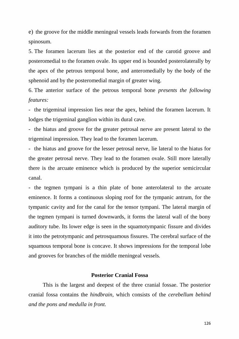
126
e) the groove for the middle meningeal vessels leads forwards from the foramen
spinosum.
5. The foramen lacerum lies at the posterior end of the carotid groove and
posteromedial to the foramen ovale. Its upper end is bounded posterolaterally by
the apex of the petrous temporal bone, and anteromedially by the body of the
sphenoid and by the posteromedial margin of greater wing.
6. The anterior surface of the petrous temporal bone presents the following
features:
- the trigeminal impression lies near the apex, behind the foramen lacerum. It
lodges the trigeminal ganglion within its dural cave.
- the hiatus and groove for the greater petrosal nerve are present lateral to the
trigeminal impression. They lead to the foramen lacerum.
- the hiatus and groove for the lesser petrosal nerve, lie lateral to the hiatus for
the greater petrosal nerve. They lead to the foramen ovale. Still more laterally
there is the arcuate eminence which is produced by the superior semicircular
canal.
- the tegmen tympani is a thin plate of bone anterolateral to the arcuate
eminence. It forms a continuous sloping roof for the tympanic antrum, for the
tympanic cavity and for the canal for the tensor tympani. The lateral margin of
the tegmen tympani is turned downwards, it forms the lateral wall of the bony
auditory tube. Its lower edge is seen in the squamotympanic fissure and divides
it into the petrotympanic and petrosquamous fissures. The cerebral surface of the
squamous temporal bone is concave. It shows impressions for the temporal lobe
and grooves for branches of the middle meningeal vessels.
Posterior Cranial Fossa
This is the largest and deepest of the three cranial fossae. The posterior
cranial fossa contains the hindbrain, which consists of the cerebellum behind
and the pons and medulla in front.
127
Boundaries
Anterior
- The superior border of the petrous temporal bone.
- The dorsum sellae of the sphenoid bone.
Posterior
- Squamous part of the occipital bone.
On each side
- Mastoid part of the temporal bone.
- The mastoid angle of the parietal bone.
Floor
Median area
- Sloping area behind the dorsum sellae or clivus in front
- The foramen magnum in the middle
- The squamous occipital behind.
Lateral area
- Condylar or lateral part of occipital bone.
- Posterior surface of the petrous temporal bone.
- Mastoid temporal bone.
- Mastoid angle of the parietal bone.
128
(Source: Gray′s anatomy for students / Richard L. Drake, A. Wayne Vogl, and Adam W. M. Mitchell;
illustrations by Richard M. Tibbitts and Paul E. Richardson; photographs by Ansell Horn; 2nd ed. // Churchill
Livingstone, 2012.)
Fig. 76. Posterior cranial fossa.
Other Features median area
1. The clivus is the sloping surface in front of the foramen magnum. It is formed
by fusion of the posterior part of the body of the sphenoid including the dorsum
sellae with the basilar part of the occipital bone or basiocciput. It is related to the
basilar plexus of veins, and supports the pons and medulla. On each side, the
clivus is separated from the petrous temporal bone by the petro-occipital fissure,
which is grooved by the inferior petrosal sinus, and is continuous behind with
the jugular foramen.
2. The foramen magnum lies in the floor of the fossa. It is bounded anteriorly by
the basiocciput, posteriorly by the squamous part of the occipital bone, and on
each side by the condylar part of the occipital bone. The anterior part of the
foramen is narrow because it is overlapped by the medial surfaces of the
oçcipital condyles.
3. The squamous part of the occipital bone shows the following features.
129
The internal occipital crest runs in the median plane from the internal
occipital protuberance to the foramen magnum where it forms a shallow
depression, the vermian fossa.
The internal occipital protuberance lies opposite the external occipital
protuberance. It is related to the confluence of sinuses, and is grooved on each
side by the beginning of transverse sinuses.
The transverse groove is quite wide and runs laterally from the internal
occipital protuberance to the mastoid angle of the parietal bone where it
becomes continuous with the sigmoid sulcus. The transverse sulcus lodges the
transverse sinus. The right transverse sulcus is usually wider than the left and is
continuous medially with the superior sagittal sulcus.
On each side of the internal occipital crest there are deep fossae which
lodge the cerebellar hemispheres.
Lateral area
1. The condylar part of the occipital bone is marked by the following:
- The jugular tubercle lies over the occipital condyle.
- The hypoglossal canal pierces the bone posteroanterior to the jugular
tubercle and runs obliquely forwards and laterally along the line of fusion
between the basilar and the condylar parts of the occipital bone.
- The condylar canal opens in the lower part of the sigmoid sulcus which
indents the jugular process of occipital bone.
2. The posterior surface of the petrous part of the temporal bone forms the
anterolateral wall of the posterior cranial fossa. The following features may be
noted:
- The internal acoustic meatus opens above the anterior part of the jugular
foramen. It is about 1 cm long and runs transversely in a lateral direction. It is
closed laterally by a perforated plate of bone known as lamina cribrosa, which
separates it from the internal ear.
130
- The orifice of the aqueduct of the vestibule is a narrow slit lying behind
the internal acoustic meatus.
- The subarcuate fossa lies below the arcuate eminence, lateral to the
internal acoustic meatus.
3. The jugular foramen lies at the posterior end of the petro-occipital fissure
between the deep jugular notch of the petrous temporal bone above and the
shallow jugular notch of the occipital bone below. The upper margin is sharp
and irregular, and presents the glossopharyngeal notch. The lower margin is
smooth and regular.
4. The mastoid part of the temporal bone forms the lateral wall of the posterior
cranial fossa just behind the petrous part of the bone. Anteriorly, it is marked by
the sigmoid groove, which begins as a downward continuation of the transverse
groove at the mastoid angle of the parietal bone, and ends at the jugular
foramen. The sigmoid sulcus lodges the sigmoid sinus, which become the
internal jugular vein at the jugular foramen. The sulcus is related anteriorly to
the tympanic antrum. The mastoid foramen opens into the upper part of the
sulcus.
Attachments and relations of the interior of the skull
Attachment on Vault
1. The frontal crest gives attachment to the falx cerebri.
2. The lips of the sagittal sulcus give attachment to the falx cerebri.
Anterior cranial fossa
1. The crista galli gives attachment to the falx cerebri.
2. The orbital surface of the frontal bone supports the frontal lobe of the brain.
3. The anterior clinoid processes give attachment to the free margin of the
tentorium cerebelli.
Middle cranial fossa
1. The middle cranial fossa lodges the temporal lobe of the cerebral hemisphere.
2. The tuberculum sellae provides attachment to the diaphragma sellae.
131
3. The hypophyseal fossa lodges the hypophysis cerebri.
4. Upper margin of the dorsum sellae provides attachment to the diaphragma
sellae, and the posterior clinoid process to anterior end of the attached margin of
tentorium cerebelli.
5. One cavernous sinus lies on each side of the body of the sphenoid. The
internal carotid artery passes through the cavernous sinus.
6. The superior border of the petrous temporal bone is grooved by the superior
petrosal sinus and provides attachment to the attached margin of the tentorium
cerebelli. It is grooved in its medial part by the trigeminal nerve.
Posterior cranial fossa
1. The posterior cranial fossa contains the hindbrain, which consists of the
cerebellum behind, and the pons and medulla in front.
2. The lower part of the clivus provides attachment to the apical ligament of the
dens near the foramen magnum, upper vertical band of cruciate ligament and to
the membrana tectoria just above the apical ligament.
3. The internal occipital crest gives attachment to the falx cerebelli.
4. The jugular tubercle is grooved by the ninth, tenth and eleventh cranial nerves
as they pass to the jugular foramen.
5. The subarcuate fossa on the posterior surface of petrous temporal bone
lodges the flocculus of the cerebellum.
Structures passing through foramina. The following foramina seen in the
cranial fossae have been dealt with under the normal basalis: foramen ovale,
foramen spinosum, emissary sphenoidal foramen, foramen lacerum, foramen
magnum, jugular foramen, hypoglossal canal, and posterior condylar canal.
Additional foramina seen in the cranial fossae are as follows.
1. The foramen caecum in the anterior cranial fossa is usually blind, but
occasionally it transmits a vein from the upper part of nose to the superior
sagittal sinus.
2. The posterior ethmoidal canals transmit the vessels of the same name.
132
3. The anterior ethmoidal canals transmit the corresponding nerves and vessels.
4. The optic canal transmits the optic nerve and the ophthalmic artery.
5. The three parts of the superior orbital fissure transmit the following
structures:
Lateral part:
- lacrimal nerve;
- frontal nerve;
- trochlear nerve;
- superior ophthalmic vein;
- meningeal branch of the lacrimal artery;
- anastomotic branch of the middle meningeal artery, which anastomoses
with the recurrent branch of the lacrimal arteiy.
Middle part:
- upper and lower divisions of the oculomotor nerve;
- nasociliary nerve in between the two divisions of the oculomotor;
- the abducent nerve, inferolateral to the foregoing nerves.
Medial part:
- inferior ophthalmic vein;
- sympathetic nerves from the plexus around the internal carotid artery.
6. The foramen rotundum transmits the maxillary nerve.
7. The internal acoustic meatus transmits the seventh and eighth cranial nerves
and the labyrinthine vessels.
Principles governing fractures of the skull
1. Fractures of the skull are prevented by: its elasticity, rounded shape,
construction from a number of secondary elastic arches, each made up of a
single bone, the muscles covering the thin areas.
2. Since the skull is an elastic sphere filled with the semifluid brain, a violent
blow on the skull produces a splitting effect commencing at the site of the blow
and tending to pass along the lines of least resistance.
133
3. The base of the skull is more fragile than the vault, and is more commonly
involved in such fractures, particularly along the foramina.
4. The inner table is more brittle than the outer table. Therefore, fractures are
more extensive on the inner table. Occasionally only the inner table is fractured
and the outer table remains intact.
5. The common sites of fracture in the skull are: the parietal area of the vault and
the middle cranial fossa of the base. This fossa is weakened by numerous
foramina and canals. The facial bones commonly fractured are: the nasal bone
and the mandible.
FOETAL SKULL
Dimensions
1. Skull is large in proportion to the other parts of skeleton.
2. Foetal skeleton is small as compared to calvaria. In foetal skull, the facial
skeleton is l/8th of calvaria; in adults, it is half of calvaria. The foetal skeleton is
small due to rudimentary mandible and maxillae, non-eruption of teeth, and
small size of maxillary sinus and nasal cavity. The large size of calvaria is due to
precocious growth of brain.
3. Base of the skull is short and narrow, though internal ear is almost of adult
size the petrous temporal has not reached the adult length.
Structure of bones
The bones of cranial vault are smooth; there is no diploe. The tables and
diploe appear by fourth year of age.
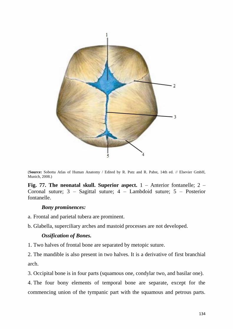
134
(Source: Sobotta Atlas of Human Anatomy / Edited by R. Putz and R. Pabst, 14th ed. // Elsevier GmbH,
Munich, 2008.)
Fig. 77. The neonatal skull. Superior aspect. 1 – Anterior fontanelle; 2 –
Coronal suture; 3 – Sagittal suture; 4 – Lambdoid suture; 5 – Posterior
fontanelle.
Bony prominences:
a. Frontal and parietal tubera are prominent.
b. Glabella, superciliary arches and mastoid processes are not developed.
Ossification of Bones.
1. Two halves of frontal bone are separated by metopic suture.
2. The mandible is also present in two halves. It is a derivative of first branchial
arch.
3. Occipital bone is in four parts (squamous one, condylar two, and basilar one).
4. The four bony elements of temporal bone are separate, except for the
commencing union of the tympanic part with the squamous and petrous parts.
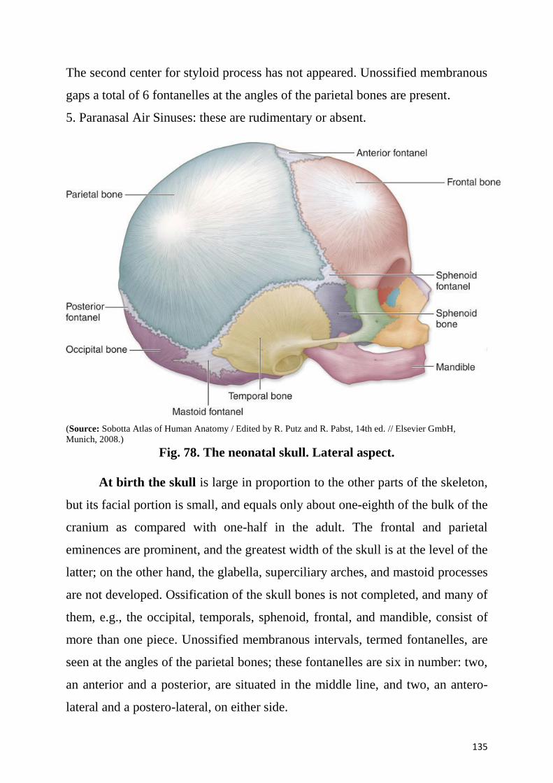
135
The second center for styloid process has not appeared. Unossified membranous
gaps a total of 6 fontanelles at the angles of the parietal bones are present.
5. Paranasal Air Sinuses: these are rudimentary or absent.
(Source: Sobotta Atlas of Human Anatomy / Edited by R. Putz and R. Pabst, 14th ed. // Elsevier GmbH,
Munich, 2008.)
Fig. 78. The neonatal skull. Lateral aspect.
At birth the skull is large in proportion to the other parts of the skeleton,
but its facial portion is small, and equals only about one-eighth of the bulk of the
cranium as compared with one-half in the adult. The frontal and parietal
eminences are prominent, and the greatest width of the skull is at the level of the
latter; on the other hand, the glabella, superciliary arches, and mastoid processes
are not developed. Ossification of the skull bones is not completed, and many of
them, e.g., the occipital, temporals, sphenoid, frontal, and mandible, consist of
more than one piece. Unossified membranous intervals, termed fontanelles, are
seen at the angles of the parietal bones; these fontanelles are six in number: two,
an anterior and a posterior, are situated in the middle line, and two, an antero-
lateral and a postero-lateral, on either side.
136
The anterior or bregmatic fontanelle is the largest, and is placed at the
junction of the sagittal, coronal, and frontal sutures; it is lozenge-shaped, and
measures about
4 cm. in its antero-posterior and 2.5 cm. in its transverse diameter. The
posterior fontanelle is triangular in form and is situated at the junction of the
sagittal and lambdoidal sutures. The lateral fontanelles are small, irregular in
shape, and correspond respectively with the sphenoidal and mastoid angles of
the parietal bones.
An additional fontanelle is sometimes seen in the sagittal suture at the
region of the obelion. The fontanelles are usually closed by the growth and
extension of the bones, which surround them, but sometimes they are the sites of
separate ossific centers, which develop into sutural bones. The posterior and
lateral fontanelles are obliterated within a month or two after birth, but the
anterior is not completely closed until about the middle of the second year.
The smallness of the face at birth is mainly accounted for by the
rudimentary condition of the maxillae and mandible, the non-eruption of the
teeth, and the small size of the maxillary air sinuses and nasal cavities. At birth
the nasal cavities lie almost entirely between the orbits, and the lower border of
the anterior nasal aperture is only a little below the level of the orbital floor.
With the eruption of the deciduous teeth, there is an enlargement of the face and
jaws, and these changes are still more marked after the second dentition.
The skull grows rapidly from birth to the seventh year, by which time the
foramen magnum and petrous parts of the temporals have reached their full size
and the orbital cavities are only a little smaller than those of the adult. Growth is
slow from the seventh year until the approach of puberty, when a second period
of activity occurs: this results in an increase in all directions, but it is especially
marked in the frontal and facial regions, where it is associated with the
development of the air sinuses. Obliteration of the sutures of the vault of the
skull takes place as age advances. This process may commence between the
137
ages of thirty and forty, and is first seen on the inner surface, and some ten years
later on the outer surface of the skull. The dates given are, however, only
approximate, as it is impossible to state with anything like accuracy the time at
which the sutures are closed. Obliteration usually occurs first in the posterior
part of the sagittal suture, next in the coronal, and then in the lambdoidal.
POSTNATAL GROWTH OF SKULL
The growth of calvaria and facial skeleton proceeds at different rates and
over different periods. Growth of calvaria is related to growth of brain, whereas
that of the facial skeleton is related to the develop-ment of dentition, muscles of
mastication, and of the tongue. The rates of growth of the base and vault are also
different.
Growth of the Vault
1. Rate: rapid during first year, and then slow up to the seventh year when it is
almost of adult size.
2. Growth in breadth: this growth occurs at the sagittal suture, sutures bordering
greater wings, occipitomastoid suture, and the petro-occipital suture at the base.
3. Growth in height: this growth occurs at the frontozygomatic suture, pterion,
squamosal suture, and asterion.
4. Growth in anteroposterior diameter: This growth occurs at the coronal and
lambdoid sutures.
Growth of the Base
The base grows in anteroposterior diameter at three cartilaginous plates
situated between the occipital and sphenoid bones, between the pre- and post-
sphenoids, and between the sphenoid and ethmoid.
Growth of the Face
1. Growth of orbits and ethmoid is complete by seventh year.
2. In the face, the growth occurs mostly during first year, although it continues
till puberty and even later.
Closure of Fontanelles
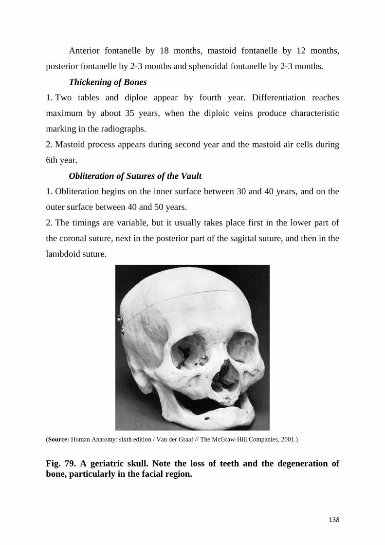
138
Anterior fontanelle by 18 months, mastoid fontanelle by 12 months,
posterior fontanelle by 2-3 months and sphenoidal fontanelle by 2-3 months.
Thickening of Bones
1. Two tables and diploe appear by fourth year. Differentiation reaches
maximum by about 35 years, when the diploic veins produce characteristic
marking in the radiographs.
2. Mastoid process appears during second year and the mastoid air cells during
6th year.
Obliteration of Sutures of the Vault
1. Obliteration begins on the inner surface between 30 and 40 years, and on the
outer surface between 40 and 50 years.
2. The timings are variable, but it usually takes place first in the lower part of
the coronal suture, next in the posterior part of the sagittal suture, and then in the
lambdoid suture.
(Source: Human Anatomy: sixth edition / Van der Graaf // The McGraw-Hill Companies, 2001.)
Fig. 79. A geriatric skull. Note the loss of teeth and the degeneration of
bone, particularly in the facial region.
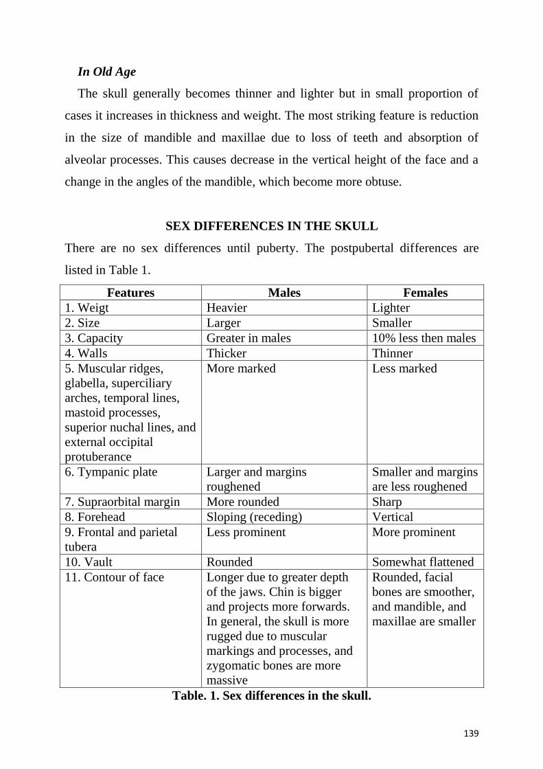
139
In Old Age
The skull generally becomes thinner and lighter but in small proportion of
cases it increases in thickness and weight. The most striking feature is reduction
in the size of mandible and maxillae due to loss of teeth and absorption of
alveolar processes. This causes decrease in the vertical height of the face and a
change in the angles of the mandible, which become more obtuse.
SEX DIFFERENCES IN THE SKULL
There are no sex differences until puberty. The postpubertal differences are
listed in Table 1.
Features Males Females
1. Weigt Heavier Lighter
2. Size Larger Smaller
3. Capacity Greater in males 10% less then males
4. Walls Thicker Thinner
5. Muscular ridges,
glabella, superciliary
arches, temporal lines,
mastoid processes,
superior nuchal lines, and
external occipital
protuberance
More marked Less marked
6. Tympanic plate Larger and margins
roughened
Smaller and margins
are less roughened
7. Supraorbital margin More rounded Sharp
8. Forehead Sloping (receding) Vertical
9. Frontal and parietal
tubera
Less prominent More prominent
10. Vault Rounded Somewhat flattened
11. Contour of face Longer due to greater depth
of the jaws. Chin is bigger
and projects more forwards.
In general, the skull is more
rugged due to muscular
markings and processes, and
zygomatic bones are more
massive
Rounded, facial
bones are smoother,
and mandible, and
maxillae are smaller
Table. 1. Sex differences in the skull.
140
Until the age of puberty, there is little difference between the skull of the
female and that of the male. The skull of an adult female is, as a rule, lighter and
smaller, and its cranial capacity about 10 percent less, than that of the male. Its
walls are thinner and its muscular ridges less strongly marked; the glabella,
superciliary arches, and mastoid processes are less prominent, and the
corresponding air sinuses are small or rudimentary. The upper margin of the
orbit is sharp, the forehead vertical, the frontal and parietal eminences
prominent, and the vault somewhat flattened. The contour of the face is more
rounded, the facial bones are smoother, and the maxillae and mandible and their
contained teeth smaller. From what has been said it will be seen that more of the
infantile characteristics are retained in the skull of the adult female than in that
of the adult male. A well-marked male or female skull can easily be recognized
as such, but in some cases the respective characteristics are so indistinct that
the determination of the sex may be difficult or impossible.
Wormian or Sutural Bones
These are small irregular bones found in the region of the fontanelles, and
are formed by additional ossification centres. They are most common at the
lambda and at the asterion; common at the pterion (epipteric bone); and rare at
the bregma (os Kerckring). Wormian bones are common in hydrocephalic
skulls.
CRANIOMETRY
Cephalic Index
It expresses the shape of the head, and is the proportion of breadth to
length of the skull. The length or longest diameter is measured from the glabella
to the occipital point, the breadth or widest diameter is measured usually a little
below the parietal tubera. It is calculated as a ration of maximal breadth to
maximal length multiplied by 100. The obtained result is called the cranial (or
cephalic) index. Three variations of human skull are distinguished depending on
the magnitude of the cranial index:
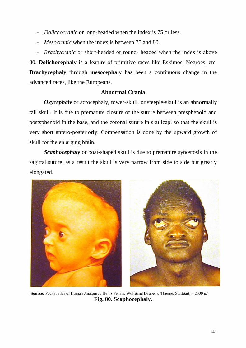
141
- Dolichocranic or long-headed when the index is 75 or less.
- Mesocranic when the index is between 75 and 80.
- Brachycranic or short-headed or round- headed when the index is above
80. Dolichocephaly is a feature of primitive races like Eskimos, Negroes, etc.
Brachycephaly through mesocephaly has been a continuous change in the
advanced races, like the Europeans.
Abnormal Crania
Oxycephaly or acrocephaly, tower-skull, or steeple-skull is an abnormally
tall skull. It is due to premature closure of the suture between presphenoid and
postsphenoid in the base, and the coronal suture in skullcap, so that the skull is
very short antero-posteriorly. Compensation is done by the upward growth of
skull for the enlarging brain.
Scaphocephaly or boat-shaped skull is due to premature synostosis in the
sagittal suture, as a result the skull is very narrow from side to side but greatly
elongated.
(Source: Pocket atlas of Human Anatomy / Heinz Feneis, Wolfgang Dauber // Thieme, Stuttgart. – 2000 p.)
Fig. 80. Scaphocephaly.
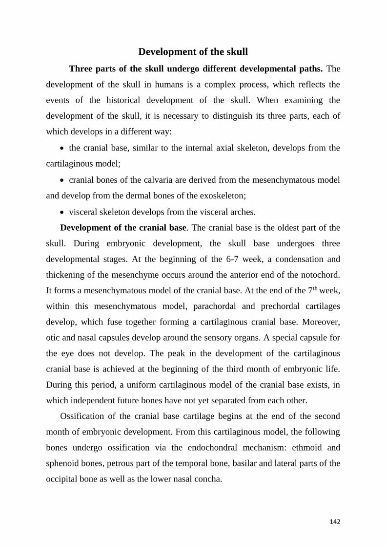
142
Development of the skull
Three parts of the skull undergo different developmental paths. The
development of the skull in humans is a complex process, which reflects the
events of the historical development of the skull. When examining the
development of the skull, it is necessary to distinguish its three parts, each of
which develops in a different way:
the cranial base, similar to the internal axial skeleton, develops from the
cartilaginous model;
cranial bones of the calvaria are derived from the mesenchymatous model
and develop from the dermal bones of the exoskeleton;
visceral skeleton develops from the visceral arches.
Development of the cranial base. The cranial base is the oldest part of the
skull. During embryonic development, the skull base undergoes three
developmental stages. At the beginning of the 6-7 week, a condensation and
thickening of the mesenchyme occurs around the anterior end of the notochord.
It forms a mesenchymatous model of the cranial base. At the end of the 7th week,
within this mesenchymatous model, parachordal and prechordal cartilages
develop, which fuse together forming a cartilaginous cranial base. Moreover,
otic and nasal capsules develop around the sensory organs. A special capsule for
the eye does not develop. The peak in the development of the cartilaginous
cranial base is achieved at the beginning of the third month of embryonic life.
During this period, a uniform cartilaginous model of the cranial base exists, in
which independent future bones have not yet separated from each other.
Ossification of the cranial base cartilage begins at the end of the second
month of embryonic development. From this cartilaginous model, the following
bones undergo ossification via the endochondral mechanism: ethmoid and
sphenoid bones, petrous part of the temporal bone, basilar and lateral parts of the
occipital bone as well as the lower nasal concha.
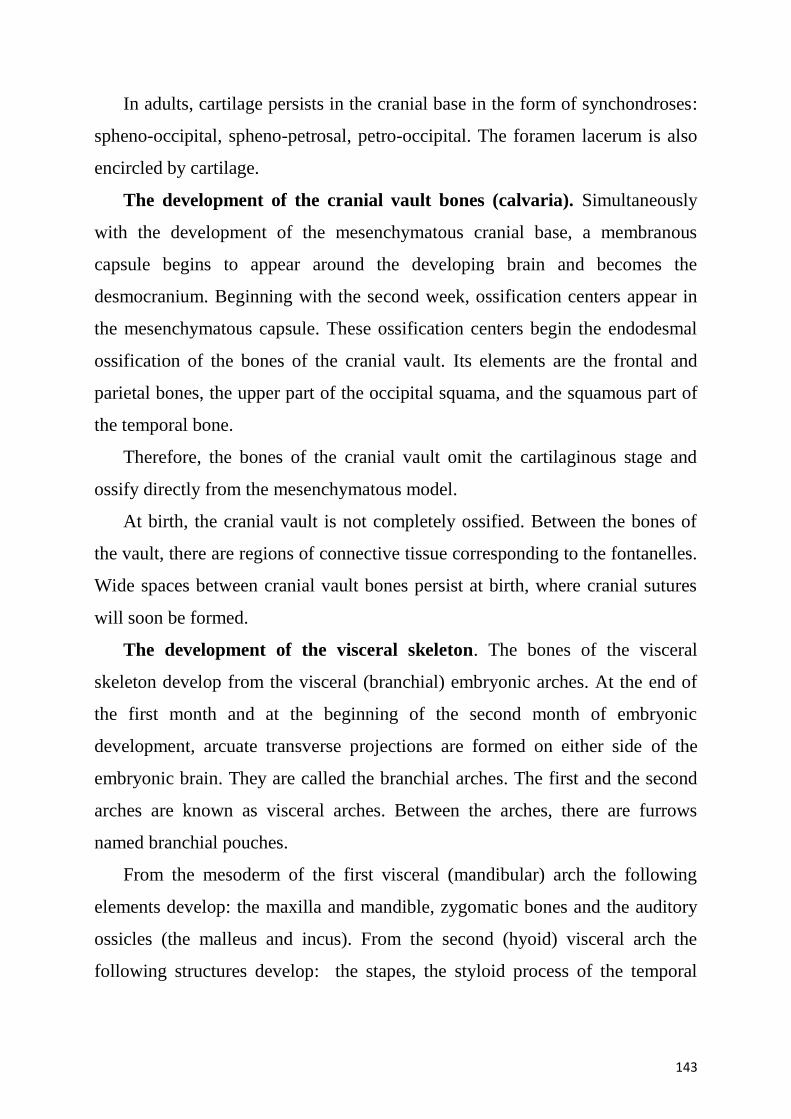
143
In adults, cartilage persists in the cranial base in the form of synchondroses:
spheno-occipital, spheno-petrosal, petro-occipital. The foramen lacerum is also
encircled by cartilage.
The development of the cranial vault bones (calvaria). Simultaneously
with the development of the mesenchymatous cranial base, a membranous
capsule begins to appear around the developing brain and becomes the
desmocranium. Beginning with the second week, ossification centers appear in
the mesenchymatous capsule. These ossification centers begin the endodesmal
ossification of the bones of the cranial vault. Its elements are the frontal and
parietal bones, the upper part of the occipital squama, and the squamous part of
the temporal bone.
Therefore, the bones of the cranial vault omit the cartilaginous stage and
ossify directly from the mesenchymatous model.
At birth, the cranial vault is not completely ossified. Between the bones of
the vault, there are regions of connective tissue corresponding to the fontanelles.
Wide spaces between cranial vault bones persist at birth, where cranial sutures
will soon be formed.
The development of the visceral skeleton. The bones of the visceral
skeleton develop from the visceral (branchial) embryonic arches. At the end of
the first month and at the beginning of the second month of embryonic
development, arcuate transverse projections are formed on either side of the
embryonic brain. They are called the branchial arches. The first and the second
arches are known as visceral arches. Between the arches, there are furrows
named branchial pouches.
From the mesoderm of the first visceral (mandibular) arch the following
elements develop: the maxilla and mandible, zygomatic bones and the auditory
ossicles (the malleus and incus). From the second (hyoid) visceral arch the
following structures develop: the stapes, the styloid process of the temporal
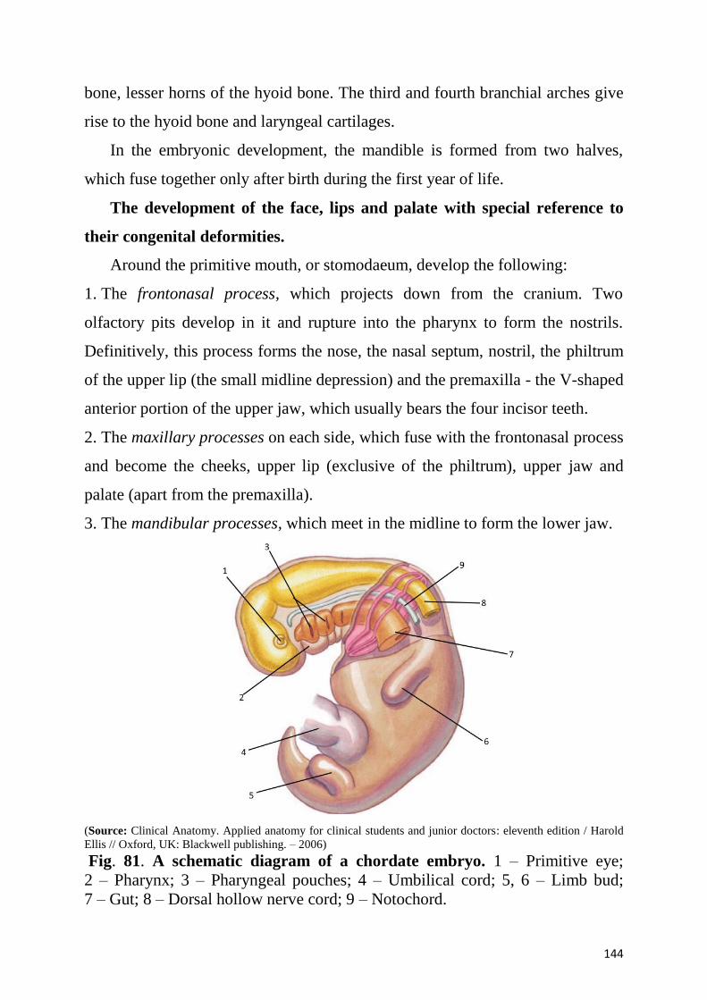
144
bone, lesser horns of the hyoid bone. The third and fourth branchial arches give
rise to the hyoid bone and laryngeal cartilages.
In the embryonic development, the mandible is formed from two halves,
which fuse together only after birth during the first year of life.
The development of the face, lips and palate with special reference to
their congenital deformities.
Around the primitive mouth, or stomodaeum, develop the following:
1. The frontonasal process, which projects down from the cranium. Two
olfactory pits develop in it and rupture into the pharynx to form the nostrils.
Definitively, this process forms the nose, the nasal septum, nostril, the philtrum
of the upper lip (the small midline depression) and the premaxilla - the V-shaped
anterior portion of the upper jaw, which usually bears the four incisor teeth.
2. The maxillary processes on each side, which fuse with the frontonasal process
and become the cheeks, upper lip (exclusive of the philtrum), upper jaw and
palate (apart from the premaxilla).
3. The mandibular processes, which meet in the midline to form the lower jaw.
(Source: Clinical Anatomy. Applied anatomy for clinical students and junior doctors: eleventh edition / Harold
Ellis // Oxford, UK: Blackwell publishing. – 2006)
Fig. 81. A schematic diagram of a chordate embryo. 1 – Primitive eye;
2 – Pharynx; 3 – Pharyngeal pouches; 4 – Umbilical cord; 5, 6 – Limb bud;
7 – Gut; 8 – Dorsal hollow nerve cord; 9 – Notochord.
145
(Source: Clinical Anatomy. Applied anatomy for clinical students and junior doctors: eleventh edition / Harold
Ellis // Oxford, UK: Blackwell publishing. – 2006)
Fig. 82. The ventral aspect of a fetal head showing the three processes,
frontonasal, maxillary and mandibular, from which the face, nose and jaws
are derived.
(Source: Clinical Anatomy. Applied anatomy for clinical students and junior doctors: eleventh edition / Harold
Ellis // Oxford, UK: Blackwell publishing. – 2006)
Fig. 83. The embryonic skull at 12 weeks is composed of bony elements
from three developmental sources: the chondrocranium (colored blue-
gray), the neurocranium (colored light yellow), and the viscerocranium
(colored red).
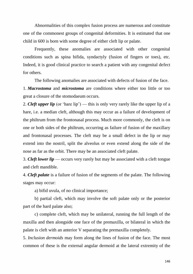
146
Abnormalities of this complex fusion process are numerous and constitute
one of the commonest groups of congenital deformities. It is estimated that one
child in 600 is born with some degree of either cleft lip or palate.
Frequently, these anomalies are associated with other congenital
conditions such as spina bifida, syndactyly (fusion of fingers or toes), etc.
Indeed, it is good clinical practice to search a patient with any congenital defect
for others.
The following anomalies are associated with defects of fusion of the face.
1. Macrostoma and microstoma are conditions where either too little or too
great a closure of the stomodaeum occurs.
2. Cleft upper lip (or ‘hare lip’) — this is only very rarely like the upper lip of a
hare, i.e. a median cleft, although this may occur as a failure of development of
the philtrum from the frontonasal process. Much more commonly, the cleft is on
one or both sides of the philtrum, occurring as failure of fusion of the maxillary
and frontonasal processes. The cleft may be a small defect in the lip or may
extend into the nostril, split the alveolus or even extend along the side of the
nose as far as the orbit. There may be an associated cleft palate.
3. Cleft lower lip — occurs very rarely but may be associated with a cleft tongue
and cleft mandible.
4. Cleft palate is a failure of fusion of the segments of the palate. The following
stages may occur:
a) bifid uvula, of no clinical importance;
b) partial cleft, which may involve the soft palate only or the posterior
part of the hard palate also;
c) complete cleft, which may be unilateral, running the full length of the
maxilla and then alongside one face of the premaxilla, or bilateral in which the
palate is cleft with an anterior V separating the premaxilla completely.
5. Inclusion dermoids may form along the lines of fusion of the face. The most
common of these is the external angular dermoid at the lateral extremity of the
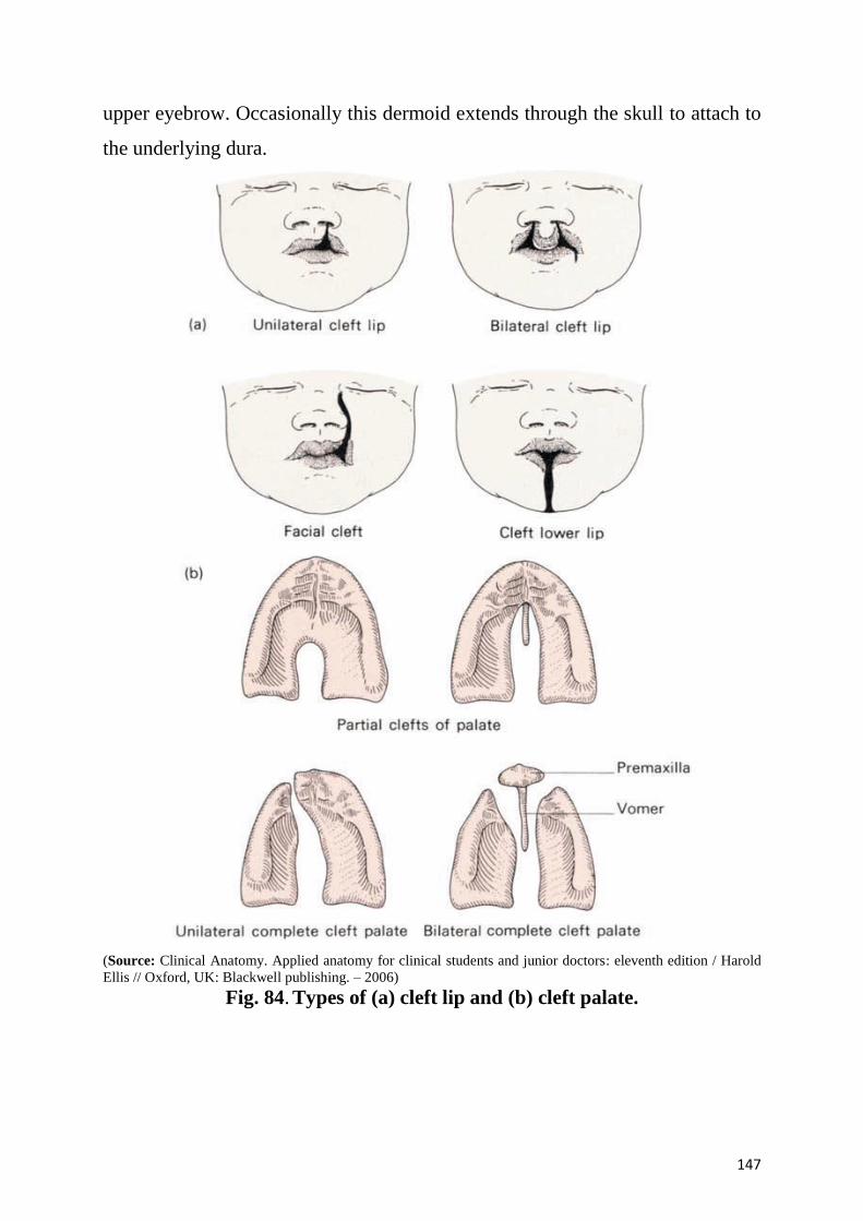
147
upper eyebrow. Occasionally this dermoid extends through the skull to attach to
the underlying dura.
(Source: Clinical Anatomy. Applied anatomy for clinical students and junior doctors: eleventh edition / Harold
Ellis // Oxford, UK: Blackwell publishing. – 2006)
Fig. 84. Types of (a) cleft lip and (b) cleft palate.
148
Control questions.
1. Name three parts of human skull, which follow different developmental path.
2. Why does the skull base ossify via endochondral mechanism?
3. Name the skull bones, which develop from cartilage?
4. Why are the cranial vault bones derived from the connective tissue?
5. What bones arise from the first visceral arch?
6. What bones develop from the second visceral arch? What bones develop from
the third and fourth branchial arches?
7. Bones of the neurocranium: the occipital bone (the structure, parts, foramens,
canals).
8. Bones of the neurocranium: the frontal bone (the structure, parts, foramens,
frontal sinus).
9. Bones of the neurocranium: the temporal bone (the structure, parts, processes,
foramens, canals).
10. The walls of the tympanic cavity.
11. Demonstrate the openings of facial canal, carotid canal and musculotubal
canal.
12. Bones of the neurocranium: the sphenoid bone (the structure, parts,
foramens).
13. Bones of the neurocranium: the ethmoid bone (the structure, plates, the
ethmoidal labyrinth).
14. Bones of the visceral skeleton: the maxilla (structure, parts, processes,
maxillary sinus).
15. Bones of the visceral skeleton: the mandible (structure, parts, processes,
foramens).
16. Bones of the visceral skeleton: the palatine bone (structure, plates, processes,
and foramens).
17. Bones of the visceral skeleton: the zygomatic bone, the lacrimal bone, the
hyoid bone (their structure).
149
18. Visceral (facial) skeleton: temporal fossa, infratemporal fossa,
pterygopalatine fossa. Their boundaries, contents, communications.
19. Neurocranium: the calvaria (bones, connections), the bony palate.
20. Neurocranium: external surface of the base of the skull (borders, foramens).
21. Neurocranium: internal surface of the base of the skull (borders, structure of
the anterior, middle and posterior cranial fossae).
22. Nasal cavity: walls, the bony nasal septum, nasal conchae, nasal meatuses
and its communications.
23. Orbital cavity: walls, the communications of the orbit. To describe and
demonstrate its on the preparation.
24. Name cranial fontanelles. When do they ossify?
25. What is the clinical significance of the dates of fontanelle closure?
26. What happens to the skull bones during the birth process?
27. What changes take place after birth?
28. When do the cranial sutures form and when do they begin to obliterate?
29. What are the characteristics of the skull in elderly?
30. Name major skull anomalies. What skull anomalies occur?
31. How is the cranial index calculated?
32. What are the three variants of the skull distinguished on the bases of the
cranial index value?
33. Name basic sexual differences of the skull. How are they determined?
150
Situation problems.
1. A casualty has a fracture of the base of the skull in the region of the
hypoglossal canal. Which bone was damaged?
A. The frontal
B. The parietal
C. The occipital*
D. The temporal
E. The sphenoid
2. After collision of two cars, a driver got deformation of the right occipital
condyle. Which part of the occipital bone can be injured?
A. Basilar part
B. External surface of the squamous part
C. Internal surface of the squamous part
D. Lateral part*
E. Nasal part
3. A casualty has a trauma of soft tissues and parietal bones in the sagittal
suture area with profuse bleeding. Which formation is probably injured?
A. Sinus rectus
B. Sinus petrosus superior
C. Sinus transverse
D. Sinus sagittalis inferior
E. Sinus sagittalis superior*
4. A casualty has a fissure in the region of the groove for transverse sinus.
Which part of the occipital bone can be injured?
A. Internal surface of the squamous part*
B. External surface of the squamous part
C. Basilar part
D. Lateral part
E. Nasal part
151
5. Purulence of occipital soft tissues took place after a trauma. Through which
anatomical formation can the purulent process spread to the cranial cavity?
A. Foramen caecum
B. Condylar canal*
C. Foramen rotundum
D. Optic canal
E. Carotid canal
6. A casualty has a fracture of the base of the skull in the region of foramen
spinosum and foramen rotundum. Which bone is damaged?
A. The frontal
B. The parietal
C. The sphenoid*
D. The temporal
E. The ethmoid
7. On the radiological investigation in a patient was diagnosed fracture of the
nasal septum in the superior 1/3. Which bone is damaged?
A. The frontal
B. The parietal
C. The sphenoid
D. The ethmoid*
E. The temporal
8. Patient admitted to hospital with an injury of the base of the skull. On the
radiological investigation in a patient was diagnosed destruction and increase of
the hypophyseal fossa. Which anatomical structure is damaged?
A. Optic canal
B. Foramen rotundum
C. Pterygoid canal
D. Superior orbital fissure
E. Sphenoidal sinus*
152
9. On the radiological investigation in a patient was diagnosed increase of the
Turkish saddle's cavity, destruction of the different part of the hypophyseal fossa
and anterior clinoid processes. Which endocrine gland is damaged?
A. The pituitary gland (hypophysis)*
B. Epiphysis
C. Suprarenal gland
D. Thyroid gland
E. Parathyroid gland
10. A casualty has a fracture of the base of the skull. Line of the injure passed
through prechiasmatic sulcus and carotid sulcus. Which part of the sphenoid
bone is damaged?
A. Greater wing
B. Body*
C. Lesser wing
D. Pterygoid process
E. Posterior clinoid process
11. A child was admitted to an otolaryngologic department with inflammation of
the middle ear. Disease began with nasopharynx inflammation. It is detected that
the infection lo the tympanic cavity through the auditory tube located in:
A. Canalis caroticus
B. Canaliculus tympanicus
C. Canalis musculotubarius*
D. Canaliculus chordae tympani
E. Canaliculi caroticotympanici
12. An 8-year-old boy with purulent otitis has the infection spread from the
tympanic cavity into the bulb of internal jugular vein. Such complication
develop in case of one of the tympanic cavity walls thinning. Which wall is it?
A. Medial
B. Superior
153
C. Lateral
D. Inferior*
E. Anterior
13. A 9-year-old girl with purulent otitis has the infection spread from the
tympanic cavity into the middle cranial fossa. Such complication develop in case
of one of the tympanic cavity walls thinning. Which wall is it?
A. Medial
B. Anterior
C. Lateral
D. Inferior
E. Superior*
14. A 7-year-old boy with purulent otitis has the infection spread from the
tympanic cavity into the inner ear. Such complication develop in case of one of
the tympanic cavity walls thinning. Which wall is it?
A. Medial*
B. Anterior
C. Lateral
D. Inferior
E. Superior
15. A 6-year-old boy with purulent otitis has the infection spread from the
tympanic cavity into the mastoid antrum. Such complication develop in case of
one of the tympanic cavity walls thinning. Which wall is it?
A. Medial
B. Posterior*
C. Lateral
D. Inferior
E. Superior
16. At birth defect, mandibular midline slit. Which non-union processes of the
maxilla leads to the development of anomalies?
154
A. Mandibular
B. Maxillary
C. Palatine*
D. Frontal
E. Zygomatic
17. At the victim was the trauma of the upper jaw with a damage of the
infraorbital foramen. Which surface of the upper jaw was broken?
A. Nasal
B. Orbital
C. Anterior*
D. Infratemporal
E. Temporal
18. At the victim was the trauma of the maxilla, knocked out the first molar
dens. Which process of the maxilla is damaged?
A. Nasal
B. Frontal
C. Palatine
D. Alveolar*
E. Temporal
19. Purulence of orbit soft tissues took place after an eye′s trauma. Through
what anatomical formation can the purulent process spread to the middle cranial
fossa?
A. Through the anterior ethmoidal foramen
B. Through the posterior ethmoidal foramen
C. Through the superior orbital fissure*
D. Through the inferior orbital fissure
E. Through the zygomaticoorbital foramen
155
20. Chronic rhinitis is complicated with the signs of maxillary sinus mucous
tunic affection (maxillary sinusitis). Through what nasal cavity formation has
the infection spread?
A. Ethmoidal infundibulum
B. Sphenoethmoidal recess
C. Sphenopalatine foramen
D. Maxillary hiatus*
E. Ethmoid cells
21. During the first days of a newborn child, a pediatrician detected that milk
gets into the child's nasal cavity. What malformation does this fact indicate?
A. Esophagus constriction
B. Diverticulum of esophagus
C. Esophageal atresia
D. Cleft clip
E. Non-closed palate*
22. During a meal, milk gets into the nasal cavity of a newborn child. What is
the probable cause of this pathology?
A. Cleft palate*
B. Nasal septum deviation to the right
C. Basal skull fracture
D. Cleft clip
E. Nasal septum deviation to the left
23. Purulence of orbit soft tissues took place after an eye’s trauma. Through
what anatomical formation can the purulent process spread to the
pterygopalatine fossa?
A. Through the round foramen
B. Through the inferior orbital fissure*
C. Through the pterygoid canal
D. Through the superior orbital fissure
156
E. Through the zygomaticoorbital foramen
24. A sick has an acute inflammation of nasolacrimal duct mucous membrane. It
is known that after influenza nasal discharges had been observed for a long time.
From what part of the nasal cavity could the infection get to the nasolacrimal
duct?
A. Superior nasal meatus
B. Middle nasal meatus
C. Inferior nasal meatus*
D. Common nasal meatus
E. Sphenoethmoidal recess
25. A patient has a suppurative inflammation of the sphenoid sinus. What part of
the nasal cavity does the pus flow out into?
A. Meatus nasi inferior
B. Meatus nasi communis
C. Meatus nasi medius
D. Meatus nasi superior*
E. Infundibulum.
26. A patient has an inflammation of sphenoid sinus. Where does the aperture of
this sinus open?
A. Infundibulum
B. Common nasal meatus
C. Middle nasal meatus
D. Inferior nasal meatus
E. Sphenoethmoidal recess*
27. A 30-year-old patient with a second upper molar pulp inflammation
appealed to a doctor with complaints of headache and nose rheum. After
examination, pulpitis complicated with sinusitis was diagnosed. Which sinus did
the infection enter from this tooth root canal?
A. Maxillary sinus*
157
B. Frontal sinus
C. Sphenoidal sinus
D. Ethmoid cells
E. Mastoid cells
28. A patient has a suppurative inflammation of the frontal sinus. What part of
the nasal cavity does the pus flow out into?
A. Meatus nasi inferior
B. Meatus nasi medius*
C. Meatus nasi communis
D. Meatus nasi superior
E. Infundibulum
29. After carried flu, a patient has the labouringly nasal breath, enhanceable
temperature, head pains, lacrimation, and sickliness at palpation in area of fossa
canina on the right. Inflammation of what additional cavity of nose complicated
the disease?
A. Sinus maxillaris dexter*
B. Sinus frontalis dexter
C. Sinus shenoidalis dexter
D. Middle cells of ethmoid bone on the right
E. Bulla ethmoidalis dexter
30. After a 2-y.o. child has had flu, there appeared complaints about earache. A
doctor revealed hearing impairment and inflammation of the middle ear. How
did the infection penetrate into the middle ear?
A. Through the auditory tube*
B. Through foramen jugularis
C. Through canalis caroticus
D. Through atrium mastoideum
E. Through canalis nasolacrimalis
158
31. Examination of a patient revealed an abscess of pterygopalatine fossa.
Where can the infection spread to unless the disease is managed in time?
A. To the orbit *
B. To the interpterygoid space
C. To the frontal sinus
D. To sphenoid sinus
E. To the tympanic cavity
32. Examination of a 6-month-old child revealed a delay in closure of the
occipital fontanelle. When should it normally close?
A. Until 3 months*
B. Before the child is born
C. Until 6 months
D. Until the end of the first year of life
E. Until the end of the second year of life
33. Examination of a 2-year-old child revealed a delay in closure of the frontal
fontanelle. When should it normally close?
A. Until 3 months
B. Before the child is born
C. Until 6 months
D. Until the end of the first year of life*
E. Until the end of the second year of life
159
Literature
1.Abramson D.L. Abnormalities of the cranial base in synostotic frontal
plagiocephaly / D.L. Abramson, I.P. Janecka, B.H. Mulliken // J. Craniofac
Surg. 1996. – Vol. 7. – № 6. – P. 426-428.
2. Alfieri A. Endoscopic endonasal approach to the pterygopalatine fossa:
anatomic study / A. Alfieri, H.D. Jho, R. Schettino, M. Tschabitscher //
Neurosurgery. – 2003. – Vol. 52. – № 2. – P. 374-380.
3. Ammirati M. Analytical evaluation of complex anterior approaches to the
cranial base: an anatomic study / M. Ammirati, A. Bernardo // Neurosurgery,
1998. – Vol. 43(6). – P. 1407-1408.
4. A three-dimensional evaluation of the upper airway in adolescents / C.A.
Aboudara, D. Hatcher, I.L. Nielsen, A. Miller // Orthod. Craniofacial Res
(Suppl. l). 2003. – № 6. – P. 173-175.
5. Atlas of Human Anatomy: in three volumes. Vol.1 / R.D. Sinelnikov;
translated by Ludmila Aksenova, M.D. // MIR Publishers Moscow, 1989. –
320p.
6. Atlas of Human Anatomy / Frank H. Netter, M.D. Arthur F. Dalley; 2nd ed. //
ILS, Medimedia USA Company, 1997. – 548p.
7. Bandak F.A. An imaging-based computational and experimental study of
skull fracture: Finite element model development / F.A. Bandak, M.J. Vorst,
L.M. Stuhmiller // J. Neurotrauma, 1995. – Vol. 12. – № 4. – P. 679-688.
8. Bhatia S.N. A manual of facial growth. A computer analysis of longitudinal
cephalometric growth data / S.N. Bhatia, B.C. Leighton // Oxford, 1993. – Р.
215-243.
9. Cicekclbasl A.E. A Morphometric Evaluation of Some Important Bony
Landmarks on the Skull Base Related to Sexes /А.Е. Cicekclbasl, K.A.
Mursehed, T. Ziylan et al. // Turk J. Med. 2004. – Vol. 34. – P. 37-42.
160
10. Clinical Anatomy. Applied anatomy for clinical students and junior doctors:
eleventh edition / Harold Ellis // Oxford, UK: Blackwell publishing. – 2006. –
455p.
11. Daniels D.L. Osseous anatomy of the orbital apex / D.L. Daniels, L.P. Mar,
M.F. Mafee // AJNR: Amer. J. Neuroraiol, 1995. – Vol. 16. – P. 1929-1935.
12. Daniel E. The evolution and development of cranial form in Homo sapiens /
E. Daniel, M. Liebeman, K. Brandeis // PNAS. – 2002. – Vol. 99. – № 3. – P.
134-1139.
13. Dibbetts J. Regional size differences in four commonly used cephalometric
atlases. / J. Dibbetts, K. Nolte // The Ann Arbor, Cleveland(Bolton),
Londor(UK) and Philadelphia atlases compared // Orthod. Craniofacial Res. –
2002, vol. 5. – P. 51-58.
14. Divaris M. Comparison of anatomy and imaging of the orbital area and the
naso-lacrimal duct / M. Divaris // Ann. Chir. Plast. Esther. – 1995. – Vol. 40. –
P. 11 - 22.
15. D' Aloisio D. A comparative and correlational study of the cranial base of
North American blacks / D. D'Aloisio, V. Pangrazio-Kulbersh // J. Orthod
Dentofacial. – 1992. – Vol. 102. – P. 449-455.
16. Gray′s anatomy for students / Richard L. Drake, A. Wayne Vogl, and Adam
W. M. Mitchell; illustrations by Richard M. Tibbitts and Paul E. Richardson;
photographs by Ansell Horn; 2nd ed. // Churchill Livingstone, 2012. –– 1103p.
17. Grant′s atlas of anatomy ∕ Anne M.R., Arthur F. Dalley II, 12th ed. //
Baltimore: Wiliams & Wolters, 2009. – 864 p.
18. Human Anatomy. In three volumes. Volume 1 / Edited by V.G.
Koveshnikov. – Lugansk: LTD «Virtualnaya realnost», 2009. – 328p.
19. Human Anatomy: sixth edition / Van der Graaf // The McGraw-Hill
Companies, 2001. – 855p.
161
20. Karisen, A.T. Morphology and growth in convex profile facial patterns: a
longitudinal study / A.T. Karisen, O. Krogstad // J. Angle Orthod., 1999. – Vol.
69. – № 4. – P. 334-344.
21. Kasai K. Relationship between cranial base and maxillofacial morphology /
K. Kasai, Т. Moro, E. Kanazawa et al. // Eur J Orthod., 1995. – Vol. 17. – № 5 –
P. 403-410.
22. Kerr W.J. The variability of some craniofacialdimensions / W .J. Kerr, I.
Ford // J. Angle Orthod., 1991. – Vol. 61. 3. – P. 205-210.
23. Lieberman D.E. How and why humans grow thin skulls: Experimental
evidence for systemic cortical robusticity / D.E. Lieberman // Amer. J. Phys.
Anthropol., 1996. – Vol. 101. – № 2. – P. 217-236.
24. Lieberman E.D. The evolution and development of cranial form in Homo
sapiens / E.D. Lieberman, M. Brandeis, G. Krovitz // Anthropol. – 2002. – Vol.
99. – № 3. – P. 1134-1139.
25. Low W.K. Middle Cranial Fossa Approach to the Internal Auditory Meatus:
A Chinese. Temporal Bone Study / W.K. Low // J. Department of
Otolaryngology. –1999. – Vol. 61. – № 3. – P. 142-145.
26. Luboqa S.A. Position and orientation of the foramen magnum in higher
primates / S.A. Luboqa, B.A. Wood // Am J. Phys Anthropol. – 1990. – Vol. 81.
–№ 1. – P. 67-76.
27. Nadery S. Morthometric analysis of human occipital condyle / S. Naderi, E.
Korman, G. Citak et al. // Clin. Neural Neurosung., 2005. – Vol. 3. – № 32. – P.
191-199.
28. Pocket atlas of Human Anatomy / Heinz Feneis, Wolfgang Dauber //
Thieme, Stuttgart. – 2000. – 510p.
29. Ricciardelli E.J. Embryology and anatomy of the cranial base / E.J.
Ricciardelli // Clin Plast Surg., 1995. – Vol. 22. – № 3. – P. 361 -372.
30. Sobotta Atlas of Human Anatomy / Edited by R. Putz and R. Pabst, 14th ed.
// Elsevier GmbH, Munich, 2008. – 895p.
162
31. Surgical anatomy of the infratemporal fossa using the transmaxillary
approach / P.H. Roche, H.D. Fournier, L. Laccourreye, P. Mercier // Surg Radiol
Anat. –2001. – Vol. 23. – № 4. – P. 209-213.
32.Sherwood T.F. Sagittal Growth of the Cranial Base in Normal and Cleft
Fetuses / T.F. Sherwood, N.P. Mooney, T.D. Smith, et al. // Depts. Of
Orthodontics and Antropology. – 1998. – Vol. 22. – № 5. – P. 28-36.
33. Yang W.S. Soft X-ray study on fetal cranio-facial growth and development /
W.S. Yang, C.H. Suhr, D.S. Nahm et al. // Taehan Chikkwa Uisa Hyophoe Chi.
– 1990. – Vol. 28. – №3. – P. 265-277.
34.Leonetti G. Variation of sphenoidal angle of human skull in the course of
aging / G. Leonetti, M. Signoli, I. Hershkovitz et al. // C.R. Acad Sci III. – 1997.
– Vol. 320. – № 11. – P. 943-947.
35. Yoganandan N. Biomechanics of skull fracture / N. Yoganandan, F.A.
Pintar, A. Sances et al. // J. Neurotrauma. – 1995. – Vol. 12. – № 4. – P. 659-
668.
36. Zhang Wohua Metrical characteristics analysis of the middle cranial fossa /
Wohua Zhang //Asta anat. 1988. – Vol. 19, N 3. – P. 244-249.
37. Anatomy at Pirogov (Atlas of Human Anatomy). T. 2. Head and Neck /
Shilkin V. V., Filimonov V. I. // 2013. – P. 725.
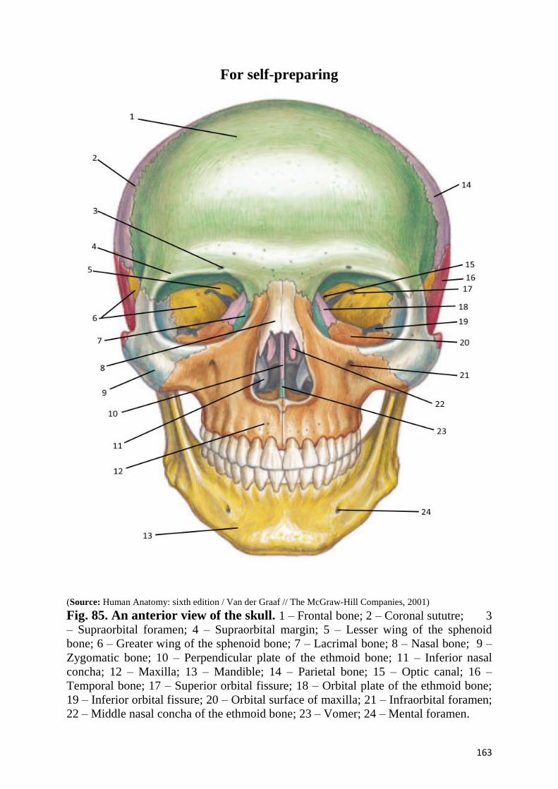
163
For self-preparing
(Source: Human Anatomy: sixth edition / Van der Graaf // The McGraw-Hill Companies, 2001)
Fig. 85. An anterior view of the skull. 1 – Frontal bone; 2 – Coronal sututre; 3
– Supraorbital foramen; 4 – Supraorbital margin; 5 – Lesser wing of the sphenoid
bone; 6 – Greater wing of the sphenoid bone; 7 – Lacrimal bone; 8 – Nasal bone; 9 –
Zygomatic bone; 10 – Perpendicular plate of the ethmoid bone; 11 – Inferior nasal
concha; 12 – Maxilla; 13 – Mandible; 14 – Parietal bone; 15 – Optic canal; 16 –
Temporal bone; 17 – Superior orbital fissure; 18 – Orbital plate of the ethmoid bone;
19 – Inferior orbital fissure; 20 – Orbital surface of maxilla; 21 – Infraorbital foramen;
22 – Middle nasal concha of the ethmoid bone; 23 – Vomer; 24 – Mental foramen.
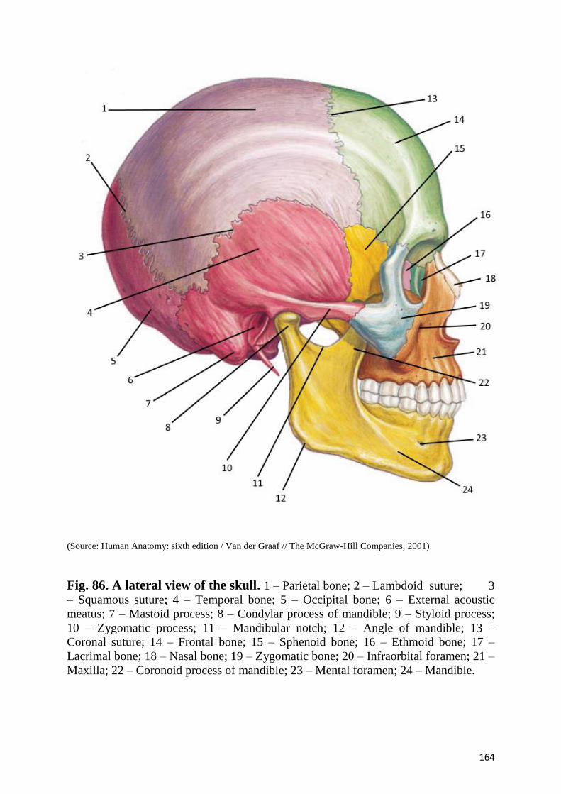
164
(Source: Human Anatomy: sixth edition / Van der Graaf // The McGraw-Hill Companies, 2001)
Fig. 86. A lateral view of the skull. 1 – Parietal bone; 2 – Lambdoid suture; 3
– Squamous suture; 4 – Temporal bone; 5 – Occipital bone; 6 – External acoustic
meatus; 7 – Mastoid process; 8 – Condylar process of mandible; 9 – Styloid process;
10 – Zygomatic process; 11 – Mandibular notch; 12 – Angle of mandible; 13 –
Coronal suture; 14 – Frontal bone; 15 – Sphenoid bone; 16 – Ethmoid bone; 17 –
Lacrimal bone; 18 – Nasal bone; 19 – Zygomatic bone; 20 – Infraorbital foramen; 21 –
Maxilla; 22 – Coronoid process of mandible; 23 – Mental foramen; 24 – Mandible.
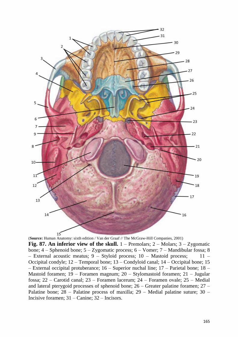
165
(Source: Human Anatomy: sixth edition / Van der Graaf // The McGraw-Hill Companies, 2001)
Fig. 87. An inferior view of the skull. 1 – Premolars; 2 – Molars; 3 – Zygomatic
bone; 4 – Sphenoid bone; 5 – Zygomatic process; 6 – Vomer; 7 – Mandibular fossa; 8
– External acoustic meatus; 9 – Styloid process; 10 – Mastoid process; 11 –
Occipital condyle; 12 – Temporal bone; 13 – Condyloid canal; 14 – Occipital bone; 15
– External occipital protuberance; 16 – Superior nuchal line; 17 – Parietal bone; 18 –
Mastoid foramen; 19 – Foramen magnum; 20 – Stylomastoid foramen; 21 – Jugular
fossa; 22 – Carotid canal; 23 – Foramen lacerum; 24 – Foramen ovale; 25 – Medial
and lateral pterygoid processes of sphenoid bone; 26 – Greater palatine foramen; 27 –
Palatine bone; 28 – Palatine process of maxilla; 29 – Medial palatine suture; 30 –
Incisive foramen; 31 – Canine; 32 – Incisors.
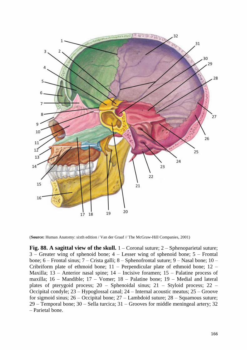
166
(Source: Human Anatomy: sixth edition / Van der Graaf // The McGraw-Hill Companies, 2001)
Fig. 88. A sagittal view of the skull. 1 – Coronal suture; 2 – Sphenoparietal suture;
3 – Greater wing of sphenoid bone; 4 – Lesser wing of sphenoid bone; 5 – Frontal
bone; 6 – Frontal sinus; 7 – Crista galli; 8 – Sphenofrontal suture; 9 – Nasal bone; 10 –
Cribriform plate of ethmoid bone; 11 – Perpendicular plate of ethmoid bone; 12 –
Maxilla; 13 – Anterior nasal spine; 14 – Incisive foramen; 15 – Palatine process of
maxilla; 16 – Mandible; 17 – Vomer; 18 – Palatine bone; 19 – Medial and lateral
plates of pterygoid process; 20 – Sphenoidal sinus; 21 – Styloid process; 22 –
Occipital condyle; 23 – Hypoglossal canal; 24 – Internal acoustic meatus; 25 – Groove
for sigmoid sinus; 26 – Occipital bone; 27 – Lambdoid suture; 28 – Squamous suture;
29 – Temporal bone; 30 – Sella turcica; 31 – Grooves for middle meningeal artery; 32
– Parietal bone.
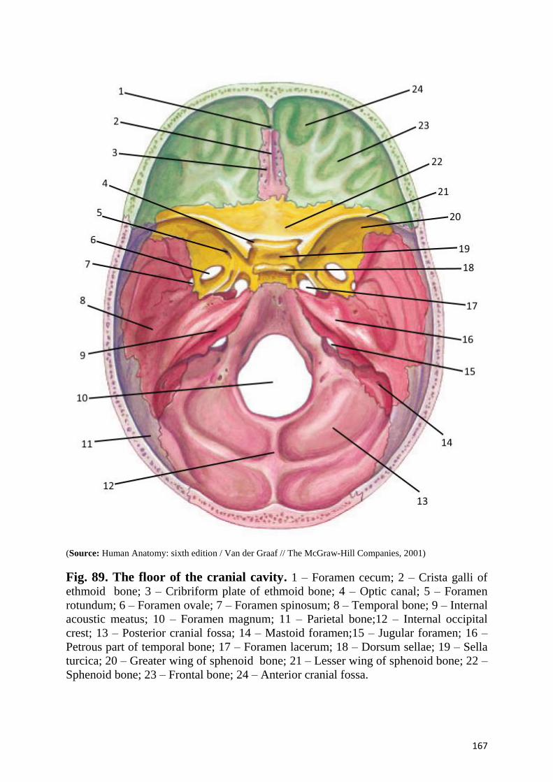
167
(Source: Human Anatomy: sixth edition / Van der Graaf // The McGraw-Hill Companies, 2001)
Fig. 89. The floor of the cranial cavity. 1 – Foramen cecum; 2 – Crista galli of
ethmoid bone; 3 – Cribriform plate of ethmoid bone; 4 – Optic canal; 5 – Foramen
rotundum; 6 – Foramen ovale; 7 – Foramen spinosum; 8 – Temporal bone; 9 – Internal
acoustic meatus; 10 – Foramen magnum; 11 – Parietal bone;12 – Internal occipital
crest; 13 – Posterior cranial fossa; 14 – Mastoid foramen;15 – Jugular foramen; 16 –
Petrous part of temporal bone; 17 – Foramen lacerum; 18 – Dorsum sellae; 19 – Sella
turcica; 20 – Greater wing of sphenoid bone; 21 – Lesser wing of sphenoid bone; 22 –
Sphenoid bone; 23 – Frontal bone; 24 – Anterior cranial fossa.
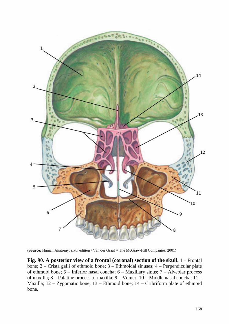
168
(Source: Human Anatomy: sixth edition / Van der Graaf // The McGraw-Hill Companies, 2001)
Fig. 90. A posterior view of a frontal (coronal) section of the skull. 1 – Frontal
bone; 2 – Crista galli of ethmoid bone; 3 – Ethmoidal sinuses; 4 – Perpendicular plate
of ethmoid bone; 5 – Inferior nasal concha; 6 – Maxillary sinus; 7 – Alveolar process
of maxilla; 8 – Palatine process of maxilla; 9 – Vomer; 10 – Middle nasal concha; 11 –
Maxilla; 12 – Zygomatic bone; 13 – Ethmoid bone; 14 – Cribriform plate of ethmoid
bone.
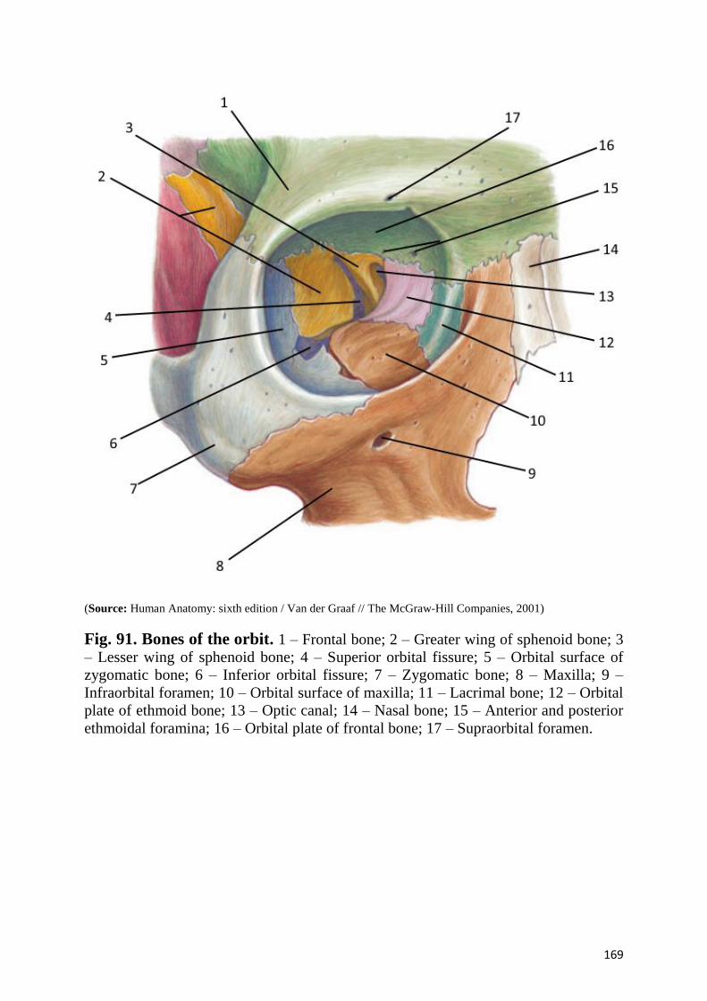
169
(Source: Human Anatomy: sixth edition / Van der Graaf // The McGraw-Hill Companies, 2001)
Fig. 91. Bones of the orbit. 1 – Frontal bone; 2 – Greater wing of sphenoid bone; 3
– Lesser wing of sphenoid bone; 4 – Superior orbital fissure; 5 – Orbital surface of
zygomatic bone; 6 – Inferior orbital fissure; 7 – Zygomatic bone; 8 – Maxilla; 9 –
Infraorbital foramen; 10 – Orbital surface of maxilla; 11 – Lacrimal bone; 12 – Orbital
plate of ethmoid bone; 13 – Optic canal; 14 – Nasal bone; 15 – Anterior and posterior
ethmoidal foramina; 16 – Orbital plate of frontal bone; 17 – Supraorbital foramen.
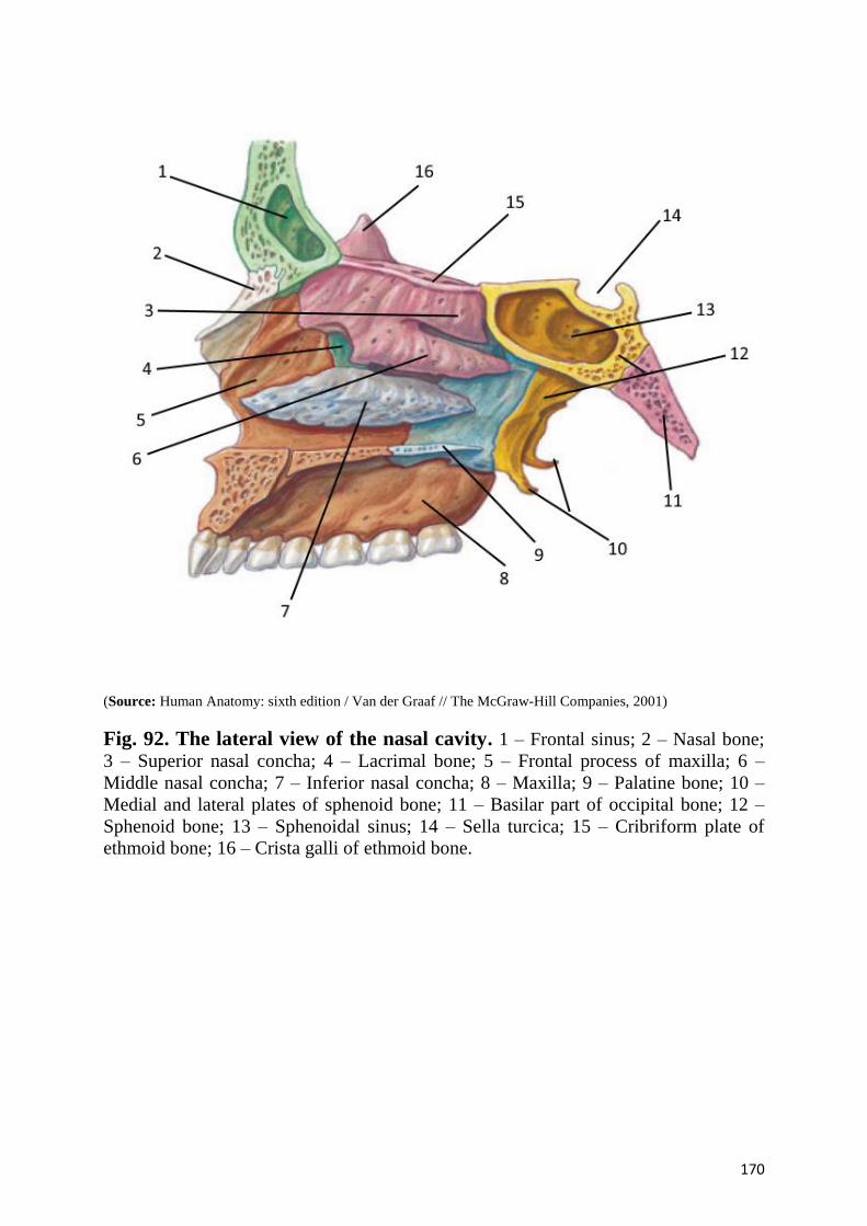
170
(Source: Human Anatomy: sixth edition / Van der Graaf // The McGraw-Hill Companies, 2001)
Fig. 92. The lateral view of the nasal cavity. 1 – Frontal sinus; 2 – Nasal bone;
3 – Superior nasal concha; 4 – Lacrimal bone; 5 – Frontal process of maxilla; 6 –
Middle nasal concha; 7 – Inferior nasal concha; 8 – Maxilla; 9 – Palatine bone; 10 –
Medial and lateral plates of sphenoid bone; 11 – Basilar part of occipital bone; 12 –
Sphenoid bone; 13 – Sphenoidal sinus; 14 – Sella turcica; 15 – Cribriform plate of
ethmoid bone; 16 – Crista galli of ethmoid bone.
171
HRYN Volodymyr Hrigorovich
Associate Professor at the Department of Human
Anatomy, PhD in Medicine.
In 2007 he graduated from HSEE of Ukraine «Ukrainian
Medical Stomatological Academy» with distinction for
specialty «General medcine». In 2010 he defended a scientific
thesis for the degree of master of medicine, majoring in
surgery on the subject «Modern state of the problem of
diagnosis and treatment of perforated ulcers of stomach and
duodenum». Since 2010 he has been working as a lecturer at
the Department of Human Anatomy.
In 2013 he defended the dissertation on competition of a
scientific degree a Doctor of Philosophy in Medicine on the
topic «The structure of the ileocecal intestine of a human with
intact appendix and after appendectomy».
The autor of 30 scientific and methodical works, 1
manuals, 3 patents for utility model, 5 rationalization proposals, 1 innovations.
SVINTSYTSKA Nataliya Leonidivna
Associate Professor at the Department of Human
Anatomy, PhD in Medicine.
In 2003 she graduated from HSEE of Ukraine
«Ukrainian Medical Stomatological Academy» with
distinction for specialty «General medcine». Since 2004
she has been working as a lecturer at the Department of
Human Anatomy.
In 2007 she defended the dissertation on competition
of a scientific degree a Doctor of Philosophy in Medicine
on the topic «The structural organization of the blood bed
of the pyloric part of a human stomach in norm».
The autor of 65 scientific and methodical works, 4
manuals, 5 patents for utility model, 3 rationalization
proposals, 4 innovations, 2 methodical recommendations.
Related Documents


