Molecular Profile and Partial Functional Analysis of Novel Endothelial Cell-Derived Growth Factors that Regulate Hematopoiesis JOHN P. CHUTE, a GARRETT G. MURAMOTO, a HOLLY K. DRESSMAN, b GARY WOLFE, c NELSON J. CHAO, a SIMON LIN d a Division of Cellular Therapy, Department of Internal Medicine, b Institute for Genome Sciences and Policy, Department of Molecular Genetics and Microbiology, d Department of Bioinformatics and Biostatistics, Duke University, Durham, North Carolina, USA; c Predictive Diagnostics Incorporated, Vacaville, California, USA Key Words. Endothelial cell • Stem cell expansion • Hematopoietic stem cell ABSTRACT Recent progress has been made in the identification of the osteoblastic cellular niche for hematopoietic stem cells (HSCs) within the bone marrow (BM). Attempts to identify the soluble factors that regulate HSC self-renewal have been less successful. We have demonstrated that primary human brain endothelial cells (HUBECs) support the ex vivo am- plification of primitive human BM and cord blood cells capable of repopulating non-obese diabetic/severe combined immunodeficient repopulating (SCID) mice (SCID repopu- lating cells [SRCs]). In this study, we sought to characterize the soluble hematopoietic activity produced by HUBECs and to identify the growth factors secreted by HUBECs that contribute to this HSC-supportive effect. Extended noncon- tact HUBEC cultures supported an eight-fold increase in SRCs when combined with thrombopoietin, stem cell factor, and Flt-3 ligand compared with input CD34 cells or cyto- kines alone. Gene expression analysis of HUBEC biological replicates identified 65 differentially expressed, nonredun- dant transcripts without annotated hematopoietic activity. Gene ontology studies of the HUBEC transcriptome revealed a high concentration of genes encoding extracellular proteins with cell-cell signaling function. Functional analyses demon- strated that adrenomedullin, a vasodilatory hormone, syner- gized with stem cell factor and Flt-3 ligand to induce the proliferation of primitive human CD34 CD38 lin cells and promoted the expansion of CD34 progenitors in culture. These data demonstrate the potential of primary HUBECs as a reservoir for the discovery of novel secreted proteins that regulate human hematopoiesis. STEM CELLS 2006;24:1315–1327 INTRODUCTION Hematopoietic stem cells (HSCs) possess the unique capacity to undergo self-renewal in vivo throughout the life of an individual while also providing the complete repertoire of mature hemato- poietic and immune cells [1–3]. Currently, transplantation of human HSCs from adult bone marrow (BM), mobilized periph- eral blood, and umbilical cord blood (CB) is applied in the curative treatment of both malignant and nonmalignant diseases [4 – 6]. More recently, the potential contribution of transplanted HSCs toward immune tolerance induction [7], vascular repair [8], and in vivo tissue regeneration [9] has been suggested. Since HSCs comprise 0.1% of the CD34 population within the bone marrow of adults [10], numerous studies have focused on the development of methods to expand HSC numbers in vitro with a goal of generating larger numbers of transplantable repopulating cells [11–14]. Concordantly, strategies have been applied to identify novel growth factors that stimulate HSC self-renewal in vivo [15–18]. Despite these efforts, few hemato- poietic growth factors have achieved clinical application [19 –21]. One strategy to identify HSC growth factors involves ex- amination of candidate niches wherein HSCs are known to reside physiologically [22–24]. Two recent studies have dem- onstrated that HSCs reside in contact with osteoblasts in the BM niche [23, 24] and these cells provide signaling through Notch ligand and cadherin interactions to maintain quiescent HSCs in vivo. A vascular niche within the marrow has also been postu- lated, comprised of sinusoidal endothelial cells, in which HSC proliferation and differentiation are thought to occur [22]. The role of endothelial cells (ECs) as regulators of hematopoiesis is supported by evidence from embryogenesis, in which Correspondence: John P. Chute, M.D., Division of Cellular Therapy, Duke University, 2400 Pratt Street, Suite 1100, Box 3961, Durham, North Carolina 27710, USA. Telephone: 919-668-4706; Fax: 919-668-1091; e-mail: [email protected] Received January 21, 2005; accepted for publication December 14, 2005; first published online in STEM CELLS EXPRESS December 22, 2005. ©AlphaMed Press 1066-5099/2006/$20.00/0 doi: 10.1634/stemcells.2005-0029 THE STEM CELL NICHE S TEM CELLS 2006;24:1315–1327 www.StemCells.com

Welcome message from author
This document is posted to help you gain knowledge. Please leave a comment to let me know what you think about it! Share it to your friends and learn new things together.
Transcript
Molecular Profile and Partial Functional Analysis of NovelEndothelial Cell-Derived Growth Factors that RegulateHematopoiesis
JOHN P. CHUTE,a GARRETT G. MURAMOTO,a HOLLY K. DRESSMAN,b GARY WOLFE,c NELSON J. CHAO,a
SIMON LINd
aDivision of Cellular Therapy, Department of Internal Medicine, bInstitute for Genome Sciences and Policy,Department of Molecular Genetics and Microbiology, dDepartment of Bioinformatics and Biostatistics, DukeUniversity, Durham, North Carolina, USA; cPredictive Diagnostics Incorporated, Vacaville, California, USA
Key Words. Endothelial cell • Stem cell expansion • Hematopoietic stem cell
ABSTRACTRecent progress has been made in the identification of theosteoblastic cellular niche for hematopoietic stem cells(HSCs) within the bone marrow (BM). Attempts to identifythe soluble factors that regulate HSC self-renewal have beenless successful. We have demonstrated that primary humanbrain endothelial cells (HUBECs) support the ex vivo am-plification of primitive human BM and cord blood cellscapable of repopulating non-obese diabetic/severe combinedimmunodeficient repopulating (SCID) mice (SCID repopu-lating cells [SRCs]). In this study, we sought to characterizethe soluble hematopoietic activity produced by HUBECsand to identify the growth factors secreted by HUBECs thatcontribute to this HSC-supportive effect. Extended noncon-tact HUBEC cultures supported an eight-fold increase inSRCs when combined with thrombopoietin, stem cell factor,
and Flt-3 ligand compared with input CD34! cells or cyto-kines alone. Gene expression analysis of HUBEC biologicalreplicates identified 65 differentially expressed, nonredun-dant transcripts without annotated hematopoietic activity.Gene ontology studies of the HUBEC transcriptome revealed ahigh concentration of genes encoding extracellular proteinswith cell-cell signaling function. Functional analyses demon-strated that adrenomedullin, a vasodilatory hormone, syner-gized with stem cell factor and Flt-3 ligand to induce theproliferation of primitive human CD34!CD38"lin" cellsand promoted the expansion of CD34! progenitors inculture. These data demonstrate the potential of primaryHUBECs as a reservoir for the discovery of novel secretedproteins that regulate human hematopoiesis. STEM CELLS2006;24:1315–1327
INTRODUCTIONHematopoietic stem cells (HSCs) possess the unique capacity toundergo self-renewal in vivo throughout the life of an individualwhile also providing the complete repertoire of mature hemato-poietic and immune cells [1–3]. Currently, transplantation ofhuman HSCs from adult bone marrow (BM), mobilized periph-eral blood, and umbilical cord blood (CB) is applied in thecurative treatment of both malignant and nonmalignant diseases[4–6]. More recently, the potential contribution of transplantedHSCs toward immune tolerance induction [7], vascular repair[8], and in vivo tissue regeneration [9] has been suggested.Since HSCs comprise !0.1% of the CD34" population withinthe bone marrow of adults [10], numerous studies have focusedon the development of methods to expand HSC numbers in vitrowith a goal of generating larger numbers of transplantable
repopulating cells [11–14]. Concordantly, strategies have beenapplied to identify novel growth factors that stimulate HSCself-renewal in vivo [15–18]. Despite these efforts, few hemato-poietic growth factors have achieved clinical application [19–21].
One strategy to identify HSC growth factors involves ex-amination of candidate niches wherein HSCs are known toreside physiologically [22–24]. Two recent studies have dem-onstrated that HSCs reside in contact with osteoblasts in the BMniche [23, 24] and these cells provide signaling through Notchligand and cadherin interactions to maintain quiescent HSCs invivo. A vascular niche within the marrow has also been postu-lated, comprised of sinusoidal endothelial cells, in which HSCproliferation and differentiation are thought to occur [22]. Therole of endothelial cells (ECs) as regulators of hematopoiesis issupported by evidence from embryogenesis, in which
Correspondence: John P. Chute, M.D., Division of Cellular Therapy, Duke University, 2400 Pratt Street, Suite 1100, Box 3961,Durham, North Carolina 27710, USA. Telephone: 919-668-4706; Fax: 919-668-1091; e-mail: [email protected] ReceivedJanuary 21, 2005; accepted for publication December 14, 2005; first published online in STEM CELLS EXPRESS December 22, 2005.©AlphaMed Press 1066-5099/2006/$20.00/0 doi: 10.1634/stemcells.2005-0029
THE STEM CELL NICHE
STEM CELLS 2006;24:1315–1327 www.StemCells.com
development of blood islands is critically dependent upon thepresence of flk-1-positive vascular precursor cells [25, 26].Gene marking studies have also suggested a common precursorcell, the hemangioblast, which appears to give rise to both HSCsand endothelial precursor cells [27]. Yolk sac ECs supporthematopoietic progenitor cell growth ex vivo [28], and adultBM ECs support the in vitro proliferation of erythroid, myeloid,and megakaryocytic progenitors [29, 30]. Anatomically, humanHSCs embed within the intimal layer of the aorta at day 35 ofembryogenesis [31] and reside in association with ECs in thefetal liver [26] and, ultimately, in the adult BM [22, 32]. There-fore, ECs are a logical source of growth factors that regulateHSC growth and differentiation.
Several studies have examined the capacity for stromal celllines to support the ex vivo maintenance of HSCs [14, 33, 34].Whereas studies of primary human BM stroma have been dis-appointing [33], a murine fetal liver stromal cell line, AFT024,has been shown to support the maintenance of human CB severecombined immunodeficient-repopulating cell (SRCs) in vitro[34, 35]. Conversely, our laboratory has shown that coculturewith brain-derived porcine microvascular endothelial cells sup-ports the expansion of human BM CD34" and CD34"CD38#
cells during short-term culture [36]. We subsequently showedthat coculture with porcine brain ECs augmented the geneticmodification of human HSCs [37] and, remarkably, induced thefunctional repair and expansion of lethally irradiated murineHSCs [38]. In collaboration with Brandt et al. [39], we alsoshowed that the progeny of BM cells cultured with porcine brainECs were capable of providing long term repopulation in le-thally irradiated baboons. Our recent studies indicate that thisHSC-supportive activity is conserved within primary humanbrain ECs (HUBECs) as well, whereas nonbrain ECs fail tomaintain human CD34"CD38# cells in culture [13]. Coculturewith HUBECs supports a four fold expansion of both humanBM SRCs [13] and CB SRCs [40] in 7-day cultures, and incontrast to comparative stromal cell lines [34], cell-to-cell con-tact does not appear to be required for HUBECs to stimulate theexpansion of human HSCs [40, 41]. These data suggest thatsoluble factors elaborated by HUBECs account for the uniquehematopoietic activity that we have observed.
In this study, we have developed a molecular profile ofHUBECs via comparative gene expression analysis to identifythe candidate novel molecules responsible for this HSC-support-ive activity. Secreted factors, extracellular proteins, and cell-cell signaling proteins are highly overrepresented within theHUBEC transcriptome. Moreover, initial functional analysesindicate that a vasoactive peptide, adrenomedullin, synergizeswith other cytokines to induce human progenitor cell prolifer-ation and expansion.
MATERIALS AND METHODS
Noncontact Cultures of Human CB CD34! Cellswith Primary HUBECsPrimary human cord blood CD34" cells were procured fromCambrex (Cambrex, Walkersville, MD, http://www.cambrex.com). Briefly, 1 $ 105 CD34" cells were placed in six-wellculture plates with Iscove’s modified Dulbecco’s medium(IMDM) (Gibco-BRL, Gaithersburg, MD, http://www.gibcobrl.com) with 10% fetal calf serum and 1% penicillin/streptomycin
(pcn/strp) (Gibco-BRL) supplemented with 20 ng/ml thrombo-poietin, 120 ng/ml stem cell factor, and 50 ng/ml Flt-3 ligand(TSF) (R&D Systems Inc., Minneapolis, http://www.rndsystem-s.com) or in noncontact cultures with primary human brainendothelial cells (RMLS-01) supplemented with TSF for 14days. HUBECs and primary CD34" cells were separated by0.4-!m transwell inserts (Gibco-BRL). At day 14, nonadherentcells were collected from each culture condition and washed,and cell counts were obtained. Immunophenotypic analysis us-ing fluorescent monoclonal antibodies CD34 and CD38 and appro-priate isotype controls (Becton, Dickinson and Company, FranklinLakes, NJ, http://www.bd.com) was performed at day 0 and day 14to compare the hematopoietic content at each time point.
Transplantation of Human Hematopoietic Cells intoNOD/SCID MiceSix- to 8-week-old nonobese diabetic severe combined immu-nodeficient (NOD/SCID) mice (Jackson Laboratory, Bar Har-bor, ME, http://www.jax.org) were used for all experiments[42]. All animal studies were performed under protocols ap-proved by the Duke University Institutional Animal Care andUse Committee. Briefly, mice were irradiated with 300 cGyfrom a Cs137 source. Four hours postirradiation, mice weretransplanted via tail vein injection with either 2 $ 104 day 0 CBCD34" cells or their progeny following 14-day culture witheither TSF alone or HUBEC transwell cultures supplementedwith TSF. Eight weeks post-transplantation, all mice were sac-rificed, bilateral femurs were harvested, and BM cells werecollected. Immunophenotypic analysis of human cell engraft-ment and lineage repopulation within the murine marrow wasperformed using antibodies against human CD45PerCP, anti-murine CD45 fluorescein isothiocyanate (FITC), anti-huCD34phycoerythrin (PE), anti-huCD38FITC, anti-huCD33FITC,anti-huCD13PE, anti-huCD19PE, anti-huCD3FITC, anti-huCD56FITC, and anti-huCD71PE, along with isotype controls(Becton, Dickinson and Company). Estimation of SRC fre-quency in each cell source was calculated using the maximumlikelihood estimator as described previously by Taswell [43] forthe single-hit Poisson model [43, 44].
Isolation of RNA from HUBECs and HumanUmbilical Vein Endothelial Cells and GeneExpression AnalysisPrimary human brain endothelial cells were placed in culture aspreviously described [13]. Briefly, 1 $ 105 HUBECs werecultured on gelatin-coated six-well plates (Corning IncorporatedLife Sciences, Acton, MA, http://www.corning.com) in com-plete endothelial cell culture medium (5 ml per well) containingM199 (Invitrogen, Carlsbad, CA, http://www. invitrogen.com),10% fetal bovine serum (FBS), 100 !g/ml L-glutamine (Invitro-gen), 50 !g/ml heparin, 60 !g/ml endothelial cell growth sup-plement (Sigma-Aldrich, St. Louis, http://www.sigmaaldrich.com), and 1% pcn/strp at 37°C in 5% CO2 atmosphere. Foranalysis of HUBEC gene expression, confluent HUBECs werecultured for 72 hours, washed twice, and trypsinized, and thecells were pelleted and resuspended in TRIzol reagent (Sigma-Aldrich) for RNA preservation.
Human umbilical vein endothelial cells (HUVECs) (ATCC,Manassas, VA, http://www.atcc.org) were used as control cellsand were cultured primarily as previously described [45].
1316 Molecular Profile of Human Endothelial Cells

Briefly, 1 $ 105 HUVECs were plated in gelatin-coatedsix-well plates in medium containing F12K medium (ATCC)with 2 mM L-glutamine, 0.1 mg/ml heparin, 0.05 mg/mlendothelial cell growth supplement, and 10% FBS. After 72hours, the confluent HUVECs were trypsinized, washedtwice, and resuspended in TRIzol reagent for RNA preservation.
RNA isolation from HUBECs and HUVECs was performedas follows. Briefly, 5 $ 106 endothelial cells were pelleted andincubated with 1 ml of TRIzol reagent and incubated for 5minutes. Cells were then mixed with 0.2 ml of chloroform for 3minutes at room temperature and then centrifuged at 11,500 rpmfor 15 minutes at 4o C. The upper aqueous phase of the samplewas then collected into RNase-free Eppendorf tubes and mixedwith 0.5 ml of isopropanol for 10 minutes. Samples were thencentrifuged at 11,500 rpm for 15 minutes at 4°C. The superna-tant was then aspirated, and the pellet was resuspended in 75%ethanol in DEPC-H20 by vortexing. Samples were then air-dried, and RNA quantity was measured via spectrophotometry.
After RNA isolation, samples were run through an RNeasycolumn to eliminate potential DNA and protein contaminationas previously described [46]. The samples were then precipi-tated with ethanol. Following ethanol precipitation, sampleswere analyzed via spectrophotometry and TBE ethidium bro-mide gel electrophoresis to verify the presence of highly pureRNA. Total RNA was used to develop the targets for Affymetrixmicroarray analysis and probes were prepared according to themanufacturer’s instructions. Briefly, biotin-labeled cRNA wasproduced by in vitro transcription, fragmented, and hybridizedto the Human 133A and 133B arrays (Affymetrix, Santa Clara,CA, http://www.affymetrix.com) containing %47,000 represen-tative human gene sequences, as previously described [47].Arrays were hybridized at 45o C for 16 hours and then washedand stained using the GeneChip Fluidics and scanned on theAffymetrix scanner. The hybridization signals from each arraywere normalized against the signals from human maintenancegenes, which show consistent levels of expression across avariety of tissues prior to comparisons with other array results[48, 49]. To verify the consistency of gene expression within theendothelial cell samples, multiple biological replicates weresubjected to microarray hybridization in an identical manner.Unified gene lists for each endothelial cell group, representingonly those genes consistently up- or downregulated, were thengenerated. Collection of probe list data and analysis followedthe Microarray Gene Expression Database Group/Minimum In-formation About a Microarray Experiment (MGED/MIAMI)guidelines [50]. All genes on U133A and B chips were reanno-tated into 26,570 nonredundant Unigene identifiers. An unsu-pervised cluster analysis using all of these genes suggested adifference between the transcriptional programs of HUBECsand HUVECs. To statistically identify these differentially ex-pressed genes, a total of 4,477,203 probes in 18 hybridizationswere fitted using gene-by-gene analysis of variance (ANOVA)linear models. All calculations were conducted using the R/Bio-conductor package [51]. The top 65 candidate genes were ana-lyzed by gene ontology using the EASE algorithm [52].
Quantitative Real-Time RT-PCR Analysis ofHUBEC Gene ExpressionTotal RNA was isolated from 1 $ 106 HUBECs or HUVECs(ATCC) using the RNeasy Mini kit (Qiagen, Valencia, CA,
http://www1.qiagen.com), according to the manufacturer’s pro-tocol. Total RNA was quantified using a SmartSpec 3000 spec-trophotometer (Bio-Rad, Hercules, CA, http://www.bio-rad.com), and 2 !g per sample was reverse transcribed using theHigh Capacity cDNA Archive kit (Applied BioSystems, FosterCity, CA, http://www.appliedbiosystems.com), using the rec-ommended reaction conditions. Fifty-nanogram equivalents ofcDNA were then used for quantitative real-time PCR usingTaqMan Gene Expression Assays (Applied Biosystems) fordecorin, insulin-like growth factor binding protein 2 (IGFBP-2),myocardin, adrenomedullin, and glyceraldehyde-3-phosphatedehydrogenase (GAPDH), with an ABI Prism 7700 SequenceDetection System (Applied Biosystems). Relative gene expressionbetween HUBECs and HUVECs was calculated using the &&Ctmethod, using GAPDH expression as a normalization reference.
CB Processing, Lineage Depletion, andFACS SortingUmbilical cord blood units were obtained from the Duke Uni-versity Stem Cell Laboratory within 48 hours of collection.Volume reduction was accomplished by 10-minute incubation atroom temperature with 1% Hetastarch (Abbott Laboratories,North Chicago, IL), followed by centrifugation at 700 rpm for10 minutes without brake, to facilitate component separation.The buffy coat was collected and washed twice with Dulbecco’sphosphate-buffered saline (DPBS) (Invitrogen) containing 10%heat-inactivated FBS (HyClone, Logan, UT, http://www.hyclone.com), 100 U/ml penicillin, and 100 !g/ml streptomycin(1% pcn/strp; Invitrogen). Cell pellets were thoroughly resus-pended in DPB " 10% FBS " 1% pcn/strp and overlaid ontoLymphoprep (Axis-Shield, Olso, Norway) and centrifuged at1,500 rpm for 30 minutes without brake to isolate the mononu-clear cell (MNC) fraction. MNC monolayers were collected andwashed twice before proceeding to lineage marker depletion.
Lineage depletion was conducted using the Human Progen-itor Enrichment Cocktail (Stem Cell Technologies, Vancouver,BC, Canada), which contains monoclonal antibodies to humanCD2, CD3, CD14, CD16, CD9, CD56, CD66b, and Glycoph-orin A, according to the manufacturer’s suggested protocol.Briefly, CB MNCs were resuspended at 5–8 $ 107 cells per mlin DPBS " 10% FBS " 1% pcn/strp, and incubated with 100!l/ml antibody cocktail for 30 minutes on ice, followed byincubation with 60 !l/ml magnetic colloid for 30 minutes on ice.Cells were then magnetically depleted on a pump-fed negativeselection column (Stem Cell Technologies), using the manufactur-er’s recommended procedure. Lin# cells were washed twice, quan-tified by manual hemacytometer count using trypan blue exclusiondye (Invitrogen), and cryopreserved in 90% FBS " 10% dimeth-ylsulfoxide (Sigma-Aldrich) or used for further experimentation.
Lin# CB cells were thawed, washed once in IMDM (In-vitrogen) containing 10% FBS and 1% pcn/strp, counted, andresuspended at 5 $ 106 to 1 $ 107 cells per ml. Immunofluo-rescent staining was conducted using anti-human CD34-FITCand anti-human CD38-PE monoclonal antibodies (Becton,Dickinson and Company) for 30 minutes on ice. Stained cellswere washed twice and resuspended at 1 $ 107 cells per ml inIMDM " 10% FBS " 1% pcn/strp. Sterile cell sorting wasconducted using a FACSvantage flow cytometer (Becton, Dick-inson and Company) to isolate CD34"CD38# and CD34"CD38"
subsets. For proliferation experiments, cells were automatically
1317Chute, Muramoto, Dressman et al.
www.StemCells.com

sorted into 60-well Terasaki plates (Nunclon, Rochester, NY),containing 5 !l per well of the appropriate growth factor media.The CD34"CD38# sort gate was set to collect only those CD34"
events falling in the lowest 5% of PE fluorescence within the totalCD34" population, as determined by staining with isotype-matched mouse IgG1 controls (BD Biosciences), to ensure acqui-sition of highly purified CD34"CD38# cells.
To screen for hematopoietic activity of HUBEC-secretedgrowth factors, we placed human CB CD34" cells in culturewith 50 ng/ml thrombopoietin, 100 ng/ml stem cell factor, and50 ng/ml Flt-3 ligand (TSF) for 7 days with and without sup-plementation with the following recombinant proteins that wefound to be differentially overexpressed by HUBECs: IGFBP2(R&D Systems), IGFBP3 (R&D Systems), follistatin (R&DSystems), and adrenomedullin (R&D Systems).
RESULTS
Soluble Factors Elaborated by HUBECs Support theExpansion of Human HSCTo extend our previous observations that primary HUBECsuniquely induce the expansion of human HSCs [13, 40, 41], wecompared the in vitro expansion and in vivo repopulating ca-pacity of human CB CD34" cells following extended (14-day)culture with an optimal cytokine combination, thrombopoietin,stem cell factor, and Flt-3 ligand (TSF), versus transwell (non-contact) HUBEC cultures supplemented with TSF. As shown inFigure 1A and 1B, total hematopoietic cell and CD34" cellexpansion at day 14 was significantly increased in the noncon-tact HUBEC cultures compared with TSF alone (p ' .008 andp ' .001, respectively; t test). More importantly, the progeny of14-day noncontact cultures with HUBECs " TSF containedapproximately eight fold increased numbers of SRCs comparedwith both input CD34" cells and TSF-cultured cells (1 SRC in8,200 cells [confidence interval: 1/3,800 to 1/19,000] versus 1 in64,000 cells [1/14,000 to 1/1,140,000], respectively; p ' .01,likelihood estimator model; Fig. 1C). These studies demonstratethe distinctly soluble hematopoietic activity elaborated by pri-mary HUBECs and indicate its synergistic effect on SRC ex-pansion when combined with thrombopoietin, stem cell factor(SCF), and Flt-3 ligand.
Gene Expression Analysis Identifies UniqueHUBEC TranscriptsTo identify with a high degree of certainty the novel HUBEC-derived factors involved in HSC regulation, we applied a repet-itive cDNA microarray analysis using Affymetrix human 133Aand 133B chips, representing %47,000 annotated human genes.Highly purified RNA was isolated from biological replicates ofprimary HUBECs (n ' 5) and HUVECs (n ' 4) at 72 hours ofculture and provided for array hybridization. Transcript listsgenerated from each sample were collected and analyzed fol-lowing the MGED/MIAMI guidelines and subsequently rean-notated into 26,570 nonredundant Unigene identifiers. To sta-tistically identify the genes that were differentially expressedbetween HUBECs and HUVECs, a total of 4,477,203 probes in18 hybridizations were fitted by using gene-by-gene ANOVAmodels. Volcano plot analysis revealed a highly consistent andnonredundant list of genes that were differentially expressedbetween HUBECs and HUVECs (Fig. 2).
ANOVA identified 65 nonredundant transcripts that weremost consistently and highly over- or underexpressed withinHUBECs. The minimum fold change of these genes was %2-fold, and the p value for each gene, corrected by the Bonferronimethod, was !.01. Figure 3 is a colorimetric plot demonstratingthe differential expression of these genes within the HUBECand HUVEC sample sets. As anticipated, subtraction of theHUBEC transcriptome against that of HUVECs eliminatedmany housekeeping endothelial cell genes that we hypothesizedwere unlikely to play a role in HSC regeneration. In addition,this analysis revealed that primary HUBECs do not differen-tially express many established hematopoietic growth factors,including granulocyte colony stimulating factor, Flt-3 ligand,stem cell factor, thrombopoietin, interleukin (IL)-1, and IL-3.Table 1 shows the fold enrichment for various gene ontologycategories within the top 65 transcripts. Fold enrichment wascalculated by comparing each gene ontology category in the65-gene set against all the genes on the chip. When organized bybiological process, molecules involved in cell growth or theregulation of cell growth were %8-fold enriched within the top
Figure 1. Noncontact culture with HUBECs increases total cells,CD34" cells, and severe combined immunodeficient-repopulating cells(SRCs) compared with cytokines alone. (A): Total cell expansion isshown comparing input cord blood (CB) CD34" cells versus day 14TSF-cultured progeny versus noncontact HUBEC culture supplementedwith TSF. (B): CD34" cell expansion is shown demonstrating a signif-icant increase in CD34" cells following HUBEC culture compared withTSF alone at day 14. (C): SRC activity of day 0 CB CD34" cells versusthe progeny of CB CD34" cells following 14-day culture with TSFalone versus the progeny of noncontact HUBEC-culture plus TSF at day14. Human CD45" cell engraftment was significantly higher in thenonobese diabetic severe combined immunodeficient (NOD/SCID)mice transplanted with the progeny of noncontact HUBEC culturescompared with either input or the progeny of TSF cultures. Abbrevia-tions: HUBEC, human brain endothelial cell; SRC, severe combinedimmunodeficient-repopulating cell; TSF, thrombopoietin, stem cell fac-tor, and Flt-3 ligand.
1318 Molecular Profile of Human Endothelial Cells

65 transcripts, and 25 of the 65 genes (38%) were annotated tohave a cell communication function (Table 1). Transcripts an-notated to have extracellular location, extracellular activity, cellgrowth activity, and collagen structure were significantly en-riched (p ! .001) within the genes upregulated in HUBECs.Conversely, transcripts annotated for function in cell adhesionand proteins integral to the membrane were significantly en-riched (p ! .001) within the most downregulated genes withinHUBECs. The Unigene identifiers and fold changes for each ofthe top 65 up- or downregulated HUBEC transcripts, along withtheir common gene names, are shown in Table 2. Fifteen of the32 (47%) upregulated transcripts have extracellular activity orsecreted protein properties, consistent with the soluble hemato-poietic activity detected in our functional studies. As shown inTable 2, certain gene families were overrepresented withinHUBECs, including insulin-like growth factor binding proteins(2 and 3), collagens (type I "2, IV "1, and VI "3), bonemorphogenetic protein (BMP) antagonists (gremlin I homologand follistatin), and stanniocalcins (1 and 2). The establishedinteractions of these genes in other biological systems, such asfolliculogenesis (IGFBPs and follistatin) [53], suggest that theseoverrepresented genes within HUBECs may participate in acoordinated process. Interestingly, none of the most upregulatedtranscripts within HUBECs are known to have definitive func-tion in hematopoiesis or HSC self-renewal. Conversely, celladhesion molecules, including ICAM 2, protocadherin, VE cad-herin, and PECAM (CD31), were significantly downregulatedwithin HUBECs compared with HUVECs (Table 2). Using less
stringent fold change-only criteria, we extended our analysis toinclude all transcripts with cell-cell signaling activity, hormoneactivity, and extracellular location that were %1.5-fold in-creased within HUBECs. These molecules are shown in supple-mental online Table 1. The raw data from the complete HUBECgene expression studies (Affymetrix CEL files) and all analyzeddata (ratio of all genes) can be accessed directly at the DukeBioinformatics Shared Resource web site (http://dbsr.duke.edu/pub/hubec). This web site provides accessible links to allowinvestigators to readily examine the complete HUBEC database.
The murine fetal liver stromal cell line, AFT024, has beenshown to support the ex vivo maintenance of murine and humanHSCs in cell-to-cell contact cultures [34, 54] The molecularprofile of the AFT024 cell line has recently been published [54].We hypothesized that common transcripts between HUBECsand AFT024 might represent an informatically validated list ofHSC regulatory molecules. We interrogated the public StroCDBdatabase (http://stromalcell.princeton.edu) [54] against the full-length sequences of all overrepresented transcripts withinHUBECs, as shown in Table 2. Whereas the majority of theupregulated HUBEC transcripts failed to match with geneswithin the AFT024 transcriptome, 7 of the 32 sequences (22%)were found to be exact sequence matches (Table 3), includingthe soluble proteins IGFBP3, pregnancy-associated plasma pro-tein A, autotaxin, and phosphodiesterase Ia. Autotaxin is ofparticular interest since this is a secreted phosphodiesterase thatinhibits the cell adhesion of normal and malignant cells andpromotes their motility [55]. Autotaxin and phosphodiesterase1a fall within the same family of phospholipases, suggesting thatthe action of these phospholipases on target HSCs may contrib-ute independently to their maintenance in vitro.
Validation of Differential Expression ofHUBEC-Specific TranscriptsTo validate the results of the gene array analyses, quantitativereal-time reverse transcription (RT)-PCR was performed forseveral genes identified to be overexpressed by HUBECs com-pared with HUVECs. Table 4 shows the expression of each genewithin HUBECs and HUVECs relative to GAPDH control.IGFBP2, myocardin, and decorin were expressed in HUBECsbut were below the level of detection within HUVECs, whereasadrenomedullin was 40-fold greater within HUBECs thanHUVECs. These results demonstrated a good correlation be-tween “present” and “absent” determinations within the genearray datasets and measurements of transcription by quantitativereal-time PCR.
Functional Assay of HUBEC-DerivedSoluble ProteinsTo begin to define the hematopoietic capacity of the novelproteins produced by HUBECs, we first assayed the activity offour HUBEC-derived proteins against primary human CBCD34" cells based upon their fold upregulation (IGFBP2 andIGFBP3), their annotated soluble or extracellular activity (ad-renomedullin and follistatin), and their collective lack of definedhematopoietic activity. As shown in Figure 4, neither IGFBP2,IGFBP3, nor follistatin demonstrated any additive hematopoi-etic effect with regard to total cell or CD34" cell expansionwhen combined with TSF. However, the addition of 50–100ng/ml adrenomedullin to TSF caused a significant increase in
Figure 2. Volcano plot of the nonconvergent nature of transcriptsidentified within human brain endothelial cells (HUBECs) and humanumbilical vein endothelial cells (HUVECs). The average fold change ofgene expression was calculated by comparing HUBECs (n ' 5) toHUVECs (n ' 4) in replicated experiments. Statistical significance wasestimated by analysis of variance models. For each gene, the t score wasplotted against the average fold change. Using stringent statistical cutoffvalues (gray line), 32 upregulated genes (red) and 33 downregulated(green) were identified as differentially expressed. The minimum foldchange of these selected genes was 2, and the Bonferroni corrected pvalue is !.01.
1319Chute, Muramoto, Dressman et al.
www.StemCells.com

total cell and CD34" cell expansion compared with TSF alone(p ' .001 and p ' .002, respectively), suggesting a potentiallydirect effect of adrenomedullin on human hematopoietic pro-genitor cells. Moreover, when we assayed HSC-enrichedCD34"CD38#lin# cells alone, the addition of 100 ng/ml ad-renomedullin significantly increased the proliferation of thisprimitive population when combined with SCF or Flt-3 ligand,as compared with either cytokine alone (p ' .01 and p ' .003,respectively; Fig. 5). Again, neither IGFBP2 nor follistatinsupported an additive effect upon SCF, Flt-3 ligand, or TSF.However, IGFBP3 at 50 ng/ml was associated with an increase
in the proliferation of CD34"CD38#lin# cells in combinationwith Flt-3 ligand as compared with Flt-3 ligand alone (p ' .01).Taken together, these data suggested that IGFBP3 and, in par-ticular, adrenomedullin, are candidate endothelial cell-derivedgrowth factors with hematopoietic activity.
DISCUSSIONOne strategy that has been employed to characterize the biologyof HSCs has involved the molecular analysis of purified cellpopulations enriched for HSCs compared with committed pro-genitors. Ivanova et al. examined the gene expression profile of
Figure 3. Expression pattern of the top 65differentially expressed genes within HU-BECs versus HUVECs. Each column repre-sents one independent experiment and eachrow represents a distinct gene. The relativeexpression ratio between HUBECs versusHUVECs is represented by color (red, high-er; green, lower; black, no change). Abbre-viations: HUBEC, human brain endothelialcell; HUVEC, human umbilical vein endo-thelial cell.
1320 Molecular Profile of Human Endothelial Cells

murine BM Lin#c-kit" Sca-1"Rholow cells (HSCs) versus mu-rine fetal liver HSC, human fetal liver HSC, embryonic neuralstem cells, and an embryonic stem cell line and found 283transcripts enriched within all three stem cell populations [56].Ramahlo-Santos et al. [57] similarly identified 216 transcriptsthat were enriched within murine HSC, neural stem cells, andembryonic stem cells. Interestingly, comparison of the lists ofstem cell-associated genes from the two studies revealed onlysix genes in common between the two [58]. An additionalanalysis by Georgantas et al. comparing the unique genes withinthe transcriptome of human CD34"CD38#lin# cells versus thereported findings of overrepresented genes within three otherdata sets of purified murine and human HSC populations deter-mined that only one gene, the GATA3 transcription factor, wascommon to all data sets [58]. One explanation for the lack ofcommonalities between these studies may be the inherent lim-itations in methods to isolate pure HSC populations in theabsence of contaminating cells, as well as the different methodsused to isolate stem cell populations across these studies. In thisstudy, we chose to examine the transcriptional profile of ahomogeneous population of primary human endothelial cellsthat support the ex vivo expansion of human HSCs [40, 41].Molecular analyses of effector cells that support the mainte-nance or expansion of HSCs have been much less frequentlyreported [54], particularly due to a lack of effector cells capableof inducing HSC expansion. The approach we have taken offersthe benefit of identifying human genes that have a likelihood of
direct involvement in signaling the maintenance and expansionof human HSCs. Concordantly, we are pursuing studies todetermine whether the conditioned medium alone from HU-BECs is capable of inducing HSC expansion, as well as theidentification of HUBEC-secreted proteins via high-throughputchromatographic separation.
Our analysis identified 65 genes that were significantlyover- or underexpressed within HUBECs compared withHUVECs, with consistency across multiple biological repli-cates. Gene ontology studies demonstrated that the majority ofthe upregulated genes within HUBECs were extracellular and/orinvolved in triggering cell growth. The identification of trans-forming growth factor-#-induced protein as one of the mosthighly overexpressed HUBEC gene products is noteworthy inlight of the established function of transforming growth factor-#in inhibiting HSC cycling and proliferation [59]. Many of theoverexpressed transcripts include families of genes, such asIGFBPs, which regulate mesodermal cell fate decisions.IGFBP2 has been shown to inhibit embryonic fibroblast prolif-eration and can induce growth arrest of type II alveolar epithe-lial stem cells [60, 61]. IGFBP3 inhibits the proliferation ofmesenchymal progenitors and fibroblasts in an IGF-1-indepen-dent manner [62]. IGFBP3 levels have also been positivelyassociated with effective erythropoiesis in children, suggesting apotential physiologic role for this secreted protein in hemato-poiesis [63]. Collagen family subtypes, specifically collagentype I "2 and collagen type IV "1, were also significantlyoverrepresented within HUBECs. Although the adhesion ofHSCs to extracellular matrix molecules, such as fibronectin andcollagen type I [64], has been associated with short-term main-tenance of repopulating cells, the soluble hematopoietic activityof collagen moieties has not been demonstrated. Interestingly,adiponectin, which is a member of the family of soluble defensecollagens, has recently been shown to inhibit colony-formingcell activity in suspension cultures [65], raising the possibilitythat collagen moieties produced by HUBECs may contribute tothe soluble hematopoietic activity we have observed.
The upregulation of two BMP antagonists, follistatin andgremlin 1 homolog, was somewhat surprising in light of thepreviously demonstrated contribution of BMP signaling in em-bryonic hematopoiesis [66]. Follistatin is an inhibitor of follicle-stimulating hormone and activin [67] and causes lethality inknockout mice via failure of brain, lung, and soft tissue devel-opment at day 15.5 [68]. A potential role for the activin/follista-tin pathway in hematopoiesis has been implied, but not con-firmed, by studies indicating that activin exposure promoted redblood cell differentiation in mice [69]. Gremlin 1 homolog, likefollistatin, also inhibits the activity of BMPs, specificallyBMP2, BMP4, and BMP 7. Interestingly, a gremlin 1 nullmutation in the mouse induces neonatal lethality secondary tofailure of nephric and lung organ development, and exogenousgremlin 1 has been shown to have antiapoptotic effects onmesodermal cells in vitro [70]. The direct role for gremlin 1 andfollistatin in hematopoiesis has yet to be demonstrated, butgiven that these molecules inhibit progenitor cell differentiationin other organ systems [71], it is plausible that either mightinhibit differentiation of proliferating HSCs.
Two of the most highly overexpressed transcripts withinHUBECs, stanniocalcin 1 and 2, regulate calcium/phosphorushomeostasis in fish and humans and induce proliferation and
Table 1. Gene ontology categories of top 65 transcripts
CategoryNo. of genes
in top 65Fold
enrichment p value
Cellular componentExtracellular 17 3.86 !.001a
Extracellular matrix 7 5.80 .001a
Extracellular space 8 5.10 .010a
Collagen 4 26.40 !.001a
Integral tomembrane
19 1.41 .077b
Biological processCell communication 25 1.96 !.001Cell-cell signaling 5 2.08 .206Cell-cell adhesion 5 4.93 .017b
Cell adhesion 11 4.31 !.001b
Organogenesis 8 2.92 .062Development 14 2.05 .011Regulation of cell
growth3 8.29 .049a
Cell growth 4 8.04 .013a
Molecular functionHormone activity 3 6.52 .075Binding 34 1.11 .233Extracellular matrixstructuralconstitutent
4 10.10 .007
aFold enrichment for these gene ontology categories was mostsignificant within the transcripts that were overexpressed byhuman brain endothelial cells (HUBECs) (p ! .001 for eachcategory).bFold enrichment for these gene ontology categories was mostsignificant within the transcripts that were underexpressed byHUBECs (p ! .001).
1321Chute, Muramoto, Dressman et al.
www.StemCells.com
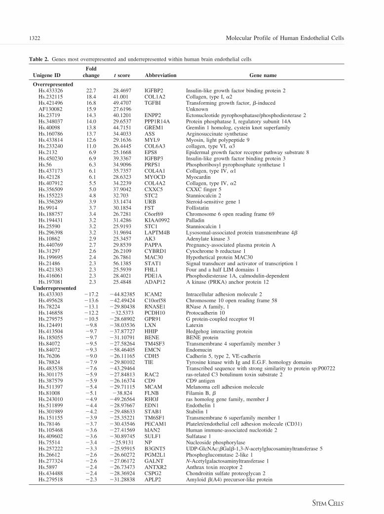
Table 2. Genes most overrepresented and underrepresented within human brain endothelial cells
Unigene IDFold
change t score Abbreviation Gene name
OverrepresentedHs.433326 22.7 28.4697 IGFBP2 Insulin-like growth factor binding protein 2Hs.232115 18.4 41.001 COL1A2 Collagen, type I, "2Hs.421496 16.8 49.4707 TGFBI Transforming growth factor, #-inducedAF130082 15.9 27.6196 UnknownHs.23719 14.3 40.1201 ENPP2 Ectonucleotide pyrophosphatase/phosphodiesterase 2Hs.348037 14.0 29.6537 PPP1R14A Protein phosphatase I, regulatory subunit 14AHs.40098 13.8 44.7151 GREM1 Gremlin 1 homolog, cystein knot superfamilyHs.160786 13.7 34.4033 ASS Arginosuccinate synthetaseHs.433814 12.6 29.1636 MYL9 Myosin, light polypeptide 9Hs.233240 11.0 26.4445 COL6A3 collagen, type VI, "3Hs.2132 6.9 25.1668 EPS8 Epidermal growth factor receptor pathway substrate 8Hs.450230 6.9 39.3367 IGFBP3 Insulin-like growth factor binding protein 3Hs.56 6.3 34.9096 PRPS1 Phosphoribosyl pyrophosphate synthetase 1Hs.437173 6.1 35.7357 COL4A1 Collagen, type IV, "1Hs.42128 6.1 28.6323 MYOCD MyocardinHs.407912 5.5 34.2239 COL4A2 Collagen, type IV, "2Hs.356509 5.0 37.9042 CXXC5 CXXC finger 5Hs.155223 4.8 32.703 STC2 Stanniocalcin 2Hs.356289 3.9 33.1474 URB Steroid-sensitive gene 1Hs.9914 3.7 30.1854 FST FollistatinHs.188757 3.4 26.7281 C6orf69 Chromosome 6 open reading frame 69Hs.194431 3.2 31.4286 KIAA0992 PalladinHs.25590 3.2 25.9193 STC1 Stanniocalcin 1Hs.296398 3.2 31.9694 LAPTM4B Lysosomal-associated protein transmembrane 4#Hs.10862 2.9 25.3457 AK3 Adenylate kinase 3Hs.440769 2.7 29.8539 PAPPA Pregnancy-associated plasma protein AHs.31297 2.6 26.2109 CYBRD1 Cytochrome b reductase 1Hs.199695 2.4 26.7861 MAC30 Hypothetical protein MAC30Hs.21486 2.3 56.1385 STAT1 Signal transducer and activator of transcription 1Hs.421383 2.3 25.5939 FHL1 Four and a half LIM domains 1Hs.416061 2.3 28.4021 PDE1A Phosphodiesterase 1A, calmodulin-dependentHs.197081 2.3 25.4848 ADAP12 A kinase (PRKA) anchor protein 12
UnderrepresentedHs.433303 #17.2 #44.82385 ICAM2 Intracellular adhesion molecule 2Hs.495628 #13.6 #42.49424 C10orf58 Chromosome 10 open reading frame 58Hs.78224 #13.1 #29.80438 RNASE1 RNase A family, 1Hs.146858 #12.2 #32.5373 PCDH10 Protocadherin 10Hs.279575 #10.5 #28.68902 GPR91 G protein-coupled receptor 91Hs.124491 #9.8 #38.03536 LXN LatexinHs.413504 #9.7 #37.87727 HHIP Hedgehog interacting proteinHs.185055 #9.7 #31.10791 BENE BENE proteinHs.84072 #9.5 #27.58264 TM4SF3 Transmembrane 4 superfamily member 3Hs.84072 #9.3 #58.46405 EMCN EndomucinHs.76206 #9.0 #26.11165 CDH5 Cadherin 5, type 2, VE-cadherinHs.78824 #7.9 #29.80102 TIE Tyrosine kinase with Ig and E.G.F. homology domainsHs.483538 #7.6 #43.29464 Transcribed sequence with strong similarity to protein sp:P00722Hs.301175 #5.9 #27.84813 RAC2 ras-related C3 botulinum toxin substrate 2Hs.387579 #5.9 #26.16374 CD9 CD9 antigenHs.511397 #5.4 #29.71115 MCAM Melanoma cell adhesion moleculeHs.81008 #5.1 #38.824 FLNB Filamin B, #Hs.243010 #4.9 #49.26564 RHOJ ras homolog gene family, member JHs.511899 #4.4 #28.97667 EDN1 Endothelin 1Hs.301989 #4.2 #29.48633 STAB1 Stabilin 1Hs.151155 #3.9 #25.35221 TM6SF1 Transmembrane 6 superfamily member 1Hs.78146 #3.7 #30.43546 PECAM1 Platelet/endothelial cell adhesion molecule (CD31)Hs.105468 #3.6 #27.41569 hIAN2 Human immune-associated nucleotide 2Hs.409602 #3.6 #30.89745 SULF1 Sulfatase 1Hs.75514 #3.4 #25.9131 NP Nucleoside phosphorylaseHs.257222 #3.3 #25.95915 B3GNT5 UDP-GlcNAc:#Gal#-1,3-N-acetylglucosaminyltransferase 5Hs.26612 #2.6 #26.60272 PGM2L1 Phosphoglucomutase 2-like 1Hs.277324 #2.6 #27.06172 GALNT N-Acetylgalactosaminyltransferase 1Hs.5897 #2.4 #26.73473 ANTXR2 Anthrax toxin receptor 2Hs.434488 #2.4 #28.36924 CSPG2 Chondroitin sulfate proteoglycan 2Hs.279518 #2.3 #31.28838 APLP2 Amyloid #(A4) precursor-like protein
1322 Molecular Profile of Human Endothelial Cells

differentiation of osteoblasts in vitro [72]. URB (steroid-sensi-tive gene 1) is a 150-kDa secreted protein and was recentlycharacterized in the mouse to have a role in skeletogenesis [73].In light of the recent demonstration of the osteoblastic niche forHSCs in the bone marrow, these data suggest the possibility thathormones elaborated by endothelial cells, possibly brain endo-thelial cells, may regulate osteoblast activity in the BM. Aneuroendocrine-hematopoietic axis has been postulated previ-ously [74], and the enrichment for osteoblast-regulatory factorswithin HUBECs further suggests this possibility. The recentdemonstration of overlapping genetic programs between neuraland hematopoietic stem cells [75] also suggests that brain-derived factors may have hematopoietic activity.
Of additional interest was the examination of transcripts thatwere downregulated in HUBECs compared with HUVECs. Ofnote, protocadherin and VE-cadherin were markedly underex-pressed in HUBECs compared with HUVECs. Since cadherin-based interactions have recently been implicated in the contact-dependent maintenance of quiescent HSCs in vivo within the
osteoblastic marrow niche [24], this implies that such interac-tions might be important for ex vivo maintenance of HSCs inculture. Despite this, noncontact HUBEC cultures and the re-sults of this gene expression analysis indicate that the cadherin-based contact interactions are not important for expansion ofHSCs in the HUBEC culture system. Taken together, these dataimplicate a novel soluble factor or factors elaborated by HU-BECs that promote the expansion of human repopulating cells.Moreover, these data suggest that the interaction of HSCs withcadherin moities may inhibit the proliferation of HSCs, therebymaintaining quiescence. Further studies will be important todelineate differences in the cell cycle status and SCID-repopu-lating capacity of HSCs cultured with osteoblasts and thosecultured under noncontact conditions with HUBECs.
The fetal liver murine stromal cell line AFT024 has beenshown, in contact cultures, to support the ex vivo maintenanceof murine and human HSCs [34, 35]. In contrast to HUBECs,which support HSC expansion equally under contact or noncon-tact conditions [43, 44], AFT024 support of LTC-IC has beenshown to decline under noncontact conditions [76]. When wequeried the most upregulated HUBEC transcripts against theAFT024 database, we identified seven transcripts in common
Table 3. Common transcripts between HUBEC and AFT024
Unigene ID Gene name E value
Hs.232115 Collagen type I, "2 !1 $ 10#5
Hs.23719 ENPP2 (Autotaxin) !1 $ 10#5
Hs.450230 IGFBP3 4 $ 10#29
Hs.443625 Collagen type VI, "3 1 $ 10#66
Hs.440769 Pregnancy-associated protein 4 $ 10#72
Hs.356289 Steroid-senstive gene 1 (URB) 2 $ 10#96
Hs.416061 PDE1A 2 $ 10#14
A Blast query search was performed to identify transcripts incommon between primary HUBEC and the murine fetal liverstromal cell line, AFT024 (57). Using the public StroCDBdatabase search engine, the entire gene sequences of the top 32HUBEC transcripts were queried against the AFT024transcriptome at http://www.stromalcell.princeton.edu. Genesequences were considered matched if the E value for thehomology was $1 $ 10#5.Abbreviation: HUBEC, human brain endothelial cell.
Table 4. Quantitative real-time RT-PCR analysis ofrepresentative genes
Gene HUBECs HUVECs
GAPDH 1.0 * 0.08 1.0 * 0.03IGFBP2 0.11 * 0.01 Not detectableDecorin 0.23 * 0.02 Not detectableAdrenomedullin 0.08 * 0.01 0.002 * 0.001Myocardin 0.04 * 0.03 Not detectable
Highly purified total RNA (2 !g per sample) was isolated fromprimary HUBECs and HUVECs and reverse-transcribed asdescribed in Materials and Methods. Fifty-nanogram equivalentsof cDNA were then used for quantitative real-time PCR fordecorin, insulin-like growth factor binding protein 2 (IGFBP2),myocardin, adrenomedullin, and glyceraldehyde-3-phosphatedehydrogenase (GAPDH). Relative gene expression betweenHUBECs and HUVECs was calculated using the &&Ct method,using GAPDH expression as a normalization reference.Abbreviations: GAPDH, glyceraldehyde-3-phosphatedehydrogenase; HUBEC, human brain endothelial cell; HUVEC,human umbilical vein endothelial cell; IGFBP2, insulin-likegrowth factor binding protein 2.
Figure 4. Adrenomedullin supports an increase in CD34" progenitorcell expansion in short-term culture. Primary human cord blood CD34"
cells (2.5 $ 104) were placed in culture with thrombopoietin, stem cellfactor, and Flt-3 ligand with and without 50–100 ng/ml IGFBP2, IG-FBP3, follistatin, or adrenomedullin for 7 days. (A): The addition ofIGFBP2, IGFBP3, or follistatin had no effect on total progenitor cellexpansion, whereas adrenomedullin caused a significant increase in totalcells (p ' .001). (B): The addition of adrenomedullin also caused asignificant increase in the number of CD34" progenitor cells over time(p ' .002). Abbreviations: ADM, adrenomedullin; Foll, follistatin;IGFBP, insulin-like growth factor binding protein; TSF, thrombopoi-etin, stem cell factor, and Flt-3 ligand.
1323Chute, Muramoto, Dressman et al.
www.StemCells.com

between HUBECs and AFT024, including collagen type I andVI, IGFBP3, URB, and autotaxin. We have prioritized func-tional assay of these genes via loss of function small inhibitoryRNA studies, since these molecules should have a high proba-bility of participation in HSC signaling. We also anticipate thatother extracellular HUBEC transcripts unique from the AFT024transcriptome will prove to be functionally important in HSCregulation in light of the distinctly soluble nature of the HUBEChematopoietic activity that we have observed.
As an initial strategy to screen for the hematopoietic activityof novel growth factors expressed by HUBECs, we have ana-lyzed a group of proteins that are available in recombinant formand have established extracellular function: IGFBP2, IGFBP3,follistatin, and adrenomedullin. Interestingly, one of these pro-teins, adrenomedullin, augments the expansion of human
CD34" cell when combined with thrombopoietin, SCF, andFlt-3 ligand, while also enhancing the individual activities ofSCF and Flt-3 ligand on HSC-enriched CD34"CD38#lin# cellsin vitro. These data indicate that further studies are merited todefine the effects of adrenomedullin on HSC fate and hemato-poiesis in general, in addition to our planned siRNA genesilencing studies to determine the precise contribution of ad-renomedullin to HUBEC-mediated HSC expansion. Although ithas been established that adrenomedullin is required for normalcardiovascular development [77], the hematopoietic activity ofadrenomedullin has not been well characterized. However, hu-man CD34" cells express the calcitonin receptor-like receptor,the receptor for adrenomedullin [78], and stromal cells express-ing adrenomedullin as well as other growth factors supporthuman colony-forming cell growth in vitro [79]. We plan to
Figure 5. Hematopoietic activity of human brain endothelial cell (HUBEC)-secreted factors on primitive human CB CD34"CD38#lin# progenitors.FACS-sorted human CB CD34"CD38#lin# cells (n ' 5 cells per well) were sorted into individual Terasaki culture wells with stem cell factor (SCF),Flt-3 ligand, or TSF with and without 50–100 ng/ml recombinant IGFBP2, IGFBP3, follistatin, or adrenomedullin. The bar graphs indicate the meantotal cell expansion under each condition at day 7. As shown, IGFBP2 failed to induce significant proliferation of human progenitors (A), whereas50 ng/ml IGFBP3 appeared to have an additive effect with Flt-3 ligand (B) (p ' .01). Follistatin had no effect on progenitor cell proliferation (C),whereas adrenomedullin demonstrated a dose-responsive additive effect on CD34"CD38#lin# progenitor cell proliferation when combined with bothSCF (p ' .01) and Flt-3 ligand (p ' .003) (D). Abbreviations: SCF, stem cell factor; TSF, thrombopoietin, stem cell factor, and Flt-3 ligand.
1324 Molecular Profile of Human Endothelial Cells
recombinantly produce and functionally assay each of the geneswith secretory or extracellular domains that are overexpressedby HUBECs and anticipate that the reproduction of HUBECstem cell-supportive activity may require the combination ofseveral proteins identified thus far.
In summary, we have presented a molecular profile ofnovel endothelial cells that support the ex vivo expansion ofhuman HSCs. Since HUBECs are unlike other establishedstromal cell lines (e.g., AFT024) in the soluble nature of theirHSC-supportive activity, it is plausible that novel solubleproteins produced by HUBECs can be identified and charac-terized. The identities of these factors may overlap withsecreted factors produced within the BM microenvironment
that support in vivo HSC maintenance and proliferation [1,22–24]. The HUBEC molecular profile is a template for theidentification of soluble factors that mediate hematopoieticstem cell fate.
ACKNOWLEDGMENTSWe thank Dr. David Venzon from the National Cancer Institutefor critical assistance with the biostatistical analysis. PrimaryHUBECs were kindly provided by the Naval Medical ResearchCenter (Silver Spring, MD) for a portion of the studies.
DISCLOSURESThe authors indicate no potential conflicts of interest.
REFERENCES
1 Sorrentino P. Clinical strategies for expansion of haematopoietic stemcells. Nat Rev Immunol 2004;4:878–888.
2 Osawa M, Hanada K, Hamada H, Nakauchi H. Long-term lymphohe-matopoietic reconstitution from a single CD34-/low hematopoietic stemcell. Science 1996;273:242–245.
3 Cashman J et al. Kinetic evidence of the regeneration of multilineagehematopoiesis from primitive cells in normal human bone marrow trans-planted into immune deficient mice. Blood 1997;80:4307–4316.
4 Rocha V, Cornish J, Sievers E et al. Comparison of outcomes of unre-lated bone marrow and umbilical cord blood transplants in children withacute leukemia. Blood 2001;97:2962–2971.
5 Storb R, Blume K, O’Donnell M et al. Cyclophosphamide and anti-thymocyte globulin to condition patients with aplastic anemia for allo-geneic marrow transplantations: The experience of 4 centers. Biol BloodMarrow Transplant 2001;7:39–44.
6 Kiehl M, Kraut L, Schwerdtfeger T et al. Outcome in hematopoietic stemcell transplantation in adult patients with acute lymphoblastic leukemia:No difference in related compared with unrelated transplant in firstcomplete remission. J Clin Oncol 2004;15:2816–2825.
7 Gur H, Krauthgamer R, Berrebi A et al. Tolerance induction bymegadose hematopoietic progenitor cells: Expansion of veto cells byshort term culture of purified human CD34" cells. Blood 2002;99:4174 – 4181.
8 Cogle C, Wainman D, Jorgensen M et al. Adult human hematopoieticcells provide functional hemangioblast activity. Blood 2004;103:133–135.
9 Rafii S, Lyden D. Therapeutic stem and progenitor cell transplanta-tion for organ vascularization and regeneration. Nat Med 2003;9:702–712.
10 Bhatia M, Wang J, Kapp U et al. Purification of primitive humanhematopoietic cells capable of repopulating immune-deficient mice. ProcNatl Acad Sci U S A 1997;94:5320–5325.
11 Krosl J, Austin P, Beslu N et al. In vitro expansion of hematopoietic stemcells by recombinant TAT-HOXB4 protein. Nat Med 2003;9:1428–1432.
12 Piacibello W, Bruno S, Sanavio F et al. Lentiviral gene transfer and exvivo expansion of human primitive stem cells capable of primary, sec-ondary, and tertiary multilineage repopulation in NOD/SCID mice.Blood 2002;15:4391–4400.
13 Chute J, Saini AA, Chute DJ et al. Co-culture with human brain endo-thelial cells increases the SCID repopulating capacity of adult humanbone marrow. Blood 2002;100:4433–4439.
14 Kawano Y, Kobune M, Yamaguchi M et al. Ex vivo expansion of humanumbilical cord hematopoietic progenitor cells using a coculture systemwith human telomerase catalytic subunit (hTERT) transfected humanstromal cells. Blood 2003;101:532–540.
15 Reya T, Duncan A, Ailles L et al. A role for Wnt signalling in the selfrenewal of hematopoietic stem cells. Nature 2003;423:409–414.
16 Varnum-Finney B, Xu L, Brashem-Stein C et al. Pluripotent, cytokine-dependent, hematopoietic stem cells are immortalized by constitutiveNotch1 signaling. Nat Med 2000;6:1278–1281.
17 Bhardwaj G, Murdoch B, Wu D et al. Sonic hedgehog induces theproliferation of primitive human hematopoietic cells via BMP regulation.Nat Immunol 2001;2:172–180.
18 Amsellem S, Pflumio F, Bardinet D et al. Ex vivo expansion of humanhematopoietic stem cells by direct delivery of the HOXB4 homeoprotein.Nat Med 2003;91423–1427.
19 Basser R, To L, Begley C et al. Rapid hematopoietic recovery aftermulticycle high dose chemotherapy: Enhancement of filgrastim-inducedprogenitor cell mobilization by recombinant human stem cell factor.J Clin Oncol 1998;16:1899–1908.
20 Rizzo J, Lichtin A, Woolf S et al. Use of epoietin in patients with cancer:Evidenced based clinical practice guidelines of the American Society ofClinical Oncology and the American Society of Hematology. J ClinOncol 2002;20:4083–4107.
21 Bennett C, Smith T, Weeks J et al. Use of hematopoietic colony stimu-lating factors: The American Society of Clinical Oncology survey. TheHealth Services Research Committee of the American Society of ClinicalOncology. J Clin Oncol 1996;14:2511–2520.
22 Avecilla S, Hattori K, Heissig B et al. Chemokine mediated interactionof hematopoietic progenitors with the bone marrow vascular niche isrequired for thrombopoiesis. Nat Med 2004;10:64–71.
23 Calvi L, Adams G, Weibrecht K et al. Osteoblastic cells regulated thehaematopoietic stem cell niche. Nature 2003;425:841–846.
24 Zhang J, Niu C, Ye L et al. Identification of the haematopoietic stem cellniche and control of the niche size. Nature 2003;425:836–841.
25 Shalaby F, Rossant J, Yamaguchi TP et al. Failure of blood islandformation and vasculogenesis in flk-1 deficient mice. Nature 1995;376:62–66.
26 Oberlin E, Tavian M, Blazsek I et al. Blood-forming potential ofvascular endothelium in the human embryo. Development 2002;129:4147– 4157.
27 Choi K, Kennedy M, Kazarov A et al. A common precursor for hema-topoietic and endothelial cells. Development 1998;125:725–732.
28 Lu L, Wang S, Auerbach R. In vitro and in vivo differentiation into Bcells, T cells, and myeloid cells of primitive yolk sac hematopoieticprogenitor cells expanded %100 fold by a clonal yolk sac endothelial cellline. Proc Natl Acad Sci U S A 1996;93:14782–14787.
29 Rafii S, Shapiro F, Rimarachin J et al. Isolation and characterization ofhuman bone marrow microvascular endothelial cells: Hematopoieticprogenitor cell adhesion. Blood 1994;84:10–19.
30 Rafii S, Shapiro F, Pettengell R et al. Human bone marrow micro-vascular endothelial cells support long term proliferation and differ-entiation of myeloid and megakaryocytic progenitors. Blood 1995;86:3353–3363.
31 Tavian M, Coulombel L, Luton D et al. Aorta associated CD34" hema-topoietic cells in the early human embryo. Blood 1996;87:67–72.
1325Chute, Muramoto, Dressman et al.
www.StemCells.com

32 Heissig B, Hattori K, Dias S et al. Recruitment of stem and progenitorcells from the bone marrow niche requires MMP-9 mediated release ofkit ligand. Cell 2002;109:625–637.
33 Gan O, Murdoch B, Larochelle A et al. Differential maintenance ofprimitive SCID-repopulating cells, clonogenic progenitors, and long-term culture initiating cells after culture on human bone marrow stromalcells. Blood 1997;90:641–650.
34 Punzel M, Liu D, Zhang T et al. The symmetry of initial divisions ofhuman hematopoietic progenitors is altered only by the cellular micro-environment. Exp Hematol 2003;31:339–347.
35 Lewis I, Almeida-Porada G, Du J et al. Umbilical cord blood cellscapable of engrafting in primary, secondary, and tertiary xenogeneichosts are preserved after ex vivo culture in a non-contact system. Blood2001;7:3441–3449.
36 Chute JP, Kampen R, Wells R et al. A comparative study of the cell cyclestatus and primitive cell adhesion molecule profile of human CD34"
cells cultured in stroma-free versus porcine microvascular endothelialcell cultures. Exp Hematol 1999;27:370–379.
37 Chute JP, Saini A, Wells M et al. Pre-incubation with endothelial cellmonolayers increases gene transfer efficiency into human bone marrowCD34"CD38# progenitor cells. Hu Gene Ther 2000;11:2515–2528.
38 Chute J, Fung J, Muramoto G et al. Ex vivo culture rescues hematopoi-etic stem cells with long term repopulating capacity following harvestfrom lethally irradiated mice. Exp Hematol 2004;32:308–317.
39 Brandt JE, Bartholemew AM, Fortman JD et al. Ex vivo expansion ofautologous bone marrow stem cells with porcine microvascular endothe-lial cells results in a graft capable of rescuing lethally irradiated baboons.Blood 1999;94:106–113.
40 Chute JP, Fung J, Muramoto G, Oxford C. Quantitative analysis dem-onstrates expansion of SCID-repopulating cells (SRC) and increasedengraftment capacity within human cord blood cells following culturewith human brain endothelial cells. STEM CELLS 2004;22:202–215.
41 Chute JP, Muramoto G, Fung J et al. Soluble factors elaborated byhuman brain endothelial cell induce the concomitant expansion of puri-fied BM CD34"CD38# cells and SCID repopulating cells. Blood 2005;105:576–583.
42 Schulz L, Schweitzer P, Christianson S et al. Multiple defects in innateand adaptive immunologic function in NOD/LtSz-scid mice. J Immunol1995;154:180–191.
43 Taswell C. Limiting dilution assays for the determination of immuno-competent cell frequencies, I: Data analysis. J Immunol 1981;126:1614–1619.
44 Wang J, Doedens M, Dick J. Primitive human hematopoietic cells areenriched in cord blood compared with adult bone marrow or mobilizedperipheral blood as measured by the quantitative in vivo SCID-repopu-lating cell assay. Blood 1997;89:3919–3924.
45 Jazwiec B, Solanilla A, Grosset C et al. Endothelial cell support ofhematopoiesis is differentially altered by IL-1 and glucocorticoids. Leu-kemia 1998;8:1210–1220.
46 Coombes B, Mahoney J. cDNA array analysis of altered gene expressionin human endothelial cells in response to Chlamydia pneumoniae infec-tion. Infect Immun 2001;69:1420–1427.
47 Gerritsen M, Soriano R, Yang S et al. Branching out: A molecularfingerprint of endothelial differentiation into tube-like structures gener-ated by Affymetrix oligonucleotide arrays. Microcirculation 2003;10:63–81.
48 Soon L, Yie T, Shvarts A et al. Overexpression of WISP-1 downregu-lated motility and invasion of lung cancer cells through inhibition of Racactivation. J Biol Chem 1003:278:11465–11470.
49 Cousins R, Blanchard R, Popp M et al. A global view of the selectivityof Zinc deprivation and excess on genes expressed in human THP-1mononuclear cells. Proc Natl Acad Sci U S A 2003;100:6952–6957.
50 Brazma A, Hingamp P, Quackenbush J et al. Minimum informationabout a microarray experiment (MIAME)-toward standards for microar-ray data. Nat Genet 2001;29:365–371.
51 Dudoit S, Gentleman R, Quackenbush J. Open source software for theanalysis of microarray data. Biotechniques 2003;suppl:45–51.
52 Hosack D, Dennis G, Sherman B et al. Identifying biological themeswithin lists of genes with EASE. Genome Biol 2003;4:R70.
53 Klein N, Battaglia D, Woodruff T et al. Ovarian follicular concentrationsof activin, follistatin, inhibin, insulin-like growth factor I (IGF-1), IGF-II, IGF binding protein 2 (IGFBP2), IGFBP3, and vascular endothelialgrowth factor in spontaneous menstrual cycles of normal women ofadvanced reproductive age. J Clin Endocrinol Metab 2000;85:4520–4525.
54 Hackney J, Charboard P, Brunk B et al. A molecular profile of ahematopoietic stem cell niche. Proc Natl Acad Sci U S A 2002;99:13061–13066.
55 Fox M, Colello R, Macklin W et al. Phosphodiesterase-1 alpha/autotaxin:A counteradhesive protein expressed by oligodendrocytes during onset ofmyelination. Mol Cell Neurosci 2003;23:507–519.
56 Ivanova N, Dimos J, Schaniel C et al. A stem cell molecular signature.Science 2002;298:601–604.
57 Ramalho-Santos M, Yoon S, Matsuzaki Y et al. “Stemness”: Transcrip-tional profiling of embryonic and adult stem cells. Science 2002;298:597–600.
58 Georgantas R, Tanavde V, Malehorn M et al. Microarray and serialanalysis of gene expression analyses identify known and novel tran-scripts overexpressed in hematopoietic stem cells. Cancer Res 2004;64:4434–4441.
59 Sitnicka E, Ruscetti F, Priestley G et al. Transforming growth factor beta1 directly and reversibly inhibits the initial cell divisions of long termrepopulating hematopoietic stem cells. Blood 1996;88:82–88.
60 Hofflich A, Lahm H, Blum W et al. Insulin like binding protein 2 inhibitsproliferation of human embryonic kidney fibroblasts and of IGF respon-sive colon carcinoma cell lines. FEBS Lett 1998;434:329–334.
61 Mouhieddine O, Cazals V, Kuto E et al. Glucocorticoid growth arrest oflung alveolar epithelial cells is associated with increased production ofinsulin like growth factor binding protein 2. Endocrinology 1996;137:287–295.
62 Longobardi L, Torello M, Buckway C et al. A novel insulin-like growthfactor I independent role for insulin like growth factor binding protein 3in mesenchymal chondroprogenitor cell apoptosis. Endocrinology 2003;144:1695–1702.
63 Vihervouri E, Virtanen M, Koistinen H et al. Hemoglobin level is linkedto growth hormone dependent proteins in short children. Blood 1996;87:2075–2081.
64 Bhatia R, Williams A, Munthe H. Contact with fibronectin enhancesmaintenance of normal but not chronic myelogenous leukemia primitivehematopoietic progenitors. Exp Hematol 2002;30:324–332.
65 Yokota T, Oritani K, Takahashi I et al. Adiponectin, a new member ofthe family of soluble defense collagens, negatively regulates the growthof myelomonocytic progenitors and the functions of macrophages. Blood2000;96:1723–1732.
66 Zon L. Self renewal versus differentiation, a job for the mighty morpho-gens. Nat Immunol 2001;2:142–143.
67 Meehan T, Schlatt S, O’Bryan M et al. Development of germ cell andSertoli cell development by activin A, follistatin, and FSH. Dev Biol2000;220:225–237.
68 Matzuk M, Lu N, Vogel H et al. Multiple defects and perinatal death inmice deficient in follistatin. 1995;374:360–363.
69 Yamashita T, Takahashi S, Ogata E. Expression of activin/erythroiddifferentiation factor in murine bone marrow stromal cells. Blood 1992;79:304–307.
70 Michos O, Panman L, Vintersten K et al. Gremlin-mediated BMP an-tagonism induces the epithelial-mesenchymal feedback signaling con-trolling metanephric kidney and limb organogenesis. Development 2004;131:3401–3410.
71 Zhang Y, Cleary M, Si Y et al. Inhibition of activin signaling inducespancreatic epithelial cell expansion and diminishes terminal differentia-tion of pancreatic beta cells. Diabetes 2004;53:2024–2033.
1326 Molecular Profile of Human Endothelial Cells

72 Yoshiko Y, Aubin J. Stanniocalcin 1 as a pleiotropic factor in mammals.Peptides 2004;25:1663–1669.
73 Liu Y, Monticone M, Tonachini L et al. URB expression in human bonemarrow stromal cells and during mouse development. Biochem BiophysRes Commun 2004;322:497–507.
74 Murphy W, Durum S, Anver M, Longo D. Immunologic and hemato-logic effects of neuroendocrine hormones: Studies on DW/J dwarf mice.J Immunol 1992;148:3799–3805.
75 Terskikh A, Easterday M, Li L et al. From hematopoiesis to neuropoiesis:Evidence of overlapping genetic programs. Proc Natl Acad Sci U S A2001;98:7934–7939.
76 Punzel M, Gupta P, Roodell M et al. Factor(s) secreted by AFT024 fetalliver cells following stimulation with human cytokines are important forhuman LTC-IC growth. Leukemia 1999;13:1079–1084.
77 Caron K, Smithies O. Extreme hydrops fetalis and cardiovascularabnormalities in mice lacking the adrenomedullin gene. Proc NatlAcad Sci U S A 2001;98:615– 619.
78 Harzenetter M, Keller U, Beer S et al. Regulation and function of theCGRP receptor complex in human granulopoiesis. Exp Hematol 2002;30:306–312.
79 De Angeli S, Di Liddo R, Buoro S et al. New immortalized humanstromal cell lines enhancing in vitro expansion of cord blood hemato-poietic stem cells. Int J Mol Med 2004;13:363–371.
See www.StemCells.com for supplemental material available online.
1327Chute, Muramoto, Dressman et al.
www.StemCells.com
Related Documents


