Molecular Modeling of 3-Arylisoquinoline Antitumor Agents Active Against A-549. A Comparative Molecular Field Analysis Study Won-Jea Cho, a, * Eui-Ki Kim, a Il Yeong Park, b Eun Young Jeong, a Tae Sung Kim, a Thanh Nguyen Le, a Dae-Duk Kim c and Eung-Seok Lee d a College of Pharmacy, Chonnam National University, Yongbong-dong Buk-gu, Kwangju 500-757, Republic of Korea b College of Pharmacy, Chungbuk National University, Cheongju 361-763, Republic of Korea c College of Pharmacy, Pusan National University, Pusan 609-735, Republic of Korea d College of Pharmacy, Yeungnam University, Kyungsan 712-749, Republic of Korea Received 15 January 2002; accepted 13 April 2002 Abstract—A series of 58 3-arylisoquinoline antitumor agents were investigated for defining the pharmacophore model using com- parative molecular field analysis (CoMFA) program. The studied compounds related to bioisostere of benzophenanthridine alka- loid were synthesized and evaluated for antitumor cytotoxicity against human lung tumor cell (A 549). In order to perform the systematic molecular modeling study of these compounds, the conformational search was carried out based on the single X-ray crystallographic structure of 7,8-dimethoxy-3-phenylisoquinolin-(2H)-one (2). Interestingly, two types of structures having different dihedral angles between the isoquinoline ring and 3-aryl ring were found in the crystals. Therefore, CoMFA was performed two different, overlapping ways. The alignments of the structures were based on the common isoquinoline ring and 3-aryl ring. The 3-D-quantitative structure–activity relationship study resulted in significant cross-validated, conventional r 2 values equal to 0.715 and 0.927, respectively. # 2002 Elsevier Science Ltd. All rights reserved. Introduction New chemotherapeutic agents for a treatment of cancer from natural compounds have been developed over the last decade. 1 With the finding of potent antitumor activity of 7,8-dimethoxy-2-methyl-3-(4,5-methylene- dioxy-2-vinylphenyl)isoquinolin-1(2H)-one (1) which is a synthetic intermediate for natural benzo[c]phen- anthridine alkaloid, chelerythrine, we tried to study structure–activity relationships of 3-arylisoquinolines against human tumor cell lines. 2 Most of the 3-aryliso- quinoline derivatives exhibited potent cytotoxicities against five different human tumor cell lines. The sty- rene (1) is considered to be a bioisostere of benzo[c]- phenanthridine which has exhibited potent antitumor activity by inhibiting DNA topoisomerase I or II. 2 From earlier studies, we could obtain the results that the amide carbonyl group of isoquinoline ring and alkyl group on aromatic rings played an important role for exhibiting the activities. 3 In order to increase water solubility of the compound, the amide carbonyl group was modified to various amines which could be con- verted to salt forms because the water solubility of the drugs should be considered for in vivo or clinical experiments. As we expected, the modification of carbonyl amide group to amines gave relatively potent cytotoxi- cites against several tumor cell lines as well as good water solubility. Among these compounds, 1-(4-methyl- piperazinyl)-3-phenylisoquinoline hydrochloride (CWJ- a-5) was tested in vivo assay using BDF1 mice (P388 leukemia) and resulted in 160 T/C% with low toxicity. 3 Moreover, the high oral bioavailability of CWJ-a-5 indi- cated the possibility of it being developed into an oral formulation. Also, the pharmacokinetic data served as useful information in future clinical studies of CWJ-a-5. 4 CoMFA, which is a ligand-based drug design skill, is not only one of the most used 3-D-QSAR methods, but also has been applied to a number of different classes of compounds. 5 0968-0896/02/$ - see front matter # 2002 Elsevier Science Ltd. All rights reserved. PII: S0968-0896(02)00137-2 Bioorganic & Medicinal Chemistry 10 (2002) 2953–2961 *Corresponding author. Tel.: +82-62-530-2933; fax: +82-62-530- 2911; e-mail: [email protected]

Welcome message from author
This document is posted to help you gain knowledge. Please leave a comment to let me know what you think about it! Share it to your friends and learn new things together.
Transcript
Molecular Modeling of 3-Arylisoquinoline Antitumor AgentsActive Against A-549. A Comparative Molecular
Field Analysis Study
Won-Jea Cho,a,* Eui-Ki Kim,a Il Yeong Park,b Eun Young Jeong,a Tae Sung Kim,a
Thanh Nguyen Le,a Dae-Duk Kimc and Eung-Seok Leed
aCollege of Pharmacy, Chonnam National University, Yongbong-dong Buk-gu, Kwangju 500-757, Republic of KoreabCollege of Pharmacy, Chungbuk National University, Cheongju 361-763, Republic of Korea
cCollege of Pharmacy, Pusan National University, Pusan 609-735, Republic of KoreadCollege of Pharmacy, Yeungnam University, Kyungsan 712-749, Republic of Korea
Received 15 January 2002; accepted 13 April 2002
Abstract—A series of 58 3-arylisoquinoline antitumor agents were investigated for defining the pharmacophore model using com-parative molecular field analysis (CoMFA) program. The studied compounds related to bioisostere of benzophenanthridine alka-loid were synthesized and evaluated for antitumor cytotoxicity against human lung tumor cell (A 549). In order to perform thesystematic molecular modeling study of these compounds, the conformational search was carried out based on the single X-raycrystallographic structure of 7,8-dimethoxy-3-phenylisoquinolin-(2H)-one (2). Interestingly, two types of structures having differentdihedral angles between the isoquinoline ring and 3-aryl ring were found in the crystals. Therefore, CoMFA was performed twodifferent, overlapping ways. The alignments of the structures were based on the common isoquinoline ring and 3-aryl ring. The3-D-quantitative structure–activity relationship study resulted in significant cross-validated, conventional r2 values equal to 0.715and 0.927, respectively. # 2002 Elsevier Science Ltd. All rights reserved.
Introduction
New chemotherapeutic agents for a treatment of cancerfrom natural compounds have been developed over thelast decade.1 With the finding of potent antitumoractivity of 7,8-dimethoxy-2-methyl-3-(4,5-methylene-dioxy-2-vinylphenyl)isoquinolin-1(2H)-one (1) which isa synthetic intermediate for natural benzo[c]phen-anthridine alkaloid, chelerythrine, we tried to studystructure–activity relationships of 3-arylisoquinolinesagainst human tumor cell lines.2 Most of the 3-aryliso-quinoline derivatives exhibited potent cytotoxicitiesagainst five different human tumor cell lines. The sty-rene (1) is considered to be a bioisostere of benzo[c]-phenanthridine which has exhibited potent antitumoractivity by inhibiting DNA topoisomerase I or II.2
From earlier studies, we could obtain the results that theamide carbonyl group of isoquinoline ring and alkyl
group on aromatic rings played an important role forexhibiting the activities.3 In order to increase watersolubility of the compound, the amide carbonyl groupwas modified to various amines which could be con-verted to salt forms because the water solubility of thedrugs should be considered for in vivo or clinicalexperiments. As we expected, themodification of carbonylamide group to amines gave relatively potent cytotoxi-cites against several tumor cell lines as well as goodwater solubility. Among these compounds, 1-(4-methyl-piperazinyl)-3-phenylisoquinoline hydrochloride (CWJ-a-5) was tested in vivo assay using BDF1 mice (P388leukemia) and resulted in 160 T/C% with low toxicity.3
Moreover, the high oral bioavailability of CWJ-a-5 indi-cated the possibility of it being developed into an oralformulation. Also, the pharmacokinetic data served asuseful information in future clinical studies of CWJ-a-5.4
CoMFA, which is a ligand-based drug design skill, isnot only one of the most used 3-D-QSAR methods, butalso has been applied to a number of different classes ofcompounds.5
0968-0896/02/$ - see front matter # 2002 Elsevier Science Ltd. All rights reserved.PI I : S0968-0896(02 )00137-2
Bioorganic & Medicinal Chemistry 10 (2002) 2953–2961
*Corresponding author. Tel.: +82-62-530-2933; fax: +82-62-530-2911; e-mail: [email protected]

In the previous paper, we reported a comparativemolecular field analysis (CoMFA) result of 29 3-aryliso-quinolines with human melanoma tumor cell (SK-MEL-2) to produce a hypothetical pharmacophoremodel.6 In the present study, the model for A-549 tumorcell line is extended to include various substituents ofthe aromatic ring as well as C1 position because it is wellrecognized that 3-D QSAR study of diverse modifiedmolecules, which show a wide range of activities, affordsa reliable pharmcophore model. Therefore, new mole-cules were designed and synthesized based on the pos-tulated pharmacophore model derived from formerCoMFA study. Moreover, we tried to determine theX-ray crystallographic structure of 7,8-dimethoxy-3-phenylisoquinolin-(2H)-one (2) for getting the infor-mation of the torsion angle of 3-aryl ring, which is con-sidered to be crucial for performing CoMFA of the3-arylisoquinoline derivatives (Scheme 1). Five differentCoMFA pharmacophore contour maps obtained usingA-549, SK-OV-3, SK-MEL-2, XF 498 and HCT 15tumor cell lines showed very similar patterns. Althoughthe target sites of action of the compounds were notclarified yet, we assumed that they share the samebinding sites because of their closely related structures.7
Methods
Synthesis
Synthesis of diverse 3-arylisoquinoline analogues isdescribed in Scheme 2. The imine chloride (5)6 derived
from the corresponding amide was treated with variousamines or thioacetic acid in refluxing DMF to providethe 1-substituted isoquinolines in good yield. For thepreparation of primary amines (6f–6h), the benzyl-amines (6a–6c) was performed to a catalytic hydro-genation reaction with 5% Pd/C in 80 psi hydrogenatmosphere to afford the corresponding amines inmoderate yield. The coupling reaction of 2,3-dimethoxy-6,N-dimethylbenzamide (8) with benzonitrile (9) wasaccomplished via dilithio species using n-BuLi in 62%yield. The cyclization reaction of 3,4-methylenedioxy-6,N-dimethylbenzamide (10) and 2-methylbenzonitrile(11) was also performed to yield the corresponding iso-quinoline which was then treated with MeI to give theN-methylated isoquinoline (12) in 63% yield.
Biological data
Cytotoxicity (IC50 value) was obtained by the followingmethod: All experimental procedures were followed upusing the NCI (USA)’s protocol based on the sulforho-damine B (SRB) method.8 Briefly, tumor cells were cul-tured to maintain logarithmic growth by changing themedium 24 h before cytotoxicity assays. On the day ofthe assay, the cells were harvested by trysinization,counted, diluted in media and added to 96-well plates.The concentration of tumor cells (A-549) used was 1 �104. The cells were then preincubated for 24 h in a 5%CO2 incubator at 37 �C. The compounds dissolved inDMSO were added to the wells in six 2-fold dilutionsstarting from the highest concentrations, and incubatedfor 48 h in a 5% CO2 incubator at 37 �C. The final
Scheme 1. The structure of 3-arylisoquinolines.
2954 W.-J. Cho et al. / Bioorg. Med. Chem. 10 (2002) 2953–2961

DMSO concentration was 0.05%. At the termination ofthe incubation, the culture medium in each well wasremoved, and the cells were fixed with cold 10% tri-chloroacetic acid (TCA) for 1 h at room temperature.The microplates were washed, dried, and stained with0.4% SRB in 1% acetic acid for 30min at room tem-perature. The cells were washed again and the boundstain was solubilized with 10mM Tris base solution (pH10.5), and the absorbances were measured spectro-photometrically at 520 nm on a microtiter plate reader(Molecular Devices, Sunnyvale, CA, USA). The datawas transformed into Lotus-123 format and survivalfractions were calculated by regression analysis (plottingthe cell viability versus the concentration of the testcompound). The IC50 values represent the concen-trations of the compounds that inhibit 50% of cellgrowth. All data represents the average values for aminimum of three wells (Table 1).
X-ray crystallographic structures and superimposition
In order to assign the torsion angle of 3-arylisoquino-lines, we carried out a single crystal X-ray analysis of
7,8-dimethoxy-3-phenylisoquinolin-1(2H)-one (2). Yel-lowish transparent prisms were obtained from an ethyl-acetate solution. The crystal was mounted on aMacScience KAPPA goniometer equipped with 18KWrotating anode. Cell constants were determined as fol-lows: monoclinic crystal system, a=13.804(2),b=15.683(4), c=12.632(2) A, �=93.87(2)�, space group;P21/a, Z=8. A total of 4403 independent reflectionswere measured. The structure was solved by directmethod and refined by full matrix least-squares proce-dure to the final R-value of 0.054 with maXus1.1 soft-ware package.9 There are two independent molecules inan asymmetric unit of the crystal as shown in Fig. 1.Two types of conformers having different dihedralangles between isoquinoline and 3-aryl rings werefound in crystal. As depicted, there are three torsionangles on the molecules such as conformer 1 (t1:�150.0�, t2: 95.0�, t3: �179.4�) and conformer 2 (t1:174.6�, t2: 99.1�, t3: 2.2�) shown on the right and leftside, respectively, in Figure 1.
As the initial conformations of the compounds, theX-ray crystallographic data of 2 was utilized. Due to the
Scheme 2. Synthesis of 3-arylisoquinoline derivatives.
W.-J. Cho et al. / Bioorg. Med. Chem. 10 (2002) 2953–2961 2955

two different conformations of the molecule 2, we triedto use both of them as lead templates for determiningthe conformation of the molecules as shown in Figure 2.The conformations of the compounds seem to be affec-ted by t1 value of 3-aryl ring rather than others. Toinvestigate the conformation profile, we carried out an
extensive conformational search of the compoundsusing the torsion angle of conformers 1 and 2. First, thetorsion angle of 3-aryl ring (t1) in the compounds wasfixed as the value (�150.0�) of conformer 1 and theconformation of each molecule was obtained after per-forming the energy minimization process. For the opti-mized coordinates, atomic charges were calculated usingGasteiger–Huckel. Secondly, by performing the aboveenergy minimization process using t1 (174.6�) of con-former 2, the active conformers of the compounds werealso obtained. Each active conformer of the 58 com-pounds was superimposed with that of the referencemolecules (conformer 1 and conformer 2 in Fig. 1) sothat each structural component is as close as possible tothe corresponding components of the reference. Thus,nine atoms, that is A ring and C3–C4 atoms and theC14 atom of C ring were selected, and the sum of the
Table 1. Observed and calculated cytotoxicity values against A-549
of the 3-arylisoquinoline derivatives
PIC50
No Compd R1 R2 R3 Obsd Calcd Diff
1 1a — — — 8.05 8.15 �0.102 3aa Me Et H2 5.89 6.20 �0.313 3ba Me Vinyl H2 5.72 5.80 �0.084 3ca Me CH2CH(OMe)2 O 6.49 6.52 �0.035 3da Me Et O 7.57 6.90 0.676 3ea H Et O 4.93 5.27 �0.347 4ab 5-NMe2 4-OMe H 4.90 4.53 0.378 4bb 6-Me 4-Cl H 5.24 4.85 0.399 4cb H 2-PMB H 4.12 4.29 �0.1710 4db H 3-Me H 4.39 4.45 �0.0611 4eb H 4-Br H 4.21 4.43 �0.2212 4fb H 4-Me H 4.52 4.42 0.1013 4gb H 4-OMe H 3.81 4.32 �0.5114 4hb H H H 3.91 3.43 0.4815 4ic H 2-CHO Me 3.87 3.75 0.1216 4jc H 2-CH2OH Me 3.84 3.95 �0.1117 4kc H 2-CH2OPMB Me 4.67 4.66 0.0118 4lc H 2-CH2CH(OMe)2 Me 3.69 3.66 0.0319 4mc H 2-vinyl Me 4.22 4.15 0.0720 4nb H H Me 4.07 4.25 �0.1821 5ab 5-NMe2 4-Cl — 3.84 3.89 �0.0522 5bb 6-Me 2-Cl — 3.88 3.85 0.0323 5cb 6-Me 2-Me — 3.58 3.86 �0.2824 5db 6-Me 4-Me — 3.95 4.24 �0.2925 5eb 6-Me H — 3.80 3.95 �0.1526 5fb H 3-Me — 3.77 3.75 0.0227 5gb H 4-Br — 3.95 3.71 0.2428 5hb H H — 3.82 3.44 0.3829 6ad 6-Me 2-Me Bn 4.56 4.39 0.1730 6bd H 2-Me Bn 3.85 3.78 0.0731 6cd H H Bn 3.76 3.82 �0.0632 6dd 6-Me 3-Me Bn 6.13 6.28 �0.1533 6ed H H morpholine 3.91 3.96 �0.0534 6fd 6-Me 2-Me NH2 6.76 6.25 0.5135 6gd H 2-Me NH2 5.50 5.70 �0.2036 6hd H H NH2 5.56 5.80 �0.2437 6id H H NH-PMB 3.61 3.59 0.0238 6jd H H piperidine 3.83 4.25 �0.4239 7ab 5-NMe2 2-Me — 4.67 4.86 �0.1940 7bb 5-NMe2 3-Me — 5.03 4.91 0.1241 7cb 5-NMe2 4-Br — 5.04 5.17 �0.1342 7db 5-NMe2 4-Cl — 5.13 5.13 043 7eb 5-NMe2 4-OMe — 4.72 4.77 �0.0544 7fb 5-NMe2 H — 4.83 4.62 0.2145 7gb 6-Me 2-Me — 5.03 4.86 0.1746 7hb 6-Me 4-Cl — 5.41 5.29 0.1247 7ib 6-Me 4-Me — 4.95 5.29 �0.3448 7jb 6-Me H — 5.13 5.09 0.0449 7kb H 2-Me — 4.23 4.30 �0.0750 7lb H 3-Me — 4.67 4.94 �0.2751 7mb H 4-Br — 4.73 4.65 0.0852 7nb H 4-Cl — 4.68 4.59 0.0953 7ob H 4-Me — 4.90 4.68 0.2254 7pb H 4-OMe — 4.75 4.53 0.2255 7qb H H — 4.47 4.38 0.0956 12d — — — 4.63 4.52 0.1157 13d — — — 3.69 3.70 �0.0158 14d — — — 4.60 4.77 �0.17
aFrom ref 2.bFrom ref 6.cFrom ref 13.dNewly synthesized compounds.
Figure 1. The crystal structure of 7,8-dimethoxy-3-phenylisoquinolin-1(2H)-one (2) drawn by ORTEP.10 The thermal ellipsoids are drawn atthe 50% probability level. Hydrogen atoms are drawn as small circlesof arbitrary radii. Two independent molecules in the asymmetric unitare designated as Conformer 1 and 2, respectively.
Figure 2. Torsion angles of 7,8-dimethoxy-3-phenylisoquinolin-1(2H)-one (2).
2956 W.-J. Cho et al. / Bioorg. Med. Chem. 10 (2002) 2953–2961

squares of the distance of the nine atomic positionsfrom the corresponding atomic positions of the refer-ence compound was made as small as possible, as shownin Figure 3.
Computer modeling
All computations were done with the molecular model-ing software package Sybyl 6.6 version.11 The coordi-nates of the substructures to be modified in substituedderivatives were calculated with the Sybyl standardvalues for bond lengths and angles using the buildingmodule. For the conventional CoMFA, the region wasfirst created automatically with the dimension of 18 �18 � 18 A (X=�7 to 11, Y=�12 to 6, Z=�6 to 12).When the region was manually defined for the mole-cules to be embedded in the grid box with dimension of24 � 22 � 32 A (X=�10 to 14, Y=�13 to 9, Z=�18to 14), more reliable statistical values such as rcv
2 andSEP were obtained as shown in Table 2. From thisresult, it is suggested that the grid size should be definedconsidering that all of the molecules were adequatelyfitted in the lattice box. The steric and electrostaticpotential energies at each lattice point were calculatedusing the Coulombic and Lennard–Jones potentialfunction, respectively. The lattice spacing was 2.0 A andthe +1 charge and the sp3 carbon were used as probesto estimate the electronic and steric fields, respectivelywith an energetic cutoff of 30 kcal/mol. Regression ana-lyses were performed using the Sybyl implementation ofthe PLS algorithm, initially with cross-validation (theleave-one-out technique) to reduce the probability ofobtaining chance correlation. The number of groups ofcross-validation was set as equal to the number of com-ponents of the training set. The optimal number of
latent variables (components) to be used in conven-tional analyses was chosen on the basis of the highestcross-validated r2 (r2cv) value, the smallest standard errorof prediction (SEP), and the minimum number of com-ponents. All leave-one-out calculations were carried outselecting a 2.0 kcal/mol energy column filter in order toenhance the signal-to-noise ratio. The steric and elec-trostatic field columns were obtained according to theCOMFA-STD default scaling option. In this method, afield is considered as a whole and every CoMFA vari-able is affected by the overall field mean and standarddeviation. Final PLS (non-cross-validated models) cali-bration equations were then derived using the optimalnumber of components so identified.
PKa values of amines
To validate whether or not amines are protonated underthe physiological condition, we calculated their pKa
values using the empirical correlation eqs 1 and 2 foraliphatic amines (NR1R2R3) reported by Takayama etal. and Hall, respectively.12 Eq. 1 is for the primaryand secondary amines, while eq 2 is for the tertiaryamines.
pKa ¼ �3:606 � s� þ 0:335 Ecs R2ð Þ þ Ec
s R3ð Þ� �
þ 1:427 nHþ 9:545 ð1Þ
pKa ¼ �3:30 � s� þ 9:61 ð2Þ
In these equations, � s* is the summation of the Taftvalues of three N-substituents. Es
c(R2)+Esc(R3) are,
respectively, the Hancock corrected steric constants oftwo bulkier substituents R2 and R3 among threeN-substituents, in which R1=H and Es
c(R2)�Esc(R3),
and nH is the number of hydrogen atoms in respectiveconjugate ammonium ions. s* and E s
c values used inthe calculations were taken from the literature.12 Thecalculation with eqs (1) and (2) estimated the pKa valueas follows: 7a–7q, 10.23; 6j, 11.34. Thus, all the aminescontaining the N-methylpiperazinyl group, or primaryamine, are reasonably considered to exist mostly in theirprotonated forms under the physiological pH condi-tions. Therefore, molecular modeling study of amineswas done in their protonated forms.
CoMFA results
The CoMFA statistical results for the cytotoxic activ-ities on A-549 are summarized in Table 3. In theseequations, m indicates the number of latent variablesand SEP is the standard deviation obtained from theleave-one-out cross-validation. RC refers to the relativecontribution of steric and electrostatic effects to varia-tions in antitumor activity. To determine whether thehydrophobic parameter is significantly involved, cLogPvalues were inserted in the CoMFA analyses. However,their contribution for the equation was less than 2% in
Figure 3. Template molecule for molecular superimposition.
Table 2. Statistical comparison of the region defined method and
Auto CoMFA with conformer 1 superposition
Principal components Alignment Ia Alignment IIb
r2 cv SEP r2 cv SEP
1 0.023 0.855 0.001 0.8652 0.331 0.714 0.281 0.7413 0.537 0.600 0.517 0.6134 0.624 0.546 0.540 0.6045 0.697 0.495 0.616 0.5586 0.715 0.485 0.632 0.5527 0.685 0.491 0.639 0.552
aRegion defined method; X, �7 to 11; Y, �12 to 6; Z, �6 to 12.bAuto CoMFA method; X, �10 to 14; Y, �13 to 9; Z, �18 to 14.
W.-J. Cho et al. / Bioorg. Med. Chem. 10 (2002) 2953–2961 2957

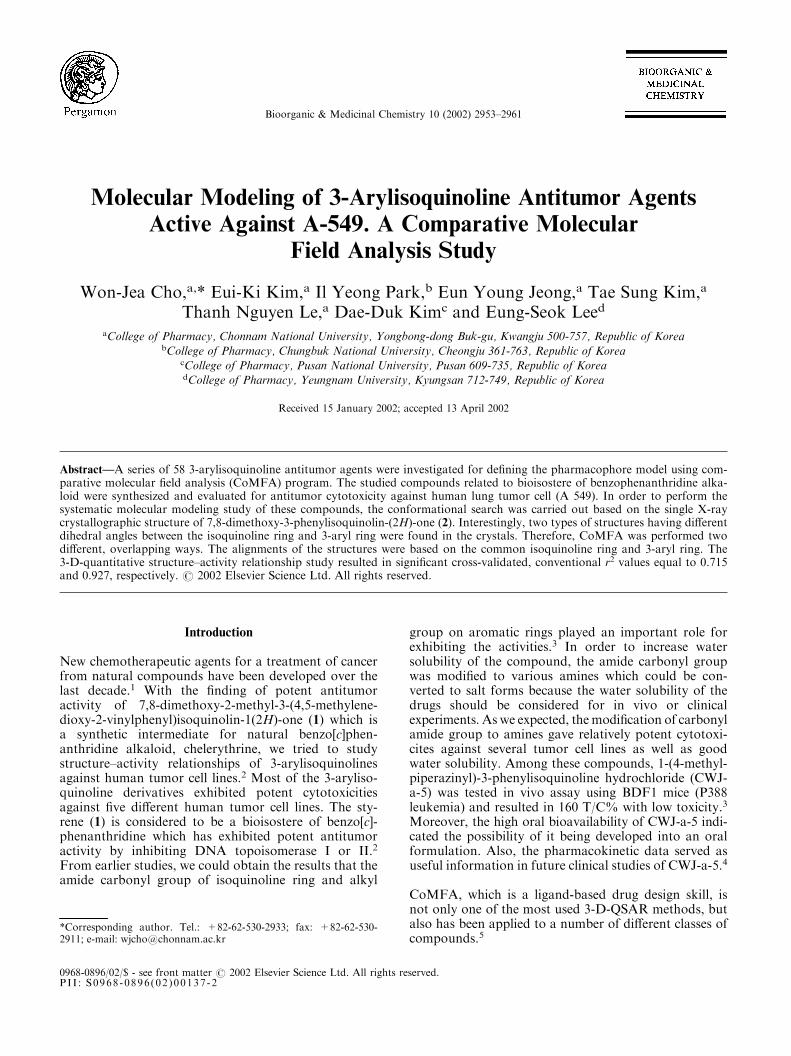
most cases. So, we initially analyzed for 58 compoundsand obtained eqs 3–5. Among these equations, eq 3,which was derived from the overlapping of the com-pounds on X-ray conformer 1 as for a lead template,was the most significant. On the other hand, eq 4 wasobtained from X-ray conformer 2 which gave poorerstatistical results. Therefore, we assumed that con-former 1 is the active form for showing the cytotoxicityand the molecules should have a �150� torsion angle of3-aryl ring. Therefore, the observed pIC50 values inTable 1 were calculated from eq 3. Figure 4 representsthe major steric and electrostatic potential contourmaps drawn according to eq 3 after CoMFA. The greenzone is more important and located around the benzenering of the isoquinoline skeleton according to the higheractivity of the molecules. That area indicates regionswhere submolecular bulk is well accommodated with anincrease in cytotoxicity, whereas the yellow areas indi-cate regions where the submolecular bulk is unfavour-able for activity. The red area indicates regions wherethe more negative electrostatic interaction with thereceptor increases the activity, whereas the blue areasshow regions where the reverse is the case.
In a contour map, a positive electrostatic-potentialregion, favoring to activity, appears around the nitrogen
atoms of the N-methylpiperazinyl group or primaryamines, whereas a negative electrostatic region, favor-able to the activity, is located at the oxygen atoms of thecarbonyl group and 7,8-dimethoxy group. A stericallyforbidden region exists at the C-5 position whichincludes the dimethylamino group and N-methylpiper-azinyl group at C-1. The compounds which have sub-stituents on C-5 position are less active because theirresidues are extended away from the favored region ofinteraction. On the other hand, a sterically permissibleregion appears at the C-6 position. It is well explainedthat methyl substitution at the C-6 position contributesto the increase of activity.
Discussion
The application of the comparative molecular fieldanalysis to a structurally varied set of 58 antitumoragents resulted in a significant 3-D QSAR model. Forgetting higher statistical data, a grid box was constructedwithout using the Auto CoMFA method. This workextends our previous analysis performed on a congenericset of isoquinoline analogues and constitutes a furtherstep towards a comprehensive 3-D QSAR model of thecytotoxic activities against A 549 tumor cell line.
Table 3. CoMFA correlation statistics for 3-arylisoquinolines (n=58)
Type ma Nb Sc F r2 Cross-validd RCe
SEP q2 Stericf Elecg Eq no.
Conformer 1 Superposition/ defined region 6 59 0.251 96.804 0.924 0.485 0.715 40.1 59.9 (3)Conformer 2 Superposition/ defined region 6 59 0.237 109.752 0.932 0.652 0.486 46.2 53.8 (4)Conformer 1 Superposition/ auto CoMFA 7 59 0.251 96.804 0.924 0.552 0.639 43.2 56.8 (5)
aNumber of components.bNumber of compounds.cStandard deviation.dObtained from the leave-one-out cross-validation.eRelative contribution.fSteric field descriptors.gElectrostatic field descriptors.
Figure 4. Steric and electrostatic contour map from the CoMFA model for antitumor 3-arylisoquinolines. Favoring activity: green, bulky group;yellow, less bulky group; blue, positive charge; red, negative charge.
2958 W.-J. Cho et al. / Bioorg. Med. Chem. 10 (2002) 2953–2961

The descriptive characteristics of the present model aregood and the plot of the calculated versus the observedpIC50 for the CoMFA of the 58 compounds is shown inFigure 5.
As regards the steric contours, they show a main regionof positive (green) contribution to the activity that sur-rounds the 8-methyl group area and this is in agreementwith the cytotoxic activities of the compounds. Themost active compounds such as 1, 3d and 6f could alsoindicate favorable steric interactions of these structuralelements with a large hydrophobic pocket in the activesite. The 7,8-dimethoxy group and the amide carbonylgroup at the C-1 position were indicated as suitable forelectronegative substitution. The reliability of a QSARmodel is usually evaluated by testing its ability to pre-dict the biological activity of newly designed moleculesacting on the same biological system. From this studythe CoMFA model not only exhibited that the cyto-toxicities of the 58 isoquinoline derivatives had anexcellent correlation with the electrostatic and stericfield, but also provided us with an important pharma-cophore model which could be useful to consider thereceptor sites three dimensionally.
Experimental
Melting points were determined on an ElectrothermalIA9200 melting point apparatus and are uncorrected.Nuclear magnetic resonance spectra (1H NMR) wererecorded on Varian 300 spectrometers, using TMS asthe internal standard ; chemical shifts are reported inparts per million (d) and signals are quoted as s (singlet),d (doublet), t (triplet), q (quartet), and m (multiplet). IRspectra were recorded on a Perkin-Elmer 783 spectro-meter and a Nicolet instrument using KBr pellets. Ele-mental analyses were performed on a CaHo Erbaelemental analyser. Anhydrous tetrahydrofuran (THF)was distilled from sodium-benzophenon ketyl. Solventswere routinely distilled prior to use. Column chroma-
tography was performed on Merck silica gel 60 (70–230mesh). TLC was carried out using plates coatedwith silicagel 60F 254 purchased from Merck Co.Reagents were obtained from commercial suppliers andwere used without purification.
Benzyl(6-methyl-3-o-tolylisoquinolin-1-yl)amine (6a)
A mixture of 1-chloro-6-methyl-3-o-tolylisoquinoline6
(460mg, 1.72mmol), benzylamine (402mg, 3.75mmol),and potassium carbonate (400mg, 2.85mmol) in DMFwas refluxed at 6 h. The reaction mixture was cooled toroom temperature, diluted with water, and extractedwith CH2Cl2. The combined organic extracts werewashed with water, dried, and concentrated. The residuewas purified by column chromatography on silica gelwith CH2Cl2/MeOH (200:1 to 25:1) to give 6a (420mg,72%) as a yellow solid; mp 99–101 �C; IR KBr cm�1
3400; 1H NMR (CDCl3) d 8.00–7.01 (13H, m, Ar–H),5.40 (1H, s, NH), 4.96 (2H, d, J=5.2Hz, –CH2–), 2.48,2.43 (each 3H, each s, Me � 2); MS, m/e (%) 338 (M+,39), 340 (27), 336 (23). Anal. C24H22N2 (C, H, N) calcd:85.17, 6.55, 8.28; found: 85.06, 6.33, 8.56.
Benzyl(6-methyl-3-phenylisoquinolin-1-yl)amine (6b). Thesame procedure as described in the preparation of 6a togive 6b (75%) as a yellow solid; mp 68–69 �C; IR KBrcm�1 3370; 1H NMR (CDCl3) d 8.18–6.91 (14H, m,aromatic–H), 5.33 (1H, s, NH), 4.86 (2H, d, J=4.5Hz,–CH2–), 2.36 (3H, s, Me); MS, m/e (%) 324 (M+, 39),321 (19). Anal. C23H20N2 (C, H, N) calcd: 85.15, 6.21,8.63; found: 85.43, 6.03, 8.78.
Benzyl(3-phenylisoquinolin-1-yl)amine (6c). The sameprocedure as described in the preparation of 6a to give6c (68%) as a yellow solid; mp 122–128 �C; IR KBrcm�1 3400, 1520; 1H NMR (CDCl3) d 8.20–7.21 (15H,m, aromatic–H), 5.43 (1H, s, NH), 4.96 (2H, d,J=5.0Hz, –CH2–); MS, m/e (%) 310 (M+, 100), 301(46). Anal. C22H18N2 (C, H, N) calcd: 85.13, 5.85, 9.03;found: 85.42, 5.63, 9.28.
Figure 5. Plot of the calculated versus observed pIC50 for the CoMFA analysis of the 58 compounds aligned according to eq 3.
W.-J. Cho et al. / Bioorg. Med. Chem. 10 (2002) 2953–2961 2959

Benzyl(6-methyl-3-m-tolylisoquinolin-1-yl)amine (6d). Thesame procedure as described in the preparation of 6a togive 6d (65%) as a yellow solid; mp 67–68 �C; IR KBrcm�1 3340; 1H NMR (CDCl3) d 8.00–7.01 (13H, m,aromatic–H), 5.45 (1H, s, NH), 4.90 (2H, d, J=5.0Hz,–CH2–), 2.54, 2.45 (each 3H, each s, Me � 2); MS, m/e(%) 338 (M+, 100), 305 (76), 258 (54). Anal. C24H22N2 (C,H, N) calcd: 85.17, 6.55, 8.28; found: 85.42, 6.63, 8.55.
1-Morpholin-4-yl-3-phenylisoquinoline (6e). The sameprocedure as described in the preparation of 6a to give6e (95%) as a yellow solid; mp 113–114 �C; IR KBrcm�1 3150, 1650, 1475; 1H NMR (CDCl3) d 8.20–7.39(9H, m, aromatic–H), 7.23 (1H, s, C4–H), 4.01 (4H, t,J=4.5Hz, –CH2–), 3.54 (4H, t, J=4.8Hz, –CH2–); MS,m/e (%) 290 (M+, 100), 289 (87), 259 (54). Anal.C19H18N2O (C, H, N) calcd: 78.59, 6.25, 9.65; found:78.38, 6.46, 9.58.
6-Methyl-3-o-tolylisoquinolin-1-ylamine (6f). The reac-tion mixture of the benzylamine (6a, 1.14 g, 3.37mmol)and 5% Pd/C (300mg) in acetic acid (15mL) was sha-ken at 66 �C under H2 (90 psi) using Parr apparatus for3 days. The reaction mixture was cooled down to roomtemperature and neutralized with satd NaHCO3 solu-tion. The extration was done with ethyl acetate and thecombined organic layer was washed with water, brineand dried over sodium sulfate. The solvent was evapo-rated off to give the residue which was purified by col-umn chromatography on silica gel with CH2Cl2/MeOH(100:3) to afford the free amine as an oil (600mg, 72%).The free amine was dissolved in acetone and was addedseveral drops of c-HCl to afford the hydrochloric acidsalt of the amine. The resulting precipitate was collectedand dried in vacuo; mp 269.1–270.2 �C; IR KBr cm�1
3300, 1650; 1H NMR (DMSO-d6) d 13.84, 9.40 (1H and2H, each s, NH3), 8.79–7.57 (7H, m, aromatic–H), 7.38(1H, s, C4–H), 2.75, 2.61 (each 3H, each s, Me � 2); 13CNMR (DMSO-d6) d 153.4, 144.4, 137.0, 135.9, 134.8,131.6, 129.4, 128.7, 128.5, 128.4, 125.7, 124.8, 124.0,113.0, 109.5; MS, m/e (%) 248 (M+, 100), 232 (87), 230(54). Anal. C17H17ClN2 (C, H, N) calcd: 71.70, 6.02,9.84; found: 71.58, 6.27, 9.58.
3-o-Tolylisoquinolin-1-ylamine (6g). The same procedureas described in the preparation of 6f to give 6g (63%) asa yellow oil; mp 232–234 �C (HCl salt); IR CHCl3 cm
�1
3350, 1650, 1475; 1H NMR (CDCl3) d 7.90–6.85 (9H,m, aromatic–H), 8.93 (2H, s, NH2), 2.42 (3H, s, Me);MS, m/e (%) 234 (M+, 100), 232 (87), 215 (54). Anal.C16H15ClN2 (C, H, N) calcd: 70.98, 5.58, 10.35; found:70.88, 5.56, 10.56.
3-Phenylisoquinolin-1-ylamine (6h). The same procedureas described in the preparation of 6f to give 6h (62%) asa yellow oil; mp 242–245 �C (HCl salt); IR CHCl3 cm
�1
3500; 1H NMR (CDCl3) d 7.90–6.85 (9H, m, aromatic–H), 8.92 (2H, s, NH2), 2.40 (3H, s, Me); MS, m/e (%) 220(M+, 100), 210 (87), 168 (54). Anal. C15H13ClN2 (C, H,N) calcd: 70.18, 5.10, 10.91; found: 70.34, 5.09, 10.66.
(4-Methoxybenzyl)-(3-phenylisoquinolin-1-yl)amine (6i).The same procedure as described in the preparation of
6a to give 6i (62%) as a yellow solid; mp 130–131 �C; IRKBr cm�1 3300, 1640, 1476; 1H NMR (CDCl3) d 8.20–6.81 (13H, m, aromatic–H), 5.30 (1H, s, NH), 4.87 (2H,d, J=5.0Hz, –CH2–), 3.79 (3H, s, OMe); MS, m/e (%)340 (M+, 100), 339 (32). Anal. C23H20N2O (C, H, N)calcd: 81.15, 5.92, 8.23; found: 81.35, 5.88, 8.46.
3-Phenyl-1-piperidin-1-yl-isoquinoline (6j). The sameprocedure as described in the preparation of 6a to give6j (88%) as a yellow solid; mp 100–101 �C; IR KBrcm�1 3250, 1650; 1H NMR (CDCl3) d 8.20–7.34 (10H,m, aromatic–H), 3.50 (4H, t, J=4.0Hz, CH2), 1.87 (4H,t, J=5.6Hz, CH2), 1.74 (2H, q, J=4.0Hz, CH2); MS,m/e (%) 288 (M+, 100), 287 (87), 259 (84). Anal.C20H20N2 (C, H, N) calcd: 83.30, 6.99, 9.71; found:83.45, 6.78, 9.66. The solid was dissolved in acetone andc-HCl was added to this mixture to afford the pre-cipitate which was collected and dried in vacuo to givethe hydrochloric acid salt; mp 198–201 �C.
7,8-Dimethoxy-3-phenyl-2H-isoquinolin-1-one (2). To asolution of 2,3-dimethoxy-6,N-dimethylbenzamide(10.25 g, 36.5mmol) in THF (250mL) was addedn-BuLi (2.5M in hexane, 43mL, 107.5mmol) at �20 �Cmaintaining the reaction mixture not exceeding 0 �Cunder nitrogen. After adding the n-BuLi, the mixturewas stirred for 1 h at the same temperature. Then, thecolor of reaction mixture was turned to red-purple. Thecooling bath was removed and the mixture was movedto �50 �C cooling bath. Benzonitrile (6.13 g, 59.5mmol)in THF (20mL) was added to the reaction mixture andthen the cooling bath was removed and the mixture wasstirred overnight at room temperature. The reactionmixture was quenched with excess water at room tem-perature and the organic layer was separated, washedwith water and brine, dried, and concentrated to dry-ness to give the crude isoquinoline as a yellow solid. Theresidue was recrystallized from ethyl acetate to give 2 asa pale yellow needles (6.36 g, 62%); mp 226.4–228.5 �C;IR KBr cm�1 1651; 1H NMR (CDCl3) d 11.07 (1H, s,NH), 7.76–7.42 (7H, m, Ar–H), 6.79 (1H, s, C4–H),3.86, 3.77 (each 3H, each s, OMe � 2); MS, m/e (%) 281(M+, 100), 264 (32). Anal. C17H15NO3 (C, H, N) calcd:72.58, 5.37, 4.98; found: 72.74, 5.42, 4.83.
6-Methyl-7-o-tolyl-6H-[1,3]dioxolo[4,5-g]isoquinolin-5-one (12). To a solution of 6-methylbenzo[1,3]dioxole-5-carboxylic acid methylamide (5.25 g, 27.2mmol) inTHF (100mL) was added n-BuLi (2.5M in hexane,22mL, 54.5mmol) at �20 �C maintaining the reactionmixture not exceeding 2 �C under nitrogen. After addingthe n-BuLi, the mixture was stirred for 4 h at the sametemperature. The mixture was cooled to �50 �C.2-Methylbenzonitrile (3.05 g, 29.5mmol) in THF(10mL) was added to the reaction mixture and then thecooling bath was removed and the mixture was stirredovernight at room temperature. The reaction mixturewas quenched with excess water at room temperatureand the organic layer was separated, washed with waterand brine, dried, and concentrated to dryness to give thecrude isoquinoline as a yellow solid. The residue wasdissolved in THF (20mL) and NaH (60% dispersion inoil, 8.7 g, 35mmol) was added at 0 �C with MeI (4.98 g,
2960 W.-J. Cho et al. / Bioorg. Med. Chem. 10 (2002) 2953–2961

35mmol) under nitrogen. The mixture was stirred for4 h at the same temperature. The reaction mixture wasquenched with water and extracted with ethyl acetatethe combined organic layer was washed with water,brine and dried over sodium sulfate. The solvent wasevaporated off to afford the residue which was purifiedby column chromatography on silica gel with CH2Cl2/MeOH (100:2) to afford the desired compound 12(5.82 g, 73%) as a pale yellow solid; mp 148.5–150 �C;IR KBr cm�1 1650; 1H NMR (CDCl3) d 7.81, 6.81 (each1H, each s, Ar–H), 7.51–7.20 (4H, m, Ar–H), 6.27 (1H,s, C4–H), 6.01 (2H, s, –OCH2O–), 3.27 (3H, s, Me), 2.18(3H, s, –NMe).; MS, m/e (%) 293 (M+, 70), 268 (100).Anal. C18H15NO3 (C, H, N) calcd: 73.71, 5.15, 4.78;found: 73.45, 5.38, 4.75.
(3-Phenylisoquinolin-1-ylsulfanyl)acetic acid ethyl ester(13). The same procedure as described in the prepara-tion of 6a to give 13 (85%) as a yellow solid; mp 69–70 �C; IR KBr cm�1 1730, 1640; 1H NMR (CDCl3) d8.14–7.37 (10H, m, aromatic–H), 4.18 (2H, s, –SCH2–),4.17 (2H, q, J=7.2Hz, –OCH2CH3), 1.23 (3H, t,J=7.2Hz, –OCH2CH3); MS, m/e (%) 323 (M+, 100),257 (67), 249 (50). Anal. C19H17NO2S (C, H, N) calcd:70.56, 5.30, 4.33; found: 70.35, 5.38, 4.47.
N,N,N 0 -Trimethyl-N 0 -(3-phenylisoquinolin-1-yl)ethane-1,2-diamine (14). The same procedure as described inthe preparation of 6a to give 14 (86%) as a yellow solid;mp 75–77 �C; IR KBr cm�1 1690, 1350; 1H NMR(CDCl3) d 8.21–7.26 (10H, m, aromatic–H), 3.86 (2H, t,J=6.5Hz, –NCH2CH2–), 3.21 (3H, s, NMe), 3.00 (2H,t, J=6.8Hz, –CH2CH2�), 2.51 (6H, s, NMe2); MS, m/e(%) 305 (M+, 45), 256 (69), 249 (100). Anal. C20H23N3
(C, H, N) calcd: 78.65, 7.59, 13.76; found: 78.45, 7.38,13.66.
Acknowledgements
This work was supported by the research grant from theMinistry of Health and Welfare in Korea (HMP-00-CH-15–0014).
References and Notes
1. Mackay, S. P.; Meth-Cohn, O.; Waich, R. D. In Advancesin Heterocyclic Chemistry, Katritzky A. R., Ed.; Academic:1997; Vol. 67, p 345.2. (a) Cho, W. J.; Yoo, S. J.; Chung, B. H.; Cheon, S. H.;Whang, S. H.; Kim, S. K.; Kang, B. H.; Lee, C. O.Arch. Pharm.Res. 1996, 19, 321. (b) Cho, W. J.; Yoo, S. J.; Park, M. J.;Chung, B. H.; Lee, C. O. Arch. Pharm. Res. 1997, 20, 264.3. Cho, W. J.; Park, M. J.; Chung, B. H.; Lee, C. O. Bioorg.Med. Chem. Lett. 1998, 8, 41.4. Kim, K. E.; Cho, W. J.; Chang, S. J.; Yong, C. S.; Lee,C. H.; Kim, D. D. Int. J. Pharmaceutics 2001, 217, 101.5. (a) Cramer, R. D. III; Patterson, D. E.; Bunce, J. D. J. Am.Chem. Soc. 1988, 110, 5959. (b) Raghavan, K.; Buolamwini,J. K.; Fesen, M. R.; Pommier, Y.; Kohn, K. W.; Weinstein,J. N. J. Med. Chem. 1995, 38, 890.6. Cho, W. J.; Kim, E. K.; Park, M. J.; Choi, S. U.; Lee, C. O.;Cheon, S. H.; Choi, B. G.; Chung, B. H. Bioorg. Med. Chem.1998, 6, 2449.7. During the preparation of this manuscript, we found thesecompounds show DNA topoisomerase I inhibitory activities.Detailed investigation related to the active sites of the enzymeis now in progress.8. (a) Rubinstein, L. V.; Shoemaker, R. H.; Paull, K. D.;Simon, R. M.; Tosini, S.; Skehan, P.; Scudiero, D. A.; Monks,A.; Boyd, M. R. J. Natl. Cancer Inst. 1990, 82, 1113. (b) Ske-han, P.; Storeng, R.; Scudiero, D.; Monks, A.; McMahon, J.;Vistica, D.; Warren, J. T.; Bokesch, H.; Kenney, S.; Boyd,M. R. J. Natl. Cancer Inst. 1990, 82, 1107.9. Details of structural results were deposited to the Cam-bridge Crystallographic Data Center as deposit No. of CCDC177289. These data can be obtained free of charge viawww.ccdc.cam.ac.uk/conts/retrieving.html (or from theCCDC, 12 Union Road, Cambridge CB2 1EZ, UK; fax: +441223 336033; e-mail: [email protected]).10. Johnson, C. K. ORTEPII: A FORTRAN Thermal-EllipsoidPlot Program for Crystal Structure Illustrations (ORNL-5138);Oak Ridge National Laboratory: Tennessee, USA, 1976.11. The Sybyl program (Version 6.6) was supplied by TriposAssociates, 1699 South Hanley Road, Suite 303, St. Louis, MI63144, USA.12. (a) Takayama, C.; Fujita, T.; Nakajima, M. J. Org. Chem.1979, 44, 2871. (b) Hall, H. K., Jr. J. Am. Chem. Soc. 1957, 79,5441.13. Cho, W. J.; Kim, I. J.; Park, S. J. Bull. Korean Chem. Soc.2000, 21, 1035.
W.-J. Cho et al. / Bioorg. Med. Chem. 10 (2002) 2953–2961 2961
Related Documents