Antitumor Immunity Induced after α Irradiation 1,2,3 Jean-Baptiste Gorin* , †, ‡ , Jérémie Ménager* , †, ‡ , Sébastien Gouard* , †, ‡ , Catherine Maurel* , †, ‡ , Yannick Guilloux* , †, ‡ , Alain Faivre-Chauvet* , †, ‡,¶ , Alfred Morgenstern # , Frank Bruchertseifer f , Michel Chérel* , †, ‡,§ , François Davodeau* , †, ‡ and Joëlle Gaschet* , †, ‡ *CRCNA–UMR 892 INSERM, Nantes, France; † CNRS, Nantes, France; ‡ University of Nantes, Nantes, France; § Institut de Cancérologie de l'Ouest, Saint-Herblain, France; ¶ Nuclear Medicine Department, CHU Nantes, Nantes, France; # Institute for Transuranium Elements, Karlsruhe, Germany Abstract Radioimmunotherapy (RIT) is a therapeutic modality that allows delivering of ionizing radiation directly to targeted cancer cells. Conventional RIT uses β-emitting radioisotopes, but recently, a growing interest has emerged for the clinical development of α particles. α emitters are ideal for killing isolated or small clusters of tumor cells, thanks to their specific characteristics (high linear energy transfer and short path in the tissue), and their effect is less dependent on dose rate, tissue oxygenation, or cell cycle status than γ and X rays. Several studies have been performed to describe α emitter radiobiology and cell death mechanisms induced after α irradiation. But so far, no investigation has been undertaken to analyze the impact of α particles on the immune system, when several studies have shown that external irradiation, using γ and X rays, can foster an antitumor immune response. Therefore, we decided to evaluate the immunogenicity of murine adenocarcinoma MC-38 after bismuth-213 ( 213 Bi) irradiation using a vaccination approach. In vivo studies performed in immunocompetent C57Bl/6 mice induced a protective antitumor response that is mediated by tumor-specific T cells. The molecular mechanisms potentially involved in the activation of adaptative immunity were also investigated by in vitro studies. We observed that 213 Bi-treated MC-38 cells release “danger signals” and activate dendritic cells. Our results demonstrate that α irradiation can stimulate adaptive immunity, elicits an efficient antitumor protection, and therefore is an immu- nogenic cell death inducer, which provides an attractive complement to its direct cytolytic effect on tumor cells. Neoplasia (2014) 16, 319–328 www.neoplasia.com Volume 16 Number 4 April 2014 pp. 319–328 319 Abbreviations: BMDC, bone marrow-derived dendritic cell; CTL, cytotoxic T lymphocyte; DAMP, danger-associated molecular pattern; RIT, radioimmunotherapy Address all correspondence to: Joëlle Gaschet, PhD, Institut de Recherche en Santé de l'Université de Nantes, CRCNA–UMR 892 INSERM, 6299 CNRS, 8 quai Moncousu, BP 70721, 44007 Nantes cedex 1, France. E-mail: [email protected] 1 Conflict of interest statement: The authors declare that the research was conducted in the absence of any commercial or financial relationships that could be construed as a potential conflict of interest. 2 Authors' contributions: Conception and design—J.-B.G., F.D., and J.G. Development of methodology—J.-B.G. and J.G. Acquisition of data—J.-B.G., J.M., S.G., C.M., A.F.-C., A.M., and F.B. Analysis and interpretation of data—J.-B.G., J.M., S.G., M.C., F.D., and J.G. Writing, review, and/or revision of the manuscript—J.-B.G., J.M., S.G., Y.G., M.C., F.D., and J.G. Study supervision—F.D. and J.G. 3 Grant support: This work was supported by grants from La Ligue Contre le Cancer and from the Pays de la Loire Council “Nucléaire pour la Santé” (NucSan). J.-B.G. and J.M. are supported by grants from the French Ministry of Research and Higher Education. A.M. and F.B. are supported by the European Commission. Received 22 January 2014; Revised 25 March 2014; Accepted 26 March 2014 © 2014 Neoplasia Press, Inc. All rights reserved 1476-5586/14 http://dx.doi.org/10.1016/j.neo.2014.04.002

Welcome message from author
This document is posted to help you gain knowledge. Please leave a comment to let me know what you think about it! Share it to your friends and learn new things together.
Transcript
www.neoplasia.com
Volume 16 Number 4 April 2014 pp. 319–328 319
AbbreviatiAddress all70721, 441Conflictpotential c2Authors'A.M., andand J.G. S3Grant supare supporReceived 2
© 2014 Nhttp://dx.d
Antitumor Immunity Induced afterα Irradiation1,2,3
ons: BMDC, bone marrow-derived dendritic cell; CTL, cytotoxic T lymphoccorrespondence to: Joëlle Gaschet, PhD, Institut de Recherche en Santé de l'U007 Nantes cedex 1, France. E-mail: [email protected] interest statement: The authors declare that the research was conducted inonflict of interest.contributions: Conception and design—J.-B.G., F.D., and J.G. Development oF.B. Analysis and interpretation of data—J.-B.G., J.M., S.G.,M.C., F.D., and J.Gtudy supervision—F.D. and J.G.port: This work was supported by grants from La Ligue Contre le Cancer andted by grants from the French Ministry of Research and Higher Education. A2 January 2014; Revised 25 March 2014; Accepted 26 March 2014
eoplasia Press, Inc. All rights reserved 1476-5586/14oi.org/10.1016/j.neo.2014.04.002
Jean-Baptiste Gorin*,†,‡, Jérémie Ménager*,†,‡,Sébastien Gouard*,†,‡, Catherine Maurel*,†,‡,Yannick Guilloux*,†,‡, Alain Faivre-Chauvet*,†,‡,¶,Alfred Morgenstern#, Frank Bruchertseiferf,Michel Chérel*,†,‡,§, François Davodeau*,†,‡
and Joëlle Gaschet*,†,‡
*CRCNA–UMR892 INSERM,Nantes, France; †CNRS, Nantes,France; ‡University of Nantes, Nantes, France; §Institut deCancérologie de l'Ouest, Saint-Herblain, France; ¶NuclearMedicine Department, CHU Nantes, Nantes, France;#Institute for Transuranium Elements, Karlsruhe, Germany
AbstractRadioimmunotherapy (RIT) is a therapeutic modality that allows delivering of ionizing radiation directly to targetedcancer cells. Conventional RIT uses β-emitting radioisotopes, but recently, a growing interest has emerged forthe clinical development of α particles. α emitters are ideal for killing isolated or small clusters of tumor cells,thanks to their specific characteristics (high linear energy transfer and short path in the tissue), and their effectis less dependent on dose rate, tissue oxygenation, or cell cycle status than γ and X rays. Several studies havebeen performed to describe α emitter radiobiology and cell death mechanisms induced after α irradiation. But sofar, no investigation has been undertaken to analyze the impact of α particles on the immune system, when severalstudies have shown that external irradiation, using γ and X rays, can foster an antitumor immune response.Therefore, we decided to evaluate the immunogenicity of murine adenocarcinoma MC-38 after bismuth-213 (213Bi)irradiation using a vaccination approach. In vivo studies performed in immunocompetent C57Bl/6 mice induced aprotective antitumor response that is mediated by tumor-specific T cells. The molecular mechanisms potentiallyinvolved in the activation of adaptative immunity were also investigated by in vitro studies. We observed that213Bi-treated MC-38 cells release “danger signals” and activate dendritic cells. Our results demonstrate that αirradiation can stimulate adaptive immunity, elicits an efficient antitumor protection, and therefore is an immu-nogenic cell death inducer, which provides an attractive complement to its direct cytolytic effect on tumor cells.
Neoplasia (2014) 16, 319–328
yte; DAMP, danger-associated molecular pattern; RIT, radioimmunotherapyniversité de Nantes, CRCNA–UMR 892 INSERM, 6299 CNRS, 8 quai Moncousu, BP
the absence of any commercial or financial relationships that could be construed as a
f methodology—J.-B.G. and J.G. Acquisition of data—J.-B.G., J.M., S.G., C.M., A.F.-C.,.Writing, review, and/or revision of themanuscript—J.-B.G., J.M., S.G., Y.G.,M.C., F.D.,
from the Pays de la Loire Council “Nucléaire pour la Santé” (NucSan). J.-B.G. and J.M..M. and F.B. are supported by the European Commission.
320 Antitumor Immunity and α Irradiation Gorin et al. Neoplasia Vol. 16, No. 4, 2014
IntroductionRadiotherapy is one of the most common treatments of cancer andhas been efficiently used for decades. Ionizing radiation is used toeradicate cancer cells through direct cytotoxicity potentially associatedwith a bystander and other nontargeted effects [1–3]. Numerousradiotherapy modalities have been developed, among which radio-immunotherapy (RIT) is one of the most promising for the treatmentof disseminated cancers. RIT is an internal form of radiotherapy usingradiolabeled vectors to target antigens expressed on tumor cells [4].This therapeutic approach has significantly progressed for the past20 years with the development of new vectors, improvement oflabeling efficiencies, and availability of new radionuclides [5]. Amongthose, α particles are very attractive for clinical development. Indeed,the physical and biologic characteristics of those radioisotopes ap-pear to be especially suited for targeting isolated cancer cells, smallclusters of tumor cells, or micrometastasis. α particles exhibit a highlinear energy transfer (~100 keV/μm) with an energy comprised inbetween 5 and 9 MeV and a short path of 50 to 90 μm in the tissues.Like other high linear energy transfer particles, α emitters inducemore DNA double-strand breaks than γ or X rays and provoke a cellcycle arrest in the G2 phase that is more marked than with γ rays[6,7]. It has been shown that irradiation of the nucleus with a fewα particles is sufficient to result in cell death [8] and only a fewdozens are needed when the membrane is targeted, whereas severalthousands of β emitters are required for the same effect [9]. Fur-thermore, radiobiologic effects associated to α radionuclides areadvantageously less sensitive to dose rate, hypoxia, and cell cycledistribution than β particles or γ rays [10]. Other aspects of α emitterradiobiology have been less investigated. For instance, few studieshave analyzed cell death mechanisms, and those are sometimesconflicting. Some groups showed that cells undergo apoptosisfollowing exposure to α particles [11,12] when others observed celldeath independent from apoptosis [13,14].
However, it has become increasingly clear that direct cytotoxicityis not the only factor accounting for tumor destruction by ionizingradiation in vivo. Indirect effects such as radiation-induced biologicbystander effects significantly contribute to the effectiveness of irra-diation [15], and numerous studies have demonstrated that externaltreatment with γ and X rays can have a beneficial effect at distancefrom the field of irradiation. This phenomenon is called abscopaleffect and has been shown to be mediated by the immune system[16,17]. Accumulating evidence also shows that the immuneresponse may play an important role in patient response to radiation[18]. Several mechanisms have been proposed to explain the im-plementation of such an antitumor response after radiotherapy.First, irradiation induces local inflammation of tumor sites andmicroenvironment that favors the recruitment of immune cells, inparticular dendritic cells (DCs). Additionally, DCs are capable ofcross-presenting antigens from the tumor cells killed by irradiationto stimulate a specific T cell response. Finally, the stress induced byionizing radiation provides the immune system with signals, called“danger signals” or danger-associated molecular patterns (DAMPs),needed for activation of antigen-presenting cells (APCs) such as DCs[19]. These results, obtained in animals after external irradiation,underline the importance of studying the impact of ionizing radia-tion on immune cells and their potential in stimulating an immuneresponse that could complement the direct effect of irradiation andestablish a long-term antitumor response. Nevertheless, the influenceof α radiation on immunity has not been investigated so far.
Therefore, our study aims to investigate the potential of bismuth-213(213Bi), an α emitter generated from an actinium-225/213Bi generator,in stimulating immune cells. We used MC-38 tumor cells, a murineadenocarcinoma, which has been reported to be weakly immunogenicand a good model for immunotherapy studies [20,21]. To study theimpact of the radioelement on the tumor cells only, without irradiatingthe microenvironment and without any vectorization that could also acton the tumor cells, we chose a vaccination approach in immunocom-petent C57Bl/6 mice. Additional in vitro studies were conducted toinvestigate the molecular mechanisms involved after MC-38 irradiationon the activation of adaptative immunity, in particular DCs and T cells,and the establishment of long-termprotection toward tumor cells.Here,we report for the first time that tumor cells irradiated with an α particleemitter lead to the development of a long-lasting antitumor immuneresponse mediated by specific T cells in vivo. We also demonstratein vitro that irradiation of MC-38 cells with 213Bi induces the release ofDAMPs [i.e., heat shock protein 70 (Hsp70) and homeostatic groupbox protein 1 (HMGB1)] and triggers the activation of DCs.
Materials and Methods
Cell CultureMC-38 murine colon carcinoma (established by Rosenberg's
laboratory, National Cancer Institute, Bethesda, MD [20] and kindlyprovided by Dr Pèlegrin, CRLC Val d'Aurelle-Paul Lamarque,Montpellier, France) and B16-F10 murine melanoma (ATCC®: CRL-6475, LGC Standards, Molsheim, France) were maintained inDulbecco's modified Eagle's medium (Gibco) supplemented with10% fetal calf serum (PAA Laboratories, Velizy-Villacoublay, France),2 mM glutamine (Invitrogen, Cergy Pontoise, France), 100 U/mlpenicillin, and 100 μg/ml streptomycin (Gibco) at 37°C and 5%CO2.
213Bi IrradiationCyclohexyl diethylene triamine penta-acetic acid (Macrocyclics,
Dallas, Texas) was conjugated to BSA as previously described [22] andcontrolled by indium labeling. For labeling with 213Bi, conjugatedBSA was incubated with 213Bi eluted from a actinium-225/213Bigenerator (Institute for Transuranium Elements, Karlsruhe, Germany)for 10 minutes at 37°C in 0.6 M sodium acetate (pH 5.3) and 0.01%ascorbic acid. The resulting 213Bi-BSA conjugate was purified fromunbound 213Bi by size exclusion chromatography using a PD-10 column(GEHealthcare, Velizy-Villacoublay, France). Radiochemical purity wasN95%, as determined by Instant Thin Layer Chromatography - SilicaGel using 0.1Mcitrate buffer (pH4.5). A solution containing 213Bi-BSAdiluted in culture media was then added to the cells at a final activity of2.22 MBq/ml 213Bi for vaccination and 0.74 MBq/ml for in vitrostudies. 213Bi-BSA was removed after 6 hours, by centrifugation andwashing the cells with fresh medium, when used for vaccination.
Annexin V and 4′, 6-Diamidino-2-phenylindol (DAPI) StainingStaining was performed according to manufacturer's instructions
(Annexin V-APC Apoptosis detection kit, BD, Le Pont de Claix,France). Briefly, cells were washed in phosphate-buffered saline (PBS)and resuspended at 1 × 10⁶ cells per milliliter in 1× Annexin bindingbuffer [10 mMHepes/NaOH (pH 7.4), 140 mMNaCl, and 2.5 mMCaCl2]. A population of 105 cells was then stained with 5 μl of AnnexinV-APC and/or 1 μl of DAPI for 2 minutes at room temperature in thedark. At least 10,000 events were analyzed using BD FACSCanto IIflow cytometer and FlowJo software (TreeStar, Ashland, OR).

Neoplasia Vol. 16, No. 4, 2014 Antitumor Immunity and α Irradiation Gorin et al. 321
Caspase-3 AssayCaspase-3 assay was performed using the PE Active Caspase-3
Apoptosis kit (BD Pharmingen, Le Pont de Claix, France) according tothe manufacturer's instructions. Briefly, cells were washed once in PBSand resuspended in BD Cytofix/Cytoperm on ice for 20 minutes. Aftertwo washes in BD Perm/Wash, cells were stained with the PEantiactive caspase-3 antibody for 30 minutes at room temperature. Atleast 50,000 events were analyzed using BD FACSCalibur flowcytometer and FlowJo software.
Mice and VaccinationWild-type C57Bl/6 (H-2b) mice aged between 11 and 17 weeks and
14-week-old nudemice (RjOrl:NMRI-Foxn1nu/Foxn1nu; Janvier Labs,Le Genest-Saint-Isle, France) were immunized with 3 × 10⁶ irradiatedMC-38 injected subcutaneously in the left flank. The injection wasperformed after 6 hours of incubation with the radioconjugate to allowfor complete radioactive decay of 213Bi before injection, thus limitingirradiation of the mice. Seven days later, the mice were challenged with2 × 105 live MC-38 s.c. in the right flank. Tumor progression was thenassessed regularly using calipers, and mice were killed when tumorvolume reached 2500 mm3 or when signs of tumor necrosis wereobserved. Experiments performed in this study were approved by thelocal veterinary committee (License No. CEEA.2013.2).
T Cell PreparationMice were killed 5 days after their last injection, and axillary,
inguinal, popliteal, and mesenteric lymph nodes were recovered.T cells were then purified using Pan T Cell Isolation Kit II (MiltenyiBiotec, Bergish Gladbach, Germany) and stimulated in vitro with5 μg/ml anti-CD3ε (clone 145-2C11; eBioscience) and 2 μg/mlanti-CD28 (clone 37.51; eBioscience). T cells were then cultured untilthey went back to a resting state (no proliferation).
Cytotoxicity AssayCytotoxic activity was tested using a standard 51Cr release assay.
Briefly, MC-38 or B16-F10 autologous tumor cell lines were used as atarget and labeled with 2.77 MBq of Na2
51CrO4 for 1 hour at 37°C,washed five times, and then plated onto 96-well U-bottom plates.Effector-to-target ratios (E:T ratios) were 20:1, 10:1, 5:1, and 2.5:1.After 16 hours of incubation at 37°C, 25 μl of supernatant from eachwell was removed, mixed with 100 μl of Betaplate Scint(PerkinElmer, Waltham, MA), and read using 1450 MicroBetaPlus counter (Wallac, Gaithersburg, MD). Each test was performedin triplicate. Results were expressed as a percentage of lysis accordingto the following formula: (experimental release − spontaneousrelease)/(maximal release − spontaneous release) × 100, whereexperimental release represents mean cpm release from target cellsin the presence of effector cells, spontaneous release represents thatfrom targets incubated without effectors, and maximum releaserepresents that from target cells incubated with 1% triton.
DAMP Detection in Culture MediaDAMP release in cell culture was assessed using ELISA directed against
Hsp70 (R&D Systems, Abingdon, UK) and HMGB1 (IBL Interna-tional, Hamburg, Germany) following the manufacturers' protocols.
Bone Marrow-Derived Dendritic Cell ProductionBone marrow cells were flushed with RPMI medium from C57Bl/
6 thighbones and filtered through a 70-μm cell strainer. Adherentcells were cultured in RPMI 1640 (Gibco) supplemented with 10%
fetal calf serum (PAA Laboratories), 2 mM glutamine (Gibco),100 U/ml penicillin, 100 μg/ml streptomycin (Gibco), 100 μM2-mercaptoethanol (Carl Roth, Karlsruhe, Germany), minimumessential medium nonessential amino acids (Gibco), and murine GM-CSF (purified from the supernatant of a transgenic GM-CSF(Granulocyte macrophage colony-stimulating factor)–producing cellline [23]) to induce bone marrow-derived dendritic cell (BMDC)differentiation. On day 3, fresh medium was added; on days 6 and 8,half of the medium was removed and replaced by fresh medium; andBMDCs were used on day 10.
Coculture AssaysFor BMDC coculture assay with tumor cells, 1 × 10⁶ murine
BMDCs were plated onto six-well dishes with 5 × 10⁶ MC-38 andincubated 24 hours at 37°C. Maturation was then analyzed byimmunofluorescence phenotyping. For BMDC maturation withtumor cell supernatant, 1 ml of tumor cell supernatant was added to1 × 106 BMDCs and plated onto 12-well dishes. BMDC phenotypewas analyzed by immunofluorescence after 24 hours of incubation.
Immunofluorescence AnalysisCells were washed once in PBS-BSA (0.1%) and then stained for
1 hour at 4°C with primary antibody. When secondary antibody wasneeded, cells were washed three times in PBS-BSA (0.1%) beforeincubation with secondary antibody. After staining, cells were washedtwice in PBS-BSA (0.1%) and once in PBS before acquisition in flowcytometer. The following antibodies and their respective control iso-types were used in this study: Alexa 647 anti-mouse CD11c (N418;eBioscience), fluorescein isothiocyanate (FITC) anti-mouse CD11b(M1/70; BD), FITC anti-mouse CD34 (RAM34; BD), FITC anti-mouse CD80 (16-10A1; BD), APC anti-mouse CD86 (GL1; BD),and APC anti-mouse CD40 (1C10; eBioscience). Immunofluo-rescence analyses were performed using BD FACSCalibur flow cyto-meter and analyzed with FlowJo software.
Data AnalysisData are expressed as the means ± SD. Survival data were analyzed
using the log-rank test and Kaplan-Meier method. Comparisons ofcontinuous variables were done using nonparametric Mann-Whitneytests or two-way analysis of variance (GraphPad Prism version 5.0, LaJolla, CA).P values of less than or equal to .05 are considered significant.
Results
Vaccination of Mice with Irradiated Cells Induces aProtective Antitumor Response In Vivo
To determine whether α-irradiated cells could foster an antitumorresponse in vivo, we conducted vaccination studies on syngeneicC57Bl/6 mice. Mice were vaccinated 7 days before MC-38 engraft-ment by s.c. injection of 3 × 10⁶ 213Bi-treated MC-38 (withoutany adjuvant), in the left flank. Irradiation was performed using213Bi-BSA conjugates; however, BSA does not target tumor cells.This approach was chosen to avoid any interaction that specificvectors such as antibodies could have on tumor cells as well as withthe immune response. This way, we only studied the effects of theradioelement and not the effects of the vector. Cells were incubatedfor 6 hours with the radioconjugate and then washed with freshmedium before injection. This allowed for complete radioactive decayand elimination of 213Bi, thus limiting irradiation of the mice. Atthe time of injection, viability of 213Bi-treated MC-38 was controlled
322 Antitumor Immunity and α Irradiation Gorin et al. Neoplasia Vol. 16, No. 4, 2014
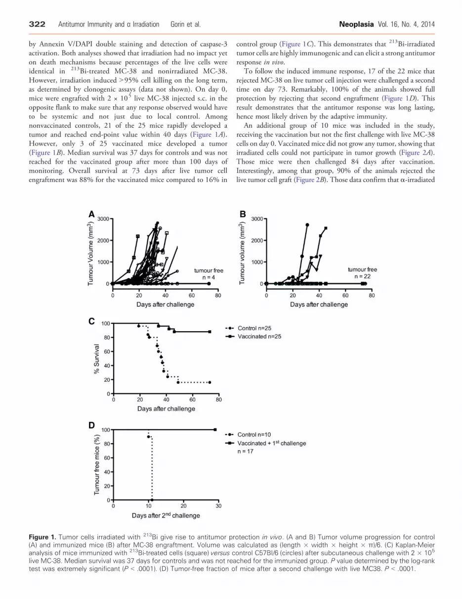
by Annexin V/DAPI double staining and detection of caspase-3activation. Both analyses showed that irradiation had no impact yeton death mechanisms because percentages of the live cells wereidentical in 213Bi-treated MC-38 and nonirradiated MC-38.However, irradiation induced N95% cell killing on the long term,as determined by clonogenic assays (data not shown). On day 0,mice were engrafted with 2 × 10⁵ live MC-38 injected s.c. in theopposite flank to make sure that any response observed would haveto be systemic and not just due to local control. Amongnonvaccinated controls, 21 of the 25 mice rapidly developed atumor and reached end-point value within 40 days (Figure 1A).However, only 3 of 25 vaccinated mice developed a tumor(Figure 1B). Median survival was 37 days for controls and was notreached for the vaccinated group after more than 100 days ofmonitoring. Overall survival at 73 days after live tumor cellengraftment was 88% for the vaccinated mice compared to 16% in
Figure 1. Tumor cells irradiated with 213Bi give rise to antitumor pr(A) and immunized mice (B) after MC-38 engraftment. Volume wasanalysis of mice immunized with 213Bi-treated cells (square) versus clive MC-38. Median survival was 37 days for controls and was not reatest was extremely significant (P b .0001). (D) Tumor-free fraction o
control group (Figure 1C). This demonstrates that 213Bi-irradiatedtumor cells are highly immunogenic and can elicit a strong antitumorresponse in vivo.
To follow the induced immune response, 17 of the 22 mice thatrejected MC-38 on live tumor cell injection were challenged a secondtime on day 73. Remarkably, 100% of the animals showed fullprotection by rejecting that second engraftment (Figure 1D). Thisresult demonstrates that the antitumor response was long lasting,hence most likely driven by the adaptive immunity.
An additional group of 10 mice was included in the study,receiving the vaccination but not the first challenge with live MC-38cells on day 0. Vaccinated mice did not grow any tumor, showing thatirradiated cells could not participate in tumor growth (Figure 2A).Those mice were then challenged 84 days after vaccination.Interestingly, among that group, 90% of the animals rejected thelive tumor cell graft (Figure 2B). Those data confirm that α-irradiated
otection in vivo. (A and B) Tumor volume progression for controlcalculated as (length × width × height × π)/6. (C) Kaplan-Meierontrol C57Bl/6 (circles) after subcutaneous challenge with 2 × 10⁵
ched for the immunized group. P value determined by the log-rankf mice after a second challenge with live MC38. P b .0001.
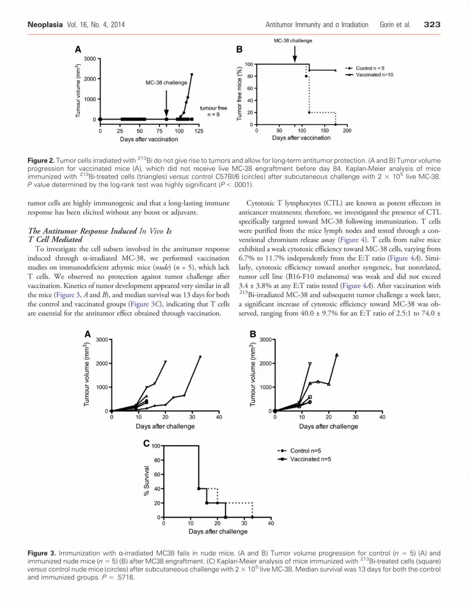
Figure 2. Tumor cells irradiated with 213Bi do not give rise to tumors and allow for long-term antitumor protection. (A and B) Tumor volumeprogression for vaccinated mice (A), which did not receive live MC-38 engraftment before day 84. Kaplan-Meier analysis of miceimmunized with 213Bi-treated cells (triangles) versus control C57Bl/6 (circles) after subcutaneous challenge with 2 × 10⁵ live MC-38.P value determined by the log-rank test was highly significant (P b .0001).
Neoplasia Vol. 16, No. 4, 2014 Antitumor Immunity and α Irradiation Gorin et al. 323
tumor cells are highly immunogenic and that a long-lasting immuneresponse has been elicited without any boost or adjuvant.
The Antitumor Response Induced In Vivo IsT Cell MediatedTo investigate the cell subsets involved in the antitumor response
induced through α-irradiated MC-38, we performed vaccinationstudies on immunodeficient athymic mice (nude) (n = 5), which lackT cells. We observed no protection against tumor challenge aftervaccination. Kinetics of tumor development appeared very similar in allthe mice (Figure 3, A and B), and median survival was 13 days for boththe control and vaccinated groups (Figure 3C), indicating that T cellsare essential for the antitumor effect obtained through vaccination.
Figure 3. Immunization with α-irradiated MC38 fails in nude mice.immunized nude mice (n = 5) (B) after MC38 engraftment. (C) Kaplanversus control nudemice (circles) after subcutaneous challenge with 2and immunized groups. P = .5716.
Cytotoxic T lymphocytes (CTL) are known as potent effectors inanticancer treatments; therefore, we investigated the presence of CTLspecifically targeted toward MC-38 following immunization. T cellswere purified from the mice lymph nodes and tested through a con-ventional chromium release assay (Figure 4). T cells from naïve miceexhibited a weak cytotoxic efficiency towardMC-38 cells, varying from6.7% to 11.7% independently from the E:T ratio (Figure 4A). Simi-larly, cytotoxic efficiency toward another syngeneic, but nonrelated,tumor cell line (B16-F10 melanoma) was weak and did not exceed3.4 ± 3.8% at any E:T ratio tested (Figure 4A). After vaccination with213Bi-irradiated MC-38 and subsequent tumor challenge a week later,a significant increase of cytotoxic efficiency toward MC-38 was ob-served, ranging from 40.0 ± 9.7% for an E:T ratio of 2.5:1 to 74.0 ±
(A and B) Tumor volume progression for control (n = 5) (A) and-Meier analysis of mice immunized with 213Bi-treated cells (square)× 105 live MC-38. Median survival was 13 days for both the control

Figure 4. Antitumor response is dependent on cytotoxic T cells. (A and B) 51Cr release assay against MC-38 or the irrelevant cell lineB16-F10 was performed on T cells purified from naïve mice (A) or mice that had been immunized with 213Bi-treated MC-38 andsubsequently challenged with live MC-38 (B). Assays were performed at different E:T ratios ; data points represent means ± SD oftriplicate measurements for three naïve mice (A) and four vaccinated + challenged mice (B). (C) Scatterplot of mean percentage of T cellcytotoxicity against MC-38 at the E:T ratio of 10:1 for different groups of mice. In parentheses, number of mice in each group. P b .05between naïve mice and mice that have been immunized and challenged. P value was determined by nonparametric Mann-Whitneytest. Mean cytotoxicity was 7.87% for naïve mice, 19.15% for challenged mice, 16.2% for vaccinated mice, and 55.85% forvaccinated + challenged mice.
324 Antitumor Immunity and α Irradiation Gorin et al. Neoplasia Vol. 16, No. 4, 2014
24.7% for an E:T ratio of 20:1 (Figure 4B), indicating that anantitumor T cell response had been raised. On the contrary, cytotoxicefficiency toward B16-F10 melanoma did not increase and was only1.0 ± 1.8% at the highest E:T ratio, indicating that the T cell responsewas specific to MC-38. Comparison of naïve, challenged, vaccinated,and vaccinated + challenged mice showed that CTL activity was onlysignificantly increased in vaccinated + challenged mice at the E:T ratioof 10:1 with 55.9% cytotoxicity compared to 7.9% in naïve animals(Figure 4C). Together with our data on the vaccination of nude mice,these results suggest that CTL is the main effector of the antitumorresponse raised after vaccination with α-irradiated tumor cells.
Tumor Cells Treated with 213Bi Activate DCs In VitroTo further explore the mechanisms supporting the antitumor
immune response to α particles, we analyzed the DC phenotype afterin vitro irradiation of MC-38 cells. DCs express costimulatorymolecules such as CD40, CD80, and CD86 on their cell surface,which are needed for activation of naïve T cells. Activation ofimmature DCs results in increased expression of those costimulatorymolecules. Immature BMDCs derived from syngeneic C57Bl/6 micehave been incubated for 48 hours with conditioned media fromcontrol or 213Bi -treated MC-38. The media of irradiated tumor cellselicited a significant increase of 32% in CD40 expression [ratio offluorescence intensity (RFI) increased from 4.5 to 5.9] and 44.8%in CD86 expression (22.7 to 32.6) and a slight increase, howevernot significant, of 4.4% in CD80 expression (3.3 to 3.4) on BMDCs(Figure 5, A–C). Besides, we observed that BMDCs aggregated in
clumps (Figure 5, D and E) when incubated in culture inserts withirradiated MC-38, which is a phenotypic characteristic of DCactivation [24]. No activation was observed when immature BMDCswere exposed to irradiated culture medium only (data not shown).These results suggest that 213Bi induces the release of soluble agentsfrom MC-38 capable of activating DCs in vitro.
213Bi Irradiation Causes Release of DAMPs fromMC-38 Cells
Then, we tested the conditioned media of 213Bi-treated MC-38for the presence of DAMPs. DAMPs such as Hsp70 or HMGB1 areself-molecules normally expressed intracellularly, which can be re-leased in the extracellular space upon cell stress or damage as a dangersignal to the immune system [25,26]. Those molecules are known toactivate DCs and have been implicated in the establishment ofefficient antitumor response [27]. ELISA on MC-38 conditionedmedia showed a significant increase of HMGB1 and Hsp70 levelsfollowing 213Bi irradiation (Figure 6). This increase in both DAMPconcentrations started 24 and 48 hours post-irradiation for Hsp70and HMGB1, respectively. Altogether, these data show that 213Biinduces the release of DAMPs from irradiated tumor cells, which maycontribute to the antitumor response by activating DCs.
DiscussionRadiotherapy is traditionally used for its cytotoxic effect on cancercells. There is however growing evidence showing that direct cyto-toxicity is not the only process through which radiation may
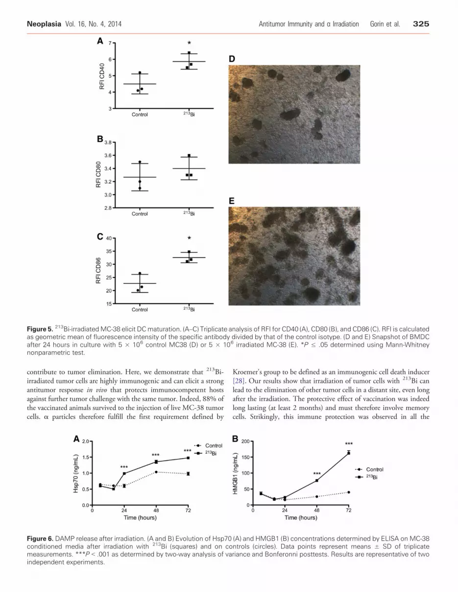
Figure 5. 213Bi-irradiatedMC-38 elicit DCmaturation. (A–C) Triplicate analysis of RFI for CD40 (A), CD80 (B), and CD86 (C). RFI is calculatedas geometric mean of fluorescence intensity of the specific antibody divided by that of the control isotype. (D and E) Snapshot of BMDCafter 24 hours in culture with 5 × 10⁶ control MC38 (D) or 5 × 10⁶ irradiated MC-38 (E). *P ≤ .05 determined using Mann-Whitneynonparametric test.
Neoplasia Vol. 16, No. 4, 2014 Antitumor Immunity and α Irradiation Gorin et al. 325
contribute to tumor elimination. Here, we demonstrate that 213Bi-irradiated tumor cells are highly immunogenic and can elicit a strongantitumor response in vivo that protects immunocompetent hostsagainst further tumor challenge with the same tumor. Indeed, 88% ofthe vaccinated animals survived to the injection of live MC-38 tumorcells. α particles therefore fulfill the first requirement defined by
Figure 6. DAMP release after irradiation. (A and B) Evolution of Hsp70conditioned media after irradiation with 213Bi (squares) and on comeasurements. ***P b .001 as determined by two-way analysis of vaindependent experiments.
Kroemer's group to be defined as an immunogenic cell death inducer[28]. Our results show that irradiation of tumor cells with 213Bi canlead to the elimination of other tumor cells in a distant site, even longafter the irradiation. The protective effect of vaccination was indeedlong lasting (at least 2 months) and must therefore involve memorycells. Strikingly, this immune protection was observed in all the
(A) and HMGB1 (B) concentrations determined by ELISA on MC-38ntrols (circles). Data points represent means ± SD of triplicateriance and Bonferonni posttests. Results are representative of two
326 Antitumor Immunity and α Irradiation Gorin et al. Neoplasia Vol. 16, No. 4, 2014
animals subjected to a second challenge with live MC-38 cells. Theresponse was T cell mediated as demonstrated by the presence ofspecific cytotoxic T cells and by the lack of protection in nude mice.This kind of specific, systemic, and long-lasting response would behighly desirable for anticancer therapy because it should help ineliminating distant metastases and preventing relapse. However, Tcells may not be the only cells needed for an efficient antitumor effect.Nude mice have functional Natural Killer (NK) and B cells that couldparticipate in the immune response. Further experiments will help toclarify the role of each immune subset in the antitumor response.
Interestingly, tumors grew much faster in nude mice with a mediansurvival of 13 versus 37 days in C57Bl/6 immunocompetent mice.Moreover, 4 of the 25 challenged mice did not develop a tumor, andthe rest of the challenged animals showed a slight increase incytotoxicity against MC-38 compared to naïve mice (however notsignificant). These data suggest that live tumor cells exhibit some levelof immunogenicity, probably related to the fact that the MC-38 cellswe used express high levels of major histocompatibility complex classI molecules (data not shown). Although this immune response slowsdown tumor growth to some extent, it is not sufficient for tumorcontrol in 84% of the animals. This could mean two things: eithertumor cells multiply too quickly for the immune response to copewith it, or the immune system sees the tumor as harmless self andrepresses the initial antitumor response. In both cases, α radiationdelivered through RIT would be of great therapeutic interest. Indeed,the high cytotoxicity of α particles could reduce tumor load, and atthe same time, α-irradiated tumor cells could activate the immuneresponse and tip the balance toward an effective antitumor response.Additional studies using less immunogenic tumors will however berequired to determine whether α radiation would be as efficient intriggering an immune response in such settings.
To depict the mechanisms that could contribute to activation ofT cells in vivo, we pursued in vitro studies on DCs. Their role inanticancer immunity is indeed crucial. When activated with theadequate stimuli, DCs can cross-present tumor-derived antigens toT cells and secrete stimulatory cytokines, which will lead to theestablishment of an effective cell-mediated antitumor response. Con-versely, if not properly activated, DCs may promote tolerance and T cellunresponsiveness. In fact, in amajority of cancer, the antitumor responseis repressed by the host's immune regulatory cells [29]. Here, we exposedimmature BMDCs to conditionedmedia from irradiatedMC-38 tumorcells and observed significant changes in both DC morphology andexpression profile of several DC activation markers, demonstrating thepotency of α-irradiated tumor cells in triggering immune activation.
To further investigate the molecular processes that couldcontribute to the activation of DCs in vitro, we conducted ELISAtests on the conditioned media of α-irradiated tumor cells to detectthe presence of well-characterized DAMPs [30]. In vivo, as irradiatedtumor cells were washed in PBS before vaccination, the moleculesinvolved in immune cells activation cannot be irradiated moleculesfrom the culture medium and have to be secreted by the tumor cellsafter exposure to 213Bi. Besides, irradiated culture medium alone didnot induce DC activation. We showed that 213Bi causes the release ofHsp70 and HMGB1 from MC-38 cells. Such molecules, which arenormally endogenous, get released in the extracellular environmentafter a stress or nonphysiological cell death and can activate immunecells [26,31]. HSP are chaperone proteins capable of bindingnumerous peptides. The HSP-peptide complexes released fromdying cells can be taken up by APC through the common receptor
CD91 to allow for antigen processing and re-presentation to T cells[32]. HMGB1 is a ubiquitous nonhistone nuclear factor that mediatesinflammation and immune responses when released from dying cells[33]. HMGB1 production in patients with cancer has conversely beenreported in associationwith both good [34] and poor prognoses [35,36].HMGB1 has also been involved in numerous processes facilitatingtumor progression, such as proliferation [37], metastasis [38,39],angiogenesis [40], and chronic inflammation [41]. The differentialactivity of HMGB1 is related to the balance of its different redox states(i.e., all-thiol, disulfide, and sulfonated) that are produced in theextracellular environment [42]. In the case of radiotherapy, a rapid andlocal increase of HMGB1 might act differently on the immune systemthan a chronic systemic secretion.Notably,HMGB1 has been shown tobe mandatory for cross-presentation of antigen derived from dyingtumor cells to T cells after radiotherapy, leading to efficient antitumorimmune response [43]. Further studies will be required to determine theimportance of these DAMPs in the establishment of an immuneresponse in our settings. Using larger screening techniques (i.e., massspectrometry), it will also be important to identify other speciesproduced after irradiation contributing to such bystander phenomena.
All these data underline the importance of the immune system inresponse to radiotherapy of cancer. Nevertheless, most studies on RITefficacy are performed on xenograft models in immunodeficient mice.Therefore, the participation of the immune system in response totherapy is completely overseen. In the future, it will be of primeimportance to assess the response to cancer therapy in both immu-nodeficient and immunocompetent models to take into account theeffect of radiation on both tumor cells and the immune environment.
In conclusion, the data presented here show that irradiation of tumorcells with 213Bi induces the release ofDAMPs, promotesDC activation,and leads to a systemic and long-lasting antitumor response. A fewstudies have investigated the impact ofγ or β irradiation on the immunesystem. It has been shown that external beam [44–46] or β emitters like90Y [21] or 153Sm [47] could elicit effective antitumor response whencombined with vaccine or CTL transfer. Our study is in line with theseinvestigations and is the first to demonstrate that irradiation of tumorsusing an α particle emitter can also lead to the establishment of anefficient antitumor immune response in vivo. Even though the impactof α irradiation on the immune response to cancer will have to befurther characterized, this study brings new insights on the mechanismof action of α particles and further supports the interest in developingthe use of such emitters for cancer therapy.
AcknowledgmentsWe thank Marie-Hélène Gaugler for critical review of the manu-script. We also thank Sandrine Minault (CRCNA [Centre deRecherche en Cancérologie Nantes-Angers]) for her technical help, aswell as the staff of Unité Thérapeutique Expérimentale (UTE),Cytocell, and Radioactivity facilities (Structure Fédérative deRecherche [SFR] François Bonamy, Institut de Recherche en Santéde l'Université de Nantes [IRS-UN], University of Nantes).
References
[1] Goldberg Z and Lehnert BE (2002). Radiation-induced effects in unirradiatedcells: a review and implications in cancer. Int J Oncol 21, 337–349.
[2] Morgan WF (2003). Is there a common mechanism underlying genomicinstability, bystander effects and other nontargeted effects of exposure to ionizingradiation? Oncogene 22, 7094–7099.
Neoplasia Vol. 16, No. 4, 2014 Antitumor Immunity and α Irradiation Gorin et al. 327
[3] Mothersill C and Seymour CB (2004). Radiation-induced bystander effects—implications for cancer. Nat Rev Cancer 4, 158–164.
[4] Barbet J, Bardiès M, Bourgeois M, Chatal JF, Chérel M, Davodeau F,Faivre-Chauvet A, Gestin JF, and Kraeber-Bodéré F (2012). Radiolabeledantibodies for cancer imaging and therapy.Methods Mol Biol 907, 681–697.
[5] Sharkey RM and Goldenberg DM (2011). Cancer radioimmunotherapy.Immunotherapy 3, 349–370.
[6] Lücke-Huhle C (1982). α-irradiation–induced G2 delay: a period of cellrecovery. Radiat Res 89, 298–308.
[7] Yong KJ, Milenic DE, Baidoo KE, and Brechbiel MW (2012). 212Pb-radioimmunotherapy induces G2 cell-cycle arrest and delays DNA damagerepair in tumor xenografts in a model for disseminated intraperitoneal disease.Mol Cancer Ther 11, 639–648.
[8] Søyland C andHassfjell SP (2000). Survival of human lung epithelial cells followingin vitro α-particle irradiation with absolute determination of the number ofα-particle traversals of individual cells. Int J Radiat Biol 76, 1315–1322.
[9] Humm JL and Cobb LM (1990). Nonuniformity of tumor dose inradioimmunotherapy. J Nucl Med 31, 75–83.
[10] Sgouros G, Roeske JC, McDevitt MR, Palm S, Allen BJ, Fisher DR, Brill AB,Song H, Howell RW, and Akabani G, et al (2010). MIRD Pamphlet No. 22(abridged): radiobiology and dosimetry of α-particle emitters for targetedradionuclide therapy. J Nucl Med 51, 311–328.
[11] Seideman JH, Stancevic B, Rotolo JA, McDevitt MR, Howell RW, Kolesnick RN,and Scheinberg DA (2011). Alpha particles induce apoptosis through thesphingomyelin pathway. Radiat Res 176, 434–446.
[12] Friesen C, Roscher M, Hormann I, Leib O, Marx S, Moreno J, and Miltner E(2013). Anti-CD33-antibodies labelled with the alpha-emitter Bismuth-213 killCD33-positive acute myeloid leukaemia cells specifically by activation of caspasesand break radio- and chemoresistance by inhibition of the anti-apoptotic proteinsX-linked inhibitor of apoptosis protein and B-cell lymphoma-extra large. Eur JCancer 49, 2542–2554.
[13] Supiot S, Gouard S, Charrier J, Apostolidis C, Chatal JF, Barbet J, Davodeau F,and Cherel M (2005). Mechanisms of cell sensitization to α radioimmunother-apy by doxorubicin or paclitaxel in multiple myeloma cell lines. Clin Cancer Res11, 7047s–7052s.
[14] Seidl C, Port M, Gilbertz KP, Morgenstern A, Bruchertseifer F, Schwaiger M,Röper B, Senekowitsch-Schmidtke R, and AbendM (2007). 213Bi-induced deathof HSC45-M2 gastric cancer cells is characterized by G2 arrest and up-regulationof genes known to prevent apoptosis but induce necrosis and mitotic catastrophe.Mol Cancer Ther 6, 2346–2359.
[15] BoydM, Ross SC, Dorrens J, Fullerton NE, Tan KW, ZalutskyMR, andMairs RJ(2006). Radiation-induced biologic bystander effect elicited in vitro by targetedradiopharmaceuticals labeled with α-, β-, and auger electron–emitting radionu-clides. J Nucl Med 47, 1007–1015.
[16] Chakravarty PK, Alfieri A, Thomas EK, Beri V, Tanaka KE, Vikram B, andGuha C (1999). Flt3-ligand administration after radiation therapy prolongssurvival in a murine model of metastatic lung cancer. Cancer Res 59, 6028–6032.
[17] Demaria S, Ng B, Devitt ML, Babb JS, Kawashima N, Liebes L, and FormentiSC (2004). Ionizing radiation inhibition of distant untreated tumors (abscopaleffect) is immune mediated. Int J Radiat Oncol Biol Phys 58, 862–870.
[18] Golden EB, Pellicciotta I, Demaria S, Barcellos-Hoff MH, and Formenti SC(2012). The convergence of radiation and immunogenic cell death signalingpathways. Front Oncol 2, 88.
[19] Formenti SC and Demaria S (2012). Combining radiotherapy and CancerImmunotherapy: a Paradigm Shift. J Natl Cancer Inst 1–10.
[20] Cameron RB, Spiess PJ, and Rosenberg SA (1990). Synergistic antitumor activityof Tumor-Infiltrating Lymphocytes, Interleukin-2 and local tumor irradiation.J Exp Med 171, 249–263.
[21] Chakraborty M, Gelbard A, Carrasquillo JA, Yu S, Mamede M, Paik CH,Camphausen K, Schlom J, and Hodge JW (2008). Use of radiolabeledmonoclonal antibody to enhance vaccine-mediated antitumor effects. CancerImmunol Immunother 57, 1173–1183.
[22] Nikula TK,CurcioMJ, BrechbielMW,GansowOA, FinnRD, and ScheinbergDA(1995). A rapid, single vessel method for preparation of clinical grade ligandconjugated monoclonal antibodies. Nucl Med Biol 22, 387–390.
[23] Dranoff G, Jaffee E, Lazenby A, Golumbek P, Levitsky H, Brose K, Jackson V,Hamada H, Pardoll D, and Mulligan RC (1993). Vaccination with irradiatedtumor cells engineered to secrete murine granulocyte–macrophage colony-stimulating factor stimulates potent, specific, and long-lasting anti-tumorimmunity. Proc Natl Acad Sci U S A 90, 3539–3543.
[24] Delemarre FG, Hoogeveen PG, De Haan-Meulman M, Simons PJ, andDrexhage HA (2001). Homotypic cluster formation of dendritic cells, a closecorrelate of their state of maturation. Defects in the biobreeding diabetes-pronerat. J Leukoc Biol 69, 373–380.
[25] Matzinger P (2002). The danger model: a renewed sense of self. Science 296,301–305.
[26] Tang D, Kang R, Coyne CB, Zeh HJ, and Lotze M (2012). PAMPs and DAMPs:signal 0s that spur autophagy and immunity. Immunol Rev 249, 158–175.
[27] Nace G, Evankovich J, Eid R, and Tsung A (2012). Dendritic cells and damage-associated molecular patterns: endogenous danger signals linking innate andadaptive immunity. J Innate Immun 4, 6–15.
[28] Kroemer G, Galluzzi L, Kepp O, and Zitvogel L (2013). Immunogenic cell deathin cancer therapy. Annu Rev Immunol 31, 51–72.
[29] Gabrilovich D (2004). Mechanisms and functional significance of tumour-induced dendritic-cell defects. Nat Rev Immunol 4, 941–952.
[30] Bianchi ME (2007). DAMPs, PAMPs and alarmins: all we need to know aboutdanger. J Leukoc Biol 81, 1–5.
[31] Garg AD, Nowis D, Golab J, Vandenabeele P, Krysko DV, and Agostinis P(2010). Immunogenic cell death, DAMPs and anticancer therapeutics: anemerging amalgamation. Biochim Biophys Acta 1805, 53–71.
[32] Basu S, Binder RJ, Ramalingam T, and Srivastava PK (2001). CD91 is acommon receptor for heat shock proteins gp96, hsp90, hsp70, and calreticulin.Immunity 14, 303–313.
[33] Andersson U and Tracey KJ (2011). HMGB1 is a therapeutic target for sterileinflammation and infection. Annu Rev Immunol 29, 139–162.
[34] Suzuki Y, Mimura K, Yoshimoto Y, Watanabe M, Ohkubo Y, Izawa S, Murata K,Fujii H, Nakano T, and Kono K (2012). Immunogenic tumor cell death inducedby chemoradiotherapy in patients with esophageal squamous cell carcinoma.CancerRes 72, 3967–3976.
[35] Chung H, Lee SG, Kim H, Hong D, Chung J, Stroncek D, and Lim JB (2009).Serum high mobility group box-1 (HMGB1) is closely associated with theclinical and pathologic features of gastric cancer. J Transl Med 7, 38.
[36] Yang GL, Zhang LH, Bo JJ, Huo XJ, Chen HG, Cao M, Liu DM, and Huang YR(2012). Increased expression of HMGB1 is associated with poor prognosis inhuman bladder cancer. J Surg Oncol 106, 57–61.
[37] Riuzzi F, Sorci G, and Donato R (2006). The amphoterin (HMGB1)/receptorfor advanced glycation end products (RAGE) pair modulates myoblastproliferation, apoptosis, adhesiveness, migration, and invasiveness. Functionalinactivation of RAGE in L6 myoblasts results in tumor formation in vivo. J BiolChem 281, 8242–8253.
[38] Kuniyasu H, Oue N, Wakikawa A, Shigeishi H, Matsutani N, Kuraoka K, Ito R,Yokozaki H, and Yasui W (2002). Expression of receptors for advanced glycationend-products (RAGE) is closely associated with the invasive and metastaticactivity of gastric cancer. J Pathol 196, 163–170.
[39] Sasahira T, Akama Y, Fujii K, and Kuniyasu H (2005). Expression of receptor foradvanced glycation end products and HMGB1/amphoterin in colorectaladenomas. Virchows Arch 446, 411–415.
[40] van Beijnum JR, Buurman WA, and Griffioen AW (2008). Convergence andamplification of toll-like receptor (TLR) and receptor for advanced glycation endproducts (RAGE) signaling pathways via high mobility group B1 (HMGB1).Angiogenesis 11, 91–99.
[41] Campana L, Bosurgi L, and Rovere-Querini P (2008). HMGB1: a two-headedsignal regulating tumor progression and immunity. Curr Opin Immunol 20,518–523.
[42] Venereau E, Casalgrandi M, Schiraldi M, Antoine DJ, Cattaneo A, De MarchisF, Liu J, Antonelli A, Preti A, and Raeli L, et al (2012). Mutually exclusive redoxforms of HMGB1 promote cell recruitment or proinflammatory cytokine release.J Exp Med 209, 1519–1528.
[43] Apetoh L, Ghiringhelli F, Tesniere A, Obeid M, Ortiz C, Criollo A, Mignot G,Maiuri MC, Ullrich E, and Saulnier P, et al (2007). Toll-like receptor4–dependent contribution of the immune system to anticancer chemotherapyand radiotherapy. Nat Med 13, 1050–1059.
[44] Garnett CT1, Palena C, Chakraborty M, Tsang KY, Schlom J, and Hodge JW(2004). Sublethal irradiation of human tumor cells modulates phenotyperesulting in enhanced killing by cytotoxic T lymphocytes. Cancer Res 64,7985–7994.
[45] Chakraborty M, Abrams SI, Coleman CN, Camphausen K, Schlom J, andHodge JW (2004). External beam radiation of tumors alters phenotype of tumorcells to render them susceptible to vaccine-mediated T-cell killing. Cancer Res 64,4328–4337.

328 Antitumor Immunity and α Irradiation Gorin et al. Neoplasia Vol. 16, No. 4, 2014
[46] Reits EA, Hodge JW, Herberts CA, Groothuis TA, Chakraborty M,Wansley EK, Camphausen K, Luiten RM, de Ru AH, and Neijssen J, et al(2006). Radiation modulates the peptide repertoire, enhances MHC class Iexpression, and induces successful antitumor immunotherapy. J Exp Med 203,1259–1271.
[47] Chakraborty M, Wansley EK, Carrasquillo JA, Yu S, Paik CH, Camphausen K,Becker MD, Goeckeler WF, Schlom J, and Hodge JW (2008). The use ofchelated radionuclide (samarium-153-ethylenediaminetetramethylenephospho-nate) to modulate phenotype of tumor cells and enhance T cell–mediated killing.Clin Cancer Res 14, 4241–4249.
Related Documents