J. Clin. Med. 2022, 11, 632. https://doi.org/10.3390/jcm11030632 www.mdpi.com/journal/jcm Review Molecular Genetics Overview of Primary Mitochondrial Myopathies Ignazio Giuseppe Arena 1 , Alessia Pugliese 1 , Sara Volta 2 , Antonio Toscano 1 and Olimpia Musumeci 1, * 1 Unit of Neurology and Neuromuscular Disorders, Department of Clinical and Experimental Medicine, University of Messina, 98125 Messina, Italy; [email protected] (I.G.A.); [email protected] (A.P.); [email protected] (A.T.) 2 Department of Neurosciences, University of Padova, 35100 Padova, Italy; [email protected] * Correspondence: [email protected]; Tel.: +39‐0902217178 Abstract: Mitochondrial disorders are the most common inherited conditions, characterized by defects in oxidative phosphorylation and caused by mutations in nuclear or mitochondrial genes. Due to its high energy request, skeletal muscle is typically involved. According to the International Workshop of Experts in Mitochondrial Diseases held in Rome in 2016, the term Primary Mitochondrial Myopathy (PMM) should refer to those mitochondrial disorders affecting principally, but not exclusively, the skeletal muscle. The clinical presentation may include general isolated myopathy with muscle weakness, exercise intolerance, chronic ophthalmoplegia/ophthalmoparesis (cPEO) and eyelids ptosis, or multisystem conditions where there is a coexistence with extramuscular signs and symptoms. In recent years, new therapeutic targets have been identified leading to the launch of some promising clinical trials that have mainly focused on treating muscle symptoms and that require populations with defined genotype. Advantages in next‐generation sequencing techniques have substantially improved diagnosis. So far, an increasing number of mutations have been identified as responsible for mitochondrial disorders. In this review, we focused on the principal molecular genetic alterations in PMM. Accordingly, we carried out a comprehensive review of the literature and briefly discussed the possible approaches which could guide the clinician to a genetic diagnosis. Keywords: mitochondrial myopathy; exercise intolerance; ophtalmoplegia; mtDNA; nDNA; oxidative phosphorylation 1. Introduction According to the International Workshop of Experts in Mitochondrial Diseases held in Rome in 2016, Primary Mitochondrial Myopathies (PMMs) can be defined as disorders that lead to defects in oxidative phosphorylation (OXPHOS) and that mainly, but not exclusively, affect skeletal muscle [1]. Progressive external ophtalmoplegia (PEO), eyelid ptosis, exercise intolerance and muscle weakness are the most common symptoms of myopathy that occur in mitochondrial diseases. Myopathy can be isolated but more frequently is associated with other clinical manifestations [2]. PMM are mostly the expression of genetic molecular alterations that may involve mitochondrial DNA (mtDNA) or nuclear DNA (nDNA), or may be due to an impairment of the intergenomic communications [3]. In recent years, there was a spread of new technologies, such as Next‐Generation Sequencing (NGS), leading to the discovery and individualization of more and more mutations. To date, more than 350 genes in both mitochondrial and nuclear genomes are known to cause primary mitochondrial diseases [4]. In the present review we will give an overview of the principal molecular genetic defects linked to PMM (Table 1). We focus searched PubMed for articles published in Citation: Arena, I.G.; Pugliese, A.; Volta, S.; Toscano, A.; Musumeci, O. Molecular Genetics Overview of Primary Mitochondrial Myopathies. J. Clin. Med. 2022, 11, 632. https://doi.org/10.3390/jcm11030632 Academic Editors: Daniele Orsucci and Sylvia Lee‐Huang Received: 8 December 2021 Accepted: 20 January 2022 Published: 26 January 2022 Publisher’s Note: MDPI stays neutral with regard to jurisdictional claims in published maps and institutional affiliations. Copyright: © 2022 by the authors. Licensee MDPI, Basel, Switzerland. This article is an open access article distributed under the terms and conditions of the Creative Commons Attribution (CC BY) license (https://creativecommons.org/license s/by/4.0/).

Welcome message from author
This document is posted to help you gain knowledge. Please leave a comment to let me know what you think about it! Share it to your friends and learn new things together.
Transcript
J. Clin. Med. 2022, 11, 632. https://doi.org/10.3390/jcm11030632 www.mdpi.com/journal/jcm
Review
Molecular Genetics Overview of Primary Mitochondrial
Myopathies
Ignazio Giuseppe Arena 1, Alessia Pugliese 1, Sara Volta 2, Antonio Toscano 1 and Olimpia Musumeci 1,*
1 Unit of Neurology and Neuromuscular Disorders, Department of Clinical and Experimental Medicine,
University of Messina, 98125 Messina, Italy; [email protected] (I.G.A.); [email protected] (A.P.);
[email protected] (A.T.) 2 Department of Neurosciences, University of Padova, 35100 Padova, Italy; [email protected]
* Correspondence: [email protected]; Tel.: +39‐0902217178
Abstract: Mitochondrial disorders are the most common inherited conditions, characterized by
defects in oxidative phosphorylation and caused by mutations in nuclear or mitochondrial genes.
Due to its high energy request, skeletal muscle is typically involved. According to the International
Workshop of Experts in Mitochondrial Diseases held in Rome in 2016, the term Primary
Mitochondrial Myopathy (PMM) should refer to those mitochondrial disorders affecting
principally, but not exclusively, the skeletal muscle. The clinical presentation may include general
isolated myopathy with muscle weakness, exercise intolerance, chronic
ophthalmoplegia/ophthalmoparesis (cPEO) and eyelids ptosis, or multisystem conditions where
there is a coexistence with extramuscular signs and symptoms. In recent years, new therapeutic
targets have been identified leading to the launch of some promising clinical trials that have mainly
focused on treating muscle symptoms and that require populations with defined genotype.
Advantages in next‐generation sequencing techniques have substantially improved diagnosis. So
far, an increasing number of mutations have been identified as responsible for mitochondrial
disorders. In this review, we focused on the principal molecular genetic alterations in PMM.
Accordingly, we carried out a comprehensive review of the literature and briefly discussed the
possible approaches which could guide the clinician to a genetic diagnosis.
Keywords: mitochondrial myopathy; exercise intolerance; ophtalmoplegia; mtDNA; nDNA;
oxidative phosphorylation
1. Introduction
According to the International Workshop of Experts in Mitochondrial Diseases held
in Rome in 2016, Primary Mitochondrial Myopathies (PMMs) can be defined as disorders
that lead to defects in oxidative phosphorylation (OXPHOS) and that mainly, but not
exclusively, affect skeletal muscle [1]. Progressive external ophtalmoplegia (PEO), eyelid
ptosis, exercise intolerance and muscle weakness are the most common symptoms of
myopathy that occur in mitochondrial diseases. Myopathy can be isolated but more
frequently is associated with other clinical manifestations [2].
PMM are mostly the expression of genetic molecular alterations that may involve
mitochondrial DNA (mtDNA) or nuclear DNA (nDNA), or may be due to an impairment
of the intergenomic communications [3].
In recent years, there was a spread of new technologies, such as Next‐Generation
Sequencing (NGS), leading to the discovery and individualization of more and more
mutations. To date, more than 350 genes in both mitochondrial and nuclear genomes are
known to cause primary mitochondrial diseases [4].
In the present review we will give an overview of the principal molecular genetic
defects linked to PMM (Table 1). We focus searched PubMed for articles published in
Citation: Arena, I.G.; Pugliese, A.;
Volta, S.; Toscano, A.; Musumeci, O.
Molecular Genetics Overview of
Primary Mitochondrial Myopathies.
J. Clin. Med. 2022, 11, 632.
https://doi.org/10.3390/jcm11030632
Academic Editors: Daniele Orsucci
and Sylvia Lee‐Huang
Received: 8 December 2021
Accepted: 20 January 2022
Published: 26 January 2022
Publisher’s Note: MDPI stays
neutral with regard to jurisdictional
claims in published maps and
institutional affiliations.
Copyright: © 2022 by the authors.
Licensee MDPI, Basel, Switzerland.
This article is an open access article
distributed under the terms and
conditions of the Creative Commons
Attribution (CC BY) license
(https://creativecommons.org/license
s/by/4.0/).
J. Clin. Med. 2022, 11, 632 2 of 27
English from 1 January 2011 to 1 November 2021, using the search terms “mitochondrial
disease OR disorder”, “CPEO”, “myopathy”, “exercise intolerance”, “genetic”,
“mtDNA”, “nDNA”. We further evaluated the reference lists from relevant articles
including recent reviews (Table 2) and case series reports
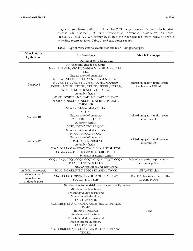
Table 1. Type of mitochondrial dysfunction and main PMM phenotypes.
Mitochondrial
Dysfunction Involved Gene Muscle Phenotype
Defects of MRC Complexes
Complex I
Mitochondrial‐encoded subunits:
Mt‐ND1, Mt‐ND2, Mt‐ND3, Mt‐ND4, Mt‐ND4L, Mt‐ND5, Mt‐
ND6
Nuclear‐encoded subunits:
NDUFA1, NDUFA2, NDUFA9, NDUFA10, NDUFA11,
NDUFA12, NDUFA13, NDUFB3, NDUFB9, NDUFB10,
NDUFB11, NDUFS1, NDUFS2, NDUFS3, NDUFS4, NDUFS6,
NDUFS7, NDUFS8, NDUFV1, NDUFV2
Assembly factors:
ACAD9, FOXRED1, NDUFAF1, NDUFAF2, NDUFAF3,
NDUFAF4, NDUFAF5, NDUFAF6, NUBPL, TIMMDC1,
TMEM126B
Isolated myopathy, multisystem
involvement, MELAS
Complex III
Mitochondrial‐encoded subunits:
Mt‐CYB
Nuclear‐encoded subunits:
CYC1, UBCRB, UQCRC2
Assembly factors:
BCSIL, LYRM7, TTC19, UQCC2
Isolated myopathy, multisystem
involvement
Complex IV
Mitochondrial‐encoded subunits:
Mt‐CO1, Mt‐CO2, Mt‐CO3
Nuclear‐encoded subunits:
COX41, COX412, NDUFA4
Assembly factors:
COA3, COA5, COA6, COA7, COX10, COX14, SCO1, SCO2,
COX15, COX20, PET100, APOFT1, SURF1, PET 11
Isolated myopathy, multisystem
involvement
Synthesis of electron carriers
COQ2, COQ4, COQ5, COQ6, COQ7, COQ8A, COQ8B, COQ9,
PDSS1, PDSS2 CYCS, HCCS
Isolated myopathy, nephropathy,
cardiomiopathy
mtDNA replication and maintenance
mtDNA homeostasis DNA2, MGME1, POLG, POLG2, RNASEH1, TWNK cPEO, cPEO plus
Maintenance of
mitochondrial
nucleotide pools
ABAT, DGUOK, MPV17, RRM2B, SAMHD1, SUCLA2,
SUCLG1, TK2, TYMP
cPEO, cPEO plus, isolated myopathy,
MNGIE, MDDS
Disorders of mitochondrial dynamics and quality control
Mitochondrial Membrane
Phospholipid Metabolism and
Protein Import Machinery
TAZ, TIMM8A XL
AGK, CHKB, DNAJC19, GFER, PAM16, SERAC1, PLA2G6,
TIMM22,
TIMM50, TIMMDC1
Mitochondrial Membrane
Phospholipid Metabolism and
Protein Import Machinery
TAZ, TIMM8A XL
AGK, CHKB, DNAJC19, GFER, PAM16, SERAC1, PLA2G6,
TIMM22,
cPEO

J. Clin. Med. 2022, 11, 632 3 of 27
TIMM50, TIMMDC1
DNM1L, MFN2, OPA1, GDAP1, MSTO1 AD/AR
MFF, STAT2, TRAK1, MIEF2
DNM1L, MFN2, OPA1, GDAP1, MSTO1, AFG32, SPG7
Table 2. Examples of reviews focusing on different aspects of mitochondrial disorders in the last
decade.
Title Main Focus Reference
Mitochondrial disease in adults Clinical aspects and diagnosis [2]
Mitochondrial energy generation
disorders: genes, mechanisms, and clues
to pathology.
Genetic discovery and functional
characterization [3]
Mitochondrial Disease Genetics Update
Recent insights into the Molecular
Diagnosis and Expanding Phenotype of
Primary Mitochondrial Disease.
Update of novel mitochondrial
disease genes and pathogenic
variants
[4]
Mitochondrial disease in adults: what’s
old and what’s new?
Disease mechanism and clinical
aspects in adults [5]
Mutations causing mitochondrial disease:
What is new and what challenges remain? Advances in mitochondrial genetics [6]
Human diseases associated with defects in
assembly of OXPHOS complexes.
Factors involved in assembly human
OXPHOS complex [7]
Complex I deficiency: clinical features,
biochemistry and molecular genetics.
Advances in the structure, function
and assembly of complex I [8]
The genetics and pathology of
mitochondrial disease.
Genetic discovery and advances in
mitochondrial pathology [9]
Nuclear gene mutations as the cause of
mitochondrial complex III deficiency
Discuss the nuclear‐encoded proteins
in which mutations have been found
to be associated to CIII deficiency
[10]
Cytochrome c oxidase deficiency Genetic etiology and clinical
manifestations in COX deficiency [11]
Mitochondrial disease in children. Clinical aspects and diagnosis [12]
Mitochondrial DNA depletion syndromes:
review and updates of genetic basis,
manifestations, and therapeutic options.
Genetic basis, clinical manifestation,
and therapeutic options [13]
Clinical and genetic spectrum of
mitochondrial neurogastrointestinal
encephalomyopathy
Symptomatology, diagnostic
procedures, and hurdles, in vitro and
in vivo models, experimental
therapies
[14]
Mitochondrial dynamics: overview of
molecular mechanisms
Overview of the molecular
mechanisms that govern
mitochondrial fission and fusion in
mammals.
[15]
Nuclear genes involved in mitochondrial
diseases caused by instability of
mitochondrial DNA
Overview of nuclear genes involved
in mitochondrial diseases [16]
2. General Aspects
Each mitochondrion has its own genetic code, constituting circular, double‐stranded
DNA of 16,569 base pairs. The mtDNA as whole contains 37 genes. Of these, there are 24
genes which code for molecules essential for the synthesis of the protein subunits of the
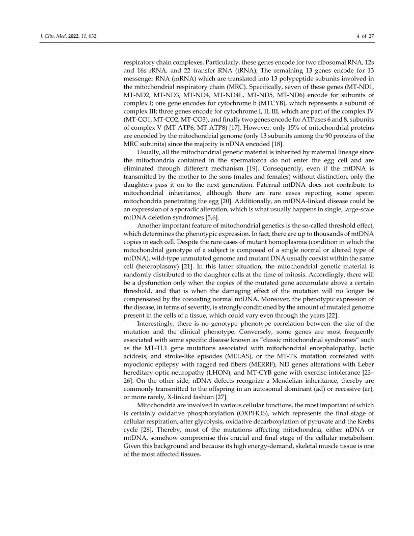
J. Clin. Med. 2022, 11, 632 4 of 27
respiratory chain complexes. Particularly, these genes encode for two ribosomal RNA, 12s
and 16s rRNA, and 22 transfer RNA (tRNA); The remaining 13 genes encode for 13
messenger RNA (mRNA) which are translated into 13 polypeptide subunits involved in
the mitochondrial respiratory chain (MRC). Specifically, seven of these genes (MT‐ND1,
MT‐ND2, MT‐ND3, MT‐ND4, MT‐ND4L, MT‐ND5, MT‐ND6) encode for subunits of
complex I; one gene encodes for cytochrome b (MTCYB), which represents a subunit of
complex III; three genes encode for cytochrome I, II, III, which are part of the complex IV
(MT‐CO1, MT‐CO2, MT‐CO3), and finally two genes encode for ATPases 6 and 8, subunits
of complex V (MT‐ATP6, MT‐ATP8) [17]. However, only 15% of mitochondrial proteins
are encoded by the mitochondrial genome (only 13 subunits among the 90 proteins of the
MRC subunits) since the majority is nDNA encoded [18].
Usually, all the mitochondrial genetic material is inherited by maternal lineage since
the mitochondria contained in the spermatozoa do not enter the egg cell and are
eliminated through different mechanism [19]. Consequently, even if the mtDNA is
transmitted by the mother to the sons (males and females) without distinction, only the
daughters pass it on to the next generation. Paternal mtDNA does not contribute to
mitochondrial inheritance, although there are rare cases reporting some sperm
mitochondria penetrating the egg [20]. Additionally, an mtDNA‐linked disease could be
an expression of a sporadic alteration, which is what usually happens in single, large‐scale
mtDNA deletion syndromes [5,6].
Another important feature of mitochondrial genetics is the so‐called threshold effect,
which determines the phenotypic expression. In fact, there are up to thousands of mtDNA
copies in each cell. Despite the rare cases of mutant homoplasmia (condition in which the
mitochondrial genotype of a subject is composed of a single normal or altered type of
mtDNA), wild‐type unmutated genome and mutant DNA usually coexist within the same
cell (heteroplasmy) [21]. In this latter situation, the mitochondrial genetic material is
randomly distributed to the daughter cells at the time of mitosis. Accordingly, there will
be a dysfunction only when the copies of the mutated gene accumulate above a certain
threshold, and that is when the damaging effect of the mutation will no longer be
compensated by the coexisting normal mtDNA. Moreover, the phenotypic expression of
the disease, in terms of severity, is strongly conditioned by the amount of mutated genome
present in the cells of a tissue, which could vary even through the years [22].
Interestingly, there is no genotype–phenotype correlation between the site of the
mutation and the clinical phenotype. Conversely, some genes are most frequently
associated with some specific disease known as “classic mitochondrial syndromes” such
as the MT‐TL1 gene mutations associated with mitochondrial encephalopathy, lactic
acidosis, and stroke‐like episodes (MELAS), or the MT‐TK mutation correlated with
myoclonic epilepsy with ragged red fibers (MERRF), ND genes alterations with Leber
hereditary optic neuropathy (LHON), and MT‐CYB gene with exercise intolerance [23–
26]. On the other side, nDNA defects recognize a Mendelian inheritance, thereby are
commonly transmitted to the offspring in an autosomal dominant (ad) or recessive (ar),
or more rarely, X‐linked fashion [27].
Mitochondria are involved in various cellular functions, the most important of which
is certainly oxidative phosphorylation (OXPHOS), which represents the final stage of
cellular respiration, after glycolysis, oxidative decarboxylation of pyruvate and the Krebs
cycle [28]. Thereby, most of the mutations affecting mitochondria, either nDNA or
mtDNA, somehow compromise this crucial and final stage of the cellular metabolism.
Given this background and because its high energy‐demand, skeletal muscle tissue is one
of the most affected tissues.
J. Clin. Med. 2022, 11, 632 5 of 27
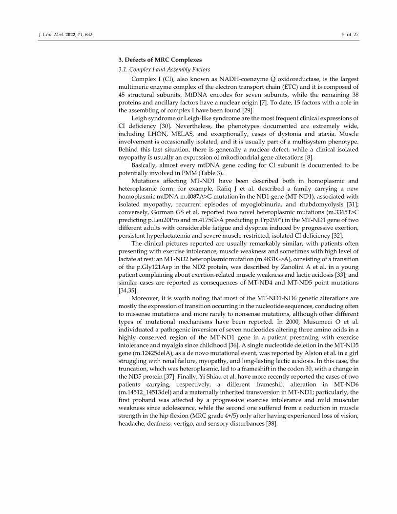
3. Defects of MRC Complexes
3.1. Complex I and Assembly Factors
Complex I (CI), also known as NADH‐coenzyme Q oxidoreductase, is the largest
multimeric enzyme complex of the electron transport chain (ETC) and it is composed of
45 structural subunits. MtDNA encodes for seven subunits, while the remaining 38
proteins and ancillary factors have a nuclear origin [7]. To date, 15 factors with a role in
the assembling of complex I have been found [29].
Leigh syndrome or Leigh‐like syndrome are the most frequent clinical expressions of
CI deficiency [30]. Nevertheless, the phenotypes documented are extremely wide,
including LHON, MELAS, and exceptionally, cases of dystonia and ataxia. Muscle
involvement is occasionally isolated, and it is usually part of a multisystem phenotype.
Behind this last situation, there is generally a nuclear defect, while a clinical isolated
myopathy is usually an expression of mitochondrial gene alterations [8].
Basically, almost every mtDNA gene coding for CI subunit is documented to be
potentially involved in PMM (Table 3).
Mutations affecting MT‐ND1 have been described both in homoplasmic and
heteroplasmic form: for example, Rafiq J et al. described a family carrying a new
homoplasmic mtDNA m.4087A>G mutation in the ND1 gene (MT‐ND1), associated with
isolated myopathy, recurrent episodes of myoglobinuria, and rhabdomyolysis [31];
conversely, Gorman GS et al. reported two novel heteroplasmic mutations (m.3365T>C
predicting p.Leu20Pro and m.4175G>A predicting p.Trp290*) in the MT‐ND1 gene of two
different adults with considerable fatigue and dyspnea induced by progressive exertion,
persistent hyperlactatemia and severe muscle‐restricted, isolated CI deficiency [32].
The clinical pictures reported are usually remarkably similar, with patients often
presenting with exercise intolerance, muscle weakness and sometimes with high level of
lactate at rest: an MT‐ND2 heteroplasmic mutation (m.4831G>A), consisting of a transition
of the p.Gly121Asp in the ND2 protein, was described by Zanolini A et al. in a young
patient complaining about exertion‐related muscle weakness and lactic acidosis [33], and
similar cases are reported as consequences of MT‐ND4 and MT‐ND5 point mutations
[34,35].
Moreover, it is worth noting that most of the MT‐ND1‐ND6 genetic alterations are
mostly the expression of transition occurring in the nucleotide sequences, conducing often
to missense mutations and more rarely to nonsense mutations, although other different
types of mutational mechanisms have been reported. In 2000, Musumeci O et al.
individuated a pathogenic inversion of seven nucleotides altering three amino acids in a
highly conserved region of the MT‐ND1 gene in a patient presenting with exercise
intolerance and myalgia since childhood [36]. A single nucleotide deletion in the MT‐ND5
gene (m.12425delA), as a de novo mutational event, was reported by Alston et al. in a girl
struggling with renal failure, myopathy, and long‐lasting lactic acidosis. In this case, the
truncation, which was heteroplasmic, led to a frameshift in the codon 30, with a change in
the ND5 protein [37]. Finally, Yi Shiau et al. have more recently reported the cases of two
patients carrying, respectively, a different frameshift alteration in MT‐ND6
(m.14512_14513del) and a maternally inherited transversion in MT‐ND1; particularly, the
first proband was affected by a progressive exercise intolerance and mild muscular
weakness since adolescence, while the second one suffered from a reduction in muscle
strength in the hip flexion (MRC grade 4+/5) only after having experienced loss of vision,
headache, deafness, vertigo, and sensory disturbances [38].
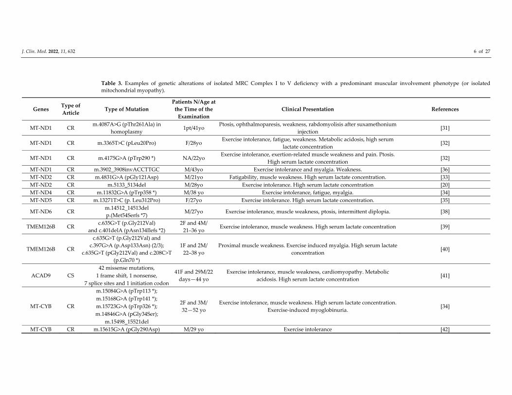
J. Clin. Med. 2022, 11, 632 6 of 27
Table 3. Examples of genetic alterations of isolated MRC Complex I to V deficiency with a predominant muscular involvement phenotype (or isolated
mitochondrial myopathy).
Genes Type of
Article Type of Mutation
Patients N/Age at
the Time of the
Examination
Clinical Presentation References
MT‐ND1 CR m.4087A>G (pThr261Ala) in
homoplasmy 1pt/41yo
Ptosis, ophthalmoparesis, weakness, rabdomyolisis after suxamethonium
injection [31]
MT‐ND1 CR m.3365T>C (pLeu20Pro) F/28yo Exercise intolerance, fatigue, weakness. Metabolic acidosis, high serum
lactate concentration [32]
MT‐ND1 CR m.4175G>A (pTrp290 *) NA/22yo Exercise intolerance, exertion‐related muscle weakness and pain. Ptosis.
High serum lactate concentration [32]
MT‐ND1 CR m.3902_3908invACCTTGC M/43yo Exercise intolerance and myalgia. Weakness. [36]
MT‐ND2 CR m.4831G>A (pGly121Asp) M/21yo Fatigability, muscle weakness. High serum lactate concentration. [33]
MT‐ND2 CR m.5133_5134del M/28yo Exercise intolerance. High serum lactate concentration [20]
MT‐ND4 CR m.11832G>A (pTrp358 *) M/38 yo Exercise intolerance, fatigue, myalgia. [34]
MT‐ND5 CR m.13271T>C (p. Leu312Pro) F/27yo Exercise intolerance. High serum lactate concentration. [35]
MT‐ND6 CR m.14512_14513del
p.(Met54Serfs *7) M/27yo Exercise intolerance, muscle weakness, ptosis, intermittent diplopia. [38]
TMEM126B CR c.635G>T (p.Gly212Val)
and c.401delA (pAsn134Ilefs *2)
2F and 4M/
21–36 yo Exercise intolerance, muscle weakness. High serum lactate concentration [39]
TMEM126B CR
c.635G>T (p.Gly212Val) and
c.397G>A (p.Asp133Asn) (2/3);
c.635G>T (pGly212Val) and c.208C>T
(p.Gln70 *)
1F and 2M/
22–38 yo
Proximal muscle weakness. Exercise induced myalgia. High serum lactate
concentration [40]
ACAD9 CS
42 missense mutations, 41F and 29M/22
days—44 yo
Exercise intolerance, muscle weakness, cardiomyopathy. Metabolic
acidosis. High serum lactate concentration [41] 1 frame shift, 1 nonsense,
7 splice sites and 1 initiation codon
MT‐CYB CR
m.15084G>A (pTrp113 *);
2F and 3M/
32—52 yo
Exercise intolerance, muscle weakness. High serum lactate concentration.
Exercise‐induced myoglobinuria. [34]
m.15168G>A (pTrp141 *);
m.15723G>A (pTrp326 *);
m.14846G>A (pGly34Ser);
m.15498_15521del
MT‐CYB CR m.15615G>A (pGly290Asp) M/29 yo Exercise intolerance [42]
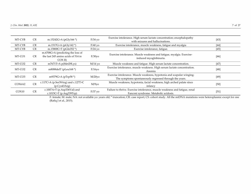
J. Clin. Med. 2022, 11, 632 7 of 27
MT‐CYB CR m.15242G>A (pGly166 *) F/34 yo Exercise intolerance. High serum lactate concentration; encephalopathy
with seizures and hallucinations. [43]
MT‐CYB CR m.1517G>A (pGly142 *) F/40 yo Exercise intolerance, muscle weakness, fatigue and myalgia [44]
MT‐CYB CR m.15800C>T (pGln352 *) F/24 yo Exercise intolerance, fatigue. [45]
MT‐CO1 CR
m.6708G>A (predicting the loss of
the last 245 amino acids of 514 in
COX II)
F/30yo Exercise intolerance. Muscle weakness and fatigue, myalgia. Exercise‐
induced myoglobinuria. [46]
MT‐CO2 CR m7671T>A p(Met29Lys) M/14 yo Muscle weakness and fatigue. High serum lactate concentration. [47]
MT‐CO2 CR m8088delT (pLeu168 *) F/16yo Exercise intolerance, muscle weakness. High serum lactate concentration.
Anemia [48]
MT‐CO3 CR m9379G>A (pTrp58 *) M/20yo Exercise intolerance. Muscle weakness, hypotonia and scapular winging;
The symptoms spontaneously regressed through the years. [49]
COX6A2 CR c.117C>A (p.Ser39Arg) and c.127T>C
(p.Cys43Arg) M/9yo
Muscle weakness, hypotonia, facial weakness, high arched palate since
infancy [50]
COX10 CR c.1007A>T (p.Asp336Val) and
c.1015C>T (p.Arg339Trp) F/37 yo
Failure to thrive. Exercise intolerance, muscle weakness and fatigue, renal
Fanconi syndrome. Metabolic acidosis. [51]
F: female; M: male; NA: not available yo: years old; * truncation; CR: case report; CS: cohort study. All the mtDNA mutations were heteroplasmic except for one
(Rafiq J et al., 2015).
J. Clin. Med. 2022, 11, 632 8 of 27
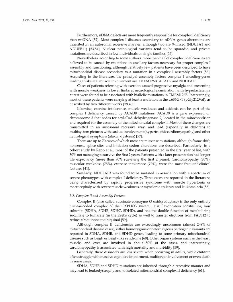
Furthermore, nDNA defects are more frequently responsible for complex I deficiency
than mtDNA [52]. Most complex I diseases secondary to nDNA genes alterations are
inherited in an autosomal recessive manner, although two are X‐linked (NDUFA1 and
NDUFB11) [53,54]. Nuclear pathological variants tend to be sporadic, and private
mutations are described in few individuals or single families [55].
Nevertheless, according to some authors, more than half of complex I deficiencies are
believed to be caused by mutations in ancillary factors necessary for proper complex I
assembly and functioning, although relatively few patients have been described to have
mitochondrial disease secondary to a mutation in a complex I assembly factors [56].
According to the literature, the principal assembly factors complex I encoding‐genes
leading to skeletal muscle involvement are TMEM126B, ACAD9 and NDUFAF3.
Cases of patients referring with exertion‐caused progressive myalgia and presenting
with muscle weakness in lower limbs at neurological examination with hyperlactatemia
at rest were found to be associated with biallelic mutations in TMEM126B. Interestingly,
most of these patients were carrying at least a mutation in the c.635G>T (pGly212Val), as
described by two different works [39,40].
Likewise, exercise intolerance, muscle weakness and acidosis can be part of the
complex I deficiency caused by ACAD9 mutations. ACAD9 is a gene expressed on
chromosome 3 that codes for acyl‐CoA dehydrogenase 9, located in the mitochondrion
and required for the assembly of the mitochondrial complex I. Most of these changes are
transmitted in an autosomal recessive way, and lead (especially in children) to
multisystem pictures with cardiac involvement (hypertrophic cardiomyopathy) and other
neurological symptoms (ataxia, dystonia) [57].
There are up to 70 cases of which most are missense mutations, although frame shift,
nonsense, splice sites and initiation codon alterations are described. Particularly, in a
cohort study by Repp et al., most of the patients presented in the first year of life, with
50% not managing to survive the first 2 years. Patients with a later presentation had longer
life expectancy (more than 90% surviving the first 2 years). Cardiomyopathy (85%),
muscular weakness (75%), exercise intolerance (72%), were the most frequent clinical
features [41].
Similarly, NDUFAF3 was found to be mutated in association with a spectrum of
severe phenotypes with complex I deficiency. Three cases are reported in the literature,
being characterized by rapidly progressive syndrome with muscle hypertonia or
macrocephaly with severe muscle weakness or myoclonic epilepsy and leukomalacia [58].
3.2. Complex II and Assembly Factors
Complex II (also called succinate‐coenzyme Q oxidoreductase) is the only entirely
nuclear‐coded complex of the OXPHOS system. It is flavoprotein constituting four
subunits (SDHA, SDHB, SDHC, SDHD), and has the double function of metabolizing
succinate to fumarate (in the Krebs cycle) as well to transfer electrons from FADH2 to
reduce ubiquinone to ubiquinol [59].
Although complex II deficiencies are exceedingly uncommon (almost 2–8% of
mitochondrial disease cases), either homozygous or heterozygous pathogenic variants are
reported in SDHA, SDHB, and SDHD genes, leading to some primary mitochondrial
disease such as Leigh or Leigh‐like syndrome [60]. Other organ systems such as the heart,
muscle, and eyes are involved in about 50% of the cases, and interestingly,
cardiomyopathy is associated with high mortality and morbidity [59].
Generally, these disorders are less severe when occurring in adults, while children
often struggle with massive cognitive impairment, multiorgan involvement or even death
in some cases.
SDHA, SDHB and SDHD mutations are inherited through a recessive manner and
may lead to leukodystrophy and to isolated mitochondrial complex II deficiency [61].
J. Clin. Med. 2022, 11, 632 9 of 27
Several complex II assembly factors have also been identified, including SDHAF2
and SDHAF1, which have been associated with autosomal recessive complex II deficiency
and leukoencephalopathy [62].
3.3. Complex III and Assembly Factors
Complex III, ubiquinone–cytochrome c oxidoreductase, is composed of 11 subunits
of which only one (cytochrome b) is encoded by the mitochondrial genome. Cytochrome
B (MT‐CYB), along with cytochrome c1 (CYC1) and the Rieske protein (UQCRFS1)
represent the catalytic center. CYC1 as well the other structural subunits (UQCRB,
UQCRQ, UQCRC2) and the assembly factors (BCS1L, LYRM7, UQCC2, and UQCC3) are
coded by nDNA [63].
Functionally, complex III represents the center of different metabolic pathways as
well as the MRC. In recent years, several new pathogenic variants have been associated
with complex III dysfunction, involving both structural subunits and assembly factors
with extremely heterogeneous clinical phenotypes (including hypoglycemia and
hyperglycemia, hepatomegaly, renal tubular acidosis, Leigh Syndrome, and other
neurological abnormalities) [64].
Myopathic involvement is reported in the form of exercise intolerance (sometimes
associated with proximal limb weakness and myoglobinuria) in more than 50% of patients
with mutation of the mitochondrial MT‐CYB gene, coding for cytochrome b [9].
The first reported cases of MT‐CYB mutations can be traced back to the 1990s,
although the correlation between CIII deficiency and muscle involvement was known for
some time before.
In fact, as early as the 1970s, Spriro AJ et al., reported the cases of a 46‐year‐old man
and his son presenting muscular weakness and progressive ataxia [65]. Afterwards,
several other cases were described: Darley‐Usmar et al. presented a patient with lactic
acidosis and muscle weakness while Hayes et al. reported the case of a Chilean girl with
ptosis and fatigue [66,67]. A progressive exercise intolerance with a low CIII activity was
also described [68].
However, only starting from the 1990s with mitochondrial DNA sequencing, was it
possible to highlight the molecular basis of these disorders.
In 1996, Dumoulin R et al. identified the first cyt b missense pathogenic mutation in
a young man with exercise intolerance. The alteration, which was heteroplasmic,
consisted of the transition of guanine to adenine in position 15,615 of mtDNA, leading to
the substitution Gly290Asp [42].
Particular importance must be given certainly to Andreu’s et al. study, with the
description of five cases of patients with severe exercise intolerance, lactic acidosis present
at rest (in four out of five patients), and biochemical evidence of complex III dysfunction.
Of these patients, there were three nonsense mutations (G15084A, G15168A, and
G15723A), one missense mutation (G14846A), and a 24‐base deletion (from nucleotides
15,498 to 15,521) in cytochrome b. All the point mutations reported involved the
substitution of adenine for guanine, but all on different locations. Moreover, there was no
maternal inheritance, and there were no other mutations in other tissue districts beside
muscle, suggesting that the disorder was due to somatic mutations in myogenic stem cells
after germline differentiation [22].
Since then, new clinical phenotypes linked to MT‐CYB have been identified such
ataxia and MELAS syndrome, while a four‐base deletion was individuated within a form
of parkinsonism [69].
Furthermore, other forms of predominantly myopathic involvement have been
reported. A stop‐codon mutation (G15242A) of the mitochondrial encoded gene for
cytochrome b predicting the loss of the last 215 amino acids of cytochrome b was identified
[43].
Wibrand F et al. described a heteroplasmic mutation changing in a highly conserved
region tyrosine to cysteine at position 278 in a patient with severe exercise intolerance in
J. Clin. Med. 2022, 11, 632 10 of 27
the context of a multisystem disorder including deafness, mental retardation, retinitis
pigmentosa, cataract, growth retardation and epilepsy [70].
Schuelke M et al. described a 25‐year‐old patient with septo‐optic dysplasia, retinitis
pigmentosa, weakness, and hypertrophic cardiomyopathy since childhood, who carried
the heteroplasmic mutation m.14849T>C predicting the p.Ser35Pro substitution within cyt
b. [71].
Legros F et al. reported two different heteroplasmic mutations (substitutions
p.Trp135Ter and p.Ser151Pro), in the muscle of two unrelated patients presenting with an
effort‐induced fatigability from their late childhood with a CIII activity below 20% in
muscle, associated with a reduced amount of cyt b protein [72].
Bruno C et al. described a 40‐year‐old woman developing progressive exercise
intolerance, lactic acidosis and muscle cramps beginning at the age of 30 years, because of
mutation m.15170G>A leading to a stop codon (p.Gly142Ter) [44].
Another heteroplasmic mutation (m.15761G>A) leading to a stop codon (cyt b
p.Gly339Ter) and thereby to a truncated protein was reported by Mancuso et al. in a 19‐
year‐old woman suffering from exercise intolerance, vomiting and lactic acidosis [73].
Another nonsense mutation m.15800C>T (substitution p.Gln352Ter) leading to a
truncation in cyt b protein is described by Lamantea E at al., in a 24‐year‐old woman who
had exercise intolerance with muscle cramps and lactic acidosis [45].
Furthermore, nuclear CIII deficiencies are caused by recessively inherited mutations
affecting nDNA genes encoding for structural subunits or assembly factors, and are
associated with a wide range of clinical presentations and in some cases reduced CIII
activity/amount in cultured fibroblasts or skeletal muscle [64].
Mutations in the BCS1L gene are reported as a cause of complex III dysfunction.
BCS1L is a gene situated on chromosome 2q35, and is involved in the synthesis of an AAA
protein (ATPase associated with diverse cellular activities) which plays a role in iron
homeostasis and in the assembly of complex III, particularly in the incorporation of
UQCRFS1. The clinical spectrum varies from purely visceral syndrome (such as GRACILE
syndrome or Bjorstand syndrome) to a pure form of encephalopathy [10,64]. In these
conditions, muscle involvement is mostly limited to only being a part of a multiorgan
disorder.
3.4. Complex IV and Assembly Factors
Complex IV, or cytochrome c oxidase (COX), is situated in the inner mitochondrial
membrane and consists of 14 structural subunits, since NDUFA4, previously assigned to
complex I, was recently added as a new peripheral subunit of COX because NDUFA4
deficiency results in a severe loss of COX activity and represents a stoichiometric
component of the individual COX complex [74]. COX consists of three catalytic subunits
encoded by mt‐DNA, MT‐CO1, MT‐CO2 and MT‐CO3. The remaining subunits (COX4,
COX5A, COX5B, COX6A, COX6B, COX6C, COX7A, COX7B, COX7C, COX8 and
NDUFA4) and ancillary factors (COX10, COX14, COX 15, COX20, COA3, COA5, COA6,
COA7, COA8, PET100, PET117, SCO1, SCO2 and SURF1) are instead entirely encoded by
nDNA. COX functions as a dimer, with two copper binding sites, two heme groups, a
magnesium ion, and a zinc ion. Remarkably, there are multiple tissue‐specific isoforms
for numerous subunits of COX (for example COX4, COX6A, COX6B, COX7A, COX8) [11].
As with other complex deficiencies, pathogenic mutations of COX have been
associated with different clinical manifestation, ranging from isolated myopathy to severe
multisystem disorder [75].
In 1999, Rahman S et al. identified the first missense mutation in the mtDNA gene
for subunit II of cytochrome c oxidase (COX) in a 14‐year‐old boy with a proximal
myopathy and lactic acidosis: particularly, mtDNA‐sequencing showed a novel
heteroplasmic transversion at nucleotide position 7671 in COII, responsible of the
substitution of methionine to lysine [47].

J. Clin. Med. 2022, 11, 632 11 of 27
In the same year, Clark KM et al. described a family with a heteroplasmic 7587TC
mutation predicting the change of methionine to threonine. Particularly, the proband,
who was the mother of the family, presented with unsteadiness of gait and fatigue, while
her son struggled from a more severe condition starting at the age of five, with optic
atrophy, pigmentary retinopathy, ataxia, and mild distal muscle atrophy [76].
Some years later, Horvath R et al., reported the case of a patient suffering from a
reversible myopathy, lactic acidosis, exercise intolerance, and delayed growth with a
heteroplasmic G9379A nonsense mutation (W58X) in the mtDNA encoding COIII subunit
gene, while a G6708A nonsense mutation in the mtDNA COI gene encoding COX subunit
I was identified by Kollberg G et al., in a 30‐year‐old woman with muscle weakness, pain,
fatigue, and one episode of rhabdomyolysis [46,49].
Another form of reversible myopathy associated with COX deficiency was firstly
described by Di Mauro et al. in 1983, but only after several years were characterized as
attributable to a homoplasmic m.14674T>C or m14674T>G in the MT‐TE gene coding the
tRNA of the glutamic acid (see Section 4) [77,78].
Nevertheless, new pathogenic variants involving either mtDNA or nDNA genes
coding for structural subunits, have emerged in recent years. A novel frame‐shift variant
in MTCO2 was recently observed in a 16‐year‐old girl with an infantile‐onset history of
exercise intolerance, in which whole‐exome sequencing revealed a single base‐pair
deletion (m.8088delT) resulting in a premature stop codon [48].
A biallelic missense mutation was identified through whole exome sequencing in
COX6A2, which is a COX‐equipping subunit isoform expressed only in the skeletal
muscle and heart, in two patients presenting with a congenital myopathy. The variants
detected were homozygous c.117C > A (p.Ser39Arg) and compound heterozygous c.117C
> A (p.Ser39Arg) and c.127T > C (p.Cys43Arg) [50].
Other symptoms evocative of skeletal muscle participation in the clinical picture are
documented in some ancillary factor genes mutations. Mutations in these genes are
usually associated with infantile onset with multisystem involvement and a fatal outcome.
SCO1 and SCO2 are metallochaperones that are essential for the assembly of the
catalytic core of COX. Mutations in SCO2 cause fatal infantile encephalomyopathy and
hypertrophic cardiomyopathy, whereas SCO1 patients presented with fatal infantile
encephalomyopathy and hepatopathy [79]. Moreover, some cases of SCO2 mutations
miming Werdnig–Hoffman syndrome are reported [80].
Finally, mutations in COX10 are rarely reported in adults with isolated COX
deficiency, associated with a relatively mild clinical phenotype comprising myopathy;
demyelinating neuropathy; premature ovarian failure; short stature; hearing loss;
pigmentary maculopathy; and renal tubular dysfunction [51].
3.5. Complex V and Assembly Factors
ATP synthase, complex V, is the multimeric molecular enzyme responsible for the
phosphorylation of ADP to obtain ATP. It consists of 13 different subunits and involves
at least three ancillary factors. Several pathogenic mutations have been observed,
especially in nDNA genes encoding complex V subunits (ATP5A1, ATP5E, USMG5) and
in the ancillary factors (TMEM70 and ATPAF2) [81,82].
One of the most known genes is MT‐ATP6, which encodes the complex V subunit
“a” that contains the proton pore that releases the proton gradient established across the
inner mitochondrial membrane. Since the discovery of MT‐ATP6 mutation m.8993T>G,
which was one of the first discovered [83], new pathogenic variants have been reported
that result in a destabilization of the complex or in an impaired stability or dysfunction of
the proton pumps, or in an increase ROS generation [84]. The most well‐known syndrome
is “NARP” (neuropathy, ataxia, and retinitis pigmentosa) syndrome due to the point
mutation T8993G) in the gene encoding ATPase 6; the full clinical syndrome manifests
when the percentage of mutated mtDNA compared to the total is 70–90% [85]. When the
percentage of heteroplasmy exceeds 90%, the clinical manifestations resume the
J. Clin. Med. 2022, 11, 632 12 of 27
maternally inherited Leigh syndrome (MILS) [86]. The two syndromes frequently coexist
in the same family, based on the level of mutation specifically present in each affected
member [87].
In any case, mutations in the mitochondrial and nuclear genes coding for proteins
and ancillary factors of complex V are mostly related to neuropathic involvement [88].
Muscle involvement is therefore unlikely to be the main clinical phenotype of these
alterations, being mostly in association with other manifestations.
Sometimes muscle involvement may be misunderstood with other neuromuscular
conditions.
Aurè et al. evidenced a homoplasmic deleterious mutation in the MT‐ATP6/8 genes
as potentially responsible for acute episodes of limb weakness mimicking periodic
paralysis, which resolved positively with acetazolamide [89].
A case of MLASA (Mitochondrial Myopathy, Lactic Acidosis, and Sideroblastic
Anemia) due to a de novo mutation (m.8969G>A) in the mitochondrially encoded ATP6
gene was described [90].
Moreover, Ganetsky et al. in a 2019 cohort study reported two individuals with
exercise intolerance and a clinical picture resembling CPEO with two ATP6 variants
(m.8608C>T predicting p.Pro28Ser and m.8723G>T predicting p. Arg66Leu) of uncertain
significance [84].
4. Defect of Translational Apparatus
The translational apparatus consists of several components, including mt‐tRNA, mt‐
rRNA, regulatory transcription factors, mitochondrial RNA polymerase, and
mitoribosomes [91]. Particularly, the 22 mt‐tRNAs play a fundamental role in the
transport of amino acids to the developing polypeptide chain during the translation of
mitochondrial proteins. If this process is altered, it can lead to a dysfunction of the ETC
complexes I, III, IV. Over 200 variants have been described in patients with mitochondrial
disorders with different phenotypes, but only a part of these variants fulfill the criteria to
be defined “pathogenic” [92]. The resulting clinical phenotypes are extremely
heterogeneous, including epilepsy, deafness, diabetes, ophthalmoparesis, myopathy,
cardiomyopathy, and encephalopathy. Some classic syndromes such as MELAS and
MERRF represent the most frequently observed. The well‐known association of the tRNA
Leu gene (MT‐TL1) and MELAS as secondary to the m.3243A>G mutation dates back to
the study by Kobayashi et al. in 1990 [93]. The m.3243A> G in the MT‐TL1 gene is very
specific, and it is still responsible for 80% of MELAS cases, followed by the m.3271T> C
(representing 10% of cases) and the m.3252A> G (which is observed in less than 5%) [94].
MELAS syndrome affects several organs, and some of its manifestations include stroke‐
like episodes, dementia, epilepsy, lactic acidemia, myopathy, recurrent headaches,
hearing impairment, diabetes, and short stature. Many patients carrying the 3243 variant
do not manifest the entire symptomatology. Recently, an isolated myopathy was
documented by Mahale RR et al., 2021, due to an m.3243A>G in the MT‐TL1 gene [95].
Similarly, MERRF, which is usually secondary to the 8344A> G mutation in MT‐TK, has
been associated with other possible pathogenic alterations in different mt‐tRNAs [96].
Beyond these typical clinical presentations, many pathogenetic variants involving mt‐
tRNAs have been described over the years as capable of leading to phenotypes with
mainly myopathic presentations [97].
A myopathic clinical presentation has been reported in two patients with muscle
weakness who were found to be carriers of the m.5631G>A and m.5610G>A mutations in
the MT‐TA gene [98].
Mt‐tRNA defects manifested also with a CPEO phenotype, and several point
mutations have been reported. A sporadic case of a heteroplasmic substitution in position
12316G>A in MT‐TL2 causing cPEO with COX‐negative fibers and RRF was described
[99]; Karadimas CL et al. described an m.12315G>A mutation in the MT‐TL2 gene in a
J. Clin. Med. 2022, 11, 632 13 of 27
woman with cPEO [100]; Soldath P et al. reported an m.12294 G>A in the MT‐TL2 gene in
an individual with cPEO and exercise intolerance [101].
More recently, an m.4414T>C in the MT‐TM gene has been described as a variant
causing PEO and myopathy in an adult patient whose muscle biopsy revealed focal
cytochrome c oxidase deficiency and ragged red fibers [102]. Finally, a case of CPEO with
a COX deficiency was also documented as due to mutation involving the MT‐TN gene
[103].
In other circumstances, mt‐tRNAs defects may lead to a benign form. Fatigue,
weakness, and exercise intolerance were the only clinical symptoms described in a family,
in which all the 13 members presented isolated complex I deficiency and an m.3250T>C
mutation in MT‐TL1 [104].
A very interesting form of myopathy related to mt‐tRNA mutation is a neonatal
mitochondrial myopathy with COX deficiency, first described by Di Mauro et al. in 1983
[77]; a genetic definition was later reached in 2009 when Horvath et al. reported a case
series of 17 individuals from 12 different families with a homoplasmic m.14674T>C or
m14674T>G in the MT‐TE gene coding the tRNA of the glutamic acid (Glu) [78,105]. This
syndrome, known as benign reversible mitochondrial myopathy, affects mostly infants
who struggle from lactic acidosis, limb myopathy and respiratory musculature
involvement leading to respiratory failure. Fortunately, most of these patients
spontaneously improve with supportive care [12]. The reason why these patients
experienced a timed spontaneous recovery was not clear, but it is hypothesized that there
is a developmental switch in the control strength of mitochondrial transfer RNAs and, in
particular, MT‐TE in mitochondrial translation, suggesting that 16–30% of steady‐state
levels of MT‐TE may have a profound effect on translation in muscles of neonates, but this
may become less critical at later stages of development.
Finally, it is worth mentioning that a myasthenia gravis‐like phenotype due to a
5728T>C mutation in the MT‐TN gene has been reported [106].
5. Defects of Electron Carriers
Coenzyme Q10 (CoQ10) is a lipophilic molecule comprising a quinone group and a 10‐
unit polyisoprenoid tail, that is located within the inner membrane of the human
mitochondria. It functions as an electron carrier in the respiratory chain, transferring
electrons from NADH:coenzyme Q reductase (complex I) and succinate: coenzyme Q
reductase (complex II) to coenzyme Q:cytochrome c reductase (complex III). CoQ10 is also
joined in pyrimidines, fatty acids and mitochondrial uncoupling proteins metabolism and
serves as antioxidant [107].
CoQ10 deficiency has been reported in literature as a primary or secondary deficiency.
The primary deficiency is associated with mutations of the at least nine genes involved in
its biosynthesis, inherited in an autosomal recessive manner (COQ2, COQ4, COQ6,
COQ7, CPOQ8A, COQ8B, COQ9, PDSS1, PDSS2) [108]. It can present with different
phenotypes, usually characterized by multisystem involvement. Firstly, it was reported
in 1988 by Ogasahara et al. who described two sisters with progressive muscle weakness,
severe fatigability, and central nervous system dysfunction since early childhood [109].
Other neurologic manifestations include hypotonia, seizures, cerebellar features,
myopathy, retinopathy or optic atrophy and sensorineural hearing loss [110].
A pure myopathic myopathy has been described with lipid storage myopathy and
respiratory chain dysfunction [111,112]. Later, Gempel et al. found mutations in the
ETFDH gene encoding electron‐transferring flavoprotein dehydrogenase; seven patients
from five families manifested exercise intolerance, fatigue, proximal myopathy, and
hyperckemia. Muscle histology showed lipid storage and subtle signs of mitochondrial
myopathy. All of the patients clinically improved after CoQ10 supplementation [113].
CoQ10 secondary deficiency is due to mutations in genes not directly involved in its
pathway or to other environmental conditions.
J. Clin. Med. 2022, 11, 632 14 of 27
Both CoQ10 primary and secondary deficiency can be improved by CoQ10 oral
administration.
6. Defects of mtDNA Replication and Homeostasis
6.1. Rearrangements of mtDNA
mtDNA molecules’ replication and maintenance are strictly regulated by a complex
apparatus. Mitochondrial diseases due to a defect in this machinery may manifest with
heterogeneous phenotypes and different modalities of inheritance. Three types of
rearrangements can be considered: singlelarge‐scale mtDNA deletions, multiple large
mtDNA deletions or mtDNA depletion; the latter two are the effects of defective mtDNA
maintenance.
Large, single mtDNA deletion is mostly noninherited and characterized by a large
nucleotide deletion from 1.3 to 7.6 kb [114]. The most “common” deletion is about 5 kb
and spans from the ATPase 8 gene to the ND5 gene. The location varied within the
mtDNA and is higher in heteroplasmy in the younger population. The mechanisms of
producing single, large deletions during development remain unclear. Single, large
deletions in mtDNA can be associated with three classic clinical syndromes: CPEO
(chronic progressive external ophtalmoplegia), KSS (Kearns–Sayre syndrome) and
Pearson syndrome [115]. A continuum of clinical phenotypes associated with single
mtDNA deletions has been recognized. Age at onset and progression of disease seems to
correlate with deletion size, heteroplasmy levels in skeletal muscle and location of the
deletion within the mtDNA [116].
CPEO is the most benign form, and generally develops in third to the fourth decade
and manifests with eyelid ptosis, ophtalmoplegia and myopathy. KSS manifest a
multiystem involvement with more severe muscular impairment (weakness and wasting)
retinopathy, ataxia, cardiac conduction defects, hearing loss, failure to thrive, ataxia and
frequently abnormal brain MRI. The mean age at onset is about 20 years and prognosis is
the worst. Cardiac conduction defects are frequent, and about 20% of these patients die of
sudden cardiac death. PEO plus is a term frequently utilized in the clinical setting to
identify patients with PEO and some degree of multisystem involvement.
Pearson syndrome is quite rare and severe; onset is during infancy, presenting as
sideroblastic anemia and exocrine pancreatic dysfunction. Interestingly, the deletion is
found in most tissues, suggesting that this event occurs very early in embryogenesis.
Frequent clinical findings include short stature, cognitive impairment, sensorineural
hearing loss, renal tubular acidosis, seizures, progressive myopathy, and
endocrinopathies. Brain MRI showed cerebral and cerebellar atrophy and white matter
changes. The disease is fatal in infancy [117].
6.2. Defects of mtDNA Mantainance
Mitochondrial DNA maintenance defects are a group of diseases caused by
pathogenic variants in the nuclear genes involved in mtDNA maintenance, resulting in
impaired mtDNA synthesis leading to quantitative (mtDNA depletion) and qualitative
(multiple mtDNA deletions) defects in mtDNA.
The genes involved encode proteins belonging to at least three pathways: mtDNA
replication and maintenance, nucleotide supply and balance, and mitochondrial
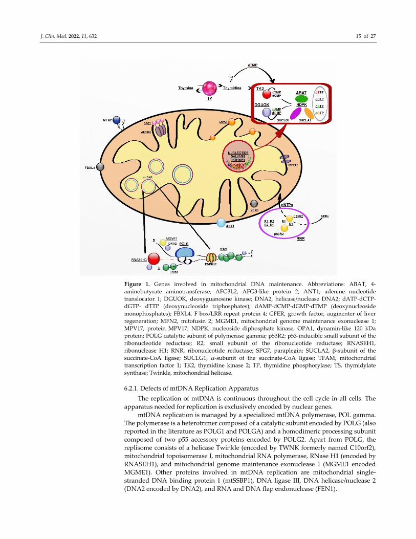
dynamics and quality control (Figure 1) [118].
J. Clin. Med. 2022, 11, 632 15 of 27
Figure 1. Genes involved in mitochondrial DNA maintenance. Abbreviations: ABAT, 4‐
aminobutyrate aminotransferase; AFG3L2, AFG3‐like protein 2; ANT1, adenine nucleotide
translocator 1; DGUOK, deoxyguanosine kinase; DNA2, helicase/nuclease DNA2; dATP‐dCTP‐
dGTP‐ dTTP (deoxynucleoside triphosphates); dAMP‐dCMP‐dGMP‐dTMP (deoxynucleoside
monophosphates); FBXL4, F‐box/LRR‐repeat protein 4; GFER, growth factor, augmenter of liver
regeneration; MFN2, mitofusin 2; MGME1, mitochondrial genome maintenance exonuclease 1;
MPV17, protein MPV17; NDPK, nucleoside diphosphate kinase, OPA1, dynamin‐like 120 kDa
protein; POLG catalytic subunit of polymerase gamma; p53R2; p53‐inducible small subunit of the
ribonucleotide reductase; R2, small subunit of the ribonucleotide reductase; RNASEH1,
ribonuclease H1; RNR, ribonucleotide reductase; SPG7, paraplegin; SUCLA2, β‐subunit of the
succinate‐CoA ligase; SUCLG1, α‐subunit of the succinate‐CoA ligase; TFAM, mitochondrial
transcription factor 1; TK2, thymidine kinase 2; TP, thymidine phosphorylase; TS, thymidylate
synthase; Twinkle, mitochondrial helicase.
6.2.1. Defects of mtDNA Replication Apparatus
The replication of mtDNA is continuous throughout the cell cycle in all cells. The
apparatus needed for replication is exclusively encoded by nuclear genes.
mtDNA replication is managed by a specialized mtDNA polymerase, POL gamma.
The polymerase is a heterotrimer composed of a catalytic subunit encoded by POLG (also
reported in the literature as POLG1 and POLGA) and a homodimeric processing subunit
composed of two p55 accessory proteins encoded by POLG2. Apart from POLG, the
replisome consists of a helicase Twinkle (encoded by TWNK formerly named C10orf2),
mitochondrial topoisomerase I, mitochondrial RNA polymerase, RNase H1 (encoded by
RNASEH1), and mitochondrial genome maintenance exonuclease 1 (MGME1 encoded
MGME1). Other proteins involved in mtDNA replication are mitochondrial single‐
stranded DNA binding protein 1 (mtSSBP1), DNA ligase III, DNA helicase/nuclease 2
(DNA2 encoded by DNA2), and RNA and DNA flap endonuclease (FEN1).
J. Clin. Med. 2022, 11, 632 16 of 27
Defects in genes involved in the replication machinery may lead to mtDNA
depletion, accumulation of multiple mtDNA deletions, or both, in critical tissues.
ANT1 encoded by SLC25A4, is the muscle‐specific isoform of the mitochondrial
adenine nucleotide translocator, and it is also expressed in the heart and brain. Several
mutations in SLC25A4 have been linked to mitochondrial disorders and fall into two
distinct clinical phenotypes: (1) Autosomal dominant CPEO [119,120] or (2) a slow
progressive mitochondrial myopathy with cardiomyopathy characterized by fatigue and
exercise intolerance and an autosomal recessive trait of inheritance [121].
Mutations of POLG are the most common pathogenic mutations and may present
with a wide range of clinical presentations
(https://tools.niehs.nih.gov/polg/index.cfm/polg, accessed on 30 November 2021). Three
main syndromes can be recognized as (1) arPEO, usually characterized by isolated PEO
and ptosis; (2) adPEO is frequently associated with myopathy and other systemic features;
(3) ataxia neuropathy spectrum (ANS) combines the previous syndromes of
mitochondrial recessive ataxia syndrome (MIRAS) and sensory ataxia neuropathy,
dysarthria and ophthalmoplegia (SANDO), myoclonic epilepsy myopathy sensory ataxia
(MEMSA) now envelops the syndrome of spinocerebellar ataxia with epilepsy (SCAE)
and includes epilepsy, myopathy, and ataxia without PEO. Two other syndromic
disorders related to POLG are recognized to present in childhood and include Alpers–
Huttenlocher syndrome (AHS) and Childhood myocerebrohepatopathy spectrum
(MCHS) disorder [122,123]
Autosomal dominant mutations in POLG2 have been shown to cause late‐onset PEO
with mtDNA deletions, but again, more complex phenotypes with ataxia, parkinsonism
and neuropathy have been associated with POLG2 variants [124,125].
Heterozygous mutations in the TWNK gene are responsible for adCPEO [126] with
accumulation of multiple mtDNA deletions. Clinical presentations include CPEO, often
associated with proximal muscle and facial weakness, dysphagia and dysphonia, mild
ataxia, and peripheral neuropathy. CPEO with parkinsonism has been rarely described in
patients with twinkle mutations [127].
Mutations in MGME1, DNA2 and RNASEH1 have been reported in patients with
PEO and accumulation of multiple mtDNA deletions. MGME1 and RNASEH1 mutations
are inherited as recessive traits, whereas DNA2 defects seem to be dominant. Clinical
manifestations appear in adulthood and more rarely in childhood, and in addition to PEO,
there is involvement of respiratory muscles and the brain, with cerebellar atrophy [128–
130].
6.2.2. Defects of Mitochondrial Deoxyribonucleosides Pools
Mitochondrial deoxynucleotides triphosphate (dNTP) pools depend on two different
pathways, the de novo synthesis due to an active transport of cytsolic dNTPs from
reduction of ribonucleotides by ribonucleotide reductase (RNR) and the salvage pathway
through the purines and pyrimidines by action of two mitochondrial deoxyribonucleoside
kinases, thymidine kinase 2 (TK2) and deoxyguanosine kinase (DGUOK). In nondividing
cells, cytosolic TK1 and dNTP synthesis is downregulated, forcing the burden of
mitochondrial dNTP pool synthesis on the two mitochondrial deoxyribonucleoside
kinases.
Mutations in the genes for enzymes involved in both pathways cause several forms
of mtDNA depletion syndromes (Table 4) [131].
MDS are autosomal recessive disorders with a broad genetic and clinical spectrum.
The salvage pathway is essential in postmitotic cells such as neurons and muscle
cells, in which dNTPs are produced by utilizing preexisting nucleosides through a
complex pathway of enzymes [132]. Among them, there are enzymes encoded by SUCLA2
(adenosine diphosphate (ADP)‐forming succinyl CoA ligase beta subunit), SUCLG1
(guanosine diphosphate (GDP)‐forming succinyl CoA ligase alpha subunit) and TYMP
(thymidine phosphorylase) [133].
J. Clin. Med. 2022, 11, 632 17 of 27
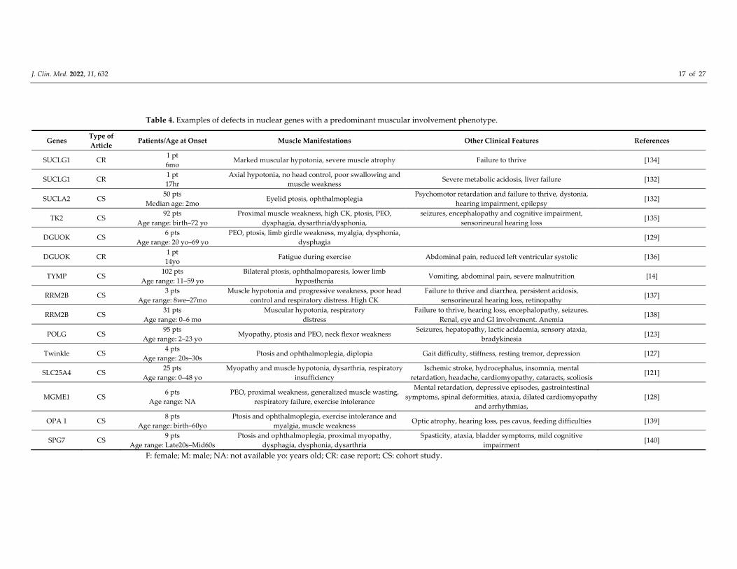
Table 4. Examples of defects in nuclear genes with a predominant muscular involvement phenotype.
Genes Type of
Article Patients/Age at Onset Muscle Manifestations Other Clinical Features References
SUCLG1 CR 1 pt
6mo Marked muscular hypotonia, severe muscle atrophy Failure to thrive [134]
SUCLG1 CR 1 pt
17hr
Axial hypotonia, no head control, poor swallowing and
muscle weakness Severe metabolic acidosis, liver failure [132]
SUCLA2 CS 50 pts
Median age: 2mo Eyelid ptosis, ophthalmoplegia
Psychomotor retardation and failure to thrive, dystonia,
hearing impairment, epilepsy [132]
TK2 CS 92 pts
Age range: birth–72 yo
Proximal muscle weakness, high CK, ptosis, PEO,
dysphagia, dysarthria/dysphonia,
seizures, encephalopathy and cognitive impairment,
sensorineural hearing loss [135]
DGUOK CS 6 pts
Age range: 20 yo–69 yo
PEO, ptosis, limb girdle weakness, myalgia, dysphonia,
dysphagia [129]
DGUOK CR 1 pt
14yo Fatigue during exercise Abdominal pain, reduced left ventricular systolic [136]
TYMP CS 102 pts
Age range: 11–59 yo
Bilateral ptosis, ophthalmoparesis, lower limb
hyposthenia Vomiting, abdominal pain, severe malnutrition [14]
RRM2B CS 3 pts
Age range: 8we–27mo
Muscle hypotonia and progressive weakness, poor head
control and respiratory distress. High CK
Failure to thrive and diarrhea, persistent acidosis,
sensorineural hearing loss, retinopathy [137]
RRM2B CS 31 pts
Age range: 0–6 mo
Muscular hypotonia, respiratory
distress
Failure to thrive, hearing loss, encephalopathy, seizures.
Renal, eye and GI involvement. Anemia [138]
POLG CS 95 pts
Age range: 2–23 yo Myopathy, ptosis and PEO, neck flexor weakness
Seizures, hepatopathy, lactic acidaemia, sensory ataxia,
bradykinesia [123]
Twinkle CS 4 pts
Age range: 20s–30s Ptosis and ophthalmoplegia, diplopia Gait difficulty, stiffness, resting tremor, depression [127]
SLC25A4 CS 25 pts
Age range: 0–48 yo
Myopathy and muscle hypotonia, dysarthria, respiratory
insufficiency
Ischemic stroke, hydrocephalus, insomnia, mental
retardation, headache, cardiomyopathy, cataracts, scoliosis [121]
MGME1 CS 6 pts
Age range: NA
PEO, proximal weakness, generalized muscle wasting,
respiratory failure, exercise intolerance
Mental retardation, depressive episodes, gastrointestinal
symptoms, spinal deformities, ataxia, dilated cardiomyopathy
and arrhythmias,
[128]
OPA 1 CS 8 pts
Age range: birth–60yo
Ptosis and ophthalmoplegia, exercise intolerance and
myalgia, muscle weakness Optic atrophy, hearing loss, pes cavus, feeding difficulties [139]
SPG7 CS 9 pts
Age range: Late20s–Mid60s
Ptosis and ophthalmoplegia, proximal myopathy,
dysphagia, dysphonia, dysarthria
Spasticity, ataxia, bladder symptoms, mild cognitive
impairment [140]
F: female; M: male; NA: not available yo: years old; CR: case report; CS: cohort study.

J. Clin. Med. 2022, 11, 632 18 of 27
SUCLA2 and SUCLG1 genes encode subunits of succinyl CoA ligase (SUCL), a
mitochondrial enzyme of the Krebs cycle that catalyzes the reversible conversion of
succinyl‐CoA and ADP or GDP to succinate and ATP or GTP [134].
SUCL deficiency is an MDS, presenting as Leigh/Leigh‐like encephalomyopathy with
variable phenotypes. It occurs in early childhood with severe hypotonia and failure to
thrive, then developing into muscular atrophy and growth retardation. Other clinical
features could be dystonia, sensorineural hearing impairment and respiratory problems.
Basal ganglia lesions, cerebral atrophy and white matter alterations are common findings
at neuroimaging as well as lipid accumulation and type I fibers’ predominance at muscle
biopsy. Additionally, it is variably associated with methylmalonic aciduria, because
accumulated succinyl‐CoA is subsequently converted to methylmalonyl‐CoA [132].
TK2 catalyzes the conversion of deoxycytidine and thymidine nucleosides to their
nucleoside monophosphates (dCMP and dTMP), that are ready to be phosphorylated to
dNTP and incorporated into mtDNA. TK2 deficiency presents predominantly as a
proximal myopathy with variable severity, leading in most cases to loss of ambulation
and respiratory insufficiency in a few years. Three main clinical forms have been
described: a rapidly progressive infantile‐onset myopathy, associated with CNS
alterations such as encephalopathy, seizures or cognitive impairment; a childhood‐onset
myopathy, resembling SMA type 3; and a late‐onset myopathy with usual facial and
extraocular muscles involvement (i.e., ptosis or PEO) [135].
To date, therapy for TK2 deficiency is still at a preclinical status. It consists of
replacement of dCMP and dTMP or their respective nucleosides deoxycytidine (dC) and
deoxythymidine (dT), directly. They were administered orally in TK2−/− mice, resulting
in reduced imbalance of mtDNA, recovery of mitochondrial functions and prolonged
lifespan [141].
DGUOK gene encodes for deoxyguanosine kinase (dGK), an enzyme that plays the
same role as TK2 in the purine nucleoside salvage pathway. It converts deoxyguanosine
and deoxyadenosine to, respectively, deoxyguanosine (dGMP) and deoxyadenosine
monophosphate (dAMP), that need another two phosphorylation steps before being
inserted into mtDNA.
dGK deficiency is mostly known as a neonatal onset multisystem disease,
characterized by hepatic and neurologic dysfunction such as hypotonia, psychomotor
retardation, and typical rotary nystagmus. Few individuals are affected later in infancy or
during childhood, when the disease presents as isolated hepatic disease [142].
Ronchi et al., 2012, reported five patients with dGK deficiency and variable skeletal
muscles’ involvement (progressive external ophthalmoplegia and ptosis, dysphonia and
dysphagia, limb girdle weakness, myalgia, cramps and rhabdomyolysis) [129].
Finally, Buchaklian et al., 2012, described a case of juvenile‐onset myopathy
presenting with weakness and fatigability [136].
Notably, recent studies have demonstrated that the nucleoside salvage pathway is
coadjuvated by the nuclear triphosphohydrolase enzyme SAMHD1. Its role is to
hydrolyze in the nucleus dNTPs synthesized de novo in the cytosol. The obtained
deoxynucleosides can be recycled by TK2 and dGK for mtDNA replication [143].
TYMP gene encodes for thymidine phosphorylase (TP), which catabolizes thymidine
(dThd) and deoxyuridine (dUrd) into their respective bases. Lack or dysfunction of TP
determines dThd and dUrd toxic accumulation with consequent mtDNA impairment,
leading to Mitochondrial Neuro–Gastro–Intestinal Encephalomyopathy (MNGIE) [144].
MNGIE is an ultrarare condition, which can present as “Early Onset” (or “Classic”)
and “Late Onset” forms. Main clinical features are represented by gastrointestinal
symptoms (diarrhea, abdominal pain, pseudo‐obstruction, weight loss/cachexia) and
neurological symptoms/signs (ptosis, ophthalmoparesis, polyneuropathy, hearing loss
and leucoencephalopathy at brain MRI imaging), with fatal evolution. Several therapeutic
options have been proposed to replace TP temporarily, for example, erythrocyte‐
encapsulated TP infusions, or to restore TP permanently such as hematopoietic stem cell
J. Clin. Med. 2022, 11, 632 19 of 27
transplantation and orthotopic liver transplantation. Recently, gene therapy was
successful in restoring biochemical homeostasis in a murine model of the disease
[14,145,146].
Another gene involved in mitochondrial nucleoside salvage is 4‐aminobutyrate
aminotransferase (ABAT), that encodes for GABA transaminase (GABA‐T). This enzyme
is responsible for both GABA neurotransmitter catabolism in the mitochondrial matrix
and conversion of dNDPs to dNTPs. Thus, ABAT deficiency causes GABA accumulation
and mitochondrial nucleoside pools imbalance [147]. The resulting MDS usually occurs
in neonatal age with hypotonia and severe psychomotor retardation. Visual impairment,
abnormal movement, hypersomnia and infantile‐onset refractory epilepsy have been
reported, too [148].
In contrast to the mitochondrial salvage pathway, nucleotide precursors can be
directly obtained through reduction in ribonucleoside diphosphates to
deoxyribonucloside diphosphates by ribonucleotide reductase. This enzyme is composed
of R1 and R2 subunits; the last one is known as p53‐inducible form (p53R2) and plays a
crucial role in de novo nucleotide synthesis [149].
It is encoded by RRM2B gene, whose mutations can cause an encephalomyopathic
form of MDS, which is clinically heterogeneous. Commonly, RRM2B deficiency presents
as neonatal or infantile myopathy with lactic acidosis and increased serum CK. Therefore,
children with combinations of hypotonia, tubulopathy, seizures, respiratory distress,
diarrhea and lactic acidosis have been described [137].
Other less common RRM2B phenotypes have been described as progressive external
ophthalmoplegia with bulbar dysfunction, fatigue, and muscle weakness with autosomal
dominant transmission in adults or with a recessive trait and childhood‐onset
[138,150,151].
6.2.3. Defects of Mitochondrial Dynamics and Quality Control
Mitochondria are highly dynamic organelles, providing themselves with different
shapes, distributions and sizes though fusion and fission reactions. The related enzymatic
machinery is called “mitochondrial dynamics” and defects in its components can be
associated with various disorders [15].
Mitochondrial fusion is the merger of two mitochondria into one, based on a
GTPases‐mediated process. Optic atrophy 1 (OPA1) is involved in the outer membrane
fusion, while GTPases for the inner membrane fusion are Mitofusins 1 and 2 (MFN1 and
MFN2) [152]. Mitochondrial fission is the opposite process—the division of a
mitochondrion into two smaller mitochondria. The main protein of the fission is dynamin‐
related protein 1 (Drp1), a GTP‐hydrolyzing enzyme [15,152].
The large majority of mutations in the OPA1 gene are related with a slowly
progressive optic neuropathy. Dominant optic atrophy (DOA) is due to OPA1 mutations
in about 60–70% of cases [139,153]. It is caused by the degeneration of optic nerve fibers
with mild visual loss and color vision alterations, usually starting during childhood. In
about 20% of patients, DOA evolves as syndromic forms with myopathy, progressive
external ophthalmoplegia, peripheral neuropathy, stroke, multiple sclerosis, spastic
paraplegia and sensorineural hearing loss [154].
MNF1 forms homomultimers and heteromultimers with MFN2. No human diseases
have been described in relation to MFN1 mutations [16]. Mutations in MFN2 are
associated with autosomal dominant or recessive Charcot–Marie–Tooth disease type 2A
and autosomal dominant hereditary motor and sensory neuropathy VIA. Charcot–Marie–
Tooth disease type 2A is characterized by distal limb muscle weakness and atrophy,
axonal degeneration/regeneration, areflexia and distal sensory loss, associated variably
with CNS involvement, optic atrophy, hearing loss and vocal cord paresis [15].
Another protein located at the inner mitochondrial membrane is paraplegin, encoded
by the SPG7 gene as part of the AAA family of ATPases. It contributes to assembly of
mitochondrial ribosomes, taking part in other proteins’ synthesis [155]. SPG7 mutations
J. Clin. Med. 2022, 11, 632 20 of 27
have been recently linked to PEO mid‐adult onset, with multiple mitochondrial DNA
deletions. Ptosis, spastic ataxia, dysphagia and proximal myopathy were other common
features [140].
7. Genetic Diagnostic Approach in PMM
In clinical practice, despite the increased awareness of mitochondrial disorders and
the introduction of deeper genetic investigations, the diagnosis, in a patient with a
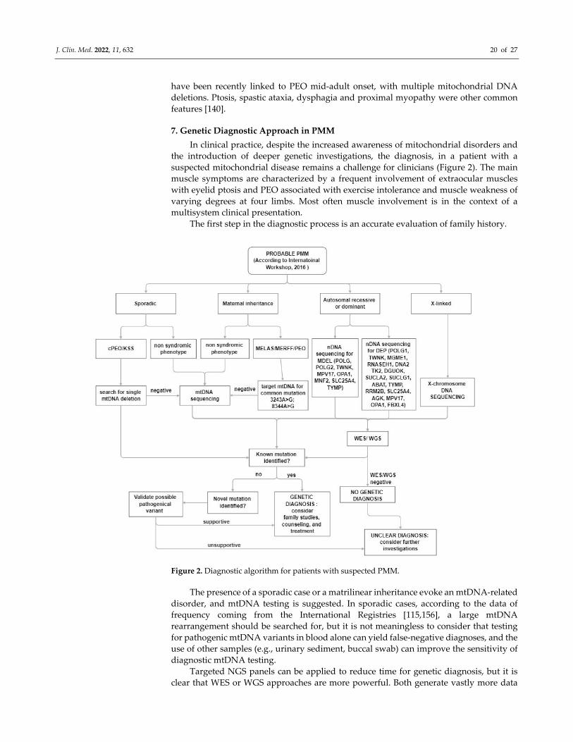
suspected mitochondrial disease remains a challenge for clinicians (Figure 2). The main
muscle symptoms are characterized by a frequent involvement of extraocular muscles
with eyelid ptosis and PEO associated with exercise intolerance and muscle weakness of
varying degrees at four limbs. Most often muscle involvement is in the context of a
multisystem clinical presentation.
The first step in the diagnostic process is an accurate evaluation of family history.
Figure 2. Diagnostic algorithm for patients with suspected PMM.
The presence of a sporadic case or a matrilinear inheritance evoke an mtDNA‐related
disorder, and mtDNA testing is suggested. In sporadic cases, according to the data of
frequency coming from the International Registries [115,156], a large mtDNA
rearrangement should be searched for, but it is not meaningless to consider that testing
for pathogenic mtDNA variants in blood alone can yield false‐negative diagnoses, and the
use of other samples (e.g., urinary sediment, buccal swab) can improve the sensitivity of
diagnostic mtDNA testing.
Targeted NGS panels can be applied to reduce time for genetic diagnosis, but it is
clear that WES or WGS approaches are more powerful. Both generate vastly more data
J. Clin. Med. 2022, 11, 632 21 of 27
and require additional analysis. These approaches can simultaneously analyze both
genomes, can identify unknown variants and novel disease genes.
Interpretation of genomic sequencing results has been greatly improved by adoption
of the American College of Medical Genetics criteria for variant classification, but requires
a multidisciplinary team comprising clinicians with expertise in mitochondrial disorders,
to evaluate the consistency with the phenotype.
In addition, unknown or uncertain variants need to be tested by functional testing
using more traditional approaches for diagnosis including morphological and
biochemical studies.
8. Conclusions
Although the heterogeneity and pleiotropy of mitochondrial disorders are not
sufficiently clarified, over recent decades the introduction of broad‐based exome
sequencing as the standard first‐line diagnostic approach has increased the yield of
definite diagnosis in patient with a suspected mitochondrial disorders. Nowadays, the
identification of the genetic basis of disease in each patient is relevant, particularly among
patients with a PMM that is becoming the main target phenotype in clinical trials.
Author Contributions: I.G.A. and O.M. participated in the design of the study. I.G.A., O.M., A.P.,
S.V. and A.T. helped in drafting the manuscript and reviewed and approved the final manuscript.
All authors have read and agreed to the published version of the manuscript.
Funding: This research received no external funding.
Institutional Review Board Statement: Not applicable.
Informed Consent Statement: Not applicable.
Data Availability Statement: Not applicable.
Acknowledgments: Three of the authors of this publication (OM and AT) are members of the
European Reference Network for ERN EURO‐NMD—Project ID No 739543.
Conflicts of Interest: The authors declare that the research was conducted in the absence of any
commercial or financial relationships that could be construed as a potential conflict of interest.
References
1. Mancuso, M.; Mcfarland, R.; Klopstock, T.; Hirano, M. International workshop: Outcome measures and clinical trial readiness
in primary mitochondrial myopathies in children and adults. Consensus recommendations. 16–18 November 2016, Rome, Italy.
Neuromuscul. Disord. 2017, 27, 1126–1137.
2. Ng, Y.S.; Bindoff, L.A.; Gorman, G.S.; Klopstock, T.; Kornblum, C.; Mancuso, M.; McFarland, R.; Sue, C.M.; Suomalainen, A.;
Taylor, R.W.; et al. Mitochondrial disease in adults: Recent advances and future promise. Lancet Neurol. 2021, 20, 573–584.
3. Frazier, A.E.; Thorburn, D.R.; Compton, A.G. Mitochondrial energy generation disorders: Genes, mechanisms, and clues to
pathology. J. Biol. Chem. 2019, 294, 5386–5395.
4. McCormick, E.M.; Zolkipli‐Cunningham, Z.; Falk, M.J. Mitochondrial Disease Genetics Update Recent insights into the
Molecular Diagnosis and Expanding Phenotype of Primary Mitochondrial Disease. Curr. Opin. Pediatr. 2018, 30, 714–724.
5. Chinnery, P.F.; Dimauro, P.S.; Shanske, S.; Schon, P.E.A.; Zeviani, M.; Mariotti, C.; Carrara, F.; Lombes, A.; Laforet, P.; Ogier,
H.; et al. Risk of developing a mitochondrial DNA deletion disorder. Lancet 2004, 364, 592–596.
6. Lightowlers, R.N.; Taylor, R.W.; Turnbull, D.M. Mutations causing mitochondrial disease: What is new and what challenges
remain? Science 2015, 349, 1494–1499.
7. Ghezzi, D.; Zeviani, M. Human diseases associated with defects in assembly of OXPHOS complexes. Essays Biochem. 2018, 62,
271–286.
8. Fassone, E.; Rahman, S. Complex I deficiency: Clinical features, biochemistry and molecular genetics. J. Med. Genet. 2012, 49,
578–590.
9. Alston, C.L.; Rocha, M.C.; Lax, N.Z.; Turnbull, D.M.; Taylor, R.W. The genetics and pathology of mitochondrial disease. J. Pathol.
2017, 241, 236–250.
10. Fernandez‐Vizarra, E.; Bugiani, M.; Goffrini, P.; Carrara, F.; Farina, L.; Procopio, E.; Donati, A.; Uziel, G.; Ferrero, I.; Zeviani, M.
Impaired complex III assembly associated with BCS1L gene mutations in isolated mitochondrial encephalopathy. Hum. Mol.
Genet. 2007, 16, 1241–1252.

J. Clin. Med. 2022, 11, 632 22 of 27
11. Brischigliaro, M.; Zeviani, M. Cytochrome c oxidase deficiency. Biochim. Biophys Acta Bioenerg. 2021, 1862, 148335.
12. Rahman, S. Mitochondrial disease in children. J. Intern. Med. 2020, 287, 609–633. https://doi.org/10.1111/joim.13054.
13. Gandhi, V.V.; Samuels, D.C. A review comparing deoxyribonucleoside triphosphate (dNTP) concentrations in the
mitochondrial and cytoplasmic compartments of normal and transformed cells. Nucleosides Nucleotides Nucleic Acids 2011, 30,
317–339.
14. Garone, C.; Tadesse, S.; Hirano, M. Clinical and genetic spectrum of mitochondrial neurogastrointestinal encephalomyopathy.
Brain : A J. Neurol. 2011, 134, 3326–3332
15. Tilokani, L.; Nagashima, S.; Paupe, V.; Prudent, J. Mitochondrial dynamics: Overview of molecular mechanisms. Essays Biochem.
2018, 62, 341–360.
16. Rusecka, J.; Kaliszewska, M.; Bartnik, E.; Tońska, K. Nuclear genes involved in mitochondrial diseases caused by instability of
mitochondrial DNA. J. Appl. Genet. 2018, 59, 43–57.
17. Garcia, I.; Jones, E.; Ramos, M.; Innis‐Whitehouse, W.; Gilkerson, R. The little big genome: The organization of mitochondrial
DNA. Front Biosci. 2017, 22, 710–721.
18. Calvo, S.E.; Mootha, V. The mitochondrial proteome and human disease. Annu. Rev. Genom. Hum. Genet. 2010, 11, 25–44.
19. Sato, M.; Sato, K. Maternal inheritance of mitochondrial DNA by diverse mechanisms to eliminate paternal mitochondrial DNA.
Biochim. Biophys. Acta—Mol. Cell Res. 2013, 1833, 1979–1984.
20. Schwartz, M.; Vissing, J. Paternal inheritance of mitochondrial DNA. N. Engl. J. Med. 2002, 347, 576–580.
21. Ballana, E.; Govea, N.; de Cid, R.; Garcia, C.; Arribas, C.; Rosell, J.; Xavier, E. Detection of Unrecognized Low‐Level mtDNA
Heteroplasmy May Explain the Variable Phenotypic Expressivity of Apparently Homoplasmic mtDNA Mutations. Hum. Mutat.
2008, 29, 248–257.
22. Rossignol, R.; Faustin, B.; Rocher, C.; Malgat, M.; Mazat, J.P.; Letellier, T. Mitochondrial threshold effects. Biochem. J. 2003, 370,
751–762.
23. Andreu, A.L.; Hanna, M.G.; Reichmann, H.; Bruno, C.; Penn, A.S.; Tanji, K.; Pallotti, F.; Iwata, S.; Bonilla, E.; Lach, B.; et al.
Exercise Intolerance Due to Mutations in the Cytochrome b Gene of Mitochondrial DNA. N. Engl. J. Med. 1999, 341, 1037–1044.
24. Rahman, S. Mitochondrial disease and epilepsy. Dev. Med. Child Neurol. 2012, 54, 397–406.
25. Meyerson, C.; van Stavern, G.; McClelland, C. Leber hereditary optic neuropathy: Current perspectives. Clin. Ophthalmol. 2015,
9, 1165–1176.
26. Mancuso, M.; Filosto, M.; Mootha, V.K.; Rocchi, A.; Pistolesi, S.; Murri, L.; DiMauro, S.; Siciliano, G. A novel mitochondrial
tRNAPhe mutation causes MERRF syndrome. Neurology 2004, 62, 2119–2121.
27. Chinnery, P.F. Mitochondrial disease in adults: What’s old and what’s new? EMBO Mol. Med. 2015, 7, 1503–1512.
28. Osellame, L.D.; Blacker, T.S.; Duchen, M.R. Cellular and molecular mechanisms of mitochondrial function. Best Pr. Res. Clin.
Endocrinol. Metab. 2012, 26, 711–723.
29. Fiedorczuk, K.; Sazanov, L.A. Mammalian Mitochondrial Complex I Structure and Disease‐Causing Mutations. Trends Cell Biol.
2018, 28, 835–867.
30. Lake, N.J.; Compton, A.G.; Rahman, S.; Thorburn, D.R. Leigh syndrome: One disorder, more than 75 monogenic causes. Ann.
Neurol. 2016, 79, 190–203.
31. Rafiq, J.; Duno, M.; Ostergaard, E.; Ravn, K.; Vissing, C.R.; Wibrand, F.; Vissing, J. Exercise intolerance and myoglobinuria
associated with a novel maternally iherited MT‐ND1 mutation. JIMD Rep. 2015, 25, 65–70.
32. Gorman, G.S.; Blakely, E.L.; Hornig‐Do, H.T.; Tuppen, H.A.; Greaves, L.C.; He, L.; Baker, A.; Falkous, G.; Newman, J.; Trenell,
M.I.; et al. Novel MTND1 mutations cause isolated exercise intolerance, complex I deficiency and increased assembly factor
expression. Clin. Sci. 2015, 128, 895–904
33. Zanolini, A.; Potic, A.; Carrara, F.; Lamantea, E.; Diodato, D.; Blasevich, F.; Marchet, S.; Mora, M.; Pallotti, F.; Morandi, L.; et al.
Pure myopathy with enlarged mitochondria associated to a new mutation in MTND2 gene. Mol. Genet. Metab. Rep. 2017, 10, 24–
27.
34. Andreu, A.L.; Tanji, K.; Bruno, C.; Hadjigeorgiou, G.M.; Sue, C.M.; Jay, C.; Ohnishi, T.; Shanske, S.; Bonilla, E.; DiMauro, S.
Exercise intolerance due to a nonsense mutation in the mtDNA ND4 gene. Ann. Neurol. 1999, 45, 820–823.
35. Downham, E.; Winterthun, S.; Nakkestad, H.L.; Hirth, A.; Halvorsen, T.; Taylor, R.W.; Bindoff, L.A. A novel mitochondrial ND5
(MTND5) gene mutation giving isolated exercise intolerance. Neuromuscul. Disord. 2008, 18, 310–314.
36. Musumeci, O.; Andreu, A.L.; Shanske, S.; Bresolin, N.; Comi, G.P.; Rothstein, R.; Schon, E.A.; DiMauro, S. Intragenic inversion
of mtDNA: A new type of pathogenic mutation in a patient with mitochondrial myopathy. Am. J. Hum. Genet. 2000, 66, 1900–
1904.
37. Alston, C.L.; Morak, M.; Reid, C.; Hargreaves, I.P.; Pope, S.A.S.; Land, J.M.; Heales, S.J.; Horvath, R.; Mundy, H.; Taylor, R.W.
A novel mitochondrial MTND5 frameshift mutation causing isolated complex I deficiency, renal failure and myopathy.
Neuromuscul. Disord. 2010, 20, 131–135.
38. Ng, Y.S.; Thompson, K.; Loher, D.; Hopton, S.; Falkous, G.; Hardy, S.A.; Schaefer, A.M.; Shaunak, S.; Roberts, M.E.; Lilleker,
J.B.; et al. Novel MT‐ND Gene Variants Causing Adult‐Onset Mitochondrial Disease and Isolated Complex I Deficiency. Front.
Genet. 2020, 11, 1–7.
J. Clin. Med. 2022, 11, 632 23 of 27
39. Alston, C.L.; Compton, A.G.; Formosa, L.E.; Strecker, V.; Oláhová, M.; Haack, T.B.; Smet, J.; Stouffs, K.; Diakumis, P.; Ciara, E.;
et al. Biallelic Mutations in TMEM126B Cause Severe Complex I Deficiency with a Variable Clinical Phenotype. Am. J. Hum.
Genet. 2016, 99, 217–227.
40. Sánchez‐Caballero, L.; Ruzzenente, B.; Bianchi, L.; Assouline, Z.; Barcia, G.; Metodiev, M.D.; Rio, M.; Funalot, B.; van den Brand,
M.A.M.; Guerrero‐Castillo, S.; et al. Mutations in Complex I Assembly Factor TMEM126B Result in Muscle Weakness and
Isolated Complex I Deficiency. Am. J. Hum. Genet. 2016, 99, 208–216.
41. Repp, B.M.; Mastantuono, E.; Alston, C.L.; Schiff, M.; Haack, T.B.; Rötig, A.; Ardissone, A.; Lombès, A.; Catarino, C.B.; Diodato,
D.; et al. Clinical, biochemical and genetic spectrum of 70 patients with ACAD9 deficiency: Is riboflavin supplementation
effective? Orphanet J. Rare Dis. 2018, 13, 1–10.
42. Dumoulin, R.; Sagnol, I.; Ferlin, T.; Bozon, D.; Stepien, G.; Mousson, B. A novel gly290asp mitochondrial cytochrome b mutation
linked to a complex III deficiency in progressive exercise intolerance. Mol. Cell Probes 1996, 10, 389–391.
43. Keightley, J.A.; Anitori, R.; Burton, M.D.; Quan, F.; Buist, N.R.M.; Kennaway, N.G. Mitochondrial encephalomyopathy and
complex III deficiency associated with a stop‐codon mutation in the cytochrome b gene. Am. J. Hum. Genet. 2000, 67, 1400–1410.
44. Bruno, C.; Santorelli, F.M.; Assereto, S.; Tonoli, E.; Tessa, A.; Traverso, M.; Scapolan, S.; Bado, M.; Tedeschi, S.; Minetti, C.
Progressive exercise intolerance associated with a new muscle‐restricted nonsense mutation (G142X) in the mitochondrial
cytochrome b gene. Muscle Nerve 2003, 28, 508–511.
45. Lamantea, E.; Carrara, F.; Mariotti, C.; Morandi, L.; Tiranti, V.; Zeviani, M. A novel nonsense mutation (Q352X) in the
mitochondrial cytochrome b gene associated with a combined deficiency of complexes I and III. Neuromuscul. Disord. 2002, 12,
49–52.
46. Kollberg, G.; Moslemi, A.R.; Lindberg, C.; Holme, E.; Oldfors, A. Mitochondrial myopathy and rhabdomyolysis associated with
a novel nonsense mutation in the gene encoding cytochrome c oxidase subunit I. J. Neuropathol. Exp. Neurol. 2005, 64, 123–128.
47. Rahman, S.; Taanman, J.W.; Cooper, J.M.; Nelson, I.; Hargreaves, I.; Meunier, B.; Hanna, M.G.; García, J.J.; Capaldi, R.A.; Lake,
B.D.; et al. A missense mutation of cytochrome oxidase subunit II causes defective assembly and myopathy. Am. J. Hum. Genet.
1999, 65, 1030–1039.
48. Roos, S.; Sofou, K.; Hedberg‐Oldfors, C.; Kollberg, G.; Lindgren, U.; Thomsen, C.; Tulinius, M.; Oldfors, A. Mitochondrial
complex IV deficiency caused by a novel frameshift variant in MT‐CO2 associated with myopathy and perturbed acylcarnitine
profile. Eur. J. Hum. Genet. 2018, 27, 331–335.
49. Horváth, R.; Lochmüller, H.; Hoeltzenbein, M.; Müller‐Höcker, J.; Schoser, B.G.; Pongratz, D.; Jaksch, M. Spontaneous recovery
of a childhood onset mitochondrial myopathy caused by a stop mutation in the mitochondrial cytochrome c oxidase III gene. J.
Med. Genet. 2004, 41, 1–5.
50. Inoue, M.; Uchino, S.; Iida, A.; Noguchi, S.; Hayashi, S.; Takahashi, T.; Fujii, K.; Komaki, H.; Takeshita, E.; Nonaka, I.; et al.
COX6A2 variants cause a muscle‐specific cytochrome c oxidase deficiency. Ann. Neurol. 2019, 86, 193–202.
51. Pitceathly, R.D.S.; Taanman, J.W.; Rahman, S.; Meunier, B.; Sadowski, M.; Cirak, S.; Hargreaves, I.; Land, J.M.; Nanji, T.; Polke,
J.M.; et al. COX10 mutations resulting in complex multisystem mitochondrial disease that remains stable into adulthood. JAMA
Neurol. 2013, 70, 1556–1561.
52. Swalwell, H.; Kirby, D.M.; Blakely, E.L.; Mitchell, A.; Salemi, R.; Sugiana, C.; Compton, A.G.; Tucker, E.J.; Ke, B.X.; Lamont, P.J.;
et al. Respiratory chain complex I deficiency caused by mitochondrial DNA mutations. Eur. J. Hum. Genet. 2011, 19, 769–775.
53. Fernandez‐Moreira, D.; Ugalde, C.; Smeets, R.; Rodenburg, R.J.T.; Lopez‐Laso, E.; Ruiz‐Falco, M.L.; Briones, P.; Martin, M.A.;
Smeitink, J.A.M.; Arenas, J. X‐linked NDUFA1 gene mutations associated with mitochondrial encephalomyopathy. Ann. Neurol.
2007, 61, 73–83.
54. Van Rahden, V.A.; Fernandez‐Vizarra, E.; Alawi, M.; Brand, K.; Fellmann, F.; Horn, D.; Zeviani, M.; Kutsche, K. Mutations in
NDUFB11, encoding a complex i component of the mitochondrial respiratory chain, cause microphthalmia with linear skin
defects syndrome. Am. J. Hum. Genet. 2015, 96, 640–650.
55. Pagniez‐Mammeri, H.; Loublier, S.; Legrand, A.; Bénit, P.; Rustin, P.; Slama, A. Mitochondrial complex I deficiency of nuclear
origin. I. Structural genes. Mol. Genet. Metab. 2012, 105, 163–172.
56. Fassone, E.; Duncan, A.J.; Taanman, J.; Pagnamenta, A.T.; Sadowski, M.I.; Holand, T.; Qasim, W.; Rutland, P.; Calvo, S.E.;
Mootha, V.K.; et al. FOXRED1, encoding an FAD‐dependent oxidoreductase complex‐I‐specific molecular chaperone, is
mutated in infantile‐onset mitochondrial encephalopathy. Hum. Mol. Genet. 2015, 24, 4183.
57. Haack, T.B.; Danhauser, K.; Haberberger, B.; Hoser, J.; Strecker, V.; Boehm, D.; Uziel, G.; Lamantea, E.; Invernizzi, F.; Poulton,
J.; et al. Exome sequencing identifies ACAD9 mutations as a cause of complex I deficiency. Nat. Genet. 2010, 42, 1131–1134.
58. Saada, A.; Vogel, R.O.; Hoefs, S.J.; Van den Brand, M.A.; Wessels, H.J.; Willems, P.H.; Venselaar, H.; Shaag, A.; Barghuti, F.;
Reish, O.; et al. Mutations in NDUFAF3 (C3ORF60), Encoding an NDUFAF4 (C6ORF66)‐Interacting Complex I Assembly
Protein, Cause Fatal Neonatal Mitochondrial Disease. Am. J. Hum. Genet. 2009, 84, 718–727.
59. Jain‐Ghai, S.; Cameron, J.M.; al Maawali, A.; Blaser, S.; Mackay, N.; Robinson, B.; Raiman, J. Complex II deficiency‐A case report
and review of the literature. Am. J. Hum. Genet. Part A 2013, 161, 285–294.
60. Renkema, G.H.; Wortmann, S.B.; Smeets, R.J.; Venselaar, H.; Antoine, M.; Visser, G.; Ben‐Omran, T.; van den Heuvel, L.P.;
Timmers, H.J.; Smeitink, J.A.; et al. SDHA mutations causing a multisystem mitochondrial disease: Novel mutations and genetic
overlap with hereditary tumors. Eur. J. Hum. Genet. 2015, 23, 202–209.
J. Clin. Med. 2022, 11, 632 24 of 27
61. Alston, C.L.; Davison, J.E.; Meloni, F.; van der Westhuizen, F.H.; He, L.; Hornig‐Do, H.T.; Peet, A.C.; Gissen, P.; Goffrini, P.;
Ferrero, I.; et al. Recessive germline SDHA and SDHB mutations causing leukodystrophy and isolated mitochondrial complex
II deficiency. J. Med. Genet. 2021, 49, 569–577.
62. Ohlenbusch, A.; Edvardson, S.; Skorpen, J.; Bjornstad, A.; Saada, A.; Elpeleg, O.; Gärtner, J.; Brockmann, K.
Leukoencephalopathy with accumulated succinate is indicative of SDHAF1 related complex II deficiency. Orphanet J. Rare Dis.
2012, 7, 7–11.
63. Xia, D.; Esser, L.; Tang, W.K.; Zhou, F.; Zhou, Y.; Yu, L.; Yu, C.A. Structural analysis of cytochrome bc1 complexes: Implications
to the mechanism of function. Biochim. Biophys. Acta Bioenerg. 2013, 1827, 1278–1294.
64. Fernández‐Vizarra, E.; Zeviani, M. Nuclear gene mutations as the cause of mitochondrial complex III deficiency. Front. Genet.
2015, 6, 1–11.
65. Spiro, A.J.; Moore, C.L.; Prineas, J.W.; Strasberg, P.M.; Rapin, I. A Cytochrome‐Related Inherited Disorder of the Nervous
System and Muscle. Arch. Neurol. 1970, 23, 103–112.
66. Darley Usmar, V.M.; Kennaway, N.G.; Buist, N.R.M.; Capaldi, R.A. Deficiency in ubiquinone cytochrome c reductase in a
patient with mitochondrial myopathy and lactic acidosis. Proc. Natl. Acad. Sci. USA 1983, 80, 5103–5106.
67. Hayes, D.J.; Lecky, B.R.F.; Landon, D.N.; Morgan Hughes, J.A.; Clark, J.B. A new Mitochondrial myopathy biochemical studies
revealing a deficiency in the cytochrome b‐c complex (complex III) of the respiratory chain. Brain 1984, 107, 1165–1177.
68. Fouad Bouzidi, M.; Schägger, H.; Collombet, J.M.; Carrier, H.; Flocard, F.; Quard, S.; Mousson, B.; Godinot, C. Decreased
expression ofubiquinol‐cytochrome c reductase subunits in patients exhibiting mitochondrial myopathy with progressive
exercise intolerance. Neuromuscul. Disord. 1993, 3, 599–604.
69. De Coo, I.F.M.; Renier, W.O.; Ruitenbeek, W.; Ter Laak, H.J.; Bakker, M.; Schagger, H.; Van Oost, B.A.; Smeets, H.J. A 4–Base
Pair Deletion in the Mitochondrial Cytochrome b Gene Associated with Parkinsonism/MELAS Overlap Syndrome. Ann. Neurol.
1999, 45, 130–133.
70. Wibrand, F.; Ravn, K.; Schwartz, M.; Rosenberg, T.; Horn, N.; Vissing, J. Multisystem Disorder Associated with a Missense
Mutation in the Mitochondrial Cytochrome b gene. Ann. Neurol. 2001, 50, 540–543.
71. Schuelke, M.; Krude, H.; Finckh, B.; Mayatepek, E.; Janssen, A.; Schmelz, M.; Trefz, F.; Trijbels, F.; Smeitink, J. Septo‐optic
Dysplasia Associated with a New Mitochondrial Cytochrome b Mutation. Ann. Neurol. 2002, 51, 388–392.
72. Legros, F.; Chatzoglou, E.; Frachon, P.; Ogier De Baulny, H.; Laforêt, P.; Jardel, C.; Godinot, C.; Lombès, A. Functional
characterization of novel mutations in the human cytochrome b gene. Eur. J. Hum. Genet. 2001, 9, 510–518.
73. Mancuso, M.; Filosto, M.; Stevens, J.C.; Patterson, M.; Shanske, S.; Krishna, S.; DiMauro, S. Mitochondrial myopathy and
complex III deficiency in a patient with a new stop‐codon mutation (G339X) in the cytochrome b gene. J. Neurol. Sci. 2003, 209,
61–63.
74. Pitceathly, R.D.S.; Rahman, S.; Wedatilake, Y.; Polke, J.M.; Cirak, S.; Foley, A.R.; Sailer, A.; Hurles, M.E.; Stalker, J.; Hargreaves,
I.; et al. NDUFA4 Mutations Underlie Dysfunction of a Cytochrome c Oxidase Subunit Linked to Human Neurological Disease.
Cell Rep. 2013, 3, 1795–1805.
75. Shoubridge, E.A. Cytochrome c oxidase deficiency. Am. J. Med. Genet. 2001, 106, 46–52.
76. Clark, K.M.; Taylor, R.W.; Johnson, M.A.; Chinnery, P.F.; Chrzanowska‐Lightowlers, Z.M.A.; Andrews, R.M.; Nelson, I.P.;
Wood, N.W.; Lamont, P.J.; Hanna, M.G.; et al. An mtDNA mutation in the initiation codon of the cytochrome C oxidase subunit
II gene results in lower levels of the protein and a mitochondrial encephalomyopathy. Am. J. Hum. Genet. 1999, 64, 1330–1339.
77. DiMauro, S.; Nicholson, J.F.; Hays, A.P.; Eastwood, A.B.; Papadimitriou, A.; Koenigsberger, R.; DeVivo, D.C. Benign infantile
mitochondrial myopathy due to reversible cytochrome c oxidase deficiency. Ann. Neurol. 1983, 14, 226–234.
78. Horvath, R.; Kemp, J.P.; Tuppen, H.A.L.; Hudson, G.; Oldfors, A.; Marie, S.K.N.; Moslemi, A.‐R.; Servidei, S.; Holme, E.;
Shanske, S.; et al. Molecular basis of infantile reversible cytochrome c oxidase deficiency myopathy. Brain 2009, 132, 3165–3174.
79. Papadopoulou, L.C.; Sue, C.M.; Davidson, M.M.; Tanji, K.; Nishino, I.; Sadlock, J.E.; Krishna, S.; Walker, W.; Selby, J.; Glerum,
D.M.; et al. Fatal infantile cardioencephalomyopathy with COX deficiency and mutations in SCO2, a COX assembly gene. Nat.
Genet. 1999, 23, 333–337.
80. Salviati, L.; Sacconi, S.; Rasalan, M.M.; Kronn, D.F.; Braun, A.; Canoll, P.; Davidson, M.; Shanske, S.; Bonilla, E.; Hays, A.P.; et
al. Cytochrome c oxidase deficiency due to a novel SCO2 mutation mimics Werdnig‐Hoffmann disease. Arch. Neurol. 2002, 59,
862–865.
81. Barca, E.; Ganetzky, R.D.; Potluri, P.; Juanola‐Falgarona, M.; Gai, X.; Li, D.; Jalas, C.; Hirsch, Y.; Emmanuele, V.; Tadesse, S.; et
al. USMG5 Ashkenazi Jewish founder mutation impairs mitochondrial complex V dimerization and ATP synthesis. Hum. Mol.
Genet. 2018, 27, 3305–3312.
82. Hejzlarová, K.; Mráček, T.; Vrbacký, M.; Kaplanová, V.; Karbanová, V.; Nůsková, H.; Pecina, P.; Houštěk, J. Nuclear genetic
defects of mitochondrial ATP synthase. Physiol. Res. 2014, 63, S57–S71.
83. Holt, I.J.; Harding, A.E.; Petty, R.K.H.; Morgan‐Hughes, J.A. A new mitochondrial disease associated with mitochondrial DNA
heteroplasmy. Am. J. Hum. Genet. 1990, 46, 428–433.
84. Ganetzky, R.D.; Stendel, C.; McCormick, E.M.; Zolkipli‐Cunningham, Z.; Goldstein, A.C.; Klopstock, T.; Falk, M.J. MT‐ATP6
mitochondrial disease variants: Phenotypic and biochemical features analysis in 218 published cases and cohort of 14 new cases.
Hum. Mutat. 2019, 40, 499–515.
J. Clin. Med. 2022, 11, 632 25 of 27
85. Gelfand, J.M.; Duncan, J.L.; Racine, C.A.; Gillum, L.A.; Chin, C.T.; Zhang, Y.; Zhang, Q.; Wong, L.J.C.; Roorda, A.; Green, A.J.
Heterogeneous patterns of tissue injury in NARP syndrome. J. Neurol. 2011, 258, 440–448.
86. D’Aurelio, M.; Vives‐Bauza, C.; Davidson, M.M.; Manfredi, G. Mitochondrial DNA background modifies the bioenergetics of
NARP/MILS ATP6 mutant cells. Hum. Mol. Genet. 2009, 19, 374–386.
87. Kara, B.; Arikan, M.; Maraş, H.; Abaci, N.; Çakiris, A.; Üstek, D. Whole mitochondrial genome analysis of a family with
NARP/MILS caused by m.8993T>C mutation in the MT‐ATP6 gene. Mol. Genet. Metab. 2012, 107, 389–393.
88. Pitceathly, R.D.; Murphy, S.M.; Cottenie, E.; Chalasani, A.; Sweeney, M.G.; Woodward, C.; Mudanohwo, E.E.; Hargreaves, I.;
Heales, S.; Land, J.; et al. Genetic dysfunction of MT‐ATP6 causes axonal Charcot‐Marie‐Tooth disease. Neurology 2012, 79, 1145–
1154.
89. Auré, K.; Dubourg, O.; Jardel, C.; Clarysse, L.; Sternberg, D.; Fournier, E.; Laforêt, P.; Streichenberger, N.; Petiot, P.; Gervais‐
Bernard, H.; et al. Episodic weakness due to mitochondrial DNA MT‐ATP6/8 mutations. Neurology 2013, 81, 1810–1818.
90. Burrage, L.C.; Tang, S.; Wang, J.; Donti, T.R.; Walkiewicz, M.; Graham, B.H.; Wong, L.; Scaglia, F. Mitochondrial Myopathy,
Lactic Acidosis, and Sideroblastic Anemia (MLASA) Plus Associated with a novel De Novo Mutation (m.8969G>A) in the
Mitochondrial Encoded ATP6 gene. Mol. Genet. Metab. 2014, 113, 207–212.
91. Boczonadi, V.; Ricci, G.; Horvath, R. Mitochondrial DNA transcription and translation: Clinical syndromes. Essays Biochem.
2018, 62, 321–340.
92. Yarham, J.W.; Al‐Dosary, M.; Blakely, E.L.; Alston, C.L.; Taylor, R.W.; Elson, J.L.; Mcfarland, R. A comparative analysis
approach to determining the pathogenicity of mitochondrial tRNA mutations Hum. Mutat. 2011, 32, 1319–1325.
93. Kobayashi, Y.; Momoi, M.Y.; Tominaga, K.; Momoi, T.; Nihei, K.; Yanagisawa, M.; Kagawa, Y.; Ohta, S. A Point Mutation in the
mitochondrial tRNALeu(UUR) gene in MELAS (Mitochondrial myopathy, encephalopathy, lactic acidosis and stroke like
episodes). Biochem. Biophys. Res. Commun. 1990, 173, 816–822.
94. Yatsuga, S.; Povalko, N.; Nishioka, J.; Katayama, K.; Kakimoto, N.; Matsuishi, T.; Kakuma, T.; Koga, Y. MELAS: A nationwide
prospective cohort study of 96 patients in Japan. Biochim. Et Biophys. Acta—Gen. Subj. 2021, 1820, 619–624.
95. Mahale, R.R.; Gautham, J.; Mailankody, P.; Padmanabha, H.; Mathuranath, P.S. Isolated mitochondrial myopathy due to
m.3243A > G mutation in MT‐TL1 gene. Acta Neurol. Belg. 2021, 2–3. https://doi.org/10.1007/s13760‐021‐01598‐1.
96. Finsterer, J.; Zarrouk‐Mahjoub, S.; Shoffner, J.M. MERRF Classification: Implications for Diagnosis and Clinical Trials. Pediatric
Neurol. 2018, 80, 8–23.
97. Lu, Y.; Zhao, D.; Yao, S.; Wu, S.; Hong, D.; Wang, Q.; Liu, J.; Smeitink, J.A.M.; Yuan, Y.; Wang, Z. Mitochondrial tRNA genes
are hotspots for mutations in a cohort of patients with exercise intolerance and mitochondrial myopathy. J. Neurol. Sci. 2017,
379, 137–143.
98. Lehmann, D.; Schubert, K.; Joshi, P.R.; Hardy, S.A.; Tuppen, H.A.L.; Baty, K.; Blakely, E.L.; Bamberg, C.; Zierz, S.; Deschauer,
M.; et al. Pathogenic mitochondrial mt‐tRNA Ala variants are uniquely associated with isolated myopathy. Eur. J. Hum. Genet.
2015, 23, 1735–1738.
99. Cardaioli, E.; da Pozzo, P.; Malfatti, E.; Gallus, G.N.; Rubegni, A.; Malandrini, A.; Gaudiano, C.; Guidi, L.; Serni, G.; Berti, G.; et
al. Chronic progressive external ophthalmoplegia: A new heteroplasmic tRNALeu(CUN) mutation of mitochondrial DNA. J.
Neurol. Sci. 2008, 272, 106–109.
100. Karadimas, C.L.; Salviati, L.; Sacconi, S.; Chronopoulou, P.; Shanske, S.; Bonilla, E.; de Vivo, D.C.; DiMauro, S. Mitochondrial
myopathy and ophthalmoplegia in a sporadic patient with the G12315A mutation in mitochondrial DNA. Neuromuscul. Disord.
2002, 12, 865–868.
101. Soldath, P.; Madsen, K.L.; Buch, A.E.; Duno, M.; Wibrand, F.; Vissing, J. Pure exercise intolerance and ophthalmoplegia
associated with the m.12,294G > A mutation in the MT‐TL2 gene: A case report. BMC Musculoskelet. Disord. 2017, 18, 1–6.
102. Hellebrekers, D.M.E.I.; Blakely, E.L.; Hendrickx, A.T.M.; Hardy, S.A.; Hopton, S.; Falkous, G.; de Coo, I.F.M.; Smeets, H.J.M.;
van der Beek, N.M.E.; Taylor, R.W. A novel mitochondrial m.4414T>C MT‐TM gene variant causing progressive external
ophthalmoplegia and myopathy. Neuromuscul. Disord. 2019, 29, 693–697.
103. Visuttijai, K.; Hedberg‐Oldfors, C.; Lindgren, U.; Nordström, S.; Elíasdóttir, Ó.; Lindberg, C.; Oldfors, A. Progressive external
ophthalmoplegia associated with novel MT‐TN mutations. Acta Neurol. Scand. 2021, 143, 103–108.
104. Darin, N.; Hedberg‐Oldfors, C.; Kroksmark, A.K.; Moslemi, A.R.; Kollberg, G.; Oldfors, A. Benign mitochondrial myopathy
with exercise intolerance in a large multigeneration family due to a homoplasmic m.3250T>C mutation in MTTL1. Eur. J. Neurol.
2017, 24, 587–593.
105. Mimaki, M.; Hatakeyama, H.; Komaki, H.; Yokoyama, M.; Arai, H.; Kirino, Y.; Suzuki, T.; Nishino, I.; Nonaka, I.; Goto, Y.I.
Reversible infantile respiratory chain deficiency: A clinical and molecular study. Ann. Neurol. 2010, 68, 845–854.
106. Tarnopolsky, M.; Brady, L.; MacNeil, L. Myasthenia graves‐like symptoms associated with rare mitochondrial mutation
(m.5728T>C). Mitochondrion 2019, 47, 139–140.
107. Hargreaves, I.P. Coenzyme Q10 in Mitochondrial and Lysosomal Disorders. J. Clin. Med. 2021, 10, 1970.
108. Quinzii, C.M.; Lopez, L.C.; Naini, A.; DiMauro, S.; Hirano, M. Human CoQ10 deficiencies. Biofactors 2008, 32, 113–118.
109. Ogasahara, S.; Engel, A.G.; Frens, D.; Mack, D. Muscle coenzyme Q deficiency in familial mitochondrial encephalomyopathy.
Proc. Natl. Acad. Sci. USA 1989, 86, 2379–2382.
J. Clin. Med. 2022, 11, 632 26 of 27
110. Salviati, L.; Trevisson, E.; Doimo, M.; Navas, P. Primary Coenzyme Q10 Deficiency. In GeneReviews® [Internet]; Adam, M.P.,
Ardinger, H.H., Pagon, R.A., Wallace, S.E., Bean, L.J.H., Stephens, K., Amemiya, A., Eds.; University of Washington, Seattle:
Seattle, WA, USA, 1993.
111. Lalani, S.R.; Vladutiu, G.D.; Plunkett, K.; Lotze, T.E.; Adesina, A.M.; Scaglia, F. Isolated mitochondrial myopathy associated
with muscle coenzyme Q10 deficiency. Arch. Neurol. 2005, 62, 317–320.
112. Horvath, R.; Schneiderat, P.; Schoser, B.G.H.; Gempel, K.; Neuen‐Jacob, E.; Plöger, H.; Müller‐Höcker, J.; Pongratz, D.E.; Naini,
A.; DiMauro, S.; et al. Coenzyme Q10 deficiency and isolated myopathy Neurology 2006, 66, 253–255.
113. Gempel, K.; Topaloglu, H.; Talim, B.; Schneiderat, P.; Schoser, B.G.; Hans, V.H.; Pálmafy, B.; Kale, G.; Tokatli, A.; Quinzii, C.; et
al. The myopathic form of coenzyme Q10 deficiency is caused by mutations in the electron‐transferring‐flavoprotein
dehydrogenase (ETFDH) gene. Brain 2007, 130, 2037–2044.
114. Schon, E.A.; Rizzuto, R.; Moraes, C.T.; Nakase, H.; Zeviani, M.; DiMauro, S. A direct repeat is a hotspot for large‐scale deletion
of human mitochondrial DNA. Science 1989, 244, 346–349.
115. Mancuso, M.; Orsucci, D.; Angelini, C.; Bertini, E.; Carelli, V.; Comi, G.P.; Donati, M.A.; Federico, A.; Minetti, C.; Moggio, M.;
et al. Redefining phenotypes associated with mitochondrial DNA single deletion. J. Neurol. 2015, 262, 1301–1309.
116. Grady, J.P.; Campbell, G.; Ratnaike, T.; Blakely, E.L.; Falkous, G.; Nesbitt, V.; Schaefer, A.M.; McNally, R.J.; Gorman, G.S.; Taylor,
R.W.; et al. Disease progression in patients with single, large‐scale mitochondrial DNA deletions. Brain 2014, 137, 323–334.
117. Wild, K.T.; Goldstein, A.C.; Muraresku, C.; Ganetzky, R.D. Broadening the phenotypic spectrum of Pearson syndrome: Five
new cases and a review of the literature. Am. J. Med. Genet. Part A 2019, 182, 365–373.
118. Viscomi, C.; Zeviani, M. MtDNA‐maintenance defects: Syndromes and genes. J. Inherit. Metab. Dis. 2017, 40, 587–599.
119. Deschauer, M.; Hudson, G.; Muller, T.; Taylor, R.W.; Chinnery, P.F.; Zierz, S. A novel ANT1 gene mutation with probable
germline mosaicism in autosomal dominant progressive external ophthalmoplegia. Neuromuscul. Disord. 2005, 15, 311–315.
120. Kaukonen, J.; Juselius, J.K.; Tiranti, V.; Kyttala, A.; Zeviani, M.; Comi, G.P.; Keranen, S.; Peltonen, L.; Suomalainen, A. Role of
adenine nucleotide translocator 1 in mtDNA maintenance. Science 2000, 289, 782–785.
121. Finsterer, J.; Zarrouk‐Mahjoub, S. Phenotypic spectrum of SLC25A4 mutations. Biomed. Rep. 2018, 9, 119–122.
122. Van Goethem, G.; Dermaut, B.; Lofgren, A.; Martin, J.J.; Van Broeckhoven, C. Mutation of POLG is associated with progressive
external ophthalmoplegia characterized by mtDNA deletions. Nat. Genet. 2001, 28, 211–212.
123. Tang, S.; Wang, J.; Lee, N.C.; Milone, M.; Halberg, M.C.; Schmitt, E.S.; Craigen, W.J.; Zhang, W.; Wong, L.J. Mitochondrial DNA
polymerase gamma mutations: an ever expanding molecular and clinical spectrum. J. Med. Genet. 2011, 48, 669–681.
124. Walter, M.C.; Czermin, B.; Muller‐Ziermann, S.; Bulst, S.; Stewart, J.D.; Hudson, G.; Schneiderat, P.; Abicht, A.; Holinski‐Feder,
E.; Lochmuller, H.; et al. Late‐onset ptosis and myopathy in a patient with a heterozygous insertion in POLG2. J. Neurol. 2010,
257, 1517–1523.
125. Van Maldergem, L.; Besse, A.; De Paepe, B.; Blakely, E.L.; Appadurai, V.; Humble, M.M.; Piard, J.; Craig, K.; He, L.; Hella, P.; et
al. POLG2 deficiency causes adult‐onset syndromic sensory neuropathy, ataxia and parkinsonism. Ann. Clin. Transl. Neurol.
2016, 4, 4–14.
126. Spelbrink, J.N.; Li, F.Y.; Tiranti, V.; Nikali, K.; Yuan, Q.P.; Tariq, M.; Wanrooij, S.; Garrido, N.; Comi, G.; Morandi, L.; et al.
Human mitochondrial DNA deletions associated with mutations in the gene encoding Twinkle, a phage T7 gene 4‐like protein
localized in mitochondria. Nat. Genet. 2001, 29, 100.
127. Baloh, R.H.; Salavaggione, E.; Milbrandt, J.; Pestronk, A. Familial parkinsonism and ophthalmoplegia from a mutation in the
mitochondrial DNA helicase twinkle. Arch. Neurol. 2007, 64, 998–1000.
128. Kornblum, C.; Nicholls, T.J.; Haack, T.B.; Scholer, S.; Peeva, V.; Danhauser, K.; Hallmann, K.; Zsurka, G.; Rorbach, J.; Iuso, A.;
et al. Loss‐of‐function mutations in MGME1 impair mtDNA replication and cause multisystemic mitochondrial disease. Nat.
Genet. 2013, 45, 214–219.
129. Ronchi, D.; Garone, C.; Bordoni, A.; Gutierrez Rios, P.; Calvo, S.E.; Ripolone, M.; Ranieri, M.; Rizzuti, M.; Villa, L.; Magri, F.; et
al. Next‐generation sequencing reveals DGUOK mutations in adult patients with mitochondrial DNA multiple deletions. Brain
2012, 135, 3404–3415.
130. Reyes, A.; Melchionda, L.; Nasca, A.; Carrara, F.; Lamantea, E.; Zanolini, A.; Lamperti, C.; Fang, M.; Zhang, J.; Ronchi, D.; et al.
RNASEH1 mutations impair mtDNA replication and cause adult‐onset mitochondrial encephalomyopathy. Am. J. Hum. Genet.
2015, 97, 186–193.
131. El‐Hattab, A.W.; Scaglia, F. Mitochondrial DNA depletion syndromes: Review and updates of genetic basis, manifestations,
and therapeutic options. Neurotherapeutics 2013, 10, 186–198.
132. Carrozzo, R.; Dionisi‐Vici, C.; Steuerwald, U.; Lucioli, S.; Deodato, F.; Di Giandomenico, S.; Bertini, E.; Franke, B.; Kluijtmans,
L.A.; Meschini, M.C.; et al. SUCLA2 mutations are associated with mild methylmalonic aciduria, Leigh‐like encephalomyopathy,
dystonia and deafness. Brain 2007, 130, 862–874.
133. El‐Hattab, A.W.; Scaglia, F. SUCLG1‐Related Mitochondrial DNA Depletion Syndrome, Encephalomyopathic Form with
Methylmalonic Aciduria. In GeneReviews® [Internet]; 1993–2021; Adam, M.P., Ardinger, H.H., Pagon, R.A., Wallace, S.E., Bean,
L.J.H., Mirzaa, G., Amemiya, A., Eds.; University of Washington, Seattle: Seattle, WA, USA, 2017.
J. Clin. Med. 2022, 11, 632 27 of 27
134. Carrozzo, R.; Verrigni, D.; Rasmussen, M.; de Coo, R.; Amartino, H.; Bianchi, M.; Buhas, D.; Mesli, S.; Naess, K.; Born, A.P.; et
al. Succinate‐CoA ligase deficiency due to mutations in SUCLA2 and SUCLG1: Phenotype and genotype correlations in 71
patients. J. Inherit. Metab. Dis. 2016, 39, 243–252.
135. Garone, C.; Taylor, R.; Nascimento, A.; Poulton, J.; Fratter, C.; Domínguez‐González, C.; Evans, J.C.; Loos, M.; Isohanni, P.;
Suomalainen, A.; et al. Retrospective natural history of thymidine kinase 2 deficiency. J. Med. Genet. 2018, 55, 515–521.
136. Buchaklian, A.H.; Helbling, D.; Ware, S.M.; Dimmock, D.P. Recessive deoxyguanosine kinase deficiency causes juvenile onset
mitochondrial myopathy. Mol. Genet. Metab. 2012, 107, 92–94.
137. Bornstein, B.; Area, E.; Flanigan, K.M.; Ganesh, J.; Jayakar, P.; Swoboda, K.J.; Coku, J.; Naini, A.; Shanske, S.; Tanji, K.; et al.
Mitochondrial DNA depletion syndrome due to mutations in the RRM2B gene. Neuromuscul. Disord. 2008, 18, 453–459.
138. Keshavan, N.; Abdenur, J.; Anderson, G.; Assouline, Z.; Barcia, G.; Bouhikbar, L.; Chakrapani, A.; Cleary, M.; Cohen, M.C.;
Feillet, F.; et al. The natural history of infantile mitochondrial DNA depletion syndrome due to RRM2B deficiency. Genet. Med.
2020, 22, 199–209.
139. Amati‐Bonneau, P.; Valentino, M.L.; Reynier, P.; Gallardo, M.E.; Bornstein, B.; Boissière, A.; Campos, Y.; Rivera, H.; de la Aleja,
J.G.; Carroccia, R.; et al. OPA1 mutations induce mitochondrial DNA instability and optic atrophy ‘plus’ phenotypes. Brain 2008,
131, 338–351.
140. Pfeffer, G.; Gorman, G.S.; Griffin, H.; Kurzawa‐Akanbi, M.; Blakely, E.L.; Wilson, I.; Sitarz, K.; Moore, D.; Murphy, J.L.; Alston,
C.L.; et al. Mutations in the SPG7 gene cause chronic progressive external ophthalmoplegia through disordered mitochondrial
DNA maintenance. Brain 2014, 137, 1323–1336.
141. Lopez‐Gomez, C.; Levy, R.J.; Sanchez‐Quintero, M.J.; Juanola‐Falgarona, M.; Barca, E.; Garcia‐Diaz, B.; Tadesse, S.; Garone, C.;
Hirano, M. Deoxycytidine and Deoxythymidine Treatment for Thymidine Kinase 2 Deficiency. Ann. Neurol. 2017, 81, 641–652.
142. El‐Hattab, A.W.; Scaglia, F.; Wong, L.J. Deoxyguanosine Kinase Deficiency. In GeneReviews® [Internet]; 1993–2021; Adam, M.P.,
Ardinger, H.H., Pagon, R.A., Wallace, S.E., Bean, L.J.H., Mirzaa, G., Amemiya, A., Eds.; University of Washington, Seattle:
Seattle, WA, USA, 2009.
143. Franzolin, E.; Salata, C.; Bianchi, V.; Rampazzo, C. The Deoxynucleoside Triphosphate Triphosphohydrolase Activity of
SAMHD1 Protein Contributes to the Mitochondrial DNA Depletion Associated with Genetic Deficiency of Deoxyguanosine
Kinase. J. Biol. Chem. 2015, 290, 25986–25996.
144. Nishino, I.; Spinazzola, A.; Hirano, M. MNGIE: From nuclear DNA to mitochondrial DNA. Neuromuscul. Disord. 2001, 11, 7–10.
145. Halter, J.P.; Michael, W.; Schüpbach, M.; Mandel, H.; Casali, C.; Orchard, K.; Collin, M.; Valcarcel, D.; Rovelli, A.; Filosto, M.; et
al. Allogeneic haematopoietic stem cell transplantation for mitochondrial neurogastrointestinal encephalomyopathy. Brain 2015,
138, 2847–2858.
146. Torres‐Torronteras, J.; Cabrera‐Pérez, R.; Vila‐Julià, F.; Viscomi, C.; Cámara, Y.; Hirano, M.; Zeviani, M.; Martí, R. Long‐Term
Sustained Effect of Liver‐Targeted Adeno‐Associated Virus Gene Therapy for Mitochondrial Neurogastrointestinal
Encephalomyopathy. Hum. Gene Ther. 2018, 29, 708–718.
147. Besse, A.; Wu, P.; Bruni, F.; Donti, T.; Graham, B.H.; Craigen, W.J.; McFarland, R.; Moretti, P.; Lalani, S.; Scott, K.L.; et al. The
GABA transaminase, ABAT, is essential for mitochondrial nucleoside metabolism. Cell Metab. 2015, 21, 417–427.
148. Besse, A.; Petersen, A.K.; Hunter, J.V.; Appadurai, V.; Lalani, S.R.; Bonnen, P.E. Personalized medicine approach confirms a
milder case of ABAT deficiency. Mol. Brain 2016, 9, 93.
149. Bourdon, A.; Minai, L.; Serre, V.; Jais, J.P.; Sarzi, E.; Aubert, S.; Chrétien, D.; de Lonlay, P.; Paquis‐Flucklinger, V.; Arakawa, H.;
et al. Mutation of RRM2B, encoding p53‐controlled ribonucleotide reductase (p53R2), causes severe mitochondrial DNA
depletion. Nat. Genet. 2007, 39, 776–780.
150. Fratter, C.; Raman, P.; Alston, C.L.; Blakely, E.L.; Craig, K.; Smith, C.; Evans, J.; Seller, A.; Czermin, B.; Hanna, M.G.; et al.
RRM2B mutations are frequent in familial PEO with multiple mtDNA deletions. Neurology 2011, 76, 2032–2034.
151. Takata, A.; Kato, M.; Nakamura, M.; Yoshikawa, T.; Kanba, S.; Sano, A.; Kato, T. Exome sequencing identifies a novel missense
variant in RRM2B associated with autosomal recessive progressive external ophthalmoplegia. Genome Biol. 2011, 28, R92.
152. Meyer, J.N.; Leuthner, T.C.; Luz, A.L. Mitochondrial fusion, fission, and mitochondrial toxicity. Toxicology 2017, 391, 42–53.
153. Lenaers, G.; Hamel, C.; Delettre, C.; Amati‐Bonneau, P.; Procaccio, V.; Bonneau, D.; Reynier, P.; Milea, D. Dominant optic
atrophy. Orphanet J. Rare Dis. 2012, 7, 46.
154. Yu‐Wai‐Man, P.; Griffiths, P.G.; Gorman, G.S.; Lourenco, C.M.; Wright, A.F.; Auer‐Grumbach, M.; Toscano, A.; Musumeci, O.;
Valentino, M.L.; Caporali, L.; et al. Multi‐system neurological disease is common in patients with OPA1 mutations. Brain 2010,
133, 771–786.
155. Wedding, I.M.; Koht, J.; Tran, G.T.; Misceo, D.; Selmer, K.K.; Holmgren, A.; Frengen, E.; Bindoff, L.; Tallaksen, C.M.; Tzoulis, C.
Spastic paraplegia type 7 is associated with multiple mitochondrial DNA deletions. PLoS ONE 2014, 9, 86340.
156. Barca, E.; Long, Y.; Cooley, V.; Schoenaker, R.; Emmanuele, V.; DiMauro, S.; Cohen, B.H.; Karaa, A.; Vladutiu, G.D.; Haas, R.;
et al. Mitochondrial diseases in North America: An analysis of the NAMDC Registry. Neurol. Genet. 2020, 6, e402.
Related Documents