Molecular Epidemiology of the Pertussis Epidemic in Washington State in 2012 Katherine E. Bowden, a Margaret M. Williams, a Pamela K. Cassiday, a Andrea Milton, a Lucia Pawloski, a Marsenia Harrison, a Stacey W. Martin, a Sarah Meyer, a Xuan Qin, b Chas DeBolt, c Azadeh Tasslimi, c Nusrat Syed, d Ronald Sorrell, d Mike Tran, d Brian Hiatt, d Maria Lucia Tondella a Centers for Disease Control and Prevention, Atlanta, Georgia, USA a ; Seattle Children’s Hospital, Seattle, Washington, USA b ; Washington State Department of Health, Communicable Disease Epidemiology, Shoreline, Washington, USA c ; Washington State Department of Health, Public Health Laboratories, Office of Microbiology, Shoreline, Washington, USA d Although pertussis disease is vaccine preventable, Washington State experienced a substantial rise in pertussis incidence begin- ning in 2011. By June 2012, the reported cases reached 2,520 (37.5 cases per 100,000 residents), a 1,300% increase compared with the same period in 2011. We assessed the molecular epidemiology of this statewide epidemic using 240 isolates collected from case patients reported from 19 of 39 Washington counties during 2012 to 2013. The typing methods included pulsed-field gel electrophoresis (PFGE), multilocus variable number tandem repeat analysis (MLVA), multilocus sequence typing (MLST), and pertactin gene (prn) mutational analysis. Using the scheme PFGE-MLVA-MLST-prn mutations-Prn deficiency, the 240 isolates comprised 65 distinct typing profiles. Thirty-one PFGE types were found, with the most common types, CDC013 (n 51), CDC237 (n 44), and CDC002 (n 42), accounting for 57% of them. Eleven MLVA types were observed, mainly comprising type 27 (n 183, 76%). Seven MLST types were identified, with the majority of the isolates typing as prn2-ptxP3-ptxA1-fim3-1 (n 157, 65%). Four different prn mutations accounted for the 76% of isolates exhibiting pertactin deficiency. PFGE provided the highest discriminatory power (D 0.87) and was found to be a more powerful typing method than MLVA and MLST com- bined (D 0.67). This study provides evidence for the continued predominance of MLVA 27 and prn2-ptxP3-ptxA1 alleles, along with the reemergence of the fim3-1 allele. Our results indicate that the Bordetella pertussis population causing this epi- demic was diverse, with a few molecular types predominating. The PFGE, MLVA, and MLST profiles were consistent with the predominate types circulating in the United States and other countries. For prn, several mutations were present in multiple mo- lecular types. O nce a major cause of illness and death among infants and children, with over 160,000 cases annually during the 1920s and 1930s, pertussis, or whooping cough, has become the most frequently reported vaccine-preventable bacterial disease in the United States (1). With the introduction of whole-cell vaccines in the 1940s, the number of reported cases decreased by 99% to a record low in the late 1970s (1–3). Acellular vaccines that con- tained inactivated pertussis toxin (Ptx), one or more additional bacterial components (i.e., filamentous hemagglutinin [Fha], pertactin [Prn], and fimbriae [Fim] types 2 and 3), and diphtheria and tetanus toxoids were subsequently licensed. These acellular vaccines were recommended for the entire childhood booster se- ries of DTaP (diphtheria, tetanus, and acellular pertussis vaccine) by the end of the 1990s, and in 2005, an additional single dose of diphtheria, tetanus, and acellular pertussis adolescent and adult booster (Tdap) was recommended (4–6). Recently, the Advisory Committee on Immunization Practices (ACIP) expanded booster recommendations to include vaccination of pregnant women to protect mothers and infants (7). Despite these recommendations, pertussis notifications have been steadily increasing since the 1980s, especially among adolescents and adults (8, 9). During late 2011 and early 2012, several counties in western Washington State observed marked and sustained increases in pertussis over several weeks, prompting the Washington State De- partment of Health (WA DOH) to declare a statewide epidemic in April 2012. By 16 June, the reported number of cases in 2012 reached 2,520 (37.5 cases per 100,000 residents), a 1,300% in- crease compared to the same time period in 2011. By the end of the year, over 4,900 cases were reported, the highest number in 70 years. The incidence was greatest in infants of 1 year and in 10-year-olds and was elevated in 13- and 14-year-olds, the first birth cohort vaccinated solely with acellular vaccines for the child- hood series and adolescent booster (10). Because WA DOH school entry data for 2011 to 2012 showed that only 3.5% of students were exempted from pertussis-containing vaccine, suggesting high immunization coverage, and the majority of pediatric cases were reported as vaccinated in this epidemic, it was important to assess how current populations of Bordetella pertussis were chang- ing at a molecular level in comparison to the current vaccine (10). To evaluate the molecular epidemiology of circulating strains of B. pertussis in this epidemic, we implemented 3 molecular typ- ing methods that are currently used in the United States and other countries (11–15): pulsed-field gel electrophoresis (PFGE), mul- tilocus variable number tandem repeat analysis (MLVA), and Received 6 May 2014 Returned for modification 30 May 2014 Accepted 10 July 2014 Published ahead of print 16 July 2014 Editor: M. J. Loeffelholz Address correspondence to Maria Lucia Tondella, [email protected]. Supplemental material for this article may be found at http://dx.doi.org/10.1128 /JCM.01189-14. Copyright © 2014, American Society for Microbiology. All Rights Reserved. doi:10.1128/JCM.01189-14 October 2014 Volume 52 Number 10 Journal of Clinical Microbiology p. 3549 –3557 jcm.asm.org 3549 on June 19, 2018 by guest http://jcm.asm.org/ Downloaded from

Welcome message from author
This document is posted to help you gain knowledge. Please leave a comment to let me know what you think about it! Share it to your friends and learn new things together.
Transcript

Molecular Epidemiology of the Pertussis Epidemic in WashingtonState in 2012
Katherine E. Bowden,a Margaret M. Williams,a Pamela K. Cassiday,a Andrea Milton,a Lucia Pawloski,a Marsenia Harrison,a
Stacey W. Martin,a Sarah Meyer,a Xuan Qin,b Chas DeBolt,c Azadeh Tasslimi,c Nusrat Syed,d Ronald Sorrell,d Mike Tran,d Brian Hiatt,d
Maria Lucia Tondellaa
Centers for Disease Control and Prevention, Atlanta, Georgia, USAa; Seattle Children’s Hospital, Seattle, Washington, USAb; Washington State Department of Health,Communicable Disease Epidemiology, Shoreline, Washington, USAc; Washington State Department of Health, Public Health Laboratories, Office of Microbiology,Shoreline, Washington, USAd
Although pertussis disease is vaccine preventable, Washington State experienced a substantial rise in pertussis incidence begin-ning in 2011. By June 2012, the reported cases reached 2,520 (37.5 cases per 100,000 residents), a 1,300% increase compared withthe same period in 2011. We assessed the molecular epidemiology of this statewide epidemic using 240 isolates collected fromcase patients reported from 19 of 39 Washington counties during 2012 to 2013. The typing methods included pulsed-field gelelectrophoresis (PFGE), multilocus variable number tandem repeat analysis (MLVA), multilocus sequence typing (MLST), andpertactin gene (prn) mutational analysis. Using the scheme PFGE-MLVA-MLST-prn mutations-Prn deficiency, the 240 isolatescomprised 65 distinct typing profiles. Thirty-one PFGE types were found, with the most common types, CDC013 (n � 51),CDC237 (n � 44), and CDC002 (n � 42), accounting for 57% of them. Eleven MLVA types were observed, mainly comprisingtype 27 (n � 183, 76%). Seven MLST types were identified, with the majority of the isolates typing as prn2-ptxP3-ptxA1-fim3-1(n � 157, 65%). Four different prn mutations accounted for the 76% of isolates exhibiting pertactin deficiency. PFGE providedthe highest discriminatory power (D � 0.87) and was found to be a more powerful typing method than MLVA and MLST com-bined (D � 0.67). This study provides evidence for the continued predominance of MLVA 27 and prn2-ptxP3-ptxA1 alleles,along with the reemergence of the fim3-1 allele. Our results indicate that the Bordetella pertussis population causing this epi-demic was diverse, with a few molecular types predominating. The PFGE, MLVA, and MLST profiles were consistent with thepredominate types circulating in the United States and other countries. For prn, several mutations were present in multiple mo-lecular types.
Once a major cause of illness and death among infants andchildren, with over 160,000 cases annually during the 1920s
and 1930s, pertussis, or whooping cough, has become the mostfrequently reported vaccine-preventable bacterial disease in theUnited States (1). With the introduction of whole-cell vaccines inthe 1940s, the number of reported cases decreased by �99% to arecord low in the late 1970s (1–3). Acellular vaccines that con-tained inactivated pertussis toxin (Ptx), one or more additionalbacterial components (i.e., filamentous hemagglutinin [Fha],pertactin [Prn], and fimbriae [Fim] types 2 and 3), and diphtheriaand tetanus toxoids were subsequently licensed. These acellularvaccines were recommended for the entire childhood booster se-ries of DTaP (diphtheria, tetanus, and acellular pertussis vaccine)by the end of the 1990s, and in 2005, an additional single dose ofdiphtheria, tetanus, and acellular pertussis adolescent and adultbooster (Tdap) was recommended (4–6). Recently, the AdvisoryCommittee on Immunization Practices (ACIP) expanded boosterrecommendations to include vaccination of pregnant women toprotect mothers and infants (7). Despite these recommendations,pertussis notifications have been steadily increasing since the1980s, especially among adolescents and adults (8, 9).
During late 2011 and early 2012, several counties in westernWashington State observed marked and sustained increases inpertussis over several weeks, prompting the Washington State De-partment of Health (WA DOH) to declare a statewide epidemic inApril 2012. By 16 June, the reported number of cases in 2012reached 2,520 (37.5 cases per 100,000 residents), a 1,300% in-crease compared to the same time period in 2011. By the end of the
year, over 4,900 cases were reported, the highest number in 70years. The incidence was greatest in infants of �1 year and in10-year-olds and was elevated in 13- and 14-year-olds, the firstbirth cohort vaccinated solely with acellular vaccines for the child-hood series and adolescent booster (10). Because WA DOH schoolentry data for 2011 to 2012 showed that only 3.5% of studentswere exempted from pertussis-containing vaccine, suggestinghigh immunization coverage, and the majority of pediatric caseswere reported as vaccinated in this epidemic, it was important toassess how current populations of Bordetella pertussis were chang-ing at a molecular level in comparison to the current vaccine (10).
To evaluate the molecular epidemiology of circulating strainsof B. pertussis in this epidemic, we implemented 3 molecular typ-ing methods that are currently used in the United States and othercountries (11–15): pulsed-field gel electrophoresis (PFGE), mul-tilocus variable number tandem repeat analysis (MLVA), and
Received 6 May 2014 Returned for modification 30 May 2014Accepted 10 July 2014
Published ahead of print 16 July 2014
Editor: M. J. Loeffelholz
Address correspondence to Maria Lucia Tondella, [email protected].
Supplemental material for this article may be found at http://dx.doi.org/10.1128/JCM.01189-14.
Copyright © 2014, American Society for Microbiology. All Rights Reserved.
doi:10.1128/JCM.01189-14
October 2014 Volume 52 Number 10 Journal of Clinical Microbiology p. 3549 –3557 jcm.asm.org 3549
on June 19, 2018 by guesthttp://jcm
.asm.org/
Dow
nloaded from

multilocus sequence typing (MLST). These methods are used todifferentiate B. pertussis isolates based on whole-genome analysisand sequencing at variable loci throughout the genome (11, 12, 14,15). Considering that prn mutational analysis for this statewideepidemic has been previously described (16), here we combinedthat prn analysis with other molecular typing methods to assesswhether those prn mutations corresponded to specific moleculartypes. Our aims were to characterize the molecular epidemiologyof the circulating strains of B. pertussis during the 2012 Washing-ton State epidemic and for our findings to contribute to the on-going discussion concerning possible reasons for the reemergenceand increased incidence of pertussis in the United States over thepast 30 years.
MATERIALS AND METHODSBacterial isolate and case reporting. According to the Washington Ad-ministrative Code (WAC), health care providers, laboratories, and healthcare facilities in Washington State are required to report cases of suspectedand confirmed pertussis to the local health jurisdiction (LHJ) where thepatient resides. In addition, clinical laboratories are also required to sub-mit pertussis isolates to the Washington State Public Health Laboratories(WAPHL). The LHJ performs case investigations and transmits data oncase demographics, clinical and laboratory characteristics, and vaccina-tion status to WA DOH using the state’s electronic disease reporting sur-veillance system. Epidemiologists in the WA DOH Office of Communi-cable Disease Epidemiology (CDE) review these electronic case reports,monitor the reported vaccination status of cases, assign case classifica-tions, transmit data to CDC, and monitor statewide pertussis activity andtrends.
Data from surveillance reports and the percentage of cases with iso-lates submitted to WA PHL suggest that most WA health care providersorder pertussis PCR only, reflecting clinical testing trends at the nationallevel. During the study period, the majority of pertussis isolates in WAwere obtained at a pediatric academic hospital laboratory, two laborato-ries serving large hospital systems, and WAPHL.
Two hundred thirty B. pertussis isolates from 2012 were forwardedfrom the WAPHL to the Centers for Disease Control and Prevention(CDC) for molecular characterization, along with 13 isolates submitted inlate 2011 and early 2013. Of the 243 isolates sent, 3 were obtained fromout-of-state residents and were not included in the analysis. Isolates werecultured on Regan-Lowe agar without cephalexin for 72 h at 35°C.
PFGE. PFGE was conducted using previously described methods (16–18). Colonies were suspended in agarose and formed into plugs for PFGE.Plugs were treated with 20 mg/ml proteinase K for 2 h and then washedseveral times to remove excess proteinase K. Slices of each plug weredigested with XbaI restriction endonuclease for 1.5 h at 37°C. Electropho-resis was performed with a CHEF-Mapper (Bio-Rad Laboratories, Her-cules, CA) using the following conditions: gradient, 6 V/cm; includedangle, 120°; initial switch time, 2.16 s; final switch time, 35.07 s; rampingfactor, linear; temperature, 14°C; run time, 18 h. After electrophoresis,gels were stained with ethidium bromide and DNA bands were visualizedwith UV light. Tagged image file format (TIFF) images of gels were ana-lyzed, and PFGE profiles were assigned to isolates based on a database ofU.S. isolates maintained at CDC, using BioNumerics software version5.01 (Applied Maths, Austin, TX).
MLVA. MLVA was performed using a duplex reaction targeting vari-able number tandem repeats (VNTRs) 3 and 4 and a multiplex reactiontargeting VNTRs 1, 5, and 6 as previously described (11–13). A HotStar-Taq polymerase kit (Qiagen, Valencia, CA) was used to amplify theVNTRs from 60 ng total DNA in each reaction mixture, yielding a finalvolume of 24 �l (see Table S1 in the supplemental material for primersequences and concentrations). The amplified products were diluted 1:50and 1:100 and mixed with 0.5 �l of MapMarker X-rhodamine-labeled400-bp ladder (BioVentures, Murfreesboro, TN). Sizes were determined
by using the Prism 3130xl genetic analyzer (Applied Biosystems, FosterCity, CA); VNTR sizes were determined using GeneMapper version 4.0software (Applied Biosystems). Amplified product sizes for all strainswere compared with those found for B. pertussis reference strain TohamaI to determine the repeat count for each locus. The assignment of MLVAtype was based on the combination of repeat counts for VNTRs 1, 3a, 3b,4, 5, and 6 and was consistent with international nomenclature (http://www.mlva.net/).
MLST and prn sequence analysis. Our MLST algorithm consisted of 4DNA targets: fragments of the pertactin gene (prn), the first gene in thepertussis toxin operon and its promoter (ptxA and ptxP), and the fimbrialprotein-encoding gene (fim3). The primers used for PCR and sequencingare described in Table S1 in the supplemental material. For prn typing,PCR was performed using the Expand high-fidelity PCR system (RocheApplied Sciences, Indianapolis, IN). After a 15-min incubation at 95°C,prn was amplified, using the primers PRN-AF and PRN-BR, to produce a1.4-kb PCR product (see Fig. S1 and Table S1) in 30 cycles, each cycleconsisting of 30 s at 95°C, 30 s at 55°C, and 2.5 min at 72°C, with a finalextension at 72°C for 5 min. For fim3, ptxP, and ptxA typing, PCR ampli-fication was performed using HotStarTaq master mix with the primersindicated in the supplemental material (see Table S1). After a 15-minincubation at 95°C, ptxP, ptxA, and fim3 were amplified in 25 cycles con-sisting of 45 s at 94°C, 45 s at 57°C (ptxP and fim3) or 58°C (ptxA), and a1-min extension at 72°C. The ptxP, ptxA, fim3, and prn sequencing wasperformed with the primers indicated in Table S1, using the BigDye ter-minator version 3.1 sequencing kit (Applied Biosystems) as previouslydescribed (19). Products were separated on an AB Prism 3130xl geneticanalyzer (Applied Biosystems), and sequences were compared to knownalleles for each locus. The compositions of these loci were expressed in anallelic profile designated prn-ptxP-ptxA-fim3.
Sequencing of prn for identification of mutations leading to the lack ofpertactin expression was conducted as described previously (16). To iden-tify the G insert at nucleotide 1185 and the stop codon at nucleotide 1273,additional prn sequencing was performed with the primers PRN-1627Rand PRN-A2F individually for complete coverage of nt 982 to nt 1627from the PRN-AF and PRN-BR 1.4-kb PCR product (reference to To-hama I) (see Fig. S1 and Table S1 in the supplemental material) (20).
The final molecular typing profile is represented as PFGE-MLVA-MLST-prn mutations-Prn deficiency.
Population analysis and index of diversity. The geographic distribu-tion of 2012 cases with and without isolates, by ZIP Code, was analyzedusing ArcMap, version 10.1 (ESRI, Redlands, CA). Typing data were com-piled using Bionumerics version 5.01. Minimum spanning trees weregenerated using default settings and either the categorical or binary coef-ficient, as indicated in the figure legends. The Simpson index of diversity(DI) was calculated as described by Hunter and Gaston (21). The DI is theassessment of the relative frequencies of types defined by a specific molec-ular typing method (21). In this study, this value indicates the probabilityof two isolates having different types for each molecular typing methodand any combination of typing methods. DI was calculated for MLVA,MLST, PFGE, prn mutations, Prn deficiency, and the combinations of allmolecular typing methods. All typing data were combined into overarch-ing molecular typing profiles for this study. For example, for thecombination PFGE plus MLVA plus MLST, CDC002-MLVA 27-prn2-ptxP3-ptxA1-fim3-1 was considered a unique type different fromCDC002-MLVA 27-prn2-ptxP3-ptxA1-fim3-2.
RESULTSDescriptive characteristics of the epidemic. Of the 4,935 pertus-sis cases included in this analysis, 4,427 cases were classified asconfirmed (84.8% had laboratory confirmation, and 15.2% wereepidemiologically linked). Of the 3,602 laboratory-confirmedcases, 90.7% were confirmed by PCR alone, 2.3% by culture alone,and 7.0% by both PCR and culture. Among 2,020 cases aged 3months to 10 years and 367 cases aged 11 to 12 years, 71.2% and
Bowden et al.
3550 jcm.asm.org Journal of Clinical Microbiology
on June 19, 2018 by guesthttp://jcm
.asm.org/
Dow
nloaded from

74.6%, respectively, were up to date for age on their pertussisvaccinations. Among 1,038 adolescents aged 13 to 19 years, 77.5%reported receipt of a Tdap dose. The case patients with isolates(240/4,935) are representative of the overall epidemic case patientpopulation with respect to gender, age, and geographic distribu-tion (Fig. 1 and Table 1). A majority of the isolates were collectedduring the peak months of the epidemic, April through June of2012 (Table 1). Cases that were hospitalized were more likely tohave had an isolate obtained than cases that were tested in anoutpatient setting (37.5% and 18.4%, respectively) (Table 1),which may be consistent with provider practices to conduct PCRtesting, as well as more specific testing via culture for infants withsevere illness. Differences by race/ethnicity reflect the distributionof the catchment area served by Seattle Children’s Hospital Labo-ratory, which submitted the majority of isolates (59%). Geo-graphic region is defined as counties of case residence east (East-ern Washington) and west (Western Washington) of the CascadeMountains. Cases residing in Eastern Washington were less likelyto have had an isolate obtained than cases in Western Washington,which reflects differences in clinic provider testing practices (e.g.,reliance on clinical diagnosis or confirmation by PCR only) be-tween the two regions and the primary catchment area served bySeattle Children’s Hospital Laboratory, which is located in West-ern Washington. However, among the 223 isolates with the casepatient’s ZIP Code known, the isolates tested were representativeof the geographic distribution of the epidemic. Spatial clusteringof PFGE types is observed among isolates from Central and East-ern Washington (Fig. 1).
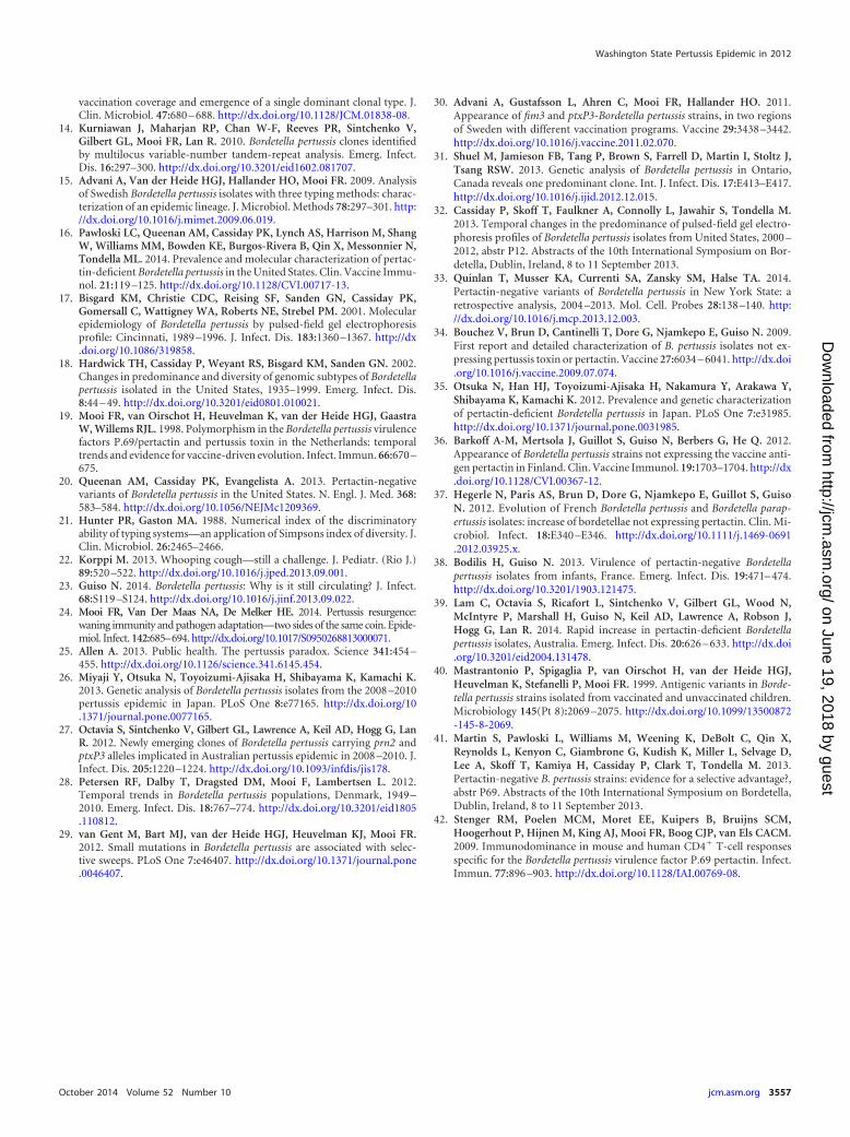
Molecular typing with PFGE, MLST, and MLVA. Sixty-fivemolecular typing profiles were identified. WA1 and WA2 were themost common profiles, representing 18% and 12% of the totalisolates, respectively (Table 2). Forty-two profiles (65%) were rep-resented by a single isolate (Table 2). Thirty-one PFGE types wereidentified, with the most common types, CDC013 (n � 51; 10profiles), CDC237 (n � 44; 2 profiles), and CDC002 (n � 42; 6profiles), accounting for 57% of total isolates (Table 2; Fig. 2). Fora majority of PFGE profiles, only a small number of isolates wereidentified (Table 2; Fig. 2).
Seven MLST types were identified, with the majority of theisolates typing as prn2-ptxP3-ptxA1-fim3-1 (n � 157, 65%; 32profiles) or prn2-ptxP3-ptxA1-fim3-2 (n � 76, 32%; 27 profiles)(Table 2; Fig. 2 and 3). Isolates with the PFGE type CDC013 werepredominately prn2-ptxP3-ptxA1-fim3-2 (n � 38, 75%), whileisolates with the PFGE types CDC002 and CDC0237 were pre-dominately prn2-ptxP3-ptxA1-fim3-1 (n � 35, 83%, and n � 42,95%, respectively) (Table 2; Fig. 2).
Eleven MLVA types were observed, mainly comprising MLVA27 (n � 183, 76%; 46 profiles) and MLVA 158 (n � 37, 15%; 7profiles) (Table 2; Fig. 2 and 3). Of the isolates with PFGE typesCDC013 and CDC237, 92% and 100%, respectively, were MLVA27. Seventy-six percent of isolates with the PFGE type CDC002were MLVA 158 (n � 32). Twenty-two of 31 PFGE types were of asingle MLVA type.
In comparing MLVA and MLST, we found that a majority ofisolates with MLVA 27 and MLVA 158 had the MLST type prn2-ptxP3-ptxA1-fim3-1 (66% and 84%, respectively) (Table 2; Fig. 3).
FIG 1 Map of the geographic distribution of cases with and without isolates collected in 2012. Cases were mapped by ZIP Code of residence. Squares representthe locations of case patients with isolates for whom the ZIP Codes and PFGE profiles are known (n � 233), with the color corresponding to the PFGE profile ofthe isolate. Gray circles represent the locations of cases without isolates but with the ZIP Code known (n � 4,681). The symbols are placed only within the ZIPCode and do not correspond to the actual location of each case patient’s residence.
Washington State Pertussis Epidemic in 2012
October 2014 Volume 52 Number 10 jcm.asm.org 3551
on June 19, 2018 by guesthttp://jcm
.asm.org/
Dow
nloaded from

The 3 isolates typing as WA22 and WA51 were the only isolatesobserved with the ptxP1 allele (Table 2; Fig. 2 and 3).
Among pertussis isolates from this statewide epidemic, PFGEalone (D � 0.87) accounted for a higher discriminatory powerthan the combination of MLVA and MLST (D � 0.67) (Table 3).The fim3 locus contributed the most to the discriminatory powerof MLST (D � 0.45).The combination of PFGE, MLVA, andMLST had an index of diversity of 0.92 (Table 3).
prn mutations. Seventy-six percent (n � 183) of the isolatescollected during this statewide epidemic were pertactin deficient,as previously described (16). Of the 65 molecular typing profilesidentified, only 25 (38%) produced Prn (Table 2). Of the 3 majorPFGE profiles, all PFGE type CDC237 isolates (n � 44) and 88%of the isolates with PFGE profile CDC002 (n � 37) had an IS481insertion in the prn gene (Table 2). In contrast, 61% of the isolateswith PFGE type CDC013 were wild type and produced Prn (n �31). The IS481 insertion, which can be found in 3 different loca-tions within prn, was seen in 35 molecular profiles and associatedwith five different MLVA types (27, 158, 95, 32, and 5), along with
three MLST types (Table 2; see also Fig. S1 in the supplementalmaterial). The prn 22-kb promoter inversion, at nucleotide �74,was present in profiles WA5 and WA17, which corresponded to84% of isolates with PFGE type CDC010 and 5% of isolates withPFGE type CDC002 (Table 2; see also Fig. S1). Furthermore, thepromoter inversion was only seen in the 18 isolates with MLVA 27and two MLST types that differ by the fim3 allele (Table 2). Thesignal sequence (SS) deletion of nucleotides 22 to 109 in prn wasonly seen in two isolates with profile WA22 (Table 2; see also Fig.S1). The prn promoter and 5=-end deletion was only seen in thetwo isolates with profiles WA28 and WA36 (Table 2). These iso-lates were MLVA 27 and harbored two MLST profiles that differby the fim3 allele (Table 2). A point mutation in prn resulting in aneutral amino acid change was seen in the isolate with the profileWA32 and did not lead to Prn deficiency (Table 2; see also Fig. S1).Figure 2 shows the clustering of predominately Prn-deficientPFGE types (indicated by red shadows) and predominately Prn-expressing PFGE types (indicated by green shadows).
The combination of PFGE and the prn mutations (D � 0.91)demonstrated a higher discriminatory power than the combina-tions of MLVA and the prn mutations (D � 0.70) and MLST andthe prn mutations (D � 0.71) (Table 3). The combination of allmolecular typing data into the scheme PFGE-MLVA-MLST-prnmutations-Prn expression accounted for the highest discrimina-tory power of all combinations of typing methods (D � 0.94)(Table 3).
DISCUSSION
We investigated the molecular epidemiology of the largest pertus-sis epidemic in Washington State in 70 years. Given that the ma-jority of cases were fully vaccinated, we assessed the current pop-ulation of B. pertussis at a molecular level and compared the resultsto the current vaccine. By PFGE, we found 65 distinct typing pro-files that demonstrated high strain diversity with no predominateclone and a large proportion of isolates with pertactin deficiency,and we found a higher discriminatory power of PFGE alone thanof MLVA plus MLST. Although it is likely that multiple, possiblyinterrelated reasons for the recent resurgence of pertussis will ul-timately be identified, this study sheds light on the importance ofconsidering bacterial adaption to current vaccines as one possiblefactor (22–25). This is evident with the surge in strains deficient inpertactin, a known pertussis acellular vaccine immunogen.
The predominate MLVA-MLST type circulating in Washing-ton State was MLVA 27 prn2-ptxP3-ptxA1-fim3-1, which asidefrom the fim3 allele, coincides with the predominate strain foundduring the early 1990s to 2009 in a similar study conducted in theUnited States (11). This strain is currently predominate in Japan,Australia, and parts of Europe (11, 13, 26–28). The molecularprofile for 2 strains used to produce the current U.S. vaccine areMLVA 38 prn1-ptxP1-ptxA2-fim3-1 (Tohama I) and MLVA 167prn1-ptxP1-ptxA4-fim3-1 (strain 10536). Few isolates were ob-served with the ptxP1 allele, which coincides with the decreasedfrequency of this allele in circulating strains in the United States,Netherlands, Canada, Sweden, Australia, and Denmark (27–31).MLVA 186 was first seen in the United States in 2007, yet the twoisolates with this MLVA type in this epidemic are associated withthe MLST profile prn1-ptxP1-ptxA2-fim3-1 that was observed inthe Tohama I strain (11).
Three PFGE profiles predominated within the WashingtonState epidemic: CDC013, CDC237, and CDC002 (Fig. 1; see also
TABLE 1 Demographic and clinical characteristics of 2012 WashingtonState pertussis cases with and without an isolate available for molecularcharacterization testing
Characteristic
No. (%) withoutisolate(n � 4,695)a
No. (%) withisolate tested(n � 240) P valueb
GenderFemale 2,529 (53.9) 135 (56.3) 0.470Male 2,166 (46.1) 105 (43.8)
Age group (yr)�1 359 (7.7) 24 (10.0) 0.5231–6 909 (19.4) 34 (14.2)7–10 972 (20.7) 46 (19.2)11–12 378 (8.1) 21 (8.8)13–14 559 (11.9) 32 (13.3)15–19 543 (11.6) 31 (12.9)�20 975 (20.8) 52 (21.7)
Race/EthnicityWhite 2,441 (52.0) 141 (59.0) 0.0002Hispanic 872 (18.6) 33 (13.8)Other 226 (4.8) 23 (9.6)Unknown 1,156 (24.6) 43 (17.9)
Geographic regionWestern WA 3,641 (77.6) 163 (67.9) 0.0005Eastern WA 1,054 (22.5) 77 (32.1)
Quarter of onset (n � 4,918) n � 4,695 n � 228January-March 1,110 (23.6) 16 (7.0) �0.0001April-June 2,050 (43.7) 127 (55.7)July-September 1,077 (22.9) 64 (28.1)October-December 458 (9.8) 21 (9.2)
Hospitalization amonginfants (n � 383)
n � 359 n � 24
Yes 66 (18.4) 9 (37.5) 0.022No 293 (81.6) 15 (62.5)
a One hundred nine cases had a positive culture documented in the case report, but therespective isolates were not submitted to the WAPHL.b �2 test for independence was used to determine differences between cases with andwithout isolates. P values of �0.05 were considered statistically significant.
Bowden et al.
3552 jcm.asm.org Journal of Clinical Microbiology
on June 19, 2018 by guesthttp://jcm
.asm.org/
Dow
nloaded from

TABLE 2 Molecular typing profiles of B. pertussis (n � 240) isolates obtained during the 2012 Washington State epidemic
Profile PFGEa MLVAb
Allele of:
prn mutationExpressesPrn
Total no.of isolatesprn ptxP ptxA fim3
WA1 237 27 2 3 1 1 IS481 insertionc � 42WA2 2 158 2 3 1 1 IS481 insertion � 28WA3 13 27 2 3 1 2 IS481 insertion � 19WA4 13 27 2 3 1 2 Wild-type prn � 17WA5 10 27 2 3 1 1 Promoter inversion (�74 nt) � 16WA6 253 27 2 3 1 1 IS481 insertion � 12WA7 13 27 2 3 1 1 Wild-type prn � 7WA8 13 27 2 3 1 1 IS481 insertion � 2WA9 325 27 2 3 1 1 IS481 insertion � 7WA10 217 27 2 3 1 1 Wild-type prn � 6
WA11 217 27 2 3 1 1 IS481 insertion � 6WA12 2 27 2 3 1 1 IS481 insertion � 5WA13 322 95 2 3 1 2 IS481 insertion � 5WA14 2 158 2 3 1 2 IS481 insertion � 4WA15 162 27 2 3 1 2 IS481 insertion � 4WA16 217 32 2 3 1 1 Wild-type prn � 4WA17 2 27 2 3 1 2 Promoter inversion (�74 nt) � 2WA18 2 27 2 3 1 1 Wild-type prn � 2WA19 46 27 2 3 1 2 IS481 insertion � 2WA20 158 27 2 3 1 2 Wild-type prn � 2
WA21 237 27 2 3 1 2 IS481 insertion � 2WA22 260 186 1 1 2 1 Signal sequence deletion (nt 26 to 109) � 2WA23 322 27 2 3 1 2 IS481 insertion � 2WA24 2 27 2 3 1 2 IS481 insertion � 1WA25 10 27 2 3 1 2 IS481 insertion � 1WA26 10 158 2 3 1 1 IS481 insertion � 1WA27 10 158 2 3 1 2 IS481 insertion � 1WA28 13 27 2 3 1 2 Deletion (nt �2090 to �478) � 1WA29 13 27 3 3 1 2 IS481 insertion � 1WA30 13 158 2 3 1 1 Wild-type prn � 1
WA31 13 158 9 3 1 2 Wild-type prn � 1WA32 13 16 2 3 1 2 T at nt 638; neutral amino acid change � 1WA33 13 78 2 3 1 1 Wild-type prn � 1WA34 28 27 2 3 1 1 IS481 insertion � 1WA35 46 27 2 3 1 1 IS481 insertion � 1WA36 46 27 2 3 1 1 Deletion (nt �2090 to 478) � 1WA37 46 32 2 3 1 2 Wild-type prn � 1WA38 82 128 2 3 1 2 Wild-type prn � 1WA39 140 27 2 3 1 2 Wild-type prn � 1WA40 158 27 2 3 1 1 Wild-type prn � 1
WA41 162 27 2 3 1 1 IS481 insertion � 1WA42 162 32 2 3 1 2 IS481 insertion � 1WA43 168 27 2 3 1 1 IS481 insertion � 1WA44 177 27 2 3 1 2 Wild-type prn � 1WA45 217 27 2 3 1 2 Wild-type prn � 1WA46 217 27 2 3 1 2 IS481 insertion � 1WA47 217 27 2 3 2 1 Wild-type prn � 1WA48 217 158 2 3 1 1 Wild-type prn � 1WA49 253 27 2 3 1 2 IS481 insertion � 1WA50 253 5 2 3 1 1 IS481 insertion � 1
WA51 258 35 1 1 1 1*d Wild-type prn � 1WA52 270 27 2 3 1 1 Wild-type prn � 1WA53 286 27 2 3 1 1 IS481 insertion � 1WA54 295 27 2 3 1 2 IS481 insertion � 1WA55 300 27 2 3 1 1 IS481 insertion � 1WA56 323 27 9 3 1 2 Wild-type prn � 1
(Continued on following page)
Washington State Pertussis Epidemic in 2012
October 2014 Volume 52 Number 10 jcm.asm.org 3553
on June 19, 2018 by guesthttp://jcm
.asm.org/
Dow
nloaded from
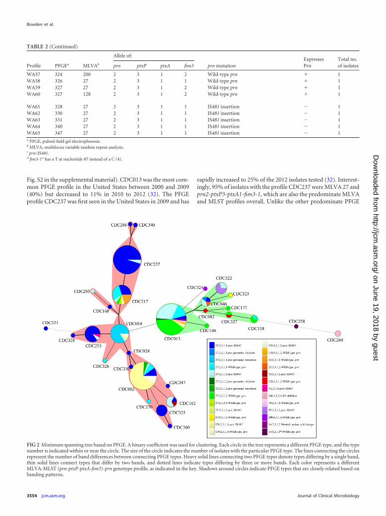
Fig. S2 in the supplemental material). CDC013 was the most com-mon PFGE profile in the United States between 2000 and 2009(40%) but decreased to 11% in 2010 to 2012 (32). The PFGEprofile CDC237 was first seen in the United States in 2009 and has
rapidly increased to 25% of the 2012 isolates tested (32). Interest-ingly, 95% of isolates with the profile CDC237 were MLVA 27 andprn2-ptxP3-ptxA1-fim3-1, which are also the predominate MLVAand MLST profiles overall. Unlike the other predominate PFGE
TABLE 2 (Continued)
Profile PFGEa MLVAb
Allele of:
prn mutationExpressesPrn
Total no.of isolatesprn ptxP ptxA fim3
WA57 324 200 2 3 1 2 Wild-type prn � 1WA58 326 27 2 3 1 1 Wild-type prn � 1WA59 327 27 2 3 1 2 Wild-type prn � 1WA60 327 128 2 3 1 2 Wild-type prn � 1
WA61 328 27 2 3 1 1 IS481 insertion � 1WA62 330 27 2 3 1 1 IS481 insertion � 1WA63 331 27 2 3 1 1 IS481 insertion � 1WA64 340 27 2 3 1 1 IS481 insertion � 1WA65 347 27 2 3 1 1 IS481 insertion � 1a PFGE, pulsed-field gel electrophoresis.b MLVA, multilocus variable tandem repeat analysis.c prn::IS481.d fim3-1* has a T at nucleotide 87 instead of a C (4).
FIG 2 Minimum spanning tree based on PFGE. A binary coefficient was used for clustering. Each circle in the tree represents a different PFGE type, and the typenumber is indicated within or near the circle. The size of the circle indicates the number of isolates with the particular PFGE type. The lines connecting the circlesrepresent the number of band differences between connecting PFGE types. Heavy solid lines connecting two PFGE types denote types differing by a single band,thin solid lines connect types that differ by two bands, and dotted lines indicate types differing by three or more bands. Each color represents a differentMLVA-MLST (prn-ptxP-ptxA-fim3)-prn genotype profile, as indicated in the key. Shadows around circles indicate PFGE types that are closely related based onbanding patterns.
Bowden et al.
3554 jcm.asm.org Journal of Clinical Microbiology
on June 19, 2018 by guesthttp://jcm
.asm.org/
Dow
nloaded from

profiles, CDC002 represented �10% of U.S. isolates from 2000 to2009 before rapidly increasing to 24% in 2010 to 2012 (32). Sixty-seven percent of isolates with the profile CDC002 were MLVA 158and prn2-ptxP3-ptxA1-fim3-1.
CDC013 continues to have a majority of isolates with the fim3-2 allele, while the majority of the CDC237 and CDC002 isolatespossess the fim3-1 allele (Fig. 2). Until 2009, increased pertussiscase reporting was correlated with the emergence of fim3-2 (des-ignated fim3B in reference 11) in the United States. That is not thecase for this epidemic, where the reemergence of the fim3-1 allele(designated fim3A in reference 11) was seen. The fim3-1 allele hasnot predominated in U.S. isolates since the early 2000s (11).
By comparing the index of diversity values of the molecular
typing methods implemented in this study, it was evident thatdifferent methods demonstrated very different snapshots of diver-sity. Similar to what is seen in the United States as a whole, as wellas in other countries, this epidemic does not demonstrate the pres-ence of a large number of alleles for the prn, ptxP, and ptxA genes,indicating a low level of diversity based on these MLST loci (11, 26,29). The fim3 locus contributed most to the diversity of the MLSTprofiles. Our finding of a higher discriminatory power for PFGEthan for the combination of MLVA and MLST was also seen in amolecular typing study conducted on Swedish isolates (15). See-ing that PFGE is a depiction of diversity on a genomic scale pro-vides evidence that these isolates are changing more rapidly on agenomic level than within the individual genes and regions tar-geted in MLVA and MLST. With the indication that diversity isoccurring at a genomic level, as seen by PFGE and prn mutations(specifically the IS481 insertion), it is important to address theneed for a whole-genome analysis to make more definitive con-clusions about correlation. With the lack of a unified typingscheme and the diversity seen in this one epidemic, it has becomeincreasingly necessary to develop a universally applicable formatfor reporting the molecular epidemiology of B. pertussis popula-tions that is consistent with the guidelines for bacterial geneticnomenclature. This will allow for more efficient comparisonsacross multiple epidemics in multiple countries.
Though it was first identified in an isolate from 1994, it was notuntil 2010 that pertactin deficiency began steadily increasing in B.pertussis isolates in the United States (16, 33). To date, the UnitedStates has seen a significantly higher number of pertactin-deficientB. pertussis strains than other countries, such as France in 2007(7%), Japan from 1990 to 2009 (27%), or Finland from 2006 to2011 (2%) (34–36). The proportion of pertactin-deficient isolateshas begun to steadily increase in other countries, includingFrance, increasing from 2% in 2005 to 14% in 2012, which is
FIG 3 Minimum spanning tree based on MLVA. A categorical coefficient was used for clustering. Each circle in the tree represents a different MLVA type, andthe type number is indicated within the circle. The size of the circle indicates the number of isolates with the particular MLVA type. Solid lines connecting circlesrepresent 1 VNTR difference between connecting MLVA types, while dotted lines represent 2 VNTR differences. Each color represents a different MLST type, asindicated in the key.
TABLE 3 Simpson’s diversity indices for typing strategies
Typing methoda
Typecount
Index ofdiversity
PFGE 31 0.87MLVA 11 0.40MLST 7 0.47PFGE � MLST 43 0.90PFGE � MLVA 43 0.91MLVA � MLST 18 0.67PFGE � MLVA � MLST 57 0.92prn mutations 6 0.51PFGE � prn mutations 40 0.91MLVA � prn mutations 16 0.70MLST � prn mutations 14 0.71Prn deficiency 2 0.36PFGE � MLVA � MLST � prn
mutations � Prn deficiency65 0.94
a PFGE, pulsed-field gel electrophoresis; MLVA, multilocus variable number tandemrepeat analysis; MLST, multilocus sequence typing; prn, pertactin gene.
Washington State Pertussis Epidemic in 2012
October 2014 Volume 52 Number 10 jcm.asm.org 3555
on June 19, 2018 by guesthttp://jcm
.asm.org/
Dow
nloaded from

similar to the progressive increases seen in the United States andAustralia (16, 37–39). A large proportion of isolates (76%) in thisstudy were pertactin deficient due to one of four different muta-tions previously identified in the prn gene (16, 20, 34–37). Thepresence of the SS deletion in the 2 isolates with PFGE profileCDC260, MLVA 186, and MLST profile prn1-ptxP1-ptxA2-fim3-1does not correlate with what was seen in Japan from 1990 to 2009,where the prn1 SS deletion predominated among pertactin-defi-cient isolates (73%) and was seen in 3 different MLVA types(MLVA 186, MLVA 194, and MLVA 226) (35). The prn1 SS dele-tion was first seen in Japan in 2000 and has shown more MLVAdiversity than was seen in the Washington State epidemic (35).One isolate (collected during 2007) out of 600 in the United Stateshad MLVA type 186 and had the same PFGE and MLST profiles asseen with the 2 isolates from the Washington State epidemic (11).The only two pertactin-deficient isolates collected in Finland from2006 to 2011 also harbored the prn1 SS deletion (36). It should benoted that while IS481 insertions are seen throughout multipleMLVA types and MLST profiles, the additional three mutationsloosely associate with two MLVA types and two MLST profiles,which may be due in part to the low number of isolates collected inthis study (Table 2).
Since the majority of cases in this statewide epidemic occurredamong school-aged children that were up to date for their age forpertussis-containing vaccine as recommended by the ACIP, it isimportant to consider what factors may have led to this surge inreported cases in Washington State, as well as in the United Statesas a whole. One current hypothesis centers on the idea that B.pertussis strains harboring prn mutations (i.e., escape mutants)possibly have a selective advantage in individuals vaccinated withacellular vaccines (40, 41). As previously mentioned, the majorityof strains circulating in the Washington State epidemic are diver-gent from the two current U.S. vaccine strains, which both expressprn1. A study conducted in Italy indicated that the prn vaccineallele (prn1, which encodes the Prn1 antigenic variant) was iden-tified at a lower frequency in vaccinated individuals and was seenin an increased number of isolates collected from areas of lowvaccination (40). prn1 and prn2 differ by the presence of 5 aminoacid repeats in variant region 1 of prn but are recognized equallyefficiently by the T-cell response in mice and humans (42). Here,we see prn2 predominating in a highly vaccinated population,which would provide evidence in support of this hypothesis. Epi-demiological analysis suggests that vaccinated persons have in-creased susceptibility to pertactin-deficient strains compared totheir susceptibility to strains expressing pertactin (40, 41). Therehas been no direct evidence that pertactin-deficient strains aremore virulent in humans than strains expressing pertactin (38,40). Through our analysis, it is clear that the currently circulatingstrains are diverse on a genomic level. The rapidly increasing avail-ability of whole-genome sequencing and analyses, specificallythrough analysis of additional virulence-related genes, will allowdirect investigation of this hypothesis by determining whetherpertactin is a major virulence-related gene affected by mutationsand will likewise provide answers with regard to what genes mayprovide these strains associated with pertussis disease with a selec-tive advantage in vaccinated populations.
In an era of increasing provider reliance on pertussis PCR forrapid diagnosis and treatment of suspected cases, this study dem-onstrates the importance of maintaining culture testing practices.With the highest number of pertussis cases reported in 2012 in the
United States since the 1950s, understanding the molecular epi-demiology of currently circulating strains of B. pertussis is critical.Providers should be made aware that isolation of B. pertussis isnecessary so that genetic characterization and analysis can be con-ducted to monitor trends in strain circulation. This evaluation ofisolates from a large pertussis epidemic in Washington State hascontributed further to our understanding of strain evolution inthe United States and suggests that as current molecular typingtechniques advance, our understanding of the B. pertussis diversityseen on a genomic level will continue to evolve.
REFERENCES1. Tanaka M, Vitek CR, Pascual FB, Bisgard KM, Tate JE, Murphy TV.
2003. Trends in pertussis among infants in the United States, 1980 –1999.JAMA 290:2968 –2975. http://dx.doi.org/10.1001/jama.290.22.2968.
2. Centers for Disease Control and Prevention. 2002. Pertussis—UnitedStates, 1997–2000. Morb. Mortal. Weekly Rep. 51:73–76.
3. Davis SF, Strebel PM, Cochi SL, Zell ER, Hadler SC. 1992. Pertussissurveillance—United States, 1989 –1991. Morb. Mortal. Wkly. Rep. 41:11–19.
4. Centers for Disease Control and Prevention. 1997. Pertussis vaccination:use of acellular pertussis vaccines among infants and young children. Rec-ommendations of the Advisory Committee on Immunization Practices(ACIP). Morb. Mortal. Weekly Rep. 416:1–25.
5. Broder KR, Cortese MM, Iskander JK, Kretsinger K, Slade BA, BrownKH, Mijalski CM, Tiwari T, Weston EJ, Cohn AC, Srivastava PU,Moran JS, Schwartz B, Murphy TV, Advisory Committee on Immuni-zation Practices (ACIP). 2006. Preventing tetanus, diphtheria, and per-tussis among adolescents: use of tetanus toxoid, reduced diphtheria toxoidand acellular pertussis vaccines recommendations of the Advisory Com-mittee on Immunization Practices (ACIP). MMWR Recomm. Rep.55(RR-3):1–34.
6. Kretsinger K, Broder KR, Cortese MM, Joyce MP, Ortega-Sanchez I,Lee GM, Tiwari T, Cohn AC, Slade BA, Iskander JK, Mijalski CM,Brown KH, Murphy TV, Centers for Disease Control and Prevention;Advisory Committee on Immunization Practices; Healthcare InfectionControl Practices Advisory Committee. 2006. Preventing tetanus, diph-theria, and pertussis among adults: use of tetanus toxoid, reduced diph-theria toxoid and acellular pertussis vaccine recommendations of theAdvisory Committee on Immunization Practices (ACIP) and recommen-dation of ACIP, supported by the Healthcare Infection Control PracticesAdvisory Committee (HICPAC), for use of Tdap among health-care per-sonnel. MMWR Recomm. Rep. 55(RR-17):1–37.
7. Sawyer M, Liang JL, Messonnier N, Clark TA. 2013. Updated recom-mendations for use of tetanus toxoid, reduced diphtheria toxoid, andacellular pertussis vaccine (Tdap) in pregnant women—Advisory Com-mittee on Immunization Practices (ACIP), 2012. MMWR Morb. Mortal.Wkly. Rep. 62:131–135.
8. Farizo KM, Cochi SL, Zell ER, Brink EW, Wassilak SG, Patriarca PA.1992. Epidemiological features of pertussis in the United States, 1980 –1989. Clin. Infect. Dis. 14:708 –719. http://dx.doi.org/10.1093/clinids/14.3.708.
9. Guris D, Strebel PM, Bardenheier B, Brennan M, Tachdjian R, Finch E,Wharton M, Livengood JR. 1999. Changing epidemiology of pertussis inthe United States: increasing reported incidence among adolescents andadults, 1990 –1996. Clin. Infect. Dis. 28:1230 –1237. http://dx.doi.org/10.1086/514776.
10. Centers for Disease Control and Prevention. 2012. Pertussis epidemic—Washington, 2012. Morb. Mortal. Weekly Rep. 61:517–522.
11. Schmidtke AJ, Boney KO, Martin SW, Skoff TH, Tondella ML, TattiKM. 2012. Population diversity among Bordetella pertussis isolates, UnitedStates, 1935–2009. Emerg. Infect. Dis. 18:1248 –1255. http://dx.doi.org/10.3201/eid1808.120082.
12. Schouls LM, van der Heide HGJ, Vauterin L, Vauterin P, Mooi FR.2004. Multiple-locus variable-number tandem repeat analysis of DutchBordetella pertussis strains reveals rapid genetic changes with clonal expan-sion during the late 1990s. J. Bacteriol. 186:5496 –5505. http://dx.doi.org/10.1128/JB.186.16.5496-5505.2004.
13. Litt DJ, Neal SE, Fry NK. 2009. Changes in genetic diversity of the Bordetellapertussis population in the United Kingdom between 1920 and 2006 reflect
Bowden et al.
3556 jcm.asm.org Journal of Clinical Microbiology
on June 19, 2018 by guesthttp://jcm
.asm.org/
Dow
nloaded from
vaccination coverage and emergence of a single dominant clonal type. J.Clin. Microbiol. 47:680 – 688. http://dx.doi.org/10.1128/JCM.01838-08.
14. Kurniawan J, Maharjan RP, Chan W-F, Reeves PR, Sintchenko V,Gilbert GL, Mooi FR, Lan R. 2010. Bordetella pertussis clones identifiedby multilocus variable-number tandem-repeat analysis. Emerg. Infect.Dis. 16:297–300. http://dx.doi.org/10.3201/eid1602.081707.
15. Advani A, Van der Heide HGJ, Hallander HO, Mooi FR. 2009. Analysisof Swedish Bordetella pertussis isolates with three typing methods: charac-terization of an epidemic lineage. J. Microbiol. Methods 78:297–301. http://dx.doi.org/10.1016/j.mimet.2009.06.019.
16. Pawloski LC, Queenan AM, Cassiday PK, Lynch AS, Harrison M, ShangW, Williams MM, Bowden KE, Burgos-Rivera B, Qin X, Messonnier N,Tondella ML. 2014. Prevalence and molecular characterization of pertac-tin-deficient Bordetella pertussis in the United States. Clin. Vaccine Immu-nol. 21:119 –125. http://dx.doi.org/10.1128/CVI.00717-13.
17. Bisgard KM, Christie CDC, Reising SF, Sanden GN, Cassiday PK,Gomersall C, Wattigney WA, Roberts NE, Strebel PM. 2001. Molecularepidemiology of Bordetella pertussis by pulsed-field gel electrophoresisprofile: Cincinnati, 1989 –1996. J. Infect. Dis. 183:1360 –1367. http://dx.doi.org/10.1086/319858.
18. Hardwick TH, Cassiday P, Weyant RS, Bisgard KM, Sanden GN. 2002.Changes in predominance and diversity of genomic subtypes of Bordetellapertussis isolated in the United States, 1935–1999. Emerg. Infect. Dis.8:44 – 49. http://dx.doi.org/10.3201/eid0801.010021.
19. Mooi FR, van Oirschot H, Heuvelman K, van der Heide HGJ, GaastraW, Willems RJL. 1998. Polymorphism in the Bordetella pertussis virulencefactors P.69/pertactin and pertussis toxin in the Netherlands: temporaltrends and evidence for vaccine-driven evolution. Infect. Immun. 66:670 –675.
20. Queenan AM, Cassiday PK, Evangelista A. 2013. Pertactin-negativevariants of Bordetella pertussis in the United States. N. Engl. J. Med. 368:583–584. http://dx.doi.org/10.1056/NEJMc1209369.
21. Hunter PR, Gaston MA. 1988. Numerical index of the discriminatoryability of typing systems—an application of Simpsons index of diversity. J.Clin. Microbiol. 26:2465–2466.
22. Korppi M. 2013. Whooping cough—still a challenge. J. Pediatr. (Rio J.)89:520 –522. http://dx.doi.org/10.1016/j.jped.2013.09.001.
23. Guiso N. 2014. Bordetella pertussis: Why is it still circulating? J. Infect.68:S119 –S124. http://dx.doi.org/10.1016/j.jinf.2013.09.022.
24. Mooi FR, Van Der Maas NA, De Melker HE. 2014. Pertussis resurgence:waning immunity and pathogen adaptation—two sides of the same coin. Epide-miol. Infect. 142:685–694. http://dx.doi.org/10.1017/S0950268813000071.
25. Allen A. 2013. Public health. The pertussis paradox. Science 341:454 –455. http://dx.doi.org/10.1126/science.341.6145.454.
26. Miyaji Y, Otsuka N, Toyoizumi-Ajisaka H, Shibayama K, Kamachi K.2013. Genetic analysis of Bordetella pertussis isolates from the 2008 –2010pertussis epidemic in Japan. PLoS One 8:e77165. http://dx.doi.org/10.1371/journal.pone.0077165.
27. Octavia S, Sintchenko V, Gilbert GL, Lawrence A, Keil AD, Hogg G, LanR. 2012. Newly emerging clones of Bordetella pertussis carrying prn2 andptxP3 alleles implicated in Australian pertussis epidemic in 2008 –2010. J.Infect. Dis. 205:1220 –1224. http://dx.doi.org/10.1093/infdis/jis178.
28. Petersen RF, Dalby T, Dragsted DM, Mooi F, Lambertsen L. 2012.Temporal trends in Bordetella pertussis populations, Denmark, 1949 –2010. Emerg. Infect. Dis. 18:767–774. http://dx.doi.org/10.3201/eid1805.110812.
29. van Gent M, Bart MJ, van der Heide HGJ, Heuvelman KJ, Mooi FR.2012. Small mutations in Bordetella pertussis are associated with selec-tive sweeps. PLoS One 7:e46407. http://dx.doi.org/10.1371/journal.pone.0046407.
30. Advani A, Gustafsson L, Ahren C, Mooi FR, Hallander HO. 2011.Appearance of fim3 and ptxP3-Bordetella pertussis strains, in two regionsof Sweden with different vaccination programs. Vaccine 29:3438 –3442.http://dx.doi.org/10.1016/j.vaccine.2011.02.070.
31. Shuel M, Jamieson FB, Tang P, Brown S, Farrell D, Martin I, Stoltz J,Tsang RSW. 2013. Genetic analysis of Bordetella pertussis in Ontario,Canada reveals one predominant clone. Int. J. Infect. Dis. 17:E413–E417.http://dx.doi.org/10.1016/j.ijid.2012.12.015.
32. Cassiday P, Skoff T, Faulkner A, Connolly L, Jawahir S, Tondella M.2013. Temporal changes in the predominance of pulsed-field gel electro-phoresis profiles of Bordetella pertussis isolates from United States, 2000 –2012, abstr P12. Abstracts of the 10th International Symposium on Bor-detella, Dublin, Ireland, 8 to 11 September 2013.
33. Quinlan T, Musser KA, Currenti SA, Zansky SM, Halse TA. 2014.Pertactin-negative variants of Bordetella pertussis in New York State: aretrospective analysis, 2004 –2013. Mol. Cell. Probes 28:138 –140. http://dx.doi.org/10.1016/j.mcp.2013.12.003.
34. Bouchez V, Brun D, Cantinelli T, Dore G, Njamkepo E, Guiso N. 2009.First report and detailed characterization of B. pertussis isolates not ex-pressing pertussis toxin or pertactin. Vaccine 27:6034 – 6041. http://dx.doi.org/10.1016/j.vaccine.2009.07.074.
35. Otsuka N, Han HJ, Toyoizumi-Ajisaka H, Nakamura Y, Arakawa Y,Shibayama K, Kamachi K. 2012. Prevalence and genetic characterizationof pertactin-deficient Bordetella pertussis in Japan. PLoS One 7:e31985.http://dx.doi.org/10.1371/journal.pone.0031985.
36. Barkoff A-M, Mertsola J, Guillot S, Guiso N, Berbers G, He Q. 2012.Appearance of Bordetella pertussis strains not expressing the vaccine anti-gen pertactin in Finland. Clin. Vaccine Immunol. 19:1703–1704. http://dx.doi.org/10.1128/CVI.00367-12.
37. Hegerle N, Paris AS, Brun D, Dore G, Njamkepo E, Guillot S, GuisoN. 2012. Evolution of French Bordetella pertussis and Bordetella parap-ertussis isolates: increase of bordetellae not expressing pertactin. Clin. Mi-crobiol. Infect. 18:E340 –E346. http://dx.doi.org/10.1111/j.1469-0691.2012.03925.x.
38. Bodilis H, Guiso N. 2013. Virulence of pertactin-negative Bordetellapertussis isolates from infants, France. Emerg. Infect. Dis. 19:471– 474.http://dx.doi.org/10.3201/1903.121475.
39. Lam C, Octavia S, Ricafort L, Sintchenko V, Gilbert GL, Wood N,McIntyre P, Marshall H, Guiso N, Keil AD, Lawrence A, Robson J,Hogg G, Lan R. 2014. Rapid increase in pertactin-deficient Bordetellapertussis isolates, Australia. Emerg. Infect. Dis. 20:626 – 633. http://dx.doi.org/10.3201/eid2004.131478.
40. Mastrantonio P, Spigaglia P, van Oirschot H, van der Heide HGJ,Heuvelman K, Stefanelli P, Mooi FR. 1999. Antigenic variants in Borde-tella pertussis strains isolated from vaccinated and unvaccinated children.Microbiology 145(Pt 8):2069 –2075. http://dx.doi.org/10.1099/13500872-145-8-2069.
41. Martin S, Pawloski L, Williams M, Weening K, DeBolt C, Qin X,Reynolds L, Kenyon C, Giambrone G, Kudish K, Miller L, Selvage D,Lee A, Skoff T, Kamiya H, Cassiday P, Clark T, Tondella M. 2013.Pertactin-negative B. pertussis strains: evidence for a selective advantage?,abstr P69. Abstracts of the 10th International Symposium on Bordetella,Dublin, Ireland, 8 to 11 September 2013.
42. Stenger RM, Poelen MCM, Moret EE, Kuipers B, Bruijns SCM,Hoogerhout P, Hijnen M, King AJ, Mooi FR, Boog CJP, van Els CACM.2009. Immunodominance in mouse and human CD4� T-cell responsesspecific for the Bordetella pertussis virulence factor P.69 pertactin. Infect.Immun. 77:896 –903. http://dx.doi.org/10.1128/IAI.00769-08.
Washington State Pertussis Epidemic in 2012
October 2014 Volume 52 Number 10 jcm.asm.org 3557
on June 19, 2018 by guesthttp://jcm
.asm.org/
Dow
nloaded from
Related Documents