1 Molecular characterization of choroid plexus tumors reveals novel clinically relevant subgroups Diana M. Merino 1 , Adam Shlien 1 , Anita Villani 1 , Malgorzata Pienkowska 1 , Stephen Mack 1 , Vijay Ramaswamy 1 , David Shih 1 , Ruth Tatevossian 2 , Ana Novokmet 1 , Sanaa Choufani 1 , Rina Dvir 4 , Myran Ben-Arush 5 , Brent T. Harris 6 , Eugene I. Hwang 7 , Rishi Lulla 8 , Stefan M. Pfister 9 , Maria Isabel Achatz 10 , Nada Jabado 11 , Jonathan L. Finlay 3 , Rosanna Weksberg 1 , Cynthia Hawkins 1 , Michael D. Taylor 1 , Uri Tabori 1 , David W. Ellison 2 , Richard J. Gilbertson 2 , David Malkin 1 1. The Hospital for Sick Children, Toronto, ON, Canada 2. St. Jude Children's Research Hospital, Memphis, TN, USA 3. Children's Hospital of Los Angeles, Los Angeles, CA, USA 4. Tel Aviv Medical Center, Tel Aviv, Israel 5. Rambam Health Care Campus, Haifa, Israel 6. Georgetown University Medical Center, Washington, DC, USA 7. Children's National Medical Center, Washington, DC, USA 8. Ann & Robert H. Lurie Children's Hospital of Chicago, Chicago, IL, USA 9. German Cancer Research Center, Heidelberg, Germany 10. A.C. Camargo Cancer Center, Sao Paulo, Brazil 11. Montreal Children’s Hospital, Montreal, QC, Canada The authors disclose no potential conflicts of interest. Corresponding author: David Malkin M.D. The Hospital for Sick Children, 686 Bay St. 18-9705, Toronto, ON M5V0A4 TEL: 416-813-5348 Research. on February 9, 2016. © 2014 American Association for Cancer clincancerres.aacrjournals.org Downloaded from Author manuscripts have been peer reviewed and accepted for publication but have not yet been edited. Author Manuscript Published OnlineFirst on October 21, 2014; DOI: 10.1158/1078-0432.CCR-14-1324

Welcome message from author
This document is posted to help you gain knowledge. Please leave a comment to let me know what you think about it! Share it to your friends and learn new things together.
Transcript

1
Molecular characterization of choroid plexus tumors reveals novel clinically
relevant subgroups
Diana M. Merino 1, Adam Shlien
1, Anita Villani
1, Malgorzata Pienkowska
1, Stephen
Mack 1, Vijay Ramaswamy
1, David Shih
1, Ruth Tatevossian
2, Ana Novokmet
1, Sanaa
Choufani 1, Rina Dvir
4, Myran Ben-Arush
5, Brent T. Harris
6, Eugene I. Hwang
7, Rishi
Lulla 8, Stefan M. Pfister
9, Maria Isabel Achatz
10, Nada Jabado
11, Jonathan L. Finlay
3,
Rosanna Weksberg 1, Cynthia Hawkins
1, Michael D. Taylor
1, Uri Tabori
1, David W.
Ellison 2, Richard J. Gilbertson
2, David Malkin
1
1. The Hospital for Sick Children, Toronto, ON, Canada
2. St. Jude Children's Research Hospital, Memphis, TN, USA
3. Children's Hospital of Los Angeles, Los Angeles, CA, USA
4. Tel Aviv Medical Center, Tel Aviv, Israel
5. Rambam Health Care Campus, Haifa, Israel
6. Georgetown University Medical Center, Washington, DC, USA
7. Children's National Medical Center, Washington, DC, USA
8. Ann & Robert H. Lurie Children's Hospital of Chicago, Chicago, IL, USA
9. German Cancer Research Center, Heidelberg, Germany
10. A.C. Camargo Cancer Center, Sao Paulo, Brazil
11. Montreal Children’s Hospital, Montreal, QC, Canada
The authors disclose no potential conflicts of interest.
Corresponding author:
David Malkin M.D.
The Hospital for Sick Children,
686 Bay St. 18-9705, Toronto, ON M5V0A4
TEL: 416-813-5348
Research. on February 9, 2016. © 2014 American Association for Cancerclincancerres.aacrjournals.org Downloaded from
Author manuscripts have been peer reviewed and accepted for publication but have not yet been edited. Author Manuscript Published OnlineFirst on October 21, 2014; DOI: 10.1158/1078-0432.CCR-14-1324

2
Email: [email protected]
ABSTRACT:
Purpose: To investigate molecular alterations in choroid plexus tumors (CPTs) using a
genome-wide high-throughput approach, in order to identify diagnostic and prognostic
signatures that will refine tumor stratification and guide therapeutic options.
Experimental Design: One hundred CPTs were obtained from a multi-institutional tissue
and clinical database. Copy number (CN), DNA methylation and gene expression
signatures were assessed for 74, 36 and 40 samples, respectively. Molecular subgroups
were correlated with clinical parameters and outcomes.
Results: Unique molecular signatures distinguished choroid plexus carcinomas (CPCs)
from choroid plexus papillomas (CPPs) and atypical choroid plexus papillomas (aCPPs);
however, no significantly distinct molecular alterations between CPPs and aCPPs were
observed. Allele-specific CN analysis of CPCs revealed two novel subgroups according
to DNA content: hypodiploid and hyperdiploid CPCs. Hyperdiploid CPCs exhibited
recurrent acquired uniparental disomy (aUPD) events. Somatic mutations in TP53 were
observed in 60% of CPCs. Investigating the number of mutated copies of p53 per sample
revealed a high-risk group of patients with CPC carrying two copies of mutant p53, who
exhibited poor 5-year event-free (EFS) and overall survival (OS) compared to patients
with CPC carrying one copy of mutant p53. (OS: 14.3%, 95% CI 0.71%-46.5% versus
66.7%, 28.2%-87.8%, respectively, p=0.04; EFS: 0% versus 44.4%, 13.6%-71.9%,
respectively, p=0.03). CPPs and aCPPs exhibited favorable survival.
Discussion: Our data demonstrates that differences in CN, gene expression and DNA
methylation signatures distinguish CPCs from CPPs and aCPPs; however molecular
similarities among the papillomas suggest that these two histological subgroups are
indeed a single molecular entity. A greater number of copies of mutated TP53 was
significantly associated to increased tumor aggressiveness and a worse survival outcome
in CPCs. Collectively, these findings will facilitate stratified approaches to the clinical
management of CPTs.
Research. on February 9, 2016. © 2014 American Association for Cancerclincancerres.aacrjournals.org Downloaded from
Author manuscripts have been peer reviewed and accepted for publication but have not yet been edited. Author Manuscript Published OnlineFirst on October 21, 2014; DOI: 10.1158/1078-0432.CCR-14-1324

3
TRANSLATIONAL RELEVANCE:
This report is the first to dissect the aberrant complexity in copy number, methylation,
and gene expression of one of the largest cohort of pediatric choroid plexus tumors
(CPTs). Our findings revealed molecular homogeneity among choroid plexus papillomas
(CPPs) and atypical choroid plexus papillomas (aCPPs), reflecting the favourable
survival of these patients and suggesting these histologically distinct subgroups are a
single tumor entity. Choroid plexus carcinomas (CPCs) were significantly different from
CPPs and aCPPs. Moreover, CPCs exhibited molecular heterogeneity, and patient
outcomes varied widely. We identified novel CPC subgroups with significantly distinct
copy number signatures suggesting different mechanisms drive CPC development. We
identified novel CPC subgroups with significantly distinct copy number signatures
suggesting different mechanisms drive CPC development. We identified that patient
overall and event-free survival significantly decreased with an increasing number of
mutated copies of p53. By defining the molecular landscape of CPTs, this study has
provided a comprehensive molecular background on which to explore mechanisms of
tumorigenesis and develop stratified approaches to the clinical management of CPTs.
Research. on February 9, 2016. © 2014 American Association for Cancerclincancerres.aacrjournals.org Downloaded from
Author manuscripts have been peer reviewed and accepted for publication but have not yet been edited. Author Manuscript Published OnlineFirst on October 21, 2014; DOI: 10.1158/1078-0432.CCR-14-1324
4
INTRODUCTION
Choroid plexus tumors (CPTs) are rare intraventricular neoplasms accounting for up to
20% of brain tumors in children under two years of age.(1,2) Three histological
subgroups have been described: choroid plexus papilloma (CPP, WHO grade I), atypical
choroid plexus papilloma (aCPP, WHO grade II), choroid plexus carcinoma (CPC, WHO
grade III). Long-term survival of CPPs is favorable with surgical resection alone
(>90%).(3) Conversely, CPCs exhibit a dismal prognosis, with an overall survival of
about 30%.(4–6) Despite aggressive treatment protocols, including surgical resection and
combination chemo- and radiation therapy,(6) the clinical behavior of CPCs is variable
and most of the few survivors exhibit long-term cognitive and developmental deficits.(6)
aCPP, a recently described pathological subgroup, exhibits an intermediate degree of
mitotic activity and outcome;(7,8) however, some cases may be difficult to distinguish
from CPC by histology alone.(9)
Over 50% of CPC tumors carry somatic TP53 mutations, and TP53 mutant CPCs have
been associated with increased genetic tumor instability and worse prognosis.(5)
Germline TP53 mutations have also been observed in CPC patients as CPC is one of the
hallmark cancers of the Li-Fraumeni syndrome (LFS), a familial cancer syndrome in
which affected family members harbor a mutant copy of the TP53 tumor suppressor gene.
Cytogenetic studies of central nervous system (CNS) tumors have revealed high
chromosomal instability in more than 90% of CPTs analyzed (Supplementary Table S1).
Defining the molecular landscape of CPTs and identifying actionable molecular
aberrations has been challenging due in part to the limited number of patients and high-
quality samples available for genome-wide studies. Here, we use an integrative molecular
Research. on February 9, 2016. © 2014 American Association for Cancerclincancerres.aacrjournals.org Downloaded from
Author manuscripts have been peer reviewed and accepted for publication but have not yet been edited. Author Manuscript Published OnlineFirst on October 21, 2014; DOI: 10.1158/1078-0432.CCR-14-1324

5
approach to characterize the genomic, transcriptomic and epigenomic landscape of the
largest cohort of CPTs to date. The information derived from these analyses creates a
molecular foundation on which to develop approaches to improve the clinical
management of this devastating disease.
MATERIALS and METHODS
Patients and Sample Preparation
CPT samples and/or clinical data were collected from institutions in Canada, the United
States of America, Brazil, Israel, and Germany (see Appendix) in accordance with each
institution’s Research Ethics Board. Informed consent was obtained from the
parents/legal guardians of all patients. We studied 100 unique tumor samples (58 CPC, 30
CPP and 12 aCPP) from 91 pediatric patients (ages 0.03-16.50 years old) for which TP53
sequence data was available (Table S2). Pathological review of CPTs was conducted by
C.H., D.W.E., and B.T.H. when samples were available. In all other institutions, expert
neuropathologists critically examined each case. In fifteen CPC cases,
immunohistochemical analysis of hHF5/INI1 was conducted and revealed
immunopositivity excluding the diagnosis of atypical teratoid/rhabdoid tumors (ATRT).
Nucleic acids were derived from fresh frozen (n=75), optimal cutting temperature
compound (OCT) (n=9), and formalin-fixed paraffin-embedded (FFPE) (n=12) samples.
We received isolated tumor DNA from 4 samples. Twenty-two nucleic acid samples
exhibited suboptimal quality and/or quantity, leaving 78 high-quality samples from 73
patients for analysis (Supplementary Fig. S1). Detailed clinical data were obtained for 68
patients. Tumor DNA was extracted using standard phenol-chloroform extraction from
Research. on February 9, 2016. © 2014 American Association for Cancerclincancerres.aacrjournals.org Downloaded from
Author manuscripts have been peer reviewed and accepted for publication but have not yet been edited. Author Manuscript Published OnlineFirst on October 21, 2014; DOI: 10.1158/1078-0432.CCR-14-1324
6
fresh-frozen samples, and the RecoverAll™ Total Nucleic Acid Isolation Kit for FFPE
(Ambion, Carlsbad, USA) from FFPE samples. Total RNA was isolated from fresh-
frozen samples using the TRIzol method (Invitrogen, Carlsbad, USA) according to the
manufacturer’s instructions.
TP53 Sequencing
Sequencing of the coding region of TP53 (exons 2-11) was performed in the molecular
diagnostic laboratory at The Hospital for Sick Children (Toronto) by direct Sanger
sequencing of whole genome DNA as previously described.(5)
Microarray Processing & Bioinformatics Analysis
Forty RNA samples were hybridized to GeneChip® Human Exon 1.0ST gene expression
microarrays (Affymetrix, Santa Clara, USA), and 36 DNA samples were hybridized to
Illumina® 450K Infinium methylation bead arrays (Illumina, San Diego, USA) as per
manufacturer’s instructions. An initial set of 55 tumor DNA samples was hybridized to
Genome-Wide Human SNP Array 6.0 (Affymetrix), while an independent set of 20
tumor DNA samples was hybridized to Affymetrix OncoScan™ FFPE Express 2.0
arrays. One technical replicate was included in both genotyping platforms and analyzed
for copy number call consistency.
Partek® Genomics Suite™ 6.5 (PGS) (Partek Inc. St. Louis, USA) and BioDiscovery
Nexus Copy Number software (Discovery Edition 7.0, BioDiscovery, Hawthorne, USA)
were used for copy number analysis as previously described(5) (see Appendix). Copy
number changes encompassing more than 75% of the chromosome were called whole-
Research. on February 9, 2016. © 2014 American Association for Cancerclincancerres.aacrjournals.org Downloaded from
Author manuscripts have been peer reviewed and accepted for publication but have not yet been edited. Author Manuscript Published OnlineFirst on October 21, 2014; DOI: 10.1158/1078-0432.CCR-14-1324
7
chromosome aberrations. Allele-specific copy number analysis of tumors (ASCAT) was
performed in R as previously described(10) and verified by Nexus software. Tumor
ploidy, where hypodiploid <1.90 and hyperdiploid >2.10, heterogeneity, and allelic
imbalances were inferred from the output. ASCAT failed to resolve the ploidy of two
samples with very low aberrant fraction, so these were excluded from further analysis.
Clustering of gene expression and methylation data was investigated in R (version 3.0.1)
by unsupervised hierarchical clustering (uHCL), non-negative matrix factorization
(NMF), and PVCLUST algorithms. Differential gene expression analysis was conducted
with PGS 6.5 (see Appendix). Gene set enrichment analysis (GSEA) was performed as
previously described(11) and its visualization was obtained by Cytoscape and Enrichment
Map using an in-house curated database containing freely available NCI, KEGG, PFAM,
Biocarta and GO databases as described in Witt et. al. (12) Differences in DNA
methylation status were analyzed with the Illumina® GenomeStudio software (see
Appendix). All probesets were annotated according to the human genome build hg19
(GRCh37). Microarray data can be accessed from GEO (GSE####, GSE####, GSE####
and GSE####).
Statistical analysis
Statistical analyses of copy number and gene expression were performed in PGS 6.5,
whereas methylation was analyzed in Genome Studio. Patient survival was calculated in
StataSE (version 12), while other statistical analyses were conducted in R (version 3.0.1)
(see Appendix). Survival estimates for tumor subgroups, and for CPCs by TP53 and
ploidy status were generated using the Kaplan-Meier method and curves were compared
Research. on February 9, 2016. © 2014 American Association for Cancerclincancerres.aacrjournals.org Downloaded from
Author manuscripts have been peer reviewed and accepted for publication but have not yet been edited. Author Manuscript Published OnlineFirst on October 21, 2014; DOI: 10.1158/1078-0432.CCR-14-1324

8
using a log-rank test. Overall survival (OS) measured time from initial diagnosis to death
from any cause or last follow-up as of December 1, 2013. Event-free survival (EFS)
measured time from initial diagnosis to tumor progression, recurrence or death from any
cause.
RESULTS
Genomic, Transcriptomic and DNA Methylation Profiling of CPTs Reveal
Significant Segregation of CPCs from CPPs and aCPPs
Unsupervised clustering analyses performed with gene expression and methylation data
revealed clear segregation of CPCs from CPPs and aCPPs (Fig. 1). NMF analysis of gene
expression (Fig. 1 A) and methylation (Fig. 1 B) data demonstrated greatest difference
between two subgroups (FDR-corrected p=2.54x10⁻⁷
and p=1.02x10⁻³⁴
, respectively),
segregating CPCs from CPPs and aCPPs. This significant molecular stratification was
also observed using a smaller number of probesets for gene expression and methylation
differences analyzed by PVCLUST (Supplementary Fig. S2). The concordance between
tumors stratified by gene expression and methylation was significant (Rand index=0.73,
p<1.0x10⁻⁴
) and revealed consistent molecular segregation of CPTs into unique molecular
subgroups.
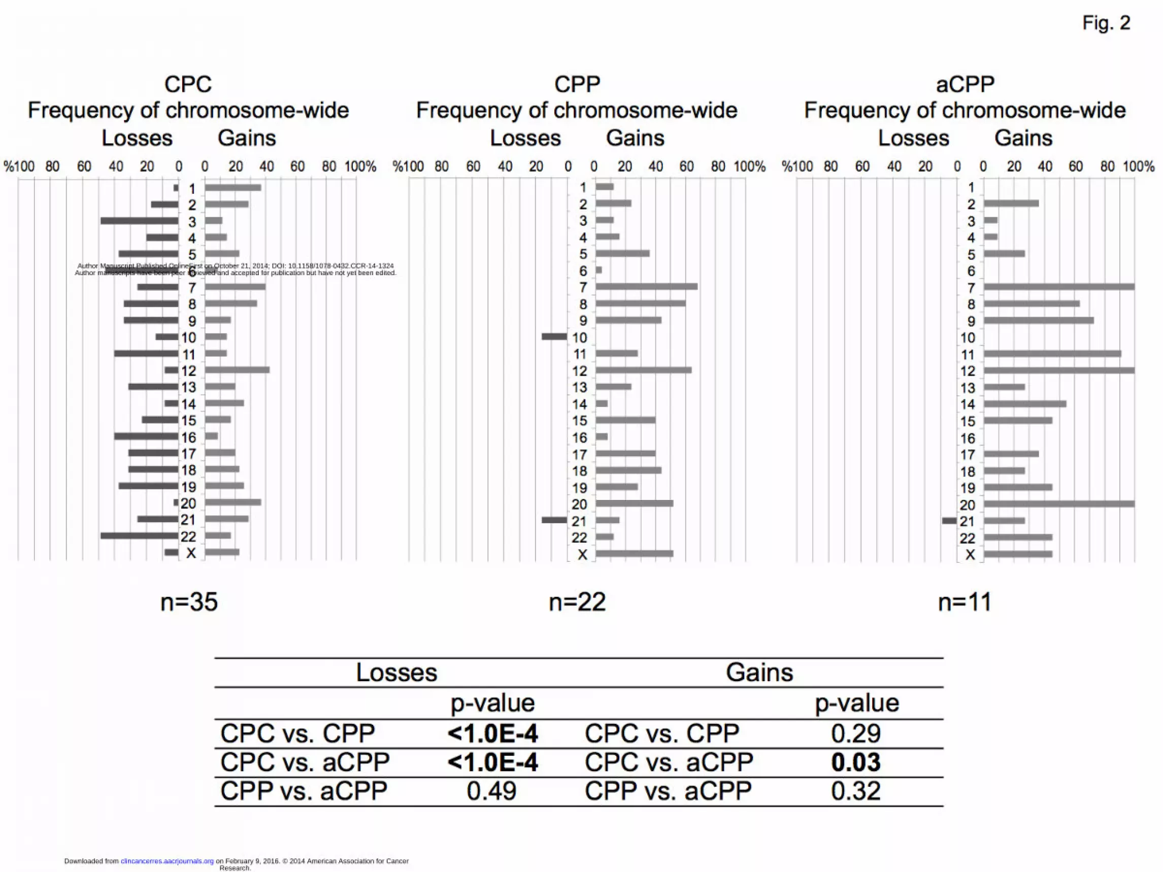
Although copy number analysis revealed widespread chromosomal instability in all
tumor subgroups (Fig. 2), a distinct signature characterized by increased frequency of
chromosome-wide gains and losses was observed in CPCs (average 5.43 chromosomes
gained and 5.65 lost per CPC), compared to increased frequency of chromosome-wide
gains but very few losses in CPPs and aCPPs (average 6.68 chromosomes gained and
Research. on February 9, 2016. © 2014 American Association for Cancerclincancerres.aacrjournals.org Downloaded from
Author manuscripts have been peer reviewed and accepted for publication but have not yet been edited. Author Manuscript Published OnlineFirst on October 21, 2014; DOI: 10.1158/1078-0432.CCR-14-1324

9
0.32 lost per CPP, and 10.00 versus 0.25 per aCPP) (Fig. 2). The frequency of
chromosome-wide losses in CPCs was significantly greater than in CPPs and aCPPs
(Two-tailed t-test p<1.00 x10⁻⁴
). Allele-specific copy number analysis allowed us to
investigate the allelic ratios in our samples. This technique revealed a striking pattern of
copy number-neutral loss of heterozygosity. This phenomenon is commonly observed in
cancer cells and may also be referred to as acquired uniparental disomy (aUPD), wherein
a chromosome pair is homozygous, thus having two copies of the same allele (13). CPCs
exhibited frequent aUPD events with an average of 2.31 aUPD events per sample while
the phenomenon occurred less frequently in CPPs and aCPPs (average 0.32 and 1 events
per sample, respectively).
Analysis of clinical variables between the three histological subgroups revealed no
significant difference of age at diagnosis (Kruskal-Wallis test p=0.30) or ratio of males to
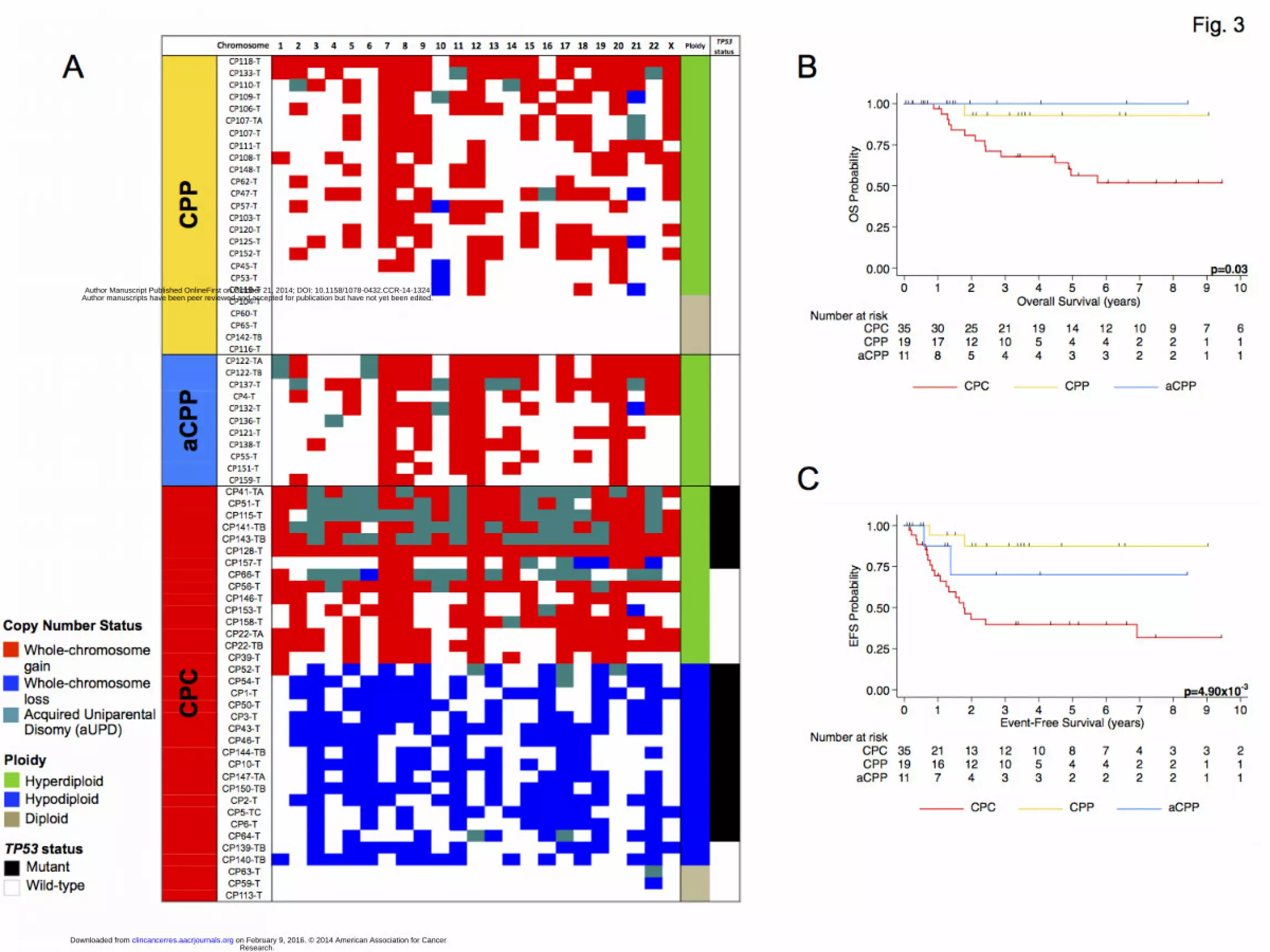
females (Two-way ANOVA p=0.26) (Table S2). Survival outcomes for CPCs were
significantly worse than for CPPs and aCPPs. Five-year OS for CPCs was 56.3% (95%
CI 36.5%-72.0%), compared to 92.9% (59.1%-99.0%) and 100% for CPPs and aCPPs,
respectively (p=0.03) (Fig. 3B). Only one patient with CPP died due to complications
from a concurrent diagnosis of ependymoma. Five-year EFS for CPCs was 39.7% (95%
CI 22.8%-56.2%), compared to 87.4% (58.1%-91.9%) and 70.0% (22.5%-91.8%) for
CPPs and aCPPs, respectively (p=4.90x10⁻³
) (Fig. 3C)
CPPs and aCPPs Share Similar Molecular Signatures Which Correlate With
Favorable Survival Outcomes
Research. on February 9, 2016. © 2014 American Association for Cancerclincancerres.aacrjournals.org Downloaded from
Author manuscripts have been peer reviewed and accepted for publication but have not yet been edited. Author Manuscript Published OnlineFirst on October 21, 2014; DOI: 10.1158/1078-0432.CCR-14-1324

10
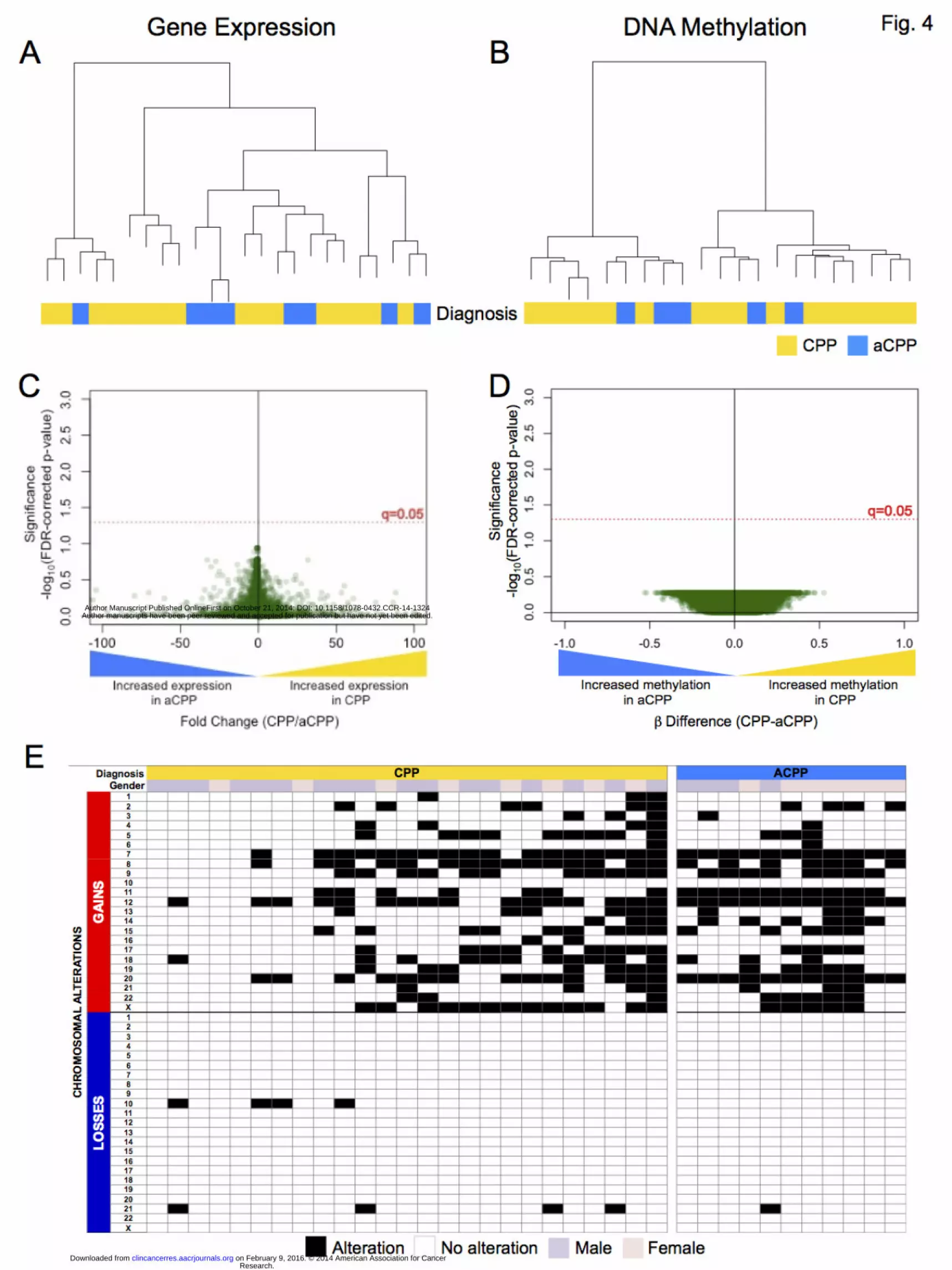
Analyzing CPPs and aCPPs independently from CPCs revealed a striking molecular
similarity between the papilloma subgroups (Fig. 4). Unsupervised clustering analysis
demonstrated that CPPs and aCPP did not segregate according to differences in gene
expression or methylation (Fig. 4 A&B). Supervised analysis using the Wilcoxon rank-
sum (WRS) test between CPPs and aCPPs revealed no significant differences in gene
expression or methylation (Fig. 4 C&D). Additionally, signatures of chromosomal
instability characterized by recurrent chromosome-wide gains and very few losses were
observed in both CPPs and aCPPs; no significant differences in the frequency of
chromosome-wide gains and losses were observed (Two-tailed t-test p=0.32 and p=0.49,
respectively) (Fig. 4E). There were no differences in age at diagnosis (Mann-Whitney test
p=0.45) or ratio of males to females (Fisher’s Exact test p=0.31) between CPPs and
aCPPs. Moreover, survival outcomes for CPP and aCPP patients were not significantly
different (Log-Rank test, OS p=0.51, EFS p=0.30).
Ploidy Analysis Reveals Novel CPC Subgroups With Unique Molecular Alterations
Ploidy analysis revealed the presence of aneuploidy in 87% of tumors (Supplementary
Fig. S3). CPPs and aCPPs exhibited ploidy greater than 2 (hyperdiploidy); however,
CPCs exhibited a wide distribution of ploidy values, with two significantly distinct
subgroups observed: hyperdiploid CPCs (average ploidy 2.76, range: 2.21-3.34) and
hypodiploid CPCs (average ploidy 1.45, range: 1.25-1.71) (Mann-Whitney test,
p<1.00x10⁻⁴
). Only three of 36 CPCs were diploid. Hypodiploid CPCs exhibited recurrent
chromosome-wide losses and very few gains with an average of 12.70 chromosomes lost
and 0.10 gained per tumor. Chromosome 3 was lost in all hypodiploid CPCs, with loss of
Research. on February 9, 2016. © 2014 American Association for Cancerclincancerres.aacrjournals.org Downloaded from
Author manuscripts have been peer reviewed and accepted for publication but have not yet been edited. Author Manuscript Published OnlineFirst on October 21, 2014; DOI: 10.1158/1078-0432.CCR-14-1324

11
chromosomes 6, 9, and 22 observed in 90% of tumors (Fig. 3A). Hyperdiploid CPCs
exhibited a high frequency of chromosome-wide gains and almost no losses (average
12.22 and 0.22 chromosomes, respectively). Chromosomes 12, 7 and 1 were gained in
more than 80% of hyperdiploid CPCs (Fig. 3A). In addition to a high frequency of
chromosomal gains, hyperdiploid CPCs also exhibited aUPD more frequently than
hypodiploid CPCs (average of 4.93 affected chromosomes per tumor compared to 0.33
chromosomes in hypodiploid CPCs) (Fisher’s Exact test p<0.0001) (Fig. 3A). Moreover,
significant enrichment in aUPD was observed in TP53 mutant hyperdiploid CPCs
compared to hyperdiploid CPCs with wild-type TP53 (Fisher’s Exact test p<0.0001).
aUPD was most frequently observed in chromosome 17, affecting 30% (10/33) of CPCs.
We conducted GSEA to identify biological pathways and processes that are differentially
expressed between hypodiploid and hyperdiploid CPCs. GSEA revealed enrichment in
RNA processing, DNA replication and repair, and chromosome segregation in
hyperdiploid CPCs. Hypodiploid CPCs exhibited enrichment in cellular metabolism,
signaling and cell migration pathways, as well as leukocyte activation and proliferation
(5% FDR, p<0.05) (Supplementary Fig. S4). The patterns of enrichment observed
suggest hyperdiploids are more proliferative than hypodiploid CPCs, and that the latter
tumors are undergoing a significant immune response. A greater understanding of these
distinct enrichment patterns will elucidate the mechanisms underlying the progression of
these molecularly distinct CPC subgroups (Supplementary Fig. S4). There were no
significant differences in DNA methylation between subgroups, although this may be due
to the low number of samples in the comparison groups (hypodiploid n=4, hyperdiploid
n=9).
Research. on February 9, 2016. © 2014 American Association for Cancerclincancerres.aacrjournals.org Downloaded from
Author manuscripts have been peer reviewed and accepted for publication but have not yet been edited. Author Manuscript Published OnlineFirst on October 21, 2014; DOI: 10.1158/1078-0432.CCR-14-1324
12
Increased Number of Copies of Mutant TP53 is Associated With Tumor
Aggressiveness and Unfavorable Survival Outcomes
Mutations in TP53 were assessed in our cohort by Sanger sequencing. Sixty percent of
CPCs (35/58) were mutant for TP53. Fifteen (15/58, 26%) of these samples belonged to
12 LFS patients carrying a germline mutation in TP53. Mutations in TP53 were observed
in both hypodiploid and hyperdiploid CPCs, however, in our cohort, the frequency of
TP53 mutations was significantly greater in hypodiploid CPCs (16/18, 89%) than
hyperdiploid CPCs (7/15, 47%) (Fisher’s Exact test p=0.02). Diploid CPCs (n=3) were
TP53 wild-type (Supplementary Table S3). No significant enrichment for LFS patients
was observed in either hypo- or hyperdiploid subgroups.
Unsupervised clustering using gene expression and methylation data segregated CPCs
into two significantly distinct clusters (p=0.05) (Fig. 5A & B). Although CPCs did not
segregate according to ploidy status, we observed two clusters, which were significantly
distinct according to TP53 status using DNA methylation data (Fisher’s Exact test,
p=0.007), but did not reach significance using gene expression data (Fisher’s Exact test,
p=0.089). LFS-CPCs did not segregate from the spontaneous CPCs, suggesting no
unique aberrations were present in tumors arising from patients with an inherited TP53
mutation. An in-depth analysis of the type of TP53 mutations revealed that a few
samples, which appeared to be miscategorized by unsupervised clustering, had an
uncharacterized intronic alteration (c.28-28G>A/wt) and mutations outside the DNA-
binding domain (ie. c.290T>A in the SH3-like/Proline-rich domain), which may account
for differences in the transcriptomic and epigenomic signature of these samples (Fig. 5).
Research. on February 9, 2016. © 2014 American Association for Cancerclincancerres.aacrjournals.org Downloaded from
Author manuscripts have been peer reviewed and accepted for publication but have not yet been edited. Author Manuscript Published OnlineFirst on October 21, 2014; DOI: 10.1158/1078-0432.CCR-14-1324

13
Combining TP53 sequencing results with allele-specific copy number status of
chromosome 17 in 33 CPCs, we estimated the number of mutated copies of TP53. We
found that 36.4% of CPCs (12/33) had 2 copies of mutant p53, 30.3% (10/33) had 1 copy
of mutant p53 and 33.3% (11/33) had zero copies of mutant p53 (wildtype). CPCs with 2
mutant copies of p53 exhibited a homozygous TP53 mutation status in all but one tumor
sample with a low aberrant cell fraction (46%), suggesting this sample was largely
contaminated with normal cells. Seventy-five percent of samples with 2 copies of
mutated p53 (9/12) exhibited aUPD in chromosome 17. Eighty-three percent of CPCs
with 2 copies of mutated p53 (10/12) had missense mutations in the DNA binding
domain, while 1 sample had a missense mutation in the SH3-like/Proline-rich domain and
the other sample, a splicing mutation. CPCs with 1 copy of mutated p53 had missense
mutations in the DNA binding (9/10) and tetramerization (1/10) domains, and carried a
single copy of chromosome 17, exhibiting LOH of the entire chromosome. Three samples
exhibited a heterozygous TP53 mutation status by sequencing, which may be a result of
normal cell contamination. Gene expression and methylation analyses revealed no
significant differences among CPCs carrying 1 or 2 mutated copies of p53 because of the
limited sample sizes (gene expression: 3 and 6 samples, respectively; methylation: 1 and
6 samples, respectively).
Examining clinical variables among CPCs revealed no differences in the age of diagnosis
(Mann-Whitney t-test p=0.80, and p=0.59) or the ratio of males to females (Fisher’s
Exact test p=1.0, and p=1.0) according to ploidy nor p53 status, respectively (Table S2).
No significant differences in OS or EFS estimates were observed between CPC patients
exhibiting a hyper- or hypodiploid tumor genome (p=0.82, p=0.94, respectively)
Research. on February 9, 2016. © 2014 American Association for Cancerclincancerres.aacrjournals.org Downloaded from
Author manuscripts have been peer reviewed and accepted for publication but have not yet been edited. Author Manuscript Published OnlineFirst on October 21, 2014; DOI: 10.1158/1078-0432.CCR-14-1324

14
(Supplementary Fig. S5A & B). TP53 status had a significant effect on the OS of our
CPC cohort (Log-rank test p=3.8x10⁻³
), however EFS was not significantly different
between TP53 mutant and wildtype CPCs (p=0.07). (Fig. 5C & D).
Investigating survival differences according to the number of mutant copies of TP53 in
CPCs revealed a significant reduction in OS (Log-rank test 2 copies vs. 1 copy, p=0.04; 2
copies vs. 0 copies, p<1.0x10⁻⁴), and EFS (Log-rank test 2 copies vs. 1 copy p=0.03; 2
copies vs. 0 copies p=0.003) in patients harboring a greater number of mutant TP53
copies. The estimated OS of patients with CPCs harboring wildtype TP53 (zero copies)
was 88.9% (95% CI 43.3%-98.4%) and EFS, 66.5% (32.9%-86.1%). Patients with CPCs
harboring a single copy of mutant TP53 exhibited an estimated OS of 66.7% (28.2%-
87.8%) and EFS of 44.4% (13.6%-71.9%), while patients with CPCs harboring two
copies of mutant TP53 showed an OS of 14.3% (0.71%-46.5%) and EFS of 0%. (Fig. 6).
DISCUSSION
Our study is the first and largest comprehensive investigation of the molecular alterations
found in CPTs, demonstrating that the molecular profile of CPCs is significantly distinct
from that of CPPs and aCPPs, and that the papillomas are not significantly distinct from
each other. In addition, using an innovative allele-specific approach in combination with
TP53 sequencing, we identified a particularly poor prognostic subgroup in TP53 mutant
CPC patients exhibiting aUPD in chromosome 17, and who as a result had an elevated
number of mutated copies of p53. This study provides evidence for the crucial role of
molecular stratification as a tool to improve the clinical management of patients with
CPT.
Research. on February 9, 2016. © 2014 American Association for Cancerclincancerres.aacrjournals.org Downloaded from
Author manuscripts have been peer reviewed and accepted for publication but have not yet been edited. Author Manuscript Published OnlineFirst on October 21, 2014; DOI: 10.1158/1078-0432.CCR-14-1324

15
Atypical CPPs are currently distinguished from CPPs by histopathology, where aCPPs
exhibit increased mitotic activity(14); yet, survival outcomes for both CPPs and aCPPs
are comparably favorable. Standard of care for these tumors consists of surgical resection
with very few aCPP cases requiring adjuvant chemotherapy. In our cohort, all aCPP
patients for which we had clinical history (6/11), were treated with surgical resection
alone, yet demonstrated favorable survival comparable to CPPs. We suggest that the
benign phenotype of aCPPs may reflect the molecular characteristics it shares with CPPs,
including very few chromosome-wide losses, and similar gene enrichment patterns and
methylation signatures. The data lend support for the conservative management of aCPP
patients with surgical resection followed by observation.
Our findings also revealed that copy number, gene expression and methylation profiles
were significantly distinct between the papillomas and CPCs, indicating the unlikelihood
that CPPs or aCPPs progress unto CPCs by the acquisition of a few additional
aberrations. Although a few studies have reported on progression from papillomas to
CPCs (9,15), we believe this unlikely scenario may have been the result of a
heterogeneous tumor sample harboring co-existing CPC and papilloma cells. Analyzing
tumor heterogeneity in CPTs, will be necessary in order to identify benign tumors more
likely to recur with an aggressive phenotype.
Wide variability in clinical outcome has been observed among CPC patients despite the
use of similar treatment protocols.(2,16,17) Our findings demonstrate that the molecular
heterogeneity of CPCs may be driving this clinical variability.
Research. on February 9, 2016. © 2014 American Association for Cancerclincancerres.aacrjournals.org Downloaded from
Author manuscripts have been peer reviewed and accepted for publication but have not yet been edited. Author Manuscript Published OnlineFirst on October 21, 2014; DOI: 10.1158/1078-0432.CCR-14-1324

16
Extensive chromosomal alterations were recurrent in CPCs. An allele-specific copy
number approach allowed us to identify aneuploidy in 91% of CPCs, and distinguish
between hypodiploid CPCs, exhibiting numerous chromosomal losses, and hyperdiploid
CPCs, exhibiting numerous chromosomal gains and concurrent aUPD. Our findings
uncovered that chromosomal instability is a common mechanism involved in CPC
development; however, further examination of the molecular differences driving hypo-
and hyperdiploid development will identify distinct mechanisms responsible for tumor
progression. Ploidy was not significantly associated with age at diagnosis, or patient
survival; nonetheless, we identified that hyperdiploid CPCs were significantly enriched in
chromosomes exhibiting aUPD. A recent study reported similar subgroupings in CPCs,
where a higher frequency of chromosomal losses were observed in younger children and
chromosomal gains in older children, and loss of 12q was associated with shorter
survival(18). In our cohort we found no significant correlations between patient age and
CPC subgroups or TP53 status. Moreover, survival differences were identified only when
TP53 copy number and mutation status were examined concomitantly.
Arising from somatic recombination errors during mitosis, aUPD is an important
mechanism leading to loss of heterozygosity with an unaffected copy number, and is
therefore associated with the enrichment of chromosomes or regions harboring
preexisting mutations, specific promoter methylation patterns, and focal deletion of genes
(13). In our study, we observed aUPD affect entire chromosomes in all tumor subgroups,
however aUPD was most frequent in CPCs harboring TP53 mutations. We identified
chromosome 17 to be the most frequent site affected by aUPD, and that 90% of CPCs
Research. on February 9, 2016. © 2014 American Association for Cancerclincancerres.aacrjournals.org Downloaded from
Author manuscripts have been peer reviewed and accepted for publication but have not yet been edited. Author Manuscript Published OnlineFirst on October 21, 2014; DOI: 10.1158/1078-0432.CCR-14-1324

17
exhibiting aUPD of chromosome 17 harbored a mutation in TP53, increasing the number
of mutant p53 copies to 2 in these tumors. Our findings suggest aUPD is a mechanism by
which CPCs accumulate deleterious aberrations, such as TP53 mutations, while retaining
the normal function of other genes due to an unaffected chromosome copy number.
Focusing on the known association between p53 mutations and CPCs, we identified that
the number of mutated copies of p53 was significantly associated with patient survival.
Our findings support the concept that in addition to the loss of tumor-suppressive activity
of p53, mutant TP53 also acquires oncogenic activities that promote CPC development.
The gain of function (GOF) properties of mutant p53 include cellular invasion,
proliferation, genomic instability, and polyploidy, among others (Reviewed in (19)).
Because of increased GOF activity, in addition to a complete loss of the tumor suppressor
functions of p53, an elevated number of mutant p53 copies could result in an aggressive
phenotype associated with decreased survival as we observed in our high risk CPC
patient cohort. Since we did not assess the mutation status of other cancer genes in
chromosome 17, we cannot infer that the number of mutant copies of p53 is the only
aberration on this locus driving CPC development and tumor aggressiveness in the high
risk CPC patient cohort. However, the significant dose-dependent correlation observed
with overall and event-free survival in CPC patients indicates a role for p53 GOF activity
in CPC aggressiveness.
TP53 mutations alone do not drive chromosomal instability in CPCs as TP53 wild-type
tumors also exhibit high levels of chromosome-wide gains and losses. However, we have
demonstrated that TP53 mutations are associated with changes in gene expression and
methylation patterns that may result in increased tumor aggressiveness and may elucidate
Research. on February 9, 2016. © 2014 American Association for Cancerclincancerres.aacrjournals.org Downloaded from
Author manuscripts have been peer reviewed and accepted for publication but have not yet been edited. Author Manuscript Published OnlineFirst on October 21, 2014; DOI: 10.1158/1078-0432.CCR-14-1324

18
the different clinical outcomes observed. Alterations affecting the p53 pathway, either
upstream or downstream of p53, may generate the molecular background necessary for
CPC development, and should be investigated further.
Recurrent lesions, such as the chromosome-wide gains of chromosome 1, which was not
only recurrently gained but also the least frequently lost in CPCs, chromosome 12, and
chromosome-wide loss of chromosome 3, may also be contributing to CPC’s unique
genotype and would need to be further investigated in order to identify unique targets for
effective therapies.
Our study demonstrates that investigating the molecular characteristics of CPTs is crucial
to further refine the molecular stratification of patients in order to improve patient care.
We suggest that the prognostic significance of TP53 mutation and copy number status in
CPCs be validated prospectively in future cooperative clinical trials. Validation of these
data in future prospective studies will inform risk stratification of CPC patients, and set
the framework for future treatment intensification for high-risk patients.
Research. on February 9, 2016. © 2014 American Association for Cancerclincancerres.aacrjournals.org Downloaded from
Author manuscripts have been peer reviewed and accepted for publication but have not yet been edited. Author Manuscript Published OnlineFirst on October 21, 2014; DOI: 10.1158/1078-0432.CCR-14-1324

19
References:
1. Ogiwara H, Dipatri Jr AJ, Alden TD, Bowman RM, Tomita T. Choroid plexus
tumors in pediatric patients. Br J Neurosurg. 2012;26:32–7.
2. Lafay-Cousin L, Keene D, Carret A-S, Fryer C, Brossard J, Crooks B, et al.
Choroid plexus tumors in children less than 36 months: the Canadian Pediatric
Brain Tumor Consortium (CPBTC) experience. Child’s Nerv Syst. 2011;27:259–
64.
3. Addo NK, Kamaly-Asl ID, Josan VA, Kelsey AM, Estlin EJ. Preoperative
vincristine for an inoperable choroid plexus papilloma: a case discussion and
review of the literature. Case Rep. 2011;8:149–53.
4. Rickert CH, Paulus W. Tumors of the choroid plexus. Microsc Res Tech.
2001;52:104–11.
5. Tabori U, Shlien A, Baskin B, Levitt S, Ray P, Alon N, et al. TP53 alterations
determine clinical subgroups and survival of patients with choroid plexus tumors. J
Clin Oncol. 2010;28:1995–2001.
6. Lafay-Cousin L, Mabbott DJ, Halliday W, Taylor MD, Tabori U, Kamaly-Asl ID,
et al. Use of ifosfamide, carboplatin, and etoposide chemotherapy in choroid
plexus carcinoma. J Neurosurg Pediatr. 2010;5:615–21.
7. Jeibmann A, Hasselblatt M, Gerss J, Wrede B, Egensperger R, Beschorner R, et al.
Prognostic implications of atypical histologic features in choroid plexus papilloma.
J Neuropathol Exp Neurol. 2006;65:1069–73.
8. Wrede B, Hasselblatt M, Peters O, Thall PF, Kutluk T, Moghrabi A, et al. Atypical
choroid plexus papilloma: clinical experience in the CPT-SIOP-2000 study. J
Neurooncol. 2009;95:383–92.
9. Jeibmann A, Wrede B, Peters O, Wolff JE, Paulus W, Hasselblatt M. Malignant
progression in choroid plexus papillomas. J Neurosurg. 2007;107:199–202.
10. Van Loo P, Nordgard SH, Lingjærde OC, Russnes HG, Rye IH, Sun W, et al.
Allele-specific copy number analysis of tumors. Proc Natl Acad Sci U S A.
2010;107:16910–5.
11. Subramanian A, Tamayo P, Mootha VK, Mukherjee S, Ebert BL, Gillette M a, et
al. Gene set enrichment analysis: a knowledge-based approach for interpreting
genome-wide expression profiles. Proc Natl Acad Sci U S A. 2005;102:15545–50.
Research. on February 9, 2016. © 2014 American Association for Cancerclincancerres.aacrjournals.org Downloaded from
Author manuscripts have been peer reviewed and accepted for publication but have not yet been edited. Author Manuscript Published OnlineFirst on October 21, 2014; DOI: 10.1158/1078-0432.CCR-14-1324

20
12. Witt H, Mack SC, Ryzhova M, Bender S, Sill M, Isserlin R, et al. Delineation of
Two Clinically and Molecularly Distinct Subgroups of Posterior Fossa
Ependymoma. Cancer Cell. Elsevier Inc.; 2011;20:143–57.
13. Tuna M, Knuutila S, Mills GB. Uniparental disomy in cancer. Trends Mol Med.
2009;15:120–8.
14. Louis DN, Ohgaki H, Wiestler OD, Cavenee WK, Burger PC, Jouvet A, et al. The
2007 WHO classification of tumours of the central nervous system. Acta
Neuropathol. 2007;114:97–109.
15. Dhillon RS, Wang YY, McKelvie P a, O’Brien B. Progression of choroid plexus
papilloma. J Clin Neurosci. Elsevier Ltd; 2013;20:1775–8.
16. Gozali AE, Britt B, Shane L, Gonzalez I, Gilles F, McComb JG, et al. Choroid
plexus tumors; management, outcome, and association with the Li-Fraumeni
syndrome: The Children’s Hospital Los Angeles (CHLA) experience, 1991-2010.
Pediatr Blood Cancer. 2012;58:905–9.
17. Bettegowda C, Adogwa O, Mehta V, Chaichana KL, Weingart J, Carson BS, et al.
Treatment of choroid plexus tumors: a 20-year single institutional experience. J
Neurosurg Pediatr. 2012;10:398–405.
18. Ruland V, Hartung S, Kordes U, Wolff JE, Paulus W, Hasselblatt M. Choroid
Plexus Carcinomas are Characterized by Complex Chromosomal Alterations
Related to Patient Age and Prognosis. 2014;53:373–80.
19. Muller PAJ, Vousden KH. Mutant p53 in Cancer: New Functions and Therapeutic
Opportunities. Cancer Cell. 2014;25:304–17.
Research. on February 9, 2016. © 2014 American Association for Cancerclincancerres.aacrjournals.org Downloaded from
Author manuscripts have been peer reviewed and accepted for publication but have not yet been edited. Author Manuscript Published OnlineFirst on October 21, 2014; DOI: 10.1158/1078-0432.CCR-14-1324
21
Figure Legends
Figure 1: Unsupervised clustering of (A) gene expression normalized intensities and (B)
methylation Beta-values by non-negative matrix factorization (NMF) demonstrate
significant segregation of CPCs (red) from CPPs (yellow) and aCPPs (light blue). No
segregation was observed between CPPs and aCPPs. NMF was conducted using 5000
probesets with the largest median absolute deviation (MAD). This clustering algorithm
identified the most significant measures of similarity (cophenetic coefficient) when the
data was at k=2 (2 clusters). In the matrix, red represents the highest measure of
similarity (1), while blue/purple represents the lowest measure of similarity (0). Any
other colors within the matrix represent a spectrum of changing measures of similarity,
from red to blue/purple. Colors: TP53 mutation status: Black: TP53 mutant, White: TP53
wildtype.
Figure 2: Characterization of recurrent chromosome-wide gains (n>2, grey) and losses
(n<2, black) for each tumor subtype. (CPC: n=37, CPP: n=25, aCPP: n=11). P-values
were calculated using the frequency of chromosome-wide copy number alterations per
subgroup and the non-parametric Mann-Whitney test.
Figure 3: (A) Genome-wide characterization of chromosome-wide gains (red), losses
(blue), and aUPD (teal) in 71 unique CPC samples. White squares represent unchanged
chromosome-wide copy number status. Chromosome-wide aberrations were defined as
aberrations that encompass more than 75% of the chromosome. Ploidy: green:
hyperdiploid, blue: hypodiploid, tan: diploid. TP53 status: black: mutant, white: wildtype.
Kaplan-Meier curves depicting (B) overall and (C) event-free survival estimates of CPT
patients by diagnosis. Statistical values were obtained with the Log-rank (Mantel-Cox)
test.
Figure 4: Unsupervised clustering of (A) gene expression normalized intensities and (B)
methylation Beta values demonstrate no segregation between CPPs (yellow) and aCPPs
(light blue). Volcano plot comparing the number of significant differentially expressed
genes (C) and significantly methylated regions (D) reveals no significant gene expression
or methylation differences between the two subgroups (after FDR adjustment). Signatures
of chromosomal instability (E), as measured by the number of chromosome-wide gains
and losses in CPPs and aCPPs, were similar between the two subgroups and characterized
by extensive chromosomal gains and very few losses. Black squares represent
chromosome-wide aberrations, while white squares represent unchanged chromosome
copy number. Gender was also depicted (pink: female, purple: male).
Figure 5: Unsupervised hierarchical clustering of (A) gene expression normalized
intensities and (B) methylation Beta values using the PVCLUST algorithm demonstrates
significant segregation among CPCs, where subgroups are characterized by differences in
TP53 status (mutant=black, wild- type=white). Red rectangles delineate statistically
significant different groups (p=0·05). PVCLUST algorithm was conducted using 1000
probesets with the largest median absolute deviation (MAD). * shows sample with an
uncharacterized intronic alteration, # shows samples with mutation in SH3-like/Proline-
rich TP53 domain. Colors: Diagnosis: Red: CPC, Yellow: CPP, Light blue: aCPP, TP53
Research. on February 9, 2016. © 2014 American Association for Cancerclincancerres.aacrjournals.org Downloaded from
Author manuscripts have been peer reviewed and accepted for publication but have not yet been edited. Author Manuscript Published OnlineFirst on October 21, 2014; DOI: 10.1158/1078-0432.CCR-14-1324
22
status: Black: TP53 mutant, White: TP53 wild-type, Ploidy: Green: hyperdiploid, Blue:
hypodiploid, Tan: Diploid, Grey: unknown. Kaplan-Meier curves depicting (C) overall
and (D) event-free survival estimates of CPC patients by TP53 mutation status. Statistical
values were obtained with the Log-rank (Mantel-Cox) test.
Figure 6: Kaplan-Meier curves depicting (A) overall and (B) event-free survival
estimates of CPC patients by number of mutated copies of p53, as estimated by Sanger
sequencing and allele-specific copy number analysis. Statistical values were obtained
with the Log-rank (Mantel-Cox) test.
Research. on February 9, 2016. © 2014 American Association for Cancerclincancerres.aacrjournals.org Downloaded from
Author manuscripts have been peer reviewed and accepted for publication but have not yet been edited. Author Manuscript Published OnlineFirst on October 21, 2014; DOI: 10.1158/1078-0432.CCR-14-1324
Published OnlineFirst October 21, 2014.Clin Cancer Res Diana M Merino, Adam Shlien, Anita Villani, et al. novel clinically relevant subgroupsMolecular characterization of choroid plexus tumors reveals
Updated version
10.1158/1078-0432.CCR-14-1324doi:
Access the most recent version of this article at:
Material
Supplementary
http://clincancerres.aacrjournals.org/content/suppl/2014/10/22/1078-0432.CCR-14-1324.DC1.html
Access the most recent supplemental material at:
Manuscript
Authoredited. Author manuscripts have been peer reviewed and accepted for publication but have not yet been
E-mail alerts related to this article or journal.Sign up to receive free email-alerts
Subscriptions
Reprints and
To order reprints of this article or to subscribe to the journal, contact the AACR Publications
Permissions
To request permission to re-use all or part of this article, contact the AACR Publications
Research. on February 9, 2016. © 2014 American Association for Cancerclincancerres.aacrjournals.org Downloaded from
Author manuscripts have been peer reviewed and accepted for publication but have not yet been edited. Author Manuscript Published OnlineFirst on October 21, 2014; DOI: 10.1158/1078-0432.CCR-14-1324
Related Documents

![Infratentorial choroid plexus tumors in children · 2020-07-12 · plexus carcinomas have a poorer prognosis thought to be due to increased local invasion [ 6]. Many patients present](https://static.cupdf.com/doc/110x72/5fb981415693b60a881c6cec/infratentorial-choroid-plexus-tumors-in-children-2020-07-12-plexus-carcinomas.jpg)