http://tpx.sagepub.com/ Toxicologic Pathology http://tpx.sagepub.com/content/39/1/186 The online version of this article can be found at: DOI: 10.1177/0192623310394214 2011 39: 186 originally published online 28 December 2010 Toxicol Pathol Conrad Johanson, Edward Stopa, Paul McMillan, Daniel Roth, Juergen Funk and Georg Krinke Toxicologic/Pathologic Phenomena, Periventricular Destabilization, and Lesion Spread The Distributional Nexus of Choroid Plexus to Cerebrospinal Fluid, Ependyma and Brain: Published by: http://www.sagepublications.com On behalf of: Society of Toxicologic Pathology can be found at: Toxicologic Pathology Additional services and information for http://tpx.sagepub.com/cgi/alerts Email Alerts: http://tpx.sagepub.com/subscriptions Subscriptions: http://www.sagepub.com/journalsReprints.nav Reprints: http://www.sagepub.com/journalsPermissions.nav Permissions: What is This? - Dec 28, 2010 OnlineFirst Version of Record - Mar 4, 2011 Version of Record >> by guest on October 11, 2013 tpx.sagepub.com Downloaded from by guest on October 11, 2013 tpx.sagepub.com Downloaded from by guest on October 11, 2013 tpx.sagepub.com Downloaded from by guest on October 11, 2013 tpx.sagepub.com Downloaded from by guest on October 11, 2013 tpx.sagepub.com Downloaded from by guest on October 11, 2013 tpx.sagepub.com Downloaded from by guest on October 11, 2013 tpx.sagepub.com Downloaded from by guest on October 11, 2013 tpx.sagepub.com Downloaded from by guest on October 11, 2013 tpx.sagepub.com Downloaded from by guest on October 11, 2013 tpx.sagepub.com Downloaded from by guest on October 11, 2013 tpx.sagepub.com Downloaded from by guest on October 11, 2013 tpx.sagepub.com Downloaded from by guest on October 11, 2013 tpx.sagepub.com Downloaded from by guest on October 11, 2013 tpx.sagepub.com Downloaded from by guest on October 11, 2013 tpx.sagepub.com Downloaded from by guest on October 11, 2013 tpx.sagepub.com Downloaded from by guest on October 11, 2013 tpx.sagepub.com Downloaded from by guest on October 11, 2013 tpx.sagepub.com Downloaded from by guest on October 11, 2013 tpx.sagepub.com Downloaded from by guest on October 11, 2013 tpx.sagepub.com Downloaded from by guest on October 11, 2013 tpx.sagepub.com Downloaded from by guest on October 11, 2013 tpx.sagepub.com Downloaded from by guest on October 11, 2013 tpx.sagepub.com Downloaded from by guest on October 11, 2013 tpx.sagepub.com Downloaded from by guest on October 11, 2013 tpx.sagepub.com Downloaded from by guest on October 11, 2013 tpx.sagepub.com Downloaded from by guest on October 11, 2013 tpx.sagepub.com Downloaded from by guest on October 11, 2013 tpx.sagepub.com Downloaded from

Welcome message from author
This document is posted to help you gain knowledge. Please leave a comment to let me know what you think about it! Share it to your friends and learn new things together.
Transcript

http://tpx.sagepub.com/Toxicologic Pathology
http://tpx.sagepub.com/content/39/1/186The online version of this article can be found at:
DOI: 10.1177/0192623310394214
2011 39: 186 originally published online 28 December 2010Toxicol PatholConrad Johanson, Edward Stopa, Paul McMillan, Daniel Roth, Juergen Funk and Georg Krinke
Toxicologic/Pathologic Phenomena, Periventricular Destabilization, and Lesion SpreadThe Distributional Nexus of Choroid Plexus to Cerebrospinal Fluid, Ependyma and Brain:
Published by:
http://www.sagepublications.com
On behalf of:
Society of Toxicologic Pathology
can be found at:Toxicologic PathologyAdditional services and information for
http://tpx.sagepub.com/cgi/alertsEmail Alerts:
http://tpx.sagepub.com/subscriptionsSubscriptions:
http://www.sagepub.com/journalsReprints.navReprints:
http://www.sagepub.com/journalsPermissions.navPermissions:
What is This?
- Dec 28, 2010 OnlineFirst Version of Record
- Mar 4, 2011Version of Record >>
by guest on October 11, 2013tpx.sagepub.comDownloaded from by guest on October 11, 2013tpx.sagepub.comDownloaded from by guest on October 11, 2013tpx.sagepub.comDownloaded from by guest on October 11, 2013tpx.sagepub.comDownloaded from by guest on October 11, 2013tpx.sagepub.comDownloaded from by guest on October 11, 2013tpx.sagepub.comDownloaded from by guest on October 11, 2013tpx.sagepub.comDownloaded from by guest on October 11, 2013tpx.sagepub.comDownloaded from by guest on October 11, 2013tpx.sagepub.comDownloaded from by guest on October 11, 2013tpx.sagepub.comDownloaded from by guest on October 11, 2013tpx.sagepub.comDownloaded from by guest on October 11, 2013tpx.sagepub.comDownloaded from by guest on October 11, 2013tpx.sagepub.comDownloaded from by guest on October 11, 2013tpx.sagepub.comDownloaded from by guest on October 11, 2013tpx.sagepub.comDownloaded from by guest on October 11, 2013tpx.sagepub.comDownloaded from by guest on October 11, 2013tpx.sagepub.comDownloaded from by guest on October 11, 2013tpx.sagepub.comDownloaded from by guest on October 11, 2013tpx.sagepub.comDownloaded from by guest on October 11, 2013tpx.sagepub.comDownloaded from by guest on October 11, 2013tpx.sagepub.comDownloaded from by guest on October 11, 2013tpx.sagepub.comDownloaded from by guest on October 11, 2013tpx.sagepub.comDownloaded from by guest on October 11, 2013tpx.sagepub.comDownloaded from by guest on October 11, 2013tpx.sagepub.comDownloaded from by guest on October 11, 2013tpx.sagepub.comDownloaded from by guest on October 11, 2013tpx.sagepub.comDownloaded from by guest on October 11, 2013tpx.sagepub.comDownloaded from
The Distributional Nexus of Choroid Plexus to Cerebrospinal Fluid,Ependyma and Brain: Toxicologic/Pathologic Phenomena,
Periventricular Destabilization, and Lesion Spread
CONRAD JOHANSON1, EDWARD STOPA
1, PAUL MCMILLAN1, DANIEL ROTH
2, JUERGEN FUNK3, AND GEORG KRINKE
4
1Brown University, Providence, Rhode Island, United States2Novartis Institutes for Biomedical Research, Basel, Switzerland
3F. Hoffmann-La Roche Ltd., Pharma Research Non-Clinical Safety, Basel, Switzerland4AnaPath GmbH, Oberbuchsiten, Switzerland
ABSTRACT
Bordering the ventricular cerebrospinal fluid (CSF) are epithelial cells of choroid plexus (CP), ependyma and circumventricular organs (CVOs)
that contain homeostatic transporters for mediating secretion/reabsorption. The distributional pathway (‘‘nexus’’) of CP-CSF-ependyma-brain
furnishes peptides, hormones, and micronutrients to periventricular regions. In disease/toxicity, this nexus becomes a conduit for infectious and
xenobiotic agents. The sleeping sickness trypanosome (a protozoan) disrupts CP and downstream CSF-brain. Piperamide is anti-trypanosomic but
distorts CP epithelial ultrastructure by engendering hydropic vacuoles; this reflects phospholipidosis and altered lysosomal metabolism. CP swelling
by vacuolation may occlude CSF flow. Toxic drug tools delineate injuries to choroidal compartments: cyclophosphamide (vasculature), methylcel-
lulose (interstitium), and piperazine (epithelium). Structurally perturbed CP allows solutes to penetrate the ventricles. There, CSF-borne pathogens
and xenobiotics may permeate the ependyma to harm neurogenic stem cell niches. Amoscanate, an anti-helmintic, potently injures rodent ependyma.
Ependymal/brain regions near CP are vulnerable to CSF-borne toxicants; this proximity factor links regional barrier breakdown to nearby
periventricular pathology. Diverse diseases (e.g., African sleeping sickness, multiple sclerosis) take early root in choroidal, circumventricular, or
perivascular loci. Toxicokinetics informs on pathogen, anti-parasitic agent, and auto-antibody distribution along the CSF nexus. CVOs are suscep-
tible to plasma-borne toxicants/pathogens. Countering the physico-chemical and pathogenic insults to the homeostasis-mediating ventricle-bordering
cells sustains brain health and fluid balance.
Keywords: pegylated drugs; anti-parasitic agents; phospholipidosis; anti-trypanosome agents; tertiary amines; lysosomal storage disease;
multiple sclerosis; epithelial vacuoles.
INTRODUCTION
Integrated Topics and Objectives
Neurons are exquisitely and adversely sensitive to toxicants
and pathogens. Normally the impermeable blood-brain barrier
(BBB) and blood-cerebrospinal fluid (CSF) barrier (BCSFB)
act together to protect the neuronal networks from potentially
injurious agents in blood. Historically, the BBB has received
more pharmacologic, toxicologic, and pathologic attention
than the BCSFB, or choroid plexus (CP). In a disease or toxi-
city context, this is ironic because BCSFB is more vulnerable
(Levine 1987) than cerebral capillaries to many foreign inva-
ders. Often the CP or an ependymal circumventricular organ
(CVO) is the first site of central penetration by a deleterious
substance or pathogen (Siso, Jeffrey, and Gonzales 2010).
CP is also of unique interest to toxicologists and pathologists
because its main secretion, the cerebrospinal fluid, rapidly and
widely disseminates throughout the central nervous system
(CNS) the various substances that have penetrated a breached
BCSFB. Thereafter, the toxicants, pathogens, and cells that
permeate the CSF are readily accessible via bulk flow circula-
tion to neurons and stem cells over wide expanses of brain.
Accordingly, the two aims of this review are to: (a) characterize
the intricate physiology of the CP, CSF, and ependyma and
This review is dedicated to the recently deceased Dr. Seymour Levine, a
neuropathologist at the New York Medical College who was a pioneer
investigator of toxico-pathological phenomena involving the choroid plexus-
cerebrospinal fluid (CSF) system. The authors express their gratitude to A.
Messier for his critique of the manuscript and to J. Johanson for constructing
diagrams. NIH support to EGS (NIH NS/AG 10682) and CEJ (NIH R01
27601 and R01 AG027910) and research support from the Departments of
Neurosurgery and Pathology at Rhode Island Hospital were instrumental for
obtaining information on blood-CSF barrier disruption in disease states.
Address correspondence and reprint requests to Conrad Johanson, Department
of Neurosurgery, Brown Medical School, Providence, RI 02903; e-mail:
Abbreviations: 3V and 4V, third and fourth ventricle; AIDS, acquired
immunodeficiency syndrome; ALA, aminolevulinic acid; AP, area postrema;
APOE4, apolipoprotein E; BBB, blood-brain barrier; BCSFB, blood-CSF bar-
rier; CAD, cationic amphiphilic drug; CBF, cerebral blood flow; CIMZIA, cer-
tolizumab; CNS, central nervous system; CP, choroid plexus; CSF,
cerebrospinal fluid; CVO, circumventricular organ; CY, cyclophosphamide;
EAE, experimental allergic encephalitis; ICV, intracerebroventricular route
of exposure; IGF, insulin-like growth factor; IP, intraperioneal route of expo-
sure; ISF, interstitial fluid; LRP, low density lipophilic receptor-related
protein; MC, methylcholanthrene; ME, median eminence; MS, multiple sclero-
sis; NLP, neural lobe of hypophysis (pituitary); OATP, organic anion transport-
ing polypeptide; OVLT, organum vasculosum of lamina terminalis; PEG,
polyethylene glycol; PEPT2, proton-coupled oligopeptide transporter; PCR,
polymerase chain reaction; PI, pineal gland; PO, per os (oral) route of expo-
sure; PT, proximal tubule; SC, subcutaneous route of exposure; SCO, subcom-
missural organ; SFO, subfornical organ; SVZ, subventricular zone; TGFb,
transforming growth factor b; UDP, uridine 5’-diphospho; VEGF, vascular
endothelial growth factor.
186
Toxicologic Pathology, 39: 186-212, 2011
Copyright # 2011 by The Author(s)
ISSN: 0192-6233 print / 1533-1601 online
DOI: 10.1177/0192623310394214
(b) assess how harmful xenobiotics and microbes (e.g., viruses,
protozoans) damage the CP and ependyma, thereby destabiliz-
ing the brain interior (Krinke et al. 1983; Levine, Sowinski, and
Nochlin 1982; Roth and Krinke 1994). Regions injured include
the delicate neurogenic niches and body-regulatory centers
near ventricular CSF.
Central Nervous System Toxicology/Pathology:
Differential Effects of Barrier Phenomena
Toxicants and pathogens vary greatly in ability to penetrate
particular regions of the central nervous system. Consequently,
knowledge of permeation patterns across diverse transport
interfaces is essential to understand specific disruptions of
brain and CSF. BBB refers to the widespread impermeable
capillaries in the CNS. However, BBB is sometimes used
inappropriately to include the BCSFB and other central sites
of impermeability. Therefore, more precise classification of
CNS transport interfaces is needed. Care should be taken to
delineate properties/reactions of the membrane boundaries that
separate blood, CSF, and brain. Bidirectional secretory and
reabsorptive phenomena should also be considered. This is true
in a physiologic-pharmacologic sense and in relation to
toxicologic-pathologic effects on barrier disruption.
Transport specialists concur that the BBB resides primarily
in the tight junctions of brain microvessels or capillaries.
BCSFB most commonly (and in this review) is used to refer
to tight junctions in CP epithelium (but should be more inclu-
sively defined if meant also to include CNS-inward flux across
arachnoid membrane into subarachnoid space). The lateral ven-
tricular CSF-brain interface, or ependyma, is permeable to
macromolecules, and therefore does not usually act as a barrier.
Certain regions of the ependymal wall contain specialized
CVOs adjacent to CSF. The CVOs, like CP, have permeable
capillaries and are thus open windows to the systemic circula-
tion. Thus, each CVO does not have a BBB.
Because BBB is endothelial and BCSFB epithelial, there are
consequently barrier differences in tight junction (imperme-
ability) properties and in types of transporters expressed at the
respective barrier cell membranes. These are important distinc-
tions in view of how toxicants and pathogens differentially
interact with BBB and BCSFB. Cerebral capillaries and chor-
oid plexuses extensively mediate homeostatic mechanisms to
protect the neuronal microenvironment. When xenobiotic
agents and viruses disrupt BBB and BCSFB, they not only
breach barriers but also may disrupt homeostatic transporter
actions that benefit neurons.
Mechanisms for Keeping a Pristine Environment for
Neurons
To function optimally, the brain requires a clean environ-
ment for the neural parenchyma. Neurons need a stable extra-
cellular or interstitial fluid (ISF) of specialized composition
to maintain an extracellular environment that promotes effi-
cient transmission of impulses along axons and through
synapses. ISF that typically bathes neurons is relatively low
in concentrations of plasma proteins, cytokines, catabolite
waste products, erythrocytes, leukocytes, immune cells, patho-
gens, drug metabolites, and toxicants. Three mechanisms
mainly accomplish CNS microenvironmental cleansing:
(1) an impermeable BBB and BCSFB that restrict diffusion
of plasma water-soluble molecules into CNS; (2) reabsorptive
organic solute transporters at the CNS-facing side of barrier
cells for actively extruding into blood certain organic acids
(anions) and bases (cations) in ISF or CSF; and (3) the sink
action of CSF to remove from the brain various metabolites and
impurities for excretion, by bulk flow, from the ventricles to
downstream venous/lymphatic exit sites.
Fundamental differences exist in the workings of BBB (cer-
ebral capillaries) versus BCSFB (choroid plexus). The latter
purifies the CSF interior and periventricular regions with
homeostatic systems to keep brain and body fluids toxicant free
and in physiologic balance. Attenuation of CP transport capac-
ity can profoundly impair cerebral metabolism and the fluid
environment of neurons (Spector and Johanson 2010a).
Accordingly, we analyze BCSFB transporters, selective CP
permeability, the CSF and peptide secretions that choroid
epithelial cells generate, as well as fluid flow through the ven-
tricular system. CSF streaming down the neuraxis from lateral
to fourth ventricles reaches many regions. The largest portion
of ventricular CSF is eventually convected to the subarachnoid
space for bulk fluid clearance across arachnoid villi and
lymphatic drainage channels that follow the olfactory nerve
and discharge their contents into the nose. However, a small but
significant portion of CSF-borne ions and organic solutes
diffuses across or is taken up by border cells at the ventricular
margins.
Compartmental Aspects of the Physiologic Nexus
Modeling features of the specific distributional pathway, or
‘‘nexus,’’ connecting the CP, CSF, ependyma, and brain are
shown in Figure 1. In a transport physiology sense, a nexus can
be viewed as a series of connected compartments within an
organ, through which substances passively move or are actively
transferred, to effect an action or function at a downstream
target. A nexus example is the facilitated diffusion of plasma
glucose via carrier proteins across brain endothelium into the
interstitial fluid, and then diffusion to nearby neurons for
carrier-mediated uptake of the sugar into axoplasm. Another
nexus pathway is the diffusion of plasma-borne angiotensin
across permeable capillaries of the subfornical organ (a CVO)
into the interstitial space, through which this peptide diffuses to
neurons. Yet another nexus, and the main theme of this review,
is the movement of a micronutrient (or hormone or xenobiotic)
across CP into ventricular CSF for bulk flow to target cells in
the ependyma and underlying periventricular brain (Figure 1).
Nexuses are convenient transport models to delineate precise
point(s) along distributional pathways interrupted by pathogens
or toxicants.
As the medium for distributing materials within ventricles
and to ependyma and subjacent brain, the CSF plays a primary
Vol. 39, No. 1, 2011 TOXICOLOGY OF VENTRICULAR CSF-BORDERING CELLS 187

role in modulating subventricular and hypothalamic regions.
Nexus is a concept applicable to CSF distributional phenomena
from uterine life to senescence. It operates in several states:
1. Ontogenetic: For fetal brain development, the neuro-
genic niches in the subventricular zone (SVZ)
depend on growth factors and neurotrophins secreted
and transported by CP-CSF for promoting stem cell
conversion to neurons (Owen-Lynch et al. 2003).
A steady supply of choroidally derived peptides
(Chodobski and Szmydynger-Chodobska 2001),
nucleosides (Redzic et al. 2005), and vitamins
(Spector and Johanson 2006, 2007b) furnished to
CSF is critical for feeding the periventricular regions
(the domain harboring neurogenic stem cells) that
drive normal brain development. Containing sparse
capillary networks, the early fetal cortex evidently
needs nutritional supplies delivered via CP-CSF as
FIGURE 1.—Central role of the choroid plexus (CP)-cerebrospinal fluid (CSF) nexus in exchanging materials with brain. Ions, water, and organic
molecules passively filter out of CP capillaries (arrow 1) into interstitial fluid (ISF). This is the first step in the distributional nexus to the brain
(green arrows). Solutes diffuse through ISF up to the basolateral membrane of the CP epithelium. Active mechanisms in membranes transfer
solutes sequentially across the basolateral (arrow 2) and apical membranes (arrow 3) into ventricular CSF. As CSF flows (arrow 4) from ventricles
into the cisterna magna, a small fraction of the CSF-borne substances diffuse across ependyma (arrow 5) into periventricular brain or are taken up
by specialized ependymal cells of circumventricular organs (CVOs) in the ventricular walls. Ependyma-penetrating substances diffuse through
brain ISF (arrow 6) for transport into neurons (N); blood-brain barrier (BBB)/capillaries, not depicted, are interspersed among neurons. Material
inflow to CNS interior thus sequentially involves CP, CSF, ependyma/CVOs, and brain. Viewed as a distributional nexus, this CNS-inward path-
way conveys solutes across the blood-CSF barrier (BCSFB) eventually to targets in the caudate nucleus, hippocampus, dentate gyrus, subventri-
cular zone (SVZ), and hypothalamus. In the opposite direction, there is a reverse nexus (red arrows) for catabolites released by neurons/glia into
the brain ISF. Accordingly, cerebral catabolites such as homovanillic acid diffuse through ISF (arrow 7), and down a transependymal concentra-
tion gradient into the CSF (arrow 8). By bulk flow of CSF (arrow 9), catabolites are convected to the subarachnoid space (not depicted) or to the
CP for active removal from the ventricles across the apical surface (arrow 10) followed by extrusion across the basolateral membrane (arrow 11).
Therefore, some endogenous or drug metabolites end up being cleared passively into the blood via microvessels (arrow 12) and venules draining
the CP (Johanson et al. 2000). Overall CSF is simultaneously a source (arrows 1 to 6) and a sink (arrows 7 to 12) for distributing molecules,
depending on the prevailing concentration gradients between ventricular CSF and brain ISF. As such, the CSF and bordering cells constitute a
nexus for mediating trophic (CSF to brain) and excretory (brain to CSF) fluxes.
188 JOHANSON ET AL. TOXICOLOGIC PATHOLOGY
well as plasma. Substantial glycogen deposition in
prenatal CP epithelium points to a BCSFB source
of substrate for energy needs by developing brain
(Kappers 1958).
2. Endocrine: As a hormonal signal relay station, the
lateral ventricle CP epithelium takes up blood-
borne hormones/peptides (Dietrich et al. 2007) by
basolateral endocytosis or carriers (arrow 2, Figure 1)
and releases them apically into CSF (arrow 3,
Figure 1). By CSF bulk flow, these plasma-derived hor-
mones are carried to the third ventricle region where
they readily permeate the arcuate nucleus (Rodriguez,
Blazquez, and Guerra 2010) and bind receptors in
specific hypothalamic nuclei. In this manner, endocrine
feedback loops modulate the hypothalamic-pituitary
axis for regulated secretion of hormones into blood.
Moreover, the co-localized fluid-balancing peptides
arginine vasopressin and basic fibroblast growth
factor, regulated in CP epithelium and hypothalamic
paraventricular nucleus, mediate osmolality homeo-
stasis in CSF and plasma (Gonzalez et al. 2010).
3. Pharmacologic: Therapeutic agents destined for
CNS targets must circumvent the neurovascular and
epithelial barrier systems that thwart agent penetra-
tion into brain. BBB has been widely manipulated
to enhance drug access to neuronal targets, albeit
with limited success. An alternative strategy is to
pharmacologically exploit the BCSFB by altering
CP permeability and transporter capability. Modifi-
cation of BCSFB can alter CP function per se or
expedite drug delivery to brain regions close to CSF
(Johanson, Duncan, et al. 2005).
4. Immunologic: Normally there is highly regulated
traffic of immune cells and molecules across BCSFB
to monitor and adjust CSF immune status. In the relapse
state of certain autoimmune diseases, leukocyte move-
ment across CP into the ventricles and adjacent
periventricular regions is augmented to the brain’s
detriment (Prendergast and Anderton 2009). Finer con-
trol of immunoglobulin and immune cell access to CSF
may prevent exacerbation of CNS immunopathology.
5. Geriatric/therapeutic: With advancing age, there is
progressive deterioration of CP secretory ability and
CSF turnover rate (Silverberg et al. 2001). This
reduces the excretory, lymph-like functions of CSF
and impairs brain metabolism. As a result, neurogenic
niches in the SVZ and dentate gyrus are disrupted.
Consequently, the altered stem cells and hippocampal
neurons near CSF can hasten the decline of cognition.
Therapeutic strategies to reduce aging damage to CP
and ependyma would likely preclude disruption of
subventricular neurogenic and CSF homeostatic-
phenomena in aging (Johanson et al. 2004).
6. Toxicologic/pathologic: CP, ependyma, and CVOs
are vulnerable, to a variable degree, to attacks by
pathogens and toxicants. Some blood-borne viruses
and parasitic agents have a predilection for binding,
damaging, and penetrating the BCSFB. Differential
abilities by xenobiotics (and pathogens) to disturb
BBB endothelial cells versus CP epithelial cells are
manifest by the specific neuropathology inflicted
downstream of the initial insult to the barrier locus.
Toxicology and Pathology Models of Damage to CSF-
Bordering Cells
The CSF fills four ventricles and the mesencephalic aque-
duct of Sylvius in the brain interior. Far from being static sacs
of fluid, the ventricular CSF exerts many dynamic actions, bio-
physical as well as biochemical, on the brain parenchyma.
Basically two types of epithelial cells form the border that
encompasses the CSF: choroidal and ependymal. Thus the
internal (ventricular) surface of the brain is comprised mainly
of a single layer of choroid plexus epithelium or ependyma
(D. E. Smith, Johanson, and Keep 2004). CP epithelial cells
do not generally contact the brain; rather, they extend into the
ventricular CSF. On the other hand, the ependyma intimately
and extensively adjoin the neural tissue. CP has its own inter-
stitium, but the ependymal cells share an underlying interstitial
space with cerebral parenchyma (Figures 1 and 7).
CP is readily dissectable as a tissue tuft for investigations.
Ependyma is not as easily separated intact as a monolayer from
underlying brain. Such architectural or topographical differ-
ences in CP versus ependyma help to explain fundamental dif-
ferences in experimental models evolving in the field. In vitro,
CP is popular due to straightforward tissue excision from the
ventricles (Crossgrove, Li, and Zheng 2005; Q. R. Smith and
Johanson 1985; Sanderson, Khan, and Thomas 2007). Alterna-
tively, the in vivo systemic modeling emphasized in this review
uses microscopy and regional tissue staining to delineate how
structures in the CP-CSF-ependyma-brain nexus (Figure 1)
respond to systemically administered xenobiotics.
Information abounds on CP and ependyma pathology, espe-
cially for tumors (Krinke et al. 2000; Netsky and Shuangshoti
1975). Spontaneous rodent tumors in the brain are less differ-
entiated than their human counterparts. Morphologic character-
istics of CNS tumors in rats and mice, including CP papillomas
and ependymomas, have been thoroughly treated (Krinke et al.
2000). Microbial and immune cell permeation of CP have also
been widely documented (Engelhardt and Sorokin 2009; Petito
2004). Toxicants of CP fall into two major categories:
inorganic agents and organic agents. For example, Zheng and
colleagues extensively recapitulated the toxic effects of the
elements Pb, Fe, Cd, Cu, Hg, and Mn on CP-CSF functions
(Aschner, Vrana, and Zheng 1999; Zheng 2001).
Toxico-Pathologic Considerations for Choroid Plexus-
Cerebrospinal Fluid
A sparsely studied distributional pathway is the transport
route from CP to CSF and then to periventricular regions
(Figure 1). This paucity of attention is surprising in view of the
critical guardian and multiprovider roles of CP-CSF for interior
Vol. 39, No. 1, 2011 TOXICOLOGY OF VENTRICULAR CSF-BORDERING CELLS 189

brain: the hippocampus (Johanson et al. 2000), neurogenic
niches (Miyan, Nabiyouni, and Zendah 2003), and hypothala-
mic nuclei (Rodriguez, Blasquez, and Guerra 2010). In addition
to CSF-mediated delivery of micronutrients, growth factors, and
neurotrophins to periventricular regions (Johanson et al. 2008),
there are toxico-pathologic issues associated with conveyance
of microbes (Levine 1987) and metals (Zheng 2001) along this
nexus. Better control of the spread of pathogens, toxic drug
metabolites, and cancer cells (Glantz and Johanson 2008)
requires additional insight for therapeutically manipulating CSF
distribution (Johanson, Duncan, et al. 2005) (Figure 1).
Due to the CP’s primary role in sustaining CSF dynamics, it
is important to uncover mechanisms by which pathogens and
toxicants alter BCSFB integrity. Upon breaching of the CP
barrier (the first line of defense for the CNS interior) by
CSF-permeating noxious agents, certain additional injuries are
caused downstream in the nexus at the ependymal boundary
(the second protective interface for deep brain regions abutting
CSF-filled cavities). Simultaneous disruption of CP, epen-
dyma, and CVOs renders the brain (and peripheral organs
under control of circumventricular brain centers) vulnerable
to invading agents. Multiple functions can be destabilized.
Therefore, ependymal/subependymal lesions have wide-ranging
effects. Ependymal wall lesions are diffuse or circumscribed.
Phenomenologically, regionalized lesions in the CNS interior
can be related to progressive damage along specific loci in the
CP-CSF-ependyma-brain nexus (Krinke et al. 1983; Levine,
Sowinski, and Nochlin 1982).
Pathogen-disrupted CP initiates the development of some
brain diseases. Phenotypically, the glycoconjugate expression
of the choroid epithelial and ventricular ependymal cells are
such that particular viruses are bound and incorporated
intracellularly (Ormerod and Raseroka 1988). Similarly,
Trypanosoma brucei, the protozoan agent that causes African
sleeping sickness, begins CNS offensive actions by penetrating
and disorganizing CP (Philip et al. 1994). Trypanosome injec-
tion into rodents or goats induces neuropathology akin to the
human African trypanosomiasis syndrome (Darsaud et al.
2003; Moulton 1986; Poltera et al. 1980). Thus, trypanosome
administration to mammals provokes localized (choroidal)
inflammatory reactions that chronically worsen and disrupt
the BCSFB (Van Marck et al. 1981). Following infection of
CP by Trypanosoma brucei, there ensue multiple sequelae that
progressively injure the CSF-brain nexus and finally the BBB
(Sanderson et al. 2008). If left pharmacologically unchecked,
FIGURE 3.—Localization and characteristics of the seven circumventricular
organs (CVOs) in the human brain. Occurring mainly as tiny ‘‘mini-
organs’’ around the ventricular system margin, the CVOs cluster
mainly around the third ventricle. Anatomically the most dorsal CVO
is the subfornical organ (SFO). Going clockwise around the third
ventricle are the subcommissural organ (SCO), pineal gland (PI),
neural lobe of the pituitary gland (NLP), median eminence (ME), and
organum vasculosum of the lamina terminalis (OVLT). Further down
the neuraxis, more distally in the cerebrospinal fluid (CSF) system,
lies the area postrema (AP) near the fourth ventricle. The CVOs are
among few sites in the central nervous system (CNS) lacking a
blood-brain barrier (BBB). Therefore the parenchymal neurons
and ependyma in CVOs sense concentrations of compounds in blood
and make homeostatic adjustments to restore fluid balance in brain
and periphery. Significantly, the diffusing blood-borne factors or
peptide signals are not restricted by the BBB in CVOs. Due to
unavoidable circumstances the above figures were not originals but
were scanned from reprints.
FIGURE 2.—Ultrastructure of typical choroid plexus (CP) epithelium.
The CP ultrastructure reflects an epithelium busily engaged in meta-
bolizing and synthesizing proteins as well as transporting solutes
between blood and cerebrospinal fluid (CSF). Organelles present at
high density include mitochondria (M), endoplasmic reticulum
(ER), lysosomes (L), and Golgi apparatus (G). Tight junctions (J) seal
apical membranes that abut at the CSF-facing pole (upper surface) of
the epithelium. Lush surface areas for transport are extant at the apical
microvilli (Mv) and basal labyrinths (BL); the latter dovetail, one
epithelial cell base with the other. C, centriole. Bar ¼ 2 mm.
190 JOHANSON ET AL. TOXICOLOGIC PATHOLOGY
this intra-CNS pathology cascade that begins with CP invasion
by trypanosomes can be fatal (Moulton 1986).
Another pathogen with a penchant for CP is human immu-
nodeficiency virus (HIV, the cause of acquired immunodeficiency
syndrome [AIDS]). Dendritic cells and monocytes in CP
interstitium are infected with HIV in AIDS patients.
The ratio of CP to brain infection with HIV is generally greater
than twofold. CP infection precedes HIV-induced encephalitis.
Even in asymptomatic AIDS, the CP involvement with
infection precedes that of brain (Petito et al. 1999). Replication-
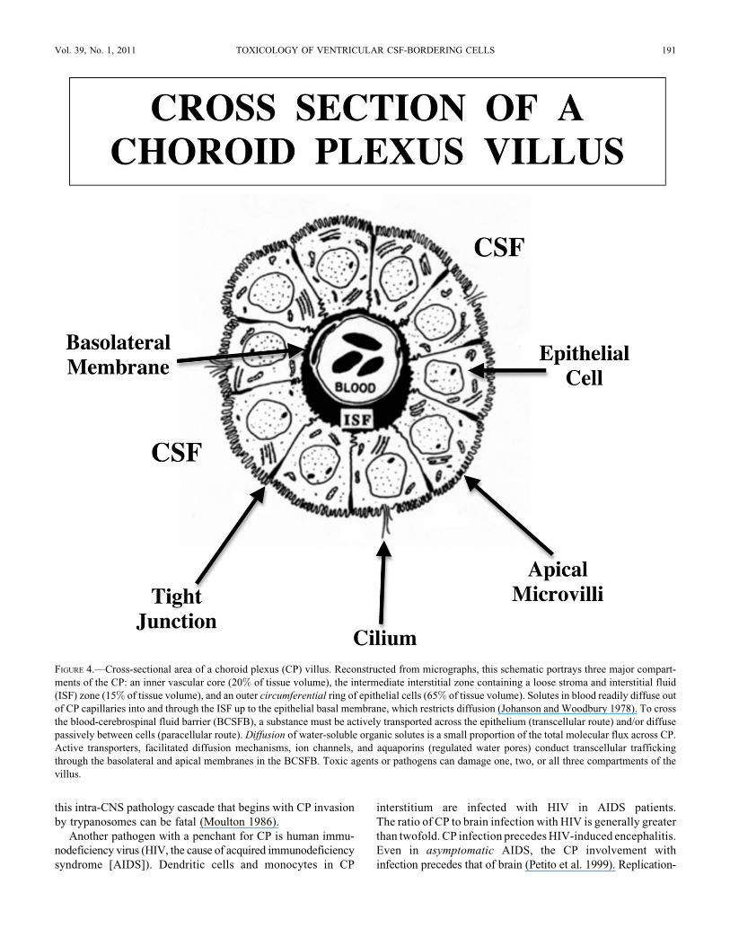
FIGURE 4.—Cross-sectional area of a choroid plexus (CP) villus. Reconstructed from micrographs, this schematic portrays three major compart-
ments of the CP: an inner vascular core (20% of tissue volume), the intermediate interstitial zone containing a loose stroma and interstitial fluid
(ISF) zone (15% of tissue volume), and an outer circumferential ring of epithelial cells (65% of tissue volume). Solutes in blood readily diffuse out
of CP capillaries into and through the ISF up to the epithelial basal membrane, which restricts diffusion (Johanson and Woodbury 1978). To cross
the blood-cerebrospinal fluid barrier (BCSFB), a substance must be actively transported across the epithelium (transcellular route) and/or diffuse
passively between cells (paracellular route). Diffusion of water-soluble organic solutes is a small proportion of the total molecular flux across CP.
Active transporters, facilitated diffusion mechanisms, ion channels, and aquaporins (regulated water pores) conduct transcellular trafficking
through the basolateral and apical membranes in the BCSFB. Toxic agents or pathogens can damage one, two, or all three compartments of the
villus.
Vol. 39, No. 1, 2011 TOXICOLOGY OF VENTRICULAR CSF-BORDERING CELLS 191
competent HIV in CP is similar to, but not identical with, that in
brain (Burkala et al. 2005). Petito and colleagues (1999)
postulated that CP is the site for hematogenous spread and a
reservoir for productive HIV infection during clinical latency.
Collectively, these histopathologic observations on CP relative
to brain (regional and temporal) implicate CP-CSF nexus
participation in disseminating HIV into CNS. Moreover,
molecular analyses of viral DNA in CP (Burkala et al. 2005)
corroborate early participation of the BCSFB pathway for later
distribution to brain (putatively by the CSF nexus). Unfortu-
nately, CP may provide an environment promoting evolution
of drug-resistant HIV strains with CNS tropism (Chen, Wood,
and Petito 2000). Involvement of CP in initial stages of HIV
infection suggest pharmacological targeting of virus trapped
in CP interstitium (i.e., not yet having accessed CSF). Reme-
dial agents in blood would then not have to pass through the
restrictive BCSFB. Rather, the drugs would readily traverse
leaky capillaries to reach the HIV-laden immune cells in the
CP interstitial space.
Neuropathologic Principles for Analyzing Choroid
Plexus and Ependyma
Pathologists regard CP in the domain of neuropathology
rather than general pathology (Levine 1987). This is understandable
because CP, an invaginating fold of pia within the ventricles, is
physically inside the CNS. However, due to absent tight junc-
tion barriers between endothelial cells in the plexus capillary
network, the CP functions more like a renal-type secretory
organ rather than a neural center (Spector and Johanson
1989). Therefore, a useful pathophysiologic concept is the
comparison between CP epithelium and the renal proximal
tubules (PT), both of which share many functional and ultra-
structural similarities. However, CP acts like a reverse kidney
in that the primary choroidal function is fluid secretion (Johan-
son et al. 2008) rather than reabsorption. This reflects Naþ
pump localization apically in CP (Parmelee and Johanson
1989) but basolaterally in PT. Still, the similar structural adap-
tations and transporter protein expression, CP versus PT,
prompt comparisons of cross-reactions in the respective base-
ment membrane-epithelial systems (Johanson, Stopa, and
McMillan 2010). Many toxicologic effects on CP mimic dam-
age to PT or other nephron regions (Levine 1987).
Great permeability of the choroidal capillaries promotes
interendothelial penetration of macromolecules (arrow 1,
Figure 1). Consequently, the choroid ISF compartment (Q. R.
Smith, Pershing, and Johanson 1981), unlike brain interstitium,
is readily accessible to plasma proteins, cytokines, and immu-
noglobulins. This makes CP parenchyma more vulnerable to
systemic disease (Levine 1987) than is the BBB-protected
brain. Plasma-borne auto-antibodies and immune complexes
(June et al. 1979) wreak havoc with CP basement membrane
and epithelium (Peress, Roxburgh, and Gelfand 1981).
This weakens the BCSFB and places the brain at risk for
CSF-transmitted infections. Immune cell and leukocyte move-
ment through CP displays transfer dynamics different from
BBB due to capillary and barrier cell (epithelial vs. endothelial)
variations (Engelhardt and Sorokin 2009). Paracellular perme-
ability in BCSFB is greater than at BBB (Thomas and Segal
1998). Thus, distinctive properties of the CP explain the differ-
ential barrier penetration of pathogens, xenobiotics, and
immune cells from blood to CSF compared with their transfer
from blood to brain.
Regional differences in structure and function occur in CP
tissues of lateral, third, and fourth ventricles. Ventricular CP
variations include epithelial cell pH, Kþ/Naþ, organic anion
transport capability, water content, and blood flow (Harbut
and Johanson 1986; Murphy and Johanson 1990; Pappenhei-
mer, Heisey, and Jordan 1961; Szmydynger-Chodobska,
Chodobski, and Johanson 1994). Inflammatory responses also
vary in third ventricle CP versus the structure of the CP in
other regions (Levine, Saltzman, and Ginsberg 2008). Given
the regional differences in CP baseline physiology and struc-
ture, one expects variations in how a particular CP responds to
pathogens and toxic agents. Neuropathologic differences
across CP regions have implications for functional variations.
Similar principles apply to comparison of the heterogeneous
ependyma that display unique profiles of cilia and junctional
apparatus. Particular toxicologic effects in ventricular regions
can often be ascribed to peculiar features of each type of
CSF-bordering cell.
Surface epithelium is a term used interchangeably with
CSF-bordering cells. Normally, and especially in disease
states, there is sloughing of choroidal and ependymal cells into
CSF. Surface epithelium was found in 5–10% of human
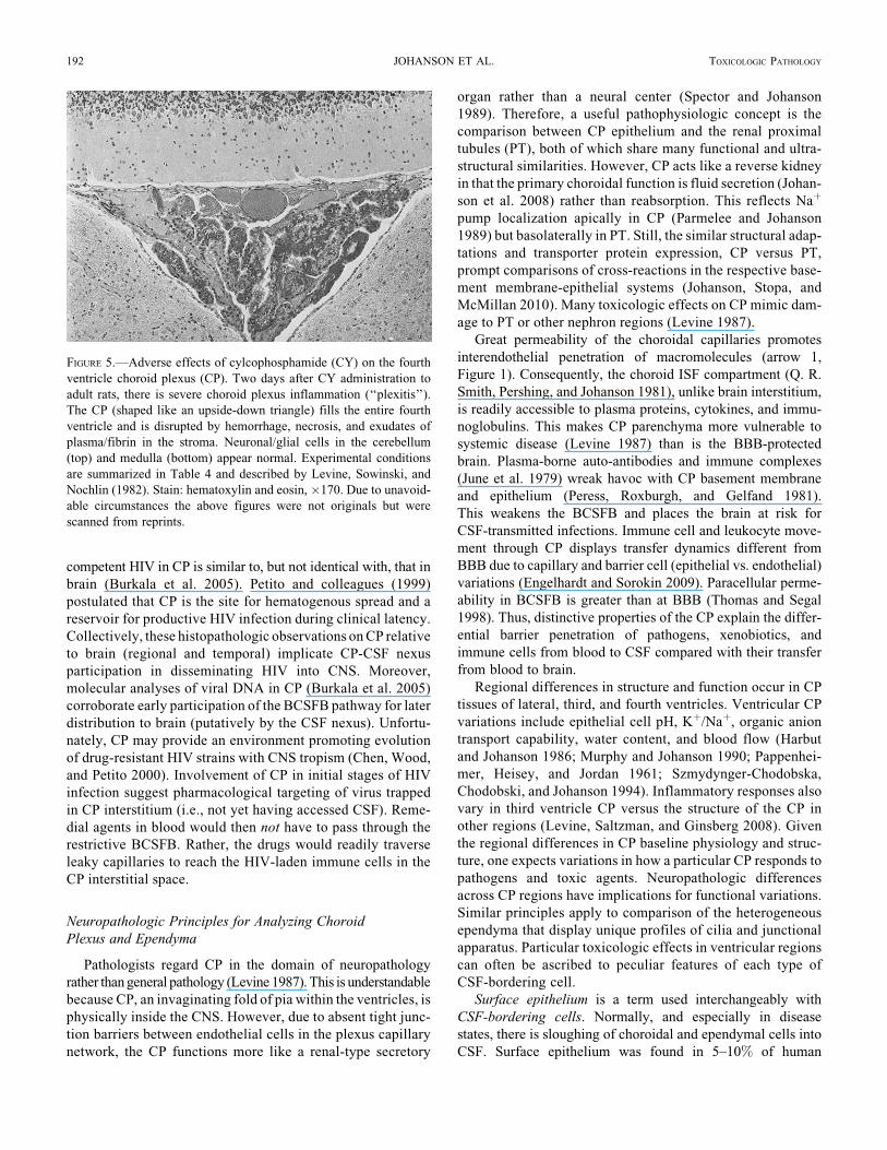
FIGURE 5.—Adverse effects of cylcophosphamide (CY) on the fourth
ventricle choroid plexus (CP). Two days after CY administration to
adult rats, there is severe choroid plexus inflammation (‘‘plexitis’’).
The CP (shaped like an upside-down triangle) fills the entire fourth
ventricle and is disrupted by hemorrhage, necrosis, and exudates of
plasma/fibrin in the stroma. Neuronal/glial cells in the cerebellum
(top) and medulla (bottom) appear normal. Experimental conditions
are summarized in Table 4 and described by Levine, Sowinski, and
Nochlin (1982). Stain: hematoxylin and eosin, �170. Due to unavoid-
able circumstances the above figures were not originals but were
scanned from reprints.
192 JOHANSON ET AL. TOXICOLOGIC PATHOLOGY
patients with CNS inflammation, neoplasms, compression, and
seizures (Wessmann et al. 2010). However, there were no sig-
nificant associations between specific disease types and the
incidence of surface epithelial cells in CSF. Further methodo-
logical refinements in analyzing surface epithelial cytology
in CSF may yield data to distinguish specific CNS disorders.
COMPLEMENTARY CIRCULATORY SYSTEMS IN THE BRAIN
Understanding differences in molecular trafficking across
BCSFB versus BBB, with patho-toxicologic implications,
depends on precise knowledge of circulatory inflow and vascu-
lar ultrastructures. CNS uses two circulations to convey
trophic/metabolic materials into and out of brain. Such a dual
arrangement of the vascular and CSF circulatory systems sets
up a complex steady state of nutrient influx and catabolite
efflux for neuronal networks (Johanson 2008). Blood supplies
the brain intrahemispherically from the inside via the cerebral
capillaries. Concurrently, from the outside of the hemispheres,
CSF provides supportive materials to neurons from fluid cav-
ities surrounding both the internal (ventricular) and external
(subarachnoid) surfaces of the brain. Both the vascular and
CSF circulations have secretory (influx) as well as reabsorptive
(efflux) components of solute distribution.
Why does the brain need CSF throughput in addition to the
primary vascular perfusion? Complex circulatory physiology,
linking CSF hydrodynamics to cerebral hemodynamics, relates
to the specialized anatomy and needs of the CNS. In a drainage
capacity, the CSF uniquely acts as a quasi-lymphatic system
(Johanson 2008). Moreover, by being a shock absorber, CSF
dampens vascular pulsations (Zou et al. 2008) that would oth-
erwise physically damage the fragile brain capillaries (Stopa
et al. 2008). Third, CSF is proximate to periventricular neuro-
genic niches. Consequently, CSF constituents support stem
cells and neurons via specialized choroidal secretions in fetal
life (Redzic et al. 2005) when brain capillary density is low.
Later in old age when cortical microvessel functions dwindle
(Miller et al. 2008; Silverberg et al. 2010), CP secretions may
become important to support cerebral angiogenesis as well as
neurogenesis. Working in tandem, the CSF and blood provide
complementary circulatory support to maintain cerebral meta-
bolism and structural integrity.
Parallel arterial input of materials to CNS is mediated by
two systems that are foremost in transferring substances: the
BBB (microvascular endothelium) and BCSFB (choroidal
epithelium). Each major transport interface features a distinc-
tive array of ions/molecules exchanged between blood and
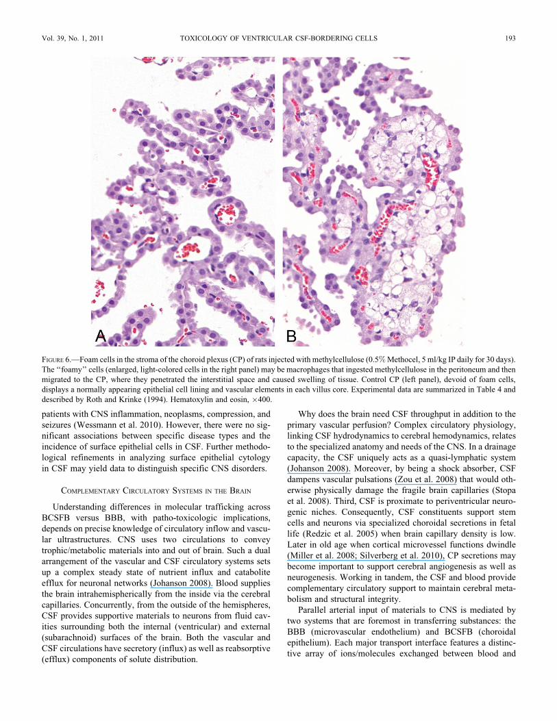
FIGURE 6.—Foam cells in the stroma of the choroid plexus (CP) of rats injected with methylcellulose (0.5% Methocel, 5 ml/kg IP daily for 30 days).
The ‘‘foamy’’ cells (enlarged, light-colored cells in the right panel) may be macrophages that ingested methylcellulose in the peritoneum and then
migrated to the CP, where they penetrated the interstitial space and caused swelling of tissue. Control CP (left panel), devoid of foam cells,
displays a normally appearing epithelial cell lining and vascular elements in each villus core. Experimental data are summarized in Table 4 and
described by Roth and Krinke (1994). Hematoxylin and eosin, �400.
Vol. 39, No. 1, 2011 TOXICOLOGY OF VENTRICULAR CSF-BORDERING CELLS 193

CNS regions (Johanson et al. 2008). A prominent feature of CP
is its great secretory abilities. In this case, the nexus mediates
material flow from the CP origin to putative targets in CNS
(Johanson, Miller, et al. 2010). Compartments of the CSF
nexus, diagrammed in Figure 1, are analyzed in the following
in the sequence of the physiological flow.
DISTRIBUTIONAL NEXUS: CHOROID PLEXUS-CSF-EPENDYMA-BRAIN
Substances secreted by CP tissues into the ventricles tra-
verse a series of boundaries and compartments to reach poten-
tial downstream receptors at neuronal and glial membranes.
For perspective on the CSF-brain interior, some distinguishing
features of the tissue compartments and systemic properties of
the nexus are addressed in the following. Fine histological fea-
tures of epithelial cells and organelles are presented further
along in the review.
Choroid Plexus as a Mini-Organ
CP is a secretory engine that actively synthesizes and trans-
ports a plethora of inorganic and organic solutes destined for
brain (Johanson 2008). Collectively, the plexus tissues are a
small fraction (*0.002) of total brain mass. Industrious activ-
ity of the epithelium, however, necessitates a choroidal blood
flow rate that is 5- to 10-fold greater than mean cerebral blood
flow (CBF) (Kadel, Heistad, and Faraci 1990; Szmydynger-
Chodobska, Chodobski, and Johanson 1994). Brisk vascular
perfusion of CP supports the energy and substrate demands
of a high degree of epithelial metabolism. As the main genera-
tor of CSF, the CP epithelia in the four ventricles transfer most
(up to 75%) of the water molecules diffusing from plasma into
CNS. Prolific water movement through CP epithelial aquaporin
1 channels (Oshio et al. 2003) into the ventricles produces the
CSF medium to convey solutes into and out of brain. Continual
fluid production by CP is the driving force behind steady CSF
streaming along the neuraxis.
Ventricular Cerebrospinal Fluid
Understanding how the CP epithelial and ependymal workings
impact the brain is predicated on knowledge of CSF dynamics.
Cells that border the ventricular system affect, and are affected
by, the CSF volume, flow, pressure, and composition. Dynamic
interplay between CP and ependyma, with CSF as the intermedi-
ary, reveals the multiple homeostatic actions of cells lining the
brain’s internal surface. Upon disruption by pathogens and toxi-
cants, the consequent CSF dyshomeostasis impairs brain metabo-
lism. This is presumably due to compromised transport and barrier
mechanisms. Salient features of CSF as a circulating medium
are discussed in the following relative to toxico-pathology.
Regular turnover of CSF in the ventricular system allows
optimal neuronal functioning. Human CSF is renewed three
to four times daily (Silverberg et al. 2002). Flow of CSF is from
lateral ventricles to the third and then through the narrow
mesencephalic aqueduct into the fourth ventricle. CSF
advances toward drainage sites (Grzybowski et al. 2006) by
bulk flow (arrows 4 and 9, Figure 1) driven by a hydrostatic
pressure gradient between CSF and venous blood (Johnston,
Boulton, and Flessner 2000; Pollay 2010). CSF bulk flow dis-
tributes endogenous materials widely throughout the CNS. The
term 3rd circulation has been coined for CSF flow in regard to
its major role in central fluid dynamics. Several minutes after
intracerebroventricular (ICV) injection, radiolabeled test tra-
cers are swept by CSF convection to regions distant from lat-
eral ventricles (Ghersi-Egea et al. 1996). Substances both
beneficial (vitamins) and harmful (cytokines) rapidly distribute
throughout the CNS after gaining access to ventricles and
downstream (subarachnoid) regions (Figure 1).
Lacking true lymphatic capillaries for drainage, the CNS
needs the CSF to continually flow in order to excrete poten-
tially harmful cerebral catabolites and to remove peptides that
leak into the brain. Concentration gradients, from brain ISF to
ventricular CSF, promote net diffusion of cerebral metabolites
and excess proteins into the ventricles (arrow 8, Figure 1). Such
CSF sink action (Parandoosh and Johanson 1982) rids the brain
of metabolic products and toxic agents/pathogens that access
CNS. Overwhelming the clearance or excretory capacity of
CSF by diseases/toxicants may injure the brain and protective
CP/ependymal cells.
Ependyma/Subependymal Regions
Ependymal and periventricular regions quickly take up toxic
as well as trophic substances transported into CSF by CP
FIGURE 7.—Vacuole formation in the choroid plexus (CP) epithelium
of rats treated with a tertiary amine. The cytoplasm of most epithelial
cells is filled with vacuoles that give the tissue the look of hydropic
pallor. This intense vacuolar response was typical of rats injected with
piperamide (500 or 750 mg/kg SC) and then necropsied one day later.
Vacuoles displace the nuclei to a basal position. Ependyma and the
adjacent periventricular brain appear normal (the slight separation
between the ependyma and the brain tissue is an artifact). Congested
CP fills the lateral ventricle. Experimental data are summarized in
Table 4 and interpreted by Levine (1994). Hematoxylin and eosin,
�450. Due to unavoidable circumstances the above figures were not
originals but were scanned from reprints.
194 JOHANSON ET AL. TOXICOLOGIC PATHOLOGY
(arrows 1–5, Figure 1). The permeable CSF-brain interface in
the lateral ventricles permits extensive, dynamic diffusion of
endogenous/xenobiotic agents into (arrow 5, Figure 1) and out
of (arrow 8, Figure 1) the brain. Ependymal destruction by tox-
icants and diseases further distorts the delicate biochemical
balance between CSF and ISF (Johanson 2008). This imbal-
ance neurochemically destabilizes the periventricular regions.
That a steady supply of micronutrients and trophic factors is
critical for ependymal well-being is exemplified by the finding
that malnutrition leads to deformation and metabolic disruption
of ependyma, at least early in development (S. P. Sharma and
Manocha 1977).
Brain Regions Adjacent to Ventricular CSF
Major neuronal networks (cholinergic, serotonergic, etc.) lie
in subependymal, neurogenic regions proximate to CSF
(Miyan, Nabiyouni, and Zendah 2003). Subventricular neurons
are accessible to endogenous and exogenous materials that dif-
fuse from CSF across the ependyma (Figure 1). Modulated
regions include the hippocampus (engaged in memory), the
SVZ and dentate gyrus (neurogenesis), hypothalamus (endo-
crine and autonomic regulation), periaqueductal gray (pain reg-
ulation), and the pons-medulla (respiratory and cardiovascular
control centers). These homeostatically sensitive areas exhibit
compensatory responses to alterations in CSF osmolality, [Kþ],
[Naþ], [Cl�], [Caþþ], pCO2, pO2, and pH. Therefore,
periventricular regions need to be carefully protected against
debilitation by toxic/pathogenic insults from foreign agents in
ventricular fluid.
CHOROID PLEXUS-EPENDYMA DISRUPTION: IMPLICATIONS FOR BRAIN
Brain health relies upon sound barrier/homeostatic mechan-
isms in CP and cerebral capillaries for protection against
blood-borne toxicants. Impermeable tight junctions (Brightman
and Reese 1969) and reabsorptive solute transporters at
BCSFB interfaces (Spector and Johanson 2010b) normally
protect the CNS against attack from foreign agents (Zheng
2001). Advanced aging, infectious agents, and toxic materials,
especially with chronic exposure, can all undermine the integ-
rity of the transport interfaces (Behl et al. 2009; Shi and Zheng
2007). Compromised barriers render the CNS vulnerable to
injury. The BCSFB in CP, as a regulated gateway for molecular
and cellular traffic into the brain (Johanson 2008), will be high-
lighted in the following.
Weakening of BCSFB augments penetration of harmful
plasma-borne materials into CSF and ultimately into the brain.
Collapse of CP-CSF results from many stresses on CNS:
arterial hypertension (Murphy and Johanson 1985), transient
forebrain ischemia (Palm et al. 1995), hyperthermia (H. S.
Sharma, Duncan, and Johanson 2006), traumatic brain injury
(H. S. Sharma, Zimmermann-Meinzingen, and Johanson
2010; Szmydynger-Chodobska et al. 2009), and advanced
aging (Preston 2001). Upon BCSFB damage in pathophysiologic
states, the increased paracellular permeability allows acces-
sion of plasma proteins and other markers to CSF. Commonly,
the elevated protein concentration in CSF leads to ventriculo-
megaly and periventricular edema (H. S. Sharma and Johanson
2007). As a consequence, cognitive/behavioral abilities in
animals are compromised when CSF-bordering hippocampal
and hypothalamic regions are injured by physical stressors
(H. S. Sharma, Duncan, and Johanson 2006). However, there
are relatively few systematic analyses of agent stress on the
CP-CSF-ependyma-SVZ-brain nexus (Figure 1).
Normally the CNS transport interfaces ward off mild to
moderate threats by invading bacteria, viruses, and xenobiotics.
Vigorous challenges by virulent pathogens and toxic drugs
(Levine 1987), though, reduce the homeostatic reserve of trans-
port interfaces. This leads to damage of CSF-contacting neural
regions. It is not feasible to predict exactly the untoward
responses by CP to potent noxious materials. A particular tox-
icant can interfere with tight junctions or with the nutritional/
trophic transporters at the BCSFB (Spector and Johanson
2010b) that vectorially direct solutes to neuronal targets via
CSF. Extreme interference with CP function causes CSF dys-
homeostasis that harms brain.
VENTRICULAR CSF-BORDERING CELLS: HISTOLOGY AND
PHYSIOLOGY
Before reviewing effects of organic agents on CSF-mediated
distribution, we describe normal ultrastructure and function
of ventricle-contacting cells. Three types of cells demarcate
ventricular CSF: choroid epithelial, ependymal, and circum-
ventricular (Table 1). Within these major groupings are
subpopulations of the parenchymal CP, ependyma, and CVOs.
Cellular heterogeneity and phenotype importantly bear on eva-
luations of specific endocrine, pharmacologic and toxicologic
responses. Cells contacting the CSF are part of a dual classifi-
cation of neuroepithelia: those suspended in CSF (CP) or those
comprising the ventricular wall (ependyma and CVOs).
Choroid Plexus Epithelial Cells
Anatomically, the base of the choroid epithelial fronds is
anchored to the brain at fixed points in the lateral, third, and
fourth ventricles. Ramifying outward from where the stalk
attaches to the ventricular wall, the tufts of CP are suspended
within CSF. Physically this arrangement provides enormous
epithelial surface area for translocation of nutrients and drugs
between plasma and CSF. However, this same large transport
area is potentially available for untoward leakage of plasma
proteins and harmful agents into CSF. The basic parenchymal
element in CP is cuboidal epithelium. An ultrastructural image
of a typical CP epithelial cell is portrayed in Figure 2. Large
surface areas promote extensive transport across the basolateral
(plasma-facing) and apical (CSF-facing) membranes. Because
the BCSFB is equipped for heavy-duty transport, severe patho-
logic damage to CP markedly distorts CSF homeostasis and
ventricular configuration.
As a multipurpose organ, the CP carries out diverse tasks:
secretory, synthetic, and reabsorptive (Table 2). Best known
among the secretory functions is CSF formation. CSF forms
Vol. 39, No. 1, 2011 TOXICOLOGY OF VENTRICULAR CSF-BORDERING CELLS 195
at the uniform rate of *0.4 ml/min/g CP in mammals. To initi-
ate CSF formation, the basolateral membrane takes up Naþ,
Kþ, Cl�, and HCO3� via several ion co-transporters and
exchangers (Johanson et al. 2008; P. D. Brown et al. 2004).
Ions and water taken up from the interstitium by epithelial
basolateral membrane (arrow 2, Figure 1) then move through
cytoplasm to the opposite (apical) side. There the apical mem-
brane extrusion mechanisms (arrow 3, Figure 1) include ion
channels, aquaporin 1, and co-transporters. Apical membrane
Naþ pumping (Q. R. Smith and Johanson 1980), by setting
up the appropriate transmembrane ion gradients, actively pro-
pels CSF production.
Drugs that induce renal natriuresis or diuresis consistently
reduce CSF turnover into the ventricles. CSF formation is
inhibited 10–30% by sympathetic, cholinergic, and serotoner-
gic neurotransmitters (Nilsson, Lindvall-Axelsson, and
Owman 1992). Neuropeptides such as arginine vasopressin,
atrial natriuretic peptide, and angiotensin II also slow down
CSF production by 15–20% (Johanson et al. 2008). Upon
secretion, the nascent CSF mixes with brain extracellular fluid
as it flows down the neuraxis. CSF flow decreases in aging.
In neurodegenerative disease (Silverberg et al. 2001) or drug
poisonings, the CSF turnover rate (i.e., formation rate / volume
can be cut in half. Adverse consequences include reduced
clearance, and hence accumulation, of proteins and cerebral
metabolites. ISF retention of uncleared waste products impairs
brain metabolism and behavior. Normally the CSF with its
debris is efficiently reabsorbed into lymph and/or venous blood
at multiple sites in arachnoid tissue (Grzybowski et al. 2006;
Johnston et al. 2004).
In addition to forming CSF, the CP homeostatically
protects the brain (Table 2). Acting like a kidney (Spector and
Johanson 1989), the CP helps to keep the chemical composi-
tion of CNS extracellular fluid in a stable state. In true kidney-
like fashion, the CP actively sets the ionic composition, pH,
and osmolality of extracellular CSF. Low permeability
imposed by tight junctions at the BCSFB allows ion and
molecular gradients to be established secondary to active
transport. For example, Caþþ and Kþ in CSF are held at
concentrations lower than plasma, while Mgþþ and Cl� are
kept at higher levels. CSF titers of numerous growth factors,
fluid-regulating peptides, and proteins are maintained at
0.01 to 0.001 of their respective concentrations in plasma.
Regulated CSF ions benefit the neurons that require finely
maintained extracellular concentrations.
Second, a plethora of reabsorptive solute transporters in the
apical membrane actively removes potentially toxic organic
acids and peptides from CSF (arrow 10, Figure 1). Organic
anion transporting polypeptides (OATPs) are part of an
increasingly appreciated gene superfamily (Spector and Johan-
son 2010b). OATP was initially immunolocalized to the CP
apical membrane (Angeletti et al. 1997), a strategic location
for clearing metabolites from CSF (Ohtsuki et al. 2003).
In BCSFB, the OATPs mediate reabsorptive (excretory) trans-
port of a wide spectrum of amphipathic organic solutes in CSF:
steroid conjugates, neurotransmitter anion catabolites (homo-
vanillic acid, a major metabolite of catecholamine neurotransmit-
ters), bile salts, anionic oligopeptides, drugs, and xenobiotics
(Hagenbuch and Meier 2003). CSF levels of certain drugs are kept
at subtherapeutic low levels by OATP and the P-glycoprotein
transporter, the potential roles of which in CP need evaluation for
therapeutic agents such as suramin and eflornithine, agents that
are used against human African trypanosomiasis (Sanderson
et al. 2008; Sanderson, Khan, and Thomas 2007).
Another CP reabsorptive transporter of organic solutes, with
a wide spectrum of substrate affinity, is the family of low
density lipophilic receptor-related proteins (LRP). Located
apically, the LRP-1 and LRP-2 clearance transporters keep
peptides and peptide fragments in the CSF from building to
toxic levels (arrow 10, Figure 1). As a promiscuous transporter,
LRP-1 can clear from CSF/brain as many as forty types of
molecules including APOE4, amyloid precursor protein, and
amyloid oligopeptide fragments (Crossgrove, Li, and Zheng
2005). LRP-2 (or megalin) has also been implicated in remov-
ing amyloid from CSF (Alvira-Botero and Carro Forthcoming).
Other apically located organic solute transporters, including
PEPT2 (proton-coupled oligopeptide transporter 2), prevent
CSF accumulation of compounds (e.g., 5-aminolevulinic acid
[ALA], a heme precursor) to toxic levels (Hu et al. 2007;
D. E. Smith, Johanson, and Keep 2004). Reabsorptive transport
from CSF is essential because ALA leaks into CSF from blood
and needs to be quickly cleared from the ventricles (arrow 10,
Figure 1) to avoid toxicity (Terr and Weiner 1983).
Third, hepatic-like metabolism in BCSFB epithelium
minimizes toxic drug accumulation in the CSF and CNS. This
liver-type role affords brain an extra line of defense against
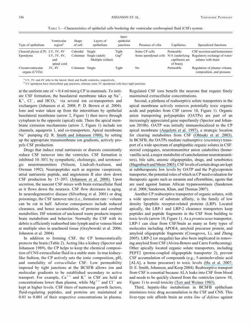
TABLE 1.—Characteristics of epithelial cells bordering the ventricular cerebrospinal fluid (CSF) system.
Type of epithelium
Ventricular
regiona
Shape
of cell
Layers of
epithelium
Inter-
epithelial
junctions Presence of cilia Capillaries Specialized functions
Choroid plexus (CP) LV, 3V, 4V Cuboidal Single Tight Some CP cells Permeable CSF secretion and homeostasis
Ependyma LV, 3V, 4V,
and
spinal cord
Columnar Single (adult)
Multiple (infant)
Gapb Some ependymal cells N/A (underlying
capillaries are
of brain)
Regulatory exchange of water/
solutes with brain
Circumventricular
organs (CVOs)
3V, 4V Columnar Single Tight No Permeable Regulation of plasma volume,
composition, and pressure
a LV, 3V, and 4V refer to the lateral, third, and fourth ventricles, respectively.b LV ependyma have intercellular gap junctions, whereas some 3V ependymal cells have tight junctions.
196 JOHANSON ET AL. TOXICOLOGIC PATHOLOGY

toxic drugs and compounds (Strazielle, Khuth, and Ghersi-
Egea 2004). Xenobiotic molecules induce drug-metabolizing
enzymes in liver and CP. In vitro, the expression of
UDP-glucuronosyltransferase (UGT1A6 isoform) is upregu-
lated in rat CP epithelium when tissue is challenged with
3-methycholanthrene (3-MC) or paraquat (Gradinaru et al.
2009), leading to a two- to threefold increase in the choroidal
1-naphthol glucuronidation activity. UGT1A6 mRNA expres-
sion (polymerase chain reaction [PCR] analysis) was also
augmented more than twofold after incubation with these two
drugs. Drug-metabolizing enzymes in the BCSFB transform
xenobiotics that might harm the brain if left unmetabolized.
Fourth, an immune-type function is subserved by CP
through the activity of antigen-presenting cells in the epithelial
lining (Nathanson and Chun 1989), thereby permitting immune
surveillance of CSF. Normally just a few immune cells migrate
from plasma into ventricles to monitor CSF immunologic
status. Under pathologic or toxicologic stress, however, the
upregulation of CP chemokines, integrins, selectins, and matrix
metalloproteinases renders the BCSFB more penetrable
(Prendergast and Anderton 2009). Consequently, many inflam-
matory cells invade CSF and provoke CNS autoimmune
disease. Since CP epithelium engages in diverse tasks to defend
and cleanse CSF, the BCSFB itself needs protection against
toxicant and foreign molecules (Johanson, Silverberg, et al.
2005). High levels of antioxidant glutathione in CP (Cooper
and Kristal 1997) comprise a biochemical defense against
oxidant molecules transported or metabolized by epithelial
cells. Overall, the CP in healthy young adult humans has
sufficient barrier, metabolic, enzymatic, and reabsorptive
capabilities to keep CSF composition sufficiently pure for
optimal neuronal performance.
Finally, over and above its homeostatic role in stabilizing
neurochemical composition and immune health of CSF-CNS,
the CP is the chief supply route for transferring plasma-borne
materials into the ventricles. As much as 70–80% of water, ion,
vitamin, and peptide transfer into CNS occurs across choroidal
epithelium (arrows 2 and 3, Figure 1) (Johanson et al. 2008).
Vitamins B (folate) and C are actively transported into CNS
preponderantly via the blood-CSF interface (Spector and
Johanson 2006). There is also significant neuroendocrine pep-
tide distribution mediated by hormonal transport mechanisms
at the BCSFB interface (Kozlowski 1986). Prolactin, insulin-
like growth factor (IGF)-1, and leptin use CP as a hormonal sig-
nal relay station in being transported from plasma to ventricles
(Dietrich et al. 2008; Redzic et al. 2005). Then these peptides
are convected by CSF bulk flow to hypothalamic regions such
as the arcuate nuclei (Rodriguez, Blazquez, and Guerra 2010).
In this manner, the CP-CSF participates in hormonal signal
transfer within neuroendocrine feedback loops for modulating
feeding/satiety and reproductive behaviors.
Translocation of plasma ions, micronutrients, and water into
CNS is mainly via CP. Serious injury of BCSFB by microbes or
xenobiotics alters brain homeostasis. When the CSF milieu is
substantially changed, brain regions close to ventricles suffer
damage to neurogenesis and cognition. Many pathogens, toxi-
cants, and neurological disorders damage the plexus. Following
stroke and trauma, not only is CSF delivery of trophic factors to
neurons compromised, but also the CSF removal of brain cat-
abolites is diminished (Johanson et al. 2000). Molecules in
TABLE 2.—Homeostatic, secretory, and metabolic functions of choroid plexus (CP) epithelial cells.
Functional aspects
Mechanisms for transport, catalysis,
or secretion
Physiological effects on, or mediation by,
cerebrospinal fluid (CSF) Significance for brain well-being
Kidney-like role (fluid balance) Naþ-Hþ, Cl�-HCO3�, and NaHCO3
transporters (inorganic ions)
Transfers Naþ, Kþ, Cl�, Hþ, or HCO3� to
maintain ion concentrations in the
ventricles
CSF turnover and flow help to buffer
[Hþ], [Kþ], and osmolality in brain
interstitial fluid (ISF)
Organic solute reabsorption
(clearance)
Organic anion transporting
polypeptide (OATP) and
low-density lipophilic
receptor-related protein
(LRP-1)
Removal of CNS catabolites (anions and
peptides) by CP apical reabsorptive trans-
port and CSF bulk flow helps to purify
brain extracellular fluid content
Cleansing effect (by CSF sink action) is
vital for optimal functioning of the
neuronal networks and glia
Liver-like role (for metabolism
of drugs)
Glutathione S-transferase and
NADPH-cyto-chrome P450
reductase in epithelium
By serving as an ‘‘enzymatic barrier,’’
hepatic-like enzymes in CP can metabolize
drugs and prevent their entry into CSF
Important to prevent neurotoxic drugs
from entering CSF and to metabolize
harmful drugs that do penetrate the
blood-brain barrier (BBB)
Immune-related functions Immune cells, such as epiplexus cells
(i.e., macrophage-like elements,
also known as Kolmer cells)
Antigen-presenting cells at the blood-CSF
epithelial interface help regulate the
immune capacity of CSF
Immunosurveillance of CSF by CP works
in concert with BBB activity to
maintain a healthy immune state for
the central nervous system
Supplier of micronutrients MTHF (methylene-tetrahydrofolate)
and SVCT2 (Naþ-activated
ascorbic acid [vitamin C]
transporter-2)
Active transport of folic acid and vitamin C
to maintain CSF concentrations that are
higher than in plasma
Folate and ascorbate in CSF are needed
by brain, but neural tissue does not
receive them by way of cerebral
capillaries
Neuro-endocrine regulation Receptor-mediated transport of
prolactin, insulin-like growth
factor (IGF)-1, and leptin
Peptides that are transported from plasma to
CSF are carried by bulk CSF flow to vari-
ous hypothalamic nuclei
Receptors in the arcuate nuclei bind
specific peptides to integrate
metabolic and reproductive
phenomena
Vol. 39, No. 1, 2011 TOXICOLOGY OF VENTRICULAR CSF-BORDERING CELLS 197
ventricular CSF, even large proteins, have access by diffusion
(down concentration gradients) to periventricular brain regions
(arrow 5, Figure 1). In the reverse direction, catabolically gen-
erated macromolecules in the hemispheres diffuse from inter-
stitium into the ependyma into the ventricular CSF (arrow 8,
Figure 1). Proper steady-state partitioning of molecules
between CP-CSF and brain requires intact ependyma.
Ependymal Cells with Gap Junctions Versus Tight
Junctions
Structurally and functionally, the ependyma lining the cere-
bral ventricles and central canal of the spinal cord is heteroge-
neous. Ontogenetically, there is progression of ependyma as a
multiple layer in the fetus to a single layer in adults. Various
cell types comprise the fetal ependymal wall (Davson and
Segal 1996). These include the tanycyte and ependymal
astrocyte as well as the true ependymal cells that persist into
adulthood. All three cell types have cilia and microvilli at their
CSF-facing apical surfaces. However, structural differentiation
occurs at the basal (brain-facing) sides. Tanycytes and radial
glia send long dense fibers to the CSF. The tanycyte’s other end
projects basally to termini in certain hypothalamic nuclei,
which receive via axonal flow those neuropeptides (‘‘endocrine
signals’’) taken up from the CSF (Rodriguez et al. 2005). The
ependymal astrocyte is a columnar cell with branching periph-
eral processes. After birth, both tanycytes and ependymal astro-
cytes dwindle in number so that the ventricular lining in adults
is mainly ependyma proper. Even in adulthood the ependyma
depend on growth factors such as vascular endothelial growth
factor (VEGF) and transforming growth factor beta (TGFb),
both secreted by CP, to maintain stable cellular structure and
fluid balance with surrounding ISF (Maharaj et al. 2008).
Diminished growth factor support for endothelial and ependy-
mal cells leads to vascular permeability changes and periventri-
cular edema.
Ependymal cells proper are not all equal. Heterogeneity
among ependyma consists of cells having cilia or not, and in
possessing leaky gap junctions or the tighter zonulae occlu-
dentes (not as prevalent). Most lateral ventricle ependymal
cells have intercellular gap junctions just under the apical sur-
face. Gap junctions are highly permeable. Large proteins, the
size of ferritin (mw *445,000), readily diffuse through the
paracellular space to cross the ependyma and thus easily move
between CSF and brain. Accordingly, the ependymal interface
between ventricular fluid and underlying brain is not regarded
as a CSF-brain barrier. Being highly permeable, the ependyma
is thus unlike the restrictive BCSFB and BBB, which impede
paracellular diffusion.
Although the lateral ventricle ependymal system is homoge-
neous, the ependyma lining the third and fourth ventricles are
heterogeneous. In specific regions of third ventricle (3V), there
are tight junctions between specialized ependymal cells. Given
the proximity of 3V to hypothalamus, the structurally modified
ependyma here reflects specialized (regulated) functional rela-
tionships between 3V CSF and the adjacent paraventricular
nucleus. In regions such as the arcuate nucleus (Rodriguez,
Blazquez, and Guerra 2010), the nearby CSF has ‘‘access’’ to
hypothalamus in that hormones exchange via diffusion across
the ependymal interface. Brain midline CVOs (Gross 1992)
in 3V and 4V are especially heterogeneous.
More than being a structural boundary between ventricular
CSF and brain, the ependyma is a dynamic interface mediating
the movement of cilia, CSF, and migrating neuroblasts. On the
ependymal apical surface is a network of cilia that beat in coor-
dination to facilitate CSF circulation. Each cilium is a subcel-
lular organelle emanating from the cell’s interior. Planar cell
polarity signaling may be controlled by cilia (Fischer and
Pontoglio 2009). Cadherin genes (Celsr2 and Celsr3) regulate
planar cell polarity. Celsr genes that are mutated or absent
(knocked out) compromise the development and planar organi-
zation of ependymal cilia. This leads to defective CSF
dynamics and hydrocephalus. Many ciliopathies render CP and
ependyma dysfunctional, thereby distorting the volume and
composition of CSF secretion (Banizs et al. 2005). Migration
of neuroblasts from the lateral ventricle wall to the olfactory
lobe relies upon ciliary-guided flow to forebrain sites for inser-
tion into circuits as interneurons (Sawamoto et al. 2006).
Proper development and operation of the brain depend on nor-
mally functioning cilia in CP and ependyma.
Currently surging research on neurogenesis in the SVZ is
generating additional insights on ependyma. As a delicate
interstitial microenvironment for stem cells (Spector and
Johanson 2007a), the neurogenic niches of dentate gyrus and
SVZ require compositional stability. This assures finely regu-
lated gliogenesis and neurogenesis, even in adulthood. Repair
mechanisms are essential to the integrity of the ependymal
lining disrupted by diseases (hydrocephalus) and disorders
(trauma). Ependyma are also damaged in advanced aging.
One restorative mechanism is SVZ-mediated repair of the
ependymal wall by astrocyte insertion in regions where
ependyma are detached. When new astrocytes incorporate
in gaps between ependymal cells, they take on antigenic and
morphologic characteristics of neighboring ependymal cells
(Luo et al. 2008). This constitutes evidence for non-neuronal
repair as well as the more established neuronal reconstitution
carried out by SVZ elements.
In addition to dynamic interaction between ependyma and
SVZ for repair mechanisms, there is homeostatic interplay
between CSF, ependyma, and the periventricular brain in fluid
balance. Even though the CSF-brain interface permits macro-
molecule diffusion between ependymal cells, implying unregu-
lated water and ion paracellular movements, there is still great
plasticity of expression of ion and water channels in ependyma.
Responding to elevated CSF pressure or a change in CSF Naþ
concentration, the ependyma upregulates aquaporin 4 water
pores and epithelial Naþ channels (H. W. Wang et al. 2010).
This may reflect regulation of ependymal cells per se, or con-
trolled fluid transfer into brain across ependymal regions with
tight junctions. Whatever the explanation for induced channel
expression, the findings point to sensitivity of the ependymal
interface to CSF changes.
198 JOHANSON ET AL. TOXICOLOGIC PATHOLOGY
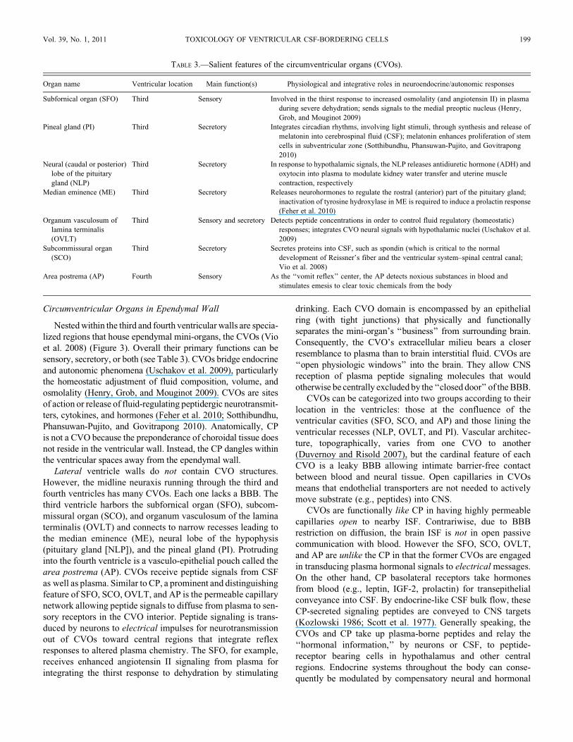
Circumventricular Organs in Ependymal Wall
Nested within the third and fourth ventricular walls are specia-
lized regions that house ependymal mini-organs, the CVOs (Vio
et al. 2008) (Figure 3). Overall their primary functions can be
sensory, secretory, or both (see Table 3). CVOs bridge endocrine
and autonomic phenomena (Uschakov et al. 2009), particularly
the homeostatic adjustment of fluid composition, volume, and
osmolality (Henry, Grob, and Mouginot 2009). CVOs are sites
of action or release of fluid-regulating peptidergic neurotransmit-
ters, cytokines, and hormones (Feher et al. 2010; Sotthibundhu,
Phansuwan-Pujito, and Govitrapong 2010). Anatomically, CP
is not a CVO because the preponderance of choroidal tissue does
not reside in the ventricular wall. Instead, the CP dangles within
the ventricular spaces away from the ependymal wall.
Lateral ventricle walls do not contain CVO structures.
However, the midline neuraxis running through the third and
fourth ventricles has many CVOs. Each one lacks a BBB. The
third ventricle harbors the subfornical organ (SFO), subcom-
missural organ (SCO), and organum vasculosum of the lamina
terminalis (OVLT) and connects to narrow recesses leading to
the median eminence (ME), neural lobe of the hypophysis
(pituitary gland [NLP]), and the pineal gland (PI). Protruding
into the fourth ventricle is a vasculo-epithelial pouch called the
area postrema (AP). CVOs receive peptide signals from CSF
as well as plasma. Similar to CP, a prominent and distinguishing
feature of SFO, SCO, OVLT, and AP is the permeable capillary
network allowing peptide signals to diffuse from plasma to sen-
sory receptors in the CVO interior. Peptide signaling is trans-
duced by neurons to electrical impulses for neurotransmission
out of CVOs toward central regions that integrate reflex
responses to altered plasma chemistry. The SFO, for example,
receives enhanced angiotensin II signaling from plasma for
integrating the thirst response to dehydration by stimulating
drinking. Each CVO domain is encompassed by an epithelial
ring (with tight junctions) that physically and functionally
separates the mini-organ’s ‘‘business’’ from surrounding brain.
Consequently, the CVO’s extracellular milieu bears a closer
resemblance to plasma than to brain interstitial fluid. CVOs are
‘‘open physiologic windows’’ into the brain. They allow CNS
reception of plasma peptide signaling molecules that would
otherwise be centrally excluded by the ‘‘closed door’’ of the BBB.
CVOs can be categorized into two groups according to their
location in the ventricles: those at the confluence of the
ventricular cavities (SFO, SCO, and AP) and those lining the
ventricular recesses (NLP, OVLT, and PI). Vascular architec-
ture, topographically, varies from one CVO to another
(Duvernoy and Risold 2007), but the cardinal feature of each
CVO is a leaky BBB allowing intimate barrier-free contact
between blood and neural tissue. Open capillaries in CVOs
means that endothelial transporters are not needed to actively
move substrate (e.g., peptides) into CNS.
CVOs are functionally like CP in having highly permeable
capillaries open to nearby ISF. Contrariwise, due to BBB
restriction on diffusion, the brain ISF is not in open passive
communication with blood. However the SFO, SCO, OVLT,
and AP are unlike the CP in that the former CVOs are engaged
in transducing plasma hormonal signals to electrical messages.
On the other hand, CP basolateral receptors take hormones
from blood (e.g., leptin, IGF-2, prolactin) for transepithelial
conveyance into CSF. By endocrine-like CSF bulk flow, these
CP-secreted signaling peptides are conveyed to CNS targets
(Kozlowski 1986; Scott et al. 1977). Generally speaking, the
CVOs and CP take up plasma-borne peptides and relay the
‘‘hormonal information,’’ by neurons or CSF, to peptide-
receptor bearing cells in hypothalamus and other central
regions. Endocrine systems throughout the body can conse-
quently be modulated by compensatory neural and hormonal
TABLE 3.—Salient features of the circumventricular organs (CVOs).
Organ name Ventricular location Main function(s) Physiological and integrative roles in neuroendocrine/autonomic responses
Subfornical organ (SFO) Third Sensory Involved in the thirst response to increased osmolality (and angiotensin II) in plasma
during severe dehydration; sends signals to the medial preoptic nucleus (Henry,
Grob, and Mouginot 2009)
Pineal gland (PI) Third Secretory Integrates circadian rhythms, involving light stimuli, through synthesis and release of
melatonin into cerebrospinal fluid (CSF); melatonin enhances proliferation of stem
cells in subventricular zone (Sotthibundhu, Phansuwan-Pujito, and Govitrapong
2010)
Neural (caudal or posterior)
lobe of the pituitary
gland (NLP)
Third Secretory In response to hypothalamic signals, the NLP releases antidiuretic hormone (ADH) and
oxytocin into plasma to modulate kidney water transfer and uterine muscle
contraction, respectively
Median eminence (ME) Third Secretory Releases neurohormones to regulate the rostral (anterior) part of the pituitary gland;
inactivation of tyrosine hydroxylase in ME is required to induce a prolactin response
(Feher et al. 2010)
Organum vasculosum of
lamina terminalis
(OVLT)
Third Sensory and secretory Detects peptide concentrations in order to control fluid regulatory (homeostatic)
responses; integrates CVO neural signals with hypothalamic nuclei (Uschakov et al.
2009)
Subcommissural organ
(SCO)
Third Secretory Secretes proteins into CSF, such as spondin (which is critical to the normal
development of Reissner’s fiber and the ventricular system–spinal central canal;
Vio et al. 2008)
Area postrema (AP) Fourth Sensory As the ‘‘vomit reflex’’ center, the AP detects noxious substances in blood and
stimulates emesis to clear toxic chemicals from the body
Vol. 39, No. 1, 2011 TOXICOLOGY OF VENTRICULAR CSF-BORDERING CELLS 199

output from CNS. Such feedback loops, involving intermediate
transport/signaling steps via CP and CVOs, constitute homeo-
static machinery for restoring distorted body fluids to a stable,
specialized composition. Hence, toxic/pathological injuries to
CP, hypothalamus, and CVOs, including periventricular
lesions, affect not only the local brain environment but also the
activity of endocrine systems throughout the body.
PERIVENTRICULAR LESIONS AND THE CHOROID PLEXUS-CSF NEXUS
Periventricular lesions are commonly downstream sequelae
of the uncontrolled passage of substances through a compro-
mised BCSFB. Materials that are inappropriately transferred
or that leak across the CP include infectious agents, blood com-
ponents (cells and proteins), and tumor cells. Representative
lesions for each group exemplify types of damage inflicted
on the ventricular wall and underlying brain. For example, the
protozoan Toxoplasma gondii frequently infects the CNS of
immunocompromised AIDS patients. Acting as a staging plat-
form, CP receives and activates Toxoplasma, which in turn
engenders choroid plexus inflammation (Falangola and Petito
1993) that promotes CSF dissemination to periventricular sites
where infectious agent and leukocytes accumulate (Stahl and
Turek 1988). Ependyma and CP in all four ventricles become
diffusely necrotic (Bourgouin et al. 1992). Another infection
of CP-CSF is by ovine lentivirus, which invades periventricular
sites as part of a more generalized encephalomyelitis (Brodie,
Bickle, and DeMartini 1995); the CSF lentiviral markers reflect
CNS pathology. Various blood cells have greatly restricted
access to CSF except in pathophysiologic conditions associated
with autoimmune disease or hemorrhage into CP-ependymal
zones. Augmented leukocyte (T-cell) penetration across the
CP into CSF occurs in relapsing multiple sclerosis (MS) in
humans or experimental allergic encephalitis (EAE) models
of MS, leading to formation of hallmark periventricular lesions
and ependymal/subventricular destabilization. Consequently,
neurogenesis is inhibited by T cell release of granzyme B
(T. Wang 2010). Recent findings in an EAE model point to
T lymphocyte antigen specificity in defining the topography
of periventricular lesions (Berger et al. 1997). Moreover,
extensive erythrocyte permeation across BCSFB distorts
CSF composition and interferes with ependyma/SVZ
functions. Thus, in neonatal brain stressed by hypoxia/ische-
mia, the choroidal/intraventricular and severe subependymal
hemorrhaging perturb the interstitial fluid environment in
periventricular white matter (Bernert et al. 1988). Tumors
(e.g., high-grade B-cell lymphoma) may form directly in the
CP (Cecchi et al. 2008) and migrate across BCSFB to become
entrenched in the periventricular brain. Metastatic CNS
lymphoma, having entered CSF via the CP, can disseminate
to periventricular sites (Kobayashi et al. 2009). Primary intra-
cerebral malignant lymphomas, albeit rarely, fulminate in CP
and putatively escape the BCSFB to permeate subependymal
sites along the lateral, third, and fourth ventricular walls
(Shibata 1989). Specific pharmacologic targeting of CP/BCSFB
may be used to thwart penetration of cancer cells (as well
as viruses and potentially harmful leukocytes) into the
CSF-ependyma-SVZ nexus. Studies of toxicant-induced
disruption to the nexus, as described in the following, furnish
useful basic data for formulating prophylactic strategies
against infection or measures to counter toxicant exposure.
ORGANIC AGENTS USED IN CHOROIDO-EPENDYMAL TOXICOLOGY
STUDIES
Drugs from different classes have been tested for toxicity to
CSF-bordering cells and underlying the brain. Tertiary amines
were extensively analyzed for deleterious effects on BCSFB
and the CSF-brain interface. A series of compounds analyzed
includes molecules with one, two, or three tertiary amines
(Levine 1977). All contain a cyclic structure, either saturated
(piperazine and piperidine) or unsaturated (naphthyridine and
pyridine). Agents used in ventricular cell-CSF investigations,
including piperidine/piperazine derivatives and amoscanate,
are compiled in Table 4. Most of these drugs are anti-parasitic.
Their CSF toxicity is fairly consistent among mammals.
Organic agents listed in Table 4 were used experimentally to
inform on the extent and location of CP-ependyma injuries.
Adult rats were commonly used for in vivo experimentation.
A typical approach involves microscopy and tissue staining,
and time course analysis (days to weeks), to assess development
and reversibility of drug-induced lesions in CP, ependyma, and
brain.
VULNERABILITY OF THE CSF-BRAIN INTERIOR TO TOXICANTS AND
PATHOGENS
A gateway duty has been ascribed to BCSFB (Johanson
et al. 2008). Thus, a significant CNS port of entry in CP is
offered to plasma molecules, pathogens, and immune cells.
Ventricular CSF nourishes and protects brain by an array of
mechanisms controlled at the CP epithelial zone and ependymal
interface. Due to dissimilar structures and molecular expres-
sions of CP versus ependyma, various toxicants and pathogens
exert differential effects on CSF-bordering epithelium.
Upon penetration of a damaged BCSFB, a given substance
flows to the ependyma and regions adjacent to CSF (arrows
2–5, Figure 1). Injury to periventricular brain sites (arrow 6,
Figure 1), causing dyshomeostasis (Aksamit, Parnell, and
Johnson 1999), is often but not always curtailed by enzymatic
defense systems at CSF interfaces that degrade penetrating
toxicants. In this manner the Pi isoform of glutathione
S-transferase that is expressed in ependymal, arachnoid, and
pial cells enzymatically buffers the brain against toxicant
penetration from CSF (Carder et al. 1990).
Many toxicology studies of CNS have observed drug-induced
formation of vacuoles within various neural cell populations.
Regionality of induced vacuoles in brain has been a topic of inter-
est (Wells and Krinke 2008). Vacuolation has clinical importance
because impaired drug elimination from neurons and epithelial
cells, which promotes vacuole generation, may lead to toxicity
and even death. With cell swelling and organelle disruption, the
protective properties of epithelial cells of BCSFB may become
200 JOHANSON ET AL. TOXICOLOGIC PATHOLOGY

seriously compromised. Therefore, it is important to understand
the mechanisms of vacuole formation in CP and ependyma and
to prevent vacuole formation and proliferation or counter any
untoward effects pharmacologically.
Injuries to Choroid Plexus
Delineating the effects of toxic agents involves consider-
ation of CP compartmental anatomy. Each CP villus has an
inner vascular core consisting of a rich network of blood ves-
sels (Figure 4). Nearly 20% of CP mass is vasculature and
blood cells. These elements impart a red cast to the tissue
(Johanson, Reed, and Woodbury 1974). Lying in the intermedi-
ate zone of the CP is the interstitial space. It has a highly devel-
oped extracellular matrix that modulates the location and
functions of both vascular and epithelial cells. The interstitium
comprises about 15% of CP mass (Q. R. Smith, Pershing, and
Johanson 1981). Notably the interstitium of the CP, like that of
brain, lacks true lymphatic capillaries. On the outside perimeter
of the choroidal villi is a single layer of epithelium.
This epithelial ring interfaces with the interstitium on the basal
side and contacts the CSF via a lush microvillous surface at the
apical pole. Severe pathologic changes to the CP may include
damage to all three compartments: blood vessels, interstitium,
and epithelium. Certain drugs destroy one specific compart-
ment, or a single toxicant can perturb all three CP regions
(Levine 1987). It is also useful to identify drug combinations
that intensify damage to multiple compartments within CP.
Vascular impairment: Cyclophosphamide (CY) at high doses
injures the CP vasculature, especially in the fourth ventricle
TABLE 4.—Organic agents used for in vivo toxicology studies of choroid plexus (CP), ependyma, and periventricular brain.
Agent Dosage/route Species
Experimental
duration Main observations Investigators
Piperamide and other
tertiary aminesa
200-1200 mg/kg (SC)
200-1200 mg/kg (oral)
Rat (Lewis)
Male/female
1 day Hydropic degeneration (severe
vacuolation) of all CP; reversal
of CP pathology after 7 days
Levine (1977)
Piperazine and
piperidine
derivatives
200-1200 mg/kg (SC)
200-1200 mg/kg (oral)
Rat (Lewis) 1 day Swollen choroid epithelium, due to
cytoplasmic vacuoles; hydropic
degeneration also in kidney distal
tubule
Levine and Sowinski (1977)
Disobutamide
(piperidine
derivative)
30-250 mg/kg (oral)
45 mg/kg (oral)
90 mg/kg (oral)
Rat
Dog
Monkey
35 days Vacuolation induced in CP epithelium
correlated with drug uptake by CP,
and the amount of drug penetration
into the cerebrospinal fluid (CSF)
Koizumi et al. (1986)
Cyclophosphamide
(alkylating agent)
125-500 mg/kg (SC or IP) Rat (Lewis) 1 to 7 days Edema, hemorrhage, and leukocyte
infiltration of CP; progression to
necrosis of individual choroidal villi
Levine and Sowinski (1973)
Triamine
(pyridine)b or
cyclophosphamide
800 mg/kg (oral)
400 mg/kg (SC)
or 250 mg/kg (SC)
Rat (Lewis)
Male
1 day or 2
days
Pyridine-induced retention of
membrane-limited vacuoles in CP
epithelium; cyclophosphamide-
induced plasma exudation, but not
epithelial vacuoles
Wenk, Levine, and Hoenig
(1979)
N-Methylpropyl-
amine (triamine)
plus cyclophos-
phamide
100-800 mg/kg (SC)
250-375 mg/kg (SC)
Rat (Lewis)
Male/female
1 day Acute necrosis of ventral folium of the
cerebellum; choroid plexitis with
fibrin exudate in the stroma as the
result of CP hemorrhage
Levine, Sowinski, and
Nochlin (1982)
Cyclophosphamide
plus endotoxinsc
250 mg/kg (SC.)
1 mg/ml (SC.)
Rat (Lewis)
Male/female
1 to 2 days Cyclophosphamide-induced hemorrhage
in CP accelerated by endotoxin-
caused thrombocytopenia
Levine and Sowinski (1973)
Amoscanate
(anti-schistosomal
agent, an
isothiocyanate
compound)
250 and 500 mg/kg (oral) Rat
(Sprague-Dawley)
Male/female
28 days Necrosis, Caþþ-positive microgranules,
pyknosis and edema localized in
ependyma/subependyma in the medial
striatum
Clark Kiel, and Parhad
(1982)
Amoscanate
(anti-schistosomal
agent, an uncoupler
of oxidative
phosphorylation)
25-500 mg/kg (oral) Rat
(Sprague-Dawley)
Male/female
Up to 20 days Mononuclear cells in ependymal wall at
20 days; progressive necrosis of
ependyma over 20 days; CP appeared
to be intact
Krinke et al. (1983)
Methycellulose
(vehicle suspender)
5 ml/kg IP of Methocel
(0.5%) daily for 30
days
Rat (LEW/
Tif RA 42)
Male/female
30 days Stroma of CP (lateral and fourth) packed
with large cells containing finely
vacuolated (‘‘foamy’’) cytoplasm
Roth and Krinke (1994)
a See tertiary amine structures in Figure 1 of Levine (1977).b (2,6-di-omega-dimethyl amino ethoxy)-pyridine.c Escherichia coli and Bordella pertussis.
Vol. 39, No. 1, 2011 TOXICOLOGY OF VENTRICULAR CSF-BORDERING CELLS 201
(Levine and Sowinski 1973). Although the plexus vessels bear
the brunt of attack, other compartments of CP eventually
become injured. CY is an experimental tool for rupturing the
BCSFB and promoting penetration of water-soluble drugs into
the ventricles. In rats, a destabilized CP vasculature leads to
hemorrhage, fibrin plugs in capillaries, focal leukocyte infiltra-
tion, and exudation from impaired vessels (Figure 5). Edema
fluid contains phagocytes and erythrocytes. In severe cases,
CY-induced hemorrhagic infarction spreads over large
expanses of CP in one or more ventricles. Substantial passage
of fluid out of choroidal capillaries compresses the epithelium
(Levine and Sowinski 1973). Destabilization of CP initiated by
the grossly ruptured vascular elements and adjacent intersti-
tium is depicted in Figure 5. Due to additive pathologic effects
and BCSFB breaching, the administration of CY followed by a
tertiary amine (acting by a different mechanism) lessens the
amine dose that damages periventricular brain (Levine,
Sowinski, and Nochlin 1982).
Interstitial impairment: The interstitium of CP is accessible to
cells of the peripheral immune system, including macrophages.
Methylcellulose, a vehicle for suspending drugs given orally,
has extensive uses in toxicology assessments. Methylcellulose
dissolved in water and injected intraperitoneally (IP) into
rats for 30 days (see Table 4) substantially modified the inter-
stitium between blood vessels and epithelium of CP (Roth and
Krinke 1994). Following methylcellulose treatment, numerous
foam-like cells appeared within the interstitium (Figure 6).
The foamy cells stained with RCA-1, reflecting a scavenger
characteristic. These large cells in the stroma contained central
nuclei surrounded by finely vacuolated, ‘‘foamy’’ cytoplasm.
This phenomenon in CP (but not in small intestine or lungs)
may be the result of uptake of methycellulose by peritoneal
macrophages that then migrate to CP and infiltrate the stroma
(Roth and Krinke 1994).
Swelling of CP results from interstitial space distension
(Figure 6). Fluid engorgement prompts consideration of two
unfavorable outcomes: edema-associated restriction to diffu-
sion of solutes between blood and CSF and a possible ‘‘mass
effect’’ of enlarged tissue in the ventricles that occludes CSF
flow. Due to macrophage distortion (enlargement) of CP inter-
stitium, a caveat is offered about using methylcellulose vehicle
to study immune cell trafficking into the CP interstitium
(Chinnery, Ruitenberg, and McMenamin 2010; Petito and
Adkins 2005) and through BCSFB (e.g., MS and EAE models).
Toxicology studies of material exchange between CP and CSF
should address the integrity of the stromal-epithelial
relationship.
Epithelial impairment: Tertiary amines cause primary damage
to CP epithelial cells. A relatively selective lesion of the
epithelium is caused by piperamide, a trypanocidal agent that
produces marked CP vacuolation in rodents (Benitz and Kramer
1968). ‘‘Hydropic degeneration’’ describes the extensive epithe-
lial vacuolation after treatment with triamine or tertiary agents:
piperidine, piperazine, disobutamide, and pyridine (Table 4).
Tilorone, a tertiary-amine potent activator of hypoxia-
inducible factor-1, at repeated high doses causes degenerative
vacuolation in CP and kidney (Levine and Sowinski 1977).
Membrane-bound vacuoles that appear in CP do not always
form in ependyma. Enhanced vacuole formation in CP of lateral,
third, and fourth ventricles relates to lysosomal degradation of
invading xenobiotic (tertiary amine).
Lysosomes are small, spherical organelles containing thin,
parallel, concentric lamellae. They break up cellular debris or
foreign agents for excretion. As a result, vacuoles are gener-
ated. Characteristics of tertiary amine-induced vacuoles
(Levine 1977; Wenk, Levine, and Hoenig 1979) are summar-
ized in Table 5. Extensive tertiary-amine elicited vacuoles
compress epithelium and alter CP tissue topography (Figure 7).
Severe vacuolar swelling of CP epithelium may fill ventricles
with enlarged CPs, thereby creating obstructive hydrocephalus
(Benitz and Kramer 1968).
Elucidating vacuole phenomena enhances understanding of
BCSFB toxicokinetics (i.e., the rate and extent of toxicant
transportation into and out of the CSF) and toxicogenomics
(e.g., phospholipidosis induction in lysosomes by cationic
amphiphilic drugs [CAD]). Intense vacuole formation usually
reflects excessive accumulation of xenobiotic material that was
not properly digested within lysosomes, thereby leading to an
iatrogenic lysosomal storage disease. Phospholipidosis in lyso-
somal disorders can be iatrogenic (drug side effects) or genetic
(mutated enzymes or membrane-bound proteins). Lamellated
inclusions characteristic of ‘‘phospholipidosis’’ are also
related to epithelial vacuolation. Various CADs induce generalized
lipidosis of rat CP epithelium, but to different degrees (Frisch and
Lullmann-Rauch 1979); such CAD agents have a hydrophobic
component and a hydrophilic side chain. For instance, chloroquine
produces large cytoplasmic vacuoles. Vacuole ultrastructure after
chloroquine treatment suggested storage of water-soluble materials
in addition to non-water-soluble polar lipids. Quinacrine,
4,4’-diethylaminoethoxyhexestrol, chlorphentermine, iprindole,
1-chloro-amitriptyline, and clomipramine caused formation of
lamellated or crystalloid inclusions as observed in drug-induced
lipidosis.
Drug-lipid complexes in lysosomes, especially those affect-
ing the CP, deserve more pharmacologic attention. Pegylated
(polyethylene glycol [PEG]-coupled) molecules help to reduce
immunogenicity and prolong bioavailabilty of biopharmaceuti-
cal drugs. However, they are typically removed from the
system by phagocytic cells (e.g., macrophages) or secretory
cells (e.g., CP epithelium in the brain, proximal tubule epithe-
lium in the kidney), a process that leads over time to vacuole
formation. This finding may have major implications for
developing pharmaceutical agents. For instance, certolizumab
(CIMZIA1) was not approved by the European Medicines
Agency for treatment of Crohn’s disease based on its look
of efficacy; however, this agent is used to treat other disor-
ders despite producing vacuolation of CP in safety studies.
Two key questions arise in such situations (Wagner et al.
2008): ‘‘How does CP epithelium metabolize pegylated
compounds?’’ and ‘‘Are there sufficient BCSFB disruptions
202 JOHANSON ET AL. TOXICOLOGIC PATHOLOGY
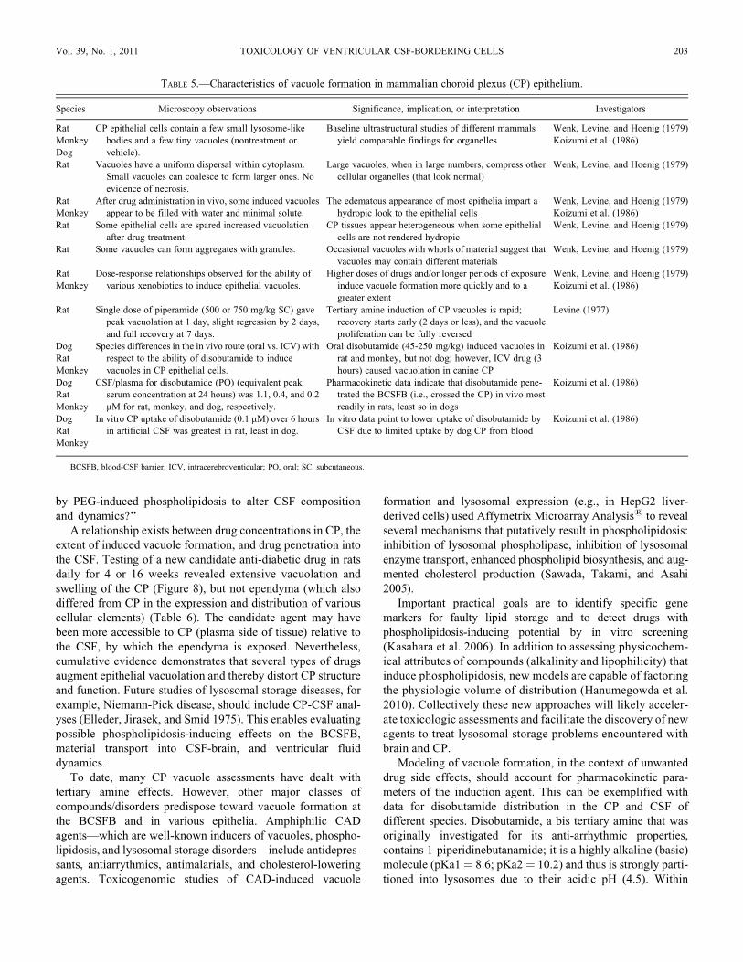
by PEG-induced phospholipidosis to alter CSF composition
and dynamics?’’
A relationship exists between drug concentrations in CP, the
extent of induced vacuole formation, and drug penetration into
the CSF. Testing of a new candidate anti-diabetic drug in rats
daily for 4 or 16 weeks revealed extensive vacuolation and
swelling of the CP (Figure 8), but not ependyma (which also
differed from CP in the expression and distribution of various
cellular elements) (Table 6). The candidate agent may have
been more accessible to CP (plasma side of tissue) relative to
the CSF, by which the ependyma is exposed. Nevertheless,
cumulative evidence demonstrates that several types of drugs
augment epithelial vacuolation and thereby distort CP structure
and function. Future studies of lysosomal storage diseases, for
example, Niemann-Pick disease, should include CP-CSF anal-
yses (Elleder, Jirasek, and Smid 1975). This enables evaluating
possible phospholipidosis-inducing effects on the BCSFB,
material transport into CSF-brain, and ventricular fluid
dynamics.
To date, many CP vacuole assessments have dealt with
tertiary amine effects. However, other major classes of
compounds/disorders predispose toward vacuole formation at
the BCSFB and in various epithelia. Amphiphilic CAD
agents—which are well-known inducers of vacuoles, phospho-
lipidosis, and lysosomal storage disorders—include antidepres-
sants, antiarrythmics, antimalarials, and cholesterol-lowering
agents. Toxicogenomic studies of CAD-induced vacuole
formation and lysosomal expression (e.g., in HepG2 liver-
derived cells) used Affymetrix Microarray Analysis1 to reveal
several mechanisms that putatively result in phospholipidosis:
inhibition of lysosomal phospholipase, inhibition of lysosomal
enzyme transport, enhanced phospholipid biosynthesis, and aug-
mented cholesterol production (Sawada, Takami, and Asahi
2005).
Important practical goals are to identify specific gene
markers for faulty lipid storage and to detect drugs with
phospholipidosis-inducing potential by in vitro screening
(Kasahara et al. 2006). In addition to assessing physicochem-
ical attributes of compounds (alkalinity and lipophilicity) that
induce phospholipidosis, new models are capable of factoring
the physiologic volume of distribution (Hanumegowda et al.
2010). Collectively these new approaches will likely acceler-
ate toxicologic assessments and facilitate the discovery of new
agents to treat lysosomal storage problems encountered with
brain and CP.
Modeling of vacuole formation, in the context of unwanted
drug side effects, should account for pharmacokinetic para-
meters of the induction agent. This can be exemplified with
data for disobutamide distribution in the CP and CSF of
different species. Disobutamide, a bis tertiary amine that was
originally investigated for its anti-arrhythmic properties,
contains 1-piperidinebutanamide; it is a highly alkaline (basic)
molecule (pKa1¼ 8.6; pKa2¼ 10.2) and thus is strongly parti-
tioned into lysosomes due to their acidic pH (4.5). Within
TABLE 5.—Characteristics of vacuole formation in mammalian choroid plexus (CP) epithelium.
Species Microscopy observations Significance, implication, or interpretation Investigators
Rat
Monkey
Dog
CP epithelial cells contain a few small lysosome-like
bodies and a few tiny vacuoles (nontreatment or
vehicle).
Baseline ultrastructural studies of different mammals
yield comparable findings for organelles
Wenk, Levine, and Hoenig (1979)
Koizumi et al. (1986)
Rat Vacuoles have a uniform dispersal within cytoplasm.
Small vacuoles can coalesce to form larger ones. No
evidence of necrosis.
Large vacuoles, when in large numbers, compress other
cellular organelles (that look normal)
Wenk, Levine, and Hoenig (1979)
Rat
Monkey
After drug administration in vivo, some induced vacuoles
appear to be filled with water and minimal solute.
The edematous appearance of most epithelia impart a
hydropic look to the epithelial cells
Wenk, Levine, and Hoenig (1979)
Koizumi et al. (1986)
Rat Some epithelial cells are spared increased vacuolation
after drug treatment.
CP tissues appear heterogeneous when some epithelial
cells are not rendered hydropic
Wenk, Levine, and Hoenig (1979)
Rat Some vacuoles can form aggregates with granules. Occasional vacuoles with whorls of material suggest that
vacuoles may contain different materials
Wenk, Levine, and Hoenig (1979)
Rat
Monkey
Dose-response relationships observed for the ability of
various xenobiotics to induce epithelial vacuoles.
Higher doses of drugs and/or longer periods of exposure
induce vacuole formation more quickly and to a
greater extent
Wenk, Levine, and Hoenig (1979)
Koizumi et al. (1986)
Rat Single dose of piperamide (500 or 750 mg/kg SC) gave
peak vacuolation at 1 day, slight regression by 2 days,
and full recovery at 7 days.
Tertiary amine induction of CP vacuoles is rapid;
recovery starts early (2 days or less), and the vacuole
proliferation can be fully reversed
Levine (1977)
Dog
Rat
Monkey
Species differences in the in vivo route (oral vs. ICV) with
respect to the ability of disobutamide to induce
vacuoles in CP epithelial cells.
Oral disobutamide (45-250 mg/kg) induced vacuoles in
rat and monkey, but not dog; however, ICV drug (3
hours) caused vacuolation in canine CP
Koizumi et al. (1986)
Dog
Rat
Monkey
CSF/plasma for disobutamide (PO) (equivalent peak
serum concentration at 24 hours) was 1.1, 0.4, and 0.2
mM for rat, monkey, and dog, respectively.
Pharmacokinetic data indicate that disobutamide pene-
trated the BCSFB (i.e., crossed the CP) in vivo most
readily in rats, least so in dogs
Koizumi et al. (1986)
Dog
Rat
Monkey
In vitro CP uptake of disobutamide (0.1 mM) over 6 hours
in artificial CSF was greatest in rat, least in dog.
In vitro data point to lower uptake of disobutamide by
CSF due to limited uptake by dog CP from blood
Koizumi et al. (1986)
BCSFB, blood-CSF barrier; ICV, intracerebroventicular; PO, oral; SC, subcutaneous.
Vol. 39, No. 1, 2011 TOXICOLOGY OF VENTRICULAR CSF-BORDERING CELLS 203
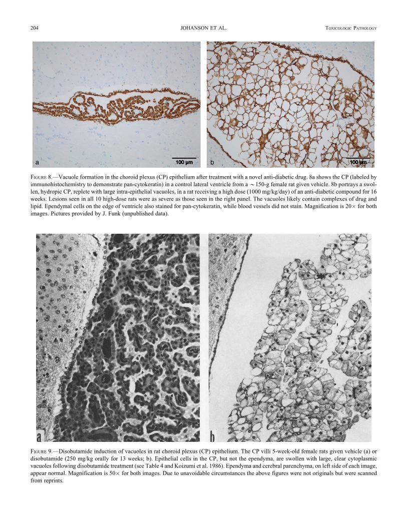
FIGURE 8.—Vacuole formation in the choroid plexus (CP) epithelium after treatment with a novel anti-diabetic drug. 8a shows the CP (labeled by
immunohistochemistry to demonstrate pan-cytokeratin) in a control lateral ventricle from a *150-g female rat given vehicle. 8b portrays a swol-
len, hydropic CP, replete with large intra-epithelial vacuoles, in a rat receiving a high dose (1000 mg/kg/day) of an anti-diabetic compound for 16
weeks. Lesions seen in all 10 high-dose rats were as severe as those seen in the right panel. The vacuoles likely contain complexes of drug and
lipid. Ependymal cells on the edge of ventricle also stained for pan-cytokeratin, while blood vessels did not stain. Magnification is 20� for both
images. Pictures provided by J. Funk (unpublished data).
FIGURE 9.—Disobutamide induction of vacuoles in rat choroid plexus (CP) epithelium. The CP villi 5-week-old female rats given vehicle (a) or
disobutamide (250 mg/kg orally for 13 weeks; b). Epithelial cells in the CP, but not the ependyma, are swollen with large, clear cytoplasmic
vacuoles following disobutamide treatment (see Table 4 and Koizumi et al. 1986). Ependyma and cerebral parenchyma, on left side of each image,
appear normal. Magnification is 50� for both images. Due to unavoidable circumstances the above figures were not originals but were scanned
from reprints.
204 JOHANSON ET AL. TOXICOLOGIC PATHOLOGY
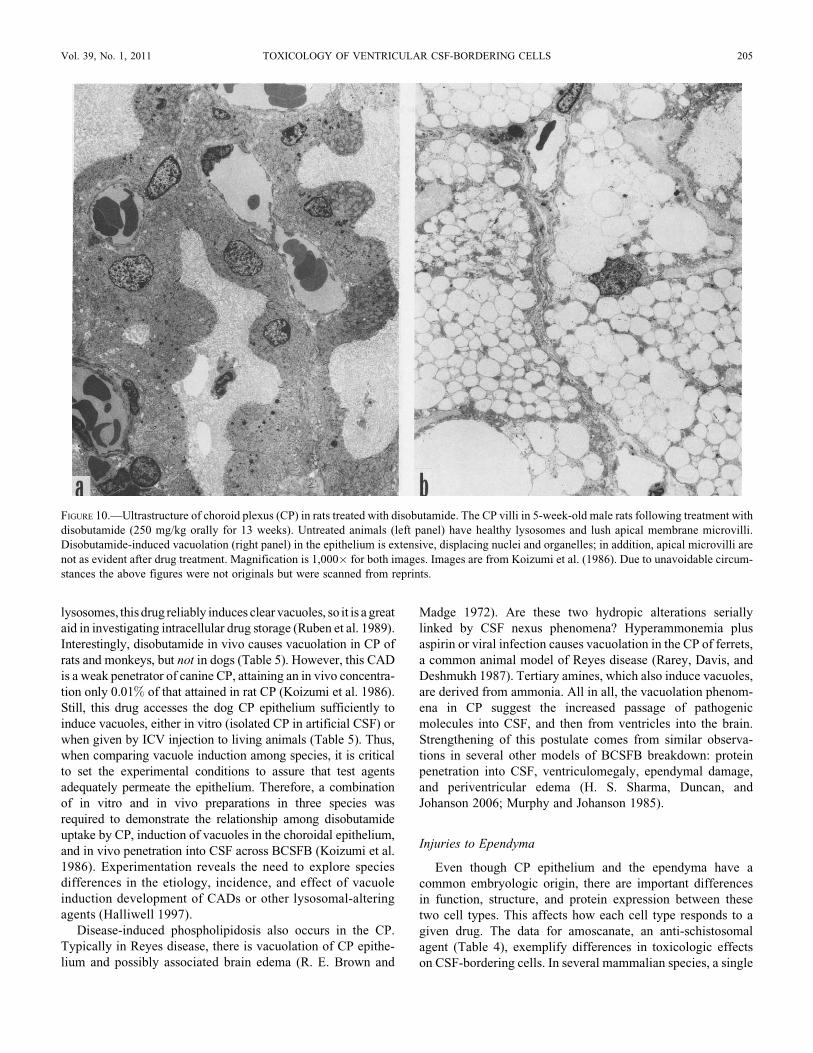
lysosomes, this drug reliably induces clear vacuoles, so it is a great
aid in investigating intracellular drug storage (Ruben et al. 1989).
Interestingly, disobutamide in vivo causes vacuolation in CP of
rats and monkeys, but not in dogs (Table 5). However, this CAD
is a weak penetrator of canine CP, attaining an in vivo concentra-
tion only 0.01% of that attained in rat CP (Koizumi et al. 1986).
Still, this drug accesses the dog CP epithelium sufficiently to
induce vacuoles, either in vitro (isolated CP in artificial CSF) or
when given by ICV injection to living animals (Table 5). Thus,
when comparing vacuole induction among species, it is critical
to set the experimental conditions to assure that test agents
adequately permeate the epithelium. Therefore, a combination
of in vitro and in vivo preparations in three species was
required to demonstrate the relationship among disobutamide
uptake by CP, induction of vacuoles in the choroidal epithelium,
and in vivo penetration into CSF across BCSFB (Koizumi et al.
1986). Experimentation reveals the need to explore species
differences in the etiology, incidence, and effect of vacuole
induction development of CADs or other lysosomal-altering
agents (Halliwell 1997).
Disease-induced phospholipidosis also occurs in the CP.
Typically in Reyes disease, there is vacuolation of CP epithe-
lium and possibly associated brain edema (R. E. Brown and
Madge 1972). Are these two hydropic alterations serially
linked by CSF nexus phenomena? Hyperammonemia plus
aspirin or viral infection causes vacuolation in the CP of ferrets,
a common animal model of Reyes disease (Rarey, Davis, and
Deshmukh 1987). Tertiary amines, which also induce vacuoles,
are derived from ammonia. All in all, the vacuolation phenom-
ena in CP suggest the increased passage of pathogenic
molecules into CSF, and then from ventricles into the brain.
Strengthening of this postulate comes from similar observa-
tions in several other models of BCSFB breakdown: protein
penetration into CSF, ventriculomegaly, ependymal damage,
and periventricular edema (H. S. Sharma, Duncan, and
Johanson 2006; Murphy and Johanson 1985).
Injuries to Ependyma
Even though CP epithelium and the ependyma have a
common embryologic origin, there are important differences
in function, structure, and protein expression between these
two cell types. This affects how each cell type responds to a
given drug. The data for amoscanate, an anti-schistosomal
agent (Table 4), exemplify differences in toxicologic effects
on CSF-bordering cells. In several mammalian species, a single
FIGURE 10.—Ultrastructure of choroid plexus (CP) in rats treated with disobutamide. The CP villi in 5-week-old male rats following treatment with
disobutamide (250 mg/kg orally for 13 weeks). Untreated animals (left panel) have healthy lysosomes and lush apical membrane microvilli.
Disobutamide-induced vacuolation (right panel) in the epithelium is extensive, displacing nuclei and organelles; in addition, apical microvilli are
not as evident after drug treatment. Magnification is 1,000� for both images. Images are from Koizumi et al. (1986). Due to unavoidable circum-
stances the above figures were not originals but were scanned from reprints.
Vol. 39, No. 1, 2011 TOXICOLOGY OF VENTRICULAR CSF-BORDERING CELLS 205

curative oral dose is typically 10 to 60 mg/kg (Bueding,
Batzinger, and Petterson 1976). Adsorbable amoscanate given
to an adult induced a lesion in the ependyma but not in CP by
three days after administration of consecutive high oral doses
(125 or 500 mg/kg) (Clark, Kiel, and Parhad 1982; Krinke
et al. 1983). Amoscanate caused necrosis by 28 days to the
lateral walls of both telencephalic ventricles; interestingly, the
lesion (Figure 11) was confined to the caudate/putamen surface
and generally spared the brain, spinal cord, and peripheral
nerves. Significantly, minute lesions occasionally occurred
close to CSF (i.e., in the fornix or septum), thereby providing
a clue about the pharmacokinetics of amoscanate distribution.
Taken together, the neuropathology pointed to CSF (rather
than plasma in periventricular capillaries) as the direct source
of drug that damages regions proximate to the ventricles.
Amoscanate likely penetrated the BCSFB and then flowed
within the CSF, inflicting damage to sensitive cells at the
ventricular border (arrows 2–5, Figure 1). Increasing the num-
ber of consecutive doses resulted in erosion of the ventricular
lining, glial reaction, and granule deposits (Krinke et al. 1983).
Amoscanate also elicited massive ultrastructural damage in
ependymal cells (Figure 12).
Another study of high-dose amoscanate in rats (250 or
500 mg/kg for 28 days) yielded similar findings of lateral
ventricle wall damage, particularly the neuropil of medial stria-
tum (Clark, Kiel, and Parhad 1982). All intoxicated animals, but
no controls, developed discrete necrotic lesions adjacent to the
lateral wall of lateral ventricle, commonly extending 1 mm into
the striatum. Immediately after initial oral dosing, rats displayed
less exploratory activity. Ependymal cells overlying necrotic
brain lesions often survived but were pyknotic. Extracellular
spaces in ependymal/subependymal regions were edematous
and spongiform (Clark, Kiel, and Parhad 1982). Less damage
was inflicted on the medial wall of the lateral ventricle than on
the lateral counterpart. Chronic treatment with high doses of
amoscanate does not injure other parts of the cerebral hemi-
spheres, cerebellum, brainstem, or CP in the third and fourth
ventricles (Clark, Kiel, and Parhad 1982; Krinke et al. 1983).
Similarly, walls of third and fourth ventricles remained intact.
Highly specific adverse responses of the ependyma (certain
regions) to amoscanate have been reproducible. Interesting
issues arise about the differential toxicity of this drug along
points in the distributional nexus: CP to CSF, ependyma, SVZ,
and ultimately the brain (Figure 1). Amoscanate, even in a
high-dose regimen, evidently does not harm CP (as examined
by microscopy). A CP capable of normal transport function
actively concentrates certain endogenous substances and drugs
in CSF relative to plasma (J. H. Wang and Takemori 1972;
Spector and Johanson 2006). One possibility is that certain
membrane transporters of organic solutes by CP (Table 2)
actively pump amoscanate, and other drugs, into CSF (arrow 3,
Figure 1). This notion needs evidence from CSF concentration
data for various therapeutic agents. Exposure of ependyma to
amoscanate in the CSF likely explains the sequential toxicity
TABLE 6.—Immunostaining of choroid plexus (CP) epithelial
and ependymal cells after treatment of rats with a candidate
anti-diabetic agent.a
Antibody
Choroid plexus
epithelium
Ependymal
cells
Pan-cytokeratin (epithelial cytoskeletal
protein)
þþ þþ
Vimentin (connective tissue filament
protein)
–* þþ
S-100 (neural crest marker) – þþFactor VIII (vascular marker) –*; ** –
Adipophilin (lipid droplet- associated
protein)
þ*** –
Ki67 (cell proliferation marker) þ/–**** –
Degree of expression, relative to control: þ ¼ increased expression; – ¼ reduced
expression.a The immunostaining data in the table correspond to the tissue images in Figure 8
(unpublished data from J. Funk).
*Only blood vessels of choroid plexus were positive.
**No increase of blood vessels labeling in high-dose animals.
***Minimal increase in high-dose animals.
****No increase in cell proliferation in high-dose animals.
FIGURE 11.—Destruction of ependyma and periventricular brain by
amoscanate. Sprague-Dawley rats treated with amoscanate (500 mg/
kg/day suspended in oil, given orally for 10 days). In the medial cau-
date/putamen, there was deposition of microgranules. Choroid plexus
was intact, but the ependyma covering the caudate/putamen was
severely damaged. Magnification is 20�. From Krinke et al. (1983).
Cresyl violet.
206 JOHANSON ET AL. TOXICOLOGIC PATHOLOGY
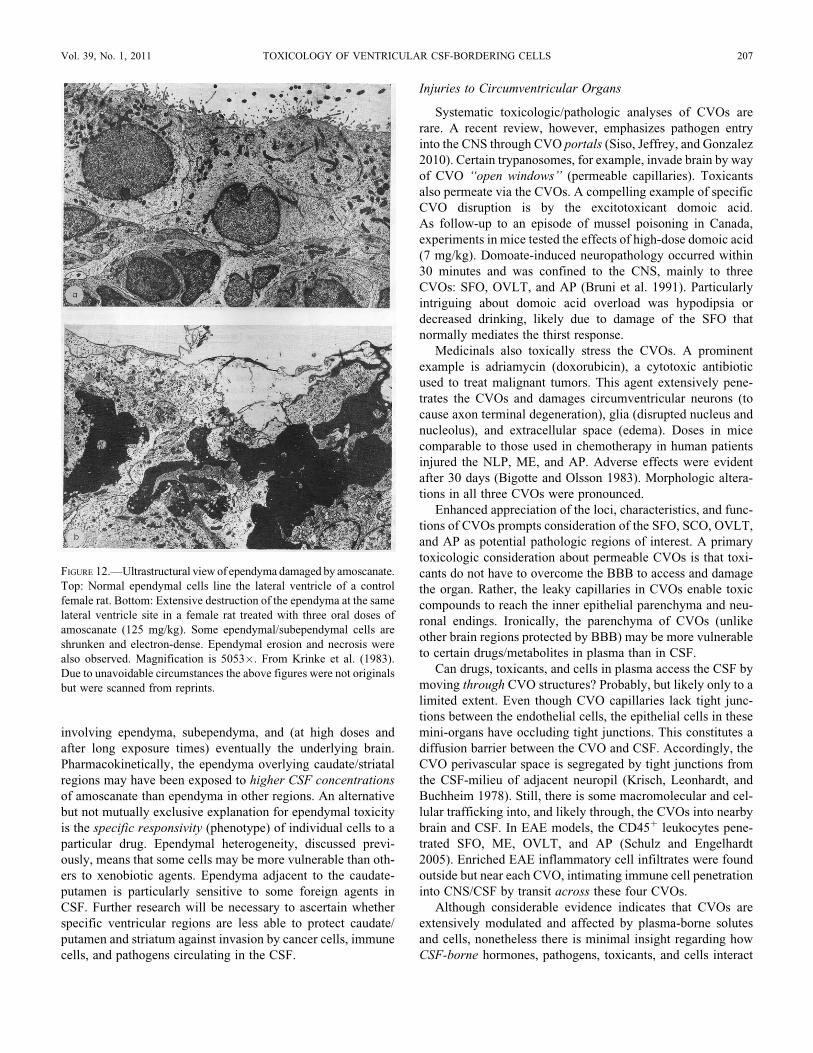
involving ependyma, subependyma, and (at high doses and
after long exposure times) eventually the underlying brain.
Pharmacokinetically, the ependyma overlying caudate/striatal
regions may have been exposed to higher CSF concentrations
of amoscanate than ependyma in other regions. An alternative
but not mutually exclusive explanation for ependymal toxicity
is the specific responsivity (phenotype) of individual cells to a
particular drug. Ependymal heterogeneity, discussed previ-
ously, means that some cells may be more vulnerable than oth-
ers to xenobiotic agents. Ependyma adjacent to the caudate-
putamen is particularly sensitive to some foreign agents in
CSF. Further research will be necessary to ascertain whether
specific ventricular regions are less able to protect caudate/
putamen and striatum against invasion by cancer cells, immune
cells, and pathogens circulating in the CSF.
Injuries to Circumventricular Organs
Systematic toxicologic/pathologic analyses of CVOs are
rare. A recent review, however, emphasizes pathogen entry
into the CNS through CVO portals (Siso, Jeffrey, and Gonzalez
2010). Certain trypanosomes, for example, invade brain by way
of CVO ‘‘open windows’’ (permeable capillaries). Toxicants
also permeate via the CVOs. A compelling example of specific
CVO disruption is by the excitotoxicant domoic acid.
As follow-up to an episode of mussel poisoning in Canada,
experiments in mice tested the effects of high-dose domoic acid
(7 mg/kg). Domoate-induced neuropathology occurred within
30 minutes and was confined to the CNS, mainly to three
CVOs: SFO, OVLT, and AP (Bruni et al. 1991). Particularly
intriguing about domoic acid overload was hypodipsia or
decreased drinking, likely due to damage of the SFO that
normally mediates the thirst response.
Medicinals also toxically stress the CVOs. A prominent
example is adriamycin (doxorubicin), a cytotoxic antibiotic
used to treat malignant tumors. This agent extensively pene-
trates the CVOs and damages circumventricular neurons (to
cause axon terminal degeneration), glia (disrupted nucleus and
nucleolus), and extracellular space (edema). Doses in mice
comparable to those used in chemotherapy in human patients
injured the NLP, ME, and AP. Adverse effects were evident
after 30 days (Bigotte and Olsson 1983). Morphologic altera-
tions in all three CVOs were pronounced.
Enhanced appreciation of the loci, characteristics, and func-
tions of CVOs prompts consideration of the SFO, SCO, OVLT,
and AP as potential pathologic regions of interest. A primary
toxicologic consideration about permeable CVOs is that toxi-
cants do not have to overcome the BBB to access and damage
the organ. Rather, the leaky capillaries in CVOs enable toxic
compounds to reach the inner epithelial parenchyma and neu-
ronal endings. Ironically, the parenchyma of CVOs (unlike
other brain regions protected by BBB) may be more vulnerable
to certain drugs/metabolites in plasma than in CSF.
Can drugs, toxicants, and cells in plasma access the CSF by
moving through CVO structures? Probably, but likely only to a
limited extent. Even though CVO capillaries lack tight junc-
tions between the endothelial cells, the epithelial cells in these
mini-organs have occluding tight junctions. This constitutes a
diffusion barrier between the CVO and CSF. Accordingly, the
CVO perivascular space is segregated by tight junctions from
the CSF-milieu of adjacent neuropil (Krisch, Leonhardt, and
Buchheim 1978). Still, there is some macromolecular and cel-
lular trafficking into, and likely through, the CVOs into nearby
brain and CSF. In EAE models, the CD45þ leukocytes pene-
trated SFO, ME, OVLT, and AP (Schulz and Engelhardt
2005). Enriched EAE inflammatory cell infiltrates were found
outside but near each CVO, intimating immune cell penetration
into CNS/CSF by transit across these four CVOs.
Although considerable evidence indicates that CVOs are
extensively modulated and affected by plasma-borne solutes
and cells, nonetheless there is minimal insight regarding how
CSF-borne hormones, pathogens, toxicants, and cells interact
FIGURE 12.—Ultrastructural view of ependyma damaged by amoscanate.
Top: Normal ependymal cells line the lateral ventricle of a control
female rat. Bottom: Extensive destruction of the ependyma at the same
lateral ventricle site in a female rat treated with three oral doses of
amoscanate (125 mg/kg). Some ependymal/subependymal cells are
shrunken and electron-dense. Ependymal erosion and necrosis were
also observed. Magnification is 5053�. From Krinke et al. (1983).
Due to unavoidable circumstances the above figures were not originals
but were scanned from reprints.
Vol. 39, No. 1, 2011 TOXICOLOGY OF VENTRICULAR CSF-BORDERING CELLS 207
with CVOs from the apical-side of each mini-organ. Tanycyte
fibers contacting CSF pick up ventricular signaling molecules
and convey these neuroendocrine hormones into hypothalamic
portal capillaries (Rodriguez et al. 2005). Putative CVO involve-
ment with tanycyte and hypothalamic homeostatic actions,
especially as modified by CSF-convected toxicants, deserves
more investigation.
PERSPECTIVES ON CEREBROSPINAL FLUID TOXICOLOGY AND
PATHOLOGY
Breakdown of CP by xenobiotic agents and pathogens
compromises CSF homeostasis with profound implications for
many CNS functions. Regulated secretions by CP represent a
lifeline of micronutritional and trophic support for neurons.
In addition, the reabsorption of brain catabolites and drug
metabolites by choroid epithelial cells pivotally maintains CSF
purity. Moreover, fine regulation of immune and neuroendo-
crine phenomena at the BCSFB bears not only on brain well-
being but also that of peripheral organs. Therefore, maintaining
integrity of the blood-CSF transport interface is vital for health.
Throughout life, the CP is challenged by blood-borne
toxicants, pathogens (viruses and protozoans), and various
xenobiotics. Usually the BCSFB remains intact as it success-
fully wards off threatening agents and protects the CNS.
Unfortunately, some stressors, given at high ‘‘doses’’ or for
long periods, overwhelm CP homeostatic capacities so that
foreign agents can invade CSF. Because the ependyma is
readily permeable in many ventricular locations, a CSF-borne
toxicant has easy access to periventricular brain regions. Such
accession challenges neurogenic niches requiring a stable,
clean environment to generate new neurons for supporting cog-
nitive function. Maintaining CP defense systems is therefore of
prime significance. A sound BCSFB thwarts penetration
of harmful molecules into CSF-brain.
Insight is needed on mechanisms underlying CP vulnerability
to certain viruses (HIV) and parasites (e.g., schistosomes and
trypanosomes). Undoubtedly one vulnerability factor is the
great permeability of the CP capillary system and high blood
flow. This assures extensive delivery of materials to intersti-
tium and the epithelial cell basolateral membrane. Another
potential susceptibility factor, quite neglected in research, is
the wide variety of epithelial cell phenotypes revealed by
differential lectin staining of glycoconjugates (McMillan et
al. 2003). Accordingly, one choroid epithelial cell may express
particular glycosylated proteins on its surface while neighbor-
ing epithelium does not; the implication is that some cells at
the BCSFB may bind a particular virus or protein, whereas neigh-
boring cells may not. Since binding is integral to endocytotic and
transcytotic phenomena, it is pertinent to obtain more information
on CP cell receptor affinities.
Certain cells of the immune system migrate through CP
(especially in autoimmune relapse states), perhaps less so
across brain capillaries. Here the epithelial cells of the BCSFB
differ markedly in structure and function from the endothelial
cells of the BBB. These unique barrier phenotypes, including
the expression of proteins (structural and enzymatic), likely
explain most differences in translocational phenomena.
Diseases such as MS and pathogen infections worsen as pro-
teins and particles migrate from CP-CSF into periventricular
regions. This likely contributes to lesion spread into the brain.
Elucidating CSF-brain immunopathology relies on new approaches
for tracking aberrant immune cell and immunoglobulin
permeation (arrows 1–3, Figure 1) at the BCSFB.
Agents for treating parasitic diseases, such as anti-trypano-
somic drugs, can be harsh on CP structure and physiology.
New antiparasitic agents are being sought that are less likely
to perturb the BCSFB. Vacuole formation in CP, provoked
by various types of drugs, is vexing due to potential
deleterious effects on CSF dynamics as well as CP epithelial
phospholipid metabolism. Preservation of lysosomal/Golgi
functions in CP is central to efficient scavenging/reabsorptive
actions to keep CSF clean. Procuring information about lyso-
somal system integrity at the BCSFB will facilitate
development of new strategies and treatments for pharma-
cotherapy and detoxification of central disorders. This includes
minimizing CSF impurities in advanced aging and early
neurodegeneration (Johanson et al. 2004).
The brain, a highly protected and specialized organ, is
exquisitely sensitive to changes in CSF composition. There is
a continuing need for systematic research to delineate and
boost protective mechanisms in CP. Therapeutic strategies
should be developed to stabilize the blood-CSF and CSF-brain
interfaces upon, or before, exposure to toxic and pathogenic
agents.
REFERENCES
Aksamit, A. J., Parnell, K. J., and Johnson, M. (1999). Diffuse leukoencepha-
lopathy and coma secondary to 5-fluorouracil, neurotoxicity. In 75th
Annual meeting of the American Association of Neuropathologists
(AANP). J Neuropathol Exp Neurol 58, 562.
Alvira-Botero, X., and Carro, E. M. (Forthcoming). Clearance of amyloid-peptide
across the choroid plexus in Alzheimer’s disease. Curr Aging Sci.
Angeletti, R. H., Novikoff, P. M., Juvvadi, S. R., Fritschy, J. M., Meier, P. J.,
and Wolkoff, A. W. (1997). The choroid plexus epithelium is the site of
the organic anion transport protein in the brain. Proc Natl Acad Sci USA
94, 283–86.
Aschner, M., Vrana, K. E., and Zheng, W. (1999). Manganese uptake and
distribution in the central nervous system (CNS). Neurotoxicology 20,
173–80.
Banizs, B., Pike, M. M., Millican, C. L., Ferguson, W. B., Komlosi, P., Sheetz,
J., Bell, P. D., Schwiebert, E. M., and Yoder, B. K. (2005). Dysfunctional
cilia lead to altered ependyma and choroid plexus function, and result in
the formation of hydrocephalus. Development 132, 5329–339.
Behl, M., Zhang, Y., Monnot, A. D., Jiang, W., and Zheng, W. (2009).
Increased beta-amyloid levels in the choroid plexus following lead
exposure and the involvement of low-density lipoprotein receptor
protein-1. Toxicol Appl Pharmacol 240, 245–54.
Benitz, K. F., and Kramer, A. W. (1968). Piperamide-induced morphological
changes in the choroid plexus. Food Cosmet Toxicol 6, 125–33.
Berger, T., Weerth, S., Kojima, K., Linington, C., Wekerle, H., and Lassmann,
H. (1997). Experimental autoimmune encephalomyelitis: The antigen
specificity of T lymphocytes determines the topography of lesions in the
central and peripheral nervous system. Lab Invest 76, 355–64.
Bernert, G., Gottling, A., Rosenkranz, A., and Zoder, G. (1988). Hemorrhagic
and hypoxic-ischemic intracranial lesions in neonates diagnosed by
208 JOHANSON ET AL. TOXICOLOGIC PATHOLOGY

realtime sonography: incidence and short-term outcome. Padiatr Padol
23, 25–37.
Bigotte, L., and Olsson, Y. (1983). Toxic effects of adriamycin on the central
nervous system. Ultrastructural changes in some circumventricular
organs of the mouse after intravenous administration of the drug.
Acta Neuropathol 61, 291–99.
Bourgouin, P. M., Melancon, D., Carpenter, S., Tampieri, D., and Ethier, R.
(1992). Hydrocephalus and prominence of the choroid plexus: An
unusual computed tomographic presentation of cerebral toxoplasmosis
in AIDS. Can Assoc Radiol J 43, 55–9.
Brightman, M. W., and Reese, T. S. (1969). Junctions between intimately
apposed cell membranes in the vertebrate brain. J Cell Biol 40, 648–77.
Brodie, S. J., Bickle, H. M., and DeMartini, J. C. (1995). Virological markers in
cerebrospinal fluid are predictive of ovine lentivirus-associated subclini-
cal encephalomyelitis. Clin Immunol Immunopathol 77, 14–8.
Brown, P. D., Davies, S. L., Speake, T., and Millar, I. D. (2004). Molecular
mechanisms of cerebrospinal fluid production. Neuroscience 129,
957–70.
Brown, R. E., and Madge, G. E. (1972). The choroid plexus in Reye’s
syndrome. Va Med Mon (1918) 99, 271–75.
Bruni, J. E., Bose, R., Pinsky, C., and Glavin, G. (1991). Circumventricular
organ origin of domoic acid-induced neuropathology and toxicology.
Brain Res Bull 26, 419–24.
Bueding, E., Batzinger, R., and Petterson, G. (1976). Antischistomal and some
toxicological properties of a nitrodiphenylaminoisothiocyanate (C 9333-
Go/CGP-4540). Experientia 32, 604–06.
Burkala, E. J., He, J., West, J. T., Wood, C., and Petito, C. K. (2005). Compart-
mentalization of HIV-1 in the central nervous system: Role of the choroid
plexus. AIDS 19, 675–84.
Carder, P. J., Hume, R., Fryer, A. A., Strange, R. C., Lauder, J., and Bell, J. E.
(1990). Glutathione S-transferase in human brain. Neuropathol Appl
Neurobiol 16, 293–303.
Cecchi, P. C., Billio, A., Colombetti, V., Rizzo, P., Ricci, U. M., and Schwarz,
A. (2008). Primary high-grade B-cell lymphoma of the choroid plexus.
Clin Neurol Neurosurg 110, 75–9.
Chen, H., Wood, C., and Petito, C. K. (2000). Comparisons of HIV-1 viral
sequences in brain, choroid plexus and spleen: Potential role of choroid
plexus in the pathogenesis of HIV encephalitis. J Neurovirol 6, 498–506.
Chinnery, H. R., Ruitenberg, M. J., and McMenamin, P. G. (2010). Novel char-
acterization of monocyte-derived cell populations in the meninges and
choroid plexus and their rates of replenishment in bone marrow chimeric
mice. J Neuropathol Exp Neurol 69, 896–909.
Chodobski, A., and Szmydynger-Chodobska, J. (2001). Choroid plexus: Target
for polypeptides and site of their synthesis. Microsc Res Tech 52, 65–82.
Clark, A. W., Kiel, S. M., and Parhad, I. M. (1982). Neuropathology of the anti-
schistosomal agent amoscanate administered in high oral doses to rats.
Neurotoxicology 3, 1–11.
Cooper, A. J., and Kristal, B. S. (1997). Multiple roles of glutathione in the cen-
tral nervous system. Biol Chem 378, 793–802.
Crossgrove, J. S., Li, G. J., and Zheng, W. (2005). The choroid plexus removes
beta-amyloid from brain cerebrospinal fluid. Exp Biol Med (Maywood)
230, 771–76.
Darsaud, A., Bourdon, L., Chevrier, C., Keita, M., Bouteille, B., Queyroy, A.,
Canini, F., Cespuglio, R., Dumas, M., and Buguet, A. (2003). Clinical
follow-up in the rat experimental model of African trypanosomiasis. Exp
Biol Med (Maywood) 228, 1355–362.
Davson, H., and Segal, M.B. (1996). Physiology of the CSF and Blood-Brain
Barriers. CRC Press, Boca Raton, FL.
Dietrich, M. O., Muller, A., Bolos, M., Carro, E., Perry, M. L., Portela, L. V.,
Souza, D. O., and Torres-Aleman, I. (2007). Western style diet impairs
entrance of blood-borne insulin-like growth factor-1 into the brain.
Neuromolecular Med 9, 324–30.
Dietrich, M. O., Spuch, C., Antequera, D., Rodal, I., de Yebenes, J. G., Molina,
J. A., Bermejo, F., and Carro, E. (2008). Megalin mediates the transport
of leptin across the blood-CSF barrier. Neurobiol Aging 29, 902–12.
Duvernoy, H. M., and Risold, P. Y. (2007). The circumventricular organs: An atlas
of comparative anatomy and vascularization. Brain Res Rev 56, 119–47.
Elleder, M., Jirasek, A., and Smid, F. (1975). Niemann-Pick disease (Crocker’s
type C): A histological study of the distribution and qualitative differ-
ences of the storage process. Acta Neuropathol 33, 191–200.
Engelhardt, B., and Sorokin, L. (2009). The blood-brain and the blood-
cerebrospinal fluid barriers: Function and dysfunction. Semin Immuno-
pathol 31, 497–511.
Falangola, M. F., and Petito, C. K. (1993). Choroid plexus infection in cerebral
toxoplasmosis in AIDS patients. Neurology 43, 2035–040.
Feher, P., Olah, M., Bodnar, I., Hechtl, D., Bacskay, I., Juhasz, B., Nagy, G. M.,
and Vecsernyes, M. (2010). Dephosphorylation/inactivation of tyrosine
hydroxylase at the median eminence of the hypothalamus is required for
suckling-induced prolactin and adrenocorticotrop hormone responses.
Brain Res Bull 82, 141–45.
Fischer, E., and Pontoglio, M. (2009). Planar cell polarity and cilia. Semin Cell
Dev Biol 20, 998–1005.
Frisch, W., and Lullmann-Rauch, R. (1979). Differential effects of chloroquine
and of several other amphiphilic cationic drugs upon rat choroid plexus.
Acta Neuropathol 46, 203–08.
Ghersi-Egea, J. F., Finnegan, W., Chen, J. L., and Fenstermacher, J. D. (1996).
Rapid distribution of intraventricularly administered sucrose into cerebrosp-
inal fluid cisterns via subarachnoid velae in rat. Neuroscience 75, 1271–288.
Glantz, M., and Johanson, C. (2008). Report on a conference analyzing the role
of cerebrospinal fluid prophylaxis for brain tumors. Cerebrospinal Fluid
Res 5, 6 (doi:10.1186/1743-8454-5-6).
Gonzalez, A. M., Taylor, W. M., Johanson, C. E., King, J. C., Leadbeater, W.
E., Stopa, E. G., and Baird, A. (2010). Co-localization and regulation of
basic fibroblast growth factor and arginine vasopressin in neuroendocrine
cells of the rat and human brain. Cerebrospinal Fluid Res 7, 13
(doi:10.1186/1743-8454-7-13).
Gradinaru, D., Minn, A. L., Artur, Y., Minn, A., and Heydel, J. M. (2009). Drug
metabolizing enzyme expression in rat choroid plexus: effects of in vivo
xenobiotics treatment. Arch Toxicol 83, 581–86.
Gross, P. M. (1992). Circumventricular organ capillaries. Prog Brain Res 91,
219–33.
Grzybowski, D. M., Holman, D. W., Katz, S. E., and Lubow, M. (2006).
In vitro model of cerebrospinal fluid outflow through human arachnoid
granulations. Invest Ophthalmol Vis Sci 47, 3664–672.
Hagenbuch, B., and Meier, P. J. (2003). The superfamily of organic anion
transporting polypeptides. Biochim Biophys Acta 1609, 1–18.
Halliwell, W. H. (1997). Cationic amphiphilic drug-induced phospholipidosis.
Toxicol Pathol 25, 53–60.
Hanumegowda, U. M., Wenke, G., Regueiro-Ren, A., Yordanova, R., Corradi,
J. P., and Adams, S. P. (2010). Phospholipidosis as a function of basicity,
lipophilicity, and volume of distribution of compounds. Chem Res
Toxicol 23, 749–55.
Harbut, R. E., and Johanson, C. E. (1986). Third ventricle choroid plexus
function and its response to acute perturbations in plasma chemistry.
Brain Res 374, 137–46.
Henry, M., Grob, M., and Mouginot, D. (2009). Endogenous angiotensin II
facilitates GABAergic neurotransmission afferent to the Naþ-responsive
neurons of the rat median preoptic nucleus. Am J Physiol Regul Integr
Comp Physiol 297, R783–92.
Hu, Y., Shen, H., Keep, R. F., and Smith, D. E. (2007). Peptide transporter
2 (PEPT2) expression in brain protects against 5-aminolevulinic acid
neurotoxicity. J Neurochem 103, 2058–065.
Johanson, C. (2008). Choroid plexus-CSF circulatory dynamics: Impact on
brain growth, metabolism and repair. In Neuroscience in Medicine
(P. Conn, ed.), pp. 173–200. The Humana Press, Totowa, NJ.
Johanson, C., McMillan, P., Tavares, R., Spangenberger, A., Duncan, J.,
Silverberg, G., and Stopa, E. (2004). Homeostatic capabilities of the
choroid plexus epithelium in Alzheimer’s disease. Cerebrospinal Fluid
Res 1, 3 (doi:10.1186/1743-8454-1-3).
Johanson, C., Silverberg, G., Donahue, J., Duncan, J., and Stopa, E. (2005).
Choroid plexus and CSF in Alzheimer’s disease: Altered expression and
transport of proteins and peptides. In The Blood-Cerebrospinal Fluid
Barrier (W. Zheng and A. Chodobski, eds.), pp. 307–39. CRC Press, Boca
Raton, FL.
Vol. 39, No. 1, 2011 TOXICOLOGY OF VENTRICULAR CSF-BORDERING CELLS 209
Johanson, C. E., Duncan, J. A., 3rd, Klinge, P. M., Brinker, T., Stopa, E. G.,
and Silverberg, G. D. (2008). Multiplicity of cerebrospinal fluid
functions: New challenges in health and disease. Cerebrospinal Fluid
Res 5, 10.
Johanson, C. E., Duncan, J. A., Stopa, E. G., and Baird, A. (2005). Enhanced
prospects for drug delivery and brain targeting by the choroid plexus-CSF
route. Pharm Res 22, 1011–037.
Johanson, C. E., Miller, M., Stopa, E., and Sharma, H. S. (2010). Disruption of
the choroid plexus-CSF-ependymal wall nexus in CNS injury and aging
models: Rescue by i.c.v. peptides. In 7th Congress of the Global College
of Neuroprotection and Neuroregeneration, Stockholm, Sweden, p. 47.
Johanson, C. E., Palm, D. E., Primiano, M. J., McMillan, P. N., Chan, P.,
Knuckey, N. W., and Stopa, E. G. (2000). Choroid plexus recovery after
transient forebrain ischemia: Role of growth factors and other repair
mechanisms. Cell Mol Neurobiol 20, 197–216.
Johanson, C. E., Reed, D. J., and Woodbury, D. M. (1974). Active transport of
sodium and potassium by the choroid plexus of the rat. J Physiol 241,
359–72.
Johanson, C. E., Stopa, E. G., and McMillan, P. N. (2010). The blood-
cerebrospinal fluid barrier: Structural and functional significance. In The
Blood-Brain and Other Neural Barriers. Biology and Research Protocols
(S. Nag, ed.), pp. Humana Press; Totowa, NJ. Methods in Molecular Biology
686, 101–31.
Johanson, C. E., and Woodbury, D. M. (1978). Uptake of [14C]urea by the in
vivo choroid plexus—cerebrospinal fluid—brain system: Identification
of sites of molecular sieving. J Physiol 275, 167–76.
Johnston, M., Boulton, M., and Flessner, M. (2000). Cerebrospinal fluid
absorption revisited: Do extracranial lymphatics play a role? The Neuros-
cientist 6, 77–87.
Johnston, M., Zakharov, A., Papaiconomou, C., Salmasi, G., and Armstrong,
D. (2004). Evidence of connections between cerebrospinal fluid and
nasal lymphatic vessels in humans, non-human primates and other
mammalian species. Cerebrospinal Fluid Res 1, 2 (doi:10.1186/
1743-8454-1-2).
June, C. H., Contreras, C. E., Perrin, L. H., Lambert, P. H., and Miescher, P. A.
(1979). Circulating and tissue-bound immune complex formation in
murine malaria. J Immunol 122, 2154–161.
Kadel, K. A., Heistad, D. D., and Faraci, F. M. (1990). Effects of endothelin on
blood vessels of the brain and choroid plexus. Brain Res 518, 78–82.
Kappers, J. A. (1958). Structural and functional changes in the telencephalic
choroid plexus during human ontogenesis. In Ciba Foundation Sympo-
sium on the Cerebrospinal Fluid (G. E. W. Wolstenholme and C. M.
O’Connor, eds.), pp. 3–25. Little, Brown, Boston.
Kasahara, T., Tomita, K., Murano, H., Harada, T., Tsubakimoto, K., Ogihara,
T., Ohnishi, S., and Kakinuma, C. (2006). Establishment of an in vitro
high-throughput screening assay for detecting phospholipidosis-
inducing potential. Toxicol Sci 90, 133–41.
Kobayashi, Z., Tsuchiya, K., Machida, A., Goto, J., Yokota, O., Miake, H.,
Watabiki, S., Taki, K., Ishizu, H., Haga, C., Arai, T., Akiyama, H., and
Mizusawa, H. (2009). Metastatic CNS lymphoma presenting with peri-
ventricular dissemination—MRI and neuropathological findings in an
autopsy case. J Neurol Sci 277, 109–13.
Koizumi, H., Watanabe, M., Numata, H., Sakai, T., and Morishita, H. (1986).
Species differences in vacuolation of the choroid plexus induced by the
piperidine-ring drug disobutamide in the rat, dog, and monkey. Toxicol
Appl Pharmacol 84, 125–48.
Kozlowski, G. P. (1986). Hormone pathways in cerebrospinal fluid. Neurol
Clin 4, 907–17.
Krinke, G., Graepel, P., Krueger, L., and Thomann, P. (1983). Early effects of
high-dosed absorbable amoscanate on rat brain. Toxicol Lett 19, 261–66.
Krinke, G. J., Kaufmann, W., Mahrous, A. T., and Schaetti, P. (2000). Morpho-
logic characterization of spontaneous nervous system tumors in mice and
rats. Toxicol Pathol 28, 178–92.
Krisch, B., Leonhardt, H., and Buchheim, W. (1978). The functional and struc-
tural border between the CSF- and blood-milieu in the circumventricular
organs (organum vasculosum laminae terminalis, subfornical organ, area
postrema) of the rat. Cell Tissue Res 195, 485–97.
Levine, S. (1977). Degeneration of choroid plexus epithelium induced by some
tertiary amines. In Neurotoxicology (L. Roizin, H. Shiraki and N. Grce-
vic, eds.), pp. 419–25. Raven Press, New York.
Levine, S. (1987). Choroid plexus: Target for systemic disease and pathway to
the brain. Lab Invest 56, 231–33.
Levine, S., Saltzman, A., and Ginsberg, S. D. (2008). Different inflammatory
reactions to vitamin D3 among the lateral, third and fourth ventricular
choroid plexuses of the rat. Exp Mol Pathol 85, 117–21.
Levine, S., and Sowinski, R. (1973). Choroid plexitis produced in rats by cyclo-
phosphamide. J Neuropathol Exp Neurol 32, 365–70.
Levine, S., and Sowinski, R. (1977). T-Lymphocyte depletion and lesions of
choroid plexus and kidney induced by tertiary amines in rats. Toxicol
Appl Pharmacol 40, 147–59.
Levine, S., Sowinski, R., and Nochlin, D. (1982). Periventricular localization
of a toxic encephalopathy induced by a mechanism involving choroid
plexus. Brain Res 242, 219–25.
Luo, J., Shook, B. A., Daniels, S. B., and Conover, J. C. (2008). Subventricular
zone-mediated ependyma repair in the adult mammalian brain. J Neu-
rosci 28, 3804–813.
Maharaj, A. S., Walshe, T. E., Saint-Geniez, M., Venkatesha, S., Maldonado,
A. E., Himes, N. C., Matharu, K. S., Karumanchi, S. A., and D’Amore, P.
A. (2008). VEGF and TGF-b are required for the maintenance of the
choroid plexus and ependyma. J Exp Med 205, 491–501.
McMillan, P. N., Jones, H. C., Nguyen, T. T., Spangenberger, A. G., Flaherty,
S. L., Duncan, J. A., and Johanson, C. E. (2003). Heterogeneity of inter-
and intraventricular glycoconjugate expression in rat choroid epithelial
cells as revealed by glycosylation probes. In Vth International Conference
on Cerebral Vascular Biology, pp. A-15. Amarillo, Texas.
Miller, M. C., Tavares, R., Johanson, C. E., Hovanesian, V., Donahue, J. E.,
Gonzalez, L., Silverberg, G. D., and Stopa, E. G. (2008). Hippocampal
RAGE immunoreactivity in early and advanced Alzheimer’s disease.
Brain Res 1230, 273–80.
Miyan, J. A., Nabiyouni, M., and Zendah, M. (2003). Development of the brain:
A vital role for cerebrospinal fluid. Can J Physiol Pharmacol 81, 317–28.
Moulton, J. E. (1986). Relapse infection after chemotherapy in goats
experimentally infected with Trypanosoma brucei: Pathological changes
in central nervous system. Vet Pathol 23, 21–8.
Murphy, V. A., and Johanson, C. E. (1985). Adrenergic-induced enhancement
of brain barrier system permeability to small nonelectrolytes: Choroid
plexus versus cerebral capillaries. J Cereb Blood Flow Metab 5, 401–12.
Murphy, V. A., and Johanson, C. E. (1990). Naþ-Hþ exchange in choroid
plexus and CSF in acute metabolic acidosis or alkalosis. Am J Physiol
258, F1528–537.
Nathanson, J. A., and Chun, L. L. (1989). Immunological function of the blood-
cerebrospinal fluid barrier. Proc Natl Acad Sci U S A 86, 1684–688.
Netsky, M., and Shuangshoti, S. (1975). The choroid plexus in health and
disease. University Press of Virginia, Charlottesville.
Nilsson, C., Lindvall-Axelsson, M., and Owman, C. (1992). Neuroendocrine
regulatory mechanisms in the choroid plexus-cerebrospinal fluid system.
Brain Res Brain Res Rev 17, 109–38.
Ohtsuki, S., Takizawa, T., Takanaga, H., Terasaki, N., Kitazawa, T., Sasaki,
M., Abe, T., Hosoya, K., and Terasaki, T. (2003). In vitro study of the
functional expression of organic anion transporting polypeptide 3 at rat
choroid plexus epithelial cells and its involvement in the cerebrospinal
fluid-to-blood transport of estrone-3-sulfate. Mol Pharmacol 63, 532–37.
Ormerod, W. E., and Raseroka, B. H. (1988). Screening compounds for sleep-
ing sickness therapy without relapse. Bull Soc Pathol Exot Filiales 81,
543–47.
Oshio, K., Song, Y., Verkman, A. S., and Manley, G. T. (2003). Aquaporin-1
deletion reduces osmotic water permeability and cerebrospinal fluid pro-
duction. Acta Neurochir Suppl 86, 525–28.
Owen-Lynch, P. J., Draper, C. E., Mashayekhi, F., Bannister, C. M., and
Miyan, J. A. (2003). Defective cell cycle control underlies abnormal cor-
tical development in the hydrocephalic Texas rat. Brain 126, 623–31.
Palm, D., Knuckey, N., Guglielmo, M., Watson, P., Primiano, M., and Johan-
son, C. (1995). Choroid plexus electrolytes and ultrastructure following
transient forebrain ischemia. Am J Physiol 269, R73–9.
210 JOHANSON ET AL. TOXICOLOGIC PATHOLOGY
Pappenheimer, J., Heisey, S., and Jordan, E. (1961). Active transport of
Diodrast and phenolsulfonphthalein from cerebrospinal fluid to blood.
Am J Physiol 200, 1–10.
Parandoosh, Z., and Johanson, C. E. (1982). Ontogeny of blood-brain barrier
permeability to, and cerebrospinal fluid sink action on, [14C]urea. Am
J Physiol 243, R400–07.
Parmelee, J. T., and Johanson, C. E. (1989). Development of potassium
transport capability by choroid plexus of infant rats. Am J Physiol
256, R786–91.
Peress, N. S., Roxburgh, V. A., and Gelfand, M. C. (1981). Binding sites for
immune components in human choroid plexus. Arthritis Rheum 24,
520–26.
Petito, C. K. (2004). Human immunodeficiency virus type 1 compartmentali-
zation in the central nervous system. J Neurovirol 10 Suppl 1, 21–4.
Petito, C. K., and Adkins, B. (2005). Choroid plexus selectively accumulates
T-lymphocytes in normal controls and after peripheral immune activa-
tion. J Neuroimmunol 162, 19–27.
Petito, C. K., Chen, H., Mastri, A. R., Torres-Munoz, J., Roberts, B., and
Wood, C. (1999). HIV infection of choroid plexus in AIDS and asympto-
matic HIV-infected patients suggests that the choroid plexus may be a
reservoir of productive infection. J Neurovirol 5, 670–77.
Philip, K. A., Dascombe, M. J., Fraser, P. A., and Pentreath, V. W. (1994).
Blood-brain barrier damage in experimental African trypanosomiasis.
Ann Trop Med Parasitol 88, 607–16.
Pollay, M. (2010). The function and structure of the cerebrospinal fluid outflow
system. Cerebrospinal Fluid Res 7, 9.
Poltera, A. A., Hochmann, A., Rudin, W., and Lambert, P. H. (1980).
Trypanosoma brucei brucei: A model for cerebral trypanosomiasis in
mice—an immunological, histological and electronmicroscopic study.
Clin Exp Immunol 40, 496–507.
Prendergast, C. T., and Anderton, S. M. (2009). Immune cell entry to central
nervous system—Current understanding and prospective therapeutic
targets. Endocr Metab Immune Disord Drug Targets 9, 315–27.
Preston, J. E. (2001). Aging choroid plexus-cerebrospinal fluid system.
Microsc Res Tech 52, 31–7.
Rarey, K. E., Davis, J. A., and Deshmukh, D. R. (1987). Response of epithelial
cells of the choroid plexus in the ferret model for Reye’s syndrome. Lab
Invest 56, 249–55.
Redzic, Z. B., Preston, J. E., Duncan, J. A., Chodobski, A., and Szmydynger-
Chodobska, J. (2005). The choroid plexus-cerebrospinal fluid system:
from development to aging. Curr Top Dev Biol 71, 1–52.
Rodriguez, E. M., Blazquez, J. L., and Guerra, M. (2010). The design of bar-
riers in the hypothalamus allows the median eminence and the arcuate
nucleus to enjoy private milieus: The former opens to the portal blood and
the latter to the cerebrospinal fluid. Peptides 31, 757–76.
Rodriguez, E. M., Blazquez, J. L., Pastor, F. E., Pelaez, B., Pena, P., Peruzzo,
B., and Amat, P. (2005). Hypothalamic tanycytes: A key component of
brain-endocrine interaction. Int Rev Cytol 247, 89–164.
Roth, D. R., and Krinke, G. J. (1994). Occurrence of foam cells in the choroid
plexus of rats injected intraperitoneally with methylcellulose. Exp Toxi-
col Pathol 45, 413–14.
Ruben, Z., Dodd, D. C., Rorig, K. J., and Anderson, S. N. (1989). Disobuta-
mide: A model agent for investigating intracellular drug storage. Toxicol
Appl Pharmacol 97, 57–71.
Sanderson, L., Dogruel, M., Rodgers, J., Bradley, B., and Thomas, S. A.
(2008). The blood-brain barrier significantly limits eflornithine entry into
Trypanosoma brucei infected mouse brain. J Neurochem 107, 1136–146.
Sanderson, L., Khan, A., and Thomas, S. (2007). Distribution of suramin, an
antitrypanosomal drug, across the blood-brain and blood-cerebrospinal
fluid interfaces in wild-type and P-glycoprotein transporter-deficient
mice. Antimicrob Agents Chemother 51, 3136–146.
Sawada, H., Takami, K., and Asahi, S. (2005). A toxicogenomic approach to drug-
induced phospholipidosis: Analysis of its induction mechanism and estab-
lishment of a novel in vitro screening system. Toxicol Sci 83, 282–92.
Sawamoto, K., Wichterle, H., Gonzalez-Perez, O., Cholfin, J. A., Yamada, M.,
Spassky, N., Murcia, N. S., Garcia-Verdugo, J. M., Marin, O., Ruben-
stein, J. L., Tessier-Lavigne, M., Okano, H., and Alvarez-Buylla, A.
(2006). New neurons follow the flow of cerebrospinal fluid in the adult
brain. Science 311, 629–32.
Schulz, M., and Engelhardt, B. (2005). The circumventricular organs partici-
pate in the immunopathogenesis of experimental autoimmune encephalo-
myelitis. Cerebrospinal Fluid Res 2, 8.
Scott, D. E., Krobisch-Dudley, G., Paull, W. K., and Kozlowski, G. P. (1977).
The ventricular system in neuroendocrine mechanisms. III. Supraependy-
mal neuronal networks in the primate brain. Cell Tissue Res 179, 235–54.
Sharma, H. S., Duncan, J. A., and Johanson, C. E. (2006). Whole-body
hyperthermia in the rat disrupts the blood-cerebrospinal fluid barrier and
induces brain edema. Acta Neurochir Suppl 96, 426–31.
Sharma, H. S., and Johanson, C. E. (2007). Blood-cerebrospinal fluid barrier in
hyperthermia. Prog Brain Res 162, 459–78.
Sharma, H. S., Zimmermann-Meinzingen, S., and Johanson, C. E. (2010).
Cerebrolysin reduces blood-cerebrospinal fluid barrier permeability
change, brain pathology, and functional deficits following traumatic
brain injury in the rat. Ann N Y Acad Sci 1199, 125–37.
Sharma, S. P., and Manocha, S. L. (1977). Experimental protein malnutrition as
a causative factor in the histological and histochemical disruption of the
ependymal cells of the third ventricla and cervical central canal of squir-
rel monkeys. Z Mikrosk Anat Forsch 91, 312–20.
Shi, L. Z., and Zheng, W. (2007). Early lead exposure increases the leakage
of the blood-cerebrospinal fluid barrier, in vitro. Hum Exp Toxicol 26,
159–67.
Shibata, S. (1989). Sites of origin of primary intracerebral malignant
lymphoma. Neurosurgery 25, 14–9.
Silverberg, G. D., Heit, G., Huhn, S., Jaffe, R. A., Chang, S. D., Bronte-
Stewart, H., Rubenstein, E., Possin, K., and Saul, T. A. (2001).
The cerebrospinal fluid production rate is reduced in dementia of the
Alzheimer’s type. Neurology 57, 1763–766.
Silverberg, G. D., Huhn, S., Jaffe, R. A., Chang, S. D., Saul, T., Heit, G., Von
Essen, A., and Rubenstein, E. (2002). Downregulation of cerebrospinal
fluid production in patients with chronic hydrocephalus. J Neurosurg
97, 1271–275.
Silverberg, G. D., Miller, M. C., Messier, A. A., Majmudar, S., Machan, J. T.,
Donahue, J. E., Stopa, E. G., and Johanson, C. E. (2010). Amyloid
deposition and influx transporter expression at the blood-brain barrier
increase in normal aging. J Neuropathol Exp Neurol 69, 98–108.
Siso, S., Jeffrey, M., and Gonzalez, L. (2010). Sensory circumventricular
organs in health and disease. Acta Neuropathol 120, 689–705.
Smith, D. E., Johanson, C. E., and Keep, R. F. (2004). Peptide and peptide
analog transport systems at the blood-CSF barrier. Adv Drug Deliv Rev
56, 1765–791.
Smith, Q. R., and Johanson, C. E. (1980). Effect of ouabain and potassium on ion
concentrations in the choroidal epithelium. Am J Physiol 238, F399–406.
Smith, Q. R., and Johanson, C. E. (1985). Active transport of chloride by lateral
ventricle choroid plexus of the rat. Am J Physiol 249, F470–77.
Smith, Q. R., Pershing, L. K., and Johanson, C. E. (1981). A comparative
analysis of extracellular fluid volume of several tissues as determined
by six different markers. Life Sci 29, 449–56.
Sotthibundhu, A., Phansuwan-Pujito, P., and Govitrapong, P. (2010). Melatonin
increases proliferation of cultured neural stem cells obtained from adult
mouse subventricular zone. J Pineal Res 49, 291–300.
Spector, R., and Johanson, C. (2006). Micronutrient and urate transport in
choroid plexus and kidney: Implications for drug therapy. Pharm Res
23, 2515–524.
Spector, R., and Johanson, C. E. (1989). The mammalian choroid plexus. Sci
Am 261, 68–74.
Spector, R., and Johanson, C. E. (2007a). The origin of deoxynucleosides in
brain: Implications for the study of neurogenesis and stem cell therapy.
Pharm Res 24, 859–67.
Spector, R., and Johanson, C. E. (2007b). Vitamin transport and homeostasis
in mammalian brain: Focus on Vitamins B and E. J Neurochem 103, 425–38.
Spector, R., and Johanson, C. E. (2010a). Choroid plexus failure in the
Kearns-Sayre syndrome. Cerebrospinal Fluid Res 7, 14.
Spector, R., and Johanson, C. E. (2010b). Vectorial ligand transport through
mammalian choroid plexus. Pharm Res 27, 2054–062.
Vol. 39, No. 1, 2011 TOXICOLOGY OF VENTRICULAR CSF-BORDERING CELLS 211
Stahl, W., and Turek, G. (1988). Chronic murine toxoplasmosis: Clinicopatho-
logic characterization of a progressive wasting syndrome. Ann Trop Med
Parasitol 82, 35–48.
Stopa, E. G., Butala, P., Salloway, S., Johanson, C. E., Gonzalez, L., Tavares, R.,
Hovanesian, V., Hulette, C. M., Vitek, M. P., and Cohen, R. A. (2008).
Cerebral cortical arteriolar angiopathy, vascular b-amyloid, smooth
muscle actin, Braak stage, and APOE genotype. Stroke 39, 814–21.
Strazielle, N., Khuth, S. T., and Ghersi-Egea, J. F. (2004). Detoxification sys-
tems, passive and specific transport for drugs at the blood-CSF barrier in
normal and pathological situations. Adv Drug Deliv Rev 56, 1717–740.
Szmydynger-Chodobska, J., Chodobski, A., and Johanson, C. E. (1994). Post-
natal developmental changes in blood flow to choroid plexuses and cere-
bral cortex of the rat. Am J Physiol 266, R1488–492.
Szmydynger-Chodobska, J., Strazielle, N., Zink, B. J., Ghersi-Egea, J. F., and
Chodobski, A. (2009). The role of the choroid plexus in neutrophil invasion
after traumatic brain injury. J Cereb Blood Flow Metab 29, 1503–516.
Terr, L., and Weiner, L. P. (1983). An autoradiographic study of d-aminolevulinic
acid uptake by mouse brain. Exp Neurol 79, 564–68.
Thomas, S.A., and Segal, M.B. (1998) The transport of the anti-HIV drug,
2’,3’-didehydro-3’-deoxythymidine (D4T), across the blood-brain and
blood-cerebrospinal fluid barriers. Br J Pharmacol 125, 49–54.
Uschakov, A., McGinty, D., Szymusiak, R., and McKinley, M. J. (2009).
Functional correlates of activity in neurons projecting from the lamina termi-
nalis to the ventrolateral periaqueductal gray. Eur J Neurosci 30, 2347–355.
Van Marck, E. A., Gigase, P. L., Beckers, A., and Wery, M. (1981). Experi-
mental infections of laboratory rodents with recently isolated stocks of
Trypanosoma brucei gambiense. 2. Histopathological investigations. Z
Parasitenkd 64, 187–93.
Vio, K., Rodriguez, S., Yulis, C. R., Oliver, C., and Rodriguez, E. M. (2008).
The subcommissural organ of the rat secretes Reissner’s fiber
glycoproteins and CSF-soluble proteins reaching the internal and external
CSF compartments. Cerebrospinal Fluid Res 5, 3.
Wagner, S., Peters, O., Fels, C., Janssen, G., Liebeskind, A. K., Sauerbrey, A.,
Suttorp, M., Hau, P., and Wolff, J. E. (2008). Pegylated-liposomal doxor-
ubicin and oral topotecan in eight children with relapsed high-grade
malignant brain tumors. J Neurooncol 86, 175–81.
Wang, H. W., Amin, M. S., El-Shahat, E., Huang, B. S., Tuana, B. S., and Leenen,
F. H. (2010a). Effects of central sodium on epithelial sodium channels in rat
brain. Am J Physiol Regul Integr Comp Physiol 299, R222–33.
Wang, J. H., and Takemori, A. E. (1972). Studies on the transport of morphine
into the cerebrospinal fluid of rabbits. J Pharmacol Exp Ther 183, 41–8.
Wang, T., Lee, M. H., Johnson, T., Allie, R., Hu, L., Calabresi, P. A., and Nath,
A. (2010b). Activated T-cells inhibit neurogenesis by releasing granzyme
B: Rescue by Kv1.3 blockers. J Neurosci 30, 5020–027.
Wells, M. Y., and Krinke, G. J. (2008). Cerebral vacuolation induced in rats
by the administration of LUP-3FDC, an anti-tuberculosis cocktail. Exp
Toxicol Pathol 59, 365–72.
Wenk, E. J., Levine, S., and Hoenig, E. M. (1979). Fine structure of contrasting
choroid plexus lesions caused by tertiary amines or cyclophosphamide.
J Neuropathol Exp Neurol 38, 1–9.
Wessmann, A., Volk, H. A., Chandler, K., Brodbelt, D., and Szladovits, B.
(2010). Significance of surface epithelial cells in canine cerebrospinal
fluid and relationship to central nervous system disease. Vet Clin Pathol
39, 358–64.
Zheng, W. (2001). Toxicology of choroid plexus: Special reference to metal-
induced neurotoxicities. Microsc Res Tech 52, 89–103.
Zou, R., Park, E. H., Kelly, E. M., Egnor, M., Wagshul, M. E., and Madsen, J.
R. (2008). Intracranial pressure waves: Characterization of a pulsation
absorber with notch filter properties using systems analysis: Laboratory
investigation. J Neurosurg Pediatrics 2, 83–94.
For reprints and permissions queries, please visit SAGE’s Web site at http://www.sagepub.com/journalsPermissions.nav.
212 JOHANSON ET AL. TOXICOLOGIC PATHOLOGY
Related Documents