Patrick Waters, PhD* Mark Woodhall, PhD* Kevin C. O’Connor, PhD Markus Reindl, PhD Bethan Lang, PhD Douglas K. Sato, MD Maciej Jury nczyk, MD George Tackley, MBBCh Joao Rocha, MD Toshiyuki Takahashi, MD Tatsuro Misu, MD Ichiro Nakashima, MD Jacqueline Palace, MD Kazuo Fujihara, MD M. Isabel Leite, DPhil Angela Vincent, FRS Correspondence to Dr. Waters: [email protected] Supplemental data at Neurology.org/nn MOG cell-based assay detects non-MS patients with inflammatory neurologic disease ABSTRACT Objective: To optimize sensitivity and disease specificity of a myelin oligodendrocyte glycoprotein (MOG) antibody assay. Methods: Consecutive sera (n 5 1,109) sent for aquaporin-4 (AQP4) antibody testing were screened for MOG antibodies (Abs) by cell-based assays using either full-length human MOG (FL-MOG) or the short-length form (SL-MOG). The Abs were initially detected by Alexa Fluor goat anti-human IgG (H 1 L) and subsequently by Alexa Fluor mouse antibodies to human IgG1. Results: When tested at 1:20 dilution, 40/1,109 sera were positive for AQP4-Abs, 21 for SL- MOG, and 180 for FL-MOG. Only one of the 40 AQP4-Ab–positive sera was positive for SL- MOG-Abs, but 10 (25%) were positive for FL-MOG-Abs (p 5 0.0069). Of equal concern, 48% (42/88) of sera from controls (patients with epilepsy) were positive by FL-MOG assay. However, using an IgG1-specific secondary antibody, only 65/1,109 (5.8%) sera were positive on FL-MOG, and AQP4-Ab– positive and control sera were negative. IgM reactivity accounted for the remain- ing anti-human IgG (H 1 L) positivity toward FL-MOG. The clinical diagnoses were obtained in 33 FL-MOG–positive patients, blinded to the antibody data. IgG1-Abs to FL-MOG were associated with optic neuritis (n 5 11), AQP4-seronegative neuromyelitis optica spectrum disorder (n 5 4), and acute disseminated encephalomyelitis (n 5 1). All 7 patients with probable multiple sclerosis (MS) were MOG-IgG1 negative. Conclusions: The limited disease specificity of FL-MOG-Abs identified using Alexa Fluor goat anti- human IgG (H 1 L) is due in part to detection of IgM-Abs. Use of the FL-MOG and restricting to IgG1-Abs substantially improves specificity for non-MS demyelinating diseases. Classification of evidence: This study provides Class II evidence that the presence of serum IgG1- MOG-Abs in AQP4-Ab–negative patients distinguishes non-MS CNS demyelinating disorders from MS (sensitivity 24%, 95% confidence interval [CI] 9%–45%; specificity 100%, 95% CI 88%–100%). Neurol Neuroimmunol Neuroinflamm 2015;2:e89; doi: 10.1212/ NXI.0000000000000089 GLOSSARY Abs 5 antibodies; ADEM 5 acute disseminated encephalomyelitis; AQP4 5 aquaporin-4; CBA 5 cell-based assay; CI 5 confidence interval; EDTA 5 ethylenediaminetetraacetic acid; FACS 5 fluorescent-activated cell sorting; FL-MOG 5 full- length human MOG; LETM 5 longitudinally extensive TM; MOG 5 myelin oligodendrocyte glycoprotein; MS 5 multiple scle- rosis; NMO 5 neuromyelitis optica; NMOSD 5 NMO spectrum disorder; ON 5 optic neuritis; PEI 5 polyethylenimine; SL- MOG 5 short-length MOG; TM 5 transverse myelitis. Antibodies (Abs) that bind the CNS-restricted membrane protein myelin oligodendrocyte gly- coprotein (MOG) were first described by ELISA or Western blot predominantly in patients with multiple sclerosis (MS), but they have also been described in patients with bacterial or viral CNS inflammation or neuromyelitis optica (NMO). 1–11 These findings were not reproducible using *These authors contributed equally to the manuscript. From the Nuffield Department of Clinical Neurosciences (P.W., M.W., B.L., M.J., G.T., J.R., J.P., M.I.L., A.V.), John Radcliffe Hospital, Oxford, UK; Department of Neurology (K.C.O.), Yale School of Medicine, New Haven, CT; Clinical Department of Neurology (M.R.), Innsbruck Medical University, Innsbruck, Austria; Department of Neurology (D.K.S., I.N.) and Department of Multiple Sclerosis Therapeutics (T.M., K.F.) Tohoku University School of Medicine, Sendai, Japan; and Department of Neurology (T.T.), Yonezawa National Hospital, Yonezawa, Japan. Funding information and disclosures are provided at the end of the article. Go to Neurology.org/nn for full disclosure forms. The Article Processing Charge was paid by the authors. This is an open access article distributed under the terms of the Creative Commons Attribution-Noncommercial No Derivative 3.0 License, which permits downloading and sharing the work provided it is properly cited. The work cannot be changed in any way or used commercially. Neurology.org/nn © 2015 American Academy of Neurology 1 ª 2015 American Academy of Neurology. Unauthorized reproduction of this article is prohibited.

Welcome message from author
This document is posted to help you gain knowledge. Please leave a comment to let me know what you think about it! Share it to your friends and learn new things together.
Transcript

Patrick Waters PhDMark Woodhall PhDKevin C OrsquoConnor PhDMarkus Reindl PhDBethan Lang PhDDouglas K Sato MDMaciej Jurynczyk MDGeorge Tackley MBBChJoao Rocha MDToshiyuki Takahashi MDTatsuro Misu MDIchiro Nakashima MDJacqueline Palace MDKazuo Fujihara MDM Isabel Leite DPhilAngela Vincent FRS
Correspondence toDr Waterspaddywatersndcnoxacuk
Supplemental dataat Neurologyorgnn
MOG cell-based assay detects non-MSpatients with inflammatory neurologicdisease
ABSTRACT
Objective To optimize sensitivity and disease specificity of a myelin oligodendrocyte glycoprotein(MOG) antibody assay
Methods Consecutive sera (n 5 1109) sent for aquaporin-4 (AQP4) antibody testing werescreened for MOG antibodies (Abs) by cell-based assays using either full-length human MOG(FL-MOG) or the short-length form (SL-MOG) The Abs were initially detected by Alexa Fluor goatanti-human IgG (H 1 L) and subsequently by Alexa Fluor mouse antibodies to human IgG1
Results When tested at 120 dilution 401109 sera were positive for AQP4-Abs 21 for SL-MOG and 180 for FL-MOG Only one of the 40 AQP4-Abndashpositive sera was positive for SL-MOG-Abs but 10 (25) were positive for FL-MOG-Abs (p 5 00069) Of equal concern 48(4288) of sera from controls (patients with epilepsy) were positive by FL-MOG assay Howeverusing an IgG1-specific secondary antibody only 651109 (58) sera were positive on FL-MOGand AQP4-Abndash positive and control sera were negative IgM reactivity accounted for the remain-ing anti-human IgG (H 1 L) positivity toward FL-MOG The clinical diagnoses were obtained in 33FL-MOGndashpositive patients blinded to the antibody data IgG1-Abs to FL-MOG were associatedwith optic neuritis (n 5 11) AQP4-seronegative neuromyelitis optica spectrum disorder (n 5 4)and acute disseminated encephalomyelitis (n 5 1) All 7 patients with probable multiple sclerosis(MS) were MOG-IgG1 negative
Conclusions The limited disease specificity of FL-MOG-Abs identified using Alexa Fluor goat anti-human IgG (H 1 L) is due in part to detection of IgM-Abs Use of the FL-MOG and restricting toIgG1-Abs substantially improves specificity for non-MS demyelinating diseases
Classification of evidence This study provides Class II evidence that the presence of serumIgG1- MOG-Abs in AQP4-Abndashnegative patients distinguishes non-MS CNS demyelinatingdisorders from MS (sensitivity 24 95 confidence interval [CI] 9ndash45 specificity100 95 CI 88ndash100) Neurol Neuroimmunol Neuroinflamm 20152e89 doi 101212
NXI0000000000000089
GLOSSARYAbs 5 antibodies ADEM 5 acute disseminated encephalomyelitis AQP4 5 aquaporin-4 CBA 5 cell-based assay CI 5confidence interval EDTA 5 ethylenediaminetetraacetic acid FACS 5 fluorescent-activated cell sorting FL-MOG 5 full-length human MOG LETM 5 longitudinally extensive TM MOG 5 myelin oligodendrocyte glycoprotein MS 5 multiple scle-rosis NMO 5 neuromyelitis optica NMOSD 5 NMO spectrum disorder ON 5 optic neuritis PEI 5 polyethylenimine SL-MOG 5 short-length MOG TM 5 transverse myelitis
Antibodies (Abs) that bind the CNS-restricted membrane protein myelin oligodendrocyte gly-coprotein (MOG) were first described by ELISA orWestern blot predominantly in patients withmultiple sclerosis (MS) but they have also been described in patients with bacterial or viral CNSinflammation or neuromyelitis optica (NMO)1ndash11 These findings were not reproducible using
These authors contributed equally to the manuscript
From the Nuffield Department of Clinical Neurosciences (PW MW BL MJ GT JR JP MIL AV) John Radcliffe Hospital OxfordUK Department of Neurology (KCO) Yale School of Medicine New Haven CT Clinical Department of Neurology (MR) InnsbruckMedical University Innsbruck Austria Department of Neurology (DKS IN) and Department of Multiple Sclerosis Therapeutics (TM KF)Tohoku University School of Medicine Sendai Japan and Department of Neurology (TT) Yonezawa National Hospital Yonezawa Japan
Funding information and disclosures are provided at the end of the article Go to Neurologyorgnn for full disclosure forms The Article ProcessingCharge was paid by the authors
This is an open access article distributed under the terms of the Creative Commons Attribution-Noncommercial No Derivative 30 License whichpermits downloading and sharing the work provided it is properly cited The work cannot be changed in any way or used commercially
Neurologyorgnn copy 2015 American Academy of Neurology 1
ordf 2015 American Academy of Neurology Unauthorized reproduction of this article is prohibited
similar methods12ndash18 but serologic findings anddifferent experimental approaches suggestedthat MOG-Abs may be pathogenic19ndash23
More-specific assays using soluble tetramerizedextracellular domain of native MOG identifiedAbs in a subset of patients with acute dissemi-nated encephalomyelitis (ADEM) but rarely inadult-onset MS cases now suggesting that thetest could be of relevance for discriminatingMSfrom other demyelinating syndromes24 Thiswas confirmed by cell-based assay (CBA) thatalso used a truncated MOG in which MOG-Abs were found in patients with aquaporin-4(AQP4)ndashseronegative NMO but not thosewith MS2526 CBA using full-length humanMOG (FL-MOG) appears to be more sensi-tive and a clinical phenotype of ADEM andAQP4-seronegative NMO spectrum disorder(NMOSD) often optic neuritis (ON) isemerging27ndash36 However positivity in healthyindividuals and patients with MS even at rela-tively high serum dilutions (up to 1640) af-fects its clinical use
Here we confirm that C-terminal truncationof the MOG antigen reduces assay sensitivityand that many of the low positive Abs foundto bind to FL-MOG result from cross-reactivity of the anti-human IgG secondaryantibody with IgM Abs Using IgG1-specificsecondary antibody allows use of lower serumdilutions with FL-MOG with improved spec-ificity for patients with ON transverse myelitis(TM) AQP4-Abndashnegative NMO or ADEM
METHODS Patients Consecutive serum samples from 1109
individuals sent for routine AQP4-Ab testing over 3 months were
studied Samples are sent to Oxford via clinical immunology
laboratories with very limited or no clinical information Sera from
118 of the 180 FL-MOGndashpositive samples were used to assess
different secondary Abs and 15180 FL-MOGndashseropositive
samples were used for flow cytometry (a flow diagram of which
samples were tested on the different assays is shown in figure 1)
To assess the clinical relevance a brief anonymized questionnaire was
sent after the analyses to 48 identifiable referring clinicians requesting
patient diagnosis treatment responses and relapses if any Controls
were sera from previously archived cohorts To validate the results 2
other cohorts were screened Patients seen at the National NMO
Specialised Services who had already been tested for AQP4-Abs were
tested for MOG-IgG1-Abs After testing was completed the
diagnoses and follow-up times from the seropositive patients were
obtained from a database A further cohort of 101 Japanese patients
with a range of demyelinating diagnoses (see Results) followed by or
referred to TohokuUniversity Hospital and who had been previously
tested for AQP4-Abs were tested for MOG-IgG1-Abs All assays
were carried out blinded to the clinical diagnoses
Ethics Ethics have been approved for the study of any patients
whose samples have been referred to the Neuroimmunology lab-
oratory in Oxford for diagnostic testing (Oxfordshire REC A
07Q160428 Immune factors in neurological disease) Since
January 2010 data on all patients seen within the Oxford clinical
NMO service have been entered prospectively into a clinical data-
base and patient serum samples have been routinely tested for
AQP4-Abs and MOG-Abs The ethics committee of Tohoku
University Graduate School of Medicine approved this study
and all participants provided written informed consent
Constructs The cloning of M23 isoform of human AQP4 has
been described previously7 FL-MOG was cloned into
pIRES2-DsRed2 using the forward primer (59-39)
gatcctcgagccaccatggcaagcttatcaagaccctctctg and the reverse
primer (59-39) gatccccgggtcagaagggatttcgtagctcttcaagg A
C-terminalndashtruncated MOG construct was created from the
full-length construct by insertion of a stop codon after Gly155
and excision of the remainder of the C-terminus The 2 forms
differ only in the intracytoplasmic domain (figure 2A)
Cell-based assays HEK293T cells polyethylenimine (PEI)
transfected with human M23-AQP4 FL-MOG or C-
terminalndashtruncated human MOG (short-length MOG SL-
MOG) were used as the substrate for live CBAs which were
performed as described elsewhere7ndash9 Patient sera were tested at
120 dilution The Alexa Fluor 488 goat anti-human IgG (H 1 L)
from Invitrogen (A1013 Carlsbad CA) was used at 1750 dilution
A semiquantitative scoring system was used 0 no binding
1 low-level binding 2ndash4 increasing level of specific binding
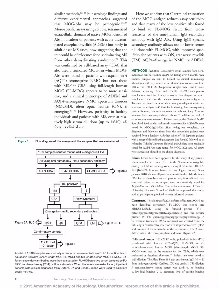
Figure 1 Flow diagram of the assays and the samples that were evaluated
A total of 1109 samples were initially screened at a serum dilution of 120 for antibodies toaquaporin-4 (AQP4) short-lengthMOG (SL-MOG) and full-length humanMOG (FL-MOG) Dif-ferent secondary antibodies were then evaluated on FL-MOGndashpositive serum samples by FL-MOG cell-based assay (CBA) or flow cytometry When the assay was established 2 patientcohorts with clinical diagnoses from Oxford UK and Sendai Japan were used to calculateassay metrics
2 Neurology Neuroimmunology amp Neuroinflammation
ordf 2015 American Academy of Neurology Unauthorized reproduction of this article is prohibited
Samples scoring 1 were considered positive The average of 2
individualrsquos scores is plotted (MW PW)
Subclasses Alexa Fluor 488 mouse anti-human IgG1 (A10631
Invitrogen) and mouse anti-human IgM (A21215 Invitrogen)
both at 1500 dilution or anti-human IgG3 (150 dilution
I7260 Sigma-Aldrich Gillingham UK) followed by Alexa
Fluor 488 goat anti-mouse IgG (H 1 L A11001 Invitrogen)
were used as secondary or tertiary Abs These assays were carried
out as described previously except cells were fixed after the final
antibody incubation
Flow cytometry A similar method to that used for detection of
AQP4-Abs (as described in Waters et al37) was used here for FL-
MOG detection Briefly HEK293T cells were transfected using
PEI for 16 hours with the pIRES2-DsRed2 plasmid that contained
the complementary DNA for FL-MOG After washing and
incubation for 24 hours at 37degC in 5 CO2 the cells were
trypsinized and resuspended in Dulbeccorsquos modified Eaglersquos
medium 1 fetal calf serum 1 mM ethylenediaminetetraacetic
acid (EDTA) (fluorescent-activated cell sorting [FACS] buffer) at
10 3 106 cellsmL The cells were rotated at 4degC for 1 hour All
further steps were carried out at 4degC Patient serum (diluted 110 in
FACS buffer) was mixed with 10 3 105 cells (100 mL) After
rocking for 1 hour the cells were washed and bound IgG was
detected with Alexa Fluor 488 goat anti-human IgG (diluted
1500 in FACS buffer) Alexa Fluor 488 anti-human IgG1 or
Alexa Fluor anti-human IgM for 30ndash45 minutes The cells were
washed resuspended in 400 mL phosphate-buffered saline2 mM
EDTA and analyzed by FACScalibur The level of transfection was
determined by measuring DsRed intensity (PE-Texas red channel)
in live cells (figure 3D y-axis) Two gates were created the upper
gate captured cells expressing high levels of DsRed the lower gate
captured untransfected or poorly transfected cells and served as a
negative control for each sample (figure 3Da) Bound IgG was
measured in the green channel (a shift to the right on the x-axis)
A score for each serum was determined by subtracting the median
green fluorescence in the lower gate from the median green
fluorescence in the upper gate
Statistics A 2-tailed Wilcoxon matched-pairs signed-rank test
was used to compare the FL-MOG and SL-MOG assays The
Mann-Whitney unpaired 2-tailed t test or Fisher exact test wasused to compare groups (p 005 was considered significant)
Primary research question Does this MOG assay using an
anti-human IgG1-specific secondary antibody identify a
Figure 2 Antibodies to MOG detected with anti-human IgG (H 1 L) as the secondary antibody
(A) Schematic of the human MOG proteins tested The extracellular and transmembrane domains are identical but the short-length MOG (SL-MOG) is 73amino acids shorter at the C-terminus than full-length MOG (FL-MOG) (B) Screening 1109 consecutive samples sent for aquaporin-4 (AQP4) antibody test-ing With anti-human IgG (H 1 L) as the secondary antibody 21 SL-MOGndash positive samples and 180 FL-MOGndashpositive samples were identified however acohort of epilepsy sera demonstrates the striking lack of specificity in the FL-MOG assay Comparing the AQP4 seropositivity in the 2 MOG assays 138AQP4-positive samples were also positive for SL-MOG antibodies (C) compared with 1038 for FL-MOG antibodies (D) CBA 5 cell-based assay
Neurology Neuroimmunology amp Neuroinflammation 3
ordf 2015 American Academy of Neurology Unauthorized reproduction of this article is prohibited
subgroup of AQP4-antibodyndashseronegative patients with a non-
MS CNS demyelinating disease
This study provides Class II evidence that the presence of
serum IgG1-Abs specific for MOG can distinguish AQP4-Abndash
negative patients with non-MS CNS demyelinating diseases from
those with MS The Japanese patients were used to calculate the
assay metrics 6 of 25 AQP4-Abndashnegative patients with non-MS
demyelinating diseases were MOG-IgG1 positive for a sensitivity
of 24 (95 confidence interval [CI] 9ndash45) and 0 of 27
patients with MS were MOG-IgG1 positive for a specificity of
100 (95 CI 88ndash100)
RESULTS Out of 1109 samples sent for diagnostictesting for AQP4-Abs 40 sera were positive at 120dilution The SL-MOG assay detected Abs in 21patients including 1 (low positive) who was stronglypositive for AQP4 (figure 2 B and C) However theFL-MOG assays detected antibodies in 180 sera (16
of the test cohort) and 10 of these sera were alsopositive for AQP4-Abs (figure 2D) Positive resultsfor FL-MOG were also found in 4288 sera frompatients with epilepsy (48 figure 2B)
Control groups and 118180 FL-MOGndashpositivesera that were available were retested by CBA usingeither anti-IgG1 or anti-IgM class-specific secondaryAbs (figure 3) With anti-IgM 101118 test sera710 healthy individuals and 1117 patients with MSwere positive The secondary antibody alone did notbind to FL-MOGndashtransfected HEK cells and the con-trol sera were negative on AQP4-transfected cells Withanti-IgG1 by contrast only 65 of 118 sera had scoresof greater than 1 and negative results were found in 49patients with MS 13 healthy sera and 14 AQP4-Abndashpositive controls (figure 3C)
Figure 3 Antibodies to MOG using different secondary antibodies Anti-human IgG (H 1 L) IgG1 or IgM
(A) Comparison of binding to full-length myelin oligodendrocyte glycoprotein (FL-MOG) using anti-human IgG (H 1 L) anti-IgM or anti-IgG1 secondaryantibodies with 3 different test sera (a-c) and a healthy control serum (con) (B) IgM and (C) IgG1 binding scores for patients and healthy controls (HC)(Da) PIRES2-DsRed2-FL-MOG transiently transfected HEK cells are separated into cells that express MOG and DsRed2 well (in the upper section of thegraph) or poorly or not at all (lowest section of the graph) (Db) Healthy control sera (upper panels) causes a specific shift in the MOG-transfected cellscompared to the untransfected cells when anti-human IgG (H1 L) or anti-human IgM secondary antibodies are used (arrows) but not when anti-human IgG1secondary antibodies are used The lower panels show higher shifts in sera positive for FL-MOG antibodies compared to controls in the upper panel (E)Fifteen samples that were IgG (H 1 L) positive and 5 healthy controls were tested on flow cytometry with anti-IgM or IgG1 A high cutoff is generated withanti-human IgM secondary antibody (DMFI of 270) vs a DMFI of 25 for the anti-human IgG1 antibody Of note one IgM-positive patient is IgG1 negative (bluecircle) Ab 5 antibody AQP4 5 aquaporin-4 CBA 5 cell-based assay MFI 5 mean fluorescence intensity MS 5 multiple sclerosis
4 Neurology Neuroimmunology amp Neuroinflammation
ordf 2015 American Academy of Neurology Unauthorized reproduction of this article is prohibited
Table 1 Antibody subclass specificity IgG (H 1 L) endpoint titration SL-MOG cell-based assay score sex age at testing and clinical description of 33 patients
IsotypeIgG (H 1 L)endpoint titer
SL-MOGscore Sex age y
Presentation or initialdiagnosis Diagnosis Treatment Recovery Relapse
IgG1 1200 40 M 32 NMO NMOSD IVMP and steroid taper PEX Substantial No
800 0 M 23 BON rON IVMP and steroid taper Partial No
1600 0 F 36 Sequential BON BON IVMP and steroid taper No Yes
800 0 F 38 BON BON IVMP Good No
200 0 F 54 CRION CRION Steroids cyclosporin MMF Yes Yes
300 0 M 55 NMO historic case NMOSD pre AQP4 Steroids azathioprine Yes Yes
3200 30 F 48 RON rON Steroids azathioprine Incomplete No
3200 10 F 45 NMOSD-like NMOSD None Substantial No
200 0 M 47 BON BON Steroids only Yes No
200 0 F 57 Tumefactive lesions CNS vasculitis Vasculitis Steroids cylcophosphamide Yes No
800 25 F 39 ON ON Steroids only Very good No
1200 0 F 33 ADEM ADEM IVMP Full No
1600 0 F 29 ON ON None Partial Not clear
800 35 F 67 NMO-like but AQP4 antibody negative rLETM Steroids only Good Not aftertreatment
300 0 F 33 BON BON Steroids only Very good No
800 0 F 28 Sequential BON rBON Steroids only Yes (third episode) No
100 0 M 30 rBON rON None Spontaneous Yes
IgG3 3200 0 M 51 ON myelitis patchy cord lesions Probable MS IVMP Full No
IgM 25 0 F 50 6 years pain aching fatigue visualdisturbance TM
Other Steroids only No
75 0 F 30 Single episode ON some focalWM lesions
ON None Spontaneous No
20 0 F 34 Tumefactive MS homonymoushemianopia ON cord lesionsOCB positive
Probable MS Steroids and DMT Yes Yes
20 0 F 30 Probable MS Probable MS None Yes but not full Yes
20 0 M 44 Probable MS many previous minor events Probable MS None Partial Yes
20 0 F 24 Pain and tingling perineal numbnessbladder disturbance patchy cordlesions
Probable MSprobable CIS
Steroids only Yes No
Continued
NeurologyN
euroimmunology
ampNeuroinflam
mation
5
ordf2015
American
Acad
emyofN
eurologyU
nauthorized
reproductio
nofthis
articleis
prohib
ited
To further examine the lack of specificity in thisassay a group of 15 FL-MOGndashseropositive samplesand 5 healthy controls were tested by flow cytometry(figure 3D) IgG in healthy control sera bound to FL-MOGndashtransfected cells when compared to the un-transfected control cells in the same test sample whenusing anti-human IgG (H 1 L) or anti-human IgMsecondary Abs but not with the anti-human IgG1secondary antibody (figure 3D horizontal arrows)Using the median score 16 SDs of the 5 healthycontrol sera very different cutoffs were generated270 for the IgM antibody and 25 for the IgG1 anti-body One sera (large blue circle) demonstrated strongpositivity using anti-human IgM secondary antibodybut was negative for IgG-Abs (IgG3 4 were also neg-ative on this sample data not shown)
Clinical phenotypesQuestionnaires on 3848 patients(selected because the referring clinician could be iden-tified) who were positive for IgG (H 1 L) Abs werereturned but complete IgG1 and IgM antibody re-sults were only available in 33 (17 IgG1-specific1 IgG3 and 15 IgM only) The isotype FL-MOGendpoint titers SL-MOG scores and clinicaldiagnoses are shown in table 1 Seven patients withMS were positive with anti-human IgG (H 1 L) oranti-IgM but not with anti-IgG1 In contrast all ofthe anti-IgG1ndashpositive patients had a clinicaldiagnosis of non-MS inflammatory demyelinatingCNS disease ON was more common with IgG1-MOG-Abs (1117 vs 315 p 5 002) In additionone 51-year-old male patient with ON and myelitiswith patchy cord lesions and high levels of IgG3antibodies (endpoint titer of 3200) was diagnosedwith probable MS
The majority of patients in each group substan-tially improved (1317 IgG1 group vs 713 IgM on-ly) and relapses were found in both groups (516IgG1 and 612 IgM only)
Confirmatory cohorts AQP4-seropositive NMOSDpatients (37 NMO 11 TM 33 ON) seen by theOxford NMO service were negative for MOG-IgG1-Abs however 23 AQP4-seronegative patients(8 NMO 1 TM 9 ON 1 ON 1 TM 4 ADEM)were MOG-IgG1 positive (figure 4A table 2)Thirteen patients with NMO were doubleseronegative Hence of the 58 patients seen inOxford that fulfill the 2006 Wingerchuk criteria forNMO 37 (638) are AQP4 seropositive 8 (138)MOG-IgG1 positive and 13 (224) doubleseronegative
A second cohort of 101 Japanese patients withinflammatory CNS diseases was screened with anti-IgG1FL-MOG without knowledge of the clinicalphenotype or AQP4 status None of the AQP4-seropositive patients (28 NMO 5 recurrent ON 6
Tab
le1
Con
tinu
ed
Isot
ype
IgG
(H1
L)en
dpoint
tite
rSL-MOG
scor
eSex
age
yPre
sent
ationor
initial
diagno
sis
Diagno
sis
Tre
atmen
tRec
over
yRelap
se
75
0F3
3MRIm
ultiplelesion
spr
ogress
ive
dise
ase
Progr
essive
MS
Steroidson
lyYes
Relap
sesno
wpr
ogress
ing
100
0F1
9Mye
litis
andON
thicke
ning
NMOSD
rLETM
MS
Steroidson
lyYes
No
100
0M7
7LE
TMLE
TMSteroidson
lyPartial
No
100
0M4
2TM
not
LETM
TM(tho
racic)
IVMP
Verygo
odNo
75
0F2
8BON2
cerebe
llarlesion
sCSF
18
WBC
BON
IVsteroids
Yes
No
400
0F5
6Rec
urrent
TMNot
clea
rSteroids
Partial
Yes
100
0M3
2Proba
bleatyp
ical
MS
mye
litis
buton
lysm
allp
atch
Proba
bleMS
IVMP
Partial
Stepw
isepr
ogress
ion
150
0F2
2Proba
blerelaps
ingMS
Proba
bleMS
Steroidson
lyNo
Yes
20
0M5
5Le
ftvisu
alloss
atypica
lON
ON
Non
ePartial
Not
clea
r
Abb
reviations
ADEM
5ac
utediss
eminated
ence
phalom
yelitisA
QP45
aqua
porin-4B
ON5
bilateralO
NC
IS5
clinically
isolated
synd
rome
CRIO
N5
chronicrelaps
inginflam
matoryOND
MT5
dise
ase-mod
ifying
therap
yIV
MP
5IV
methy
lpredn
isolon
eLE
TM5
long
itud
inally
extens
iveTM
MMF5
myc
ophe
nolate
mofetil
MS
5multiplesc
lerosis
NMO
5ne
urom
yelitis
optica
NMOSD
5NMO
spec
trum
diso
rderO
CB
5
oligoc
lona
lban
dON5
opticne
uritisP
EX5
plas
maex
chan
gerBON5
recu
rren
tBONrLE
TM5
recu
rren
tLE
TMrON5
recu
rren
tONS
L-MOG
5sh
ort-leng
thmye
linoligod
endr
ocyteglyc
oproteinT
M5
tran
sverse
mye
litisW
BC
5white
bloo
dce
llWM
5white
matter
Allpa
tien
tswerese
rone
gative
forAQP4
antibo
dies
6 Neurology Neuroimmunology amp Neuroinflammation
ordf 2015 American Academy of Neurology Unauthorized reproduction of this article is prohibited
monophasic longitudinally extensive TM [LETM] 10recurrent LETM) were MOG-IgG1 positive but 6samples had IgG1-MOG-Abs with a clinical diagnosisof bilateral ON (33) monophasic LETM (110) orADEM (211) The remainder of the cohort was dou-ble seronegative (3 NMO 4 recurrent ON 3 mono-phasic LETM 27 MS and 9 ADEM (figure 4) Incontrast to the Oxford cohort in which 823 AQP4-seronegative NMO patients were MOG-IgG1 posi-tive none of the 3 AQP4-seronegative NMO patientsfrom Tohoku were MOG-IgG1 positive The majorityof the MOG-IgG1ndashseropositive patients had a singleattack and good recovery after steroid treatment but 2children with ADEM relapsed (table 2)
DISCUSSION MOG-Abs have been detected usingdifferent methods which affects the patient groupsthat are identified as seropositive Initially using
peptide Western blots or ELISAs patients with MSor viral or bacterial encephalitis were identified asMOG seropositive More recently the extracellulardomain of native MOG has been used in immuno-precipitation assays in which the majority of patientswith MS were seronegative but one-third of patientswith ADEM were seropositive The advent of theCBA enabled native human MOG to be expressedon the cell surface as a target for these AbsUnfortunately sera from many healthy individualsdiluted 120 were seropositive using this assaytherefore a ldquohigh-titerrdquo serum cutoff of 1160 isused to differentiate patient cohorts from healthyindividuals A few patients with MS AQP4-seropositive patients and healthy controls are stillpositive using this ldquohigh-titerrdquo cutoff30
We confirm the lack of disease specificity of theMOG CBA at 120 16 of sera sent for AQP4
Figure 4 Confirmatory cohorts to assess MOG-IgG1 assay
(A) All 81 aquaporin-4 (AQP4)- seropositive patients (blue) from the Oxford National neuromyelitis optica (NMO) servicewere negative for IgG1 antibodies to myelin oligodendrocyte glycoprotein (MOG) however 23 AQP4-seronegative patientswere identified as MOG-IgG1 seropositive (red) Of the 58 patients with NMO 37 (634) were AQP4 seropositive 8(138) were MOG-IgG1 seropositive and 13 (22) were double seronegative (B) A second cohort from Japan wasscreened blinded to clinical information None of the 49 AQP4-seropositive patients (blue) or 27 patients with multiple scle-rosis (MS) were positive for MOG antibodies but 625 patients with acute disseminated encephalomyelitis (ADEM) trans-verse myelitis (TM) optic neuritis (ON) or AQP4-seronegative NMO were MOG antibody positive (red) CBA 5 cell-basedassay
Neurology Neuroimmunology amp Neuroinflammation 7
ordf 2015 American Academy of Neurology Unauthorized reproduction of this article is prohibited
testing and nearly 50 of patients with epilepsy wereMOG positive Similar positivity is seen in healthycontrol sera (data not shown) The secondary anti-body IgG (H 1 L) binds to more than the IgG anti-body class which appears to affect the MOG CBAmore than CBAs in which other targets are expressed(eg AQP4 GlyR) When examined by flow cytom-etry the ldquolow- levelrdquo binding of healthy control seravisualized by CBA is replicated by a specific shift inthe MOG-transfected cells when compared to theuntransfected or poorly transfected cells Two advan-tages of this quantitative system are that the
background binding can be quantified and a cutoffcan be generated based on healthy sera Using 6 SDsabove the mean of a group of healthy control sera theIgG (H 1 L) antibody gave a cutoff of 470 with thetop of the assay 10 times this cutoff value (data notshown) Using the same control and test samples theanti-human IgM secondary antibody gave a cutoff of270 with the top of the assay only 3 times the cutoffand with very few positive samples whereas the anti-human IgG1 antibody cutoff was just 25 and the topof the assay was 220 times this cutoff The specificityof the MOG-IgG1 assay was confirmed by CBA in
Table 2 Demographics diagnoses treatment and response to treatment in the confirmatory cohorts testedfrom Oxford and Japan
Sex age y Diagnosis Treatment Recovery Relapse
Oxford F 12 ADEM IVMP 1 steroid tapering Complete No
F 6 ADEM Acyclovir 1 IVIg followed by oral steroids Substantial No
M 3 ADEM ON Steroids IVIg and PEX then azathioprineand prednisolone
Substantial Yes
M 27 ADEM LETM IVMP then oral steroids Partial Yes
F 37 BON IVMP and oral steroids Substantial Yes
M 33 BON IVMP 1 steroid tapering and PEX Partial No
M 4 BON IVMP 1 steroid tapering Substantial No
F 59 LETM IVMP 1 steroid tapering Complete No
F 34 NMO IVMP oral steroids Substantial Yes
F 23 NMO None Complete Yes
M 16 NMO IVMP 1 steroid tapering Partial Yes
M 36 NMO IVMP 1 steroid tapering azathioprine Partial Yes
M 24 NMO IVMP oral steroids azathioprine Partial Yes
M 31 NMO IVMP PEX oral steroids Complete No
F 34 NMO IVMP oral steroids Substantial Yes
M 17 NMO IVMP oral steroids Substantial Yes
F 14 ON IVMP oral steroids Substantial No
F 54 ON brain IVMP oral steroids MMF Partial Yes
M 27 ON TM brain IVMP oral steroids interferon b Partial Yes
F 43 RION IVMP oral steroids methotrexate None Yes
F 42 RION IVMP methotrexate oral steroids Partial Yes
M 8 RION IVMP oral steroids PEX azathioprine Partial Yes
M 34 RION None Partial Yes
Japan M 28 BON IVMP 1 steroid tapering Yes No
M 70 BON IVMP 1 steroid tapering Yes No
M 37 BON IVMP 1 steroid tapering Yes No
M 50 Myelitis IVMP Yes No
M 13 ADEM IVMP 1 steroids Yes Yes
F 9 ADEM IVMP 1 steroids Yes Yes
Abbreviations ADEM 5 acute disseminated encephalomyelitis BON 5 bilateral ON Brain 5 changes seen on brain MRIIVIg 5 IV immunoglobulin IVMP 5 IV methylprednisolone LETM 5 longitudinally extensive TM MMF 5 mycophenolatemofetil NMO 5 neuromyelitis optica ON 5 optic neuritis PEX 5 plasma exchange RION 5 relapsing inflammatory ONTM 5 transverse myelitisNone of the patients were aquaporin-4 antibody positive
8 Neurology Neuroimmunology amp Neuroinflammation
ordf 2015 American Academy of Neurology Unauthorized reproduction of this article is prohibited
which 49 patients with MS 13 healthy control seraand 14 AQP4-seropositive serum samples were all neg-ative at a dilution of 120 whereas 65 of the 118samples that were positive using IgG (H1 L) second-ary antibody remained positive using the IgG1-specificantibody None of the MOG-IgG1ndashpositive patientswith an available clinical diagnosis had MS suggestingthat this assay may be valuable to help distinguishpatients with MS from those with ADEM or AQP4-Abndashnegative NMOSD Furthermore 6065 (92)IgG1-positive samples had IgG (H 1 L) endpointtiters$1200 indicating that the IgG1 assay identifiesnot only the patients above cutoff with the anti-IgG(H 1 L) but also disease-relevant Abs that fall belowthis cutoff These findings are consistent with a previ-ous report that high-titer MOG-Abs were exclusivelyof the IgG1 isotype303638
Detection of IgM by CBA at a serum dilution of120 did not distinguish different patient groups fromhealthy controls limiting its diagnostic use The flowcytometry data show that something in healthy andpatient sera binds to the surface of MOG-transfected cells at low levels and is detected byanti-human IgG (H 1 L) or IgM antibodies Thisis consistent with other studies reporting high levelsof MOG-IgM-Abs using immunoblot or ELISA11839
and might be explained by the observation that MOGbinds to components of the immune system such asC1q or DC-SIGN4041
The SL-MOG assay was previously shown to benegative in patients with MS and healthy controls2627
but here it only identified 323 of the IgG1 FL-MOG-Absndashpositive samples (see table 1) As the extra-cellular domains are identical in the 2 constructs thedifferences in assay sensitivity may be due to expressionlevel on the surface glycosylation or ability to multi-merize Two of the 21 SL-MOGndashpositive patientswere IgM positive only The low sensitivity of theSL-MOG assay limits its use in clinical practice
Although this work is retrospective with limitedclinical descriptions of the patients it does suggestthat the anti-IgG1FL-MOG antibody assay can beuseful in identifying MOG-Abs in patients withdemyelinating diseases who are unlikely to haveMS Prospective studies with longer-term follow-upare needed to establish the clinical utility of this assay
AUTHOR CONTRIBUTIONSDraftingrevising the manuscript all authors Study concept or design
PW Analysis or interpretation of data PW AV Contribution of vital
reagentstoolspatients KCO MR DKS MJ GT JR TT
TM IN KF Acquisition of data PW MW DKS AV Statis-
tical analysis PW AV Study supervision or coordination PW
Obtaining funding JP AV
STUDY FUNDINGNHS National Specialised Services for Neuromyelitis Optica (PW
MW JP MIL AV) the Oxford Biomedical Research Centre
(MIL PW AV) the ERA-net E-rare EDEN project (PW MR
AV) KAKENHI (22229008) of The Ministry of Education Culture
Sports Science and Technology (MEXT) of Japan and the Health and
Labour Sciences Research Grant on Intractable Diseases (Neuroimmuno-
logical Diseases) from the Ministry of Health Labour and Welfare of
Japan (DKS TT TM IN KF) MJ received research fellowship
from the Polish Ministry of Science and Higher Education program
Moblinosc Plus (1070MOB20130)
DISCLOSUREP Waters has received speaker honoraria from Biogen Idec Japan and
Euroimmun AG has been a review editor for Frontiers in Molecular
Innate Immunity holds a patent for assays for the detection and anti-
bodies to lGi1 Caspr2 and tag-1 and has submitted a patent for
GABARR M Woodhall reports no disclosures KC OrsquoConnor has
received travel funding and speaker honorarium from ACTRIMS-
CMSC and has received research support from NIH and Nancy Davis
Foundation for Multiple Sclerosis M Riendl is an academic editor for
PLOS ONE is on the editorial board for Current Medicinal Chemistry
and Autoimmune Diseases and has received research support from
Austrian Science Fund Austrian Federal Ministry of Science and Jubi-
laeumsfonds of the Austrian National Bank M Reindl and Medical
University of Innsbruck receive payments for antibody assays (AQP4
and antineuronal antibodies) and for AQP4 antibody validation experi-
ments organized by Euroimmun B Lang is a member of the Medical
Committee of Mayaware holds a patent for use of LGI1 as an antigen in
detection of autoantibodies and use of GABAa gamma subunit in detec-
tion of autoantibodies receives research support from Epilepsy Research
UK and received royalties for use of LGI1 as an antigen in detection of
autoantibodies Her department receives payment for running diagnostic
assays for a range of autoantibodies DK Sato has received research
support from Ministry of Education Culture Sports Science amp Tech-
nology (MEXT) in Japan Japanese Government Scholarship Program
and Ichiro Kanehara Foundation M Jurynczyk has received research
support from the Polish Ministry of Science and Higher Education
G Tackley and J Rocha report no disclosures T Takahashi has received
speaker honoraria from Biogen Idec and Cosmic Corporation T Misu
has received speaker honoraria from Bayer Schering Pharma Biogen Idec
and Mitsubishi Pharma has received research support from Bayer
Schering Pharma Biogen Idec Japan Asahi Kasei Kuraray Medical
Co The Chemo-Sero-Therapeutic Research Institute Teva Pharmaceu-
tical KK Mitsubishi Tanabe Pharma Corporation and Teijin Pharma
and has received Grants-in-Aid for Scientific Research from the Ministry
of Education Science and Technology and the Ministry of Health Labor
and Welfare of Japan I Nakashima has received travel fundingand or
speaker honoraria from Biogen Idec Japan Tanabe Mitsubishi and
Novartis Pharma is an editorial board member for Multiple Sclerosis
International and received research support from LSI Medience Corpo-
ration J Palace has been a UK advisory board participant for Merck
Serono Bayer Schering Pharma Biogen Idec Teva Pharmaceutical
Industries Ltd Novartis Pharmaceuticals UK Ltd Sanofi-Aventis and
Alexion has received travel funding andor speaker honoraria from
ECTRIMS Merck Serono Novartis Biogen Idec Bayer Schering and
ISIS Innovation Limited a wholly owned subsidiary of the University of
Oxford has filed a patent application to protect for the use of metab-
olomics as a method to diagnose and stage disease in multiple sclerosis
has consulted for Ono Pharmaceuticals Ltd Chigai Pharma Ltd CI
Consulting Biogen Idec and GlaxoSmithKline and has received research
support from Bayer Schering Merck Serono Novartis Department of
Health MS Society and UK Guthy Jackson Foundation K Fujihara
serves on scientific advisory boards for Bayer Schering Pharma Biogen
Idec Mitsubishi Tanabe Pharma Corporation Novartis Pharma Chugai
Pharmaceutical Ono Pharmaceutical Nihon Pharmaceutical Merck
Serono Alexion Pharmaceuticals Medimmune and Medical Review
has received funding for travel and speaker honoraria from Bayer Schering
Pharma Biogen Idec Eisai Inc Mitsubishi Tanabe Pharma Corporation
Novartis Pharma Astellas Pharma Inc Takeda Pharmaceutical Company
Limited Asahi Kasei Medical Co Daiichi Sankyo Nihon Pharmaceuti-
cal and Cosmic Corporation is on the editorial board for Clinical and
Experimental Neuroimmunology is an advisory board member of Sri
Lanka Journal of Neurology and has received research support from Bayer
Neurology Neuroimmunology amp Neuroinflammation 9
ordf 2015 American Academy of Neurology Unauthorized reproduction of this article is prohibited
Schering Pharma Biogen Idec Japan Asahi Kasei Medical The Chemo-
Sero-Therapeutic Research Institute Teva Pharmaceutical Mitsubishi
Tanabe Pharma Teijin Pharma Chugai Pharmaceutical Ono Pharma-
ceutical Nihon Pharmaceutical Genzyme Japan Ministry of Education
Science and Technology of Japan and Ministry of Health Welfare and
Labor of Japan MI Leite has received travel funding andor speaker
honoraria from Biogen Idec and has received research support from NHS
specialised commissioning group for neuromyelitis optica UK and
NIHR Oxford Biomedical Research Centre A Vincent has received
travel funding and speaker honoraria from Baxter International Inc and
Biogen Inc is on the editorial board for Neurology was an associate editor
for Brain holds a patent with Oxford University for LGI1CASPR2
antibodies licensed to Euroimmun AG and for GABAAR antibodies
in negotiation with Euroimmun AG received royalties from Athena
Diagnostics Euroimmun AG Blackwell Publishing and Mac Keith
Press has consulted with Athena Diagnostics and has received research
support from NIHR Go to Neurologyorgnn for full disclosure forms
Received December 13 2014 Accepted in final form January 20 2015
REFERENCES1 Berger T Rubner P Schautzer F et al Antimyelin antibod-
ies as a predictor of clinically definite multiple sclerosis after a
first demyelinating event N Engl J Med 2003349139ndash145
2 Klawiter EC Piccio L Lyons JA et al Elevated intrathecal
myelin oligodendrocyte glycoprotein antibodies in multi-
ple sclerosis Arch Neurol 2010671102ndash1108
3 Zhou D Srivastava R Nessler S et al Identification of a
pathogenic antibody response to native myelin oligoden-
drocyte glycoprotein in multiple sclerosis Proc Natl Acad
Sci USA 200610319057ndash19062
4 Zadro I Brinar V Horvat G Brinar M Clinical relevance
of antibodies against myelin oligodendrocyte glycoprotein
in different clinical types of multiple sclerosis Clin Neurol
Neurosurg 200710923ndash26
5 Lalive PH Menge T Delarasse C et al Antibodies to
native myelin oligodendrocyte glycoprotein are serologic
markers of early inflammation in multiple sclerosis Proc
Natl Acad Sci USA 20061032280ndash2285
6 Gaertner S de Graaf KL Greve B Weissert R Antibodies
against glycosylated native MOG are elevated in patients
with multiple sclerosis Neurology 2004632381ndash2383
7 Markovic M Trajkovic V Drulovic J et al Antibodies
against myelin oligodendrocyte glycoprotein in the cere-
brospinal fluid of multiple sclerosis patients J Neurol Sci
200321167ndash73
8 Reindl M Linington C Brehm U et al Antibodies
against the myelin oligodendrocyte glycoprotein and the
myelin basic protein in multiple sclerosis and other neu-
rological diseases a comparative study Brain 1999122
2047ndash2056
9 Bernard CC Johns TG Slavin A et al Myelin oligoden-
drocyte glycoprotein a novel candidate autoantigen in
multiple sclerosis J Mol Med (Berl) 19977577ndash88
10 Xiao BG Linington C Link H Antibodies to myelin-
oligodendrocyte glycoprotein in cerebrospinal fluid
from patients with multiple sclerosis and controls
J Neuroimmunol 19913191ndash96
11 Haase CG Schmidt S Detection of brain-specific autoan-
tibodies to myelin oligodendrocyte glycoprotein S100beta
and myelin basic protein in patients with Devicrsquos neuro-
myelitis optica Neurosci Lett 2001307131ndash133
12 Pittock SJ Reindl M Achenbach S et al Myelin oligo-
dendrocyte glycoprotein antibodies in pathologically
proven multiple sclerosis frequency stability and clinico-
pathologic correlations Mult Scler 2007137ndash16
13 Karni A Bakimer-Kleiner R Abramsky O Ben-Nun A
Elevated levels of antibody to myelin oligodendrocyte gly-
coprotein is not specific for patients with multiple sclero-
sis Arch Neurol 199956311ndash315
14 Lim ET Berger T Reindl M et al Anti-myelin antibodies
do not allow earlier diagnosis of multiple sclerosis Mult
Scler 200511492ndash494
15 Wang H Munger KL Reindl M et al Myelin oligoden-
drocyte glycoprotein antibodies and multiple sclerosis in
healthy young adults Neurology 2008711142ndash1146
16 Pelayo R Tintoreacute M Montalban X et al Anti-myelin
antibodies with no progression to multiple sclerosis N
Engl J Med 2007356426ndash428
17 Kuhle J Pohl C Mehling M et al Lack of association
between anti-myelin antibodies and progression to multi-
ple sclerosis N Engl J Med 2007356371ndash378
18 Lampasona V Franciotta D Furlan R et al Similar low
frequency of anti-MOG IgG and IgM in MS patients and
healthy subjects Neurology 2004622092ndash2094
19 Dale RC Tantsis EM Merheb V et al Antibodies to MOG
have a demyelination phenotype and affect oligodendrocyte
cytoskeleton Neurol Neuroimmunol Neuroinflamm 2014
1e12 doi 101212NXI0000000000000012
20 Ikeda K Kiyota N Kuroda H et al Severe demyelination
but no astrocytopathy in clinically definite neuromyelitis
optica with anti-myelin-oligodendrocyte glycoprotein anti-
body Mult Scler Epub 2014 Sep 25
21 Marta CB Bansal R Pfeiffer SE Microglial Fc receptors
mediate physiological changes resulting from antibody
cross-linking of myelin oligodendrocyte glycoprotein
J Neuroimmunol 200819635ndash40
22 von Buumldingen HC Hauser SL Fuhrmann A et al Molecular
characterization of antibody specificities against myelinoligo-
dendrocyte glycoprotein in autoimmune demyelination Proc
Natl Acad Sci USA 2002998207ndash8212
23 Marta CB Oliver AR Sweet RA Pfeiffer SE Ruddle NH
Pathogenic myelin oligodendrocyte glycoprotein antibod-
ies recognize glycosylated epitopes and perturb oligoden-
drocyte physiology Proc Natl Acad Sci USA 2005102
13992ndash13997
24 OrsquoConnor KC McLaughlin KA De Jager PL et al Self-
antigen tetramers discriminate between myelin autoanti-
bodies to native or denatured protein Nat Med 200713
211ndash217
25 Kitley J Woodhall M Waters P et al Myelin-oligoden-
drocyte glycoprotein antibodies in adults with a neuromy-
elitis optica phenotype Neurology 2012791273ndash1277
26 Kitley J Waters P Woodhall M et al Neuromyelitis
optica spectrum disorders with aquaporin-4 and myelin-
oligodendrocyte glycoprotein antibodies a comparative
study JAMA Neurol 201471276ndash283
27 Sato DK Callegaro D Lana-Peixoto MA et al Distinc-
tion between MOG antibody-positive and AQP4
antibody-positive NMO spectrum disorders Neurology
201482474ndash481
28 Reindl M Di Pauli F Rostasy K Berger T The spectrum
of MOG autoantibody-associated demyelinating diseases
Nat Rev Neurol 20139455ndash461
29 Rostaacutesy K Mader S Hennes EM et al Persisting myelin
oligodendrocyte glycoprotein antibodies in aquaporin-4
antibody negative pediatric neuromyelitis optica Mult
Scler 2013191052ndash1059
30 Mader S Gredler V Schanda K et al Complement
activating antibodies to myelin oligodendrocyte
10 Neurology Neuroimmunology amp Neuroinflammation
ordf 2015 American Academy of Neurology Unauthorized reproduction of this article is prohibited
glycoprotein in neuromyelitis optica and related disor-
ders J Neuroinflammation 20118184
31 Ramanathan S Reddel SW Henderson A et al Antibodies
to myelin oligodendrocyte glycoprotein in bilateral and recur-
rent optic neuritis Neurol Neuroimmunol Neuroinflamm
20141e40 doi 101212NXI0000000000000040
32 Mayer MC Breithaupt C Reindl M et al Distinction
and temporal stability of conformational epitopes on mye-
lin oligodendrocyte glycoprotein recognized by patients
with different inflammatory central nervous system dis-
eases J Immunol 20131913594ndash3604
33 Rostasy K Mader S Schanda K et al Anti-myelin oligo-
dendrocyte glycoprotein antibodies in pediatric patients
with optic neuritis Arch Neurol 201269752ndash756
34 Kezuka T Usui Y Yamakawa N et al Relationship
between NMO-antibody and anti-MOG antibody in
optic neuritis J Neuroophthalmol 201232107ndash110
35 Brilot F Dale RC Selter RC et al Antibodies to native
myelin oligodendrocyte glycoprotein in children with
inflammatory demyelinating central nervous system dis-
ease Ann Neurol 200966833ndash842
36 Probstel AK Dornmair K Bittner R et al Antibodies to
MOG are transient in childhood acute disseminated
encephalomyelitis Neurology 201177580ndash588
37 Waters PJ McKeon A Leite MI et al Serologic diagnosis
of NMO a multicenter comparison of aquaporin-4-IgG
assays Neurology 201278665ndash671
38 McLaughlin KA Chitnis T Newcombe J et al Age-
dependent B cell autoimmunity to a myelin surface anti-
gen in pediatric multiple sclerosis J Immunol 2009183
4067ndash4076
39 Egg R Reindl M Deisenhammer F Linington C
Berger T Anti-MOG and anti-MBP antibody subclasses
in multiple sclerosis Mult Scler 20017285ndash289
40 Johns TG Bernard CC Binding of complement compo-
nent Clq to myelin oligodendrocyte glycoprotein a novel
mechanism for regulating CNS inflammation Mol Immu-
nol 19973433ndash38
41 Garciacutea-Vallejo JJ Ilarregui JM Kalay H et al CNS mye-
lin induces regulatory functions of DC-SIGN-expressing
antigen-presenting cells via cognate interaction with
MOG J Exp Med 20142111465ndash1483
Neurology Neuroimmunology amp Neuroinflammation 11
ordf 2015 American Academy of Neurology Unauthorized reproduction of this article is prohibited
DOI 101212NXI000000000000008920152 Neurol Neuroimmunol Neuroinflamm
Patrick Waters Mark Woodhall Kevin C OConnor et al MOG cell-based assay detects non-MS patients with inflammatory neurologic disease
This information is current as of March 19 2015
ServicesUpdated Information amp
httpnnneurologyorgcontent23e89fullhtmlincluding high resolution figures can be found at
Supplementary Material httpnnneurologyorgcontentsuppl2015031923e89DC1html
Supplementary material can be found at
References httpnnneurologyorgcontent23e89fullhtmlref-list-1
This article cites 40 articles 12 of which you can access for free at
Subspecialty Collections
httpnnneurologyorgcgicollectionmultiple_sclerosisMultiple sclerosis
httpnnneurologyorgcgicollectiondevics_syndromeDevics syndrome
myelitishttpnnneurologyorgcgicollectionacute_disseminated_encephaloAcute disseminated encephalomyelitisfollowing collection(s) This article along with others on similar topics appears in the
Permissions amp Licensing
httpnnneurologyorgmiscaboutxhtmlpermissionsits entirety can be found online atInformation about reproducing this article in parts (figurestables) or in
Reprints
httpnnneurologyorgmiscaddirxhtmlreprintsusInformation about ordering reprints can be found online
2015 American Academy of Neurology All rights reserved Online ISSN 2332-7812Published since April 2014 it is an open-access online-only continuous publication journal Copyright copy
is an official journal of the American Academy of NeurologyNeurol Neuroimmunol Neuroinflamm
similar methods12ndash18 but serologic findings anddifferent experimental approaches suggestedthat MOG-Abs may be pathogenic19ndash23
More-specific assays using soluble tetramerizedextracellular domain of native MOG identifiedAbs in a subset of patients with acute dissemi-nated encephalomyelitis (ADEM) but rarely inadult-onset MS cases now suggesting that thetest could be of relevance for discriminatingMSfrom other demyelinating syndromes24 Thiswas confirmed by cell-based assay (CBA) thatalso used a truncated MOG in which MOG-Abs were found in patients with aquaporin-4(AQP4)ndashseronegative NMO but not thosewith MS2526 CBA using full-length humanMOG (FL-MOG) appears to be more sensi-tive and a clinical phenotype of ADEM andAQP4-seronegative NMO spectrum disorder(NMOSD) often optic neuritis (ON) isemerging27ndash36 However positivity in healthyindividuals and patients with MS even at rela-tively high serum dilutions (up to 1640) af-fects its clinical use
Here we confirm that C-terminal truncationof the MOG antigen reduces assay sensitivityand that many of the low positive Abs foundto bind to FL-MOG result from cross-reactivity of the anti-human IgG secondaryantibody with IgM Abs Using IgG1-specificsecondary antibody allows use of lower serumdilutions with FL-MOG with improved spec-ificity for patients with ON transverse myelitis(TM) AQP4-Abndashnegative NMO or ADEM
METHODS Patients Consecutive serum samples from 1109
individuals sent for routine AQP4-Ab testing over 3 months were
studied Samples are sent to Oxford via clinical immunology
laboratories with very limited or no clinical information Sera from
118 of the 180 FL-MOGndashpositive samples were used to assess
different secondary Abs and 15180 FL-MOGndashseropositive
samples were used for flow cytometry (a flow diagram of which
samples were tested on the different assays is shown in figure 1)
To assess the clinical relevance a brief anonymized questionnaire was
sent after the analyses to 48 identifiable referring clinicians requesting
patient diagnosis treatment responses and relapses if any Controls
were sera from previously archived cohorts To validate the results 2
other cohorts were screened Patients seen at the National NMO
Specialised Services who had already been tested for AQP4-Abs were
tested for MOG-IgG1-Abs After testing was completed the
diagnoses and follow-up times from the seropositive patients were
obtained from a database A further cohort of 101 Japanese patients
with a range of demyelinating diagnoses (see Results) followed by or
referred to TohokuUniversity Hospital and who had been previously
tested for AQP4-Abs were tested for MOG-IgG1-Abs All assays
were carried out blinded to the clinical diagnoses
Ethics Ethics have been approved for the study of any patients
whose samples have been referred to the Neuroimmunology lab-
oratory in Oxford for diagnostic testing (Oxfordshire REC A
07Q160428 Immune factors in neurological disease) Since
January 2010 data on all patients seen within the Oxford clinical
NMO service have been entered prospectively into a clinical data-
base and patient serum samples have been routinely tested for
AQP4-Abs and MOG-Abs The ethics committee of Tohoku
University Graduate School of Medicine approved this study
and all participants provided written informed consent
Constructs The cloning of M23 isoform of human AQP4 has
been described previously7 FL-MOG was cloned into
pIRES2-DsRed2 using the forward primer (59-39)
gatcctcgagccaccatggcaagcttatcaagaccctctctg and the reverse
primer (59-39) gatccccgggtcagaagggatttcgtagctcttcaagg A
C-terminalndashtruncated MOG construct was created from the
full-length construct by insertion of a stop codon after Gly155
and excision of the remainder of the C-terminus The 2 forms
differ only in the intracytoplasmic domain (figure 2A)
Cell-based assays HEK293T cells polyethylenimine (PEI)
transfected with human M23-AQP4 FL-MOG or C-
terminalndashtruncated human MOG (short-length MOG SL-
MOG) were used as the substrate for live CBAs which were
performed as described elsewhere7ndash9 Patient sera were tested at
120 dilution The Alexa Fluor 488 goat anti-human IgG (H 1 L)
from Invitrogen (A1013 Carlsbad CA) was used at 1750 dilution
A semiquantitative scoring system was used 0 no binding
1 low-level binding 2ndash4 increasing level of specific binding
Figure 1 Flow diagram of the assays and the samples that were evaluated
A total of 1109 samples were initially screened at a serum dilution of 120 for antibodies toaquaporin-4 (AQP4) short-lengthMOG (SL-MOG) and full-length humanMOG (FL-MOG) Dif-ferent secondary antibodies were then evaluated on FL-MOGndashpositive serum samples by FL-MOG cell-based assay (CBA) or flow cytometry When the assay was established 2 patientcohorts with clinical diagnoses from Oxford UK and Sendai Japan were used to calculateassay metrics
2 Neurology Neuroimmunology amp Neuroinflammation
ordf 2015 American Academy of Neurology Unauthorized reproduction of this article is prohibited
Samples scoring 1 were considered positive The average of 2
individualrsquos scores is plotted (MW PW)
Subclasses Alexa Fluor 488 mouse anti-human IgG1 (A10631
Invitrogen) and mouse anti-human IgM (A21215 Invitrogen)
both at 1500 dilution or anti-human IgG3 (150 dilution
I7260 Sigma-Aldrich Gillingham UK) followed by Alexa
Fluor 488 goat anti-mouse IgG (H 1 L A11001 Invitrogen)
were used as secondary or tertiary Abs These assays were carried
out as described previously except cells were fixed after the final
antibody incubation
Flow cytometry A similar method to that used for detection of
AQP4-Abs (as described in Waters et al37) was used here for FL-
MOG detection Briefly HEK293T cells were transfected using
PEI for 16 hours with the pIRES2-DsRed2 plasmid that contained
the complementary DNA for FL-MOG After washing and
incubation for 24 hours at 37degC in 5 CO2 the cells were
trypsinized and resuspended in Dulbeccorsquos modified Eaglersquos
medium 1 fetal calf serum 1 mM ethylenediaminetetraacetic
acid (EDTA) (fluorescent-activated cell sorting [FACS] buffer) at
10 3 106 cellsmL The cells were rotated at 4degC for 1 hour All
further steps were carried out at 4degC Patient serum (diluted 110 in
FACS buffer) was mixed with 10 3 105 cells (100 mL) After
rocking for 1 hour the cells were washed and bound IgG was
detected with Alexa Fluor 488 goat anti-human IgG (diluted
1500 in FACS buffer) Alexa Fluor 488 anti-human IgG1 or
Alexa Fluor anti-human IgM for 30ndash45 minutes The cells were
washed resuspended in 400 mL phosphate-buffered saline2 mM
EDTA and analyzed by FACScalibur The level of transfection was
determined by measuring DsRed intensity (PE-Texas red channel)
in live cells (figure 3D y-axis) Two gates were created the upper
gate captured cells expressing high levels of DsRed the lower gate
captured untransfected or poorly transfected cells and served as a
negative control for each sample (figure 3Da) Bound IgG was
measured in the green channel (a shift to the right on the x-axis)
A score for each serum was determined by subtracting the median
green fluorescence in the lower gate from the median green
fluorescence in the upper gate
Statistics A 2-tailed Wilcoxon matched-pairs signed-rank test
was used to compare the FL-MOG and SL-MOG assays The
Mann-Whitney unpaired 2-tailed t test or Fisher exact test wasused to compare groups (p 005 was considered significant)
Primary research question Does this MOG assay using an
anti-human IgG1-specific secondary antibody identify a
Figure 2 Antibodies to MOG detected with anti-human IgG (H 1 L) as the secondary antibody
(A) Schematic of the human MOG proteins tested The extracellular and transmembrane domains are identical but the short-length MOG (SL-MOG) is 73amino acids shorter at the C-terminus than full-length MOG (FL-MOG) (B) Screening 1109 consecutive samples sent for aquaporin-4 (AQP4) antibody test-ing With anti-human IgG (H 1 L) as the secondary antibody 21 SL-MOGndash positive samples and 180 FL-MOGndashpositive samples were identified however acohort of epilepsy sera demonstrates the striking lack of specificity in the FL-MOG assay Comparing the AQP4 seropositivity in the 2 MOG assays 138AQP4-positive samples were also positive for SL-MOG antibodies (C) compared with 1038 for FL-MOG antibodies (D) CBA 5 cell-based assay
Neurology Neuroimmunology amp Neuroinflammation 3
ordf 2015 American Academy of Neurology Unauthorized reproduction of this article is prohibited
subgroup of AQP4-antibodyndashseronegative patients with a non-
MS CNS demyelinating disease
This study provides Class II evidence that the presence of
serum IgG1-Abs specific for MOG can distinguish AQP4-Abndash
negative patients with non-MS CNS demyelinating diseases from
those with MS The Japanese patients were used to calculate the
assay metrics 6 of 25 AQP4-Abndashnegative patients with non-MS
demyelinating diseases were MOG-IgG1 positive for a sensitivity
of 24 (95 confidence interval [CI] 9ndash45) and 0 of 27
patients with MS were MOG-IgG1 positive for a specificity of
100 (95 CI 88ndash100)
RESULTS Out of 1109 samples sent for diagnostictesting for AQP4-Abs 40 sera were positive at 120dilution The SL-MOG assay detected Abs in 21patients including 1 (low positive) who was stronglypositive for AQP4 (figure 2 B and C) However theFL-MOG assays detected antibodies in 180 sera (16
of the test cohort) and 10 of these sera were alsopositive for AQP4-Abs (figure 2D) Positive resultsfor FL-MOG were also found in 4288 sera frompatients with epilepsy (48 figure 2B)
Control groups and 118180 FL-MOGndashpositivesera that were available were retested by CBA usingeither anti-IgG1 or anti-IgM class-specific secondaryAbs (figure 3) With anti-IgM 101118 test sera710 healthy individuals and 1117 patients with MSwere positive The secondary antibody alone did notbind to FL-MOGndashtransfected HEK cells and the con-trol sera were negative on AQP4-transfected cells Withanti-IgG1 by contrast only 65 of 118 sera had scoresof greater than 1 and negative results were found in 49patients with MS 13 healthy sera and 14 AQP4-Abndashpositive controls (figure 3C)
Figure 3 Antibodies to MOG using different secondary antibodies Anti-human IgG (H 1 L) IgG1 or IgM
(A) Comparison of binding to full-length myelin oligodendrocyte glycoprotein (FL-MOG) using anti-human IgG (H 1 L) anti-IgM or anti-IgG1 secondaryantibodies with 3 different test sera (a-c) and a healthy control serum (con) (B) IgM and (C) IgG1 binding scores for patients and healthy controls (HC)(Da) PIRES2-DsRed2-FL-MOG transiently transfected HEK cells are separated into cells that express MOG and DsRed2 well (in the upper section of thegraph) or poorly or not at all (lowest section of the graph) (Db) Healthy control sera (upper panels) causes a specific shift in the MOG-transfected cellscompared to the untransfected cells when anti-human IgG (H1 L) or anti-human IgM secondary antibodies are used (arrows) but not when anti-human IgG1secondary antibodies are used The lower panels show higher shifts in sera positive for FL-MOG antibodies compared to controls in the upper panel (E)Fifteen samples that were IgG (H 1 L) positive and 5 healthy controls were tested on flow cytometry with anti-IgM or IgG1 A high cutoff is generated withanti-human IgM secondary antibody (DMFI of 270) vs a DMFI of 25 for the anti-human IgG1 antibody Of note one IgM-positive patient is IgG1 negative (bluecircle) Ab 5 antibody AQP4 5 aquaporin-4 CBA 5 cell-based assay MFI 5 mean fluorescence intensity MS 5 multiple sclerosis
4 Neurology Neuroimmunology amp Neuroinflammation
ordf 2015 American Academy of Neurology Unauthorized reproduction of this article is prohibited
Table 1 Antibody subclass specificity IgG (H 1 L) endpoint titration SL-MOG cell-based assay score sex age at testing and clinical description of 33 patients
IsotypeIgG (H 1 L)endpoint titer
SL-MOGscore Sex age y
Presentation or initialdiagnosis Diagnosis Treatment Recovery Relapse
IgG1 1200 40 M 32 NMO NMOSD IVMP and steroid taper PEX Substantial No
800 0 M 23 BON rON IVMP and steroid taper Partial No
1600 0 F 36 Sequential BON BON IVMP and steroid taper No Yes
800 0 F 38 BON BON IVMP Good No
200 0 F 54 CRION CRION Steroids cyclosporin MMF Yes Yes
300 0 M 55 NMO historic case NMOSD pre AQP4 Steroids azathioprine Yes Yes
3200 30 F 48 RON rON Steroids azathioprine Incomplete No
3200 10 F 45 NMOSD-like NMOSD None Substantial No
200 0 M 47 BON BON Steroids only Yes No
200 0 F 57 Tumefactive lesions CNS vasculitis Vasculitis Steroids cylcophosphamide Yes No
800 25 F 39 ON ON Steroids only Very good No
1200 0 F 33 ADEM ADEM IVMP Full No
1600 0 F 29 ON ON None Partial Not clear
800 35 F 67 NMO-like but AQP4 antibody negative rLETM Steroids only Good Not aftertreatment
300 0 F 33 BON BON Steroids only Very good No
800 0 F 28 Sequential BON rBON Steroids only Yes (third episode) No
100 0 M 30 rBON rON None Spontaneous Yes
IgG3 3200 0 M 51 ON myelitis patchy cord lesions Probable MS IVMP Full No
IgM 25 0 F 50 6 years pain aching fatigue visualdisturbance TM
Other Steroids only No
75 0 F 30 Single episode ON some focalWM lesions
ON None Spontaneous No
20 0 F 34 Tumefactive MS homonymoushemianopia ON cord lesionsOCB positive
Probable MS Steroids and DMT Yes Yes
20 0 F 30 Probable MS Probable MS None Yes but not full Yes
20 0 M 44 Probable MS many previous minor events Probable MS None Partial Yes
20 0 F 24 Pain and tingling perineal numbnessbladder disturbance patchy cordlesions
Probable MSprobable CIS
Steroids only Yes No
Continued
NeurologyN
euroimmunology
ampNeuroinflam
mation
5
ordf2015
American
Acad
emyofN
eurologyU
nauthorized
reproductio
nofthis
articleis
prohib
ited
To further examine the lack of specificity in thisassay a group of 15 FL-MOGndashseropositive samplesand 5 healthy controls were tested by flow cytometry(figure 3D) IgG in healthy control sera bound to FL-MOGndashtransfected cells when compared to the un-transfected control cells in the same test sample whenusing anti-human IgG (H 1 L) or anti-human IgMsecondary Abs but not with the anti-human IgG1secondary antibody (figure 3D horizontal arrows)Using the median score 16 SDs of the 5 healthycontrol sera very different cutoffs were generated270 for the IgM antibody and 25 for the IgG1 anti-body One sera (large blue circle) demonstrated strongpositivity using anti-human IgM secondary antibodybut was negative for IgG-Abs (IgG3 4 were also neg-ative on this sample data not shown)
Clinical phenotypesQuestionnaires on 3848 patients(selected because the referring clinician could be iden-tified) who were positive for IgG (H 1 L) Abs werereturned but complete IgG1 and IgM antibody re-sults were only available in 33 (17 IgG1-specific1 IgG3 and 15 IgM only) The isotype FL-MOGendpoint titers SL-MOG scores and clinicaldiagnoses are shown in table 1 Seven patients withMS were positive with anti-human IgG (H 1 L) oranti-IgM but not with anti-IgG1 In contrast all ofthe anti-IgG1ndashpositive patients had a clinicaldiagnosis of non-MS inflammatory demyelinatingCNS disease ON was more common with IgG1-MOG-Abs (1117 vs 315 p 5 002) In additionone 51-year-old male patient with ON and myelitiswith patchy cord lesions and high levels of IgG3antibodies (endpoint titer of 3200) was diagnosedwith probable MS
The majority of patients in each group substan-tially improved (1317 IgG1 group vs 713 IgM on-ly) and relapses were found in both groups (516IgG1 and 612 IgM only)
Confirmatory cohorts AQP4-seropositive NMOSDpatients (37 NMO 11 TM 33 ON) seen by theOxford NMO service were negative for MOG-IgG1-Abs however 23 AQP4-seronegative patients(8 NMO 1 TM 9 ON 1 ON 1 TM 4 ADEM)were MOG-IgG1 positive (figure 4A table 2)Thirteen patients with NMO were doubleseronegative Hence of the 58 patients seen inOxford that fulfill the 2006 Wingerchuk criteria forNMO 37 (638) are AQP4 seropositive 8 (138)MOG-IgG1 positive and 13 (224) doubleseronegative
A second cohort of 101 Japanese patients withinflammatory CNS diseases was screened with anti-IgG1FL-MOG without knowledge of the clinicalphenotype or AQP4 status None of the AQP4-seropositive patients (28 NMO 5 recurrent ON 6
Tab
le1
Con
tinu
ed
Isot
ype
IgG
(H1
L)en
dpoint
tite
rSL-MOG
scor
eSex
age
yPre
sent
ationor
initial
diagno
sis
Diagno
sis
Tre
atmen
tRec
over
yRelap
se
75
0F3
3MRIm
ultiplelesion
spr
ogress
ive
dise
ase
Progr
essive
MS
Steroidson
lyYes
Relap
sesno
wpr
ogress
ing
100
0F1
9Mye
litis
andON
thicke
ning
NMOSD
rLETM
MS
Steroidson
lyYes
No
100
0M7
7LE
TMLE
TMSteroidson
lyPartial
No
100
0M4
2TM
not
LETM
TM(tho
racic)
IVMP
Verygo
odNo
75
0F2
8BON2
cerebe
llarlesion
sCSF
18
WBC
BON
IVsteroids
Yes
No
400
0F5
6Rec
urrent
TMNot
clea
rSteroids
Partial
Yes
100
0M3
2Proba
bleatyp
ical
MS
mye
litis
buton
lysm
allp
atch
Proba
bleMS
IVMP
Partial
Stepw
isepr
ogress
ion
150
0F2
2Proba
blerelaps
ingMS
Proba
bleMS
Steroidson
lyNo
Yes
20
0M5
5Le
ftvisu
alloss
atypica
lON
ON
Non
ePartial
Not
clea
r
Abb
reviations
ADEM
5ac
utediss
eminated
ence
phalom
yelitisA
QP45
aqua
porin-4B
ON5
bilateralO
NC
IS5
clinically
isolated
synd
rome
CRIO
N5
chronicrelaps
inginflam
matoryOND
MT5
dise
ase-mod
ifying
therap
yIV
MP
5IV
methy
lpredn
isolon
eLE
TM5
long
itud
inally
extens
iveTM
MMF5
myc
ophe
nolate
mofetil
MS
5multiplesc
lerosis
NMO
5ne
urom
yelitis
optica
NMOSD
5NMO
spec
trum
diso
rderO
CB
5
oligoc
lona
lban
dON5
opticne
uritisP
EX5
plas
maex
chan
gerBON5
recu
rren
tBONrLE
TM5
recu
rren
tLE
TMrON5
recu
rren
tONS
L-MOG
5sh
ort-leng
thmye
linoligod
endr
ocyteglyc
oproteinT
M5
tran
sverse
mye
litisW
BC
5white
bloo
dce
llWM
5white
matter
Allpa
tien
tswerese
rone
gative
forAQP4
antibo
dies
6 Neurology Neuroimmunology amp Neuroinflammation
ordf 2015 American Academy of Neurology Unauthorized reproduction of this article is prohibited
monophasic longitudinally extensive TM [LETM] 10recurrent LETM) were MOG-IgG1 positive but 6samples had IgG1-MOG-Abs with a clinical diagnosisof bilateral ON (33) monophasic LETM (110) orADEM (211) The remainder of the cohort was dou-ble seronegative (3 NMO 4 recurrent ON 3 mono-phasic LETM 27 MS and 9 ADEM (figure 4) Incontrast to the Oxford cohort in which 823 AQP4-seronegative NMO patients were MOG-IgG1 posi-tive none of the 3 AQP4-seronegative NMO patientsfrom Tohoku were MOG-IgG1 positive The majorityof the MOG-IgG1ndashseropositive patients had a singleattack and good recovery after steroid treatment but 2children with ADEM relapsed (table 2)
DISCUSSION MOG-Abs have been detected usingdifferent methods which affects the patient groupsthat are identified as seropositive Initially using
peptide Western blots or ELISAs patients with MSor viral or bacterial encephalitis were identified asMOG seropositive More recently the extracellulardomain of native MOG has been used in immuno-precipitation assays in which the majority of patientswith MS were seronegative but one-third of patientswith ADEM were seropositive The advent of theCBA enabled native human MOG to be expressedon the cell surface as a target for these AbsUnfortunately sera from many healthy individualsdiluted 120 were seropositive using this assaytherefore a ldquohigh-titerrdquo serum cutoff of 1160 isused to differentiate patient cohorts from healthyindividuals A few patients with MS AQP4-seropositive patients and healthy controls are stillpositive using this ldquohigh-titerrdquo cutoff30
We confirm the lack of disease specificity of theMOG CBA at 120 16 of sera sent for AQP4
Figure 4 Confirmatory cohorts to assess MOG-IgG1 assay
(A) All 81 aquaporin-4 (AQP4)- seropositive patients (blue) from the Oxford National neuromyelitis optica (NMO) servicewere negative for IgG1 antibodies to myelin oligodendrocyte glycoprotein (MOG) however 23 AQP4-seronegative patientswere identified as MOG-IgG1 seropositive (red) Of the 58 patients with NMO 37 (634) were AQP4 seropositive 8(138) were MOG-IgG1 seropositive and 13 (22) were double seronegative (B) A second cohort from Japan wasscreened blinded to clinical information None of the 49 AQP4-seropositive patients (blue) or 27 patients with multiple scle-rosis (MS) were positive for MOG antibodies but 625 patients with acute disseminated encephalomyelitis (ADEM) trans-verse myelitis (TM) optic neuritis (ON) or AQP4-seronegative NMO were MOG antibody positive (red) CBA 5 cell-basedassay
Neurology Neuroimmunology amp Neuroinflammation 7
ordf 2015 American Academy of Neurology Unauthorized reproduction of this article is prohibited
testing and nearly 50 of patients with epilepsy wereMOG positive Similar positivity is seen in healthycontrol sera (data not shown) The secondary anti-body IgG (H 1 L) binds to more than the IgG anti-body class which appears to affect the MOG CBAmore than CBAs in which other targets are expressed(eg AQP4 GlyR) When examined by flow cytom-etry the ldquolow- levelrdquo binding of healthy control seravisualized by CBA is replicated by a specific shift inthe MOG-transfected cells when compared to theuntransfected or poorly transfected cells Two advan-tages of this quantitative system are that the
background binding can be quantified and a cutoffcan be generated based on healthy sera Using 6 SDsabove the mean of a group of healthy control sera theIgG (H 1 L) antibody gave a cutoff of 470 with thetop of the assay 10 times this cutoff value (data notshown) Using the same control and test samples theanti-human IgM secondary antibody gave a cutoff of270 with the top of the assay only 3 times the cutoffand with very few positive samples whereas the anti-human IgG1 antibody cutoff was just 25 and the topof the assay was 220 times this cutoff The specificityof the MOG-IgG1 assay was confirmed by CBA in
Table 2 Demographics diagnoses treatment and response to treatment in the confirmatory cohorts testedfrom Oxford and Japan
Sex age y Diagnosis Treatment Recovery Relapse
Oxford F 12 ADEM IVMP 1 steroid tapering Complete No
F 6 ADEM Acyclovir 1 IVIg followed by oral steroids Substantial No
M 3 ADEM ON Steroids IVIg and PEX then azathioprineand prednisolone
Substantial Yes
M 27 ADEM LETM IVMP then oral steroids Partial Yes
F 37 BON IVMP and oral steroids Substantial Yes
M 33 BON IVMP 1 steroid tapering and PEX Partial No
M 4 BON IVMP 1 steroid tapering Substantial No
F 59 LETM IVMP 1 steroid tapering Complete No
F 34 NMO IVMP oral steroids Substantial Yes
F 23 NMO None Complete Yes
M 16 NMO IVMP 1 steroid tapering Partial Yes
M 36 NMO IVMP 1 steroid tapering azathioprine Partial Yes
M 24 NMO IVMP oral steroids azathioprine Partial Yes
M 31 NMO IVMP PEX oral steroids Complete No
F 34 NMO IVMP oral steroids Substantial Yes
M 17 NMO IVMP oral steroids Substantial Yes
F 14 ON IVMP oral steroids Substantial No
F 54 ON brain IVMP oral steroids MMF Partial Yes
M 27 ON TM brain IVMP oral steroids interferon b Partial Yes
F 43 RION IVMP oral steroids methotrexate None Yes
F 42 RION IVMP methotrexate oral steroids Partial Yes
M 8 RION IVMP oral steroids PEX azathioprine Partial Yes
M 34 RION None Partial Yes
Japan M 28 BON IVMP 1 steroid tapering Yes No
M 70 BON IVMP 1 steroid tapering Yes No
M 37 BON IVMP 1 steroid tapering Yes No
M 50 Myelitis IVMP Yes No
M 13 ADEM IVMP 1 steroids Yes Yes
F 9 ADEM IVMP 1 steroids Yes Yes
Abbreviations ADEM 5 acute disseminated encephalomyelitis BON 5 bilateral ON Brain 5 changes seen on brain MRIIVIg 5 IV immunoglobulin IVMP 5 IV methylprednisolone LETM 5 longitudinally extensive TM MMF 5 mycophenolatemofetil NMO 5 neuromyelitis optica ON 5 optic neuritis PEX 5 plasma exchange RION 5 relapsing inflammatory ONTM 5 transverse myelitisNone of the patients were aquaporin-4 antibody positive
8 Neurology Neuroimmunology amp Neuroinflammation
ordf 2015 American Academy of Neurology Unauthorized reproduction of this article is prohibited
which 49 patients with MS 13 healthy control seraand 14 AQP4-seropositive serum samples were all neg-ative at a dilution of 120 whereas 65 of the 118samples that were positive using IgG (H1 L) second-ary antibody remained positive using the IgG1-specificantibody None of the MOG-IgG1ndashpositive patientswith an available clinical diagnosis had MS suggestingthat this assay may be valuable to help distinguishpatients with MS from those with ADEM or AQP4-Abndashnegative NMOSD Furthermore 6065 (92)IgG1-positive samples had IgG (H 1 L) endpointtiters$1200 indicating that the IgG1 assay identifiesnot only the patients above cutoff with the anti-IgG(H 1 L) but also disease-relevant Abs that fall belowthis cutoff These findings are consistent with a previ-ous report that high-titer MOG-Abs were exclusivelyof the IgG1 isotype303638
Detection of IgM by CBA at a serum dilution of120 did not distinguish different patient groups fromhealthy controls limiting its diagnostic use The flowcytometry data show that something in healthy andpatient sera binds to the surface of MOG-transfected cells at low levels and is detected byanti-human IgG (H 1 L) or IgM antibodies Thisis consistent with other studies reporting high levelsof MOG-IgM-Abs using immunoblot or ELISA11839
and might be explained by the observation that MOGbinds to components of the immune system such asC1q or DC-SIGN4041
The SL-MOG assay was previously shown to benegative in patients with MS and healthy controls2627
but here it only identified 323 of the IgG1 FL-MOG-Absndashpositive samples (see table 1) As the extra-cellular domains are identical in the 2 constructs thedifferences in assay sensitivity may be due to expressionlevel on the surface glycosylation or ability to multi-merize Two of the 21 SL-MOGndashpositive patientswere IgM positive only The low sensitivity of theSL-MOG assay limits its use in clinical practice
Although this work is retrospective with limitedclinical descriptions of the patients it does suggestthat the anti-IgG1FL-MOG antibody assay can beuseful in identifying MOG-Abs in patients withdemyelinating diseases who are unlikely to haveMS Prospective studies with longer-term follow-upare needed to establish the clinical utility of this assay
AUTHOR CONTRIBUTIONSDraftingrevising the manuscript all authors Study concept or design
PW Analysis or interpretation of data PW AV Contribution of vital
reagentstoolspatients KCO MR DKS MJ GT JR TT
TM IN KF Acquisition of data PW MW DKS AV Statis-
tical analysis PW AV Study supervision or coordination PW
Obtaining funding JP AV
STUDY FUNDINGNHS National Specialised Services for Neuromyelitis Optica (PW
MW JP MIL AV) the Oxford Biomedical Research Centre
(MIL PW AV) the ERA-net E-rare EDEN project (PW MR
AV) KAKENHI (22229008) of The Ministry of Education Culture
Sports Science and Technology (MEXT) of Japan and the Health and
Labour Sciences Research Grant on Intractable Diseases (Neuroimmuno-
logical Diseases) from the Ministry of Health Labour and Welfare of
Japan (DKS TT TM IN KF) MJ received research fellowship
from the Polish Ministry of Science and Higher Education program
Moblinosc Plus (1070MOB20130)
DISCLOSUREP Waters has received speaker honoraria from Biogen Idec Japan and
Euroimmun AG has been a review editor for Frontiers in Molecular
Innate Immunity holds a patent for assays for the detection and anti-
bodies to lGi1 Caspr2 and tag-1 and has submitted a patent for
GABARR M Woodhall reports no disclosures KC OrsquoConnor has
received travel funding and speaker honorarium from ACTRIMS-
CMSC and has received research support from NIH and Nancy Davis
Foundation for Multiple Sclerosis M Riendl is an academic editor for
PLOS ONE is on the editorial board for Current Medicinal Chemistry
and Autoimmune Diseases and has received research support from
Austrian Science Fund Austrian Federal Ministry of Science and Jubi-
laeumsfonds of the Austrian National Bank M Reindl and Medical
University of Innsbruck receive payments for antibody assays (AQP4
and antineuronal antibodies) and for AQP4 antibody validation experi-
ments organized by Euroimmun B Lang is a member of the Medical
Committee of Mayaware holds a patent for use of LGI1 as an antigen in
detection of autoantibodies and use of GABAa gamma subunit in detec-
tion of autoantibodies receives research support from Epilepsy Research
UK and received royalties for use of LGI1 as an antigen in detection of
autoantibodies Her department receives payment for running diagnostic
assays for a range of autoantibodies DK Sato has received research
support from Ministry of Education Culture Sports Science amp Tech-
nology (MEXT) in Japan Japanese Government Scholarship Program
and Ichiro Kanehara Foundation M Jurynczyk has received research
support from the Polish Ministry of Science and Higher Education
G Tackley and J Rocha report no disclosures T Takahashi has received
speaker honoraria from Biogen Idec and Cosmic Corporation T Misu
has received speaker honoraria from Bayer Schering Pharma Biogen Idec
and Mitsubishi Pharma has received research support from Bayer
Schering Pharma Biogen Idec Japan Asahi Kasei Kuraray Medical
Co The Chemo-Sero-Therapeutic Research Institute Teva Pharmaceu-
tical KK Mitsubishi Tanabe Pharma Corporation and Teijin Pharma
and has received Grants-in-Aid for Scientific Research from the Ministry
of Education Science and Technology and the Ministry of Health Labor
and Welfare of Japan I Nakashima has received travel fundingand or
speaker honoraria from Biogen Idec Japan Tanabe Mitsubishi and
Novartis Pharma is an editorial board member for Multiple Sclerosis
International and received research support from LSI Medience Corpo-
ration J Palace has been a UK advisory board participant for Merck
Serono Bayer Schering Pharma Biogen Idec Teva Pharmaceutical
Industries Ltd Novartis Pharmaceuticals UK Ltd Sanofi-Aventis and
Alexion has received travel funding andor speaker honoraria from
ECTRIMS Merck Serono Novartis Biogen Idec Bayer Schering and
ISIS Innovation Limited a wholly owned subsidiary of the University of
Oxford has filed a patent application to protect for the use of metab-
olomics as a method to diagnose and stage disease in multiple sclerosis
has consulted for Ono Pharmaceuticals Ltd Chigai Pharma Ltd CI
Consulting Biogen Idec and GlaxoSmithKline and has received research
support from Bayer Schering Merck Serono Novartis Department of
Health MS Society and UK Guthy Jackson Foundation K Fujihara
serves on scientific advisory boards for Bayer Schering Pharma Biogen
Idec Mitsubishi Tanabe Pharma Corporation Novartis Pharma Chugai
Pharmaceutical Ono Pharmaceutical Nihon Pharmaceutical Merck
Serono Alexion Pharmaceuticals Medimmune and Medical Review
has received funding for travel and speaker honoraria from Bayer Schering
Pharma Biogen Idec Eisai Inc Mitsubishi Tanabe Pharma Corporation
Novartis Pharma Astellas Pharma Inc Takeda Pharmaceutical Company
Limited Asahi Kasei Medical Co Daiichi Sankyo Nihon Pharmaceuti-
cal and Cosmic Corporation is on the editorial board for Clinical and
Experimental Neuroimmunology is an advisory board member of Sri
Lanka Journal of Neurology and has received research support from Bayer
Neurology Neuroimmunology amp Neuroinflammation 9
ordf 2015 American Academy of Neurology Unauthorized reproduction of this article is prohibited
Schering Pharma Biogen Idec Japan Asahi Kasei Medical The Chemo-
Sero-Therapeutic Research Institute Teva Pharmaceutical Mitsubishi
Tanabe Pharma Teijin Pharma Chugai Pharmaceutical Ono Pharma-
ceutical Nihon Pharmaceutical Genzyme Japan Ministry of Education
Science and Technology of Japan and Ministry of Health Welfare and
Labor of Japan MI Leite has received travel funding andor speaker
honoraria from Biogen Idec and has received research support from NHS
specialised commissioning group for neuromyelitis optica UK and
NIHR Oxford Biomedical Research Centre A Vincent has received
travel funding and speaker honoraria from Baxter International Inc and
Biogen Inc is on the editorial board for Neurology was an associate editor
for Brain holds a patent with Oxford University for LGI1CASPR2
antibodies licensed to Euroimmun AG and for GABAAR antibodies
in negotiation with Euroimmun AG received royalties from Athena
Diagnostics Euroimmun AG Blackwell Publishing and Mac Keith
Press has consulted with Athena Diagnostics and has received research
support from NIHR Go to Neurologyorgnn for full disclosure forms
Received December 13 2014 Accepted in final form January 20 2015
REFERENCES1 Berger T Rubner P Schautzer F et al Antimyelin antibod-
ies as a predictor of clinically definite multiple sclerosis after a
first demyelinating event N Engl J Med 2003349139ndash145
2 Klawiter EC Piccio L Lyons JA et al Elevated intrathecal
myelin oligodendrocyte glycoprotein antibodies in multi-
ple sclerosis Arch Neurol 2010671102ndash1108
3 Zhou D Srivastava R Nessler S et al Identification of a
pathogenic antibody response to native myelin oligoden-
drocyte glycoprotein in multiple sclerosis Proc Natl Acad
Sci USA 200610319057ndash19062
4 Zadro I Brinar V Horvat G Brinar M Clinical relevance
of antibodies against myelin oligodendrocyte glycoprotein
in different clinical types of multiple sclerosis Clin Neurol
Neurosurg 200710923ndash26
5 Lalive PH Menge T Delarasse C et al Antibodies to
native myelin oligodendrocyte glycoprotein are serologic
markers of early inflammation in multiple sclerosis Proc
Natl Acad Sci USA 20061032280ndash2285
6 Gaertner S de Graaf KL Greve B Weissert R Antibodies
against glycosylated native MOG are elevated in patients
with multiple sclerosis Neurology 2004632381ndash2383
7 Markovic M Trajkovic V Drulovic J et al Antibodies
against myelin oligodendrocyte glycoprotein in the cere-
brospinal fluid of multiple sclerosis patients J Neurol Sci
200321167ndash73
8 Reindl M Linington C Brehm U et al Antibodies
against the myelin oligodendrocyte glycoprotein and the
myelin basic protein in multiple sclerosis and other neu-
rological diseases a comparative study Brain 1999122
2047ndash2056
9 Bernard CC Johns TG Slavin A et al Myelin oligoden-
drocyte glycoprotein a novel candidate autoantigen in
multiple sclerosis J Mol Med (Berl) 19977577ndash88
10 Xiao BG Linington C Link H Antibodies to myelin-
oligodendrocyte glycoprotein in cerebrospinal fluid
from patients with multiple sclerosis and controls
J Neuroimmunol 19913191ndash96
11 Haase CG Schmidt S Detection of brain-specific autoan-
tibodies to myelin oligodendrocyte glycoprotein S100beta
and myelin basic protein in patients with Devicrsquos neuro-
myelitis optica Neurosci Lett 2001307131ndash133
12 Pittock SJ Reindl M Achenbach S et al Myelin oligo-
dendrocyte glycoprotein antibodies in pathologically
proven multiple sclerosis frequency stability and clinico-
pathologic correlations Mult Scler 2007137ndash16
13 Karni A Bakimer-Kleiner R Abramsky O Ben-Nun A
Elevated levels of antibody to myelin oligodendrocyte gly-
coprotein is not specific for patients with multiple sclero-
sis Arch Neurol 199956311ndash315
14 Lim ET Berger T Reindl M et al Anti-myelin antibodies
do not allow earlier diagnosis of multiple sclerosis Mult
Scler 200511492ndash494
15 Wang H Munger KL Reindl M et al Myelin oligoden-
drocyte glycoprotein antibodies and multiple sclerosis in
healthy young adults Neurology 2008711142ndash1146
16 Pelayo R Tintoreacute M Montalban X et al Anti-myelin
antibodies with no progression to multiple sclerosis N
Engl J Med 2007356426ndash428
17 Kuhle J Pohl C Mehling M et al Lack of association
between anti-myelin antibodies and progression to multi-
ple sclerosis N Engl J Med 2007356371ndash378
18 Lampasona V Franciotta D Furlan R et al Similar low
frequency of anti-MOG IgG and IgM in MS patients and
healthy subjects Neurology 2004622092ndash2094
19 Dale RC Tantsis EM Merheb V et al Antibodies to MOG
have a demyelination phenotype and affect oligodendrocyte
cytoskeleton Neurol Neuroimmunol Neuroinflamm 2014
1e12 doi 101212NXI0000000000000012
20 Ikeda K Kiyota N Kuroda H et al Severe demyelination
but no astrocytopathy in clinically definite neuromyelitis
optica with anti-myelin-oligodendrocyte glycoprotein anti-
body Mult Scler Epub 2014 Sep 25
21 Marta CB Bansal R Pfeiffer SE Microglial Fc receptors
mediate physiological changes resulting from antibody
cross-linking of myelin oligodendrocyte glycoprotein
J Neuroimmunol 200819635ndash40
22 von Buumldingen HC Hauser SL Fuhrmann A et al Molecular
characterization of antibody specificities against myelinoligo-
dendrocyte glycoprotein in autoimmune demyelination Proc
Natl Acad Sci USA 2002998207ndash8212
23 Marta CB Oliver AR Sweet RA Pfeiffer SE Ruddle NH
Pathogenic myelin oligodendrocyte glycoprotein antibod-
ies recognize glycosylated epitopes and perturb oligoden-
drocyte physiology Proc Natl Acad Sci USA 2005102
13992ndash13997
24 OrsquoConnor KC McLaughlin KA De Jager PL et al Self-
antigen tetramers discriminate between myelin autoanti-
bodies to native or denatured protein Nat Med 200713
211ndash217
25 Kitley J Woodhall M Waters P et al Myelin-oligoden-
drocyte glycoprotein antibodies in adults with a neuromy-
elitis optica phenotype Neurology 2012791273ndash1277
26 Kitley J Waters P Woodhall M et al Neuromyelitis
optica spectrum disorders with aquaporin-4 and myelin-
oligodendrocyte glycoprotein antibodies a comparative
study JAMA Neurol 201471276ndash283
27 Sato DK Callegaro D Lana-Peixoto MA et al Distinc-
tion between MOG antibody-positive and AQP4
antibody-positive NMO spectrum disorders Neurology
201482474ndash481
28 Reindl M Di Pauli F Rostasy K Berger T The spectrum
of MOG autoantibody-associated demyelinating diseases
Nat Rev Neurol 20139455ndash461
29 Rostaacutesy K Mader S Hennes EM et al Persisting myelin
oligodendrocyte glycoprotein antibodies in aquaporin-4
antibody negative pediatric neuromyelitis optica Mult
Scler 2013191052ndash1059
30 Mader S Gredler V Schanda K et al Complement
activating antibodies to myelin oligodendrocyte
10 Neurology Neuroimmunology amp Neuroinflammation
ordf 2015 American Academy of Neurology Unauthorized reproduction of this article is prohibited
glycoprotein in neuromyelitis optica and related disor-
ders J Neuroinflammation 20118184
31 Ramanathan S Reddel SW Henderson A et al Antibodies
to myelin oligodendrocyte glycoprotein in bilateral and recur-
rent optic neuritis Neurol Neuroimmunol Neuroinflamm
20141e40 doi 101212NXI0000000000000040
32 Mayer MC Breithaupt C Reindl M et al Distinction
and temporal stability of conformational epitopes on mye-
lin oligodendrocyte glycoprotein recognized by patients
with different inflammatory central nervous system dis-
eases J Immunol 20131913594ndash3604
33 Rostasy K Mader S Schanda K et al Anti-myelin oligo-
dendrocyte glycoprotein antibodies in pediatric patients
with optic neuritis Arch Neurol 201269752ndash756
34 Kezuka T Usui Y Yamakawa N et al Relationship
between NMO-antibody and anti-MOG antibody in
optic neuritis J Neuroophthalmol 201232107ndash110
35 Brilot F Dale RC Selter RC et al Antibodies to native
myelin oligodendrocyte glycoprotein in children with
inflammatory demyelinating central nervous system dis-
ease Ann Neurol 200966833ndash842
36 Probstel AK Dornmair K Bittner R et al Antibodies to
MOG are transient in childhood acute disseminated
encephalomyelitis Neurology 201177580ndash588
37 Waters PJ McKeon A Leite MI et al Serologic diagnosis
of NMO a multicenter comparison of aquaporin-4-IgG
assays Neurology 201278665ndash671
38 McLaughlin KA Chitnis T Newcombe J et al Age-
dependent B cell autoimmunity to a myelin surface anti-
gen in pediatric multiple sclerosis J Immunol 2009183
4067ndash4076
39 Egg R Reindl M Deisenhammer F Linington C
Berger T Anti-MOG and anti-MBP antibody subclasses
in multiple sclerosis Mult Scler 20017285ndash289
40 Johns TG Bernard CC Binding of complement compo-
nent Clq to myelin oligodendrocyte glycoprotein a novel
mechanism for regulating CNS inflammation Mol Immu-
nol 19973433ndash38
41 Garciacutea-Vallejo JJ Ilarregui JM Kalay H et al CNS mye-
lin induces regulatory functions of DC-SIGN-expressing
antigen-presenting cells via cognate interaction with
MOG J Exp Med 20142111465ndash1483
Neurology Neuroimmunology amp Neuroinflammation 11
ordf 2015 American Academy of Neurology Unauthorized reproduction of this article is prohibited
DOI 101212NXI000000000000008920152 Neurol Neuroimmunol Neuroinflamm
Patrick Waters Mark Woodhall Kevin C OConnor et al MOG cell-based assay detects non-MS patients with inflammatory neurologic disease
This information is current as of March 19 2015
ServicesUpdated Information amp
httpnnneurologyorgcontent23e89fullhtmlincluding high resolution figures can be found at
Supplementary Material httpnnneurologyorgcontentsuppl2015031923e89DC1html
Supplementary material can be found at
References httpnnneurologyorgcontent23e89fullhtmlref-list-1
This article cites 40 articles 12 of which you can access for free at
Subspecialty Collections
httpnnneurologyorgcgicollectionmultiple_sclerosisMultiple sclerosis
httpnnneurologyorgcgicollectiondevics_syndromeDevics syndrome
myelitishttpnnneurologyorgcgicollectionacute_disseminated_encephaloAcute disseminated encephalomyelitisfollowing collection(s) This article along with others on similar topics appears in the
Permissions amp Licensing
httpnnneurologyorgmiscaboutxhtmlpermissionsits entirety can be found online atInformation about reproducing this article in parts (figurestables) or in
Reprints
httpnnneurologyorgmiscaddirxhtmlreprintsusInformation about ordering reprints can be found online
2015 American Academy of Neurology All rights reserved Online ISSN 2332-7812Published since April 2014 it is an open-access online-only continuous publication journal Copyright copy
is an official journal of the American Academy of NeurologyNeurol Neuroimmunol Neuroinflamm
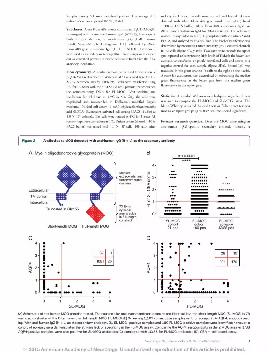
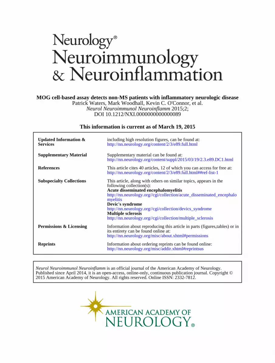
Related Documents