Garraway, Sandra M. and Shawn Hochman. Modulatory actions of serotonin, norepinephrine, dopamine, and acetylcholine in spinal cord deep dorsal horn neurons. J Neurophysiol 86: 2183–2194, 2001. The deep dorsal horn represents a major site for the integration of spinal sensory information. The bulbospinal monoamine transmit-ters, released from sero- tonergic, noradrenergic, and dopaminergic systems, exert modulatory con- trol over spinal sensory systems as does acetylcholine, an intrinsic spinal cord biogenic amine trans-mitter. Whole cell recordings of deep dorsal horn neurons in the rat spinal cord slice preparation were used to compare the cellular actions of serotonin, norepinephrine, dopamine, and acetylcholine on dorsal root stimulation-evoked afferent input and membrane cellular properties. In the majority of neurons, evoked excitatory postsynaptic po- tentials were depressed by the bulbospinal trans-mitters serotonin, norepi- nephrine, and dopamine. Although, the three descending transmitters could evoke common actions, in some neurons, individual transmitters evoked opposing actions. In comparison, acetylcholine generally facilitated the evoked re-sponses, particularly the late, presumably N-methyl-D-aspartate receptor-mediated component. None of the transmitters modified neuronal passive membrane properties. In contrast, in response to depolarizing cur- rent steps, the biogenic amines significantly in-creased the number of spikes in 14/19 neurons that originally fired phasically (P , 0.01). Together, these results demonstrate that even though the deep dorsal horn contains many functionally distinct subpopulations of neurons, the bulbospinal monoamine transmitters can act at both synaptic and cellular sites to alter neuronal sensory integrative properties in a rather predictable manner, and clearly distinct from the actions of acetylcholine. 0022-3077/01 $5.00 Copyright © 2001 The American Physiological Society INTRODUCTION Neurons within the spinal cord represent a primary site for the integration of somatosensory input. Spinal sensory integra-tion is a dynamic process regulated by factors that include multisensory convergence and pathway selection (Baldissera et al. 1981; Jan- kowska 1992; Lundberg 1979), activity-dependent plasticity (see Millan 1999), and neuromodulation (see Randic 1996). Neuro- modulatory responses within the spinal cord in-clude actions me- diated by monoaminergic systems that origi-nate in the brain stem. These bulbospinal monoaminergic nu-clei can be divided into three subtypes by their transmitter phenotype, serotonin (5-HT), norepinephrine (NA), or dopa-mine (DA). Neurons within these nuclei are characterized by The costs of publication of this article were defrayed in part by the payment of page charges. The article must therefore be hereby marked ‘‘advertisement’’ in accordance with 18 U.S.C. Section 1734 solely to indicate this fact. Modulatory Actions of Serotonin, Norepinephrine, Dopamine, and Acetylcholine in Spinal Cord Deep Dorsal Horn Neurons SANDRA M. GARRAWAY AND SHAWN HOCHMAN Department of Physiology, University of Manitoba, Winnipeg, Manitoba R3E 0W3, Canada; and Department of Physiology, Emory University, Atlanta, Georgia 30322 Received 20 December 2000; accepted in final form 25 July 2001 www.jn.org their widespread projections throughout the spinal cord (e.g., Clark and Proudfit 1991, 1993; Holstege et al. 1996; Marlier et al. 1991a). The monoaminergic modulation of two prominent spinal cord functional systems has been examined in some detail. These are the control of motor output and nociception. Generally, the monoamines have been reported to facilitate motor activity and inhibit sensory systems (Basbaum and Fields 1984; Bell and Matsumiya 1981; Jacobs and Fornal 1993; Wallis 1994; Willis and Coggeshall 1991), consistent with a general hypothesis on 5-HT function in the CNS forwarded by Jacobs and Fornal (1993). Because serotoner-gic, noradrenergic, and dopaminergic systems have a simi-larly diffuse distribution in the spinal cord (Clark and Proudfit 1991, 1993; Holstege et al. 1996; Marlier et al. 1991a; Rajaofetra et al. 1989, 1992) and their monoamine transmit- ters frequently exert similar actions (Belcher et al. 1978; Bell and Matsumiya 1981; Headley et al. 1978; Weight and Salmoiraghi 1966), it is possible that these transmitter systems act at similar spinal sites and by similar mechanisms. For example, descending monoaminergic transmitters powerfully inhibit nociceptive informa- tion in neurons by activation of serotonergic 5-HT1A, 5-HT1B, a2- adrenergic, and D2-dopaminergic receptors (Kiritsy-Roy 1994; Per- tovaara 1993; Zemlan 1994) all of which are negatively coupled to adenylate cyclase (reviewed in Barnes and Sharp 1999; Bylund et al. 1994; Vallone et al. 2000). However, the existence of many bul- bospinal monoaminer-gic systems with heterogeneous transmitter phenotypes (in-cluding co-transmitters) that act on a variety of spi- nal metabotropic receptor subtypes (e.g., Huang and Peroutka 1987; Marlier et al. 1991b; Stone et al. 1998; van Dijken et al. 1996), sug- gest that neuromodulation in the spinal cord is a highly differenti- ated process. Indeed, more recent findings indicate that different noradrenergic or serotonergic nuclei can exert opposing modulatory actions on spinal cord noci-ceptive function (Calejesan et al. 1998; Martin et al. 1999). Further, the actions of 5-HT and NA on the afferent-evoked recruitment of functionally identified spinal neu- rons can differ considerably (Bras et al. 1989; Jankowska et al. 1997, 2000). For example, the recruitment of ascending tract neu- rons following primary afferent stimulation is commonly 2183 Address for reprint requests: S. Hochman, Rm. 362, Physiology Building, Emory University School of Medicine, 1648 Pierce Dr., Atlanta, GA 30322 (E-mail: [email protected]).

Welcome message from author
This document is posted to help you gain knowledge. Please leave a comment to let me know what you think about it! Share it to your friends and learn new things together.
Transcript

Garraway, Sandra M. and Shawn Hochman. Modulatory actions of serotonin, norepinephrine, dopamine, and acetylcholine in spinal cord deep dorsal horn neurons. J Neurophysiol 86: 2183–2194, 2001. The deep dorsal horn represents a major site for the integration of spinal sensory information. The bulbospinal monoamine transmit-ters, released from sero-tonergic, noradrenergic, and dopaminergic systems, exert modulatory con-trol over spinal sensory systems as does acetylcholine, an intrinsic spinal cord biogenic amine trans-mitter. Whole cell recordings of deep dorsal horn neurons in the rat spinal cord slice preparation were used to compare the cellular actions of serotonin, norepinephrine, dopamine, and acetylcholine on dorsal root stimulation-evoked afferent input and membrane cellular properties. In the majority of neurons, evoked excitatory postsynaptic po-tentials were depressed by the bulbospinal trans-mitters serotonin, norepi-nephrine, and dopamine. Although, the three descending transmitters could evoke common actions, in some neurons, individual transmitters evoked opposing actions. In comparison, acetylcholine generally facilitated the evoked re-sponses, particularly the late, presumably N-methyl-D-aspartate receptor-mediated component. None of the transmitters modified neuronal passive membrane properties. In contrast, in response to depolarizing cur-rent steps, the biogenic amines significantly in-creased the number of spikes in 14/19 neurons that originally fired phasically (P , 0.01). Together, these results demonstrate that even though the deep dorsal horn contains many functionally distinct subpopulations of neurons, the bulbospinal monoamine transmitters can act at both synaptic and cellular sites to alter neuronal sensory integrative properties in a rather predictable manner, and clearly distinct from the actions of acetylcholine.
0022-3077/01 $5.00 Copyright © 2001 The American Physiological Society
INTRODUCTION
Neurons within the spinal cord represent a primary site for the integration of somatosensory input. Spinal sensory integra-tion is a dynamic process regulated by factors that include multisensory convergence and pathway selection (Baldissera et al. 1981; Jan-kowska 1992; Lundberg 1979), activity-dependent plasticity (see Millan 1999), and neuromodulation (see Randic 1996). Neuro-modulatory responses within the spinal cord in-clude actions me-diated by monoaminergic systems that origi-nate in the brain stem. These bulbospinal monoaminergic nu-clei can be divided into three subtypes by their transmitter phenotype, serotonin (5-HT), norepinephrine (NA), or dopa-mine (DA). Neurons within these nuclei are characterized by
The costs of publication of this article were defrayed in part by the payment of page charges. The article must therefore be hereby marked ‘‘advertisement’’ in accordance with 18 U.S.C. Section 1734 solely to indicate this fact.
Modulatory Actions of Serotonin, Norepinephrine, Dopamine, andAcetylcholine in Spinal Cord Deep Dorsal Horn Neurons
SANDRA M. GARRAWAY AND SHAWN HOCHMANDepartment of Physiology, University of Manitoba, Winnipeg, Manitoba R3E 0W3, Canada; and Department of Physiology,Emory University, Atlanta, Georgia 30322Received 20 December 2000; accepted in final form 25 July 2001
www.jn.org
their widespread projections throughout the spinal cord (e.g., Clark and Proudfit 1991, 1993; Holstege et al. 1996; Marlier et al. 1991a).
The monoaminergic modulation of two prominent spinal cord functional systems has been examined in some detail. These are the control of motor output and nociception.Generally, the monoamines have been reported to facilitate motor activity and inhibit sensory systems (Basbaum and Fields 1984; Bell and Matsumiya 1981; Jacobs and Fornal 1993; Wallis 1994; Willis and Coggeshall 1991), consistent with a general hypothesis on 5-HT function in the CNS forwarded by Jacobs and Fornal (1993). Because serotoner-gic, noradrenergic, and dopaminergic systems have a simi-larly diffuse distribution in the spinal cord (Clark and Proudfit 1991, 1993; Holstege et al. 1996; Marlier et al. 1991a; Rajaofetra et al. 1989, 1992) and their monoamine transmit-ters frequently exert similar actions (Belcher et al. 1978; Bell and Matsumiya 1981; Headley et al. 1978; Weight and Salmoiraghi 1966), it is possible that these transmitter systems act at similar spinal sites and by similar mechanisms. For example, descending monoaminergic transmitters powerfully inhibit nociceptive informa-tion in neurons by activation of serotonergic 5-HT1A, 5-HT1B, a2-adrenergic, and D2-dopaminergic receptors (Kiritsy-Roy 1994; Per-tovaara 1993; Zemlan 1994) all of which are negatively coupled to adenylate cyclase (reviewed in Barnes and Sharp 1999; Bylund et al. 1994; Vallone et al. 2000). However, the existence of many bul-bospinal monoaminer-gic systems with heterogeneous transmitter phenotypes (in-cluding co-transmitters) that act on a variety of spi-nal metabotropic receptor subtypes (e.g., Huang and Peroutka 1987; Marlier et al. 1991b; Stone et al. 1998; van Dijken et al. 1996), sug-gest that neuromodulation in the spinal cord is a highly differenti-ated process. Indeed, more recent findings indicate that different noradrenergic or serotonergic nuclei can exert opposing modulatory actions on spinal cord noci-ceptive function (Calejesan et al. 1998; Martin et al. 1999). Further, the actions of 5-HT and NA on the afferent-evoked recruitment of functionally identified spinal neu-rons can differ considerably (Bras et al. 1989; Jankowska et al. 1997, 2000). For example, the recruitment of ascending tract neu-rons following primary afferent stimulation is commonly
2183
Address for reprint requests: S. Hochman, Rm. 362, Physiology Building,Emory University School of Medicine, 1648 Pierce Dr., Atlanta, GA 30322 (E-mail: [email protected]).
O2-5% CO2. For experimentation, spinal cord slices were affixed to a record-ing chamber using platinum U frames with a parallel array of nylon fibers glued across (Edwards et al. 1989). Patch electrodes were prepared from 1.5-mm OD capillary tubes (Precision Instruments or Warner) pulled in a two-stage process (Narishige PP83) producing resistance values ranging from 4 to 7 MV with recording solution containing (in mM) 140 K-gluconate, 0.2 EGTA, 10 HEPES, 4 Mg-ATP, and 1 GTP; pH 7.3. The re-cording chamber was continu-ously superfused with oxygenated normal ACSF at a rate of ;2 ml/min. The whole cell “blind” patch-clamp recording technique (Blanton et al. 1989) was undertaken at room temperature (;20°C) using the Axopatch 1D amplifier (Axon Instruments) filtered at 5 kHz (4-pole low-pass Bessel). Voltage- and current-clamp data were ac-quired on com-puter with the pCLAMP acquisition software (v 6.0; Axon Instruments).
Determination of cell membrane propertiesImmediately following rupture of the cell membrane (in voltage clamp at
290 mV), the current-clamp recording configuration was used to determine resting membrane potential. Series resistance was subtracted in current-clamp mode (bridge balance), and junction po-tentials were measured and subtracted off-line. For the duration of the experiment, leak conductance and bridge balance were monitored; if their values were largely unaltered, the experiments were continued. Mean electrode series resistance was 33 6 4 (SD) MV (n 5 37). At an adjusted membrane potential of 270 mV, a series of hyperpolar-izing and depolarizing current steps were undertaken to obtain esti-mates of membrane time constant, cell resistance, rheobase, voltage threshold, action potential height, and action potential duration at half-maximal amplitude (half-width). For details on the estimation of these mem-brane properties, refer to Hochman et al. (1997).
Primary afferent stimulationPrimary afferents were stimulated electrically with a constant cur-rent
stimulator (Eide 1972), using bipolar tungsten electrodes. In the present comparative study, we used high stimulation intensities to recruit the highest threshold unmyelinated afferents and, hence, the majority of afferent fiber types, irrespective of age (typically $500 mA, 500 ms) (see Thompson et al. 1990). In the present sample, 29% of the neurons received synaptic responses at intensities ,500mA, 100ms; 49% received synaptic input at 500mA, 100ms, while the remaining 22% of neurons only received input at intensities $500 mA, 500 ms. Generally, the evoked synaptic responses were first char-acterized as excitatory by determining their reversal potential prior to collec-tion of baseline events. Neurons with short-latency inhibitory synaptic re-sponses were not included in this study. Excita-tory postsynaptic potentials (EPSPs) were evoked at low frequencies (once every 20–60 s) by stimulating dorsal rootlets for a baseline period of 10–15 min while maintaining the neuron at a holding potential of 290 mV. In all cases, membrane potential was carefully monitored, and any alterations in membrane potential were noted, then countered with intracellular current injection to maintain a hold-ing potential of 290 mV.
Application of agonists5-hydroxytryptamine HCl (5-HT), norepinephrine bitartrate (NA), dopa-
mine HCl (DA), and acetylcholine chloride (ACh) were obtained from RBI/Sigma. The solutions were prepared on the day of the experiment from 10 mM frozen stock solutions and bath applied at a final concentration of 10 mM. Ascorbic acid (100 mM), an antioxidant, was added to solutions con-taining 5-HT, NA, and DA to prevent their oxidation (Krnjevic et al. 1978). A 10 mM concentration was chosen based on previous studies involving bath application of NA, 5-HT, and ACh (e.g., Baba et al. 2000; Lopez-Garcia and King 1996;
2184 S. M. GARRAWAY AND S. HOCHMAN
facilitated by 5-HT yet depressed by NA (Jankowska et al.1997).
The ester amine acetylcholine (ACh) also modulates spinal sen-sory processing in the dorsal horn (Myslinski and Randic 1977; Urban et al. 1989). As it appears that there are no descending cho-linergic systems in the rat (refer to Willis and Coggeshall 1991, but see Bowker et al. 1983), these actions probably arise from a popula-tion of intrinsic cholinergic inter-neurons found in the dorsal horn (Barber et al. 1984; Todd 1991).
Several studies have compared the actions of these biogenic amine transmitters on the modulation of sensory input onto spinal neurons (Belcher et al. 1978; Bras et al. 1989; Headley et al. 1978; Jankowska et al. 1997; Skoog and Noga 1995; Todd and Millar 1983; Weight and Salmoiraghi 1966; Will-cockson et al. 1984). However, in these studies, only modifi-cations in extracellular spik-ing or field potentials were re-corded and transmitters were applied by iontophoresis (but see Bras et al. 1989). While it is apparent from these studies that the transmitters have both common and dis-tinct actions on the modulation of spinal sensory input, the effects of monoamines on intrinsic cellular properties and synaptic poten-tials in indi-vidual neurons were not studied. Clearly, additional insight into monoamine transmitter function may be achieved by a more direct examination of their actions with intracellular record-ings (e.g., Khasabov et al. 1998, 1999; Lopez-Garcia 1998; Lopez-Garcia and King 1996).
Therefore in this study, we compared the effects of bath-applied 5-HT, NA, DA, and ACh on cellular properties and primary afferent-evoked synaptic responses in individual deep dorsal horn (DDH) neurons using whole cell patch-clamp recordings (see Gar-raway et al. 1997; Hochman et al. 1997). Neurons in the DDH rep-resent a functionally heterogeneous population, which include sev-eral classes of ascending tract cells and spinal interneurons (Willis and Coggeshall 1991) and may thereby account for the diversity in intrinsic and synaptic properties observed from neurons in this re-gion (e.g., Jiang et al. 1995; Morisset and Nagy 1998, 1999; also refer to Hoch-man et al. 1997). Parts of these results have been pre-sented in abstract form (Garraway and Hochman 1999).
METHODS
Preparation of spinal cord slicesAll experimental procedures complied with the Canadian Council of
Animal Care guidelines. Neonatal rats (Sprague-Dawley postnatal days 10–14) were first anesthetized with 10% urethan (2 mg/kg ip body wt) and decapitated, and spinal segments L2–S1were removed. The isolated spinal cord was embedded in Agar, 2.5% wt/vol (Type E,Sigma), and sliced on a vibrating blade microtome in 500–600 mm trans-verse sections (Leica VT1000S or Pelco 101) in cooled (,4°C) oxygenated high sucrose-containing artificial cerebrospinal fluid (ACSF) containing (in mM) 250 sucrose, 2.5 KCl, 1 CaCl2, 3 MgCl2, 25 glucose, 1.25 NaH2PO4, and 26 NaHCO3at a pH of 7.4. Short dorsal rootlets remained attached to the spinal segments to allow for electrical stimulation of primary afferents.
ElectrophysiologySlices were incubated at 32°C for$1 h in normal ACSF containing (in
mM) 125 NaCl, 2.5 KCl, 2 CaCl2, 1 MgCl2, 25 glucose, 1.25 NaH2PO4, and 26 NaHCO3at a pH of 7.4 and oxygenated with 95%
J Neurophysiol • VOL 86 • NOVEMBER 2001 • www.jn.org
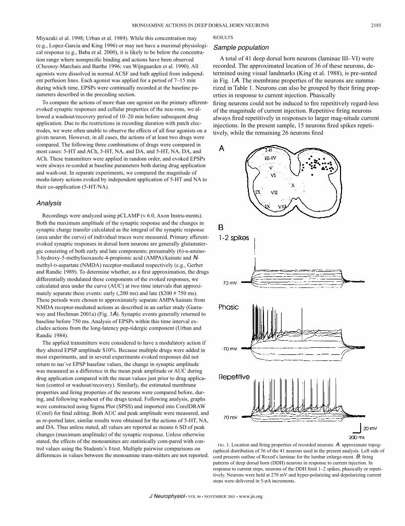
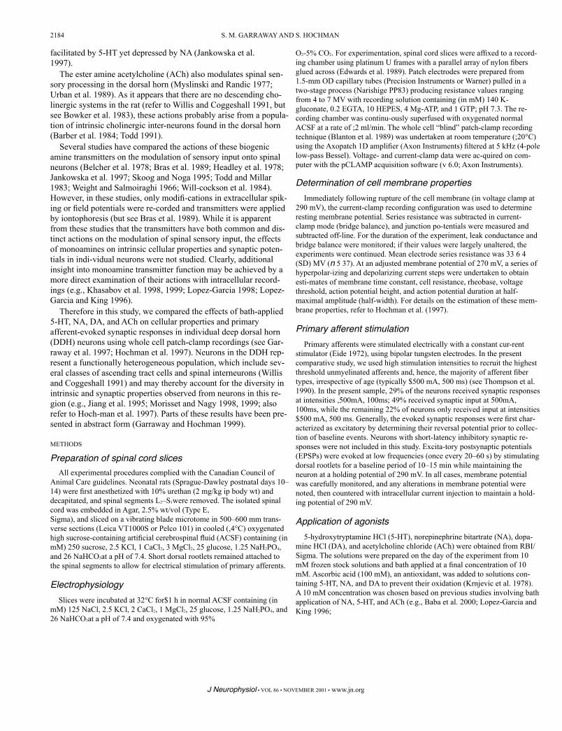
FIG. 1. Location and firing properties of recorded neurons. A: approximate topog-raphical distribution of 36 of the 41 neurons used in the present analysis. Left side of cord presents outline of Rexed’s laminae for the lumbar enlarge-ment. B: firing patterns of deep dorsal horn (DDH) neurons in response to current injection. In response to current steps, neurons of the DDH fired 1–2 spikes, phasically or repeti-tively. Neurons were held at 270 mV and hyper-polarizing and depolarizing current steps were delivered in 5-pA increments.
J Neurophysiol • VOL 86 • NOVEMBER 2001 • www.jn.org
Miyazaki et al. 1998; Urban et al. 1989). While this concentration may (e.g., Lopez-Garcia and King 1996) or may not have a maximal physiologi-cal response (e.g., Baba et al. 2000), it is likely to be below the concentra-tion range where nonspecific binding and actions have been observed (Chesnoy-Marchais and Barthe 1996; van Wijngaarden et al. 1990). All agonists were dissolved in normal ACSF and bath applied from independ-ent perfusion lines. Each agonist was applied for a period of 7–15 min during which time, EPSPs were continually recorded at the baseline pa-rameters described in the preceding section.
To compare the actions of more than one agonist on the primary afferent-evoked synaptic responses and cellular properties of the neu-rons, we al-lowed a washout/recovery period of 10–20 min before subsequent drug application. Due to the restrictions in recording duration with patch elec-trodes, we were often unable to observe the effects of all four agonists on a given neuron. However, in all cases, the actions of at least two drugs were compared. The following three combinations of drugs were compared in most cases: 5-HT and ACh, 5-HT, NA, and DA, and 5-HT, NA, DA, and ACh. These transmitters were applied in random order, and evoked EPSPs were always re-corded at baseline parameters both during drug application and wash-out. In separate experiments, we compared the magnitude of modu-latory actions evoked by independent application of 5-HT and NA to their co-application (5-HT/NA).
Analysis
Recordings were analyzed using pCLAMP (v 6.0, Axon Instru-ments). Both the maximum amplitude of the synaptic response and the changes in synaptic charge transfer calculated as the integral of the synaptic response (area under the curve) of individual traces were measured. Primary afferent-evoked synaptic responses in dorsal horn neurons are generally glutamater-gic consisting of both early and late components; presumably (6)-a-amino-3-hydroxy-5-methylisoxasole-4-propionic acid (AMPA)/kainate and N-methyl-D-aspartate (NMDA) receptor-mediated respectively (e.g., Gerber and Randic 1989). To determine whether, as a first approximation, the drugs differentially modulated these components of the evoked responses, we calculated area under the curve (AUC) at two time intervals that approxi-mately separate these events: early (,200 ms) and late ($200 # 750 ms). These periods were chosen to approximately separate AMPA/kainate from NMDA receptor-mediated actions as described in an earlier study (Garra-way and Hochman 2001a) (Fig. 3A). Synaptic events generally returned to baseline before 750 ms. Analysis of EPSPs within this time interval ex-cludes actions from the long-latency pep-tidergic component (Urban and Randic 1984).
The applied transmitters were considered to have a modulatory action if they altered EPSP amplitude $10%. Because multiple drugs were added in most experiments, and in several experiments evoked responses did not return to naı¨ve EPSP baseline values, the change in synaptic amplitude was measured as a difference in the mean peak amplitude or AUC during drug application compared with the mean values just prior to drug applica-tion (control or washout/recovery). Similarly, the estimated membrane properties and firing properties of the neurons were compared before, dur-ing, and following washout of the drugs tested. Following analysis, graphs were constructed using Sigma Plot (SPSS) and imported into CorelDRAW (Corel) for final editing. Both AUC and peak amplitude were measured, and as re-ported later, similar results were obtained for the actions of 5-HT, NA, and DA. Thus unless stated, all values are reported as means 6 SD of peak changes (maximum amplitude) of the synaptic response. Unless otherwise stated, the effects of the monoamines are statistically com-pared with con-trol values using the Students’s t-test. Multiple pairwise comparisons on differences in values between the monoamine trans-mitters are not reported.
RESULTS
Sample populationA total of 41 deep dorsal horn neurons (laminae III–VI) were
recorded. The approximated location of 36 of these neurons, de-termined using visual landmarks (King et al. 1988), is pre-sented in Fig. 1A. The membrane properties of the neurons are summa-rized in Table 1. Neurons can also be grouped by their firing prop-erties in response to current injection. Phasicallyfiring neurons could not be induced to fire repetitively regard-less of the magnitude of current injection. Repetitive firing neurons always fired repetitively in responses to larger mag-nitude current injections. In the present sample, 15 neurons fired spikes repeti-tively, while the remaining 26 neurons fired
2185 MONOAMINE ACTIONS IN DEEP DORSAL HORN NEURONS

5-HT (28) 230 6 49* 236 6 48* NA (26) 213 6 42* 220 6 66*DA (20) 210 6 33 217 6 37*
EMR,mV! Rin,mV tm,ms Rheobase, pA! VTH,mV AP Height, mV AP1⁄2-Width, ms
only phasically, 10 of which fired no more than two spikes (1–2 spikes) (refer to Lopez-Garcia and King 1994). Examples of neu-ronal firing patterns are illustrated in Fig. 1B. The effects of the monoamines on neuronal membrane properties will be considered later.
Effects of the monoamine transmitters on primary afferent-evoked EPSPs
The effects of bath-applied 5-HT, NA, DA, and ACh on the dor-sal root stimulation-evoked EPSPs are summarized in Ta-bles 2 and 3 and illustrated in Fig. 2. In the majority of neurons, 5-HT and NA depressed EPSP amplitude (P , 0.05), and a similar trend was ob-served for DA. In comparison ACh facil-itated EPSP amplitude (P , 0.05; Table 3, Fig. 2A). When EPSP AUC values were compared, 5-HT, NA, and DA de-creased while ACh increased AUC values (P , 0.05). These observations demonstrate that despite the heterogene-ous pop-ulation of neurons found in the DDH, the biogenic amine neurotransmitters altered afferent-evoked EPSPs rather pre-dictably with the bulbospinal monoamines having similarly depressant ac-tions. The relative incidence of the transmitters at modulating peak EPSP amplitude were 5-HT . ACh . NA . DA (Table 2). Following washout of NA and DA, partial recovery of EPSP amplitudes gener-ally occurred (Fig. 2B). However, in some neurons, during the washout that followed 5-HT-evoked synaptic depression, a rebound potentiation of EPSP amplitude occurred, usually exceeding control values. In addition, EPSP amplitudes generally remained facilitated fol-lowing washout of ACh.
We also compared the differential effects of the agonists on the early versus the late occurring components of the evoked EPSPs by measuring the AUC for early (,200 ms) and late ($200 # 750 ms) components of the EPSP (Table 4), periods that approximately sepa-rate AMPA/kainate from NMDA re-ceptor-mediated actions (e.g., Fig. 3A) (see also Garraway and
TABLE 2. Incidence of neurons having EPSP amplitude depres-sion or facilitation evoked by 5-HT, NA, DA, and ACh
5-HT (28) 14 79 7 NA (26) 15 62 23 DA (20) 15 55 30 ACh (20) 70 15 15
Agonist EPSP Amplitude,1 EPSP Amplitude,2 No Change
Control (33–41) 259 6 18 507 6 250 44 6 19 57 6 42 26 6 7 94 6 12 3.7 6 2.05-HT (16–28) 258 6 18 509 6 187 42 6 16 57 6 43 26 6 8 78 6 19 4.3 6 2.0NA (20–26) 259 6 18 606 6 288 46 6 26 60 6 47 30 6 12 82 6 12 4.1 6 1.3DA (16–20) 259 6 19 583 6 335 46 6 21 72 6 56 33 6 11 82 6 12 4.8 6 2.4ACh (14–18) 257 6 19 541 6 246 47 6 26 69 6 51 30 6 6 78 6 16 4.2 6 1.4
Values reported are means 6 SD. The range of sample size used to calculate mean values is bracketed in left column. EMR, resting membrane potential; VTH, voltage threshold; Rin, cell input resistance; tm, membrane time constant; AP height, action potential height; AP1⁄2 width, action potential duration at half maximal amplitude. These properties were estimated at an adjusted membrane potential of 270 mV. 5-HT, serotonin; NA, norepinephrine; DA, dopamine; ACh, acetylcholine.
Values are percentage of sample size indicated in brackets. Shaded boxes indicate predominant action for each transmitter. EPSP, excitatory postsynap-tic potential.
J Neurophysiol • VOL 86 • NOVEMBER 2001 • www.jn.org
Agonist Peak Amplitude Total AUC
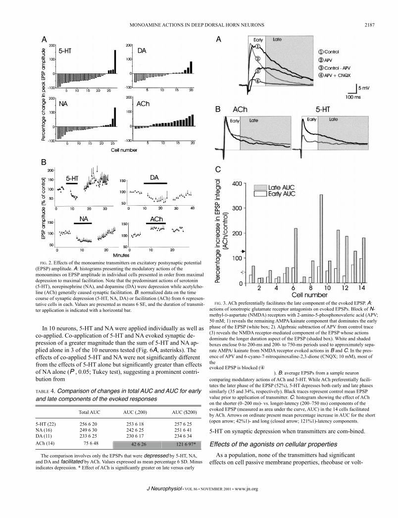
Hochman 2001a). There were no differences in depression between early and late AUC values for 5-HT, NA, and DA (Table 4), sup-porting a uniform depression of both AMPA/ kainate and NMDA receptor-mediated responses. However, AUC measures show that ACh preferentially facilitated the late component of the EPSP sup-porting a preferential facilitation of the NMDA receptor-evoked response (Table 4). Figure 3B illustrates a representative example of the effects of 5-HT and ACh on evoked EPSPs in a given cell. Fig-ure 3C compares the AUC increases for early and late components of the EPSP following application of ACh in individual neurons. Note that in many neurons, the later component of the EPSP is dramat-ically facilitated compared with the early component of the EPSP.
The modulatory actions of ACh and 5-HT were compared in12 neurons (Fig. 4). In six of the nine neurons where EPSPs were depressed by 5-HT, ACh facilitated the EPSPs. Thus 5-HT and ACh have different (P , 0.01; Wilcoxon signed-rank test) and predomi-nantly opposite actions on spinal neu-rons in the deep dorsal horn.
The generally depressant actions of the three descending trans-mitters 5-HT, NA, and DA were compared in 15 neu-rons (Fig. 5). With few exceptions, none of the transmitters had competing modu-latory actions on the evoked EPSPs. In 6 of the 15 neurons, all three transmitters produced synaptic depression while in 1 neuron, all three drugs produced synaptic facilitation. In 8 of 15 neurons, common modula-tory responses were not produced by all three transmitters, but only in 3 of these neurons were opposite actions .10% observed. An example of the common neuromodulatory ac-tions of the brain stem monoamines in a single neuron is presented in Fig. 5B. Thus unlike ACh, which generally supported synaptic facilitation, the three descending mono-amines 5-HT, NA, and DA commonly exerted similar func-tions on spinal cord sensory input to a given cell.
TABLE 3. Effects of 5-HT, NA, DA, and ACh on the evoked synap-tic responses
Values are mean percentages 6 SD of change in EPSP peak amplitude and total AUC. Minus indicates depression. Note that with the exception of DA, the transmit-ters had significant actions on both peak amplitudes and areas under the curve (AUC). * Overall effect of transmitter on changes in EPSP amplitude and AUC is
2186 S. M. GARRAWAY AND S. HOCHMAN
TABLE 1. Membrane properties of recorded neurons in the presence or absence of a biogenic amine
evoked EPSP is blocked (④). B: average EPSPs from a sample neuron
comparing modulatory actions of ACh and 5-HT. While ACh preferentially facili-tates the later phase of the EPSP (52%), 5-HT depresses both early and late phases similarly (35 and 34%, respectively). Black traces represent control mean EPSP value prior to application of transmitter. C: histogram showing the effect of ACh on the shorter (0–200 ms)- vs. longer-latency (200–750 ms) components of the evoked EPSP (measured as area under the curve, AUC) in the 14 cells facilitated by ACh. Arrows on ordinate present mean percentage increase in AUC for the short (open arrow; 42%1)- and long (closed arrow; 121%1)-latency components.
5-HT on synaptic depression when transmitters are com-bined.
Effects of the agonists on cellular propertiesAs a population, none of the transmitters had significant
effects on cell passive membrane properties, rheobase or volt-
FIG. 2. Effects of the monoamine transmitters on excitatory postsynaptic potential (EPSP) amplitude. A: histograms presenting the modulatory actions of the monoamines on EPSP amplitude in individual cells presented in order from maximal depression to maximal facilitation. Note that the predominant actions of serotonin (5-HT), norepinephrine (NA), and dopamine (DA) were depression while acetylcho-line (ACh) generally caused synaptic facilitation. B: normalized data on the time course of synaptic depression (5-HT, NA, DA) or facilitation (ACh) from 6 represen-tative cells in each. Values are presented as means 6 SE, and the duration of transmit-ter application is indicated with a horizontal bar.
ACh (14) 75 6 48
The comparison involves only the EPSPs that were depressed by 5-HT, NA, and DA and facilitated by ACh. Values expressed as mean percentage 6 SD. Minus indicates depression. * Effect of ACh is significantly greater on late versus early
Total AUC AUC (,200) AUC ($200)
FIG. 3. ACh preferentially facilitates the late component of the evoked EPSP. A: actions of ionotropic glutamate receptor antagonists on evoked EPSPs. Block of N-methyl-D-aspartate (NMDA) receptors with 2-amino-5-phosphonovaleric acid (APV; 50 mM; 1) reveals the remaining AMPA/kainate component that dominates the early phase of the EPSP (white box; 2). Algebraic subtraction of APV from control trace (3) reveals the NMDA receptor-mediated component of the EPSP whose actions dominate the longer duration aspect of the EPSP (shaded box). White and shaded boxes enclose 0-to 200-ms and 200- to 750-ms periods used to approximately sepa-rate AMPA/ kainate from NMDA receptor evoked actions in B and C. In the pres-ence of APV and 6-cyano-7-nitroquinoxaline-2,3-dione (CNQX; 10 mM), most of the
5-HT (22) 256 6 20 253 6 18 257 6 25NA (16) 249 6 30 242 6 25 251 6 41DA (11) 233 6 25 230 6 17 234 6 34
42 6 26 121 6 97*
In 10 neurons, 5-HT and NA were applied individually as well as co-applied. Co-application of 5-HT and NA evoked synaptic de-pression of a greater magnitude than the sum of 5-HT and NA ap-plied alone in 3 of the 10 neurons tested (Fig. 6A, asterisks). The effects of co-applied 5-HT and NA were not significantly different from the effects of 5-HT alone but significantly greater than effects of NA alone (P , 0.05; Tukey test), suggesting a prominent contri-bution from
TABLE 4. Comparison of changes in total AUC and AUC for early and late components of the evoked responses
2187 MONOAMINE ACTIONS IN DEEP DORSAL HORN NEURONS
J Neurophysiol • VOL 86 • NOVEMBER 2001 • www.jn.org
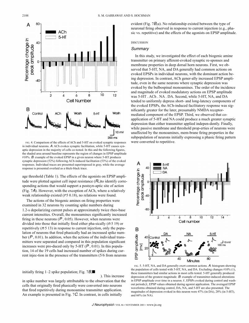
FIG. 5. 5-HT, NA, and DA generally exert common actions. A: histogram showing the population of cells tested with 5-HT, NA, and DA. Excluding changes #10% (1), these transmitters had similar actions in most cells tested. 5-HT generally produced depression of the greatest magnitude. B: example of transmitter-induced alterations in EPSP amplitude over time in a neuron. F, EPSPs evoked during control and wash-out periods;E, EPSP values obtained during agonist application. The averaged EPSP waveforms obtained during control, DA, NA, and 5-HT are also presented. The magnitudes of depression evoked in this neuron were 47% (in DA), 28% (in 5-HT), and 66% (in NA).
J Neurophysiol • VOL 86 • NOVEMBER 2001 • www.jn.org
FIG. 4. Comparison of the effects of ACh and 5-HT on evoked synaptic responses in individual neurons. A: ACh evokes synaptic facilitation, while 5-HT causes syn-aptic depression in the majority of cells co-tested. In this and the following figures, the shaded area around baseline represents the region of changes in EPSP amplitude #10%. B: example of the evoked EPSP in a given neuron where 5-HT produces synaptic depression (52%) following ACh-induced facilitation (53%) of the evoked responses. Individual traces are presented superimposed in gray, while the average response is presented overlaid as a thick-black trace.
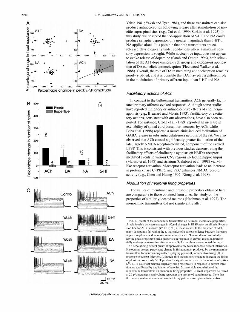
age threshold (Table 1). The effects of the agonists on EPSP ampli-tude were plotted against cell input resistance (Rin)to identify corre-sponding actions that would support a postsyn-aptic site of action (Fig. 7A). However, with the exception of ACh, where a relatively weak relationship existed (r25 0.18), no relations were found.
The actions of the biogenic amines on firing properties were examined in 32 neurons by counting spike numbers during1.2-s depolarizing current pulses at approximately twice rheo-base current intensities. Overall, the monoamines significantly increased firing in these neurons (P , 0.05). However, when neurons were divided into those that initially fired either pha-sically (n 5 19) or repetitively (n 5 13) in response to current injection, only the popu-lation of neurons that fired phasically had an increased spike num-ber (P , 0.01). In addition, when the actions of the individual trans-mitters were separated and compared in this population significant increases were pro-duced only by 5-HT (P , 0.01). In this popula-tion, 14 of the 19 cells had increased number of spikes during cur-rent injec-tion in the presence of the transmitters (5/6 from neurons
2188 S. M. GARRAWAY AND S. HOCHMAN
initially firing 1–2 spike population; Fig. 7B,■). This increase
in spike number was largely attributable to the observation that the cells that originally fired phasically were converted into neurons that fired repetitively during monoamine transmitter application. An example is presented in Fig. 7C. In contrast, in cells initially
evident (Fig. 7B,u). No relationship existed between the type of neuronal firing observed in response to current injection (e.g., pha-sic vs. repetitive) and the effects of the agonists on EPSP amplitude.
DISCUSSION
SummaryIn this study, we investigated the effect of each biogenic amine
transmitter on primary afferent-evoked synaptic re-sponses and membrane properties in deep dorsal horn neurons. First, we ob-served that 5-HT, NA, and DA generally had common actions on evoked EPSPs in individual neurons, with the dominant action be-ing depression. In contrast, ACh gener-ally increased EPSP ampli-tude, even in the same neurons where synaptic depression was evoked by the bulbospinal monoamines. The order of the incidence and magnitude of evoked modulatory actions on EPSP amplitude was 5-HT . ACh . NA . DA. Second, while 5-HT, NA, and DA tended to uniformly depress short- and long-latency components of the evoked EPSPs, the ACh-induced facilitatory response was sig-nificantly greater for the later, presumably NMDA receptor-mediated component of the EPSP. Third, we observed that co-application of 5-HT and NA could produce a much greater synaptic depression than either transmitter applied indepen-dently. Finally, while passive membrane and threshold prop-erties of neurons were unaffected by the monoamines, mem-brane firing properties in the subpopulation of neurons initially expressing a phasic firing pattern were converted to repetitive.

2189
FIG. 6. Co-application of NA and 5-HT can produce greater depression than either 5-HT or NA alone. A: histogram showing population of cells tested with NA/5-HT. In 3 of 10 cells (asterisk), NA/5-HT-induced a synaptic depression greater than the sum of each agonist applied alone. B: evoked EPSPs obtained from cell 2 in histo-gram above. Note that the depression of theEPSP is greater during co-application of 5-HT and NA (65%) than either drug ap-plied alone (31 and 23%, respectively). Individual traces are presented superimposed in gray, while the average response is presented overlaid as a thick-black trace.
Common actions of the monoamines on evoked EPSPs in individual neurons
Consistent with previous in vitro studies, we observed that 5-HT generally produced synaptic depression (e.g., Khasabov et al. 1999; Lopez-Garcia 1998; Lopez-Garcia and King 1996) although facili-tation was observed in a few cells. Like 5-HT, bath application of NA and DA also produced synaptic depres-sion in most cells, con-sistent with depressant actions observed in previous studies that monitored alterations in firing fre-quency (Fleetwood-Walker et al. 1988; Headley et al. 1978; Skoog and Noga 1995; Willcockson et al. 1984). An important observation in this study is that 5-HT, NA, and DA commonly produced the same modulatory action on EPSPs when applied independently to the same neuron. Thus it appears that despite the diverse functional heterogeneity of the spinal cord dorsal horn (Baldissera et al. 1981; Jankowska 1992; Willis and Coggeshall 1991), the bulbospinal monoamine transmitters can produce a widespread depression of sensory synaptic input onto deep dorsal horn neurons. It is, however, necessary to mention, that in addition to the common actions generally evoked by the three transmitters arising from brain stem nuclei on individual neurons, these transmitters were also capable of inducing distinct modulatory actions on some neurons.
Despite the similar actions of the three transmitters arising from the brain stem, the magnitudes of synaptic depression differed in order of 5-HT . NA . DA. In addition, the depression evoked by 5-HT was more widespread as a greater proportion of cells underwent synaptic depression by 5-HT. The differences in depression may reflect the relative effec-tiveness of different bulbospinal systems in mediating modu-latory actions. Alternatively, the relative magnitude of effects may be due to differences in potency, as the transmitters may not be working at the same relative point on their dose-response curves (e.g., Elliot and Wallace 1992).
The actions of 5-HT and NA on antinociception have been exten-sively studied (Basbaum and Fields 1984; Fitzgerald 1986; Jones 1991; Millan 1995). Previous studies have dem-onstrated that nox-ious input leads to the release of both NA and 5-HT in the spinal cord (Satoh and Omote 1996; Tyce and
In contrast to the depressant actions of the monoamines observed here, Jankowska and colleagues (1997, 2000) dem-onstrated that nonnociceptive afferent input from different afferents to different groups of spinal interneurons or ascend-ing tract cells is modulated by NA and 5-HT in a highly differentiated manner. Depending on the neuron and afferent fiber type, they observed that NA and 5-HT could have com-mon facilitatory, inhibitory, or opposing modula-tory actions on synaptic input strength as measured using peristimu-lus time histogram measures of extracellular spike latency and fre-quency. An explanation for the observed differences between their results and ours is our stimulation at high intensities to also recruit high-threshold C and Ad fibers, which comprise the largest fraction of primary afferent fibers (Snider and McMa-hon 1998; Willis and Coggeshall 1991). Therefore the strong depressant actions of the monoamines in our study may result from a dominating activation of high-threshold afferents that mask more subtle differential modu-latory actions on the low-threshold afferents studied by Jankowska and colleagues. An-other explanation for our observed differences may relate to our finding that EPSP amplitude can decrease con-comitant with increases in firing postsynaptically. Together, these stud-ies support a complex and functionally differentiated modula-tion of sensory-evoked firing properties in spinal cord neurons.
J Neurophysiol • VOL 86 • NOVEMBER 2001 • www.jn.org
MONOAMINE ACTIONS IN DEEP DORSAL HORN NEURONS
The bulbospinal monoamine transmitter systems project widely throughout the spinal cord (Holstege et al. 1996; Mar-lier et al. 1991a; Rajaofetra et al. 1992), and there are many serotonergic, noradrenergic, and dopaminergic receptors in the dorsal horn (e.g., Huang and Peroutka 1987; Marlier et al. 1991b; van Dijken et al. 1996). Hence, the effects of the bath-applied bulbospinal monoamine transmitters observed here probably reflect the actions of these transmitters on their respective families of receptors. How-ever, it is also possible that part of the observed depression of the longer-latency portion of the EPSP is due to the transmitter having a direct voltage-dependent block of the NMDA receptor ionophore (Chesnoy-Marchais and Barthe 1996). That 5-HT, NA, and DA had common actions in most cells suggests that receptors for all three transmitters are co-localized on many neurons and/or primary affer-ent terminals. In another study, we have demon-strated that the syn-aptic depression produced by 5-HT in sim-ilarly located neurons is only partly mediated by 5-HT1A receptors while 5-HT7receptors are predominantly responsible for producing synaptic facilitation (Gar-raway and Hochman 2001b).
Facilitatory actions of AChIn contrast to the bulbospinal transmitters, ACh generally facili-
tated primary afferent-evoked responses. Although some studies have reported inhibitory or antinociceptive effects of cholinergic agonists (e.g., Bleazard and Morris 1993), facilita-tory or excita-tory actions, consistent with our observations, have also been re-ported. For instance, Urban et al. (1989) reported an increase in excitability of spinal cord dorsal horn neurons by ACh, while Baba et al. (1998) reported a musca-rinic-induced facilitation of GABA release in substantia gelati-nosa neurons of the rat. We also observed that ACh caused significantly greater facilitation of the late, largely NMDA receptor-mediated, component of the evoked EPSP. This is consistent with previous studies demonstrating the facilitatory effects of cholinergic agonists on NMDA receptor-mediated events in various CNS regions including hippocampus (Marino et al. 1998) and striatum (Calabresi et al. 1998) via M1-like receptor activation. M1receptor activation leads to an increase in protein kinase C (PKC), and PKC enhances NMDA receptor activity (e.g., Chen and Huang 1992; Xiong et al. 1998).
Modulation of neuronal firing propertiesThe values of membrane and threshold properties obtained here
are comparable to those obtained from an earlier study on the properties of similarly located neurons (Hochman et al. 1997). The monoamine transmitters did not significantly alter
J Neurophysiol • VOL 86 • NOVEMBER 2001 • www.jn.org
Yaksh 1981; Yaksh and Tyce 1981), and these transmitters can also produce antinociception following release after stimula-tion of spe-cific supraspinal sites (e.g., Cui et al. 1999; Sorkin et al. 1993). In this study, we observed that co-application of 5-HT and NA could produce synaptic depression of a greater magnitude than 5-HT or NA applied alone. It is possible that both transmitters are co-released physiologically under condi-tions where a maximal sen-sory depression is sought. While nociceptive input does not appear to evoke release of dopamine (Satoh and Omote 1996), both stimu-lation of the A11 dopa-minergic cell group and exogenous applica-tion of DA can elicit antinociception (Fleetwood-Walker et al. 1988). Overall, the role of DA in mediating antinociception remains poorly stud-ied, and it is possible that DA may play a different role in the modulation of primary afferent input than 5-HT and NA.
2190 S. M. GARRAWAY AND S. HOCHMAN
FIG. 7. Effects of the monoamine transmitters on neuronal membrane prop-erties. A: relationship between changes in Rinand changes in EPSP peak amplitude. Regres-sion line for ACh is shown (r25 0.18; NS).4, mean values. In the presence of ACh, many data points fall within the 1, indicative of a correspondence between increases in peak amplitude and increases in input resistance. B: several neurons initially having phasic repetitive firing properties in response to current injection preferen-tially undergo increases in spike numbers. Spike numbers were counted during a 1.2-s depolarizing current pulses at approximately twice rheobase current intensities. Histograms present percentage change in firing number produced by the monoamine transmitters for neurons originally displaying phasic (■) or repetitive firing (1) in response to current injection. Although all 4 transmitters tended to increase the firing of phasic neurons, only 5-HT produced a significant increase in the number of spikes (P , 0.01). Note that neurons originally firing repetitively in response to current injec-tion are unaffected by application of agonist. C: reversible modulation of the monoamine transmitters on membrane firing properties. Current steps were delivered at 20-pA increments and voltage responses are presented superimposed. Note that the bulbospinal monoamines converted firing patterns from phasic to repetitive.

guinea pig (Travagli and Williams 1996), while presynaptic D2 do-pamine receptor mediates depression of spinal reflexes (Ga-jendiran et al. 1996). In addition, many monoaminergic recep-tors are pre-sent on primary afferent terminals (e.g., Daval et al. 1987; Hamon et al. 1989; Kidd et al. 1993; Ridet et al. 1994; Stone et al. 1998), and application of 5-HT has been shown to generate a primary afferent depolarization (PAD) that corre-sponds to presynaptic inhibition of primary afferents (Khas-abov et al. 1998, 1999; Lopez-Garcia and King 1996). Inter-estingly, Khasabov et al. (1998) demonstrated that capsaicin treatment, which selectively destroys unmyelinated pri-mary afferent fibers, significantly reduced 5-HT-induced PAD in the neonatal rat. Thus 5-HT may mediate spinal inhibitory actions by reducing transmitter release from nociceptors. The ability of the monoamine transmitters to reduce nociceptive transmitter release at the first CNS synaptic input site supports a critical role in antino-ciceptive function.
In contrast to the bulbospinal monoaminergic transmitters,ACh probably mediates its facilitatory actions predominantly post-synaptically. ACh had a preferential facilitatory action on the late, presumably NMDA receptor-mediated, component of the EPSP, consistent with observed modulatory actions of M1 muscarinic re-ceptor activation on NMDA receptor activity (Calabresi et al. 1998; Marino et al. 1998). If facilitation in synaptic strength involved only presynaptic mechanisms that increase glutamate release, one would expect to see a uniform facilitation of both AMPA/kainate and NMDA receptor-medi-ated responses. Interestingly, the presumed postsynaptic action of ACh in this study is consistent with the dominant postsyn-aptic localization of cholinergic receptors in the spinal cord dorsal horn (Gillberg and Askmark 1991), particularly the muscarinic receptors which are expressed about two to three times higher than the nicotinic receptors (Gillberg et al. 1988).
Experimental issuesThere are several potential limitations to our experimental ap-
proach. 1) Bath-applied transmitters will activate all their respective receptors simultaneously, whereas physiologically, it is possible that there is some degree of preferential activation of receptor subtypes by separate descending serotonergic sys-tems (Wei et al. 1999). Thus physiological conditions may exist where synaptic facilitation dominates over the depression observed in this study. Nonetheless, for 5-HT at least, the dorsal horn contains many 5-HT varicosities that end blindly and do not form conventional synapses (Poulat et al. 1992; Ridet et al. 1994) and in this regard have been suggested to modulate spinal activity via volume transmission (Zoli and Ag-nati 1996). Thus if 5-HT and the other descending mono-amine transmitters act via volume transmission, their actions would non-specifically activate all receptor subtypes. Interest-ingly, in this re-gard, the net effect in most neurons would be a significant synaptic depression. 2) Our use of high-intensity electrical stimulation to recruit the majority of afferents (Thompson et al. 1990) prevents a comparison of modulatory actions between specific afferent fiber populations. For exam-ple, Jankowska and co-workers (Jankowska et al. 1995, 1997, 2000; Noga et al. 1992; Riddell et al. 1993) dem-onstrated that 5-HT and raphe-spinal stimulation can exert a differ-ential control over primary afferents of different modalities. 3) The DDH spinal region is functionally heterogeneous, yet we
J Neurophysiol • VOL 86 • NOVEMBER 2001 • www.jn.org
MONOAMINE ACTIONS IN DEEP DORSAL HORN NEURONS 2191
the passive membrane or threshold properties of DDH neurons. However, in neurons that originally fired phasically, the mono-amines could reversibly transform firing behavior from phasic to repetitive. These neurons did not display accelerated dis-charges in response to injection of depolarizing current steps or continued membrane depolarization and firing after current step termination. Thus the increased duration of firing is un-likely to be due to the activation of a plateau potential (see Morisset and Nagy 1998). In-terestingly, Lopez-Garcia andKing (1994) showed that firing patterns in response to current injec-tion are functionally correlated to the source of primary afferent input. For example, wide dynamic range (WDR) neu-rons receive convergent input from both low- and high-thresh-old afferents, and these cells generally fire repetitively. If we applied this classification to the neuronal firing properties observed following the application of monoamines, the mono-amines could convert the functional properties of neurons from those that previously had restricted sen-sory convergence to a WDR profile. Interestingly, the majority of neurons in the dorsal horn of awake sheep, obviously having normal bul-bospinal activity, are WDR (Herrero and Headley 1995). It is also possible that the monoamines do not alter convergent proper-ties to DDH neurons but rather, the classification scheme developed by Lopez-Garcia and King (1994) does not apply to the behavior of neurons in the presence of monoamine trans-mitters.
It may seem inconsistent that the changes in neuronal firing prop-erties were unrelated to the observed changes in EPSP amplitude. However, since different receptor subtypes may be found at pre- and postsynaptic sites, it is not surprising that the monoamine transmit-ters exert different actions on the synaptic and firing properties of spinal neurons. For instance, while one class of receptor may de-press sensory input (e.g., Khasabov et al. 1999), another class may increase the excitability of these neurons (e.g., Wallis et al. 1991). Opposing pre- and postsyn-aptic actions may be a widely employed strategy to alter the network properties of spinal neurons. For ex-ample, a depressed sensory input with increased neuronal respon-siveness could support a transfer of control from peripheral to de-scending command systems.
Possible sites of actionAlthough the bulbospinal monoamines are capable of exert-ing
direct postsynaptic actions, several observations suggest that the depression they produced on EPSPs is mediated via presynaptic mechanisms. First, none of these transmitters had effects on the passive membrane properties that would support a reduction in EPSP amplitudes by postsynaptic mechanisms (i.e., a decreased tmor Rin). In addition, the identical percent depression on early, presum-able AMPA/kainate and late, pre-sumably NMDA receptor-mediated, components can be ex-plained by a reduction in gluta-mate transmitter release. Con-sistent with a presynaptic site of ac-tion, all three transmitters, 5-HT, NA, and DA, have been shown to depress synaptic responses presynaptically as a result of their ability to increase potassium conductance (e.g., North and Yoshimura 1984, see also Barnes and Sharp 1999) or by inhibiting both N- and L-type calcium channels (e.g., Wikstro¨m et al. 1999). Other studies suggested that 5-HT and NA may mediate presynaptic inhibition of glutamate release from primary afferents in the

2192 S. M. GARRAWAY AND S. HOCHMAN
treated all dorsal horn neurons as a single population. Jankowska and co-workers (2000) have demonstrated that the actions of 5-HT may depend on the functional identity of the spinal neuron studied. 4) The animals were recorded from immature rats (P10–14), and hence the actions may be differ-ent in adult rats. However, this is the preferred age range to study sensory integrative mechanisms in vitro due to its near-intact circuitry, and near mature developmental status (Fitzger-ald 1985; Fitzgerald and Koltzenburg 1986; Lopez-Garcia and King 1994; Thompson et al. 1990; Woolf and King 1987).Furthermore, the modulatory actions observed here are un-likely to differ from those seen in the adult. If we take 5-HT as an example, 5-HT immunoreactivity is found in the gray matter at all spinal cord levels at birth, and staining density increases, peaking at P7 in cer-vical and P14 in lumbar cord (Bregman 1987). In addition, Wallis et al. (1993) showed that the strong descending inhibition of the monosynaptic reflex in the P1 rat is mediated by serotonin. Thus at least for 5-HT, descending modulatory systems are present at birth (but see Fitzgerald and Koltzenburg 1986).
The aforementioned limitations notwithstanding, the present ob-servations demonstrate that the descending monoamine transmitters 5-HT, NA, and DA have broadly similar depres-sant actions in the spinal cord and on the same neurons, and these actions are opposite to the facilitatory actions of ACh, which is normally released from intrinsic spinal neurons. That these effects were similar in the ma-jority of neurons sampled suggests that descending monoaminergic systems are capable of modulating spinal sensory integration in a diffuse and gen-eral matter.
SignificanceIn conclusion, we provide the first comparative analysis of the
actions of the biogenic amine transmitters on synaptic and cellular properties of DDH spinal neurons. 5-HT, NA, and DA are involved in the descending control of spinal sensory inte-gration, and the present observations suggest that the separate brain stem monoaminergic systems can affect spinal sensory integration in a remarkably similar manner. With respect to the control of sensory input, these studies increase our understand-ing of how these trans-mitters act, perhaps to maximize antino-ciceptive actions.
We thank C. Gibbs for expert technical assistance.This work was supported by the Christopher Reeve Paralysis Foundation.
S. M. Garraway obtained studentship salary support from the Rick HansenInstitute.
REFERENCES
BABA H, KOHNO T, OKAMOTO M, GOLDSTEIN PA, SHIMOJI K, AND YOSHIMURA
M. Muscarinic facilitation of GABA release in substantia gelatinosa of the rat spinal dorsal horn. J Physiol (Lond) 508: 83–93, 1998.
BABA H, SHIMOJI K, AND YOSHIMURA M. Norepinephrine facilitates inhibitorytransmission in substantia gelatinosa of adult rat spinal cord. I. Effects on axon terminals of GABAergic and glycinergic neurons. Anesthesiology 92: 473–484, 2000.
BALDISSERA F, HULTBORN H, AND ILLERT M. Integration in spinal neuronalsystems. In: Handbook of Physiology. The Nervous System. Motor Con-
trol.Bethesda, MD: Am. Physiol. Soc., 1981, sect. 1, vol. II, p. 509–595.
BARBER RP, PHELPS PE, HOUSER CR, CRAWFORD GD, SALVATERRA PM, AND
VAUGHN JE. The morphology and distribution of neurons containing choline acetyltransferase in the adult rat spinal cord: an immunocytochemical study.J Comp Neurol 229: 329–346, 1984.
mediate facilitation of a nociceptive reflex by subcutaneous formalin injec-tion into the hindpaw in rats. Brain Res 798: 46–54, 1998.
CHEN L AND HUANG L-YM. Protein kinase C reduces Mg21block of NMDA-receptor channels as a mechanism of modulation. Nature 356: 521–523,1992.
CHESNOY-MARCHAIS D AND BARTHE JY. Voltage-dependent block of NMDAresponses by 5-HT agonists in ventral spinal cord neurones. Br J Pharmacol117: 133–141, 1996.
CLARK FM AND PROUDFIT HK. The projection of noradrenergic neurons in theA7 catecholamine cell group to the spinal cord in the rat demonstrated by antero-grade tracing combined with immunocytochemistry. Brain Res 547: 279–288, 1991.
CLARK FMAND PROUDFIT HK. The projections of noradrenergic neurons in theA5 catecholamine cell group to the spinal cord in the rat: anatomical evidence that A5 neurons modulate nociception. Brain Res 616: 200–210, 1993.
CUI M, FENG Y, MCADOO DJ, AND WILLIS WD. Periaqueductal gray stimula-tion-induced inhibition of nociceptive dorsal horn neurons in rats is associ-ated with the release of norepinephrine, serotonin, and amino acids. J Phar-macol Exp Ther 289: 868–876, 1999.
DAVAL G, VERGE D, BASBAUM AI, BOURGOIN S, AND HAMON M. Autoradio-graphic evidence of serotonin1 binding sites on primary afferent fibres in the dorsal horn of the rat spinal cord. Neurosci Lett 83: 71–76, 1987.
EDWARDS EA, KONNERTH A, SAKMANN B, AND TAKAHASHI T. A thin slicepreparation for patch clamp recordings from neurones of the mammalian central nervous system. Pfl¨ugers Arch 414: 600–612, 1989.
EIDE E. An electrically isolated stimulator for neurophysiological research(Abstract). Acta Physiol Scand 84: 3, 1972.
ELLIOTT P AND WALLIS DI. Serotonin and L-norepinephrine as mediators ofaltered excitability in neonatal rat motoneurons studied in vitro. Neuro-science 47: 533–544, 1992.
FITZGERALD M. The post-natal development of cutaneous afferent fibre inputand receptive field organization in the rat dorsal horn. J Physiol (Lond) 364:1–18, 1985.
FITZGERALD M. Monoamines and descending control of nociception. TrendsNeurosci 9: 51–52, 1986.
FITZGERALD M AND KOLTZENBURG M. The functional development of descend-ing inhibitory pathways in the dorsolateral funiculus of the newborn rat spinal cord. Brain Res 389: 270, 1986.
J Neurophysiol • VOL 86 • NOVEMBER 2001 • www.jn.org
BARNES NM AND SHARP T. A review of central 5-HT receptors and theirfunction. Neuropharmacology 38: 1083–1152, 1999.
BASBAUM AI AND FIELDS HL. Endogenous pain control systems: brainstemspinal pathways and endorphin circuitry. Annu Rev Neurosci 7: 309–338,1984.
BELCHER G, RYALL RW, AND SCHAFFNER R. The differential effects of 5-hy-droxytryptamine, noradrenaline and raphe stimulation on nociceptive and non-nociceptive dorsal horn interneurones in the cat. Brain Res 151: 307– 321, 1978.
BELL JA AND MATSUMIYA T. Inhibitory effects of dorsal horn and excitanteffects of ventral horn intraspinal microinjections of norepinephrine and serotonin in the cat. Life Sci 29: 1507–1514, 1981.
BLANTON MG, LO TORCO JJ, AND KRIEGSTEIN AR. Whole cell recording fromneurons in slices of reptilian and mammalian cerebral cortex. J NeurosciMethods 30: 203–210, 1989.
BLEAZARD L AND MORRIS R. The effects of cholinoceptor agonists and antag-onists on C-fibre evoked responses in the substantia gelatinosa of neonatal rat spinal cord slices. Br J Pharmacol 110: 1061–1066, 1993.
BOWKER RM, WESTLUND KN, SULLIVAN MC, WILBER JF, AND COULTER JD.Descending serotonergic, peptidergic and cholinergic pathways from the raphe nuclei: a multiple transmitter complex. Brain Res 288: 33–48, 1983.
BRAS H, CAVALLARI P, JANKOWSKA E, AND MCCREA D. Comparison of effectsof monoamines on transmission in spinal pathways from group I and II muscle afferents in the cat. Exp Brain Res 76: 27–37, 1989.
BREGMAN BS. Development of serotonin immunoreactivity in the rat spinalcord and its plasticity after neonatal spinal cord lesions. Dev Brain Res 34:245–263, 1987.
BYLUND DB, EIKENBERG DC, HIEBLE JP, LANGER SZ, LEFKOWITZ RJ, MINNE-MAN KP, MOLINOFF PB, RUFFOLO RR, AND TRENDELENBURG U. International Un-ion of Pharmacology nomenclature of adrenoceptors. Pharmacol Rev 46: 121–136, 1994.
CALABRESI P, CENTONZE D, GUBELLINI P, PISANI A, AND BERNARDI G. Endog-enous ACh enhances striatal NMDA-responses via M1-like muscarinic receptors and PKC activation. Eur J Neurosci 10: 2887–2895, 1998.
CALEJESAN AA, CH’ANG MH, AND ZHUO M. Spinal serotonergic receptors

J Neurophysiol • VOL 86 • NOVEMBER 2001 • www.jn.org
FLEETWOOD-WALKER SM, HOPE PJ, AND MITCHELL R. Antinociceptive actionsof descending dopaminergic tracts on cat and rat dorsal horn somatosensory neu-rones. J Physiol (Lond) 399: 335–348, 1988.
GAJENDIRAN M, SETH P, AND GANGULY DK. Involvement of the presynapticdopamine D2receptor in the depression of spinal reflex by apomorphine.Neuroreport 7: 513–516, 1996.
GARRAWAY SM AND HOCHMAN S. Comparison of synaptic and cellular actionsof serotonin, noradrenaline, dopamine and acetylcholine in deep dorsal horn neurons. Soc Neurosci Abstr 29: 922, 1999.
GARRAWAY SM, POCKETT S, AND HOCHMAN S. Primary afferent-evoked syn-aptic plasticity in deep dorsal horn neurons from neonatal rat spinal cord in vitro. Neurosci Lett 230: 61–64, 1997.
GARRAWAY SM AND HOCHMAN S. Serotonin increases the incidence of primaryafferent-evoked long-term depression in rat deep dorsal horn neurons.J Neurophysiol 85: 1864–1872, 2001a.
GARRAWAY SM AND HOCHMAN S. Pharmacological characterization of sero-tonin receptor subtypes modulating primary afferent input to deep dorsal horn neurons in the neonatal rat. Br J Pharmacol 132: 1789–1798, 2001b.
GERBER G AND RANDIC M. Excitatory amino acid-mediated components ofsynaptically evoked input from dorsal roots to deep dorsal horn neurons in the rat spinal cord slice. Neurosci Lett 106: 211–219, 1989.
GILLBERG P-G AND ASKMARK H. Changes in cholinergic and opioid receptorsin the rat spinal cord, dorsal root and sciatic nerve after ventral and dorsal root lesion. J Neural Transm 85: 31–39, 1991.
GILLBERG PG, D’ARGY R, AND AQUILONIUS SM. Autoradiographic distributionof [3H]acetylcholine binding sites in the cervical spinal cord of man and some other species. Neurosci Lett 90: 197–202, 1988.
HAMON M, GALLISSOT MC, MENARD F, GOZLAN H, BOURGOIN S, AND VERGE
D. 5-HT3 receptor binding sites are on capsaicin-sensitive fibres in the rat spinal cord. Eur J Pharmacol 164: 315–322, 1989.
HEADLEY PM, DUGGAN AW, AND GRIERSMITH BT. Selective reduction bynoradrenaline and 5-hydroxytryptamine of nociceptive responses of cat dorsal horn neurones. Brain Res 145: 185–189, 1978.
HERRERO JF AND HEADLEY PM. The dominant class of somatosensory neuronerecorded in the spinal dorsal horn of awake sheep has wide dynamic range proper-ties. Pain 61: 133–138, 1995.
HOCHMAN S, GARRAWAY SM, AND POCKETT S. Membrane properties of deepdorsal horn neurons from neonatal rat spinal cord in vitro. Brain Res 767:214–219, 1997.
HOLSTEGE JC, VAN DIJKEN H, BUIJS RM, GOEDKNEGT H, GOSENS T, AND
BONGERS CMH. Distribution of dopamine immunoreactivity in the rat, cat, and monkey spinal cord. J Comp Neurol 376: 631–652, 1996.
HUANG JC AND PEROUTKA SJ. Identification of 5-hydroxytryptamine bindingsite subtypes in rat spinal cord. Brain Res 436: 173–176, 1987.
JACOBS BL AND FORNAL CA. 5-HT and motor control: a hypothesis. TrendsNeurosci 16: 346–352, 1993.
JANKOWSKA E. Interneuronal relay in spinal pathways from proprioceptors.Prog Neurobiol 38: 335–378, 1992.
JANKOWSKA E, HAMMAR I, CHOJNICKA B, AND HEDEN CH. Effects of mono-amines on interneurons in four spinal reflex pathways from group I and/or group II muscle afferents. Eur J Neurosci 12: 701–714, 2000.
JANKOWSKA E, HAMMAR I, DJOUHRI L, HEDEN C, SZABO LZ, AND YIN XK.Modulation of responses of four types of feline ascending tract neurons by sero-tonin and noradrenaline. Eur J Neurosci 9: 1375–1387, 1997.
JANKOWSKA E, KRUTKI P, SZABO L¨ACKBERG Z, AND HAMMAR I. Effects ofserotonin on dorsal horn dorsal spinocerebellar tract neurons. Neuroscience67: 489–495, 1995.
JIANG MC, CLELAND CL, AND GEBHART GF. Intrinsic properties of deep dorsalhorn neurons in the L6–S1spinal cord of the intact rat. J Neurophysiol 74:1819–1827, 1995.
JONES SL. Descending noradrenergic influences on pain. Prog Brain Res 88:381–394, 1991.
KHASABOV SG, LOPEZ-GARCIA JA, ASGHAR AU, AND KING AE. Modulation ofafferent-evoked neurotransmission by 5-HT3 receptors in young rat dorsal horn neurones in vitro: a putative mechanism of 5-HT3 induced anti-nociception. Br J Pharmacol 127: 843–852, 1999.
KHASABOV SG, LOPEZ-GARCIA JA, AND KING AE. Serotonin-induced popula-tion primary afferent depolarization in vitro: the effects of neonatal capsa-icin treatment. Brain Res 789: 339–342, 1998.
KIDD EJ, LAPORTE AM, LANGLOIS X, FATTACCINI C-M, DOYEN C, LOMBARD
MC, GOZLAN H, AND HAMON M. 5-HT3 receptors in the rat central nervous system are mainly located on nerve fibres and terminals. Brain Res 612: 289–298, 1993.
2193 MONOAMINE ACTIONS IN DEEP DORSAL HORN NEURONS
KING AE, THOMPSON SWN, URBAN L, AND WOOLF CJ. The responses recordedin vitro of deep dorsal horn neurones to direct and orthodromic stimulation in the young rat spinal cord. Neuroscience 27: 231–242, 1988.
KIRITSY-ROY JA. Spinal antinociception mediated by a cocaine-sensitive do-paminergic supraspinal mechanism. Brain Res 644: 109–116, 1994.
KRNJEVIC K, LAMOUR Y, MACDONALD JF, AND NISTRI A. Intracellular actionsof monoamine transmitters. Can J Physiol Pharmacol 56: 896–900, 1978.
LOPEZ-GARCIA JA. Serotonergic modulation of the responses to excitatoryamino acids of rat dorsal horn neurons in vitro: implications for somato-sensory transmission. Eur J Neurosci 10: 1341–1349, 1998.
LOPEZ-GARCIA JA AND KING AE. Membrane properties of physiologicallyclassified rat dorsal horn neurons in vitro: correlation with cutaneous sen-sory afferent input. Eur J Neurosci 6: 998–1007, 1994.
LOPEZ-GARCIA JA AND KING AE. Pre- and post-synaptic actions of 5-hydroxy-tryptamine in the rat lumbar dorsal horn in vitro: implications for somato-sensory transmission. Eur J Neurosci 8: 2188–2197, 1996.
LUNDBERG A. Multisensory control of spinal reflex pathways. Prog Brain Res50: 11–28, 1979.
MARINO MJ, ROUSE ST, LEVEY AI, POTTER LT, AND CONN PJ. Activation of thegenetically defined m1 muscarinic receptor potentiates N-methyl-D-aspar-tate (NMDA) receptor currents in hippocampal pyramidal cells. Proc Natl Acad Sci USA 95: 11465–11470, 1998.
MARLIER L, SANDILLON F, POULAT P, RAJAOFETRA N, GEFFARD M, AND PRIVAT
A. Serotonergic innervation of the dorsal horn of rat spinal cord: light and electron microscopic immunocytochemical study. J Neurocytol 20: 310– 322, 1991a.
MARLIER L, TEILHAC J-R, CERRUTI C, AND PRIVAT A. Autoradiographic map-ping of 5-HT1, 5-HT1A, 5-HT1Band 5-HT2receptors in the rat spinal cord.Brain Res 550: 15–23, 1991b.
MARTIN WJ, GUPTA NK, LOO CM, ROHDE DS, AND BASBAUM AI. Differentialeffects of neurotoxic destruction of descending noradrenergic pathways on acute and persistent nociceptive processing. Pain 80: 57–65, 1999.
MILLAN MJ. Serotonin (5-HT) and pain: a reappraisal of its role in the light ofreceptor multiplicity. Semin Neurosci 7: 409–419, 1995.
MILLAN MJ. The induction of pain: an integrative review. Prog Neurobiol 57:1–164, 1999.
MIYAZAKI T, KOBAYASHI H, AND TOSAKA T. Presynaptic inhibition by nor-adrenaline of the EPSC evoked in neonatal rat sympathetic preganglionic neurons. Brain Res 790: 170–177, 1998.
MORISSET V AND NAGY F. Nociceptive integration in the rat spinal cord: roleof non-linear membrane properties of deep dorsal horn neurons. Eur J Neu-rosci 10: 3642–3652, 1998.
MORISSET V AND NAGY F. Ionic basis for plateau potentials in deep dorsal hornneurons of the rat spinal cord. J Neurosci 19: 7309–7316, 1999.
MYSLINSKI NR AND RANDIC M. Responses of identified spinal neurones toacetylcholine applied by micro-electrophoresis. J Physiol (Lond) 269: 195–219, 1977.
NOGA BR, BRAS H, AND JANKOWSKA E. Transmission from group II muscleafferents is depressed by stimulation of locus coeruleus/subcoeruleus. K¨ol-liker-Fuse and raphe nuclei in the cat. Exp Brain Res 88: 502–516, 1992.
NORTH RA AND YOSHIMURA M. The actions of noradrenaline on neurones ofthe rat substantia gelatinosa in vitro. J Physiol (Lond) 349: 43–55, 1984.
PERTOVAARA A. Antinociception induced by alpha-2-adrenoceptor agonists,with special emphasis on medetomidine studies. Prog Neurobiol 40: 691–709, 1993.
POULAT P, MARLIER L, RAJAOFETRA N, AND PRIVAT A. 5-Hydroxytryptamine,substance P and thyrotropin-releasing hormone synapses in the intermedio-lateral cell column of the rat thoracic spinal cord. Neurosci Lett 136: 19–22, 1992.
RAJAOFETRA N, RIDET J-L, POULAT P, MARLIER L, SANDILLON F, GEFFARD M,AND PRIVAT A. Immunocytochemical mapping of noradrenergic projections to the rat spinal cord with an antiserum against noradrenaline. J Neurocytol 21: 481–494, 1992.
RAJAOFETRA N, SANDILLON F, GEFFARD M, AND PRIVAT A. Pre- and post-natalontogeny of serotonergic projections to the rat spinal cord. J Neurosci Res22: 305–321, 1989.
RANDIC M. Plasticity of excitatory synaptic transmission in the spinal corddorsal horn. Prog Brain Res 113: 463–506, 1996.
RIDDELL JS, JANKOWSKA E, AND EIDE E. Depolarization of group II muscleafferents by stimuli applied in the locus coeruleus and raphe nuclei of the cat. J Physiol (Lond) 461: 723–741, 1993.
RIDET JL, TAMIR H, AND PRIVAT A. Direct immunocytochemical localization of5-hydroxytryptamine receptors in the adult rat spinal cord: a light and

WIKSTR¨OM MA, GRILLNER S, AND EL MANIRA A. Inhibition of N- and L-typeCa21currents by dopamine in lamprey spinal motoneurons. Neuroreport 10:3179–3183, 1999.
WILLCOCKSON WS, CHUNG JM, HORI Y, LEE KH, AND WILLIS WD. Effects ofiontophoretically released amino acids and amines on primate spinothalamic tract cells. J Neurosci 4: 732–740, 1984.
WILLIS WD AND COGGESHALL RE. Sensory Mechanisms of the Spinal Cord.New York: Plenum, 1991.
WOOLF CJ AND KING AE. Physiology and morphology of multireceptiveneurons with C-afferent fiber inputs in the deep dorsal horn of the rat lumbar spinal cord. J Neurophysiol 58: 460–479, 1987.
XIONG ZG, RAOUF R, LU WY, WANG LY, ORSER BA, DUDEK EM, BROWNING
MD, AND MACDONALD JF. Regulation of N-methyl-D-aspartate receptor function by constitutively active protein kinase C. Mol Pharmacol 54: 1055–1063, 1998.
YAKSH TL AND TYCE GM. Release of norepinephrine and serotonin in catspinal cord: direct in vivo evidence for the activation of descending mono-amine pathways by somatic stimulation. J Physiol (Paris) 77: 483–487, 1981.
ZEMLAN FP. 5-HT(1A) Receptors mediate the effect of the bulbospinal sero-tonin system on spinal dorsal horn nociceptive neurons. Pharmacology 48:1–10, 1994.
ZOLI M AND AGNATI LF. Wiring and volume transmission in the centralnervous system: the concept of closed and open synapses. Prog Neurobiol49: 363–380, 1996.
2194 S. M. GARRAWAY AND S. HOCHMAN
VAN WIJNGAARDEN I, TULP MT, AND SOUDIJN W. The concept of selectivity in5-HT receptor research. Eur J Pharmacol 188: 301–312, 1990.
WALLIS DI. 5-HT receptors involved in initiation or modulation of motorpatterns: opportunities for drug development. Trends Pharmacol Sci 15:288–292, 1994.
WALLIS DI, CONNELL LA, AND KVALTINOVA Z. Further studies on the action of5-hydroxytryptamine on lumbar motoneurones in the rat isolated spinal cord. Naunyn Schmiedebergs Arch Pharmacol 343: 344–352, 1991.
WALLIS DI, WU J, AND WANG X. Descending inhibition in the neonate ratspinal cord is mediated by 5-hydroxytryptamine. Neuropharmacology 32:73–83, 1993.
WEI F, DUBNER R, AND REN K. Nucleus reticularis gigantocellularis andnucleus raphe magnus in the brain stem exert opposite effects on behavioral hy-peralgesia and spinal Fos protein expression after peripheral inflamma-tion. Pain 80: 127–141, 1999.
WEIGHT FF AND SALMOIRAGHI GC. Responses of spinal cord interneurons toacetylcholine, norepinephrine and serotonin administered by microelectro-
electron microscopic study using an anti-idiotypic antiserum. J Neurosci Res38: 109–121, 1994.
SATOH O AND OMOTE K. Roles of monoaminergic glycinergic and GABAergicinhibitory systems in the spinal cord in rats with peripheral mononeuropa-thy. Brain Res 728: 27–36, 1996.
SKOOG B AND NOGA BR. Dopaminergic control of transmission from group IImuscle afferents to spinal neurones in the cat and guinea pig. Exp Brain Res105: 39–47, 1995.
SNIDER WD AND MCMAHON SB. Tackling pain at the source: new ideas aboutnociceptors. Neuron 20: 629–632, 1998.
SORKIN LS, MCADOO DJ, AND WILLIS WD. Raphe magnus stimulation-inducedantinociception in the cat is associated with release of amino acids as well as serotonin in the lumbar dorsal horn. Brain Res 618: 95–108, 1993.
STONE LS, BROBERGER C, VULCHANOVA L, WILCOX GL, HOKFELT T, RIEDL
MS, AND ELDE R. Differential distribution of alpha2A and alpha2C adren-ergic receptor immunoreactivity in the rat spinal cord. J Neurosci 18: 5928–5937, 1998.
THOMPSON SWN, KING AE, AND WOOLF CJ. Activity-dependent changes in ratventral horn neurons in vitro; summation of prolonged afferent evoked postsynap-tic depolarizations produce a D-2-amino-5-phosphonovaleric acid sensitive windup. Eur J Neurosci 2: 638–649, 1990.
TODD AJ. Immunohistochemical evidence that acetylcholine and glycine existin different populations of GABAergic neurons in lamina III of rat spinal dorsal horn. Neuroscience 44: 741–746, 1991.
TODD AJ AND MILLAR J. Receptive fields and responses to ionophoreticallyapplied noradrenaline and 5-hydroxytryptamine of units recorded in laminae I–III of cat dorsal horn. Brain Res 288: 159–167, 1983.
TRAVAGLI RA AND WILLIAMS JT. Endogenous monoamines inhibit glutamatetransmission in the spinal trigeminal nucleus of the guinea pig. J Physiol (Lond) 491: 177–185, 1996.
TYCE GM AND YAKSH TL. Monoamine release from cat spinal cord by somaticstimuli: an intrinsic modulatory system. J Physiol (Lond) 314: 513–529,1981.
URBAN L AND RANDIC M. Slow excitatory transmission in rat dorsal horn:possible mediation by peptides. Brain Res 290: 336–341, 1984.
URBAN L, WILLETTS J, MURASE K, AND RANDIC M. Cholinergic effects onspinal dorsal horn neurons in vitro: an intracellular study. Brain Res 500:12–20, 1989.
VALLONE D, PICETTI R, AND BORRELLI E. Structure and function of dopaminereceptors. Neurosci Biobehav Rev 24: 125–132, 2000.
VAN DIJKEN H, DIJK J, VOORN P, AND HOLSTEGE JC. Localization of dopamineD2receptor in rat spinal cord identified with immunocytochemistry and in situ hybridization. Eur J Neurosci 8: 621–628, 1996.
www.paingenie.com
Related Documents