ALL RIGHTS RESERVED; please do not, copy, reproduce or distribute without permission. New Frontiers in the Matrix of Neuro-musculoskeletal Pain: Integrating Pain Mechanisms with Objective Physical Findings and Needling Strategies Jay P. Shah, MD Goals and Objectives: - To gain deeper understanding of the mechanisms of central and peripheral sensitization, and investigate the critical role of these neuroplastic changes in perpetuating chronic neuro-musculoskeletal pain - Discuss the unique neurobiology of muscle pain - Demonstrate that an active myofascial trigger point (MTrP) in the upper trapezius has elevated levels of inflammatory mediators, neuropeptides, catecholamines and cytokines – substances known to be associated with pain, sensitization and inflammation - Discuss the limitations of digital palpation - Introduce novel applications of ultrasound techniques to visualize MTrPs, measure their stiffness properties and local blood flow - Demonstrate that MTrPs in the upper trapezius are stiffer than surrounding tissue and that active MTrPs can be distinguished from latent MTrPs by their high-resistance blood flow - Summarize the reproducible physical manifestations of spinal segmental sensitization (SSS) associated with chronic neuro-musculoskeletal pain - Review how improved quantitative and objective diagnostic techniques are used to determine the spinal segments involved in SSS (including dermatomes, myotomes and sclerotomes), and how such investigations are applicable in the diagnosis and treatment of chronic neuro-musculoskeletal pain - Discuss and demonstrate modalities and needling techniques used to desensitize the involved segments, eliminate chronic myofascial trigger points and alleviate chronic neuro-musculoskeletal pain Abstract Chronic pain states are characterized by profound changes in neuronal excitability and architecture in the pain matrix. These neuroplastic changes occur in the spinal cord, thalamic nuclei, cortical and limbic areas and may alter the threshold, intensity and affect of one’s pain experience. Spinal Segmental Sensitization (SSS) is a hyperactive state of the dorsal horn caused by bombardment of nociceptive impulses from sensitized and/or damaged tissue. Active (i.e., spontaneously painful myofascial trigger points [MTrPs]) are a very common source of persistent nociceptiona and sensitization of dorsal horn neurons that often results in SSS and chronic pain. Furthermore, recent studies of the biochemical milieu (using novel microanalytical techniques) and viscoelastic properties (using office-based diagnostic ultrasound) have revealed fascinating objective abnormalities of MTrPs that help explain their role in myofascial pain syndrome and SSS. The dynamic changes that occur during the initiation, amplification and perpetuation of SSS may explain the objective and reproducible segmental physical findings (e.g., dermatomal allodynia and hyperalgesia) and the effects observed following dry needling.

Welcome message from author
This document is posted to help you gain knowledge. Please leave a comment to let me know what you think about it! Share it to your friends and learn new things together.
Transcript

ALL RIGHTS RESERVED; please do not, copy, reproduce or distribute without permission.
New Frontiers in the Matrix of Neuro-musculoskeletal Pain: Integrating Pain
Mechanisms with Objective Physical Findings and Needling Strategies
Jay P. Shah, MD
Goals and Objectives:
- To gain deeper understanding of the mechanisms of central and peripheral sensitization,
and investigate the critical role of these neuroplastic changes in perpetuating chronic
neuro-musculoskeletal pain
- Discuss the unique neurobiology of muscle pain
- Demonstrate that an active myofascial trigger point (MTrP) in the upper trapezius has
elevated levels of inflammatory mediators, neuropeptides, catecholamines and cytokines
– substances known to be associated with pain, sensitization and inflammation
- Discuss the limitations of digital palpation
- Introduce novel applications of ultrasound techniques to visualize MTrPs, measure their
stiffness properties and local blood flow
- Demonstrate that MTrPs in the upper trapezius are stiffer than surrounding tissue and that
active MTrPs can be distinguished from latent MTrPs by their high-resistance blood flow
- Summarize the reproducible physical manifestations of spinal segmental sensitization
(SSS) associated with chronic neuro-musculoskeletal pain
- Review how improved quantitative and objective diagnostic techniques are used to
determine the spinal segments involved in SSS (including dermatomes, myotomes and
sclerotomes), and how such investigations are applicable in the diagnosis and treatment
of chronic neuro-musculoskeletal pain
- Discuss and demonstrate modalities and needling techniques used to desensitize the
involved segments, eliminate chronic myofascial trigger points and alleviate chronic
neuro-musculoskeletal pain
Abstract
Chronic pain states are characterized by profound changes in neuronal excitability and
architecture in the pain matrix. These neuroplastic changes occur in the spinal cord, thalamic
nuclei, cortical and limbic areas and may alter the threshold, intensity and affect of one’s pain
experience. Spinal Segmental Sensitization (SSS) is a hyperactive state of the dorsal horn
caused by bombardment of nociceptive impulses from sensitized and/or damaged tissue. Active
(i.e., spontaneously painful myofascial trigger points [MTrPs]) are a very common source of
persistent nociceptiona and sensitization of dorsal horn neurons that often results in SSS and
chronic pain. Furthermore, recent studies of the biochemical milieu (using novel microanalytical
techniques) and viscoelastic properties (using office-based diagnostic ultrasound) have revealed
fascinating objective abnormalities of MTrPs that help explain their role in myofascial pain
syndrome and SSS.
The dynamic changes that occur during the initiation, amplification and perpetuation of
SSS may explain the objective and reproducible segmental physical findings (e.g., dermatomal
allodynia and hyperalgesia) and the effects observed following dry needling.

ALL RIGHTS RESERVED; please do not, copy, reproduce or distribute without permission.
This workshop and handout will integrate emerging knowledge from the pain sciences in
a clinically accessible way by discussing how to identify findings suggestive of SSS in patients
with chronic pain. In addition, modalities and needling techniques that desensitize the involved
spinal segment will be discussed and demonstrated.
Distinct Neurobiology of Muscle Pain
Most current knowledge on pain mechanisms is derived from studies on cutaneous pain.
In actuality, muscle pain has a unique neurobiology. Its distinctive characteristics are critical in
explaining the clinical presentation of myofascial pain. Muscle pain can often be described as
aching, cramping, deep and difficult to localize. It is distinguished from cutaneous pain in that
muscle pain involves nociceptive-specific neurons in the brainstem and spinal cord[1, 2] and
activates unique cortical areas that are associated with affective or emotional components of
pain[3]. Although muscle nociception is inhibited more intensely by descending pain-modulating
pathways[4, 5], persistent muscle nociception, compared to cutaneous nociception, is more
effective at inducing maladaptive neuroplastic changes within the dorsal horn[6]. Such
neuroplastic changes support the clinical observation that muscle pain is often difficult to
resolve.
Characteristics, Evaluation, and Diagnostic Criteria of Myofascial Pain
Musculoskeletal pain is the most common manifestation of chronic pain. The term neuro-
musculoskeletal pain is preferable when describing a chronic musculoskeletal pain state because
it accurately implies fundamental alterations in the nervous system – sometimes irreversibly so.
Myofascial pain arises from myofascial trigger points (MTrPs) (see Figure 1). An MTrP has
been defined as a “hyperirritable spot, usually within a taut band of skeletal muscle or in the
muscle’s fascia, that is painful on compression and that can give rise to characteristic referred
pain, tenderness, and autonomic phenomena[7].” Accordingly, it has been found that active
MTrPs have a significantly lower pain pressure threshold than latent MTrPs and normal,
uninvolved muscle tissue[8]. Diagnosis depends exclusively upon history and physical
examination. The Trigger Point Manual contains detailed instructions for examination which
may be performed by a clinician trained in manual palpation techniques.
While MTrPs cause local pain upon palpation, it is also common for them to project pain
to distant sites, such that myofascial pain is experienced in seemingly unrelated areas. Continued
pressure over an active MTrP should increase local pain and mimic the patient’s reported referral
pain patterns. A latent MTrP, though not spontaneously painful, is usually tender and may also
be associated with referred pain upon palpation.
Another characteristic physical finding of the MTrP is the presence of a local twitch
response (LTR). This involuntary, localized contraction of muscle fibers is both transient and
rapid and can be elicited by manual palpation. In fact, the LTR is considered a criterion of an
MTrP. While controversy exists over an official list of diagnostic criteria, Gerwin (1997)
outlined essential findings of an MTrP: 1.) an exquisitely tender spot found in a taut band of
muscle, 2.) an LTR and/or referred pain to distant sites upon manual palpation or needling of the
tender spot, 3.) restricted range of motion, 4.) reproduction of the patient’s pain complaint
through pressure on the MTrP, 5.) regional muscle weakness and 6.) autonomic symptoms[9].
The fourth criterion is only applicable for active MTrPs since latent MTrPs do not cause
spontaneous pain.

ALL RIGHTS RESERVED; please do not, copy, reproduce or distribute without permission.
Despite the fact that muscle makes up more than half of the human body by weight, there
is no organized focus on student training or research in muscle pain. As a result of a lack of
understanding, awareness and/or training, muscle pain is often overlooked. MTrPs are the most
common, yet misdiagnosed and inadequately treated component of non-articular musculoskeletal
pain disorders. Clinicians tend to treat the symptoms of muscle pain (e.g., with medications)
rather than the cause, which are usually MTrPs. Muscle pain is often given little consideration
because there is neither consensus on the diagnosis nor any standardized objective measures to
verify the presence of MTrPs. To date, accurate diagnosis of myofascial pain depends
exclusively upon the palpation skills, clinical acumen and experience of the examiner.
Figure 1. Schematic of a trigger point complex. A trigger point complex in a taut band of muscle
is composed of multiple contraction knots (Adapted from Simons, D.G., Travell, J.G. Myofascial
Pain and Dysfunction: The Trigger Point Manual, vol. 1; second ed., and Användare: Chrizz.)
Background on Sensitization

ALL RIGHTS RESERVED; please do not, copy, reproduce or distribute without permission.
Chronic pain syndromes (e.g., myofascial pain syndrome, fibromyalgia, etc.) exhibit
profound neuroplastic changes, altering neuronal excitability and architecture in structures of the
pain matrix (e.g., the spinal cord, thalamic nuclei, cortical areas, amygdala and periaqueductal
gray area). This dynamic process can fundamentally alter pain threshold, pain intensity and
emotional affect[10].
Signaling in the pain matrix may begin with activation of polymodal nociceptors,
structures which can be sensitized by substances released from damaged tissue and the
nociceptor terminals themselves. Prolonged noxious input may lead to long-term changes in
gene expression, somatosensory processing and synaptic structure. For example, a continuous
barrage of noxious input into the dorsal horn (a process termed “afferent bombardment”) results
in the co-release of L-glutamate and substance P (SP). Released together, these two substances
can lower thresholds for synaptic activation and open previously ineffective synaptic connections
in wide dynamic range (WDR) neurons, thus inducing central sensitization[11, 12].
Sensitization up-regulates ion channel and receptor expression and increases the number
of these membrane proteins on nociceptors and dorsal horn neurons. Under normal
circumstances, a dynamic balance exists between pain’s role in facilitating and inhibiting
function. Neurons conveying nociceptive information are controlled by a variety of inhibitory
interneurons, structures critically involved in preventing the transition from acute to chronic
pain[10].
An understanding of segmental distribution of sensory nerve fibers is a vital component
in proper pain management[13]. Innervation patterns of the skin, muscles and deep structures
occur at an early stage of human fetal development and little variability exists among
individuals[14]. Accordingly, each spinal cord segment has a consistent segmental relationship
to its spinal nerves. This allows clinicians to attribute the pattern of dermatomal, myotomal and
sclerotomal hyperalgesia to dysfunction in its corresponding spinal segment[13, 15].
Spinal segmental sensitization (SSS) is a hyperactive state of the dorsal horn caused by
bombardment of nociceptive impulses from sensitized and/or damaged tissue (e.g., somatic
structures such as active MTrPs or visceral structures such as the gall bladder). Manifestations in
the sensitized spinal segment include dermatomal allodynia (i.e., pain to a normally non-painful
stimulus) and hyperalgesia (i.e., increased pain to a normally painful stimulus) in addition to
sclerotomal tenderness and MTrPs within the involved myotomes [13, 15]. Hyperalgesia of
central origin is so prevalent that in one study, it was found to be responsible for 61% of patients
suffering from arthrosis. This suggests that both central and peripheral mechanisms are
responsible for maintaining a chronic pain state in these individuals. Initially, hypersensitivity
occurs at a local, affected site but it is possible for central mechanisms to then begin and persist
separately from the peripheral process[16]. Further, segmental sensitization occurs through
neuron hypertrophy as well as upregulation of excitatory neurons, prohyperalgesic peptides, and
neurotransmitters at the dorsal horn. As a result, pain and inflammation occur as independent
events, as one condition is not indicative of the other.
MTrPs are Associated with Peripheral Abnormalities While the pathophysiology of myofascial pain remains enigmatic, various studies have
begun to elucidate its underlying properties. Our research team has sought to determine if there
are objective biochemical differences among active MTrPs, latent MTrPs, and normal muscle

ALL RIGHTS RESERVED; please do not, copy, reproduce or distribute without permission.
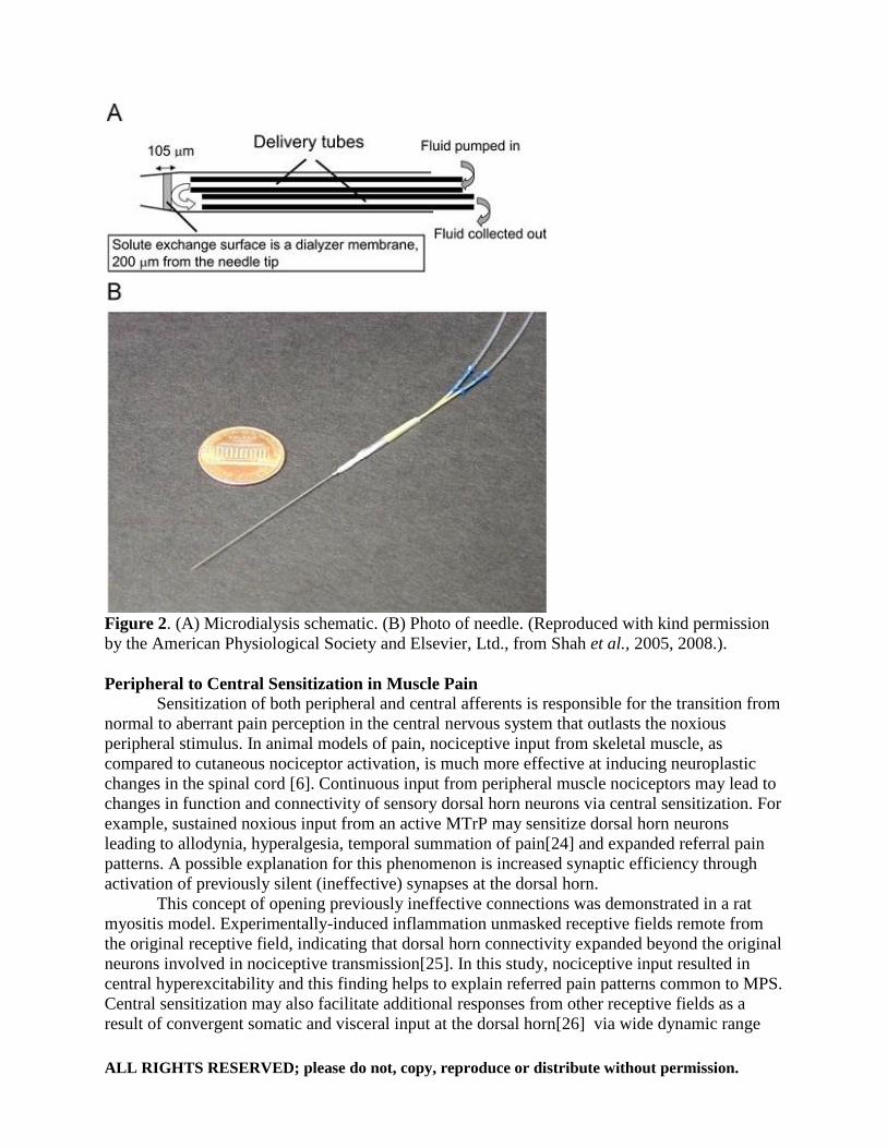
tissue. To accomplish this, we developed a novel microdialysis needle (Figures 2A and 2B) with
the same size, shape, and characteristics of an acupuncture needle. This microanalytical
technique could safely and quantitatively measure the local biochemical environment of muscle
in vivo using continuous, real-time sampling[12, 17, 18].
We chose to investigate the levels of biochemical substances (e.g., inflammatory
mediators, neuropeptides, catecholamines, cytokines, etc.) that are released from and act on
muscle, nerve, and connective tissue. These bioactive substances were selected because they are
known to be associated with sensitization, pain, inter-cellular signaling and inflammation.
Results from the upper trapezius muscle indicate that active MTrPs have a unique biochemical
milieu compared to latent MTrPs and muscle without palpable MTrPs. Subjects with neck pain
secondary to an MTrP had significantly elevated local levels of various endogenous substances,
including substance P (SP), calcitonin gene-related peptide (CGRP), bradykinin (BK),
serotonin/5-hydroxytryptamin (5-HT), norepinephrine (NE), tumor necrosis factor-alpha (TNF-
α), and interleukin-1β (IL-1β) within the active MTrP compared to carefully matched
controls[12, 17, 18]. Interestingly, compared to controls, subjects with active MTrPs in the upper
trapezius also had elevated levels of these biochemicals in a remote, unaffected muscle (the
gastrocnemius)[12, 18].
Together, these studies demonstrated and confirmed that the clinical distinction between
active and latent MTrPs is associated with a highly significant objective difference in the local
biochemical milieu. High concentrations of the biochemicals found have the ability to cause both
peripheral and central sensitization. These findings may help to explain why active MTrPs are
acutely painful, tender, and a source of referred pain. Our biochemical studies have helped to
establish the clinical importance of palpating and identifying active MTrPs. They also suggest
that myofascial pain is an objective entity in the spectrum of clinical pain states and may also
explain why specific treatments are effective. For example, studies have found that an injection
of the serotonin antagonist tropisetron was found to be more effective than lidocaine in relieving
pain from MTrPs[19, 20]. Fittingly, our research indicates elevated local levels of 5-HT within
individuals suffering from active MTrPs. CGRP, which was also found to be elevated in these
individuals [12, 17, 18], has implications for activity within the neuromuscular junction. CGRP
enhances the release of acetylcholine (ACh) from the motor end plate, decreases the
effectiveness of acetylcholinesterase[21, 22], and upregulates the ACh-receptors in the muscle.
As ACh activity becomes more effective, the frequency of miniature endplate potentials
increases as does the development of persistent focal muscle fiber contraction, a defining
characteristic of the MTrP[23].
ALL RIGHTS RESERVED; please do not, copy, reproduce or distribute without permission.
Figure 2. (A) Microdialysis schematic. (B) Photo of needle. (Reproduced with kind permission
by the American Physiological Society and Elsevier, Ltd., from Shah et al., 2005, 2008.).
Peripheral to Central Sensitization in Muscle Pain
Sensitization of both peripheral and central afferents is responsible for the transition from
normal to aberrant pain perception in the central nervous system that outlasts the noxious
peripheral stimulus. In animal models of pain, nociceptive input from skeletal muscle, as
compared to cutaneous nociceptor activation, is much more effective at inducing neuroplastic
changes in the spinal cord [6]. Continuous input from peripheral muscle nociceptors may lead to
changes in function and connectivity of sensory dorsal horn neurons via central sensitization. For
example, sustained noxious input from an active MTrP may sensitize dorsal horn neurons
leading to allodynia, hyperalgesia, temporal summation of pain[24] and expanded referral pain
patterns. A possible explanation for this phenomenon is increased synaptic efficiency through
activation of previously silent (ineffective) synapses at the dorsal horn.
This concept of opening previously ineffective connections was demonstrated in a rat
myositis model. Experimentally-induced inflammation unmasked receptive fields remote from
the original receptive field, indicating that dorsal horn connectivity expanded beyond the original
neurons involved in nociceptive transmission[25]. In this study, nociceptive input resulted in
central hyperexcitability and this finding helps to explain referred pain patterns common to MPS.
Central sensitization may also facilitate additional responses from other receptive fields as a
result of convergent somatic and visceral input at the dorsal horn[26] via wide dynamic range

ALL RIGHTS RESERVED; please do not, copy, reproduce or distribute without permission.
(WDR) neurons (Figure 3). Furthermore, afferent fibers have the ability to sprout new spinal
terminals that broaden synaptic contacts at the dorsal horn and may also contribute to expanded
pain receptive fields[27]. This change in functional connectivity may occur within a few hours,
even before metabolic and genetic alterations occur in dorsal horn neurons[28].
There is a biochemical basis to explain the development of peripheral and central
sensitization in muscle pain. Continuous activation of muscle nociceptors leads to the co-release
of L-glutamate and SP at the pre-synaptic terminals of the dorsal horn. In addition to activation
of alpha-amino-3-hydroxy-5-methyl-4-isoxazolepropionic acid (AMPA) receptors by L-
glutamate at the post-synaptic terminal, SP facilitates activation of previously dormant N-
methyl-D-aspartate (NMDA) receptors. This leads to maximal opening of calcium-permeable ion
channels, which hyperexcites nociceptive neurons and causes apoptosis of inhibitory
interneurons[29]. Consequently, a persistent noxious barrage from the periphery can create long-
lasting alterations in the central nervous system. Metabolic and gene induction changes, such as
cyclo-oxygenase 2 (COX-2) induction in dorsal horn neurons, are maximal at several hours after
an initial noxious stimulation and bolster functional changes after peripheral tissue injury[30].
Figure 3. Wide dynamic range neuron. A WDR neuron receives convergent input from
cutaneous, visceral, and deep somatic afferents and subsequently sends signals to the thalamus.
As such, the WDR neuron and higher-level brain centers can be driven by various inputs.
Accordingly, central sensitization may facilitate responses from other structures (e.g., the
shoulder, heart and skin) which share convergent input.
(Adapted from: Willard, F., "Basic Mechanisms of Pain." Future Trends in CAM Research, in
Integrative Pain Medicine: The Science and Practice of Complementary and Alternative
Medicine in Pain Management, J.F. Audette, Bailey, A., Editor 2008, Humana Press Inc.:
Totowa.)

ALL RIGHTS RESERVED; please do not, copy, reproduce or distribute without permission.
Higher Brain Centers Dynamically Modulate Muscle Pain As aforementioned, persistent afferent input from active MTrPs preferentially activates
and sensitizes WDR neurons in the dorsal horn. The stimuli then ascend the spinothalamic tract
to reach higher brain centers. In addition to activating the thalamus, muscle afferent input
preferentially activates the limbic system (i.e., the anterior cingulate gyrus, insula and
amygdala), which plays a critical role in modulating muscle pain and the emotional or affective
component to persistent pain[3]. Increased activity in the limbic system leads to greater fear,
anxiety, and stress. Furthermore, Niddam et al. (2007) demonstrated increased limbic system
(i.e., anterior insula) activity in patients with upper trapezius myofascial pain syndrome[31].
There is a dynamic balance between supraspinal descending facilitation and inhibition.
For example, the rostral ventral medulla (RVM) is a relay area between the periaqueductal gray
(a structure located in the midbrain) and the spinal cord. The RVM contains a population of “on”
cells and “off” cells which can either increase or decrease the level of pain, respectively. It does
so though projections that modulate activity in the dorsal horn. Following initial tissue injury, the
“on” cells serve a useful and protective purpose designed to prevent further damage. Under
ordinary circumstances, tissue healing would lead to a decrease in “on” cell activity and an
increase in “off” cell activity. However, in chronic musculoskeletal pain conditions, there
appears to be an overall shift to a decrease in inhibition, presumably due to an imbalance of “on”
cell and “off” cell activity[11].
Muscle pain also impairs diffuse noxious inhibitory control (DNIC)[32]. Disrupted
descending inhibition in chronic musculoskeletal pain may lead to an increased pain sensitivity
of muscle tissue[33]. Current data suggest that MTrPs are not merely a peripheral phenomenon
but rather, they activate and sensitize WDR neurons in the dorsal horn and higher brain centers
and may, in turn, be dynamically modulated by these structures[31, 34].
Dry Needling and the Local Twitch Response
Dry needling is an effective, non-pharmacological treatment of MTrPs which has
approached acceptance as the “standard of practice” for deactivating active MTrPs. It may be
performed using either a superficial or deep dry needling technique. Elicitation of one or more
local twitch responses (LTRs) is a goal of dry needling and often benefits those with pain
secondary to MTrPs. Though the mechanism of an LTR is unknown, studies suggest a
biochemical component. Five minutes after the induction of a single LTR, our group found a
dramatic change in the biochemical milieu of the upper trapezius muscle. Within minutes of the
LTR, the initially elevated levels of SP and CGRP within the active MTrP drastically decreased
to levels approaching that of normal uninvolved muscle tissue. The reduction of these
biochemicals in the local muscle area may be due to a small, localized increase in blood flow
and/or nociceptor and mechanistic changes associated with an augmented inflammatory
response[17, 18]. Though not designed as a treatment intervention, the results of these studies are
provocative in that the substances analyzed are known to be associated with sensitization,
persistent pain, and spinal facilitation. In an animal model, it appears that dry needling may, in
fact, activate the descending inhibitory pain system and cause local deactivation of the
MTrP[35].

ALL RIGHTS RESERVED; please do not, copy, reproduce or distribute without permission.
Limitations of Digital Palpation
Current diagnostic standards for myofascial pain rely on palpation for the presence of
MTrPs in a taut band of skeletal muscle[7]. However, proper diagnosis requires a highly skilled
clinician and some studies have found low inter-rater reliability among examiners in their
attempts to identify MTrPs[36, 37]. Although digital palpation is considered the gold standard
for diagnosis, it does have several limitations. Specifically, digital palpation does not 1) provide
an objective, reliable and sensitive method of diagnosis and measurement of treatment efficacy;
2) provide quantitative comparisons of the tissue properties before and after treatment; 3)
objectively differentiate among active MTrPs, latent MTrPs, and palpably normal tissue; 4)
objectively discriminate between superficial and deep MTrPs; and 5) permit objective study of
the natural history of MTrPs.
Visualization and Characterization of MTrPs
Accordinigly, there is a need to develop objective, repeatable, and reliable diagnostic
tests for evaluating MTrPs and determining treatment outcome measures. Such measures can be
used to properly diagnose MTrPs, understand their natural progression, and overcome the
subjectivity and limitations of digital palpation. Accordingly, our group has applied three types
of ultrasound diagnostic imaging techniques—grayscale (2D ultrasound), vibration
sonoelastography (Figure 4), and Doppler—to differentiate tissue characteristics of MTrPs in the
upper trapezius muscle compared to surrounding soft tissue.
These office-based measures are readily available, portable, and inexpensive imaging
modalities, suitable for use in a clinician’s office. We have demonstrated that ultrasound
elastography can serve as an objective image-based measure of MTrPs. Using ultrasound, MTrPs
can be imaged and appear as focal hypoechoic (darker) areas with a heterogeneous echotexture.
MTrPs also have reduced vibration amplitude on elastography, indicating a localized area of
stiffer tissue compared to surrounding soft tissue (Figure 4)[8, 38].
Our studies have also revealed that MTrPs have a unique vascular environment. Doppler
ultrasound was able to show differences in the microcirculation in and around active MTrPs
compared to latent MTrPs and normal tissue. For example, blood flow waveform characteristics
can be used to differentiate active and latent MTrPs. Blood flow reversal in diastole was
associated with active MTrPs, indicating a very high resistance vascular bed. This may be due to
a blood vessel compression by a local muscle contracture (e.g., an MTrP) and/or biochemically-
mediated vasoconstriction of the local blood vessels [38, 39]. Further analysis has also
demonstrated that active MTrPs have a significantly larger surface area than latent MTrPs and
normal sites[8].
Figure 4. (A) Upper trapezius muscle with a palpable MTrP. A hypoechoic region and a well-defined
focal decrease of color variance indicating a localized stiffer region are visible.

ALL RIGHTS RESERVED; please do not, copy, reproduce or distribute without permission.
Figure 4. (B) Normal upper trapezius muscle. A myofascial trigger point is not palpable and the normal
muscle appears isoechoic and has uniform color variance.
Spinal Facilitation
Spinal facilitation is an increase in activity of spinal cord neurons due to the
bombardment of nociceptive stimuli into the dorsal horn (Figure 5)[40]. Under normal
circumstances, activation of primary afferent nociceptors in the dorsal horn is modulated by
inhibitory mechanisms either locally or via descending pathways from the cerebral cortex or
brainstem. However, persistent nociceptive afferent input may result in inhibitory neuronal cell
death, wind-up and sensitization of secondary order neurons in the dorsal horn. Circuits in the
spinal cord (i.e., dorsal horn, ventral horn and lateral horn) may develop lowered thresholds of
activation, causing them to be more easily activated by minimal or no input at all. The ensuing
spinal facilitation is characterized by:
1) Increased ventral horn outflow that stimulates anterior motor horn cells, resulting in
increased muscle tone in the myotome corresponding to its segmental level of afferent
barrage;
2) Increased lateral horn outflow which results in autonomic reflexes that enhance
nociceptive activity; and
3) Increased dorsal horn outflow that causes anti-dromic electrical activity along a
sensory nerve (also known as “dorsal root reflexes”).
Dorsal root reflexes activate dorsal root ganglion cell bodies to increase production and release
vasoactive neuropeptides (e.g., SP, CGRP and somatostatin) both centrally and peripherally.
Upon release into the peripheral tissue, they may exacerbate a local inflammatory process by
stimulating vasodilation and plasma extravasation. This results in local tissue tenderness and
mechanical hyperalgesia. Furthermore, adjacent spinal segments may become progressively
sensitized upon bombardment of the central nervous system[24].
An underappreciated anatomical fact is that primary afferent nociceptive fibers actually
trifurcate upon entering the dorsal horn (Figure 6). That is, one branch enters the dorsal horn at
that segmental level, one branch ascends, and one branch descends along the dorsal margin of
the dorsal horn. Furthermore, some visceral afferents have been found to span the entire length
of the spinal cord[41, 42]. These often overlooked anatomical considerations have enormous
implications in the initiation, perpetuation and amplification of spinal facilitation and persistent
pain states.
Consider the following clinical scenario: An individual develops severe acute painful
cholecystitis. The sustained noxious input is of sufficient intensity and duration to destroy
inhibitory neurons at the segmental level of entry into the dorsal horn (T6). Fortunately, removal

ALL RIGHTS RESERVED; please do not, copy, reproduce or distribute without permission.
of the gall bladder completely alleviates the pain. Years later, the individual develops a minor
but acutely painful back injury while lifting heavy boxes. The resultant afferent nociceptive input
immediately enters the lower thoracic/upper lumbar segments and ascends/descends the spinal
cord. While ascending the cord, but prior to reaching T6, presumably intact and functional
inhibitory neurons suppress the nociceptive input and prevent the sensation of pain at those
segmental levels. However, upon reaching the T6 segment, the nociceptive signal is able to
activate dorsal horn neurons at this level since the local inhibitory neurons are dysfunctional
and/or dead. These result from prior cell death following the original intense gall bladder
stimuli associated with the acute cholecystitis. In fact, a common clinical manifestation is
reproduction of the identical pain pattern caused years before, in this case by the acute gall
bladder disease. Under these circumstances, the patient may become distressed and even
complain of acute gall bladder pain even while acknowledging that the organ had been removed,
thus resulting in a type of “phantom” pain. Accordingly, practitioners of osteopathy often
interpret the re-emergence of an old pain pattern as the possible harbinger of new disease. The
cause could be musculoskeletal in origin (as in this example) or due to an underlying visceral
problem or disease (e.g., peptic ulcer disease, pre-clinical cardiac ischemia, etc.)
Figure 5. Facilitated spinal segment.
a. Nociception originating in the L4 facet joint synapses on the dorsal horn.
b. At the L4 segmental level, a motor neuron within the ventral horn becomes activated, causing
a reflex spasm of muscles innervated by the same segment such as (i.) the paraspinal muscles
and (ii.) the rectus femoris muscle.
c. Dermatomal and sclerotomal structures sharing the L4 segmental level may become sensitized
and painful as a result of dorsal root reflexes.
d. Dorsal root reflexes in the L4 segment may also sensitize cutaneous structures, rendering them
more painful.
(Adapted from Romero Ventosilla, P., Consecuencias clínicas de la Estimulación Sensorial persistente:
Sensibilización Espinal Segmentaria, (Personal Communication), 2010.)

ALL RIGHTS RESERVED; please do not, copy, reproduce or distribute without permission.
Figure 6. Trifurcation of a neuron. Upon entry into the dorsal root of the spinal cord, primary
afferent neurons have the ability to trifurcate; one branch enters at that segmental level while
other branches may ascend and descend along the dorsal margin of the dorsal horn in Lissauer’s
tract.
Diagnosis and Implications of Spinal Segmental Sensitization in the Clinic
Spinal segmental sensitization is consistently associated with musculoskeletal pain states,
underscoring its significance. For example, involvement of thoracic spinal levels (e.g., T1-T12)
in SSS facilitates and perpetuates abdominal pain and somatovisceral symptoms commonly
mimicking gastrointestinal conditions, such as peptic ulcer disease. The development or
activation of MTrPs is one of the clinical manifestations of SSS. In other words, a latent MTrP
that is located along a sensitized segment (i.e., myotome) may become an active MTrP (i.e.,
associated with a spontaneous pain complaint).
Many treatments for myofascial pain such as physical therapy and trigger point injection
procedures are directed at the peripheral pain generators, e.g., active MTrPs. Oftentimes, the
segmental dysfunction is overlooked and practitioners fail to recognize the presence of SSS. As a
result, many individuals may only experience temporary deactivation of MTrPs and pain
frequently recurs.
An accurate diagnosis of pain distribution requires identification of the sensitized spinal
segment. SSS is determined by findings of allodynia, hyperalgesia and measurable pressure pain
sensitivity over the sensory, motor and skeletal areas along with viscera supplied by a particular
spinal segment (i.e., the dermatome, myotome, sclerotome and viscerotome, respectively).

ALL RIGHTS RESERVED; please do not, copy, reproduce or distribute without permission.
Furthermore, these objective and quantitative findings help the clinician to identify the tissues
and likely pain mechanisms involved in their patients’ chronic pain. These segmental findings
are not only reproducible, but they are often indicative of the severity of the sensitized state and
provide important clues about the underlying pathogenesis of the pain syndrome.
The requisite examination skills are easy to learn and of fundamental importance to the
evaluation and management of a chronic pain complaint. Furthermore, their application before
and after treatment, aimed at desensitizing the involved spinal segment, provides the clinician
and patient meaningful, objective and reproducible physical findings to guide treatment
outcomes.
Imamura et al. (2008) systematically evaluated individuals with refractory, disabling pain
associated with knee osteoarthritis (OA), who were scheduled to undergo total knee replacement.
The authors speculated that the pain experienced by these patients may be associated with the
presence of central nervous sensitization rather than peripheral inflammation and injury. The
presence of hyperalgesia was evaluated, and the impact of pressure pain threshold (PPT)
measurements on pain, disability and quality of life was assessed in these patients and compared
to age-matched healthy controls. PPT measurements were obtained for the subcutaneous
dermatomes of the lower extremities and over the vastus medialis, adductor longus, rectus
femoris, vastus lateralis, tibialis anterior, peroneus longus, iliacus, quadratus lumborum and
popliteus muscles and the supraspinous ligament. They found that the group with knee OA had
significantly lower PPT over all evaluated structures versus healthy control subjects. Lower PPT
values were correlated with higher pain intensity, higher disability scores and poorer quality of
life, except for the role-emotional and general health status. Combined PPT values over the
patellar tendon, at the S2 subcutaneous dermatome and at the adductor longus muscle were the
best predictors for the visual analog scale and Western Ontario and McMaster Universities
Osteoarthritis Index pain scores. They concluded that patients with knee pain due to OA who
were scheduled for total knee replacement showed hyperalgesia of nervous system origin that
negatively impacted pain, knee functional capacity and most aspects of quality of life[16].
In order to determine the presence of SSS, the patient is asked to identify with one finger
the location of his/her principal pain complaint and indicate the intensity of pain from 1-10.
Adjacent dermatomal levels are examined parapsinally by:
- Scratching the skin with the sharp edge of a paper clip or Wartenberg pinwheel – this
noxious stimulus is applied across dermatomal borders and the patient is instructed to
simultaneously report any sharpening or dulling in the sensation of pain during the
procedure. An increased painful response is indicative of hyperalgesia.
- Picking up the skin between the thumb and forefinger and rolling the tissue underneath,
also known as a “pinch and roll” test (Figure 7) – this non-noxious stimulus is applied
across dermatomal borders and the patient is instructed to simultaneously report any
sensation of pain. The sensation of pain is indicative of allodynia, a finding that is the
most sensitive indicator for the diagnosis of sensitization.
Adjacent myotomal levels are examined by:
- Palpating segmentally related musculature for tender spots, taut bands and MTrPs.
- Applying a pressure algometer to measure the local tenderness (i.e., PPT) along the
myotome.
Adjacent sclerotomal levels are examined by:
- Palpating segmentally related tendons (e.g., tendonitis), entheses (e.g., enthesitis), bursae
(e.g., bursitis) and ligaments (e.g., supraspinous ligament sprain).

ALL RIGHTS RESERVED; please do not, copy, reproduce or distribute without permission.
- Applying a pressure algometer to measure the local tenderness (i.e., PPT) along these
sclerotomal structures.
(Note: PPT is the minimum pressure that elicits pain and is considered abnormal if it is at least
2kg/cm² lower than a normosensitive control point.)
These examination techniques are used to determine the depth and breadth of the
segmental manifestations of an individual’s pain syndrome. Dermatomal, myotomal and
sclerotomal segmental findings often overlap, making diagnosis and treatment of the sensitized
segmental level relatively straightforward. However, in some cases they do not. For example, the
affected dermatomal levels may be different from the affected myotomal and/or sclerotomal
levels. When such differences arise, the affected segmental levels (whether dermatomal,
myotomal or sclerotomal) most closely corresponding to the principal pain complaint should be
treated first with paraspinal needling, a technique which addresses the centrally sensitized
component of pain.
If the patient experiences little or no pain relief, then the additional segmental levels may
be needled paraspinally until the patient reports a decrease in pain. This subjective decrease in
pain is typically accompanied by an objective improvement in the segmental findings. However,
effective management involves identification and treatment of both the peripheral and central
components of sensitization. Accordingly, the clinician should identify and eradicate all foci of
nociceptive bombardment (i.e., peripheral sensitization) responsible for initiating and/or
perpetuating the centrally sensitized segmental findings. Active MTrPs are a very common
source of peripheral nociceptive bombardment which may lead to central sensitization and
perpetuation of the pain complaint. If left unresolved, active MTrPs (or other peripheral pain
generators) will re-sensitize the dorsal horn, resulting in the re-emergence of segmental findings
(i.e. allodynia and hyperalgesia) and the reproduction of the same pain complaint even after
paraspinal needling treatment.

ALL RIGHTS RESERVED; please do not, copy, reproduce or distribute without permission.
Figure 7. “Pinch and roll” test. The skin and subcutaneous tissue is gently pinched between the
thumb and forefinger and rolled vertically across dermatomal borders. Elicitation of a painful
response is indicative of allodynia.
Modalities and Manual Therapies
Modalities and manual therapies are often clinically effective at deactivating active
MTrPs and desensitizing sensitized spinal segments and are commonly employed as a first line
of treatment before attempting more invasive therapies. For example, various forms of electrical
stimulation including microcurrent, transcutaneous electrical nerve stimulation (TENS),
percutaneous electrical nerve stimulation (PENS), manual therapies and spray and stretch are
commonly used to treat myofascial pain and SSS. However, if pain relief is only partial or pain
persists despite several treatments using such various modalities, then needling and injection
techniques should be considered, particularly in chronic cases in which the physical examination

ALL RIGHTS RESERVED; please do not, copy, reproduce or distribute without permission.
reveals severe and persistent allodynia and hyperalgesia, suggesting dense dermatomal,
myotomal and sclerotomal manifestations of SSS.
Paraspinal Dry Needling Techniques
Fischer et al. (2002) developed a technique utilizing injection of 1% lidocaine into the
paraspinal muscles adjacent to the spinous processes. A 25-gauge needle, of sufficient length to
reach the deep layers up to the vertebral lamina, is inserted in the sagittal plane. Injection is
performed between the levels of the spinous processes corresponding to the affected segmental
levels of sensitization as identified on physical examination. The needle is inserted through the
paraspinal muscle to a maximal depth but before contacting the vertebral lamina. The needle is
aspirated (in order to avoid blood vessels) and then approximately 0.1mL of anesthetic is
injected; the needle is then withdrawn to a subcutaneous level and redirected in the caudal
direction, ending about 5mm from the previous deposit of anesthetic solution. One continues this
procedure, going as far as the needle reaches. The same procedures are then repeated going in the
cephalad direction. The result of this technique (which Fischer calls a “paraspinous block”) is to
effectively block the medial branch of the posterior primary rami at affected segmental
levels[43].
Coincidentally, acupuncture practitioners utilize similar anatomical locations (e.g.,
traditional Chinese medicine “Hua Tuo Jia Ji” points). According to the Acupuncture Energetics
textbook definition, these are “a collection of points on either side of the ligament attaching the
transverse processes of the vertebral column, from T1 to L5, described as one-half Cun lateral to
the spinous process; to be needled as deeply as possible into the ligaments just as they are
accessible lateral to the midline; very useful as local points for axial or peripheral pain problems,
and as points to reinforce the qualities of the back Shu points at the same vertebral levels” for
needle insertion in order to achieve pain relief[44]. Many clinicians, including myself (as I have
received training in both medical acupuncture and Fischer’s injection techniques), have adapted
a dry needling technique to Fischer’s model of paraspinous blocks. Instead of a hypodermic
syringe, my preference is to insert acupuncture needles sagitally into the paraspinal muscles as
aforementioned (Figure 8). The needle is manipulated by using an up and down (i.e., “pistoning”
action) accompanied by clock-wise and counter-clock wise rotations. Multiple acupuncture
needles may be similarly inserted and manipulated creating a “paraspinous block” at each of the
affected segmental levels, corresponding to the principal pain complaint. Furthermore, the
segmentally corresponding supraspinous ligaments may be needled using a superficial needle
insertion technique accompanied by just a clockwise and counter-clockwise rotation of the
needle (i.e., “pistoning” of the needle should be avoided). The needles may be left in place for
15-20 minutes (similar to standard practice in acupuncture) while the relevant dermatomes,
myotomes and sclerotomes are re-examined to determine whether the selected treated segments
eliminate the objective signs of SSS (i.e. allodynia, hyperalgesia, etc.). This finding is often
accompanied by a significant reduction in pain.
There are several advantages associated with this dry needling technique when compared
with Fischer’s injection technique. First, affected segments may be treated without concern for
the possible side effects associated with injection of local anaesthetics. In addition, more
segments may be treated in one visit with dry needling because the number of injections is
limited by the total dosage of anaesthetic that may be administered at one time. Furthermore,
acupuncture needles are minimally invasive and better tolerated by patients because they have
rounded-tips, designed to painlessly pass around cells, blood vessels and other tissues. On the

ALL RIGHTS RESERVED; please do not, copy, reproduce or distribute without permission.
other hand, hypodermic needles are bevel-edged and sharp, designed to cut through blood
vessels (thereby damaging to cells and tissues); this attribute results in more inflammation,
bleeding and pain. Due to its fine construction and smaller diameter, an acupuncture needle
provides the practitioner superior kinesthetic feedback when compared to a hypodermic needle.
This permits more accurate manipulation and easier placement of the needle at desired tissue
depths.
Though many practitioners can attest to improvement in pain levels as a result of dry
needling in the paraspinal muscles, these merely arise from clinical observation. To date, there
have been no randomized, double-blinded, placebo-controlled clinical trials examining the
effects of paraspinal dry needling. A recent study offers a method that may be used to determine
the effectiveness of this treatment. Mayoral del Moral et al. designed a study in which subjects
scheduled for total knee replacement surgery were examined several hours before surgery. They
were then randomly assigned to one of two groups: true dry needling or sham dry needling.
Upon the induction of general anesthesia but before surgery began, those assigned to true dry
needling of MTrPs were treated by a physiotherapist. Since subjects were unconscious at the
time of true or sham treatment, they were unaware of their group assignment. Post-surgery, the
true dry needling group reported less pain, demanded significantly less analgesics and rated their
visual analog scale significantly better than the sham needling group[45]. A similar protocol
could be used to systematically assess the outcome of paraspinal dry needling and lend support
to its use. Knee joint and related muscles are innervated by the L3-L4 segmental levels.
Accordingly, it can be speculated that paraspinal needling within these segments prior to total
knee replacement for osteoarthritis may provide better pain relief than surgery alone and could
also be used as an effective and conservative first line of treatment.

ALL RIGHTS RESERVED; please do not, copy, reproduce or distribute without permission.
Figure 8. Paraspinal dry needling. An acupuncture needle is inserted sagitally into the spinalis
muscle and then manipulated as described. Multiple acupuncture needles may be inserted to
create a “paraspinous block” at each affected segmental level.
Conclusion
Current understanding of chronic pain mechanisms, particularly in the unique properties
of the neuraxis, is changing rapidly as knowledge emerges in molecular and cellular biology. For
example, active MTrPs function as dynamic foci of peripheral nociception that can initiate,
accentuate and maintain central sensitization and chronic pain states. Continuous nociceptive
input from MTrPs can increase excitability of dorsal horn neurons, leading to hyperalgesia and
allodynia, and open previously ineffective synaptic connections, resulting in new receptive fields
and pain referral[12].
Although MTrPs are a ubiquitous and under-diagnosed component of many acute and
chronic pain complaints, they are also a common physical finding in asymptomatic individuals.
This dichotomy challenges pain management practitioners to learn how to carefully palpate the
soft tissue in order to distinguish active from latent MTrPs. Making this distinction is critical in
order to adequately identify and treat a myofascial component of pain.
Histological, neurophysiological, ultrasound imaging, and somatosensory studies of
MTrPs have found objective abnormalities. Novel biochemical sampling techniques demonstrate

ALL RIGHTS RESERVED; please do not, copy, reproduce or distribute without permission.
that active MTrPs have elevated levels of bradykinin, serotonin, substance P, CGRP,
norepinephrine, IL-6, IL-8, TNF-α, and IL-1β. Active sites also have a more acidic pH. Together
with observed motor and sensory abnormalities, these studies implicate peripheral and central
mechanisms in the development of myofascial pain and associated MTrPs. These biochemical
findings validate the clinical observation that active MTrPs are a source of nociceptive foci that
continuously bomard dorsal horn neurons, leading to central sensitization and SSS.
Dorsal horn neurons may undergo neuroplastic changes as a result of chronic
nociception. However, these changes may also occur in higher centers of the brain if pain is left
unresolved for an extended period of time. Alterations in cortical areas may help to maintain and
amplify the pain state and thereby create a vicious cycle that is increasingly difficult to resolve.
At this point, removal of the etiological factors may be insufficient to relieve pain[24].
Once the presence of central sensitization has been established in a patient with chronic
pain, the central nervous system should be targeted in addition to the musculoskeletal
component, which is primarily treated with anti-inflammatory agents. Thorough understanding
and identification of central nervous system sensitization has the ability to provide innovative
and cost-effective therapeutic tools to control pain, reduce disability and improve quality of life.
Comprehensive management should focus on the removal of perpetuating factors (e.g., active
MTrPs) and by addressing SSS early in its development through methods such as electrical
modalities, manual therapies, paraspinal dry needling, paraspinous blocks, centrally-acting
pharmacologic agents, biofeedback, behavioral therapy, etc.
Spinal segmental sensitization offers an important paradigm to explain the nature of
neuro-musculoskeletal pain. Though it essentially has the same origin as peripheral sensitization,
the central phenomenon is distinguished by its clinical characteristics. The presence of specific
dermatomal, myotomal and sclerotomal distribution patterns are objective, reproducible and
reliable hallmarks of SSS. Paraspinal dry needling provides physiotherapists and other clinicians
a clinically effective and minimally invasive treatment for neuro-musculoskeletal pain.
There is a need to develop objective, repeatable, and reliable diagnostic tests for
evaluation and treatment outcome measures for MTrPs. Such measures can be used to properly
diagnose and understand the natural history of MTrPs and to determine the underlying
mechanisms and relevance to the development and resolution of myofascial pain. They may also
be used as outcome measures in treatment trials of various interventions including manual
therapies, electrical modalities, etc. Do manual techniques result in “softening” and eventually
elimination of the MTrP? Do they improve blood flow in the vicinity of MTrPs, presumably
washing out noxious, sensitizing and painful biochemicals? These office-based ultrasound
techniques will help answer these questions.
Future Directions Our group is currently developing a model for the peripheral and central mechanisms
involved in myofascial pain. Now that we have identified objective differences which distinguish
active MTrPs from latent MTrPs and normal tissue, we plan to further study the nature of MTrPs
and surrounding soft tissue over time. Although painful MTrPs activate muscle nociceptors that,
upon sustained noxious stimulation, initiate peripheral and central sensitization, what is their
etiology and pathophysiology? What is the mechanism by which the pain state begins, evolves,

ALL RIGHTS RESERVED; please do not, copy, reproduce or distribute without permission.
and persists? What are the levels of anti-inflammatory substances, analgesic substances and
muscle metabolites in the local biochemical milieu of muscle with and without MTrPs? How
does a tender nodule progress to a myofascial pain syndrome? Which soft tissues are involved?
Are there objective measures for assessing therapeutic outcomes? What is the mechanism by
which active MTrPs contribute to SSS? What effects do local and central treatments of MTrPs
have on SSS?
Future clinical research studies should focus on identifying the mechanisms responsible
for the pathogenesis and pathophysiology of both myofascial pain, SSS and neuro-
musculoskeletal pain by linking the symptoms and objective physical findings to the physical
properties and biochemical changes in the muscle tissue.

ALL RIGHTS RESERVED; please do not, copy, reproduce or distribute without permission.
References 1. Sessle, B.J., Acute and chronic craniofacial pain: brainstem mechanisms of nociceptive
transmission and neuroplasticity, and their clinical correlates. Critical Reviews in Oral Biology & Medicine, 2000. 11(1): p. 57-91.
2. Arendt-Nielsen, L., Graven-Nielsen, T., Deep tissue hyperalgesia. Journal of Musculoskeletal Pain, 2002. 10(1/2): p. 97-119.
3. Svensson, P., Minoshima, S., Beydoun, A., Morrow, T.J., Casey, K.L., Cerebral processing of acute skin and muscle pain in humans. Journal of Neurophysiology, 1997. 78(1): p. 450-460.
4. XianMin, Y., Mense, S., Response properties and descending control of rat dorsal horn neurons with deep receptive fields. Neuroscience, 1990. 39: p. 823-831.
5. Fields, H.L., Basbaum, A.I., Central nervous system mechanisms of pain modulation, in Textbook of Pain, R. Melzack, Wall, P.D., Editor 1999, Churchill Livingstone: Edinburgh. p. 309-329.
6. Wall, P.D., Woolf, C.J., Muscle but not cutaneous c-afferent input produces prolonged increases in the excitability of the flexion reflex in the rat. Journal of Physiology, 1984. 356: p. 443-458.
7. Travell, J.G., Simons, D.G., Myofascial Pain and Dysfunction: The Trigger Point Manual. VI and VII ed1999, Baltimore: Williams & Wilkins.
8. Ballyns, J.J., Shah, J.P., Hammond, J., Gebreab, T., Gerber, L.H., Sikdar, S., Objective sonographic measures for characterizing myofascial trigger points associated with cervical pain. Journal of Ultrasound in Medicine, 2011. 30: p. 1331-1340.
9. Gerwin, R.D., Shannon, S., Hong, C.Z., Hubbard, D., Gevirtz, R., Interrater reliability in myofascial trigger point examination. Pain, 1997. 69(1-2): p. 65-73.
10. Zieglgänsberger, W., Berthele, A., Tölle, T.R. , Understanding neuropathic pain. CNS Spectrums, 2005. 10: p. 298-308.
11. Willard, F., "Basic Mechanisms of Pain." Future Trends in CAM Research, in Integrative Pain Medicine: The Science and Practice of Complementary and Alternative Medicine in Pain Management, J.F. Audette, Bailey, A., Editor 2008, Humana Press Inc.: Totowa.
12. Shah, J.P., Gilliams, E.A., Uncovering the biochemical milieu of myofascial trigger points using in-vivo microdialysis: An application of muscle pain concepts to myofascial pain syndrome. Journal of Bodywork and Movement Therapies, 2008. 12(4): p. 371-384.
13. Fischer, A.A., Functional diagnosis of musculoskeletal pain by quantitative and objective methods, in Myofascial pain and Fibromyalgia: Trigger Point Management, E.S. Rachlin, Rachlin, I.S., Editor 2002, Mosby: St. Louis. p. 145-173.
14. Waldman, S.D., Physical diagnosis of pain: an atlas of signs and symptoms. 1st ed, ed. S.D. Waldman2006, Philadelphia: Saunders & Elsevier.
15. Fischer, A.A., Imamura, M., New concepts in the diagnosis and management of musculoskeletal pain, in Pain procedures in clinical practice, T.A. Lennard, Editor 2000, Henley & Belfus: Philadelphia. p. 213-229.
16. Imamura, M., Imamura, S.T., Kaziyama, H.H.S., Targino, R.A., Hsing, W.T., De Souza, L.P.M., Cutait, M.M., Fregni, F., Camanho, G.L., Impact of nervous system hyperalgesia on pain, disability, and quality of life in patients with knee osteoarthritis: A controlled analysis. Arthritis Care & Research, 2008. 59(10): p. 1424-1431.
17. Shah, J.P., Phillips, T.M., Danoff, J.V., Gerber, L., An in vivo microanalytical technique for measuring the local biochemical milieu of human skeletal muscle. Journal of Applied Physiology, 2005. 99: p. 1977-1984.
18. Shah, J.P., Danoff, J.V., Desai, M., Parikh, S., Nakamura, L.Y., Phillips, T.M., Gerber, L.H., Biochemicals associated with pain and inflammation are elevated in sites near to and remote from active myofascial trigger points. Archives of Physical Medicine and Rehabilitation, 2008. 89: p. 16-23.

ALL RIGHTS RESERVED; please do not, copy, reproduce or distribute without permission.
19. Ettlin, T., Trigger point injection treatment with the 5-HT3 receptor antagonist tropisetron in patients with late whiplast-associated disorder. First results of a multiple case study. Scandinavian Journal of Rheumatology, 2004. 11(9): p. 49-50.
20. Müller, W., Stratz, T., Local treatment of tendinopathies and myofascial pain syndromes with the 5-HT3 receptor antagonist tropisetron. Scandinavian Journal of Rheumatology, 2004. 11(9): p. 44-48.
21. Fernandez, H.L., Hodges-Savola, C.A., Physiological regulation of G4 AChE in fast-twitch muscle: Effects of exercise and CGRP. Journal of Applied Physiology, 1996. 80(1): p. 357-362.
22. Hodges-Savola, C.A., Fernandez, H.L., A role for calcitonin generelated peptide in the regulation of rat skeletal muscle G4 acetylcholinesterase. Neuroscience Letters, 1995. 190(2): p. 117-120.
23. Gerber, L.H., Sikdar, S., Hammond, J., Shah, J.P., A Brief Overview and Update of Myfascial Pain Syndrome and Myofascial Trigger Points. Journal of The Spinal Research Foundation, 2011. 6: p. 55-64.
24. Camanho, L., G., Imamura, M., Arendt-Nielsen, L., Genesis of pain in arthrosis. Revista Brasileira de Ortopedia, 2011. 46(1): p. 14-17.
25. Hoheisel, U., Koch, K., Mense, S., Functional reorganization in the rat dorsal horn during an experimental myositis. Pain, 1994. 59: p. 111-118.
26. Sato, A., Somatovisceral reflexes. Journal of Manipulative Physiological Therapeutics, 1995. 18: p. 597-602.
27. Sperry, M.A., Goshgarian, H.G., Ultrastructural changes in the rat phrenic nucleus developing within 2 h after cervical spinal cord hemisection. Experimental Neurology, 1993. 120: p. 233-244.
28. Mense, S., Hoheisel, U., Central nervous sequelae of local muscle pain. Journal of Musculoskeletal Pain 2004. 12: p. 101-109.
29. Mense, S., The pathogenesis of muscle pain. Current Pain and Headache Reports, 2003. 7: p. 419-425.
30. Woolf, C.J., Central sensitization: uncovering the relation between pain and plasticity. Anesthesiology, 2007. 106(4): p. 864-867.
31. Niddam, D.M., Chan, R.C., Lee, S.H., Yeh, T.C., Hsieh, J.C., Central modulation of pain evoked from myofascial trigger point. Clinical Journal of Pain, 2007. 23: p. 440-448.
32. Arendt-Nielsen, L., Sluka, K.A., Nie, H.L., Experimental muscle pain impairs descending inhibition. Pain, 2008. 140: p. 465-471.
33. Ge, H.Y., Fernández-de-las-Peñas, C., Yue, S.W., Myofascial trigger points: spontaneous electrical activity and its consequences for pain induction and propagation. Chinese Medicine, 2011. 6(13).
34. Mense, S., How Do Muscle Lesions such as Latent and Active Trigger Points Influence Central Nociceptive Neurons? Journal of Musculoskeletal Pain, 2010. 18(4): p. 348-353.
35. Hsieh, Y.L., Chou, L.W., Joe, Y.S., Hong, C.Z., Spinal cord mechanism involving the remote effects of dry needling on the irritability of myofascial trigger spots in rabbit skeletal muscle. Archives of Physical Medicine and Rehabilitation, 2011. 92(7): p. 1098-1105.
36. Njoo, K.H., Van der Does E., The occurrence and inter-rater reliability of myofascial trigger points in the quadratus lumborum and gluteus medius: a prospective study in non-specific low back pain patients and controls in general practice. Pain, 1994. 58(3): p. 317-323.
37. Wolfe, F., Simons, D.G., Friction, J.R., Bennett, R.M., Goldenberg, D.L., Gerwin, R.D., Hathaway, D.E., McCain, G.A., Russell, I.J., Sanders, H., Skootsky, S.A., The fibromyalgia and myofascial pain syndromes: a preliminary study of tender points and trigger points in persons with fibromyalgia, myofascial pain syndrome and no disease. Journal of Rheumatology, 1992. 19: p. 944-951.
38. Sikdar, S., Shah, J.P., Gebreab, T., Yen, R., Gilliams, E., Danoff, J., Gerber, L.H., Novel Applications of Ultrasound Technology to Visualize and Characterize Myofascial Trigger Points (MTrPs) and

ALL RIGHTS RESERVED; please do not, copy, reproduce or distribute without permission.
Surrounding Soft Tissue. Archives of Physical Medicine and Rehabilitation, 2009. 90: p. 1829-1838.
39. Sikdar, S., Ortiz, R., Gebreab, T., Gerber, L.H., Shah, J.P., Understanding the vascular environment of myofascial trigger points using ultrasonic imaging and computational modeling, in 32nd Annual International Conference of the IEEE EMBS2010: Buenos Aires, Argentina.
40. Romero Ventosilla, P., Consecuencias clínicas de la Estimulación Sensorial persistente: Sensibilización Espinal Segmentaria, 2010.
41. Sugiura, Y., Terui, N., Hosoya, Y., Difference in distribution of central terminals between visceral and some somatic unmyelinated (C) primary afferent fibers. Journal of Neurophysiology, 1989. 62: p. 834-840.
42. Wall, P.D., Bennett, D.L., Postsynaptic effects of long-range afferents in distant segments caudal to their entry point in the rat spinal cord under the influence of picrotoxin or strychnine. Journal of Neurophysiology, 1994. 72: p. 2703-2713.
43. Fischer, A.A., New injection techniques for treatment of musculoskeletal pain, in Myofascial pain and Fibromyalgia: Trigger Point Management, E.S. Rachlin, Rachlin, I.S., Editor 2002, Mosby. p. 403-419.
44. Helms, J.M., Acupuncture energetics : a clinical approach for physicians. 1st ed1995, Berkeley: Medical Acpuncture Publishers.
45. Mayoral del Moral, O., Dry Needling Treatments for Myofascial Trigger Points. Journal of Musculoskeletal Pain, 2010. 18(4): p. 411-416.
Related Documents