Original Contribution Modulation of Trypanosoma rangeli ecto-phosphatase activity by hydrogen peroxide Daniela Cosentino-Gomes, Thais Russo-Abrahão, André Luiz Fonseca-de-Souza, Clara Rodrigues Ferreira, Antonio Galina, José Roberto Meyer-Fernandes ⁎ Instituto de Bioquímica Médica and Instituto Nacional de Ciência e Tecnologia de Biologia Estrutural e Bioimagem, Centro de Ciências da Saúde, Universidade Federal do Rio de Janeiro, Ilha do Fundão, 21941-590 Rio de Janeiro, RJ, Brazil abstract article info Article history: Received 28 November 2008 Revised 14 April 2009 Accepted 14 April 2009 Available online 21 April 2009 Keywords: Trypanosoma rangeli Ecto-phosphatase Hydrogen peroxide Mitochondrial ROS formation Free radicals As a protozoan parasite of hematophagous insects, Trypanosoma rangeli epimastigotes are exposed to reactive oxygen species during development in hosts. In this work, we investigated the role of H 2 O 2 as a modulator of the ecto-phosphatase activity present in living T. rangeli. We observed that H 2 O 2 inhibits ecto- phosphatase activities in the short and long epimastigote forms of T. rangeli. Ecto-phosphatase activity found in the short form was more sensitive than that found in the long form. Moreover, H 2 O 2 inhibited ecto- phosphatase activity of the short form in a dose-dependent manner and this inhibition was reversible after H 2 O 2 removal. This effect was not observed for T. rangeli ecto-ATPase, another ecto-enzyme present on the external surface of T. rangeli. Cysteine, β-mercaptoethanol, and reduced glutathione were able to revert the enzyme inhibition promoted by H 2 O 2 . Catalase and glutathione peroxidase stimulated this ecto-phosphatase activity, whereas superoxide dismutase was not able to modulate this activity. The ecto-phosphatase activity was also activated by FCCP and inhibited by oligomycin. It seems that H 2 O 2 plays a fundamental role in the regulation of cellular processes of these organisms. We showed, for the first time, that these parasites can produce H 2 O 2 , and it is able to regulate ecto-phosphatase activity. © 2009 Elsevier Inc. All rights reserved. All aerobic organisms must tolerate reactive oxygen species (ROS) 1 , including hydrogen peroxide (H 2 O 2 ), which are generated by oxidative metabolism. Parasitic protozoa not only have to eliminate endogenous toxic metabolites but must also cope with the oxidative (or respiratory) burst of the host immune system [1]. Trypanosoma rangeli is a hemoflagellate protozoan parasite that infects not only humans and a great number of other mammals but also its triatomine vectors, insects of the genus Rhodnius [2,3]. Even though T. rangeli is apathogenic for vertebrate hosts, its biological and epidemiological characteristics are studied within the context of the biology and epidemiology of Trypanosoma cruzi (the causative agent of Chagas disease). Both species produce mixed infections in vectors and vertebrates, generating serological cross reactions that may lead to misdiagnosis [3]. T. rangeli has a complex life cycle that involves distinct morpho- logical and functional forms in the insect vector. Interaction of the parasite with its invertebrate host begins with ingestion of the trypomastigote form by the insect. After ingestion, T. rangeli trans- forms into short epimastigotes, multiplies in the midgut, and invades the hemocoel. In a few days after infection, short epimastigotes appear in the hemocoel of the vector but soon they disappear and are replaced by a massive colonization of long epimastigotes [4–6]. The long epimastigotes survive in the hemolymph and/or inside the hemocytes and migrate to and complete their development in the salivary glands [7]. To complete its biological cycle in nature, T. rangeli is transmitted to the vertebrate host through salivary secretion during feeding [4]. The series of morphological and biochemical transformations in the life cycle may occur in response to external stimuli and their transduction into an appropriate response [8]. Protozoan parasites present major differences in terms of antioxidant defenses, not only compared to their hosts, but also among themselves [9]. ROS sensing is likely to be an important mechanism for the adaptation and interaction of trypanosomatids with their environment [8,10]. Recently, H 2 O 2 was shown to be a ubiquitous intracellular messenger at subtoxic concentrations. In T. cruzi, for example, low H 2 O 2 concentrations led to an increase in cell proliferation [10]. Moreover, it has been postulated that H 2 O 2 can affect the function of various proteins, including transcription factors, phospholipases, protein kinases and phosphatases, ion channels, and G proteins [11]. In trypanosomatids, as in other cells, the roles of molecules in the detection of H 2 O 2 and ROS and the associated environmental signals are not yet fully understood [8]. Regulated reversible phosphorylation of proteins and other cellular molecules plays an important role in the control of cellular behavior. Phosphatases that utilize a nucleophilic cysteine residue in Free Radical Biology & Medicine 47 (2009) 152–158 Abbreviations: ROS, reactive oxygen species; SOD, superoxide dismutase; LIT, liver infusion tryptose; p-NPP, substrate p-nitrophenyl phosphate; GSH, reduced glu- tathione; GSH-Px, glutathione peroxidase; β-Mer, β-mercaptoethanol; PTP, protein- tyrosine phosphatase; FCCP, carbonyl cyanide p-trifluoromethoxyphenylhydrazone. ⁎ Corresponding author. Fax: +5521 22708647. E-mail address: [email protected] (J.R. Meyer-Fernandes). 0891-5849/$ – see front matter © 2009 Elsevier Inc. All rights reserved. doi:10.1016/j.freeradbiomed.2009.04.020 Contents lists available at ScienceDirect Free Radical Biology & Medicine journal homepage: www.elsevier.com/locate/freeradbiomed

Welcome message from author
This document is posted to help you gain knowledge. Please leave a comment to let me know what you think about it! Share it to your friends and learn new things together.
Transcript

Free Radical Biology & Medicine 47 (2009) 152–158
Contents lists available at ScienceDirect
Free Radical Biology & Medicine
j ourna l homepage: www.e lsev ie r.com/ locate / f reeradb iomed
Original Contribution
Modulation of Trypanosoma rangeli ecto-phosphatase activity by hydrogen peroxide
Daniela Cosentino-Gomes, Thais Russo-Abrahão, André Luiz Fonseca-de-Souza, Clara Rodrigues Ferreira,Antonio Galina, José Roberto Meyer-Fernandes ⁎Instituto de Bioquímica Médica and Instituto Nacional de Ciência e Tecnologia de Biologia Estrutural e Bioimagem, Centro de Ciências da Saúde, Universidade Federal do Rio de Janeiro,Ilha do Fundão, 21941-590 Rio de Janeiro, RJ, Brazil
Abbreviations: ROS, reactive oxygen species; SOD, sinfusion tryptose; p-NPP, substrate p-nitrophenyl phtathione; GSH-Px, glutathione peroxidase; β-Mer, β-mtyrosine phosphatase; FCCP, carbonyl cyanide p-trifluor⁎ Corresponding author. Fax: +5521 22708647.
E-mail address: [email protected] (J.R. Meyer-
0891-5849/$ – see front matter © 2009 Elsevier Inc. Adoi:10.1016/j.freeradbiomed.2009.04.020
a b s t r a c t
a r t i c l e i n f oArticle history:Received 28 November 2008Revised 14 April 2009Accepted 14 April 2009Available online 21 April 2009
Keywords:Trypanosoma rangeliEcto-phosphataseHydrogen peroxideMitochondrial ROS formationFree radicals
As a protozoan parasite of hematophagous insects, Trypanosoma rangeli epimastigotes are exposed toreactive oxygen species during development in hosts. In this work, we investigated the role of H2O2 as amodulator of the ecto-phosphatase activity present in living T. rangeli. We observed that H2O2 inhibits ecto-phosphatase activities in the short and long epimastigote forms of T. rangeli. Ecto-phosphatase activity foundin the short form was more sensitive than that found in the long form. Moreover, H2O2 inhibited ecto-phosphatase activity of the short form in a dose-dependent manner and this inhibition was reversible afterH2O2 removal. This effect was not observed for T. rangeli ecto-ATPase, another ecto-enzyme present on theexternal surface of T. rangeli. Cysteine, β-mercaptoethanol, and reduced glutathione were able to revert theenzyme inhibition promoted by H2O2. Catalase and glutathione peroxidase stimulated this ecto-phosphataseactivity, whereas superoxide dismutase was not able to modulate this activity. The ecto-phosphatase activitywas also activated by FCCP and inhibited by oligomycin. It seems that H2O2 plays a fundamental role in theregulation of cellular processes of these organisms. We showed, for the first time, that these parasites canproduce H2O2, and it is able to regulate ecto-phosphatase activity.
© 2009 Elsevier Inc. All rights reserved.
All aerobic organisms must tolerate reactive oxygen species (ROS)1,including hydrogen peroxide (H2O2), which are generated by oxidativemetabolism. Parasitic protozoa not only have to eliminate endogenoustoxic metabolites but must also copewith the oxidative (or respiratory)burst of the host immune system [1].
Trypanosoma rangeli is a hemoflagellate protozoan parasite thatinfects not only humans and a great number of other mammals butalso its triatomine vectors, insects of the genus Rhodnius [2,3]. Eventhough T. rangeli is apathogenic for vertebrate hosts, its biological andepidemiological characteristics are studied within the context of thebiology and epidemiology of Trypanosoma cruzi (the causative agentof Chagas disease). Both species produce mixed infections in vectorsand vertebrates, generating serological cross reactions that may leadto misdiagnosis [3].
T. rangeli has a complex life cycle that involves distinct morpho-logical and functional forms in the insect vector. Interaction of theparasite with its invertebrate host begins with ingestion of thetrypomastigote form by the insect. After ingestion, T. rangeli trans-forms into short epimastigotes, multiplies in the midgut, and invadesthe hemocoel. In a few days after infection, short epimastigotes appear
uperoxide dismutase; LIT, liverosphate; GSH, reduced glu-ercaptoethanol; PTP, protein-omethoxyphenylhydrazone.
Fernandes).
ll rights reserved.
in the hemocoel of the vector but soon they disappear and arereplaced by a massive colonization of long epimastigotes [4–6]. Thelong epimastigotes survive in the hemolymph and/or inside thehemocytes and migrate to and complete their development in thesalivary glands [7]. To complete its biological cycle in nature, T. rangeliis transmitted to the vertebrate host through salivary secretion duringfeeding [4].
The series of morphological and biochemical transformations inthe life cycle may occur in response to external stimuli and theirtransduction into an appropriate response [8]. Protozoan parasitespresent major differences in terms of antioxidant defenses, not onlycompared to their hosts, but also among themselves [9]. ROS sensingis likely to be an important mechanism for the adaptation andinteraction of trypanosomatids with their environment [8,10].
Recently, H2O2 was shown to be a ubiquitous intracellularmessenger at subtoxic concentrations. In T. cruzi, for example, lowH2O2 concentrations led to an increase in cell proliferation [10].Moreover, it has been postulated that H2O2 can affect the function ofvarious proteins, including transcription factors, phospholipases,protein kinases and phosphatases, ion channels, and G proteins [11].In trypanosomatids, as in other cells, the roles of molecules in thedetection of H2O2 and ROS and the associated environmental signalsare not yet fully understood [8].
Regulated reversible phosphorylation of proteins and othercellular molecules plays an important role in the control of cellularbehavior. Phosphatases that utilize a nucleophilic cysteine residue in
153D. Cosentino-Gomes et al. / Free Radical Biology & Medicine 47 (2009) 152–158
catalysis have been shown to be mediators of redox signaling throughthe reversible oxidative inactivation of their active site [12–18]. Thereversibility of redox-mediated inactivation of phosphatases is animportant mechanism in the maintenance of signaling. In a way, theantioxidative responsemay have to be sufficient to compensate for theimbalance in ROS production. In addition to enzymatic antioxidantssuch as superoxide dismutase (SOD), catalase, and peroxidases, cellscan count on nonenzymatic compounds, such as α-tocopherol(vitamin E), β-carotene, ascorbate (vitamin C), glutathione, and freeamino acids [19].
Cell surface components play a key role in the survival of protozoanparasites in hostile insect and vertebrate environments and areconfronted with the host immune responses [20]. In this context, thepresence of enzymes with catalytic sites facing the extracellularmedium, such as ecto-phosphatases and ecto-ATPase, seem to beextremely important to the living parasite [21]. Membrane-boundecto-phosphatases have been characterized and reported to bepathogenic factors in several members of the Trypanosoma genus[22–26], the Leishmania genus [27–29], Leptomonas collosoma [30],Herpetomonas muscarum muscarum [31], H. samuelpessoai [32], Phy-tomonas spp. [33], and Crithidia deanei [34]. Ecto-phosphatases arealso supposed to be involved in nutrition [35], protection [35,36],virulence [22,28], and cellular differentiation [21,32].
Recently, our laboratory characterized ecto-phosphatase activitiespresent in intact cells of T. rangeli [37,38]. In this parasite, theseenzymes seem to be involved in differentiation [37] and phosphateacquisition [38]. Because the functionality of membrane-bound ecto-phosphatases of these organisms is not completely understood, in thiswork we studied the sensitivity of ecto-phosphatase activity to H2O2.We postulate that this enzyme should be sensitive to the action ofH2O2, because it is modulated by either external addition of H2O2 orendogenous mitochondrial production of H2O2 by the parasite.
Materials and methods
Materials
All reagents were purchased from E. Merk (Darmstadt, Germany)or Sigma Chemical Co. (St. Louis, MO, USA). Water used in thepreparation of all solutions was filtered through a four-stage Milli-Qsystem (Millipore Corp., Bedford, MA, USA).
Cell culture
Epimastigote forms of T. rangeli strain H14 (supplied by Dr. MariaAuxiliadora Sousa, from Coleção de Tripanossomatídeos, InstitutoOswaldo Cruz, Rio de Janeiro, Brazil) were maintained in liver infusiontryptose (LIT) medium supplemented with 20% heat-inactivated fetalcalf serum (Gibco) at 28±2°C. Epimastigote forms of T. cruzi strain Ywere maintained in the same culture medium and under the sameconditions used for T. rangeli.
Generation of long and short epimastigote forms of T. rangeli in vitro
Long epimastigotes of T. rangeli were obtained from the latestationary growth phase (14 days of culture) of short epimastigoteforms, which originated from the log growth phase in LIT mediumsupplemented with 20% fetal calf serum for 7 days as previouslydescribed [37]. For the experiments, the parasites were harvestedfrom the culture medium by centrifugation at 1500 g at 4°C for 15 minand washed three times in a cold buffer solution containing 100 mMsucrose, 20 mMKCl, and 50mM Tris, pH 7.2. Growth was estimated bydetermining the cell number in a Neubauer chamber. Cellular viabilitywas assessed, before and after incubation, by trypan blue dyeexclusion [38]. For trypan staining, the cells were incubated in thepresence of 0.01% trypan blue for 10 min in the buffer used in each
experiment. The viability was not affected under the conditions andtreatments employed here.
Ecto-phosphatase activity determination
Phosphatase activity was quantified by the release of the p-nitrophenolate ion after addition of the substrate p-nitrophenylphosphate (p-NPP). Living short or long epimastigotes of T. rangeli(1.0 × 10⁷ cells/ml) were preincubated at 25°C for 30min in a reactionmixture containing 50 mM Tris buffer, pH 7.2, 100 mM sucrose, and20 mM KCl in the presence or absence of 500 μM H2O2, unlessotherwise stated in the figure legends. After incubation, assays wereinitiated by the addition of 5 mM p-NPP as substrate to a final volumeof 0.2 ml and were carried out at 25°C for 60 min. The reaction wasstoppedwith 0.4 ml of 1.0 N NaOH. The tubes were then centrifuged at1500 g for 15 min at 4°C. The phosphatase activity was calculated bysubtracting nonspecific p-NPP hydrolysis measured in the absence ofcells. The concentration of the p-nitrophenolate ion released in thereaction was measured spectrophotometrically at 405 nm, using astandard curve of p-nitrophenolate ion for comparison [26].
Reversibility of the inhibition promoted by H2O2 on ecto-phosphataseactivity
To verify the reversibility of the inhibition of ecto-phosphataseactivity promoted by H2O2, cells were washed twice with 50 mM Trisbuffer, pH 7.2, 100 mM sucrose, and 20 mM KCl after preincubationwith H2O2. After this, the cells were assayed as described above for60 min with the addition of the substrate p-NPP. The reversibility ofecto-phosphatase activity was also determined with the antioxidants1.0 mM GSH, 1.0 mM β-Mer, or 1.0 mM Cys. Immediately afterpreincubation of the cells with 500 μM H2O2 for 30 min at 25°C, theantioxidant and the substrate p-NPP were added to the reactionmedium and the reaction was carried out as described before.
Effect of hydrogen peroxide on ecto-ATPase activity
Short epimastigotes of T. rangeli (1.0 × 10⁷ cells/ml) werepreincubated at 25°C for 30 min in a reaction mixture containing50 mM Tris buffer, pH 7.2, 100 mM sucrose, and 20 mM KCl in thepresence or absence of 500 μMH2O2. Assays were carried out with theaddition of 5 mM ATP as substrate at 25°C for 60 min in a final volumeof 0.5 ml. The experiments were started by the addition of living cellsand terminated by the addition of 1.0 ml of ice-cold 25% charcoal in0.1 M HCl. This charcoal suspensionwas washed at least 20 times with0.1 M HCl before use to remove inorganic phosphate (Pi) contamina-tion [39]. After the reaction, the tubes were centrifuged at 1500 g for15 min at 4°C and 0.5 ml of the supernatant was added to 0.5 ml ofFiske and Subbarow reactive mixture [40]. Briefly this method is basedon the production of a transition complex (ammonium phosphomo-lybdate) between inorganic phosphate released by the phosphatasereaction and ammoniummolybdate catalyzed by ferrous iron in acidicmedium. The absorbance was measured spectrophotometrically at650 nm. The ATPase activity was calculated by subtracting thenonspecific ATP hydrolysis measured in the absence of cells. Theconcentration of released Pi in the reaction medium was measuredspectrophotometrically at 650 nm, using a standard curve of Pi forcomparison.
Effects of catalase, glutathione peroxidase (GSH-Px), and SOD onecto-phosphatase activity
Short epimastigotes of T. rangeli (1.0 × 10⁷ cells/ml) wereincubated at 25°C for 60 min in a reaction mixture containing50 mM Tris buffer, pH 7.2, 100 mM sucrose, 20 mM KCl, p-NPP, and25 U/ml catalase (from bovine liver) or 25 U/ml glutathione

Fig. 2. Effects of increasing hydrogen peroxide concentrations on T. rangeli ecto-phosphatase activity. Living parasites (1.0×10⁷ cells/ml) were preincubated for 30 minat 25°C in a reaction mixture containing 30 mM KCl, 100 mM sucrose, and 50 mM Trisbuffer, pH 7.2, with the addition of increasing concentrations of hydrogen peroxide, asindicated on the abscissa. After this time, 5 mM p-NPP was added to the reaction at afinal volume of 0.2 ml for 60 min. The parasites were viable during the course of allexperiments under all conditions used. The values represent the means±standarderror of at least three independent experiments.
154 D. Cosentino-Gomes et al. / Free Radical Biology & Medicine 47 (2009) 152–158
peroxidase (from bovine liver) or 25 U/ml superoxide dismutase(from bovine liver) in a final volume of 0.2 ml. All experiments withGSH-Px were done in the presence of 1.0 mM GSH. Controls in whichcells and the enzymes were added after the interruption of thereaction were used as blanks [26]. The reaction was stopped with0.4 ml of 1.0 N NaOH and the p-nitrophenolate ion released in thereaction was measured spectrophotometrically at 405 nm, asdescribed before.
Determination of endogenous hydrogen peroxide generation
H2O2 released by T. rangeli intact cells was determined by theAmplex red oxidation method [41]. Cells (1.0 × 10⁸ cells/ml) wereincubated in 50 mM Tris buffer, pH 7.2, 100 mM sucrose, and 20 mMKCl with 1.7 μM Amplex red and 6.7 U/ml horseradish peroxidase.Fluorescence was monitored at excitation and emission wavelengthsof 563 (slit 5 nm) and 587 nm (slit 5 nm), respectively. Calibrationwasperformed by the addition of known quantities of H2O2. Otheradditions are indicated in the figure legends.
Statistical analysis
All experiments were performed in triplicate, with similar resultsobtained from at least three separate cell suspensions. Data wereanalyzed statistically using Student's t test. Statistical significance wasconsidered attained at pb0.05.
Results
Inhibition of ecto-phosphatase activity in T. rangeli intact cells byhydrogen peroxide
Preincubation of short and long epimastigote forms of T. rangeliintact cells with 500 μMH2O2 for 30 min at 25°C resulted in around 90and 55% loss of ecto-phosphatase activity, respectively (Fig. 1). Theecto-phosphatase activity present on the external surface of T. rangelicells was measured in living parasites at physiological pH (pH 7.2)using p-NPP as the substrate. Cellular viability was assessed beforeand after incubation by trypan blue dye exclusion. The viability (N98%)was not affected by the experimental conditions.
Incubation of living parasites with increasing concentrations ofH2O2 (0–500 μM) for 30 min at 25°C resulted in a loss of ecto-phosphatase activity in a dose-dependent manner (Fig. 2). To verifythe reversibility of the inhibition promoted by H2O2 on the ecto-
Fig. 1. Effects of hydrogen peroxide on the ecto-phosphatase activities of short and longepimastigotes of T. rangeli. Living short or long epimastigotes (1.0×10⁷ cells/ml) werepreincubated for 30 min at 25°C in a reaction mixture containing 30 mM KCl, 100 mMsucrose, 50 mM Tris buffer, pH 7.2, and 500 μM H2O2. After this time, 5 mM p-NPP wasadded to the reaction at a final volume of 0.2 ml for 60 min. The parasites were viableduring the course of all experiments under all conditions used. The values represent themeans±standard error of at least three independent experiments. The asterisk denotessignificant difference (pb0.05) in comparison with control.
phosphatase activity, cells were washed twice with buffer (describedunder Materials and methods) to remove the remaining H2O2 fromthe reaction medium after a preincubation for 30 min at 25°C with500 μM H2O2, the highest concentration tested (Fig. 3). This resultshows that T. rangeli ecto-phosphatase activity was entirely recovered,even after the maximal inhibition promoted by H2O2 for 30 min.
We also tested the effects of known phosphatase oxidants otherthan H2O2 to verify the ROS specificity of the inhibition of T. rangeliecto-phosphatase activity. It has been described that hydroxyl radicalsare formed when transition metals react with H2O2 [42]. Preincuba-tion of cells with 500 μMascorbate or 200 μMFeSO4 for 30min did notresult in alterations in the previously detected enzyme activity (datanot shown). However, preincubation of cells with 500 μM ascorbateprotected enzyme activity from inhibition by H2O2. The same effectwas observed when 200 μM FeSO4 and 500 μM H2O2 were addedtogether to the reaction medium (data not shown). To enhance thehydroxyl radical generation of FeSO4 and H2O2, we preincubated cellsfor 30 min with these two reagents in the presence of 500 μMascorbate. Again, no effect was observed on ecto-phosphatase activity.Probably, the protective effects of ascorbate and FeSO4 are associatedwith degrading H2O2, becausewhen thesemolecules were added afterthe preincubation of cells for 30 min with 500 μM H2O2, no protectiveeffect was observed (data not shown).
Fig. 3. Reversible oxidation of T. rangeli ecto-phosphatase activity. Living parasites(1.0×10⁷ cells/ml) were preincubated for 30 min at 25°C in a reaction mixturecontaining 30 mM KCl, 100 mM sucrose, 50 mM Tris buffer, pH 7.2, and 500 μM H2O2.After this time, cells were washed or not as indicated on the abscissa and 5 mM p-NPPwas added to the reaction at a final volume of 0.2 ml for 60 min. The parasites wereviable during the course of all experiments under all conditions used. The valuesrepresent the means±standard errors of at least three independent experiments. Theasterisk denotes significant difference (pb0.05) compared with control.

Fig. 4. Effect of hydrogen peroxide on T. rangeli ecto-ATPase activity. Living parasites(1.0×10⁷ cells/ml) were preincubated for 30 min at 25°C in a reaction mixturecontaining 30 mM KCl, 100 mM sucrose, 50 mM Tris buffer, pH 7.2, and 500 μM H2O2.After this time, 5 mM p-NPP was added to the ecto-phosphatase activity assay and5 mM ATP was added to the ecto-ATPase activity assay, at final volumes of 0.5 ml for60 min. The parasites were viable during the course of all experiments under allconditions used. The values represent the means±standard error of at least threeindependent experiments. The asterisk denotes significant difference (pb0.05)compared with control.
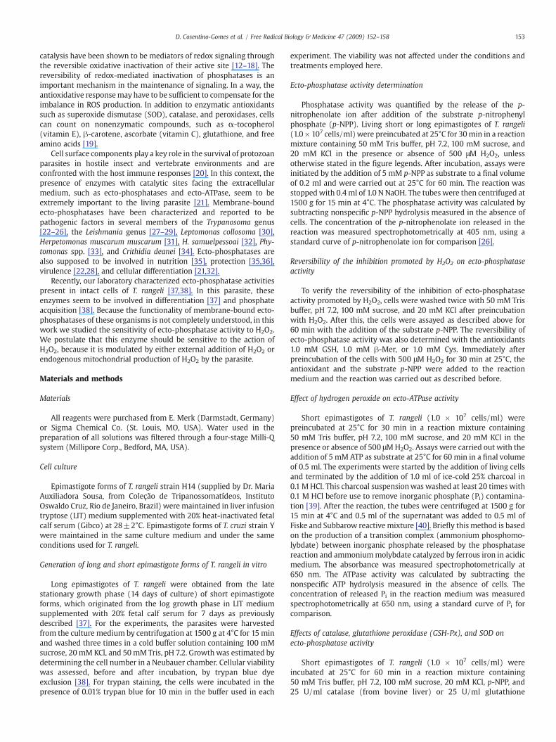
Fig. 6. Effects of enzymatic antioxidants on T. rangeli and T. cruzi ecto-phosphataseactivities. T. rangeli living parasites (1.0×10⁷ cells/ml) were incubated for 60 min at 25°Cin a reaction mixture containing 30 mM KCl, 100 mM sucrose, 50 mM Tris buffer, pH 7.2,and 5 mM p-NPP, in the absence (control) or in the presence of 10, 25, or 100 U/mlcatalase, or 25 U/ml glutathione peroxidase, or 25 U/ml superoxide dismutase asindicated on the abscissa. Inset: T. cruzi living parasites (1.0×10⁷ cells/ml) werepreincubated under the same conditions as in Fig.1, with 500 μMH2O2, or in the presenceof 25 U/ml catalase as described for T. rangeli. The parasites were viable during the courseof all experiments under all conditions used. The values represent the means±standarderror of at least three independent experiments. The asterisk denotes significant difference(pb0.05) compared with control.
155D. Cosentino-Gomes et al. / Free Radical Biology & Medicine 47 (2009) 152–158
Effect of hydrogen peroxide on ecto-ATPase activity
Another ecto-enzyme present on the external surface of T. rangeliis an ecto-ATPase [43]. Using ATP as a substrate, we tested the effectsof H2O2 on the ecto-ATPase activity in intact cells of T. rangeli. Asshown in Fig. 4, preincubation of cells for 30 min in the presence of500 μMH2O2 did not affect ecto-ATPase activity. This result shows thatthe inhibition promoted by H2O2 is specific to ecto-phosphataseactivity.
Effect of molecular and enzymatic antioxidants on ecto-phosphataseactivity
Some molecular antioxidants have been described as modulatingphosphatase activity [44,45]. Fig. 5A shows that β-Mer, Cys, and GSHat 1 mM did not modulate ecto-phosphatase activity, although at3 mM these antioxidants stimulated the ecto-phosphatase activity(Fig. 5A, inset). Interestingly, these antioxidants at 1 mMwere able torevert the inhibition induced by 500 μM H2O2. Intact cells of T. rangeliwere preincubated with 500 μM H2O2 for 30 min. Immediatelyafterward, each antioxidant was added at 1 mM to the reactionmedium with the substrate p-NPP (Fig. 5B). We also assayed the
Fig. 5. Effects of antioxidant agents on T. rangeli ecto-phosphatase activity. (A) Living parasite30mM KCl, 100 mM sucrose, 50 mM Tris buffer, pH 7.2, and 5mM p-NPP, with the addition ofof T. rangeli ecto-phosphatase activity by molecular antioxidants. Living parasites (1.0×10⁷ cKCl, 100 mM sucrose, 50 mM Tris buffer, pH 7.2, and 500 μM H2O2. Afterward, 1 mM GSH, β-60 min. The parasites were viable during the course of all experiments under all conditionexperiments. CTRL, control; β-Mer, β-mercaptoethanol; Cys, L-cysteine; GSH, reduced gluta
effects of the enzymatic antioxidants catalase, GSH-Px, and SOD on theecto-phosphatase activity (Fig. 6). Catalase and glutathione perox-idase at 25 U/ml enhanced the ecto-phosphatase activity by 54 and27%, respectively (Fig. 6). Nevertheless, no effect was observed on theenzyme activity for SOD (Fig. 6). In addition, when catalase andglutathione peroxidase were boiled at 100°C for 5 min, no effect wasobserved on the ecto-phosphatase activity (data not shown). Inter-estingly, the ecto-phosphatase activity of T. cruzi epimastigote forms isnot inhibited by H2O2 and is not stimulated by catalase (Fig. 6, inset).These results reinforce the importance of H2O2 in the modulation ofecto-phosphatase activity of T. rangeli and consequently the cellsignaling response. The stimulatory effects of catalase and glutathioneperoxidase on ecto-phosphatase activity could be related to the factthat the phosphatase activity was already inhibited by endogenousH2O2 production from cellular metabolism.
s (1.0×10⁷ cells/ml) were incubated for 60 min at 25°C in a reaction mixture containing1 or 3 mM (inset) GSH, β-Mer, or Cys as shown on the abscissa. (B) Reversible oxidationells/ml) were preincubated for 30 min at 25°C in a reaction mixture containing 30 mMMer, or Cys was added to the reaction with 5 mM p-NPP in a final volume of 0.2 ml fors used. The values represent the means±standard error of at least three independentthione. The asterisk denotes significant difference (pb0.05) compared with control.

Fig. 7. Effects of increasing concentrations of FCCP and oligomycin on the production of hydrogen peroxide by T. rangeli. (A) Living parasites (1.0×10⁸ cells/ml) were incubated for40min at 28°C in a reactionmedium containing 30mMKCl, 100mM sucrose, and 50mM Tris buffer, pH 7.2, with the addition of increasing concentrations of FCCP, as indicated on theabscissa, in a final volume of 0.2 ml. (B) Living parasites (1.0×10⁸ cells/ml) were incubated as in (A) but with the addition of increasing concentrations of oligomycin, as indicated onthe abscissa, in a final volume of 0.2 ml. The parasites were viable during the course of all experiments under all conditions used. The values represent the means±standard error ofat least three independent experiments. The asterisk denotes significant difference (pb0.05) compared with control.
156 D. Cosentino-Gomes et al. / Free Radical Biology & Medicine 47 (2009) 152–158
Endogenous hydrogen peroxide generation in vivo
The mitochondrial electron transport chain is the major andcontinuous source of cellular ROS, including H2O2, that can easilydiffuse through the plasma membrane [41,46] and interact withmembrane-bound components. We investigated the capacity ofintact cells of T. rangeli to produce H2O2 in the extracellular mediumfrom mitochondrial oxidative metabolism (Fig. 7). The addition ofFCCP, a proton ionophore that abolishes mitochondrial ROS genera-tion [47], inhibited H2O2 production in a dose-dependent manner(Fig. 7A). On the other hand, oligomycin, a blocker of proton trans-location through the F0F1ATP synthase [48], stimulated H2O2
production in a dose-dependent manner (Fig. 7B). Accordingly, theaddition of 5 μM FCCP increased and oligomycin inhibited the ecto-phosphatase activity (Fig. 8).
Discussion
Recent studies have demonstrated that H2O2 could play a key rolein intracellular signal transduction through the reversible inactivationof the active site of several enzymes [11–14,16,17,44,46], includingphosphatases [17]. Little is known about ecto-phosphatases and theirregulatory mechanism. Most studies of phosphatases have been doneeither with crude cell lysates or with purified enzymes. Because ecto-phosphatases present outward-facing active sites to the extracellularmedium, we studied the behavior of this enzyme in response to redoxreactions in intact cells.
Fig. 8. Effects of FCCP and oligomycin on T. rangeli ecto-phosphatase activity. Livingparasites (1.0×10⁷ cells/ml) were incubated for 60 min at 25°C in a reaction mixturecontaining 30 mM KCl, 100 mM sucrose, 50 mM Tris buffer, pH 7.2, and 5 mM p-NPP,with or without the addition of 5 μM FCCP or 5 μg/ml oligomycin, as indicated on theabscissa, in a final volume of 0.2 ml. The parasites were viable during the course of allexperiments under all conditions used. The values represent the means±standarderror of at least three independent experiments. The asterisk denotes significantdifference (pb0.05) compared with control.
We have described the sensitivity of the T. rangeli epimastigoteecto-phosphatase activity to H2O2 and antioxidants. The firstenvironment encountered by T. rangeli epimastigotes is the midgutof the vector, which is described as having more ROS than othercompartments in the insect [49]. The concentration range of H2O2
found in the midgut of Rhodnius prolixus is between 100 and 400 μM[49], the same inhibitory concentration range for T. rangeli ecto-phosphatase activity (Fig. 2). It seemed possible that the apparentsensitivity to oxidation and reduction of the ecto-phosphatase activitycould play a role in the regulation of cell signaling, because theseenzymes are supposed to be involved in differentiation and nutritionof these parasites [37,38].
Initial experiments identified differences in the sensitivity of theecto-phosphatase activities of the short and long epimastigote formsof T. rangeli to H2O2. Because these two forms live in differentcompartments and show striking biochemical differences in theirecto-phosphatase activities [37], it is plausible that differences insusceptibility to oxidation exist. The ecto-phosphatase activity ofshort epimastigotes was around 90% inhibited by 500 μM H2O2,whereas the long-form activity was inhibited by only 55% (Fig. 1).
We observed a loss in ecto-phosphatase activity of the short formwith increasing H2O2 concentrations. Biochemical characterization ofthe T. rangeli ecto-phosphatase activity indicated the presence of aprotein-tyrosine phosphatase-like activity [37]. This protein familyutilizes a nucleophilic cysteine residue in catalysis. Owing to theirmicroenvironment, the catalytic cysteines have low pKa's. Undernormal conditions, the active-site cysteines are in the thiolate anionform and are therefore highly susceptible to oxidation [11–15,17–19].
In this work, we demonstrated that even in the presence of sub-lethal doses of H2O2 (500 μM), the ecto-phosphatase inhibition wasreversible when the oxidantwas removed from themedium (Fig. 3) orin the presence of molecular antioxidants like β-mercaptoethanol,GSH, and the amino acid cysteine (Fig. 5B). The effect of GSH andcysteine must be due to the cysteinyl groups present in their structure[45]. The thiol group of these two structures competes with thecysteine in the active site of the ecto-phosphatase. These results showthat within cells, the ecto-phosphatase would form a stable oxidizedspecies, which can be reduced in vitro to recover the total activity andcontinue cell signaling.
GSH is found in the midgut of R. prolixus, one of the T. rangelivectors [50], and it would play a role in enzyme activity andconsequently in many cellular processes. Various phosphataseactivities have been shown to be reduced after oxidation by H2O2.Phosphatases, like PTEN of the PTP family, are reduced by dithio-threitol and β-mercaptoethanol, but are not reduced efficiently byGSH. On the other hand, PTPs such as PTPL1 could be efficientlyreduced by all of these agents, including GSH [44]. On the whole,

157D. Cosentino-Gomes et al. / Free Radical Biology & Medicine 47 (2009) 152–158
phosphatases as well as ecto-phosphatases can be modulated, notonly by oxidants, but also by reducing agents. The specificity of H2O2
for ecto-phosphatase activity was reinforced by the result with ecto-ATPase activity. With this other ecto-enzyme, H2O2 did not have aneffect (Fig. 4).
Fenton's reagent did not modulate T. rangeli ecto-phosphataseactivity. Preincubation of cells with ascorbate or FeSO4 did not alterthe total enzyme activity. However, ascorbate and FeSO4 were able toprotect the enzyme from oxidation by H2O2 (data not shown). Thisresult is opposed to that described for purple acid phosphatase, onwhich ascorbate alone had an inhibitory effect and this effect wasenhanced in the presence of H2O2 [51]. Nevertheless, our assays are inagreement with the protective effect of ascorbate on calcineurin,protein phosphatase type 1, and protein phosphatase type 2, threephosphatases of the serine/threonine phosphatase family [46].
We also tested enzymatic antioxidants, such as catalase, glu-tathione peroxidase, and SOD, three enzymes present in the midgut ofR. prolixus [49,50], the natural environment of T. rangeli shortepimastigotes. Catalase and glutathione peroxidase stimulated ecto-phosphatase activity, whereas no effect was observed with SOD (Fig. 6).Even though catalase and GSH-Px are absent from trypanosomatids[9,52], these enzymes are present in their environment and, like GSH,would play a role in enzyme activity and consequently in many cellularprocesses. The stimulatory effects of enzymatic scavengers of H2O2
such as catalase and glutathione peroxidase, such as those observed inthe presence of molecular antioxidants (Fig. 5A, inset), suggest thehypothesis that the enzyme was already inhibited. This inhibition isprobably due to endogenous H2O2 production from cellular metabo-lism in vitro. But, to oxidize the enzyme, H2O2 should be outside of theplasma membrane.
To evaluate this possibility, we measured the ability of cells toproduce H2O2 in the extracellular medium. Fig. 7 shows that smallamounts of H2O2 are found in the extracellular medium; probablymost of it is lost in oxidative reactions when crossing the membrane.This production can be modulated by either the protonophore FCCP orthe F0F1ATPase inhibitor oligomycin. Both compounds can modulatethe production of H2O2 by mitochondria, which may be partiallyrelated to the endogenous H2O2 production of the cell. Interestingly,the increases in ecto-phosphatase activity promoted by catalase andFCCP treatment were similar (Figs. 6 and 8). The mitochondrialformation of H2O2 is dependent on many factors and may beinfluenced by subcellular organization and mitochondrial morphol-ogy. In addition, there is evidence that the proton-motive force maynot be the same in reticular mitochondria, being localized in specificregions of a mitochondrion. Additionally, the morphology of mito-chondria can alter the rate of ROS production, flux, and diffusion ofmetabolites at specific regions of the organelle [53].
To verify whether the rate of external H2O2 production by the cellwould affect ecto-phosphatase activity, FCCP and oligomycin wereadded to the reaction for ecto-phosphatase activity. Accordingly, FCCPcaused a stimulation of the ecto-phosphatase activity, whereasoligomycin inhibited this activity (Fig. 8). However, we cannotdiscard the fact that uncouplers and poisons of mitochondrialrespiration may make changes in mitochondrial morphology [53].Cells or tissues are in a stable state if the rates of ROS production andscavenging capacity are appropriately balanced for cell survival andfunction. Redox signaling requires the disturbance of this balance,either by an increase in ROS concentrations or by a decrease in theactivity of one or more of the antioxidant systems [19]. For aprotozoan parasite, an imbalance in its natural environment must bedetected as soon as possible to facilitate a rapid protective response.These data suggest that the ecto-phosphatase is an importantmolecule in the detection of H2O2 for two reasons: (1) the ecto-phosphatase activity is readily inactivated by low concentrations ofH2O2 and (2) the active site faces the extracellular medium. Althoughthe consequences of inactivation of ecto-phosphatase activity by
H2O2 are not known, more studies have to be done to elucidate thepotential of these enzymes in the regulation of cellular signalingpathways.
Acknowledgments
We thank Dra. Maria Auxiliadora, from the TrypanosomatidCollection Fiocruz, for supplying the T. rangeli. We also thank Mr.Fabiano Ferreira Esteves and Ms. Rosangela Rosa de Araújo for theexcellent technical assistance. This work was supported by grantsfrom the Brazilian agencies Conselho Nacional de DesenvolvimentoCientífico e Tecnológico (CNPq), Coordenação de Aperfeiçoamento dePessoal de Nível Superior (CAPES), and Fundação de Amparo aPesquisa do Estado do Rio de Janeiro (FAPERJ).
References
[1] Müller, S.; Liebau, E.; Walter, R. D.; Krauth-Siegel, R. L. Thiol-based redoxmetabolism of protozoan parasites. Trends Parasitol. 19:320–328; 2003.
[2] Watkins, R. Histology of Rhodnius prolixus infected with Trypanosoma rangeli.J. Invertebr. Pathol. 17:59–66; 1971.
[3] Guhl, F.; Vallejo, A. Trypanosoma (Herpetosoma) rangeli Tejera, 1920: an updatedreview. Mem. Inst. Oswaldo Cruz 98:435–442; 2003.
[4] Machado, E. M. M.; Azambuja, P.; Garcia, E. S. WEB 2086, a platelet-activatingfactor antagonist, inhibits prophenoloxidase-activating system and hemocytemicroaggregation reactions induced by Trypanosoma rangeli infection in Rhodniusprolixus hemolymph. J. Insect Physiol. 52:685–692; 2006.
[5] Gomes, S. A. O.; Feder, D.; Thomas, N. E. S.; Garcia, E. S.; Azambuja, P. Rhodniusprolixus infected with Trypanosoma rangeli: in vivo and in vitro experiments.J. Invertebr. Pathol. 73:289–293; 1999.
[6] Mello, C. B.; Garcia, E. S.; Ratcliffe, N. A.; Azambuja, P. Trypanosoma cruzi andTrypanosoma rangeli: interplay with hemolymph components of Rhodnius prolixus.J. Invertebr. Pathol. 65:261–268; 1995.
[7] Takle, G. B. Studies on the cellular immune responses of insects toward the insectpathogen Trypanosoma rangeli. J. Invertebr. Pathol. 51:64–72; 1988.
[8] Steenkamp, D. J. Trypanosomal antioxidants and emerging aspects of redoxregulation in the trypanosomatids. Antioxid. Redox Signaling 4:105–121; 2002.
[9] Turrens, J. F. Oxidative stress and antioxidant defenses: a target for the treat-ment of diseases caused by parasitic protozoa. Mol. Aspects Med. 25:211–220;2004.
[10] Finzi, J. K.; Chiavegatto, C.W.M.; Corat, K. F.; Lopez, J. A.; Cabrera, O. G.; Mielniczki-Pereira, A. A.; Colli, W.; Alves, M. J. M.; Gadelha, F. R. Trypanosoma cruzi responseto the oxidative stress generated by hydrogen peroxide. Mol. Biochem. Parasitol.133:37–43; 2004.
[11] Rhee, S. G.; Kang, S. W.; Jeong, W.; Chang, T. S.; Yang, K. S.; Woo, H. A. Intracellularmessenger function of hydrogen peroxide and its regulation by peroxiredoxins.Curr. Opin. Cell Biol. 17:183–189; 2005.
[12] Leslie, N. R.; Lindsay, Y.; Ross, S. H.; Downes, C. P. Redox regulation of phosphatasefunction. Biochem. Soc. Trans. 32:1018–1020; 2004.
[13] Cho, S. H.; Lee, C. H.; Ahn, Y.; Kim, H.; Ahn, C. Y.; Yang, K. S.; Lee, S. R. Redoxregulation of PTEN and protein tyrosine phosphatases in H(2)O(2) mediated cellsignaling. FEBS Lett. 560:7–13; 2004.
[14] Tonks, N. K. Redox redux: revisiting PTPs and the control of cell signaling. Cell 121:667–670; 2005.
[15] Van der Wijk, T.; Blanchelot, C.; Den Hertog, J. Regulation of receptor protein-tyrosine phosphatase dimerization. Methods 35:73–79; 2005.
[16] Rao, R. K.; Clayton, L. W. Regulation of protein phosphatase 2A by hydrogenperoxide and glutathionylation. Biochem. Biophys. Res. Commun. 293:610–616;2002.
[17] Den Hertog, J.; Groen, A.; Van der Wijk, T. Redox regulation of protein-tyrosinephosphatases. Arch. Biochem. Biophys. 434:11–15; 2005.
[18] Yang, J.; Groen, A.; Lemeer, S.; Jans, A.; Slijper, M.; Roe, S. M.; Den Hertog, J.;Barford, D. Reversible oxidation of membrane distal domain of receptor PTPα ismediated by a cyclic sulfonamide. Biochemistry 46:709–719; 2007.
[19] Dröge, W. Free radicals in the physiological control of cell function. Physiol. Rev.82:47–95; 2002.
[20] Colli, W.; Alves, M. J. M. Relevant glycoconjugates on the surface of Trypanosomacruzi. Mem. Inst. Oswaldo Cruz 94:37–49; 1999.
[21] Meyer-Fernandes, J. R. Ecto-ATPases in protozoa parasites: looking for a function.Parasitol. Int. 51:299–303; 2002.
[22] Bakalara, N.; Seyfang, A.; Baltz, T. Davis, C. Trypanosoma brucei and Trypanosomacruzi: life cycle-regulated protein tyrosine phosphatase activity. Exp. Parasitol. 81:302–312; 1995.
[23] Furuya, T.; Zhong, L.; Meyer-Fernandes, J. R.; Lu, H. G.; Moreno, S. N. J.; Docampo, R.Ecto-protein tyrosine phosphatase activity in Trypanosoma cruzi infective stages.Mol. Biochem. Parasitol. 92:339–348; 1998.
[24] Meyer-Fernandes, J. R.; da Silva-Neto, M. A.; Soares, M. S.; Fernandes, E.; Vercesi,A. E.; Oliveira, M. M. Ecto-phosphatase activities on the cell surface of theamastigote forms of Trypanosoma cruzi. Z. Naturforsch. 54:977–984; 1999.
[25] Bakalara, N.; Santarelli, X.; Davis, C.; Baltz, T. Purification, cloning andcharacterization of an acidic ectoprotein phosphatase differentially expressed in

158 D. Cosentino-Gomes et al. / Free Radical Biology & Medicine 47 (2009) 152–158
the infectious bloodstream form of Trypanosoma brucei. J. Biol. Chem. 275:8863–8871; 2000.
[26] Fernandes, E. C.; Granjeiro, J. M.; Taga, E. M.; Meyer-Fernandes, J. R.; Aoyama, H.Phosphatase activity characterization on the surface of intact bloodstream formsof Trypanosoma brucei. FEMS Microbiol. Lett. 220:197–206; 2003.
[27] Shakarian, A. M.; Joshi, M. B.; Ghedin, E.; Dwyer, D. M. Molecular dissection of thefunctional domains of a unique, tartrate-resistant, surface membrane acidphosphatase in the primitive human pathogen Leishmania donovani. J. Biol.Chem. 277:17994–18001; 2002.
[28] Vannier-Santos, M. A.; Martiny, A.; Meyer-Fernandes, J. R.; de Souza, W.Leishmanial protein kinase C modulates host cell infection via secreted acidphosphatase. Eur. J. Cell. Biol. 67:112–119; 1995.
[29] Wiese, M.; Berger, O.; Stierhof, Y. D.; Wolfram, M.; Fuchs, M.; Overath, P. Genecloning and cellular localization of a membrane-bound acid phosphatase ofLeishmania mexicana. Mol. Biochem. Parasitol. 82:153–165; 1996.
[30] Hunt, R. C.; Ellar, D. J. Isolation of the plasma membrane of a trypanosomatidflagellate: general characterisation and lipid composition. Biochim. Biophys. Acta339:173–189; 1974.
[31] Dutra, P. M. L.; Rodrigues, C. O.; Jesus, J. B.; Lopes, A. H.; Souto-Padrón, T.; Meyer-Fernandes, J. R. A novel ecto-phosphatase activity of Herpetomonas muscarummuscarum inhibited by platelet-activating factor. Biochem. Biophys. Res. Commun.253:164–169; 1998.
[32] Santos, A. L. S.; Souto-Padrón, T.; Alviano, C. S.; Lopes, A. H.; Soares, R. M.; Meyer-Fernandes, J. R. Secreted phosphatase activity induced by dimethyl sulfoxide inHerpetomonas samuelpessoai. Arch. Biochem. Biophys. 405:191–198; 2002.
[33] Dutra, P. M. L.; Dias, F. A.; Santos, M. A. A.; Rodrigues, C. O.; Romeiro, A.; Attias, M.;De Souza, W.; Lopes, A. H. C. S.; Meyer-Fernandes, J. R. Secreted phosphataseactivities in Trypanosomatid parasites of plants modulated by platelet-activatingfactor. Phytopathology 91:408–414; 2001.
[34] Lemos, A. D.; de Souza, A. L. F.; Pinheiro, A. A. D.; Berrêdo-Pinho, M.; Meyer-Fernandes, J. R. Ecto-phosphatase activity on the cell surface of Crithidia deanei.Z. Naturforsch. 57:500–505; 2002.
[35] Gottlieb, M.; Dwyer, D. M. Leishmania donovani: surface membrane acidphosphatase activity of promastigotes. Exp. Parasitol. 52:117–128; 1981.
[36] Remaley, A. T.; Das, S.; Campbell, P. I.; LaRocca, G. M.; Pope, M. T.; Glew, R. H.Characterization of Leishmania donovani acid phosphatases. J. Biol. Chem. 260:880–886; 1985.
[37] Gomes, S. A. O.; de Souza, A. L. F.; Silva, B. A.; Kiffer-Moreira, T.; Santos-Mallet, J. R.;Santos, A. L. S.; Meyer-Fernandes, J. R. Trypanosoma rangeli: differential expressionof cell surface polypeptides and ecto-phosphatase activity in short and longepimastigote forms. Exp. Parasitol. 112:253–262; 2006.
[38] Fonseca-de-Souza, A. L.; Dick, C. F.; Dos Santos, A. L. A.; Meyer-Fernandes, J. R. AMg2+-dependent ecto-phosphatase activity on the external surface of Trypano-
soma rangeli modulated by exogenous inorganic phosphate. Acta Trop. 107:153–158; 2008.
[39] Guilherme, A.; Meyer-Fernandes, J. R.; Vieyra, A. Reversible inhibition by 4,4′-diiso-thiocyanatostilbene-2,2′disulfonic acid of the plasma membrane (Ca2++Mg2+)ATPase from kidney proximal tubules. Biochemistry 30:5700–5706; 1991.
[40] Fiske, C. H.; Subbarow, J. W. The colorimetric determination of phosphorous. J. Biol.Chem. 66:375–392; 1925.
[41] Meyer, L. E.; Machado, L. B.; Santiago, A. P. S. A.; Da-Silva, W. S.; De Felice, F. G.;Holub, O.; Oliveira, M. F.; Galina, A. Mitochondrial creatine kinase activity preventsreactive oxygen species generation. J. Biol. Chem. 281:37361–37371; 2006.
[42] Spear, N.; Aust, S. D. Effects of glutathione on Fenton reagent-dependent radicalproduction and DNA oxidation. Arch. Biochem. Biophys. 324:111–116; 1995.
[43] Fonseca, F. V.; Fonseca de Souza, A. L.; Mariano, A. C.; Entringer, P. F.; Gondim, K. C.;Meyer-Fernandes, J. R. Trypanosoma rangeli: characterization of a Mg-dependentecto ATP-diphosphohydrolase activity. Exp. Parasitol. 112:76–84; 2006.
[44] Ross, S. H.; Lindsay, Y.; Safrany, S. T.; Lorenzo, O.; Villa, F.; Toth, R.; Clague, M. J.;Downes, C. P.; Leslie, N. R. Differential redox regulation within the PTPsuperfamily. Cell. Signalling 19:1521–1530; 2007.
[45] de Sá Pinheiro, A. A.; Amazonas, J. N.; Barros, F. S.; Menezes, L. F.; Batista, E. J. O.;Silva, E. F.; De Souza, W.; Meyer-Fernandes, J. R. Entamoeba histolytica: an ecto-phosphatase activity regulated by oxidation–reduction reactions. Exp. Parasitol.115:352–358; 2007.
[46] Sommer, D.; Coleman, S.; Swanson, S. A.; Stemmer, P. M. Differential susceptibi-lities of serine/threonine phosphatases to oxidative and nitrosative stress. Arch.Biochem. Biophys. 404:271–278; 2002.
[47] Boveris, A.; Chance, B. The mitochondrial generation of hydrogen peroxide:general properties and effect of hyperbaric oxygen. Biochem. J.134:707–716; 1973.
[48] Korshunov, S. S.; Skulachev, V. P.; Starkov, A. A. High protonic potential actuates amechanism of production of reactive oxygen species in mitochondria. FEBS Lett.416:15–18; 1997.
[49] Paes, M. C.; Oliveira, M. B.; Oliveira, P. L. Hydrogen peroxide detoxification in themidgut of the blood-sucking insect, Rhodnius prolixus. Arch. Insect Biochem.Physiol. 48:63–71; 2001.
[50] Paes, M. C.; Oliveira, P. L. Extracellular glutathione peroxidase from the blood-sucking bug, Rhodnius prolixus. Arch. Insect Biochem. Physiol. 4:171–177; 1999.
[51] Beck, J. L.; Durack, M. C. A.; Hamilton, S. E.; Jersey, J. Irreversible inactivation ofpurple acid phosphatase by hydrogen peroxide and ascorbate. J. Inorg. Biochem.73:245–252; 1999.
[52] Boveris, A.; Sies, H.; Martino, E. E.; Docampo, R.; Turrens, J. F.; Stoppani, A. O. M.Deficient metabolic utilization of hydrogen peroxide in Trypanosoma cruzi.Biochem. J. 188:643–648; 1980.
[53] Bernard, G.; Rossignol, R. Ultrastructure of the mitochondrion and its bearing onfunction and bioenergetics. Antioxid. Redox Signaling 10:1313–1342; 2008.
Related Documents