Modulation of the canonical Wnt activity by androgen signaling in prostate epithelial basal stem cells Yueli Liu 1 , Jiawen Wang 2 , Corrigan Horton 1 , Sol Katzman 3 , Tao Cai 2 , Zhu A. Wang 1† 1 Department of Molecular, Cell, and Developmental Biology, University of California, Santa Cruz, CA 95064, USA 2 Sequencing Center, National Institute of Biological Sciences, Beijing, 102206, China 3 Jack Baskin School of Engineering, University of California, Santa Cruz † Author for correspondence: [email protected] author/funder. All rights reserved. No reuse allowed without permission. The copyright holder for this preprint (which was not peer-reviewed) is the . https://doi.org/10.1101/2020.01.10.902270 doi: bioRxiv preprint

Welcome message from author
This document is posted to help you gain knowledge. Please leave a comment to let me know what you think about it! Share it to your friends and learn new things together.
Transcript

Modulation of the canonical Wnt activity by androgen signaling in prostate
epithelial basal stem cells
Yueli Liu1, Jiawen Wang2, Corrigan Horton1, Sol Katzman3, Tao Cai2, Zhu A.
Wang1†
1Department of Molecular, Cell, and Developmental Biology, University of
California, Santa Cruz, CA 95064, USA
2Sequencing Center, National Institute of Biological Sciences, Beijing, 102206, China
3Jack Baskin School of Engineering, University of California, Santa Cruz
†Author for correspondence: [email protected]
author/funder. All rights reserved. No reuse allowed without permission. The copyright holder for this preprint (which was not peer-reviewed) is the. https://doi.org/10.1101/2020.01.10.902270doi: bioRxiv preprint

Abstract
Both the canonical Wnt signaling and androgen signaling are important factors
regulating prostate organogenesis. How these two pathways crosstalk to regulate
prostate stem cell functions remain unclear. Here, we show that while canonical Wnt
activity is required for prostate basal stem cell multipotency in vivo, ectopic Wnt
activity does not promote basal-to-luminal cell differentiation. We provide evidence
that androgen signaling may keep Wnt activity in check. In prostate organoid culture
from basal cells, dihydrotestosterone (DHT) antagonizes R-spondin-stimulated
organoid growth in a concentration-dependent manner. Molecular analyses of
organoids under different treatment conditions showed that androgen signaling down-
regulated the expressions of a Wnt reporter as well as many Wnt target genes.
Pathway analysis and gene set enrichment analysis of organoid RNA-seq data also
revealed the canonical Wnt signaling as a key pathway distinguishing organoids
treated with or without DHT. Notably, DHT treatment enhanced AR and β–catenin
binding in the nuclei of prostate organoids, providing possible mechanistic clues. Our
results reveal a critical role of AR signaling in modulating canonical Wnt activity in
prostate basal cells to regulate their multipotency.
author/funder. All rights reserved. No reuse allowed without permission. The copyright holder for this preprint (which was not peer-reviewed) is the. https://doi.org/10.1101/2020.01.10.902270doi: bioRxiv preprint
Introduction
Signaling crosstalks are crucial for regulating adult stem cells in an organ. The
canonical Wnt signaling pathway is a key player in stem cell self-renewal and
differentiation in multiple tissues (Clevers et al., 2014; Holland et al., 2013). Its
interaction with organ-specific hormone signals may be important for orchestrating
the functions of adult stem cells in that tissue. In the prostate, both the canonical Wnt
signaling and androgen signaling pathways have been shown to be essential in driving
organogenesis (Toivanen and Shen, 2017). Specifically, stromal Wnt secretion
promotes prostate bud branching morphogenesis and expression of the prostate-
specific transcription factor Nkx3.1 in the epithelium (Francis et al., 2013; Julio et al.,
2013; Simons et al., 2012). Classical tissue recombination experiments and
conditional knockout experiments have revealed the role of stromal androgen receptor
(AR) in instructing the specification of prostate epithelium through paracrine signals
(Cunha et al., 1992; Lai et al., 2012). However, how Wnt and androgen signals
interact to promote prostate organogenesis and whether their interaction affects
prostate stem cells remain unknown.
Basal and luminal cells are the two major cell types lining the prostate
epithelium. During prostate organogenesis, basal cells behave as stem cells to
generate luminal cells and rare neuroendocrine cells (Ousset et al., 2012). Although
basal stem cell multipotency become restricted in the mature prostate (Choi et al.,
2012; Wang et al., 2013), isolated adult basal cells can be reactivated to generate
prostatic tissues in the renal-grafting assay (Wang et al., 2014; Xin et al., 2003), and
are more efficient in generating prostate organoids in cultures compared to luminal
author/funder. All rights reserved. No reuse allowed without permission. The copyright holder for this preprint (which was not peer-reviewed) is the. https://doi.org/10.1101/2020.01.10.902270doi: bioRxiv preprint

cells (Chua et al., 2014; Karthaus et al., 2014). The organoid culture medium
contains R-spondin, an agonist of the canonical Wnt pathway (Binnerts et al., 2007;
Carmon et al., 2011; de Lau et al., 2011), demonstrating a positive role of Wnt
signaling for prostate organoid formation and expansion (Karthaus et al., 2014). On
the other hand, conditional knockout of β-catenin in adult prostate basal cells
appeared to negatively affect their luminal differentiation, as revealed by lineage
tracing during a 6-week period (Lu and Chen, 2015). Recently, our lab performed
long-term basal cell lineage tracing with AR conditional knockout, and demonstrated
that AR within basal cells is required for basal-to-luminal cell differentiation in vivo
(Xie et al., 2017). Despite these findings, how Wnt and androgen signaling pathways
cooperate to promote basal cell multipotency is unclear. In this study, we show that
ectopic Wnt activity leads to prostate basal cell over-proliferation, but not luminal
differentiation, and that androgen signaling can down-regulate Wnt activity in basal
cells, possibly through binding with β-catenin to interfere with target gene
transcription.
author/funder. All rights reserved. No reuse allowed without permission. The copyright holder for this preprint (which was not peer-reviewed) is the. https://doi.org/10.1101/2020.01.10.902270doi: bioRxiv preprint
Results and Discussion
Intermediate Wnt activity is essential for prostate basal-to-luminal cell
differentiation
Although a previous lineage-tracing study suggested a role of canonical Wnt
activity in promoting basal-to-luminal differentiation (Lu and Chen, 2015), the long-
term effect of β-catenin deletion in basal cells is unknown. To this end, we utilized
the CK5-CreERT2; R26R-CAG-YFP/+ reporter for basal cell lineage tracing, for
which we showed previously marks almost all prostate basal cells (Xie et al., 2017).
We tamoxifen-induced CK5-CreERT2; Ctnnb1fl/fl; R26R-CAG-YFP/+ adult male mice
(termed Basbcat-/-) and CK5-CreERT2; R26R-CAG-YFP/+ controls (termed BasWT), and
performed long-term lineage tracing under homeostasis (Fig. 1A). Since β-catenin is a
structural protein expressed in luminal cells contacting the basal layer, its loss in
individual basal cells could not be easily distinguished by immunofluorescence (IF)
staining (Fig. S1A). Nevertheless, efficient β-catenin deletion driven by CK5-CreERT2
was confirmed by flow-sorting of YFP+ prostate basal cells (Fig. S1B) for qRT-PCR
analysis (Fig. S1C), and inferred by observing hair loss through time (Fig. S1D)
(Kishimoto et al., 2000). Six months after induction, basal-to-luminal cell
differentiation was significantly reduced in the Basbcat-/- group compared to BasWT
(Fig. 1B, 1C), while basal cell proliferation rates were unchanged as measured by
Ki67 staining and BrdU incorporation assay (Fig. 1D, 1E). These results are
consistent with the previous 6-week tracing data in the Lu and Chen study. However,
contrary to their finding in the tumor models (Lu and Chen, 2015), we found that β-
catenin deletion did not rescue the progression of basal-derived Pten-null tumors, as
author/funder. All rights reserved. No reuse allowed without permission. The copyright holder for this preprint (which was not peer-reviewed) is the. https://doi.org/10.1101/2020.01.10.902270doi: bioRxiv preprint

CK5-CreERT2; ctnnb1fl/fl; Ptenfl/fl; R26R-CAG-YFP/+ tumors (Basbcat-/-Pten-/-) were
morphologically indistinguishable from those of CK5-CreERT2; Ptenfl/fl; R26R-CAG-
YFP/+ (BasPten-/-) (Fig. 1F). The double knockout tumors still contain cribriform
pattern and luminal differentiation (Fig. 1G), despite their lack of β-catenin
expression (Fig. 1H). The cause of the discrepancy is unclear. We conclude that Pten
loss can override the requirement of β-catenin in promoting basal-to-luminal cell
differentiation.
To assess whether gain of Wnt signaling activity could enhance basal-to-
luminal cell differentiation, we tamoxifen-induced CK5-CreERT2; APCfl/fl; R26R-
CAG-YFP/+ mice (termed BasAPC-/-) and performed 3-week lineage tracing (Fig. 1A).
We had to analyze the prostate morphology at this early time point due to mice
succumbing to rapidly developing basal tumors growing elsewhere. While the
majority of prostate epithelial cells appeared normal, we observed clones of basal
cells stacked with each other, resembling squamous tumors (Fig. 1I). Those cells were
strictly basal, and did not express the luminal marker CK18 (Fig. 1J). We then tried to
propagate the basal-APC-deleted prostate tissues by renal grafting those cells onto
immunodeficient mice (Fig. 1A), and again observed such basal cell over-
proliferation phenotype after three months of growth (Fig. 1K). Taken together, these
results suggest that Wnt signaling activity needs to be tightly controlled in vivo since
intermediate Wnt activity is essential for basal-to-luminal cell differentiation.
Ectopic Wnt activity enhances basal stem cell activities in organoid assay
We next investigated the effects of different Wnt activity levels on prostate
basal stem cells using a defined organoid culture protocol (Drost et al., 2016). Wild-
type and Wnt-activated basal cells were isolated from induced BasWT and BasAPC-/-
author/funder. All rights reserved. No reuse allowed without permission. The copyright holder for this preprint (which was not peer-reviewed) is the. https://doi.org/10.1101/2020.01.10.902270doi: bioRxiv preprint

mice, respectively, and were cultured under three conditions: (1) “basement medium”
(BM condition) containing all the factors as described in the protocol except for R-
spondin and dihydrotestosterone (DHT), (2) BM plus R-spondin (R condition), and
(3) BM plus the Wnt antagonist Dkk-1 (Dkk condition) (Glinka et al., 1998).
Representative images of organoids growing for 7 days under different conditions are
shown in Fig. 2A. These organoids typically exhibit CK5-expressing basal cells on
the outskirt and CK18-expressing luminal cells in the center (Fig. 2A). Organoids of
BasAPC-/- cells were significantly larger than those of BasWT (Fig. 2B), and contained
significantly more complex structures such as increased branching (Fig. 2C),
suggesting ectopic Wnt activity enhances basal stem cell activities and organoid
growth in vitro. Consistently, R-spondin treatment increased organoid size and
branching morphogenesis compared to BM for both BasAPC-/- and BasWT organoids,
while Dkk-1 treatment had the opposite effects (Fig. 2B, 2C). Notably, robust
organoid growth under the BM or R condition was achieved without adding DHT, a
component described in the defined prostate organoid protocol (Drost et al., 2016),
suggesting that DHT is not required for basal stem cell functions in vitro.
DHT suppresses prostate basal stem cell activities in organoid assay
Given the pivotal role of androgen signaling in prostate development, we next
tested how adding DHT affects organoid growth from wild-type basal cells. The
basement medium was supplemented with DHT at two concentrations 10-7 M and 10-9
M as the DHT-9 and DHT-7 conditions. In another two conditions, DHT was also
added together with R-spondin (RD-9 and RD-7 conditions) to test the combinatory
effects of activating Wnt and androgen signaling pathways on prostate organoid
growth. Representative images of organoids growing for 7 days under these
author/funder. All rights reserved. No reuse allowed without permission. The copyright holder for this preprint (which was not peer-reviewed) is the. https://doi.org/10.1101/2020.01.10.902270doi: bioRxiv preprint

conditions are shown in Fig. 3A. Surprisingly, our data consistently showed that DHT
suppressed basal stem cell activities in organoid culture in a concentration-dependent
manner. When organoid size and structural complexity as measured by the number of
branching were quantified, DHT-9 condition led to significantly reduced growth and
branching morphogenesis compared to BM alone, and DHT-7 condition further
exacerbated such phenotypes as the organoids were significantly stunted in growth
(Fig. 3A-C). Adding R-spondin was able to rescue these phenotypes to some extent,
but DHT could clearly antagonize the growth-promoting effects of R-spondin since
organoids under the RD-9 condition were significantly smaller and contained less
branching than those under the R condition (Fig. 3B, 3C). These data imply that Wnt
and androgen signaling pathways have opposite effects on prostate basal stem cell
activities and that their combined effects may be subtractive.
DHT decreases Wnt signaling activity in prostate organoids
Based on the results above, we hypothesized that DHT inhibits basal organoid
growth through modulation of the Wnt signaling pathway. To test it, we isolated basal
cells from the TCF/Lef-H2B.GFP reporter mice, in which GFP signal can serves as
readout for canonical Wnt signaling activity (Ferrer-Vaquer et al., 2010). We then
cultured these cells as organoids using 6 medium conditions: BM, R, RD-9, RD-7,
DHT-9, and DHT-7. After 7 days of culture, we found that the GFP signals were
usually present in the center of the organoids, and were most prominent in the R
group (Fig. 4A). When we quantified the percentages of GFP area size of the total
organoid size, we found the trend as follows: R>BM>RD-9>RD-7>DHT-9>DHT-7,
and adding DHT significantly reduced GFP area even in the presence of R-spondin
(Fig. 4B). Expression reduction upon DHT treatment was also observed for several
author/funder. All rights reserved. No reuse allowed without permission. The copyright holder for this preprint (which was not peer-reviewed) is the. https://doi.org/10.1101/2020.01.10.902270doi: bioRxiv preprint

Wnt target genes Axin2, Lef1, Ccnd2, Cd44, and Myc, as quantified by qRT-PCR
(Fig. 4C), confirming that DHT decreases Wnt signaling activity.
To further explore the mechanism, we isolated mRNAs from BM, R, and RD-
7 organoids and performed RNA-sequencing. Principal component analysis (PCA)
and unsupervised hierarchical clustering analysis demonstrated the consistency of the
samples within each group and the distinct transcriptomes of the RD-7 group from the
BM and R groups (Fig. 5A, 5B). Gene expression differential analysis revealed the
numbers of upregulated and downregulated genes between each groups as shown in
the volcano plots (Fig. S2A). Many genes, including Wnt targets, were upregulated in
R compared to BM, but were downregulated in RD-7 compared to R (Fig. S2B).
David GO analysis (Huang da et al., 2009) of differentially expressed gene set
between R and RD-7 groups identified key pathways involved, among which were
cell cycle regulation and Wnt signaling pathway (Fig. 5C), supporting our model that
the inhibitory effects of androgen on basal organoids were mediated through
modulation of the canonical Wnt activity. In further support of this, gene set
enrichment analysis (GSEA) (Subramanian et al., 2005) showed that Wnt signaling
pathway genes were highly enriched in the genes that were downregulated in the RD-
7 group compared to the R group (Fig. 5D). Similarly, genes of the GO term Organ
Morphogenesis were also highly enriched in the downregulated RD-7 genes, while
two gene signatures that were previously shown to be upregulated by androgen
(Mulholland et al., 2011; Schaeffer et al., 2008) were highly enriched in the
upregulated RD-7 genes (Fig. 5D).
Increased AR and β-catenin binding in organoid cells upon R-spondin and DHT
treatment
author/funder. All rights reserved. No reuse allowed without permission. The copyright holder for this preprint (which was not peer-reviewed) is the. https://doi.org/10.1101/2020.01.10.902270doi: bioRxiv preprint

Previous studies in prostate cancer cell lines have shown that AR can compete
with TCF for β-catenin binding (Song et al., 2003), and the binding of AR and β-
catenin can enhance AR signaling output (Amir et al., 2003; Mulholland et al., 2002;
Truica et al., 2000). How this binding affects Wnt signaling activity is unclear. We
hypothesize that DHT inhibition of Wnt activity in prostate organoids may be
mediated through AR binding to β-catenin in the nucleus to hamper the normal
function of β-catenin as a Wnt signaling transcription factor. To visualize their
interactions in organoids, we performed Duolink Proximity Ligation Assay (PLA)
(Fredriksson et al., 2002; Soderberg et al., 2008), in which antibodies recognizing
both AR and β-catenin in close proximity can yield an amplified signal dot in situ.
We cultured prostate organoids for 7 days and performed quality check experiments
to confirm that PLA signals can only be detected when both AR and β-catenin
antibodies were applied (Fig. 6A). Interestingly, when the organoids were cultured
under BM, R, RD-9, and RD-7 conditions, we observed significantly more AR and β-
catenin protein interactions per nucleus or per cell under the RD treatment conditions
(Fig. 6B, 6C), and the interactions increased with the concentration of DHT (Fig. 6C).
These data are consistent with our hypothesis and call for further functional
investigation of the consequences of increased AR and β-catenin binding in prostate
organoids. Previous studies have only focused on the effects of such interactions on
androgen signaling output, and cancer cell lines were often used as materials (Amir et
al., 2003; Mulholland et al., 2002; Song et al., 2003; Truica et al., 2000). Analyzing
the AR and β-catenin cistromes through ChIP-seq under various treatment conditions
for the basal-derived organoids should provide mechanistic insights into how Wnt
signaling output is modified by enhanced AR binding in the context of normal
prostate stem cell function.
author/funder. All rights reserved. No reuse allowed without permission. The copyright holder for this preprint (which was not peer-reviewed) is the. https://doi.org/10.1101/2020.01.10.902270doi: bioRxiv preprint

Methods
Mouse strains and genotyping
The CK5-CreERT2, Ptenflox, and R26R-CAG-YFP lines were used previously (Xie et
al., 2017). The Ctnnb1flox (Brault et al., 2001) and APCflox (Cheung et al., 2010) lines
were obtained from JAX. The TCF/Lef-H2B.GFP line (Ferrer-Vaquer et al., 2010)
was obtained from Dr. Hadjantonakis. Animals were maintained in C57BL/6N or
mixed background. Genotyping was performed by PCR using tail genomic DNA,
with the following primer sequences: CreERT2 allele: 5’-
CAGATGGCGCGGCAACACC-3’ and 5’-GCGCGGTCTGGCAGTAAAAAC-3’;
TCF/Lef-H2B.GFP allele: 5’-ACAACAAGCGCTCGACCATCAC-3’ and 5’-
AGTCGATGCCCTTCAGCTCGAT-3’; Ctnnb1flox allele: 5’-
ACTGCCTTTGTTCTCTTCCCTTCTG-3’ and 5’-
CAGCCAAGGAGAGCAGGTGAGG-3’; APCflox allele: 5’-
GAGAAACCCTGTCTCGAAAAAA-3’ and 5’-
AGTGCTGTTTCTATGAGTCAAC-3’; R26R-CAG-YFP allele: 5’-
AAGGGAGCTGCAGTGGAGTA-3’ (wild-type forward), 5’-
CCGAAAATCTGTGGGAAGTC-3’ (wild-type reverse), 5’-
ACATGGTCCTGCTGGAGTTC-3’ (mutated forward), 5’-
GGCATTAAAGCAGCGTATCC-3’ (mutated reverse).
Mouse procedures
For tamoxifen induction, mice were administered 9 mg per 40 g body weight
tamoxifen (Sigma) suspended in corn oil by oral gavage once daily for 4 consecutive
author/funder. All rights reserved. No reuse allowed without permission. The copyright holder for this preprint (which was not peer-reviewed) is the. https://doi.org/10.1101/2020.01.10.902270doi: bioRxiv preprint

days. For BrdU incorporation assay, BrdU (Sigma) was dissolved in PBS (10 mg/ml)
and administered by intraperitoneal injection twice daily (0.1 ml per dose) for 11
consecutive days during homeostasis to label proliferating cells. All animal
experiments received approval from the Institutional Animal Care and Use
Committee at UCSC.
Tissue collection and flow cytometry
Mouse prostate tissues were dissected and fixed in 4% paraformaldehyde for
subsequent cryo-embedding in OCT compound (Sakura), or fixed in 10% formalin
followed by paraffin embedding. For flow cytometry, prostate tissues were dissected
and minced to small clumps, followed by enzymatic dissociation with 0.2%
Collagenase/Hyaluronidase (StemCell Technologies) in DMEM/F12 media with 5%
FBS for 3 h at 37°C. Tissues were digested with 0.25% Trypsin-EDTA (StemCell
Technologies) for 1 h at 4°C, passed through 21- to 26-gauge syringes and filtered
through a 40-mm cell strainer to obtain single-cell suspensions. Dissociated prostate
cells were suspended in Hanks’ Balanced Salt Solution Modified/2% FBS. Dead cells
were excluded by propidium iodide staining. Lineage-marked basal cells were sorted
based on YFP positivity. Antibodies (Table S1) were used for sorting Lin−Sca-1+
CD49fhi basal cells from TCF/Lef-H2B.GFP mice. Cell sorting was performed on a
BD FACS Aria II instrument in the Flow Cytometry Shared Facility of UCSC.
Renal grafting assay
Flow-sorted APCfl/fl YFP+ basal cells were mixed with 2.5 × 10⁵ dissociated
urogenital sinus mesenchyme (UGM) cells from embryonic day 18.0 rat embryos.
UGM cells were obtained from dissected urogenital sinus treated for 30 min in 1%
author/funder. All rights reserved. No reuse allowed without permission. The copyright holder for this preprint (which was not peer-reviewed) is the. https://doi.org/10.1101/2020.01.10.902270doi: bioRxiv preprint
trypsin, followed by mechanical dissociation and treatment with 0.1% collagenase B
(Roche) for 30 min at 37°C, and washing in PBS. Pelleted cell mixtures were
resuspended in 10 μl of 1:9 collagen/setting buffer (10× Earle’s Balanced Salt
Solution (Life Technologies), 0.2 M NaHCO3 and 50 mM NaOH), and gelatinized at
37°C for 20 min. Tissue recombinants were cultured in DMEM/10% FBS
supplemented with 10−7 M DHT overnight, followed by transplantation under the
kidney capsules of immunodeficient NCRNU-M sp/sp nude mice (Taconic
Biosciences). Grafts were collected after 3 months of growth and embedded in OCT.
Prostate organoids culture
Flow-sorted basal cells were washed with advanced DMEM/ F12 (Life
Technologies), and resuspended in 10 μl advanced DMEM/F12 and 30 μl Matrigel
per well in the Nunc Lab-Tek II CC2 Chamber Slide System (Thermo Fisher
Scientific). Chamber slide was put upside down in the 37°C cell culture incubator for
15 min to let the matrigel solidify. Mouse prostate organoid culture medium was
prepared according to a previous protocol (Drost et al., 2016). Briefly, the following
components were added to advanced DMEM/F12 medium: B27 (50× diluted),
HEPES 1 M (100× diluted), GlutaMAX (100× diluted), Penicillin-streptomycin (100×
diluted), N-acetylcysteine (1.25 mM), EGF (50 ng/ml), A83-01 (200 nM), Noggin
(100 ng/ml), Y-27632 dihydrochloride (10 μM) as basement medium (BM). R-
spondin1 (500 ng/ml) and different concentrations of DHT (10-7M, 10-9M) were
added alone or in combinations to BM as different treatments. Dkk-1 (GenScript) was
added to BM at final concentration of 100 ng/ml. Organoid culture medium was pre-
warmed before adding to the wells. The medium was changed every 3 days.
Organoids were fixed in 4% PFA for 20 min at room temperature before
author/funder. All rights reserved. No reuse allowed without permission. The copyright holder for this preprint (which was not peer-reviewed) is the. https://doi.org/10.1101/2020.01.10.902270doi: bioRxiv preprint

immunofluorescence staining. In situ organoid images were taken using the Keyence
microscope, and immunofluorescence images were taken using Zeiss AxioImager
microscope in the UCSC Microscopy Shared Facility. Organoid sizes and GFP area
percentage were quantified using ImageJ.
Histology and immunofluorescence staining
H&E staining was performed using standard protocols as previously described, and
visualized using a Zeiss AxioImager. Immunofluorescence staining was performed
using 6 μm cryosections or on organoids in situ. Samples were incubated with 10%
normal goat serum (NGS) and primary antibodies diluted in 10% NGS overnight at
4°C. Samples were then incubated with secondary antibodies (diluted 1:500 in PBST)
labeled with Alexa Fluor 488, 555, or 647 (Invitrogen/Molecular Probes). Slides were
mounted with VectaShield mounting medium with DAPI (Vector Labs), and images
were taken on a Leica TCS SP5 spectral confocal microscope in the UCSC
Microscopy Shared Facility. All primary antibodies and dilutions used are listed in
Table S1.
Quantitative real-time PCR
Cultured organoids in matrigel were scraped from the incubation chamber with spoon
to exclude fibroblasts contamination. Organoids mRNA was isolated using the
RNeasy Micro Kit (Qiagen). RNA was reverse transcribed and amplified into cDNA
using SuperScrip III kit (Life Technology). Quantitative real-time PCR was carried
out using Power SYBR Green PCR Master Mix (Life Technology) in the ViiA 7
Real-Time PCR instrument. Expression values were obtained using the ΔΔCT method
author/funder. All rights reserved. No reuse allowed without permission. The copyright holder for this preprint (which was not peer-reviewed) is the. https://doi.org/10.1101/2020.01.10.902270doi: bioRxiv preprint
and normalized to β-actin (Actb) expression; average values are shown as mean±s.d.
Primer sequences are provided in Table S2.
Duolink proximity ligation assay
Organoids were fixed in 4% PFA for 20 min at room temperature before performing
Duolink PLA. Assay was performed as per the kit instruction (DUO92101, Sigma).
The antibodies used were AR (Sigma A9853, Rabbit, 1:500 dilution) and β-catenin
(BD Bioscience 610153, mouse, 1:500 dilution). Images were taken using the Zeiss
AxioImager microscope in the UCSC Microscopy Shared Facility.
Lineage Analysis and Statistics
For lineage-tracing analysis, cell numbers were counted manually using confocal ×40
photomicrographs across tissue sections. Basal cells were determined based on
positive CK5 staining and location at the basement membrane. Luminal cells were
determined based on positive CK18 staining, the columnar shape, and location at the
apical side of the epithelium. Statistical analyses for lineage tracing and organoids
experiments were performed using the two-sided student’s t-test or Mann-Whitney U
test as appropriate. At least three biological replicates for each experiment or
genotype were analyzed. The variances were similar between the groups that were
being statistically compared.
Organoid bulk RNA-seq
Organoid mRNA was isolated using the RNeasy Micro Kit (Qiagen). RNA was
reverse transcribed and amplified into cDNA using the Takara SmartSeq kit at the UC
Berkeley QB3 Genomics Center, where library construction and sequencing were
author/funder. All rights reserved. No reuse allowed without permission. The copyright holder for this preprint (which was not peer-reviewed) is the. https://doi.org/10.1101/2020.01.10.902270doi: bioRxiv preprint

performed. The 2x150 bp paired-end sequencing was performed on the NovaSeq
6000 platform. bc and bcl2fastq was used for converting BCL to fastq format, coupled
with adaptor trimming. Sequencing reads were then mapped to mouse genome
(mm10) using the STAR package. Mapped sequencing reads were assigned to genes
using ‘featurecounts’ function of Rsubread package (version 1.30.7). Expression of
genes was measured by calculating fragments per kilobase of exon model per million
mapped reads (FPKM value) using edgeR package (version 3.24.1) with default
settings.
Principal components analysis and clustering analysis
Genes with extremely low or high expression (the mean FPKM in all samples < 0.3 or
> 6000) were filtered out to decrease data noise and potential outliers. The log-
transformation was performed on the data. PCA was then performed on the data with
‘prcomp’ function in R v3.5.0 with the parameter scale. = TRUE. The gene
hierarchical clustering was done by using ‘heatmap.2’ function of gplots package
(version 3.0.1 in R v3.5.0). Here, the Spearman correlation distance was calculated
and the complete linkage clustering algorithm was chosen.
Gene expression and pathway analyses
Differential expression was estimated using the empirical Bayes methods (edgeR
v3.24.1 in R v3.5.0) to obtain false discovery rate (FDR) and fold change. The
differentially expressed genes (FDR < 0.05, and fold change > 2) were extracted and
fed to the DAVID website for the enriched pathway analysis.
Gene set enrichment analysis
author/funder. All rights reserved. No reuse allowed without permission. The copyright holder for this preprint (which was not peer-reviewed) is the. https://doi.org/10.1101/2020.01.10.902270doi: bioRxiv preprint
The significantly differentially expressed genes (FDR < 0.05) were ranked by their
log-transformed fold change value. Gene Set Enrichment Analysis (GSEA) was
conducted using GSEA software (Version 4.0.3). The pre-ranked gene list and
MousePath_All_gmt-Format.gmt or MousePath_GO_gmt.gmt gene set (both were
downloaded from http://ge-lab.org/gskb/) were used for running the tool “Run GSEA
Preranked” with default parameters.
author/funder. All rights reserved. No reuse allowed without permission. The copyright holder for this preprint (which was not peer-reviewed) is the. https://doi.org/10.1101/2020.01.10.902270doi: bioRxiv preprint
Acknowledgements
We thank the microscopy and FACS shared facilities at UCSC for technical
support. This work was supported by a post-doctoral fellowship from the TRDRP
(Y.L.), a Santa Cruz Cancer Benefit Group award (Z.A.W.), and the NIH grant
R01GM116872 (Z.A.W.).
Competing interests
The authors declare no competing interests.
Author contributions
Y.L. and Z.A.W. designed the study. Y.L. performed organoid experiments.
C.H. performed lineage tracing and renal grafting experiments. J.W., S.K., and T.C.
performed bioinformatic analyses. All the authors discussed data, and contributed to
figures and tables. Z.A.W. wrote the manuscript with input from Y.L., J.W., and C.H.
author/funder. All rights reserved. No reuse allowed without permission. The copyright holder for this preprint (which was not peer-reviewed) is the. https://doi.org/10.1101/2020.01.10.902270doi: bioRxiv preprint
References Amir, A.L., Barua, M., McKnight, N.C., Cheng, S., Yuan, X., and Balk, S.P. (2003).
A direct beta-catenin-independent interaction between androgen receptor and T
cell factor 4. The Journal of biological chemistry 278, 30828-30834.
Binnerts, M.E., Kim, K.A., Bright, J.M., Patel, S.M., Tran, K., Zhou, M., Leung, J.M.,
Liu, Y., Lomas, W.E., 3rd, Dixon, M., et al. (2007). R-Spondin1 regulates Wnt
signaling by inhibiting internalization of LRP6. Proceedings of the National
Academy of Sciences of the United States of America 104, 14700-14705.
Brault, V., Moore, R., Kutsch, S., Ishibashi, M., Rowitch, D.H., McMahon, A.P.,
Sommer, L., Boussadia, O., and Kemler, R. (2001). Inactivation of the beta-
catenin gene by Wnt1-Cre-mediated deletion results in dramatic brain
malformation and failure of craniofacial development. Development 128, 1253-
1264.
Carmon, K.S., Gong, X., Lin, Q., Thomas, A., and Liu, Q. (2011). R-spondins
function as ligands of the orphan receptors LGR4 and LGR5 to regulate Wnt/beta-
catenin signaling. Proceedings of the National Academy of Sciences of the United
States of America 108, 11452-11457.
Cheung, A.F., Carter, A.M., Kostova, K.K., Woodruff, J.F., Crowley, D., Bronson,
R.T., Haigis, K.M., and Jacks, T. (2010). Complete deletion of Apc results in
severe polyposis in mice. Oncogene 29, 1857-1864.
Choi, N., Zhang, B., Zhang, L., Ittmann, M., and Xin, L. (2012). Adult murine
prostate basal and luminal cells are self-sustained lineages that can both serve as
targets for prostate cancer initiation. Cancer cell 21, 253-265.
author/funder. All rights reserved. No reuse allowed without permission. The copyright holder for this preprint (which was not peer-reviewed) is the. https://doi.org/10.1101/2020.01.10.902270doi: bioRxiv preprint

Chua, C.W., Shibata, M., Lei, M., Toivanen, R., Barlow, L.J., Bergren, S.K., Badani,
K.K., McKiernan, J.M., Benson, M.C., Hibshoosh, H., et al. (2014). Single
luminal epithelial progenitors can generate prostate organoids in culture. Nature
cell biology.
Clevers, H., Loh, K.M., and Nusse, R. (2014). Stem cell signaling. An integral
program for tissue renewal and regeneration: Wnt signaling and stem cell control.
Science 346, 1248012.
Cunha, G.R., Alarid, E.T., Turner, T., Donjacour, A.A., Boutin, E.L., and Foster, B.A.
(1992). Normal and abnormal development of the male urogenital tract. Role of
androgens, mesenchymal-epithelial interactions, and growth factors. Journal of
andrology 13, 465-475.
de Lau, W., Barker, N., Low, T.Y., Koo, B.K., Li, V.S., Teunissen, H., Kujala, P.,
Haegebarth, A., Peters, P.J., van de Wetering, M., et al. (2011). Lgr5 homologues
associate with Wnt receptors and mediate R-spondin signalling. Nature 476, 293-
297.
Drost, J., Karthaus, W.R., Gao, D., Driehuis, E., Sawyers, C.L., Chen, Y., and
Clevers, H. (2016). Organoid culture systems for prostate epithelial and cancer
tissue. Nature protocols 11, 347-358.
Ferrer-Vaquer, A., Piliszek, A., Tian, G., Aho, R.J., Dufort, D., and Hadjantonakis,
A.K. (2010). A sensitive and bright single-cell resolution live imaging reporter of
Wnt/ss-catenin signaling in the mouse. BMC developmental biology 10, 121.
Francis, J.C., Thomsen, M.K., Taketo, M.M., and Swain, A. (2013). beta-catenin is
required for prostate development and cooperates with Pten loss to drive invasive
carcinoma. PLoS genetics 9, e1003180.
author/funder. All rights reserved. No reuse allowed without permission. The copyright holder for this preprint (which was not peer-reviewed) is the. https://doi.org/10.1101/2020.01.10.902270doi: bioRxiv preprint
Fredriksson, S., Gullberg, M., Jarvius, J., Olsson, C., Pietras, K., Gustafsdottir, S.M.,
Ostman, A., and Landegren, U. (2002). Protein detection using proximity-
dependent DNA ligation assays. Nature biotechnology 20, 473-477.
Glinka, A., Wu, W., Delius, H., Monaghan, A.P., Blumenstock, C., and Niehrs, C.
(1998). Dickkopf-1 is a member of a new family of secreted proteins and
functions in head induction. Nature 391, 357-362.
Holland, J.D., Klaus, A., Garratt, A.N., and Birchmeier, W. (2013). Wnt signaling in
stem and cancer stem cells. Current opinion in cell biology 25, 254-264.
Huang da, W., Sherman, B.T., and Lempicki, R.A. (2009). Bioinformatics enrichment
tools: paths toward the comprehensive functional analysis of large gene lists.
Nucleic acids research 37, 1-13.
Julio, M.K., Shibata, M., Desai, N., Reynon, M., Halili, M.V., Hu, Y.P., Price, S.M.,
Abate-Shen, C., and Shen, M.M. (2013). Canonical Wnt signaling regulates
Nkx3.1 expression and luminal epithelial differentiation during prostate
organogenesis. Developmental dynamics : an official publication of the American
Association of Anatomists.
Karthaus, W.R., Iaquinta, P.J., Drost, J., Gracanin, A., van Boxtel, R., Wongvipat, J.,
Dowling, C.M., Gao, D., Begthel, H., Sachs, N., et al. (2014). Identification of
Multipotent Luminal Progenitor Cells in Human Prostate Organoid Cultures. Cell.
Kishimoto, J., Burgeson, R.E., and Morgan, B.A. (2000). Wnt signaling maintains the
hair-inducing activity of the dermal papilla. Genes & development 14, 1181-1185.
Lai, K.P., Yamashita, S., Vitkus, S., Shyr, C.R., Yeh, S., and Chang, C. (2012).
Suppressed prostate epithelial development with impaired branching
morphogenesis in mice lacking stromal fibromuscular androgen receptor. Mol
Endocrinol 26, 52-66.
author/funder. All rights reserved. No reuse allowed without permission. The copyright holder for this preprint (which was not peer-reviewed) is the. https://doi.org/10.1101/2020.01.10.902270doi: bioRxiv preprint
Lu, T.L., and Chen, C.M. (2015). Differential requirements for beta-catenin in murine
prostate cancer originating from basal versus luminal cells. The Journal of
pathology 236, 290-301.
Mulholland, D.J., Cheng, H., Reid, K., Rennie, P.S., and Nelson, C.C. (2002). The
androgen receptor can promote beta-catenin nuclear translocation independently
of adenomatous polyposis coli. The Journal of biological chemistry 277, 17933-
17943.
Mulholland, D.J., Tran, L.M., Li, Y., Cai, H., Morim, A., Wang, S., Plaisier, S.,
Garraway, I.P., Huang, J., Graeber, T.G., et al. (2011). Cell autonomous role of
PTEN in regulating castration-resistant prostate cancer growth. Cancer cell 19,
792-804.
Ousset, M., Van Keymeulen, A., Bouvencourt, G., Sharma, N., Achouri, Y., Simons,
B.D., and Blanpain, C. (2012). Multipotent and unipotent progenitors contribute
to prostate postnatal development. Nature cell biology 14, 1131-1138.
Schaeffer, E.M., Marchionni, L., Huang, Z., Simons, B., Blackman, A., Yu, W.,
Parmigiani, G., and Berman, D.M. (2008). Androgen-induced programs for
prostate epithelial growth and invasion arise in embryogenesis and are reactivated
in cancer. Oncogene 27, 7180-7191.
Simons, B.W., Hurley, P.J., Huang, Z., Ross, A.E., Miller, R., Marchionni, L.,
Berman, D.M., and Schaeffer, E.M. (2012). Wnt signaling though beta-catenin is
required for prostate lineage specification. Developmental biology 371, 246-255.
Soderberg, O., Leuchowius, K.J., Gullberg, M., Jarvius, M., Weibrecht, I., Larsson,
L.G., and Landegren, U. (2008). Characterizing proteins and their interactions in
cells and tissues using the in situ proximity ligation assay. Methods 45, 227-232.
author/funder. All rights reserved. No reuse allowed without permission. The copyright holder for this preprint (which was not peer-reviewed) is the. https://doi.org/10.1101/2020.01.10.902270doi: bioRxiv preprint
Song, L.N., Herrell, R., Byers, S., Shah, S., Wilson, E.M., and Gelmann, E.P. (2003).
Beta-catenin binds to the activation function 2 region of the androgen receptor and
modulates the effects of the N-terminal domain and TIF2 on ligand-dependent
transcription. Molecular and cellular biology 23, 1674-1687.
Subramanian, A., Tamayo, P., Mootha, V.K., Mukherjee, S., Ebert, B.L., Gillette,
M.A., Paulovich, A., Pomeroy, S.L., Golub, T.R., Lander, E.S., et al. (2005).
Gene set enrichment analysis: a knowledge-based approach for interpreting
genome-wide expression profiles. Proceedings of the National Academy of
Sciences of the United States of America 102, 15545-15550.
Toivanen, R., and Shen, M.M. (2017). Prostate organogenesis: tissue induction,
hormonal regulation and cell type specification. Development 144, 1382-1398.
Truica, C.I., Byers, S., and Gelmann, E.P. (2000). Beta-catenin affects androgen
receptor transcriptional activity and ligand specificity. Cancer research 60, 4709-
4713.
Wang, Z.A., Mitrofanova, A., Bergren, S.K., Abate-Shen, C., Cardiff, R.D., Califano,
A., and Shen, M.M. (2013). Lineage analysis of basal epithelial cells reveals their
unexpected plasticity and supports a cell-of-origin model for prostate cancer
heterogeneity. Nature cell biology 15, 274-283.
Wang, Z.A., Toivanen, R., Bergren, S.K., Chambon, P., and Shen, M.M. (2014).
Luminal Cells Are Favored as the Cell of Origin for Prostate Cancer. Cell reports.
Xie, Q., Liu, Y., Cai, T., Horton, C., Stefanson, J., and Wang, Z.A. (2017). Dissecting
cell-type-specific roles of androgen receptor in prostate homeostasis and
regeneration through lineage tracing. Nature communications 8, 14284.
Xin, L., Ide, H., Kim, Y., Dubey, P., and Witte, O.N. (2003). In vivo regeneration of
murine prostate from dissociated cell populations of postnatal epithelia and
author/funder. All rights reserved. No reuse allowed without permission. The copyright holder for this preprint (which was not peer-reviewed) is the. https://doi.org/10.1101/2020.01.10.902270doi: bioRxiv preprint

urogenital sinus mesenchyme. Proceedings of the National Academy of Sciences
of the United States of America 100 Suppl 1, 11896-11903.
Figure legends
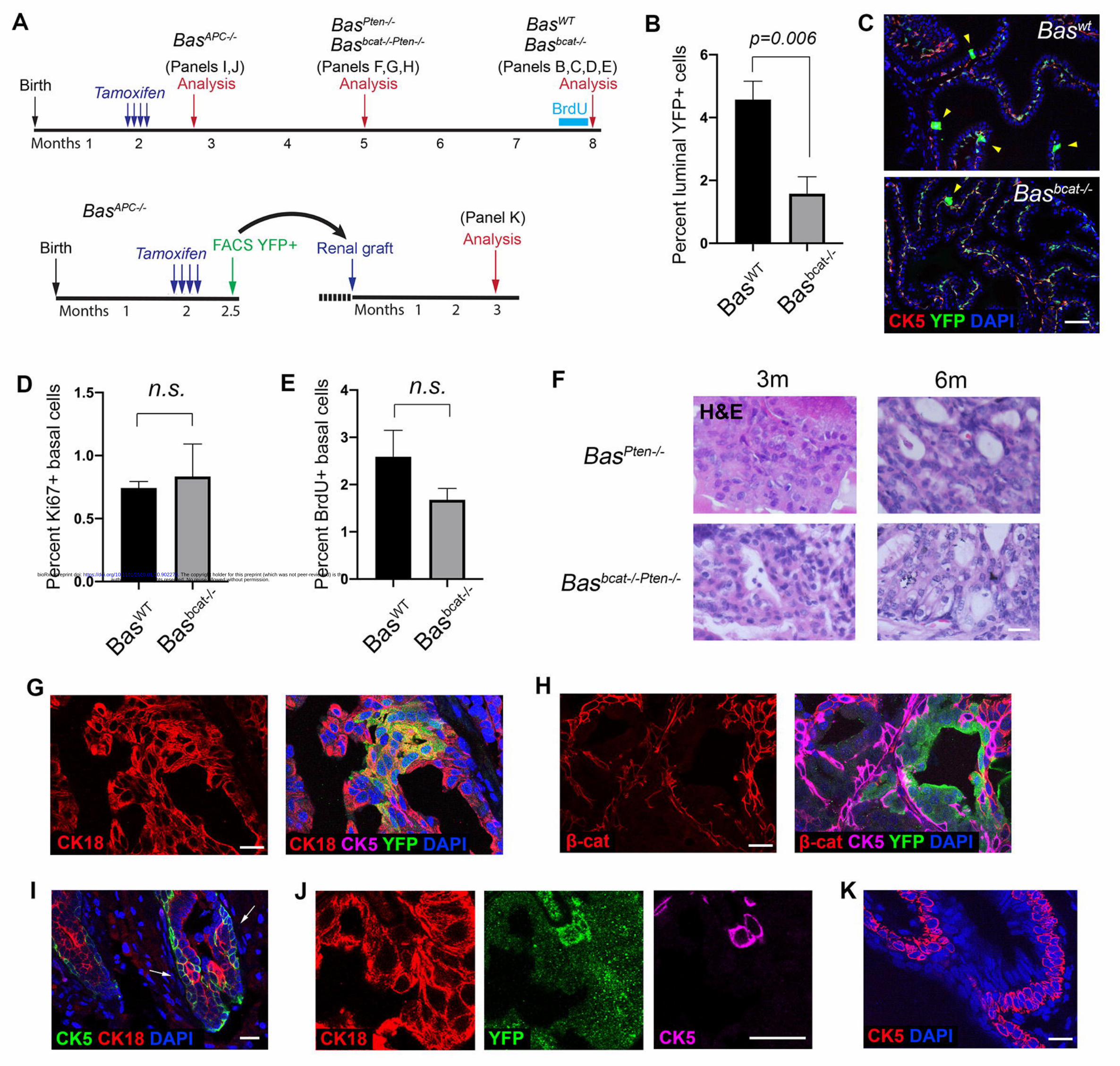
Figure 1. Lineage tracing analysis of basal cells of different levels of Wnt activity.
(A) Timeline of experiments for lineage tracing and renal grafting of Wnt-mutant-
related basal cells. (B) Percentage of YFP+ luminal cells decreased significantly in
Basbcat-/- mice (N=5) compared to BasWT mice (N=4) at 6 months post induction by
student’s t-test. (C) Representative IF images of YFP+ luminal cells (arrowheads) in
the BasWT and Basbcat-/- prostate 6 months post tamoxifen induction. (D, E) Bar graphs
showing proliferation rates in BasWT and Basbcat-/- basal cells by Ki67 (D) and BrdU
(E). n.s., not significant by student’s t-test. (F) H&E staining of tissue sections from
BasPten-/- and BasPten-/-;Bcat-/- mice at 3 months and 6 months post tamoxifen induction.
(G, H) IF image showing increased basal-to-luminal differentiation (G) and loss of β-
catenin (H) in BasPten-/-;Bcat-/- tumor regions at 3 months post induction. (I) Squamous
basal cell phenotype (arrows) seen in BasAPC-/- mice at 2 weeks post induction. (J) IF
staining showing the strict basal cell identity of the squamous-like cells. (K) IF
showing BasAPC-/- basal cell over-proliferation 3 months after renal grafting. Scale
bars in C correspond to 50 μm, and in F-K to 20 μm. Error bars correspond to one s.d.
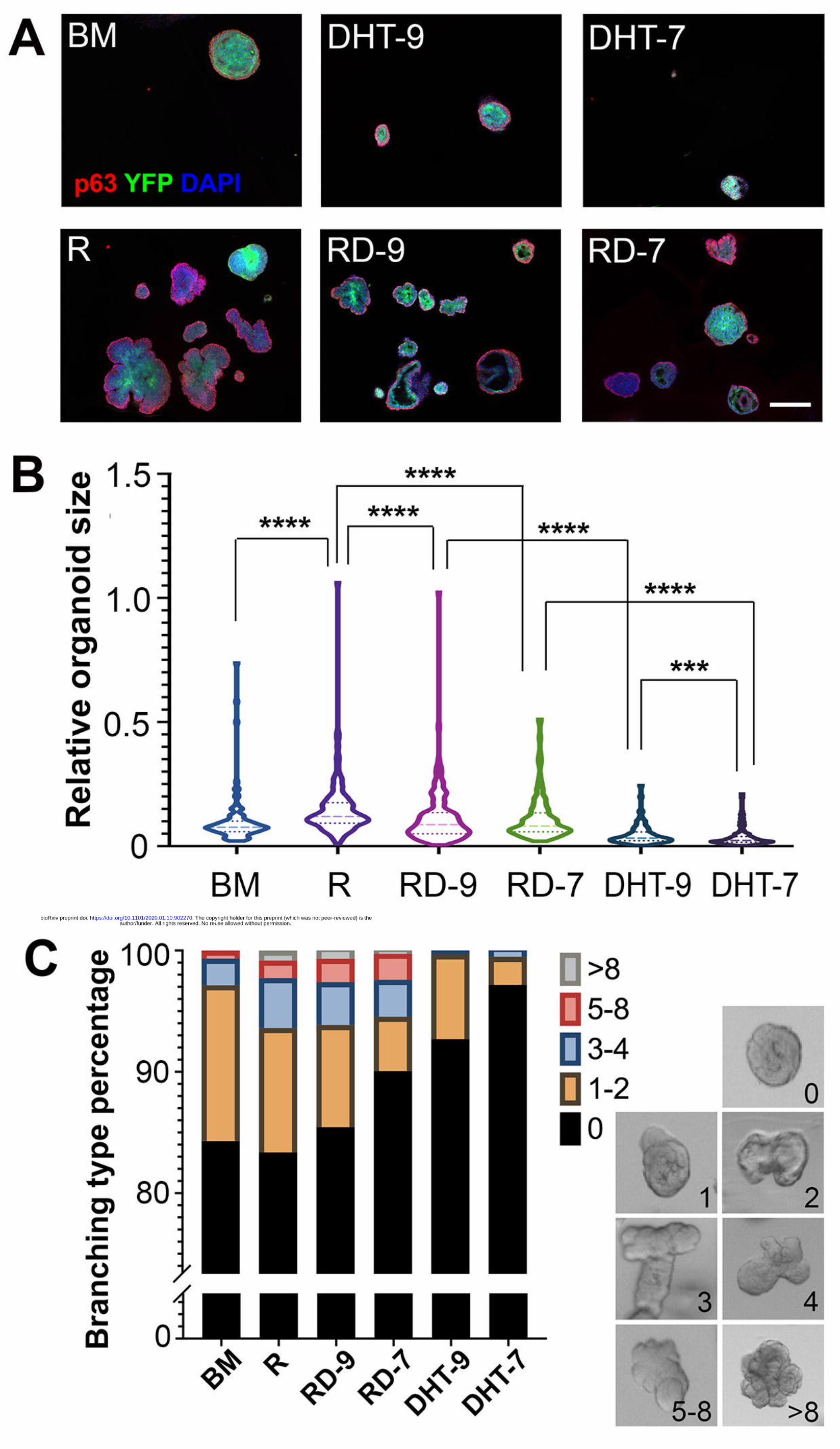
Figure 2. Analysis of organoids derived from basal cells of different Wnt
activities.
author/funder. All rights reserved. No reuse allowed without permission. The copyright holder for this preprint (which was not peer-reviewed) is the. https://doi.org/10.1101/2020.01.10.902270doi: bioRxiv preprint
(A) Representative IF images showing BasWT and BasAPC-/- organoid morphology after
7 days under different culturing conditions. Scale bar, 200 μm. (B) Violin plot
comparing relative sizes of BasWT and BasAPC-/- organoids under different culturing
conditions by Mann-Whitney U-test. **** p<0.0001, ** p<0.01. (C) Quantification
of BasWT and BasAPC-/- organoid branching under different conditions. Examples of
different numbers of branching are shown on the right.
Figure 3. Analysis of basal-derived organoids under combined R-spondin and
DHT treatment.
(A) Representative IF images showing organoid morphology after 7 days under
different R-spondin and DHT conditions. Scale bar, 200 μm. (B) Violin plot
comparing relative organoid sizes under different treatments by Mann-Whitney U-
test. **** p<0.0001, *** p<0.001. (C) Quantification of organoid branching under
different R-spondin and DHT conditions. Examples of different numbers of branching
are shown on the right.
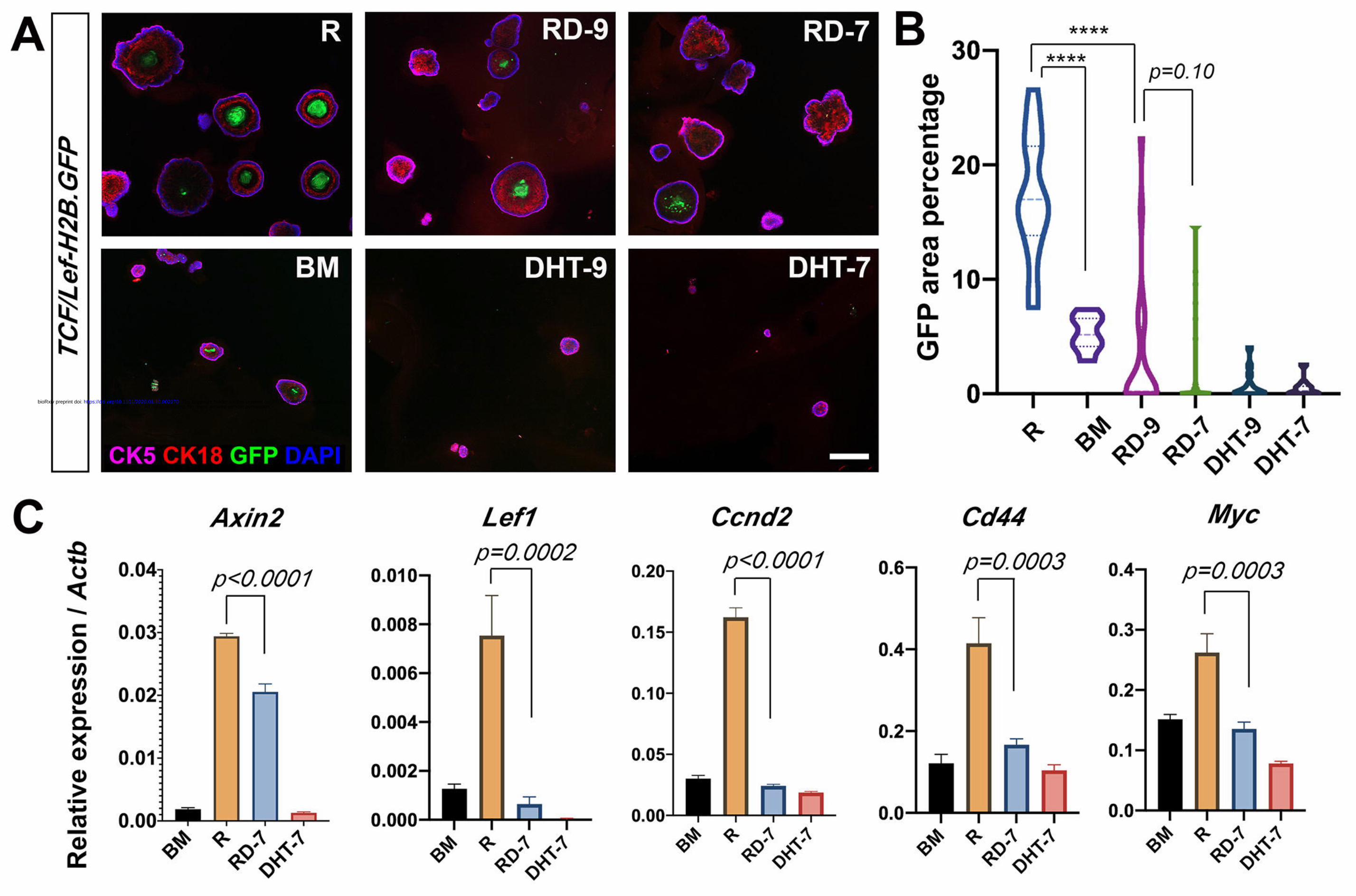
Figure 4. DHT decreases Wnt signaling activities in organoids.
(A) Representative IF images showing TCF/Lef-H2B.GFP reporter signals in basal-
derived organoids cultured under different conditions. Scale bar, 200 μm. (B) Violin
plot showing GFP area percentages for different treatment conditions. **** P<0.0001
by using Mann-Whitney U-test. (C) Quantitative real-time PCR analysis of Wnt
target genes under different treatments. Gene expression levels were normalized to β-
actin expression. P values were analyzed by unpaired student’s t-test. Error bars
correspond to one s.d.
author/funder. All rights reserved. No reuse allowed without permission. The copyright holder for this preprint (which was not peer-reviewed) is the. https://doi.org/10.1101/2020.01.10.902270doi: bioRxiv preprint
Figure 5. RNA-seq analysis of basal-derived organoids under different treatment
conditions.
(A) Scatter-plot of the two main components from a principle component analysis of
the BM (purple), R (blue), and RD-7 (orange) samples. N=3 in each group. (B)
Unsupervised hierarchical clustering analysis showing distinct transcriptomes of RD-
7 from BM and R. (C) David GO analysis showing the enriched pathways in the
comparison of R vs. RD-7, with the number of genes in each pathway. (D) GSEA
comparison of RD-7 vs. R showing Wnt pathway genes and organ morphogenesis
genes are enriched in the downregulated genes in the RD-7 group, and androgen
responsive genes enriched in the upregulated genes in the RD-7 group.
Figure 6. Duolink PLA of AR and β-catenin binding in organoids.
(A) Representative IF images showing Duolink PLA signals (red dots) only in the
presence of both β-catenin and AR antibodies. (B) Representative IF images showing
Duolink PLA signals under R, BM, RD-9 and RD-7 conditions. Cyan represents
nuclear DAPI staining. Scale bars, 50 μm. (C) Violin plots showing quantitation of
dots per nucleus (left) and dots per cell (right) in each organoid under different
treatment conditions. P values were analyzed by Mann-Whitney U-test.
Figure S1. Analysis of Basbcat-/- basal cells.
(A) IF images showing β-catenin expression pattern in Basbcat-/- mice. Scale bar, 20
μm. (B) FACS plot of sorting YFP+ Basbcat-/- basal cells. (C) qRT-PCR showing
significantly reduced β-catenin expression in Basbcat-/- basal cells compared to
wildtype basal cells by student’s t-test. Gene expression levels were normalized to β-
author/funder. All rights reserved. No reuse allowed without permission. The copyright holder for this preprint (which was not peer-reviewed) is the. https://doi.org/10.1101/2020.01.10.902270doi: bioRxiv preprint
actin expression. (D) A mouse losing hair due to β-catenin deletion by the CK5-
CreERT2 driver expression in skin basal cells.
Figure S2. Comparison of organoid RNA-seq data of different treatments.
(A) Volcano plots showing the numbers of upregulated and downregulated genes
between R vs. BM, RD-7 vs. R, and BM vs. RD-7 (FDR<0.05 and fold change >2).
(B) Heatmap showing expression levels of selected genes in different samples.
author/funder. All rights reserved. No reuse allowed without permission. The copyright holder for this preprint (which was not peer-reviewed) is the. https://doi.org/10.1101/2020.01.10.902270doi: bioRxiv preprint
author/funder. All rights reserved. No reuse allowed without permission. The copyright holder for this preprint (which was not peer-reviewed) is the. https://doi.org/10.1101/2020.01.10.902270doi: bioRxiv preprint

author/funder. All rights reserved. No reuse allowed without permission. The copyright holder for this preprint (which was not peer-reviewed) is the. https://doi.org/10.1101/2020.01.10.902270doi: bioRxiv preprint
author/funder. All rights reserved. No reuse allowed without permission. The copyright holder for this preprint (which was not peer-reviewed) is the. https://doi.org/10.1101/2020.01.10.902270doi: bioRxiv preprint
author/funder. All rights reserved. No reuse allowed without permission. The copyright holder for this preprint (which was not peer-reviewed) is the. https://doi.org/10.1101/2020.01.10.902270doi: bioRxiv preprint

author/funder. All rights reserved. No reuse allowed without permission. The copyright holder for this preprint (which was not peer-reviewed) is the. https://doi.org/10.1101/2020.01.10.902270doi: bioRxiv preprint

author/funder. All rights reserved. No reuse allowed without permission. The copyright holder for this preprint (which was not peer-reviewed) is the. https://doi.org/10.1101/2020.01.10.902270doi: bioRxiv preprint
Related Documents