Modulation of the cAMP signaling pathway after traumatic brain injury Coleen M. Atkins a,b,c , Anthony A. Oliva Jr. a,c , Ofelia F. Alonso a,b,c , Damien D. Pearse a,c , Helen M. Bramlett a,b,c, ⁎ , W. Dalton Dietrich a,b,c a Department of Neurological Surgery, University of Miami Miller School of Medicine, Miami, FL 33101, USA b The Neurotrauma Research Center, University of Miami Miller School of Medicine, Miami, FL 33101, USA c The Miami Project to Cure Paralysis, University of Miami Miller School of Medicine, Miami, FL 33101, USA Received 26 April 2007; revised 3 August 2007; accepted 20 August 2007 Available online 29 August 2007 Abstract Traumatic brain injury (TBI) results in both focal and diffuse brain pathologies that are exacerbated by the inflammatory response and progress from hours to days after the initial injury. Using a clinically relevant model of TBI, the parasagittal fluid-percussion brain injury (FPI) model, we found injury-induced impairments in the cyclic AMP (cAMP) signaling pathway. Levels of cAMP were depressed in the ipsilateral parietal cortex and hippocampus, as well as activation of its downstream target, protein kinase A, from 15 min to 48 h after moderate FPI. To determine if preventing hydrolysis of cAMP by administration of a phosphodiesterase (PDE) IV inhibitor would improve outcome after TBI, we treated animals intraperitoneally with rolipram (0.3 or 3.0 mg/kg) 30 min prior to TBI, and then once per day for 3 days. Rolipram treatment restored cAMP to sham levels and significantly reduced cortical contusion volume and improved neuronal cell survival in the parietal cortex and CA3 region of the hippocampus. Traumatic axonal injury, characterized by β-amyloid precursor protein deposits in the external capsule, was also significantly reduced in rolipram-treated animals. Furthermore, levels of the pro-inflammatory cytokines, interleukin-1β (IL-1β) and tumor necrosis factor-α (TNF-α), were significantly decreased with rolipram treatment. These results demonstrate that the cAMP–PKA signaling cascade is downregulated after TBI, and that treatment with a PDE IV inhibitor improves histopathological outcome and decreases inflammation after TBI. © 2007 Elsevier Inc. All rights reserved. Keywords: cAMP; Fluid-percussion; Inflammation; Interleukin-1β; PKA; Phosphodiesterase; Rolipram; TNF-α; Traumatic brain injury; TBI Introduction Traumatic brain injury (TBI) is a prevalent, debilitating health problem, occurring in 1.4 million people each year and disabling 5 million people in the United States (Langlois et al., 2004). The subsequent progressive injury after brain trauma develops from hours to days after the initiating insult, providing an accessible time window for pharmacological therapies. Despite intense efforts, research in TBI has not yielded a therapy that has passed Phase III clinical trials (Doppenberg et al., 2004). Brain trauma results in contusion formation, neuronal apop- tosis, and axonal tract damage. These pathologies are worsened by the inflammatory cascade set into motion by the initial injury (Morganti-Kossmann et al., 2002; Dietrich et al., 2004). Two pro- inflammatory cytokines released after TBI are tumor necrosis factor-α (TNF-α) and interleukin-1β (IL-1β). Numerous studies have documented rapid increases in TNF-α and IL-1β levels after TBI (Taupin et al., 1993; Shohami et al., 1994; Fan et al., 1996; Kinoshita et al., 2002; Vitarbo et al., 2004). IL-1β synergistically acts with TNF-α to induce cell death after TBI. These pro-inflammatory cytokines stimulate inflam- matory cells to release damaging reactive oxygen and nitrogen species, raise glutamate levels to excitotoxic levels, impair the ability of glia cells to buffer extracellular potassium, compro- mise the blood–brain barrier, and attract more inflammatory cells into the brain (Tanaka et al., 1994; Meda et al., 1995; Soares et al., 1995; Hu et al., 1997; Keeling et al., 2000). Once Available online at www.sciencedirect.com Experimental Neurology 208 (2007) 145 – 158 www.elsevier.com/locate/yexnr ⁎ Corresponding author. The Miami Project to Cure Paralysis, University of Miami Miller School of Medicine, P.O. Box 016960 (R-48), Miami, FL 33101, USA. Fax: +1 305 243 3207. E-mail address: [email protected] (H.M. Bramlett). 0014-4886/$ - see front matter © 2007 Elsevier Inc. All rights reserved. doi:10.1016/j.expneurol.2007.08.011

Welcome message from author
This document is posted to help you gain knowledge. Please leave a comment to let me know what you think about it! Share it to your friends and learn new things together.
Transcript
Available online at www.sciencedirect.com
08 (2007) 145–158www.elsevier.com/locate/yexnr
Experimental Neurology 2
Modulation of the cAMP signaling pathway after traumatic brain injury
Coleen M. Atkins a,b,c, Anthony A. Oliva Jr. a,c, Ofelia F. Alonso a,b,c, Damien D. Pearse a,c,Helen M. Bramlett a,b,c,⁎, W. Dalton Dietrich a,b,c
a Department of Neurological Surgery, University of Miami Miller School of Medicine, Miami, FL 33101, USAb The Neurotrauma Research Center, University of Miami Miller School of Medicine, Miami, FL 33101, USA
c The Miami Project to Cure Paralysis, University of Miami Miller School of Medicine, Miami, FL 33101, USA
Received 26 April 2007; revised 3 August 2007; accepted 20 August 2007Available online 29 August 2007
Abstract
Traumatic brain injury (TBI) results in both focal and diffuse brain pathologies that are exacerbated by the inflammatory response and progressfrom hours to days after the initial injury. Using a clinically relevant model of TBI, the parasagittal fluid-percussion brain injury (FPI) model, wefound injury-induced impairments in the cyclic AMP (cAMP) signaling pathway. Levels of cAMP were depressed in the ipsilateral parietal cortexand hippocampus, as well as activation of its downstream target, protein kinase A, from 15 min to 48 h after moderate FPI. To determine ifpreventing hydrolysis of cAMP by administration of a phosphodiesterase (PDE) IV inhibitor would improve outcome after TBI, we treatedanimals intraperitoneally with rolipram (0.3 or 3.0 mg/kg) 30 min prior to TBI, and then once per day for 3 days. Rolipram treatment restoredcAMP to sham levels and significantly reduced cortical contusion volume and improved neuronal cell survival in the parietal cortex and CA3region of the hippocampus. Traumatic axonal injury, characterized by β-amyloid precursor protein deposits in the external capsule, was alsosignificantly reduced in rolipram-treated animals. Furthermore, levels of the pro-inflammatory cytokines, interleukin-1β (IL-1β) and tumornecrosis factor-α (TNF-α), were significantly decreased with rolipram treatment. These results demonstrate that the cAMP–PKA signalingcascade is downregulated after TBI, and that treatment with a PDE IV inhibitor improves histopathological outcome and decreases inflammationafter TBI.© 2007 Elsevier Inc. All rights reserved.
Keywords: cAMP; Fluid-percussion; Inflammation; Interleukin-1β; PKA; Phosphodiesterase; Rolipram; TNF-α; Traumatic brain injury; TBI
Introduction
Traumatic brain injury (TBI) is a prevalent, debilitatinghealth problem, occurring in 1.4 million people each year anddisabling 5 million people in the United States (Langlois et al.,2004). The subsequent progressive injury after brain traumadevelops from hours to days after the initiating insult,providing an accessible time window for pharmacologicaltherapies. Despite intense efforts, research in TBI has notyielded a therapy that has passed Phase III clinical trials(Doppenberg et al., 2004).
⁎ Corresponding author. The Miami Project to Cure Paralysis, University ofMiami Miller School of Medicine, P.O. Box 016960 (R-48), Miami, FL 33101,USA. Fax: +1 305 243 3207.
E-mail address: [email protected] (H.M. Bramlett).
0014-4886/$ - see front matter © 2007 Elsevier Inc. All rights reserved.doi:10.1016/j.expneurol.2007.08.011
Brain trauma results in contusion formation, neuronal apop-tosis, and axonal tract damage. These pathologies are worsenedby the inflammatory cascade set into motion by the initial injury(Morganti-Kossmann et al., 2002; Dietrich et al., 2004). Two pro-inflammatory cytokines released after TBI are tumor necrosisfactor-α (TNF-α) and interleukin-1β (IL-1β). Numerous studieshave documented rapid increases in TNF-α and IL-1β levels afterTBI (Taupin et al., 1993; Shohami et al., 1994; Fan et al., 1996;Kinoshita et al., 2002; Vitarbo et al., 2004).
IL-1β synergistically acts with TNF-α to induce cell deathafter TBI. These pro-inflammatory cytokines stimulate inflam-matory cells to release damaging reactive oxygen and nitrogenspecies, raise glutamate levels to excitotoxic levels, impair theability of glia cells to buffer extracellular potassium, compro-mise the blood–brain barrier, and attract more inflammatorycells into the brain (Tanaka et al., 1994; Meda et al., 1995;Soares et al., 1995; Hu et al., 1997; Keeling et al., 2000). Once
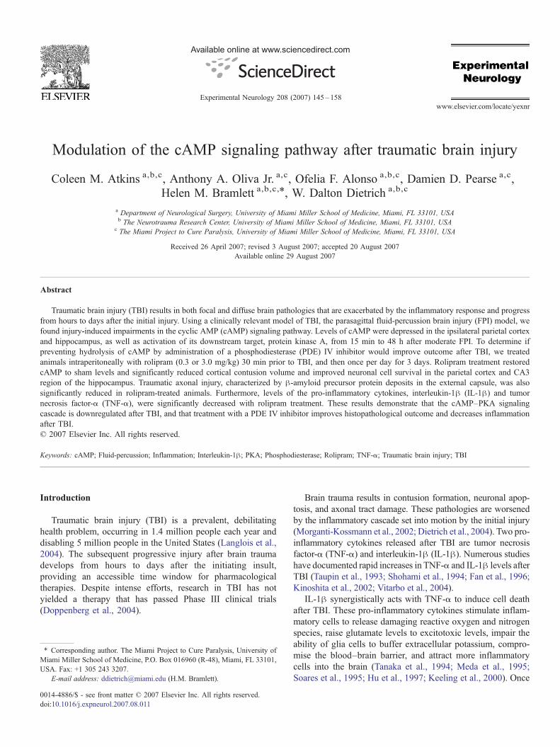
Fig. 1. cAMP levels decrease after TBI. The ipsilateral parietal cortex (A; n=4–10), hippocampus (B; n=4–11), and thalamus (C; n=5–10) were assayed byELISA for cAMP levels after moderate parasagittal FPI. cAMP levels weresignificantly decreased in the cortex at 24 h (n=4, ⁎pb0.05) and 48 h (n=6,⁎⁎pb0.01) after TBI, and at 15 min (n=6, ⁎⁎⁎pb0.001), 1 h (n=6, ⁎⁎pb0.01),and 4 h (n=5, ⁎pb0.05) after TBI in the hippocampus as compared to shamlevels. Data represent mean±S.E.M.
146 C.M. Atkins et al. / Experimental Neurology 208 (2007) 145–158
initiated, the inflammatory cascade becomes a toxic positive-feedback loop, further exacerbating brain pathology.
In other models of CNS injury, several studies havedemonstrated that restoration of cyclic AMP (cAMP) levelsimproves outcome. In spinal cord injury, application of rolipramto inhibit the degradation of cAMP promotes axon sparing andresults in locomotor improvements (Nikulina et al., 2004;Pearse et al., 2004). Similarly, in transient global ischemiarolipram improves neuronal survival in the hippocampus andhippocampal-dependent learning (Kato et al., 1995; Block et al.,1997; Imanishi et al., 1997; Block et al., 2001).
The effects of cAMP are short-lived because phosphodies-terases (PDEs) rapidly degrade cAMP (Manganiello et al.,1995). Of the 10 classes of PDEs, two isoforms are highlyselective for degrading cAMP, PDE IV and VII. Rolipram, aselective inhibitor of PDE IV, reduces inflammation in a numberof diseases including asthma, multiple sclerosis, septic shock,rheumatoid arthritis, and inflammatory bowel disease (Dal Piazand Giovannoni, 2000; Castro et al., 2005). Consequently, PDEIV inhibitors are widely-utilized by the pharmaceutical industryas anti-inflammatory drugs.
A primary action of cAMP is activation of protein kinase A(PKA). PKA phosphorylates transcription factors, includingcAMP-responsive element binding (CREB) protein andnuclear factor-κB (NF-κB) p50 (Montminy and Bilezikjian,1987; Hou et al., 2003). Phosphorylation of CREB stimulatestranscription of cell survival genes (Mayr and Montminy,2001). Phosphorylation of NF-κB p50 subunit suppressestranscription of genes with IκB elements in their promoters;this includes the pro-inflammatory cytokines TNF-α and IL-1β (Cogswell et al., 1994; Verghese et al., 1995; Hou et al.,2003). Thus, we hypothesized that rolipram treatment mayimprove TBI outcome by decreasing pro-inflammatorycytokine production.
Materials and methods
Traumatic brain injury
All experimental procedures were in compliance with theNIH Guide for the Care and Use of Laboratory Animals andapproved by the University of Miami Animal Care and UseCommittee. Male Sprague–Dawley rats (270–320 g; CharlesRiver Laboratories, Raleigh, NC, USA) were anesthetized with3% halothane, 70% N2O, and 30% O2, then intubatedendotracheally and mechanically ventilated (Harvard Appara-tus, Holliston, MA, USA) with 1.5% halothane, 70% N2O, and30% O2. To immobilize the animals and facilitate mechanicalventilation, pancuronium bromide (0.5 mg/kg) was intrave-nously administered through the femoral artery. On the dayprior to TBI, animals received a 4.8 mm craniotomy (3.8 mmposterior to bregma, 2.5 mm lateral to the midline) and amodified plastic 18 gauge syringe hub (8 mm length,PrecisionGlide needle, Becton Dickinson, Franklin Lakes, NJ,USA) was secured over the right parietal cortex. The next day,animals were anesthetized, intubated, and then placed under afluid-percussion brain injury (FPI) device. A moderate fluid-
percussion pulse (2.0±0.2 atmospheres) was delivered to theright parietal cortex. Sham-operated rats received all surgicalmanipulations, but without the fluid-percussion pulse, and weremonitored under anesthesia for 15–30 min after the sham injury.Rectal and temporalis muscle thermistors were used to maintaincore and brain temperatures at 36.8–37.3 °C using self-
147C.M. Atkins et al. / Experimental Neurology 208 (2007) 145–158
adjusting feedback warming lamps. Blood gases (pO2 andpCO2), blood pH, and mean arterial pressure were monitored15 min before TBI and up to 4 h after TBI and maintainedwithin normal physiological ranges.
cAMP assays
Six experimental groups (n=47) were used tomeasure cAMPlevels by ELISA. Animals received either sham surgery (n=11)or moderate parasagittal FPI followed by recovery for 15 min
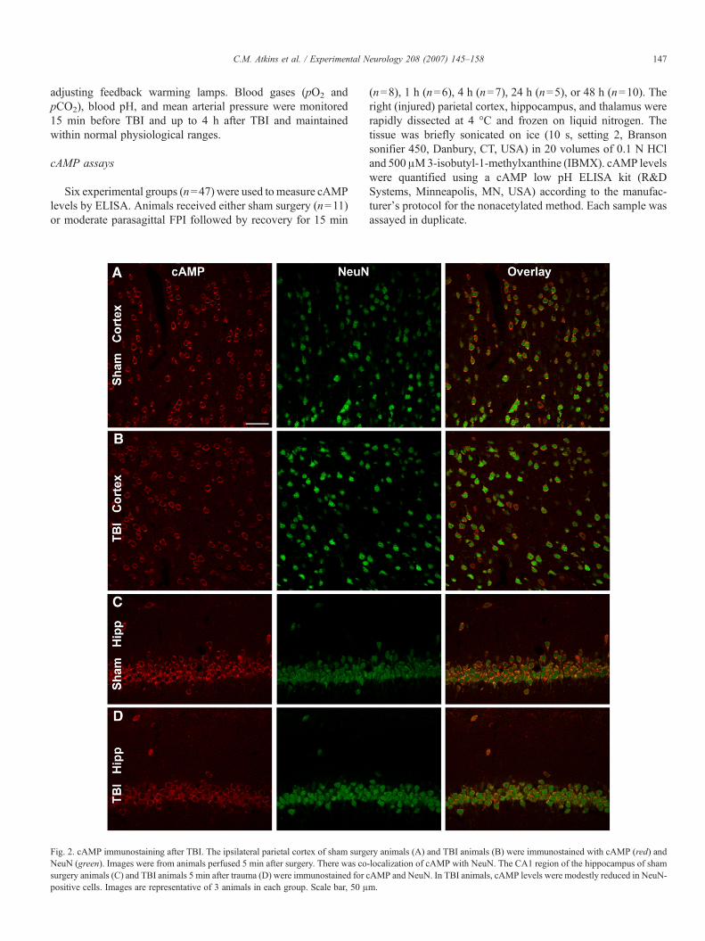
Fig. 2. cAMP immunostaining after TBI. The ipsilateral parietal cortex of sham surgeNeuN (green). Images were from animals perfused 5 min after surgery. There was cosurgery animals (C) and TBI animals 5 min after trauma (D) were immunostained for cpositive cells. Images are representative of 3 animals in each group. Scale bar, 50 μ
(n=8), 1 h (n=6), 4 h (n=7), 24 h (n=5), or 48 h (n=10). Theright (injured) parietal cortex, hippocampus, and thalamus wererapidly dissected at 4 °C and frozen on liquid nitrogen. Thetissue was briefly sonicated on ice (10 s, setting 2, Bransonsonifier 450, Danbury, CT, USA) in 20 volumes of 0.1 N HCland 500 μM3-isobutyl-1-methylxanthine (IBMX). cAMP levelswere quantified using a cAMP low pH ELISA kit (R&DSystems, Minneapolis, MN, USA) according to the manufac-turer's protocol for the nonacetylated method. Each sample wasassayed in duplicate.
ry animals (A) and TBI animals (B) were immunostained with cAMP (red) and-localization of cAMP with NeuN. The CA1 region of the hippocampus of shamAMP and NeuN. In TBI animals, cAMP levels were modestly reduced in NeuN-m.
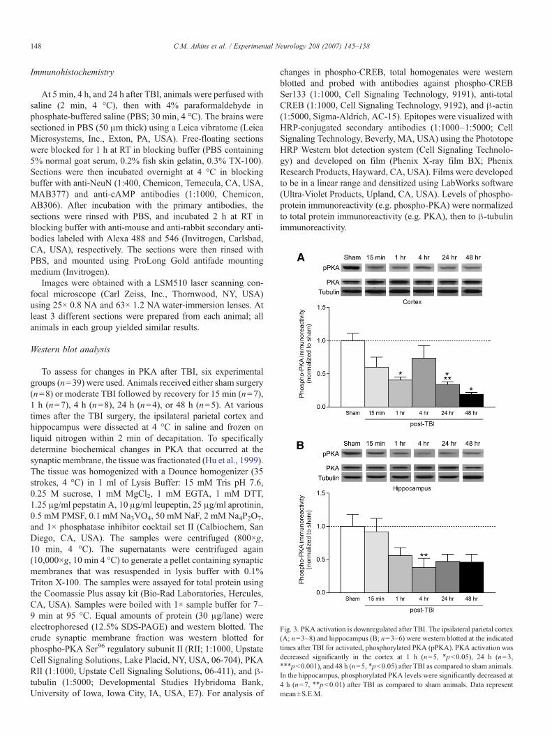
Fig. 3. PKA activation is downregulated after TBI. The ipsilateral parietal cortex(A; n=3–8) and hippocampus (B; n=3–6) were western blotted at the indicatedtimes after TBI for activated, phosphorylated PKA (pPKA). PKA activation wasdecreased significantly in the cortex at 1 h (n=5, ⁎pb0.05), 24 h (n=3,⁎⁎⁎pb0.001), and 48 h (n=5, ⁎pb0.05) after TBI as compared to sham animals.In the hippocampus, phosphorylated PKA levels were significantly decreased at4 h (n=7, ⁎⁎pb0.01) after TBI as compared to sham animals. Data representmean±S.E.M.
148 C.M. Atkins et al. / Experimental Neurology 208 (2007) 145–158
Immunohistochemistry
At 5 min, 4 h, and 24 h after TBI, animals were perfused withsaline (2 min, 4 °C), then with 4% paraformaldehyde inphosphate-buffered saline (PBS; 30 min, 4 °C). The brains weresectioned in PBS (50 μm thick) using a Leica vibratome (LeicaMicrosystems, Inc., Exton, PA, USA). Free-floating sectionswere blocked for 1 h at RT in blocking buffer (PBS containing5% normal goat serum, 0.2% fish skin gelatin, 0.3% TX-100).Sections were then incubated overnight at 4 °C in blockingbuffer with anti-NeuN (1:400, Chemicon, Temecula, CA, USA,MAB377) and anti-cAMP antibodies (1:1000, Chemicon,AB306). After incubation with the primary antibodies, thesections were rinsed with PBS, and incubated 2 h at RT inblocking buffer with anti-mouse and anti-rabbit secondary anti-bodies labeled with Alexa 488 and 546 (Invitrogen, Carlsbad,CA, USA), respectively. The sections were then rinsed withPBS, and mounted using ProLong Gold antifade mountingmedium (Invitrogen).
Images were obtained with a LSM510 laser scanning con-focal microscope (Carl Zeiss, Inc., Thornwood, NY, USA)using 25× 0.8 NA and 63× 1.2 NAwater-immersion lenses. Atleast 3 different sections were prepared from each animal; allanimals in each group yielded similar results.
Western blot analysis
To assess for changes in PKA after TBI, six experimentalgroups (n=39) were used. Animals received either sham surgery(n=8) or moderate TBI followed by recovery for 15 min (n=7),1 h (n=7), 4 h (n=8), 24 h (n=4), or 48 h (n=5). At varioustimes after the TBI surgery, the ipsilateral parietal cortex andhippocampus were dissected at 4 °C in saline and frozen onliquid nitrogen within 2 min of decapitation. To specificallydetermine biochemical changes in PKA that occurred at thesynaptic membrane, the tissue was fractionated (Hu et al., 1999).The tissue was homogenized with a Dounce homogenizer (35strokes, 4 °C) in 1 ml of Lysis Buffer: 15 mM Tris pH 7.6,0.25 M sucrose, 1 mM MgCl2, 1 mM EGTA, 1 mM DTT,1.25 μg/ml pepstatin A, 10 μg/ml leupeptin, 25 μg/ml aprotinin,0.5 mM PMSF, 0.1 mM Na3VO4, 50 mM NaF, 2 mM Na4P2O7,and 1× phosphatase inhibitor cocktail set II (Calbiochem, SanDiego, CA, USA). The samples were centrifuged (800×g,10 min, 4 °C). The supernatants were centrifuged again(10,000×g, 10 min 4 °C) to generate a pellet containing synapticmembranes that was resuspended in lysis buffer with 0.1%Triton X-100. The samples were assayed for total protein usingthe Coomassie Plus assay kit (Bio-Rad Laboratories, Hercules,CA, USA). Samples were boiled with 1× sample buffer for 7–9 min at 95 °C. Equal amounts of protein (30 μg/lane) wereelectrophoresed (12.5% SDS-PAGE) and western blotted. Thecrude synaptic membrane fraction was western blotted forphospho-PKA Ser96 regulatory subunit II (RII; 1:1000, UpstateCell Signaling Solutions, Lake Placid, NY, USA, 06-704), PKARII (1:1000, Upstate Cell Signaling Solutions, 06-411), and β-tubulin (1:5000; Developmental Studies Hybridoma Bank,University of Iowa, Iowa City, IA, USA, E7). For analysis of
changes in phospho-CREB, total homogenates were westernblotted and probed with antibodies against phospho-CREBSer133 (1:1000, Cell Signaling Technology, 9191), anti-totalCREB (1:1000, Cell Signaling Technology, 9192), and β-actin(1:5000, Sigma-Aldrich, AC-15). Epitopes were visualized withHRP-conjugated secondary antibodies (1:1000–1:5000; CellSignaling Technology, Beverly, MA, USA) using the PhototopeHRP Western blot detection system (Cell Signaling Technolo-gy) and developed on film (Phenix X-ray film BX; PhenixResearch Products, Hayward, CA, USA). Films were developedto be in a linear range and densitized using LabWorks software(Ultra-Violet Products, Upland, CA, USA). Levels of phospho-protein immunoreactivity (e.g. phospho-PKA) were normalizedto total protein immunoreactivity (e.g. PKA), then to β-tubulinimmunoreactivity.
149C.M. Atkins et al. / Experimental Neurology 208 (2007) 145–158
Rolipram administration
Rolipram (Sigma-Aldrich, St. Louis, MO, USA), wasdissolved in 100% DMSO at 10 mg/ml, and then diluted with0.9% NaCl for a final concentration of either 0.5 mg/ml or0.05 mg/ml in 5% DMSO and 95% saline. The drug wasadministered intraperitoneally (i.p.) 30 min prior to TBI at 6 ml/kg. For each group, rolipram or vehicle (5% DMSO/95% saline)was administered once every 24 h and on the final day of theexperiment, 30 min prior to sacrifice.
Histopathological analysis
TBI- and sham-operated animals were anesthetized (3%halothane for 5 min) and perfused transcardially with isotonicsaline for 2 min (75 ml) and then 30 min of 4% paraformal-dehyde in 0.1 M sodium phosphate buffer, pH 7.4 (350 ml). The
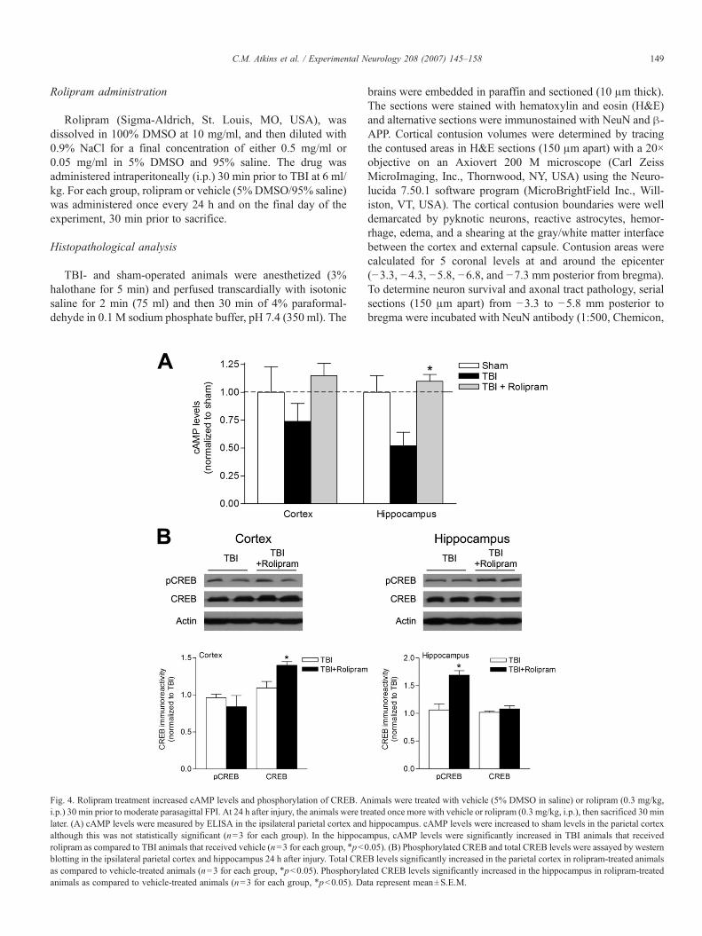
Fig. 4. Rolipram treatment increased cAMP levels and phosphorylation of CREB. Ai.p.) 30 min prior to moderate parasagittal FPI. At 24 h after injury, the animals were trlater. (A) cAMP levels were measured by ELISA in the ipsilateral parietal cortex andalthough this was not statistically significant (n=3 for each group). In the hippocarolipram as compared to TBI animals that received vehicle (n=3 for each group, ⁎pb0blotting in the ipsilateral parietal cortex and hippocampus 24 h after injury. Total CREas compared to vehicle-treated animals (n=3 for each group, ⁎pb0.05). Phosphorylaanimals as compared to vehicle-treated animals (n=3 for each group, ⁎pb0.05). Da
brains were embedded in paraffin and sectioned (10 μm thick).The sections were stained with hematoxylin and eosin (H&E)and alternative sections were immunostained with NeuN and β-APP. Cortical contusion volumes were determined by tracingthe contused areas in H&E sections (150 μm apart) with a 20×objective on an Axiovert 200 M microscope (Carl ZeissMicroImaging, Inc., Thornwood, NY, USA) using the Neuro-lucida 7.50.1 software program (MicroBrightField Inc., Will-iston, VT, USA). The cortical contusion boundaries were welldemarcated by pyknotic neurons, reactive astrocytes, hemor-rhage, edema, and a shearing at the gray/white matter interfacebetween the cortex and external capsule. Contusion areas werecalculated for 5 coronal levels at and around the epicenter(−3.3, −4.3, −5.8, −6.8, and −7.3 mm posterior from bregma).To determine neuron survival and axonal tract pathology, serialsections (150 μm apart) from −3.3 to −5.8 mm posterior tobregma were incubated with NeuN antibody (1:500, Chemicon,
nimals were treated with vehicle (5% DMSO in saline) or rolipram (0.3 mg/kg,eated once more with vehicle or rolipram (0.3 mg/kg, i.p.), then sacrificed 30 minhippocampus. cAMP levels were increased to sham levels in the parietal cortexmpus, cAMP levels were significantly increased in TBI animals that received.05). (B) Phosphorylated CREB and total CREB levels were assayed by westernB levels significantly increased in the parietal cortex in rolipram-treated animalsted CREB levels significantly increased in the hippocampus in rolipram-treatedta represent mean±S.E.M.
150 C.M. Atkins et al. / Experimental Neurology 208 (2007) 145–158
MAB377) or β-APP antibody (1:500, Chemicon, MAB348),respectively. Immunostaining was developed with anti-mouseIgG (1:1000), ABC Elite (Vector Laboratories, Burlingame,CA, USA), and NiDAB (2.5% Nickle Ammonium SulfateAcetate-Imidasole Buffer, 0.05% DAB, 0.001% H2O2, VectorLaboratories). NeuN-positive neurons were quantified in anunbiased, systematic manner using stereology with an Axiovert200 M microscope (Carl Zeiss MicroImaging, Inc.) by a blindobserver (Suzuki et al., 2003, 2004). The parietal cortexoverlying the contusion area and the CA3 region of thehippocampus were contoured at 20×, then a counting grid of250×200 μm was placed in the parietal cortex or a grid of140×70 μm was placed in the CA3 region. Using a 35×35 μmcounting frame, NeuN-positive cells were counted in 25–40randomly-placed sampling sites with Stereoinvestigator 7.50.1software (MicroBrightField, Inc.) with a 63×, 1.4 NA objective.NeuN counts were measured from bregma levels −3.3 mm to−5.8 mm in sections spaced 150 μm apart. For cortical cellcounts, Q values ranged from 446 to 715 and CE2/CV2 valueswere 0.04, 0.06, and 0.11 for the vehicle, 3.0 mg/kg rolipramand 0.3 mg/kg rolipram groups, respectively. For CA3hippocampal cell counts, the Q range was 325–550 and CE2/CV2 values were 0.39, 0.11, and 0.11 for the vehicle, 3.0 mg/kgrolipram and 0.3 mg/kg rolipram groups, respectively. To quan-
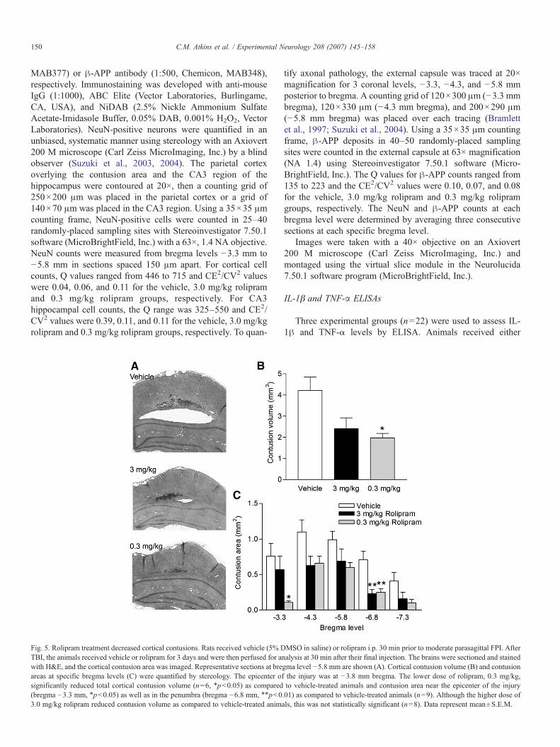
Fig. 5. Rolipram treatment decreased cortical contusions. Rats received vehicle (5% DTBI, the animals received vehicle or rolipram for 3 days and were then perfused for anwith H&E, and the cortical contusion area was imaged. Representative sections at breareas at specific bregma levels (C) were quantified by stereology. The epicenter osignificantly reduced total cortical contusion volume (n=6, ⁎pb0.05) as compared(bregma −3.3 mm, ⁎pb0.05) as well as in the penumbra (bregma −6.8 mm, ⁎⁎pb03.0 mg/kg rolipram reduced contusion volume as compared to vehicle-treated anim
tify axonal pathology, the external capsule was traced at 20×magnification for 3 coronal levels, −3.3, −4.3, and −5.8 mmposterior to bregma. A counting grid of 120×300 μm (−3.3 mmbregma), 120×330 μm (−4.3 mm bregma), and 200×290 μm(−5.8 mm bregma) was placed over each tracing (Bramlettet al., 1997; Suzuki et al., 2004). Using a 35×35 μm countingframe, β-APP deposits in 40–50 randomly-placed samplingsites were counted in the external capsule at 63× magnification(NA 1.4) using Stereoinvestigator 7.50.1 software (Micro-BrightField, Inc.). The Q values for β-APP counts ranged from135 to 223 and the CE2/CV2 values were 0.10, 0.07, and 0.08for the vehicle, 3.0 mg/kg rolipram and 0.3 mg/kg rolipramgroups, respectively. The NeuN and β-APP counts at eachbregma level were determined by averaging three consecutivesections at each specific bregma level.
Images were taken with a 40× objective on an Axiovert200 M microscope (Carl Zeiss MicroImaging, Inc.) andmontaged using the virtual slice module in the Neurolucida7.50.1 software program (MicroBrightField, Inc.).
IL-1β and TNF-α ELISAs
Three experimental groups (n=22) were used to assess IL-1β and TNF-α levels by ELISA. Animals received either
MSO in saline) or rolipram i.p. 30 min prior to moderate parasagittal FPI. Afteralysis at 30 min after their final injection. The brains were sectioned and stainedgma level −5.8 mm are shown (A). Cortical contusion volume (B) and contusionf the injury was at −3.8 mm bregma. The lower dose of rolipram, 0.3 mg/kg,to vehicle-treated animals and contusion area near the epicenter of the injury.01) as compared to vehicle-treated animals (n=9). Although the higher dose ofals, this was not statistically significant (n=8). Data represent mean±S.E.M.
151C.M. Atkins et al. / Experimental Neurology 208 (2007) 145–158
sham surgery (n=5) or moderate parasagittal FPI andtreatment with vehicle (n=8) or 0.3 mg/kg rolipram (n=9)30 min prior to FPI and 30 min prior to sacrifice. At 3 h afterFPI, the animals were sacrificed and the ipsilateral parietalcortex, hippocampus and thalamus were rapidly dissected onice in saline. The tissue was briefly sonicated on ice (10 s,setting 2, Branson sonifier 450, Danbury, CT, USA) in10 volumes/weight of Lysis Buffer supplemented with 0.1%Igepal CA-630 (Sigma-Aldrich). Total protein was measuredusing the Coomassie Plus assay kit (Bio-Rad Laboratories).Each sample was assayed in duplicate according to themanufacturer's protocol (R&D Systems, Inc.).
Statistical analysis
Data presented are mean±S.E.M. Statistical analyses areStudent's t test or one-way ANOVAs with post-hoc Tukey HSDt tests.
Results
To ascertain if the cAMP–PKA pathway is a potentialtherapeutic target after TBI, we first determined if the cAMP–
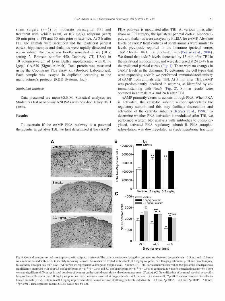
Fig. 6. Cortical neuron survival was improved with rolipram treatment. The parietal cowas immunostained with NeuN to identify surviving neurons. Animals were treated wfollowed by once per day for 3 days. (A) Shown are representative images at bregma lsignificantly improved with both 0.3 mg/kg rolipram (n=5, ⁎⁎pb0.01) and 3.0 mg/kgwere no significant differences in total numbers of neurons on the contralateral side wibregma levels illustrates that 3.0 mg/kg rolipram increased neuronal survival at bregtreated animals (n=9). Rolipram at 0.3 mg/kg improved cortical neuron survival at al⁎⁎pb0.01). Data represent mean±S.E.M. Scale bar, 50 μm.
PKA pathway is modulated after TBI. At various times aftersham or FPI surgery, the ipsilateral parietal cortex, hippocam-pus, and thalamus were assayed by ELISA for cAMP. Absolutelevels of cAMP from cortices of sham animals were similar tolevels previously reported in the literature (parietal cortexcAMP levels 184.1±5.6 pmol/ml, n=6) (Pearse et al., 2004).We found that cAMP levels decreased by 15 min after TBI inthe ipsilateral hippocampus, and were depressed at 24 to 48 h inthe ipsilateral parietal cortex (Fig. 1). There were no changes incAMP levels in the thalamus. To determine the cell types thatwere expressing cAMP, we performed immunohistochemistryof cAMP from animals after TBI. At 5 min after TBI, cAMPwas predominantly localized in neurons, as identified by co-immunostaining with NeuN (Fig. 2). Similar results wereobtained in animals at 4 and 24 h after TBI.
cAMP primarily exerts its actions through PKA. When PKAis activated, the catalytic subunit autophosphorylates theregulatory subunit and this may facilitate dissociation andactivation of the catalytic subunits (Keryer et al., 1998). Todetermine whether PKA activation is modulated after TBI, weperformed western blot analysis with antibodies to phosphor-ylated, activated PKA regulatory subunit II. PKA autopho-sphorylation was downregulated in crude membrane fractions
rtex overlying the contusion area between bregma levels −3.3 mm and −6.8 mmith vehicle, 0.3 mg/kg rolipram, or 3.0 mg/kg rolipram i.p. 30 min prior to injury,evel −5.8 mm. (B) Total cortical neuron survival on the ipsilateral side (Ipsi) wasrolipram (n=8, ⁎⁎pb0.01) as compared to vehicle-treated animals (n=9). Thereth rolipram treatment (Contra). (C) Quantification of neuronal survival at specificma levels −4.3 mm and −5.8 mm (n=8, ⁎⁎pb0.01) when compared to vehicle-l bregma levels tested (n=6; −3.3 mm, ⁎pb0.05; −4.3 mm, ⁎pb0.05; −5.8 mm,
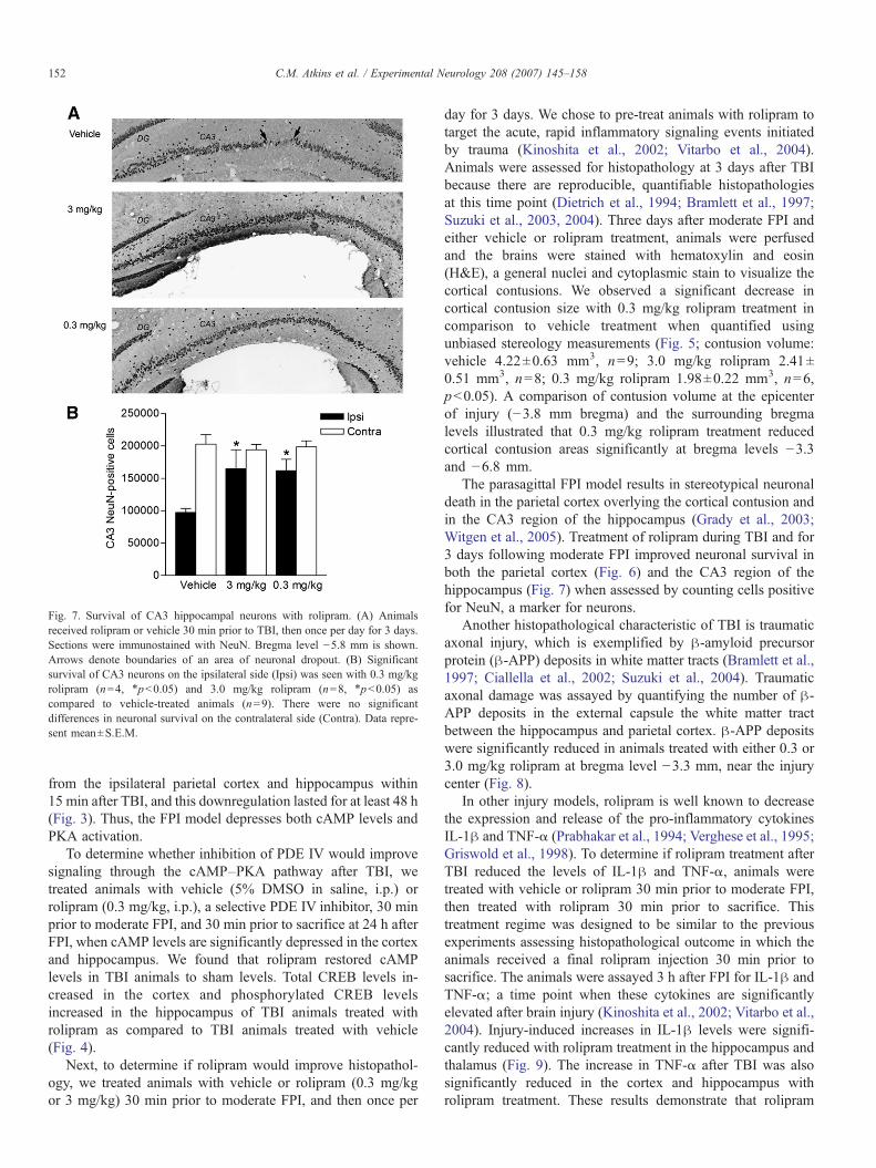
Fig. 7. Survival of CA3 hippocampal neurons with rolipram. (A) Animalsreceived rolipram or vehicle 30 min prior to TBI, then once per day for 3 days.Sections were immunostained with NeuN. Bregma level −5.8 mm is shown.Arrows denote boundaries of an area of neuronal dropout. (B) Significantsurvival of CA3 neurons on the ipsilateral side (Ipsi) was seen with 0.3 mg/kgrolipram (n=4, ⁎pb0.05) and 3.0 mg/kg rolipram (n=8, ⁎pb0.05) ascompared to vehicle-treated animals (n=9). There were no significantdifferences in neuronal survival on the contralateral side (Contra). Data repre-sent mean±S.E.M.
152 C.M. Atkins et al. / Experimental Neurology 208 (2007) 145–158
from the ipsilateral parietal cortex and hippocampus within15 min after TBI, and this downregulation lasted for at least 48 h(Fig. 3). Thus, the FPI model depresses both cAMP levels andPKA activation.
To determine whether inhibition of PDE IV would improvesignaling through the cAMP–PKA pathway after TBI, wetreated animals with vehicle (5% DMSO in saline, i.p.) orrolipram (0.3 mg/kg, i.p.), a selective PDE IV inhibitor, 30 minprior to moderate FPI, and 30 min prior to sacrifice at 24 h afterFPI, when cAMP levels are significantly depressed in the cortexand hippocampus. We found that rolipram restored cAMPlevels in TBI animals to sham levels. Total CREB levels in-creased in the cortex and phosphorylated CREB levelsincreased in the hippocampus of TBI animals treated withrolipram as compared to TBI animals treated with vehicle(Fig. 4).
Next, to determine if rolipram would improve histopathol-ogy, we treated animals with vehicle or rolipram (0.3 mg/kgor 3 mg/kg) 30 min prior to moderate FPI, and then once per
day for 3 days. We chose to pre-treat animals with rolipram totarget the acute, rapid inflammatory signaling events initiatedby trauma (Kinoshita et al., 2002; Vitarbo et al., 2004).Animals were assessed for histopathology at 3 days after TBIbecause there are reproducible, quantifiable histopathologiesat this time point (Dietrich et al., 1994; Bramlett et al., 1997;Suzuki et al., 2003, 2004). Three days after moderate FPI andeither vehicle or rolipram treatment, animals were perfusedand the brains were stained with hematoxylin and eosin(H&E), a general nuclei and cytoplasmic stain to visualize thecortical contusions. We observed a significant decrease incortical contusion size with 0.3 mg/kg rolipram treatment incomparison to vehicle treatment when quantified usingunbiased stereology measurements (Fig. 5; contusion volume:vehicle 4.22±0.63 mm3, n=9; 3.0 mg/kg rolipram 2.41±0.51 mm3, n=8; 0.3 mg/kg rolipram 1.98±0.22 mm3, n=6,pb0.05). A comparison of contusion volume at the epicenterof injury (−3.8 mm bregma) and the surrounding bregmalevels illustrated that 0.3 mg/kg rolipram treatment reducedcortical contusion areas significantly at bregma levels −3.3and −6.8 mm.
The parasagittal FPI model results in stereotypical neuronaldeath in the parietal cortex overlying the cortical contusion andin the CA3 region of the hippocampus (Grady et al., 2003;Witgen et al., 2005). Treatment of rolipram during TBI and for3 days following moderate FPI improved neuronal survival inboth the parietal cortex (Fig. 6) and the CA3 region of thehippocampus (Fig. 7) when assessed by counting cells positivefor NeuN, a marker for neurons.
Another histopathological characteristic of TBI is traumaticaxonal injury, which is exemplified by β-amyloid precursorprotein (β-APP) deposits in white matter tracts (Bramlett et al.,1997; Ciallella et al., 2002; Suzuki et al., 2004). Traumaticaxonal damage was assayed by quantifying the number of β-APP deposits in the external capsule the white matter tractbetween the hippocampus and parietal cortex. β-APP depositswere significantly reduced in animals treated with either 0.3 or3.0 mg/kg rolipram at bregma level −3.3 mm, near the injurycenter (Fig. 8).
In other injury models, rolipram is well known to decreasethe expression and release of the pro-inflammatory cytokinesIL-1β and TNF-α (Prabhakar et al., 1994; Verghese et al., 1995;Griswold et al., 1998). To determine if rolipram treatment afterTBI reduced the levels of IL-1β and TNF-α, animals weretreated with vehicle or rolipram 30 min prior to moderate FPI,then treated with rolipram 30 min prior to sacrifice. Thistreatment regime was designed to be similar to the previousexperiments assessing histopathological outcome in which theanimals received a final rolipram injection 30 min prior tosacrifice. The animals were assayed 3 h after FPI for IL-1β andTNF-α; a time point when these cytokines are significantlyelevated after brain injury (Kinoshita et al., 2002; Vitarbo et al.,2004). Injury-induced increases in IL-1β levels were signifi-cantly reduced with rolipram treatment in the hippocampus andthalamus (Fig. 9). The increase in TNF-α after TBI was alsosignificantly reduced in the cortex and hippocampus withrolipram treatment. These results demonstrate that rolipram
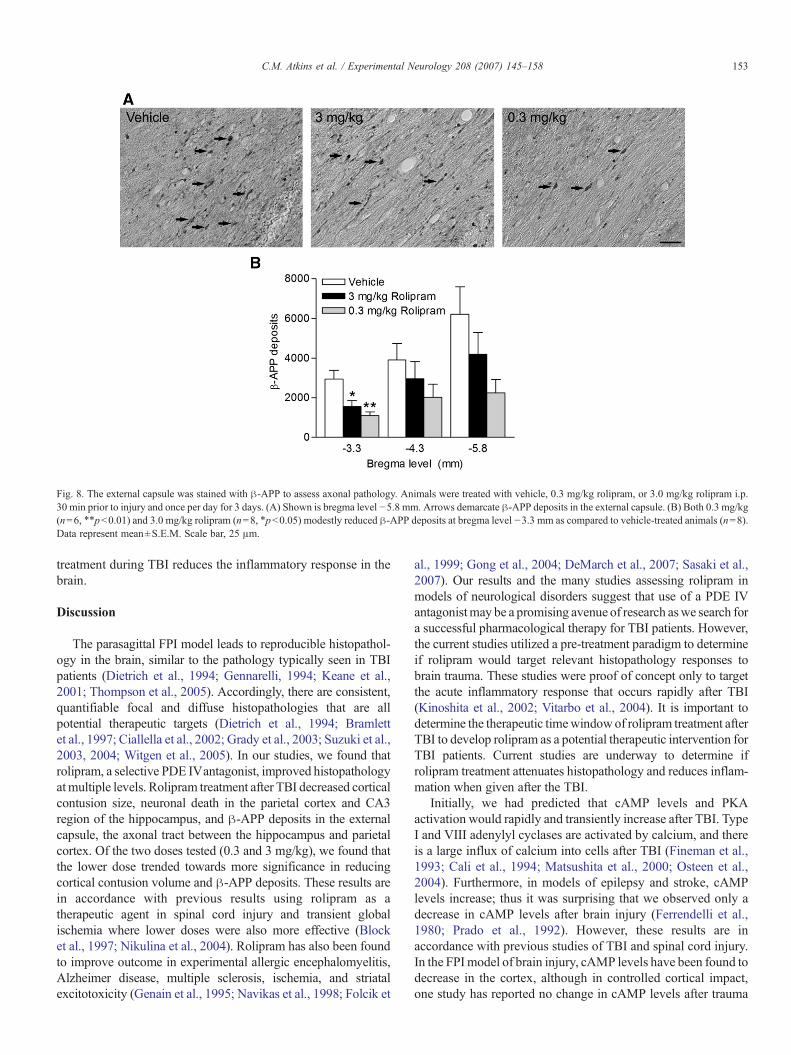
Fig. 8. The external capsule was stained with β-APP to assess axonal pathology. Animals were treated with vehicle, 0.3 mg/kg rolipram, or 3.0 mg/kg rolipram i.p.30 min prior to injury and once per day for 3 days. (A) Shown is bregma level −5.8 mm. Arrows demarcate β-APP deposits in the external capsule. (B) Both 0.3 mg/kg(n=6, ⁎⁎pb0.01) and 3.0 mg/kg rolipram (n=8, ⁎pb0.05) modestly reduced β-APP deposits at bregma level −3.3 mm as compared to vehicle-treated animals (n=8).Data represent mean±S.E.M. Scale bar, 25 μm.
153C.M. Atkins et al. / Experimental Neurology 208 (2007) 145–158
treatment during TBI reduces the inflammatory response in thebrain.
Discussion
The parasagittal FPI model leads to reproducible histopathol-ogy in the brain, similar to the pathology typically seen in TBIpatients (Dietrich et al., 1994; Gennarelli, 1994; Keane et al.,2001; Thompson et al., 2005). Accordingly, there are consistent,quantifiable focal and diffuse histopathologies that are allpotential therapeutic targets (Dietrich et al., 1994; Bramlettet al., 1997; Ciallella et al., 2002; Grady et al., 2003; Suzuki et al.,2003, 2004; Witgen et al., 2005). In our studies, we found thatrolipram, a selective PDE IVantagonist, improved histopathologyatmultiple levels. Rolipram treatment after TBI decreased corticalcontusion size, neuronal death in the parietal cortex and CA3region of the hippocampus, and β-APP deposits in the externalcapsule, the axonal tract between the hippocampus and parietalcortex. Of the two doses tested (0.3 and 3 mg/kg), we found thatthe lower dose trended towards more significance in reducingcortical contusion volume and β-APP deposits. These results arein accordance with previous results using rolipram as atherapeutic agent in spinal cord injury and transient globalischemia where lower doses were also more effective (Blocket al., 1997; Nikulina et al., 2004). Rolipram has also been foundto improve outcome in experimental allergic encephalomyelitis,Alzheimer disease, multiple sclerosis, ischemia, and striatalexcitotoxicity (Genain et al., 1995; Navikas et al., 1998; Folcik et
al., 1999; Gong et al., 2004; DeMarch et al., 2007; Sasaki et al.,2007). Our results and the many studies assessing rolipram inmodels of neurological disorders suggest that use of a PDE IVantagonistmay be a promising avenue of research aswe search fora successful pharmacological therapy for TBI patients. However,the current studies utilized a pre-treatment paradigm to determineif rolipram would target relevant histopathology responses tobrain trauma. These studies were proof of concept only to targetthe acute inflammatory response that occurs rapidly after TBI(Kinoshita et al., 2002; Vitarbo et al., 2004). It is important todetermine the therapeutic timewindowof rolipram treatment afterTBI to develop rolipram as a potential therapeutic intervention forTBI patients. Current studies are underway to determine ifrolipram treatment attenuates histopathology and reduces inflam-mation when given after the TBI.
Initially, we had predicted that cAMP levels and PKAactivation would rapidly and transiently increase after TBI. TypeI and VIII adenylyl cyclases are activated by calcium, and thereis a large influx of calcium into cells after TBI (Fineman et al.,1993; Cali et al., 1994; Matsushita et al., 2000; Osteen et al.,2004). Furthermore, in models of epilepsy and stroke, cAMPlevels increase; thus it was surprising that we observed only adecrease in cAMP levels after brain injury (Ferrendelli et al.,1980; Prado et al., 1992). However, these results are inaccordance with previous studies of TBI and spinal cord injury.In the FPImodel of brain injury, cAMP levels have been found todecrease in the cortex, although in controlled cortical impact,one study has reported no change in cAMP levels after trauma
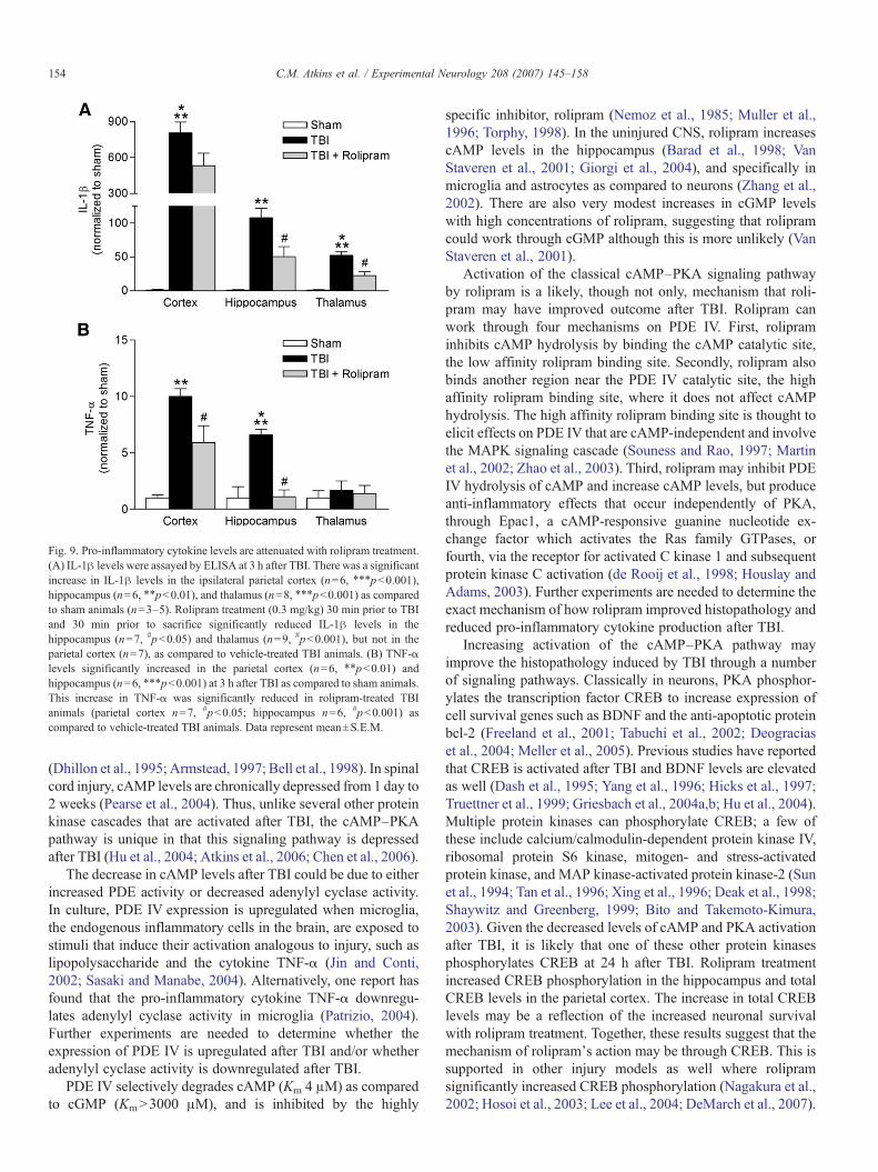
Fig. 9. Pro-inflammatory cytokine levels are attenuated with rolipram treatment.(A) IL-1β levels were assayed by ELISA at 3 h after TBI. There was a significantincrease in IL-1β levels in the ipsilateral parietal cortex (n=6, ⁎⁎⁎pb0.001),hippocampus (n=6, ⁎⁎pb0.01), and thalamus (n=8, ⁎⁎⁎pb0.001) as comparedto sham animals (n=3–5). Rolipram treatment (0.3 mg/kg) 30 min prior to TBIand 30 min prior to sacrifice significantly reduced IL-1β levels in thehippocampus (n=7, #pb0.05) and thalamus (n=9, #pb0.001), but not in theparietal cortex (n=7), as compared to vehicle-treated TBI animals. (B) TNF-αlevels significantly increased in the parietal cortex (n=6, ⁎⁎pb0.01) andhippocampus (n=6, ⁎⁎⁎pb0.001) at 3 h after TBI as compared to sham animals.This increase in TNF-α was significantly reduced in rolipram-treated TBIanimals (parietal cortex n=7, #pb0.05; hippocampus n=6, #pb0.001) ascompared to vehicle-treated TBI animals. Data represent mean±S.E.M.
154 C.M. Atkins et al. / Experimental Neurology 208 (2007) 145–158
(Dhillon et al., 1995; Armstead, 1997; Bell et al., 1998). In spinalcord injury, cAMP levels are chronically depressed from 1 day to2 weeks (Pearse et al., 2004). Thus, unlike several other proteinkinase cascades that are activated after TBI, the cAMP–PKApathway is unique in that this signaling pathway is depressedafter TBI (Hu et al., 2004; Atkins et al., 2006; Chen et al., 2006).
The decrease in cAMP levels after TBI could be due to eitherincreased PDE activity or decreased adenylyl cyclase activity.In culture, PDE IV expression is upregulated when microglia,the endogenous inflammatory cells in the brain, are exposed tostimuli that induce their activation analogous to injury, such aslipopolysaccharide and the cytokine TNF-α (Jin and Conti,2002; Sasaki and Manabe, 2004). Alternatively, one report hasfound that the pro-inflammatory cytokine TNF-α downregu-lates adenylyl cyclase activity in microglia (Patrizio, 2004).Further experiments are needed to determine whether theexpression of PDE IV is upregulated after TBI and/or whetheradenylyl cyclase activity is downregulated after TBI.
PDE IV selectively degrades cAMP (Km 4 μM) as comparedto cGMP (KmN3000 μM), and is inhibited by the highly
specific inhibitor, rolipram (Nemoz et al., 1985; Muller et al.,1996; Torphy, 1998). In the uninjured CNS, rolipram increasescAMP levels in the hippocampus (Barad et al., 1998; VanStaveren et al., 2001; Giorgi et al., 2004), and specifically inmicroglia and astrocytes as compared to neurons (Zhang et al.,2002). There are also very modest increases in cGMP levelswith high concentrations of rolipram, suggesting that rolipramcould work through cGMP although this is more unlikely (VanStaveren et al., 2001).
Activation of the classical cAMP–PKA signaling pathwayby rolipram is a likely, though not only, mechanism that roli-pram may have improved outcome after TBI. Rolipram canwork through four mechanisms on PDE IV. First, rolipraminhibits cAMP hydrolysis by binding the cAMP catalytic site,the low affinity rolipram binding site. Secondly, rolipram alsobinds another region near the PDE IV catalytic site, the highaffinity rolipram binding site, where it does not affect cAMPhydrolysis. The high affinity rolipram binding site is thought toelicit effects on PDE IV that are cAMP-independent and involvethe MAPK signaling cascade (Souness and Rao, 1997; Martinet al., 2002; Zhao et al., 2003). Third, rolipram may inhibit PDEIV hydrolysis of cAMP and increase cAMP levels, but produceanti-inflammatory effects that occur independently of PKA,through Epac1, a cAMP-responsive guanine nucleotide ex-change factor which activates the Ras family GTPases, orfourth, via the receptor for activated C kinase 1 and subsequentprotein kinase C activation (de Rooij et al., 1998; Houslay andAdams, 2003). Further experiments are needed to determine theexact mechanism of how rolipram improved histopathology andreduced pro-inflammatory cytokine production after TBI.
Increasing activation of the cAMP–PKA pathway mayimprove the histopathology induced by TBI through a numberof signaling pathways. Classically in neurons, PKA phosphor-ylates the transcription factor CREB to increase expression ofcell survival genes such as BDNF and the anti-apoptotic proteinbcl-2 (Freeland et al., 2001; Tabuchi et al., 2002; Deograciaset al., 2004; Meller et al., 2005). Previous studies have reportedthat CREB is activated after TBI and BDNF levels are elevatedas well (Dash et al., 1995; Yang et al., 1996; Hicks et al., 1997;Truettner et al., 1999; Griesbach et al., 2004a,b; Hu et al., 2004).Multiple protein kinases can phosphorylate CREB; a few ofthese include calcium/calmodulin-dependent protein kinase IV,ribosomal protein S6 kinase, mitogen- and stress-activatedprotein kinase, and MAP kinase-activated protein kinase-2 (Sunet al., 1994; Tan et al., 1996; Xing et al., 1996; Deak et al., 1998;Shaywitz and Greenberg, 1999; Bito and Takemoto-Kimura,2003). Given the decreased levels of cAMP and PKA activationafter TBI, it is likely that one of these other protein kinasesphosphorylates CREB at 24 h after TBI. Rolipram treatmentincreased CREB phosphorylation in the hippocampus and totalCREB levels in the parietal cortex. The increase in total CREBlevels may be a reflection of the increased neuronal survivalwith rolipram treatment. Together, these results suggest that themechanism of rolipram's action may be through CREB. This issupported in other injury models as well where rolipramsignificantly increased CREB phosphorylation (Nagakura et al.,2002; Hosoi et al., 2003; Lee et al., 2004; DeMarch et al., 2007).
155C.M. Atkins et al. / Experimental Neurology 208 (2007) 145–158
Another signaling pathway regulated by cAMP–PKA isthrough the transcription factor NF-κB p50 to reduce expressionof pro-inflammatory cytokines such as IL-1β and TNF-α(Montminy and Bilezikjian, 1987; Hou et al., 2003). Transcrip-tion of the tnf-α gene is suppressed by the NF-κB p50 subunitwhich constitutively binds the IκB element in the promoterregion (Kuprash et al., 1995; Jimenez et al., 2001; Takahashiet al., 2002; Foey et al., 2003). PKA phosphorylation of Ser337on the NF-κB p50 subunit increases its binding and repressionof transcription of IκB-containing gene promoter of the tnf-αgene (Ollivier et al., 1996; Baer et al., 1998; Hou et al., 2003).How the cAMP pathway regulates IL-1β expression is an activearea of investigation. Several studies have shown that raisingcAMP levels with either neurotransmitters or phosphodiesteraseinhibitors reduce IL-1β levels, but the exact mechanism is stillunclear (Cogswell et al., 1994; Verghese et al., 1995; Si et al.,1998; Caggiano and Kraig, 1999; Cho et al., 2001; Dello Russoet al., 2004).
Reducing pro-inflammatory cytokine levels after TBI toimprove outcome has met with varying success. Administrationof an inhibitor of IL-1β receptors, IL-1 receptor antagonist (IL-1ra), reduces contusion volume and transgenic mice over-expressing IL-1ra have improved behavioral recovery after TBI(Sanderson et al., 1999; Tehranian et al., 2002). Similarly,knockout mice of tnfα have improved behavior recovery 1 weekafter TBI, but worsened histopathology and behavioral outcome2–4 weeks after injury (Scherbel et al., 1999). These studiesindicate that inflammation is a complex, evolving series ofbiochemical events that can be both detrimental and beneficial forfunctional outcome after injury. Thus, targeting the inflammatorycascade as a therapeutic intervention requires careful consider-ation of the optimal time window, dosage, and mechanism ofaction.
Although these studies demonstrate an improvement inhistopathology after TBI, in consideration of the many failedclinical trials of other neuroprotective agents for the treatment ofTBI, these preliminary studies are only proof of concept for theFPI model. It is important to extend these observations to a post-injury treatment paradigm and determine the therapeuticwindow for rolipram treatment after TBI. Furthermore, whetherthese improvements in histopathology are accompanied by animprovement in behavioral deficits remains to be determined.Another important consideration is to understand the conse-quences of decreased cAMP levels after TBI: what cell typesexhibit decreases in cAMP–PKA signaling and whether this canbe rescued with rolipram treatment. And finally, understandingthe mechanism of how rolipram leads to an improvement infunctional outcome, possibly by increasing CREB-regulatedgene expression and decreasing the inflammatory response, isnecessary to develop PDE IV inhibition into a potential therapy.
Acknowledgments
This work was supported by NIH grants NS30291 andNS42133 (W.D.D.). We thank the Dietrich and Pearse labs forhelpful discussions and Beata Frydel and Jarret Weinrich fortechnical assistance.
References
Armstead, W.M., 1997. Role of impaired cAMP and calcium-sensitive K+channel function in altered cerebral hemodynamics following brain injury.Brain Res. 768, 177–184.
Atkins, C.M., Chen, S., Alonso, O.F., Dietrich, W.D., Hu, B.R., 2006.Activation of calcium/calmodulin-dependent protein kinases after traumaticbrain injury. J. Cereb. Blood Flow Metab. 26, 1507–1518.
Baer, M., Dillner, A., Schwartz, R.C., Sedon, C., Nedospasov, S., Johnson, P.F.,1998. Tumor necrosis factor-α transcription in macrophages is attenuated byan autocrine factor that preferentially induces NF-κB p50. Mol. Cell. Biol.18, 5678–5689.
Barad, M., Bourtchouladze, R., Winder, D.G., Golan, H., Kandel, E., 1998.Rolipram, a type IV-specific phosphodiesterase inhibitor, facilitates theestablishment of long-lasting long-term potentiation and improves memory.Proc. Natl. Acad. Sci. U. S. A. 95, 15020–15025.
Bell, M.J., Kochanek, P.M., Carcillo, J.A., Mi, Z., Schiding, J.K., Wisniewski,S.R., Clark, R.S., Dixon, C.E., Marion, D.W., Jackson, E., 1998. Interstitialadenosine, inosine, and hypoxanthine are increased after experimentaltraumatic brain injury in the rat. J. Neurotrauma 15, 163–170.
Bito, H., Takemoto-Kimura, S., 2003. Ca(2+)/CREB/CBP-dependent generegulation: a shared mechanism critical in long-term synaptic plasticityand neuronal survival. Cell Calcium 34, 425–430.
Block, F., Tondar, A., Schmidt, W., Schwarz, M., 1997. Delayed treatment withrolipram protects against neuronal damage following global ischemia in rats.Neuroreport 8, 3829–3832.
Block, F., Bozdag, I., Nolden-Koch, M., 2001. Inflammation contributes to thepostponed ischemic neuronal damage following treatment with a glutamateantagonist in rats. Neurosci. Lett. 298, 103–106.
Bramlett, H.M., Kraydieh, S., Green, E.J., Dietrich, W.D., 1997. Temporaland regional patterns of axonal damage following traumatic brain injury:A β-amyloid precursor protein immunocytochemical study in rats.J. Neuropathol. Exp. Neurol. 56, 1132–1141.
Caggiano, A.O., Kraig, R.P., 1999. Prostaglandin E receptor subtypes incultured rat microglia and their role in reducing lipopolysaccharide-inducedinterleukin-1beta production. J. Neurochem. 72, 565–575.
Cali, J.J., Zwaagstra, J.C., Mons, N., Cooper, D.M., Krupinski, J., 1994. TypeVIII adenylyl cyclase. A Ca2+/calmodulin-stimulated enzyme expressed indiscrete regions of rat brain. J. Biol. Chem. 269, 12190–12195.
Castro, A., Jerez, M.J., Gil, C., Martinez, A., 2005. Cyclic nucleotidephosphodiesterases and their role in immunomodulatory responses:advances in the development of specific phosphodiesterase inhibitors.Med. Res. Rev. 25, 229–244.
Chen, S., Atkins, C.M., Liu, C.L., Alonso, O.F., Dietrich, W.D., Hu, B.R., 2006.Alterations in mammalian target of rapamycin signaling pathways aftertraumatic brain injury. J. Cereb. Blood Flow Metab. 27, 939–949.
Cho, S., Kim, Y., Cruz, M.O., Park, E.M., Chu, C.K., Song, G.Y., Joh, T.H.,2001. Repression of proinflammatory cytokine and inducible nitric oxidesynthase (NOS2) gene expression in activated microglia by N-acetyl-O-methyldopamine: protein kinase A-dependent mechanism. Glia 33,324–333.
Ciallella, J.R., Ikonomovic, M.D., Paljug, W.R., Wilbur, Y.I., Dixon, C.E.,Kochanek, P.M., Marion, D.W., DeKosky, S.T., 2002. Changes inexpression of amyloid precursor protein and interleukin-1β after experi-mental traumatic brain injury in rats. J. Neurotrauma 19, 1555–1567.
Cogswell, J.P., Godlevski, M.M., Wisely, G.B., Clay, W.C., Leesnitzer, L.M.,Ways, J.P., Gray, J.G., 1994. NF-kappa B regulates IL-1 beta transcriptionthrough a consensus NF-kappa B binding site and a nonconsensus CRE-likesite. J. Immunol. 153, 712–723.
Dal Piaz, V., Giovannoni, M.P., 2000. Phosphodiesterase 4 inhibitors,structurally unrelated to rolipram, as promising agents for the treatment ofasthma and other pathologies. Eur. J. Med. Chem. 35, 463–480.
Dash, P.K., Moore, A.N., Dixon, C.E., 1995. Spatial memory deficits, increasedphosphorylation of the transcription factor CREB, and induction of the AP-1complex following experimental brain injury. J. Neurosci. 15, 2030–2039.
de Rooij, J., Zwartkruis, F.J., Verheijen, M.H., Cool, R.H., Nijman, S.M.,Wittinghofer, A., Bos, J.L., 1998. Epac is a Rap1 guanine-nucleotide-exchange factor directly activated by cyclic AMP. Nature 396, 474–477.
156 C.M. Atkins et al. / Experimental Neurology 208 (2007) 145–158
Deak, M., Clifton, A.D., Lucocq, L.M., Alessi, D.R., 1998. Mitogen- and stress-activated protein kinase-1 (MSK1) is directly activated by MAPK andSAPK2/p38, and may mediate activation of CREB. EMBO J. 17,4426–4441.
Dello Russo, C., Boullerne, A.I., Gavrilyuk, V., Feinstein, D.L., 2004. Inhibitionof microglial inflammatory responses by norepinephrine: effects on nitricoxide and interleukin-1β production. J. Neuroinflammation 1, 9–24.
DeMarch, Z., Giampa, C., Patassini, S., Martorana, A., Bernardi, G., Fusco, F.R.,2007. Beneficial effects of rolipram in a quinolinic acid model of striatalexcitotoxicity. Neurobiol. Dis. 25, 266–273.
Deogracias, R., Espliguero, G., Iglesias, T., Rodriguez-Pena, A., 2004. Expressionof the neurotrophin receptor trkB is regulated by the cAMP/CREB pathway inneurons. Mol. Cell. Neurosci. 26, 470–480.
Dhillon, H.S., Yang, L., Padmaperuma, B., Dempsey, R.J., Fiscus, R.R., RenukaPrasad, M., 1995. Regional concentrations of cyclic nucleotides afterexperimental brain injury. J. Neurotrauma 12, 1035–1043.
Dietrich, W.D., Alonso, O., Halley, M., 1994. Early microvascular and neuronalconsequences of traumatic brain injury: a light and electron microscopicstudy in rats. J. Neurotrauma 11, 289–301.
Dietrich, W.D., Chatzipanteli, K., Vitarbo, E., Wada, K., Kinoshita, K., 2004.The role of inflammatory processes in the pathophysiology and treatment ofbrain and spinal cord trauma. Acta Neurochir., Suppl. 89, 69–74.
Doppenberg, E.M., Choi, S.C., Bullock, R., 2004. Clinical trials in traumaticbrain injury: lessons for the future. J. Neurosurg. Anesthesiol. 16, 87–94.
Fan, L., Young, P.R., Barone, F.C., Feuerstein, G.Z., Smith, D.H., McIntosh, T.K.,1996. Experimental brain injury induces differential expression of tumornecrosis factor-αmRNA in the CNS. Brain Res. Mol. Brain Res. 36, 287–291.
Ferrendelli, J.A., Blank, A.C., Gross, R.A., 1980. Relationships between seizureactivity and cyclic nucleotide levels in brain. Brain Res. 200, 93–103.
Fineman, I., Hovda, D.A., Smith, M., Yoshino, A., Becker, D.P., 1993.Concussive brain injury is associated with a prolonged accumulation ofcalcium: A 45Ca autoradiographic study. Brain Res. 624, 94–102.
Foey, A.D., Field, S., Ahmed, S., Jain, A., Feldmann, M., Brennan, F.M.,Williams, R., 2003. Impact of VIP and cAMP on the regulation of TNF-αand IL-10 production: Implications for rheumatoid arthritis. Arthritis Res.Ther. 5, R317–R328.
Folcik, V.A., Smith, T., O'Bryant, S., Kawczak, J.A., Zhu, B., Sakurai, H.,Kajiwara, A., Staddon, J.M., Glabinski, A., Chernosky, A.L., Tani, M.,Johnson, J.M., Tuohy, V.K., Rubin, L.L., Ransohoff, R.M., 1999. Treatmentwith BBB022A or rolipram stabilizes the blood–brain barrier in experimentalautoimmune encephalomyelitis: an additional mechanism for the therapeuticeffect of type IV phosphodiesterase inhibitors. J. Neuroimmunol. 97, 119–128.
Freeland, K., Boxer, L.M., Latchman, D.S., 2001. The cyclic AMP responseelement in the Bcl-2 promoter confers inducibility by hypoxia in neuronalcells. Brain Res. Mol. Brain Res. 92, 98–106.
Genain, C.P., Roberts, T., Davis, R.L., Nguyen, M.H., Uccelli, A., Faulds, D.,Li, Y., Hedgpeth, J., Hauser, S.L., 1995. Prevention of autoimmunedemyelination in non-human primates by a cAMP-specific phosphodiester-ase inhibitor. Proc. Natl. Acad. Sci. U. S. A. 92, 3601–3605.
Gennarelli, T.A., 1994. Animate models of human head injury. J. Neurotrauma11, 357–368.
Giorgi, M., Modica, A., Pompili, A., Pacitti, C., Gasbarri, A., 2004. Theinduction of cyclic nucleotide phosphodiesterase 4 gene (PDE4D) impairsmemory in a water maze task. Behav. Brain Res. 154, 99–106.
Gong, B., Vitolo, O.V., Trinchese, F., Liu, S., Shelanski, M., Arancio, O., 2004.Persistent improvement in synaptic and cognitive functions in an Alzheimermouse model after rolipram treatment. J. Clin. Invest. 114, 1624–1634.
Grady, M.S., Charleston, J.S., Maris, D., Witgen, B.M., Lifshitz, J., 2003.Neuronal and glial cell number in the hippocampus after experimentaltraumatic brain injury: analysis by stereological estimation. J. Neurotrauma20, 929–941.
Griesbach, G.S., Gomez-Pinilla, F., Hovda, D.A., 2004a. The upregulation ofplasticity-related proteins following TBI is disrupted with acute voluntaryexercise. Brain Res. 1016, 154–162.
Griesbach, G.S., Hovda, D.A., Molteni, R., Wu, A., Gomez-Pinilla, F., 2004b.Voluntary exercise following traumatic brain injury: brain-derived neuro-trophic factor upregulation and recovery of function. Neuroscience 125,129–139.
Griswold, D.E., Webb, E.F., Badger, A.M., Gorycki, P.D., Levandoski, P.A.,Barnette, M.A., Grous, M., Christensen, S., Torphy, T.J., 1998. SB 207499(Ariflo), a second generation phosphodiesterase 4 inhibitor, reduces tumornecrosis factor α and interleukin-4 production in vivo. J. Pharmacol. Exp.Ther. 287, 705–711.
Hicks, R.R., Numan, S., Dhillon, H.S., Prasad, M.R., Seroogy, K.B., 1997.Alterations in BDNF and NT-3 mRNAs in rat hippocampus afterexperimental brain trauma. Brain Res. Mol. Brain Res. 48, 401–406.
Hosoi, R., Ishikawa, M., Kobayashi, K., Gee, A., Yamaguchi, M., Inoue, O.,2003. Effect of rolipram on muscarinic acetylcholine receptor binding in theintact mouse brain. J. Neural Transm. 110, 363–372.
Hou, S., Guan, H., Ricciardi, R.P., 2003. Phosphorylation of serine 337 of NF-κB p50 is critical for DNA binding. J. Biol. Chem. 278, 45994–45998.
Houslay, M.D., Adams, D.R., 2003. PDE4 cAMP phosphodiesterases: modularenzymes that orchestrate signalling cross-talk, desensitization and compart-mentalization. Biochem. J. 370, 1–18.
Hu, S., Peterson, P.K., Chao, C.C., 1997. Cytokine-mediated neuronalapoptosis. Neurochem. Int. 30, 427–431.
Hu, B.R., Fux, C.M., Martone, M.E., Zivin, J.A., Ellisman, M.H., 1999.Persistent phosphorylation of cyclic AMP responsive element-bindingprotein and activating transcription factor-2 transcription factors followingtransient cerebral ischemia in rat brain. Neuroscience 89, 437–452.
Hu, B., Liu, C., Bramlett, H., Sick, T.J., Alonso, O.F., Chen, S., Dietrich, W.D.,2004. Changes in trkB-ERK1/2-CREB/Elk-1 pathways in hippocampalmossy fiber organization after traumatic brain injury. J. Cereb. Blood FlowMetab. 24, 934–943.
Imanishi, T., Sawa, A., Ichimaru, Y., Miyashiro, M., Kato, S., Yamamoto, T.,Ueki, S., 1997. Ameliorating effects of rolipram on experimentally inducedimpairments of learning and memory in rodents. Eur. J. Pharmacol. 321,273–278.
Jimenez, J.L., Punzon, C., Navarro, J., Munoz-Fernandez, M.A., Fresno, M.,2001. Phosphodiesterase 4 inhibitors prevent cytokine secretion by Tlymphocytes by inhibiting nuclear factor-κB and nuclear factor of activatedT cells activation. J. Pharmacol. Exp. Ther. 299, 753–759.
Jin, S.L., Conti, M., 2002. Induction of the cyclic nucleotide phosphodiesterasePDE4B is essential for LPS-activated TNF-α responses. Proc. Natl. Acad.Sci. U. S. A. 99, 7628–7633.
Kato, H., Araki, T., Itoyama, Y., Kogure, K., 1995. Rolipram, a cyclic AMP-selective phosphodiesterase inhibitor, reduces neuronal damage followingcerebral ischemia in the gerbil. Eur. J. Pharmacol. 272, 107–110.
Keane, R.W., Kraydieh, S., Lotocki, G., Alonso, O.F., Aldana, P., Dietrich,W.D.,2001. Apoptotic and antiapoptotic mechanisms after traumatic brain injury.J. Cereb. Blood Flow Metab. 21, 1189–1198.
Keeling, K.L., Hicks, R.R., Mahesh, J., Billings, B.B., Kotwal, G.J., 2000. Localneutrophil influx following lateral fluid-percussion brain injury in rats isassociated with accumulation of complement activation fragments of thethird component (C3) of the complement system. J. Neuroimmunol. 105,20–30.
Keryer, G., Yassenko, M., Labbe, J.C., Castro, A., Lohmann, S.M., Evain-Brion,D., Tasken, K., 1998. Mitosis-specific phosphorylation and subcellularredistribution of the RIIα regulatory subunit of cAMP-dependent proteinkinase. J. Biol. Chem. 273, 34594–34602.
Kinoshita, K., Chatzipanteli, K., Vitarbo, E., Truettner, J.S., Alonso, O.F.,Dietrich, W.D., 2002. Interleukin-1βmessenger ribonucleic acid and proteinlevels after fluid-percussion brain injury in rats: importance of injuryseverity and brain temperature. Neurosurgery 51, 195–203.
Kuprash, D.V., Udalova, I.A., Turetskaya, R.L., Rice, N.R., Nedospasov, S.A.,1995. Conserved κ B element located downstream of the tumor necrosisfactor α gene: distinct NF-κ B binding pattern and enhancer activity in LPSactivated murine macrophages. Oncogene 11, 97–106.
Langlois, J.A., Rutland-Brown,W., Thomas, K.E., 2004. Traumatic Brain Injuryin the United States: Emergency Department Visits, Hospitalizations, andDeaths. Centers for Disease Control and Prevention. National Center forInjury Prevention and Control, Atlanta, GA, pp. 1–68.
Lee, H.T., Chang, Y.C., Wang, L.Y., Wang, S.T., Huang, C.C., Ho, C.J., 2004.cAMP response element-binding protein activation in ligation precondition-ing in neonatal brain. Ann. Neurol. 56, 611–623.
Manganiello, V.C., Murata, T., Taira, M., Belfrage, P., Degerman, E., 1995.
157C.M. Atkins et al. / Experimental Neurology 208 (2007) 145–158
Diversity in cyclic nucleotide phosphodiesterase isoenzyme families. Arch.Biochem. Biophys. 322, 1–13.
Martin, C., Goggel, R., Dal Piaz, V., Vergelli, C., Giovannoni, P., Ernst, M.,Uhlig, S., 2002. Airway relaxant and anti-inflammatory properties of aPDE4 inhibitor with low affinity for the high-affinity rolipram binding site.Naunyn Schmiedebergs Arch. Pharmacol. 365, 284–289.
Matsushita, Y., Shima, K., Nawashiro, H., Wada, K., Tsuzuki, N., Miyazawa, T.,2000. Real time monitoring of glutamate following fluid percussion braininjury with hypoxia in the rat. Acta Neurochir. Suppl. 76, 207–212.
Mayr, B., Montminy, M., 2001. Transcriptional regulation by the phosphory-lation-dependent factor CREB. Nat. Rev. Mol. Cell Biol. 2, 599–609.
Meda, L., Cassatella, M.A., Szendrei, G.I., Otvos Jr., L., Baron, P., Villalba, M.,Ferrari, D., Rossi, F., 1995. Activation of microglial cells by β-amyloidprotein and interferon-γ. Nature 374, 647–650.
Meller, R., Minami, M., Cameron, J.A., Impey, S., Chen, D., Lan, J.Q.,Henshall, D.C., Simon, R.P., 2005. CREB-mediated Bcl-2 proteinexpression after ischemic preconditioning. J. Cereb. Blood Flow Metab.25, 234–246.
Montminy, M.R., Bilezikjian, L.M., 1987. Binding of a nuclear protein to thecyclic-AMP response element of the somatostatin gene. Nature 328,175–178.
Morganti-Kossmann, M.C., Rancan, M., Stahel, P.F., Kossmann, T., 2002.Inflammatory response in acute traumatic brain injury: a double-edgedsword. Curr. Opin. Crit. Care 8, 101–105.
Muller, T., Engels, P., Fozard, J.R., 1996. Subtypes of the type 4 cAMPphosphodiesterases: structure, regulation and selective inhibition. TrendsPharmacol. Sci. 17, 294–298.
Nagakura, A., Niimura, M., Takeo, S., 2002. Effects of a phosphodiesterase IVinhibitor rolipram on microsphere embolism-induced defects in memoryfunction and cerebral cyclic AMP signal transduction system in rats. Br.J. Pharmacol. 135, 1783–1793.
Navikas, V., Matusevicius, D., Soderstrom, M., Pirskanen, R., Fredrikson, S.,Link, H., 1998. The phosphodiesterase i.v. inhibitor rolipram in vitro reducesthe numbers of MBP-reactive IFN-gamma and TNF-alpha mRNAexpressing blood mononuclear cells in patients with multiple sclerosis.Clin. Neuropharmacol. 21, 236–244.
Nemoz, G., Prigent, A.F., Moueqqit, M., Fougier, S., Macovschi, O., Pacheco,H., 1985. Selective inhibition of one of the cyclic AMP phosphodiesterasesfrom rat brain by the neurotropic compound rolipram. Biochem. Pharmacol.34, 2997–3000.
Nikulina, E., Tidwell, J.L., Dai, H.N., Bregman, B.S., Filbin, M.T., 2004. Thephosphodiesterase inhibitor rolipram delivered after a spinal cord lesionpromotes axonal regeneration and functional recovery. Proc. Natl. Acad. Sci.U. S. A. 101, 8786–8790.
Ollivier, V., Parry, G.C., Cobb, R.R., de Prost, D., Mackman, N., 1996. Elevatedcyclic AMP inhibits NF-kB-mediated transcription in human monocyticcells and endothelial cells. J. Biol. Chem. 271, 20828–20835.
Osteen, C.L., Giza, C.C., Hovda, D.A., 2004. Injury-induced alterations inN-methyl-D-aspartate receptor subunit composition contribute to prolonged45calcium accumulation following lateral fluid percussion. Neuroscience 128,305–322.
Patrizio, M., 2004. Tumor necrosis factor reduces cAMP production in ratmicroglia. Glia 48, 241–249.
Pearse, D.D., Pereira, F.C., Marcillo, A.E., Bates, M.L., Berrocal, Y.A.,Filbin, M.T., Bunge, M.B., 2004. cAMP and Schwann cells promoteaxonal growth and functional recovery after spinal cord injury. Nat. Med.10, 610–616.
Prabhakar, U., Lipshutz, D., Bartus, J.O., Slivjak, M.J., Smith III, E.F., Lee, J.C.,Esser, K.M., 1994. Characterization of cAMP-dependent inhibition of LPS-induced TNF α production by rolipram, a specific phosphodiesterase IV(PDE IV) inhibitor. Int. J. Immunopharmacol. 16, 805–816.
Prado, R., Busto, R., Globus, M.Y., 1992. Ischemia-induced changes inextracellular levels of striatal cyclic AMP: role of dopamine neurotrans-mission. J. Neurochem. 59, 1581–1584.
Sanderson, K.L., Raghupathi, R., Saatman, K.E., Martin, D., Miller, G.,McIntosh, T.K., 1999. Interleukin-1 receptor antagonist attenuates regionalneuronal cell death and cognitive dysfunction after experimental braininjury. J. Cereb. Blood Flow Metab. 19, 1118–1125.
Sasaki, K., Manabe, H., 2004. KF19514, a phosphodiesterase 4 and 1 inhibitor,inhibits TNF-α-induced GM-CSF production by a human bronchialepithelial cell line via inhibition of PDE4. Inflamm. Res. 53, 31–37.
Sasaki, T., Kitagawa, K., Omura-Matsuoka, E., Todo, K., Terasaki, Y., Sugiura,S., Hatazawa, J., Yagita, Y., Hori, M., 2007. The phosphodiesterase inhibitorrolipram promotes survival of newborn hippocampal neurons after ischemia.Stroke 38, 1597–1605.
Scherbel, U., Raghupathi, R., Nakamura, M., Saatman, K.E., Trojanowski, J.Q.,Neugebauer, E., Marino, M.W., McIntosh, T.K., 1999. Differential acute andchronic responses of tumor necrosis factor-deficient mice to experimentalbrain injury. Proc. Natl. Acad. Sci. U. S. A. 96, 8721–8726.
Shaywitz, A.J., Greenberg, M.E., 1999. CREB: A stimulus-induced transcrip-tion factor activated by a diverse array of extracellular signals. Ann. Rev.Biochem. 68, 821–861.
Shohami, E., Novikov, M., Bass, R., Yamin, A., Gallily, R., 1994. Closed headinjury triggers early production of TNF α and IL-6 by brain tissue. J. Cereb.Blood Flow Metab. 14, 615–619.
Si, Q., Nakamura, Y., Ogata, T., Kataoka, K., Schubert, P., 1998. Differentialregulation of microglial activation by propentofylline via cAMP signaling.Brain Res. 812, 97–104.
Soares, H.D., Hicks, R.R., Smith, D., McIntosh, T.K., 1995. Inflammatoryleukocytic recruitment and diffuse neuronal degeneration are separatepathological processes resulting from traumatic brain injury. J. Neurosci. 15,8223–8233.
Souness, J.E., Rao, S., 1997. Proposal for pharmacologically distinctconformers of PDE4 cyclic AMP phosphodiesterases. Cell. Signal. 9,227–236.
Sun, P., Enslen, H., Myung, P.S., Maurer, R.A., 1994. Differential activation ofCREB by Ca2+/calmodulin-dependent protein kinase type II and type IVinvolves phosphorylation of a site that negatively regulates activity. GenesDev. 8, 2527–2539.
Suzuki, T., Bramlett, H.M., Dietrich, W.D., 2003. The importance of gender onthe beneficial effects of posttraumatic hypothermia. Exp. Neurol. 184,1017–1026.
Suzuki, T., Bramlett, H.M., Ruenes, G., Dietrich, W.D., 2004. The effects ofearly post-traumatic hyperthermia in female and ovariectomized rats.J. Neurotrauma 21, 842–853.
Tabuchi, A., Sakaya, H., Kisukeda, T., Fushiki, H., Tsuda, M., 2002.Involvement of an upstream stimulatory factor as well as cAMP-responsiveelement-binding protein in the activation of brain-derived neurotrophicfactor gene promoter I. J. Biol. Chem. 277, 35920–35931.
Takahashi, N., Tetsuka, T., Uranishi, H., Okamoto, T., 2002. Inhibition of theNF-κB transcriptional activity by protein kinase A. Eur. J. Biochem. 269,4559–4565.
Tan, Y., Rouse, J., Zhang, A., Cariati, S., Cohen, P., Comb, M.J., 1996. FGF andstress regulate CREB and ATF-1 via a pathway involving p38 MAP kinaseand MAPKAP kinase-2. EMBO J. 15, 4629–4642.
Tanaka, M., Sotomatsu, A., Yoshida, T., Hirai, S., Nishida, A., 1994. Detectionof superoxide production by activated microglia using a sensitive andspecific chemiluminescence assay and microglia-mediated PC12h celldeath. J. Neurochem. 63, 266–270.
Taupin, V., Toulmond, S., Serrano, A., Benavides, J., Zavala, F., 1993.Increase in IL-6, IL-1 and TNF levels in rat brain following traumaticlesion. Influence of pre- and post-traumatic treatment with Ro5 4864, aperipheral-type (p site) benzodiazepine ligand. J. Neuroimmunol. 42,177–185.
Tehranian, R., Andell-Jonsson, S., Beni, S.M., Yatsiv, I., Shohami, E., Bartfai,T., Lundkvist, J., Iverfeldt, K., 2002. Improved recovery and delayedcytokine induction after closed head injury in mice with central over-expression of the secreted isoform of the interleukin-1 receptor antagonist.J. Neurotrauma 19, 939–951.
Thompson, H.J., Lifshitz, J., Marklund, N., Grady, M.S., Graham, D.I., Hovda,D.A., McIntosh, T.K., 2005. Lateral fluid percussion brain injury: a 15-yearreview and evaluation. J. Neurotrauma 22, 42–75.
Torphy, T.J., 1998. Phosphodiesterase isozymes: molecular targets for novelantiasthma agents. Am. J. Respir. Crit. Care Med. 157, 351–370.
Truettner, J., Schmidt-Kastner, R., Busto, R., Alonso, O.F., Loor, J.Y., Dietrich,W.D., Ginsberg, M.D., 1999. Expression of brain-derived neurotrophic
158 C.M. Atkins et al. / Experimental Neurology 208 (2007) 145–158
factor, nerve growth factor, and heat shock protein HSP70 following fluidpercussion brain injury in rats. J. Neurotrauma 16, 471–486.
van Staveren, W.C., Markerink-van Ittersum, M., Steinbusch, H.W., de Vente, J.,2001. The effects of phosphodiesterase inhibition on cyclic GMP and cyclicAMP accumulation in the hippocampus of the rat. Brain Res. 888, 275–286.
Verghese, M.W., McConnell, R.T., Strickland, A.B., Gooding, R.C., Stimpson,S.A., Yarnall, D.P., Taylor, J.D., Furdon, P.J., 1995. Differential regulation ofhuman monocyte-derived TNF α and IL-1 β by type IV cAMP–phosphodiesterase (cAMP–PDE) inhibitors. J. Pharmacol. Exp. Ther. 272,1313–1320.
Vitarbo, E.A., Chatzipanteli, K., Kinoshita, K., Truettner, J.S., Alonso, O.F.,Dietrich, W.D., 2004. Tumor necrosis factor α expression and protein levelsafter fluid percussion injury in rats: the effect of injury severity and braintemperature. Neurosurgery 55, 416–424.
Witgen, B.M., Lifshitz, J., Smith, M.L., Schwarzbach, E., Liang, S.L., Grady,M.S., Cohen, A.S., 2005. Regional hippocampal alteration associated with
cognitive deficit following experimental brain injury: a systems, networkand cellular evaluation. Neuroscience 133, 1–15.
Xing, J., Ginty, D.D., Greenberg, M.E., 1996. Coupling of the RAS–MAPKpathway to gene activation by RSK2, a growth factor-regulated CREBkinase. Science 273, 959–963.
Yang, K., Perez-Polo, J.R., Mu, X.S., Yan, H.Q., Xue, J.J., Iwamoto, Y., Liu, S.J.,Dixon, C.E., Hayes, R.L., 1996. Increased expression of brain-derivedneurotrophic factor but not neurotrophin-3 mRNA in rat brain after corticalimpact injury. J. Neurosci. Res. 44, 157–164.
Zhang, B., Yang, L., Konishi, Y., Maeda, N., Sakanaka, M., Tanaka, J., 2002.Suppressive effects of phosphodiesterase type IV inhibitors on rat culturedmicroglial cells: comparison with other types of cAMP-elevating agents.Neuropharm. 42, 262–269.
Zhao, Y., Zhang, H.T., O'Donnell, J.M., 2003. Antidepressant-induced increase inhigh-affinity rolipram binding sites in rat brain: dependence on noradrenergicand serotonergic function. J. Pharmacol. Exp. Ther. 307, 246–253.
Related Documents