430 D ifferentiated vascular smooth muscle cells (VSMC) are major constituents of the blood vessel wall and play a vital role in the maintenance of vessel homeostasis. These highly specialized cells regulate vessel tone, blood pressure, and blood flow distribution. 1,2 Mature VSMC that are fully differentiated exhibit a contractile phenotype characterized by low protein: DNA synthetic activity, reduced proliferation rate, and a unique set of contractile proteins and signaling molecules. 3,4 Unlike skeletal and cardiac muscle cells, mature VSMC retain a remarkable ability to modulate their pheno- type and dedifferentiate into a synthetic phenotype in response to changes in local environmental cues. 5,6 The synthetic phe- notype is characterized by increased VSMC migration, loss of contractility, and abnormal extracellular matrix production. These hallmarks have been observed clinically and in animal models of vascular injury and diseases, including systemic hypertension, angioplasty-induced restenosis, atherosclerosis, and aortic aneurysm formation. 7,8 VSMC phenotype is influenced by diverse hormonal and environmental cues, including cytokine stimulation, cell–cell contact, cellular adhesions, vascular injury, and increased mechanical force. In vivo, VSMC are constantly subjected to mechanical forces as a consequence of pulsatile blood flow and shear stress. Among multiple hemodynamic forces, VSMC are primarily subjected to pulsatile cyclic stretch (CS) in response to systolic–diastolic fluctuations in pressure. As such, in vitro CS serves as a model of pressure fluctuations in the vasculature with 10%, 1 Hz stretch mim- icking hypertension. 9 Indeed, CS is a well-established stimu- lus for VSMC dedifferentiation and a switch to the synthetic phenotype, 10–12 yet the mechanism involved is incompletely understood. © 2014 American Heart Association, Inc. Arterioscler Thromb Vasc Biol is available at http://atvb.ahajournals.org DOI: 10.1161/ATVBAHA.114.304936 Objective—Blood vessel hemodynamics have profound influences on function and structure of vascular cells. One of the main mechanical forces influencing vascular smooth muscle cells (VSMC) is cyclic stretch (CS). Increased CS stimulates reactive oxygen species (ROS) production in VSMC, leading to their dedifferentiation, yet the mechanisms involved are poorly understood. This study was designed to test the hypothesis that pathological CS stimulates NADPH oxidase isoform 1 (Nox1)–derived ROS via MEF2B, leading to VSMC dysfunction via a switch from a contractile to a synthetic phenotype. Approach and Results—Using a newly developed isoform-specific Nox1 inhibitor and gene silencing technology, we demonstrate that a novel pathway, including MEF2B-Nox1-ROS, is upregulated under pathological stretch conditions, and this pathway promotes a VSMC phenotypic switch from a contractile to a synthetic phenotype. We observed that CS (10% at 1 Hz) mimicking systemic hypertension in humans increased Nox1 mRNA, protein levels, and enzymatic activity in a time-dependent manner, and this upregulation was mediated by MEF2B. Furthermore, we show that stretch-induced Nox1-derived ROS upregulated a specific marker for synthetic phenotype (osteopontin), whereas it downregulated classical markers for contractile phenotype (calponin1 and smoothelin B). In addition, our data demonstrated that stretch-induced Nox1 activation decreases actin fiber density and augments matrix metalloproteinase 9 activity, VSMC migration, and vectorial alignment. Conclusions—These results suggest that CS initiates a signal through MEF2B that potentiates Nox1-mediated ROS production and causes VSMC to switch to a synthetic phenotype. The data also characterize a new Nox1 inhibitor as a potential therapy for treatment of vascular dysfunction in hypertension. (Arterioscler Thromb Vasc Biol. 2015;35:430- 438. DOI: 10.1161/ATVBAHA.114.304936.) Key Words: MEF2B ◼ Nox1 ◼ oxidative stress ◼ vascular remodeling Received on: April 22, 2013; final version accepted on: December 4, 2014. From the Department of Pharmacology and Chemical Biology and Vascular Medicine Institute (A.I.R., G.C., D.J.R, D.M.F., L.A., P.J.P), and Departments of Bioengineering, Surgery, and Cardiothoracic Surgery and Center for Vascular Remodeling and Regeneration (K.J.B., D.A.V), University of Pittsburgh, PA; and Department of Basic Sciences, Faculty of Science, Universidad del Bío-Bío, Chillán, Chile (A.I.R). *These authors contributed equally to this article. The online-only Data Supplement is available with this article at http://atvb.ahajournals.org/lookup/suppl/doi:10.1161/ATVBAHA.114.304936/-/DC1. Correspondence to Patrick J. Pagano, PhD, FAHA, BST E1247, 200 Lothrop St, University of Pittsburgh, Pittsburgh, PA 15261. E-mail [email protected] MEF2B-Nox1 Signaling Is Critical for Stretch-Induced Phenotypic Modulation of Vascular Smooth Muscle Cells Andrés I. Rodríguez,* Gábor Csányi,* Daniel J. Ranayhossaini, Douglas M. Feck, Kory J. Blose, Lillian Assatourian, David A. Vorp, Patrick J. Pagano

Welcome message from author
This document is posted to help you gain knowledge. Please leave a comment to let me know what you think about it! Share it to your friends and learn new things together.
Transcript

430
Differentiated vascular smooth muscle cells (VSMC) are major constituents of the blood vessel wall and play a
vital role in the maintenance of vessel homeostasis. These highly specialized cells regulate vessel tone, blood pressure, and blood flow distribution.1,2 Mature VSMC that are fully differentiated exhibit a contractile phenotype characterized by low protein: DNA synthetic activity, reduced proliferation rate, and a unique set of contractile proteins and signaling molecules.3,4 Unlike skeletal and cardiac muscle cells, mature VSMC retain a remarkable ability to modulate their pheno-type and dedifferentiate into a synthetic phenotype in response to changes in local environmental cues.5,6 The synthetic phe-notype is characterized by increased VSMC migration, loss of contractility, and abnormal extracellular matrix production. These hallmarks have been observed clinically and in animal models of vascular injury and diseases, including systemic
hypertension, angioplasty-induced restenosis, atherosclerosis, and aortic aneurysm formation.7,8
VSMC phenotype is influenced by diverse hormonal and environmental cues, including cytokine stimulation, cell–cell contact, cellular adhesions, vascular injury, and increased mechanical force. In vivo, VSMC are constantly subjected to mechanical forces as a consequence of pulsatile blood flow and shear stress. Among multiple hemodynamic forces, VSMC are primarily subjected to pulsatile cyclic stretch (CS) in response to systolic–diastolic fluctuations in pressure. As such, in vitro CS serves as a model of pressure fluctuations in the vasculature with 10%, 1 Hz stretch mim-icking hypertension.9 Indeed, CS is a well-established stimu-lus for VSMC dedifferentiation and a switch to the synthetic phenotype,10–12 yet the mechanism involved is incompletely understood.
© 2014 American Heart Association, Inc.
Arterioscler Thromb Vasc Biol is available at http://atvb.ahajournals.org DOI: 10.1161/ATVBAHA.114.304936
Objective—Blood vessel hemodynamics have profound influences on function and structure of vascular cells. One of the main mechanical forces influencing vascular smooth muscle cells (VSMC) is cyclic stretch (CS). Increased CS stimulates reactive oxygen species (ROS) production in VSMC, leading to their dedifferentiation, yet the mechanisms involved are poorly understood. This study was designed to test the hypothesis that pathological CS stimulates NADPH oxidase isoform 1 (Nox1)–derived ROS via MEF2B, leading to VSMC dysfunction via a switch from a contractile to a synthetic phenotype.
Approach and Results—Using a newly developed isoform-specific Nox1 inhibitor and gene silencing technology, we demonstrate that a novel pathway, including MEF2B-Nox1-ROS, is upregulated under pathological stretch conditions, and this pathway promotes a VSMC phenotypic switch from a contractile to a synthetic phenotype. We observed that CS (10% at 1 Hz) mimicking systemic hypertension in humans increased Nox1 mRNA, protein levels, and enzymatic activity in a time-dependent manner, and this upregulation was mediated by MEF2B. Furthermore, we show that stretch-induced Nox1-derived ROS upregulated a specific marker for synthetic phenotype (osteopontin), whereas it downregulated classical markers for contractile phenotype (calponin1 and smoothelin B). In addition, our data demonstrated that stretch-induced Nox1 activation decreases actin fiber density and augments matrix metalloproteinase 9 activity, VSMC migration, and vectorial alignment.
Conclusions—These results suggest that CS initiates a signal through MEF2B that potentiates Nox1-mediated ROS production and causes VSMC to switch to a synthetic phenotype. The data also characterize a new Nox1 inhibitor as a potential therapy for treatment of vascular dysfunction in hypertension. (Arterioscler Thromb Vasc Biol. 2015;35:430-438. DOI: 10.1161/ATVBAHA.114.304936.)
Key Words: MEF2B ◼ Nox1 ◼ oxidative stress ◼ vascular remodeling
Received on: April 22, 2013; final version accepted on: December 4, 2014.From the Department of Pharmacology and Chemical Biology and Vascular Medicine Institute (A.I.R., G.C., D.J.R, D.M.F., L.A., P.J.P), and Departments
of Bioengineering, Surgery, and Cardiothoracic Surgery and Center for Vascular Remodeling and Regeneration (K.J.B., D.A.V), University of Pittsburgh, PA; and Department of Basic Sciences, Faculty of Science, Universidad del Bío-Bío, Chillán, Chile (A.I.R).
*These authors contributed equally to this article.The online-only Data Supplement is available with this article at http://atvb.ahajournals.org/lookup/suppl/doi:10.1161/ATVBAHA.114.304936/-/DC1.Correspondence to Patrick J. Pagano, PhD, FAHA, BST E1247, 200 Lothrop St, University of Pittsburgh, Pittsburgh, PA 15261. E-mail [email protected]
MEF2B-Nox1 Signaling Is Critical for Stretch-Induced Phenotypic Modulation of Vascular Smooth Muscle CellsAndrés I. Rodríguez,* Gábor Csányi,* Daniel J. Ranayhossaini, Douglas M. Feck, Kory J. Blose,
Lillian Assatourian, David A. Vorp, Patrick J. Pagano
Rodríguez et al MEF2B, Nox, and Vascular Smooth Muscle Phenotype 431
NADPH oxidase (Nox) isozymes are expressed in vascular endothelial cells, smooth muscle cells, and adventitial fibro-blasts, in which they are major reactive oxygen species (ROS) producers and mediators of cardiovascular physiology and pathophysiology.13 Consistent with a role for Nox in stretch-induced dedifferentiation, smooth muscle Nox isoform 1 (Nox1)-derived superoxide anion (O
2−) production is elevated
in neointimal growth in response to balloon angioplasty.14,15
Myocyte-enhanced factor 2 (MEF2) proteins are a family of transcription factors that play a pivotal role in the transduction of extracellular signals to the genome that control cell differen-tiation, proliferation, morphogenesis, survival, and apoptosis.16 MEF2s are evolutionarily conserved and serve as lynchpins in the transcriptional circuits that control cell differentiation and organogenesis of an ancient regulatory differentiation net-work. Importantly, MEF2s are involved in morphogenesis and myogenesis of skeletal, cardiac, and smooth muscle cells.17,18 Moreover, MEF2s are established contributors to growth, pro-liferation, and hypertrophy of multiple cell types.18 However, their role in VSMCs is less clear.
MEF2s are subject to multiple positive and negative control mechanisms, which serve to fine-tune the diverse transcrip-tional circuits in which these factors participate. In adult rat aortic VSMC (RASMC), 3 MEF2 isoforms (MEF2A, MEF2B, and MEF2D) are expressed, whose levels are increased in vas-cular injury.19,20 Interestingly, studies using RT-PCR (5′RACE) suggest that the Nox1 promoter region possesses a cis-regula-tory element that is a consensus site for MEF2B.21 Despite the evidence for a role of MEF2s in developmental myogenesis and their upregulation in vessel injury, the role of MEF2s and their link to Nox/ROS and adult smooth muscle cell differen-tiation in vascular disease are entirely unknown.
We postulated that CS via induction of MEF2B activity stimulates Nox1-derived O
2− production, leading to a switch
from a contractile to synthetic smooth muscle cell pheno-type. This includes marked changes in phenotypic markers, including calponin 1 (CNN1), smoothelin B, and osteopontin, in concert with a decrease in F-actin fiber density, enhanced matrix metalloproteinase (MMP) activity, cell migration, and aberrant vectorial cell alignment.
ResultsUniaxial CS Induces Nox1 Expression and ActivityTo determine the effect of uniaxial mechanical CS on Nox1 mRNA, protein levels, and activity in RASMC, cells were
subjected to 10% CS (1 Hz) for different time periods. Nox1 mRNA expression increased in a time-dependent response, reaching a plateau of 3.8-fold versus static after 3 hours of stimulation (Figure 1A). Likewise, Nox1 protein levels increased in a time-dependent manner, yielding a maximum signal of 1.45-fold versus static after 24 hours of stimula-tion (Figure 1B). As 24 hours yielded maximum Nox1 pro-tein upregulation, we chose this time point for the remainder of experiments in this study. After 24 hours of mechanical stretch, we observed an ≈2-fold increase in O
2− production in
stretched cells versus static control. Preincubation of RASMC with a recently developed isoform-specific Nox1 peptidic inhibitor, NoxA1ds,22 completely inhibited CS-induced O
2−
generation to values observed in static conditions (Figure 1C). Additionally, a time course of optimal NoxA1ds effectiveness was tested (Figure I in the online-only Data Supplement), showing that administration of NoxA1ds 4 hours before the end of CS was maximally effective. The inhibitory effect of NoxA1ds in RASMC under control conditions and in response to classical Nox agonists, such as phorbol myristate acetate and platelet-derived growth factor, is shown in Figure II in the online-only Data Supplement. For detailed evidence of NoxA1ds isoform-specificity, efficacy, and mechanism of action, refer Ranayhossaini et al.22
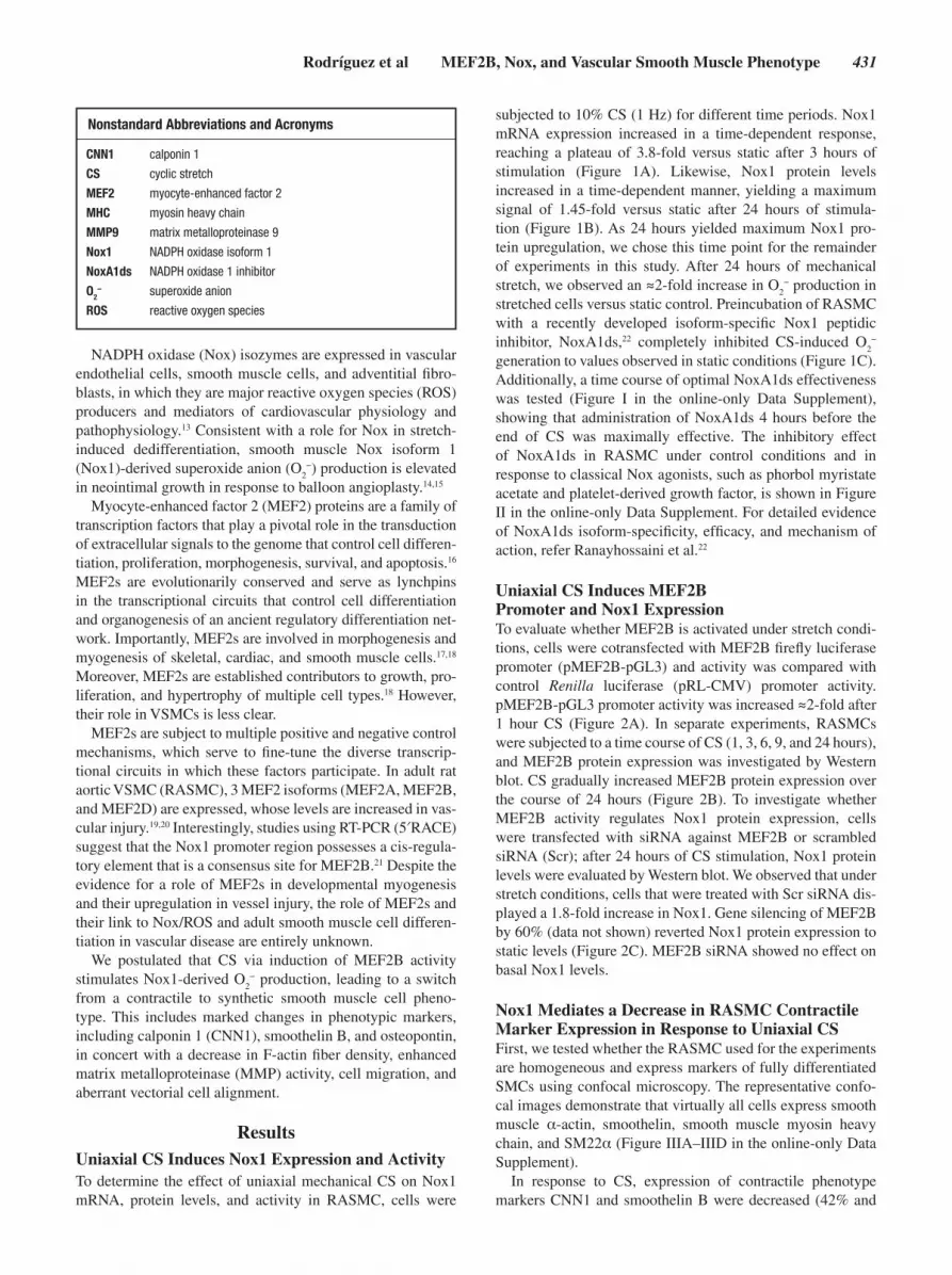
Uniaxial CS Induces MEF2B Promoter and Nox1 ExpressionTo evaluate whether MEF2B is activated under stretch condi-tions, cells were cotransfected with MEF2B firefly luciferase promoter (pMEF2B-pGL3) and activity was compared with control Renilla luciferase (pRL-CMV) promoter activity. pMEF2B-pGL3 promoter activity was increased ≈2-fold after 1 hour CS (Figure 2A). In separate experiments, RASMCs were subjected to a time course of CS (1, 3, 6, 9, and 24 hours), and MEF2B protein expression was investigated by Western blot. CS gradually increased MEF2B protein expression over the course of 24 hours (Figure 2B). To investigate whether MEF2B activity regulates Nox1 protein expression, cells were transfected with siRNA against MEF2B or scrambled siRNA (Scr); after 24 hours of CS stimulation, Nox1 protein levels were evaluated by Western blot. We observed that under stretch conditions, cells that were treated with Scr siRNA dis-played a 1.8-fold increase in Nox1. Gene silencing of MEF2B by 60% (data not shown) reverted Nox1 protein expression to static levels (Figure 2C). MEF2B siRNA showed no effect on basal Nox1 levels.
Nox1 Mediates a Decrease in RASMC Contractile Marker Expression in Response to Uniaxial CSFirst, we tested whether the RASMC used for the experiments are homogeneous and express markers of fully differentiated SMCs using confocal microscopy. The representative confo-cal images demonstrate that virtually all cells express smooth muscle α-actin, smoothelin, smooth muscle myosin heavy chain, and SM22α (Figure IIIA–IIID in the online-only Data Supplement).
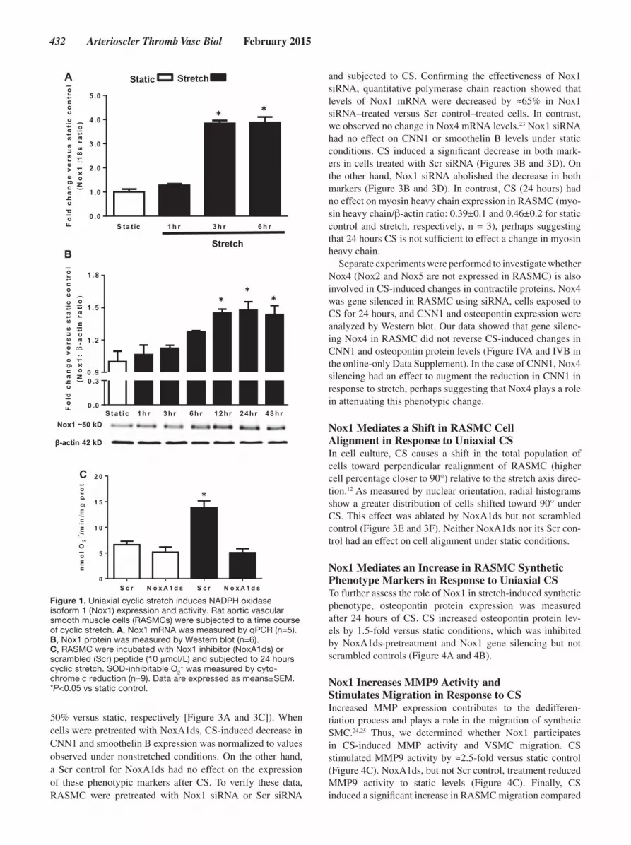
In response to CS, expression of contractile phenotype markers CNN1 and smoothelin B were decreased (42% and
Nonstandard Abbreviations and Acronyms
CNN1 calponin 1
CS cyclic stretch
MEF2 myocyte-enhanced factor 2
MHC myosin heavy chain
MMP9 matrix metalloproteinase 9
Nox1 NADPH oxidase isoform 1
NoxA1ds NADPH oxidase 1 inhibitor
O2− superoxide anion
ROS reactive oxygen species
432 Arterioscler Thromb Vasc Biol February 2015
50% versus static, respectively [Figure 3A and 3C]). When cells were pretreated with NoxA1ds, CS-induced decrease in CNN1 and smoothelin B expression was normalized to values observed under nonstretched conditions. On the other hand, a Scr control for NoxA1ds had no effect on the expression of these phenotypic markers after CS. To verify these data, RASMC were pretreated with Nox1 siRNA or Scr siRNA
and subjected to CS. Confirming the effectiveness of Nox1 siRNA, quantitative polymerase chain reaction showed that levels of Nox1 mRNA were decreased by ≈65% in Nox1 siRNA–treated versus Scr control–treated cells. In contrast, we observed no change in Nox4 mRNA levels.23 Nox1 siRNA had no effect on CNN1 or smoothelin B levels under static conditions. CS induced a significant decrease in both mark-ers in cells treated with Scr siRNA (Figures 3B and 3D). On the other hand, Nox1 siRNA abolished the decrease in both markers (Figure 3B and 3D). In contrast, CS (24 hours) had no effect on myosin heavy chain expression in RASMC (myo-sin heavy chain/β-actin ratio: 0.39±0.1 and 0.46±0.2 for static control and stretch, respectively, n = 3), perhaps suggesting that 24 hours CS is not sufficient to effect a change in myosin heavy chain.
Separate experiments were performed to investigate whether Nox4 (Nox2 and Nox5 are not expressed in RASMC) is also involved in CS-induced changes in contractile proteins. Nox4 was gene silenced in RASMC using siRNA, cells exposed to CS for 24 hours, and CNN1 and osteopontin expression were analyzed by Western blot. Our data showed that gene silenc-ing Nox4 in RASMC did not reverse CS-induced changes in CNN1 and osteopontin protein levels (Figure IVA and IVB in the online-only Data Supplement). In the case of CNN1, Nox4 silencing had an effect to augment the reduction in CNN1 in response to stretch, perhaps suggesting that Nox4 plays a role in attenuating this phenotypic change.
Nox1 Mediates a Shift in RASMC Cell Alignment in Response to Uniaxial CSIn cell culture, CS causes a shift in the total population of cells toward perpendicular realignment of RASMC (higher cell percentage closer to 90°) relative to the stretch axis direc-tion.12 As measured by nuclear orientation, radial histograms show a greater distribution of cells shifted toward 90° under CS. This effect was ablated by NoxA1ds but not scrambled control (Figure 3E and 3F). Neither NoxA1ds nor its Scr con-trol had an effect on cell alignment under static conditions.
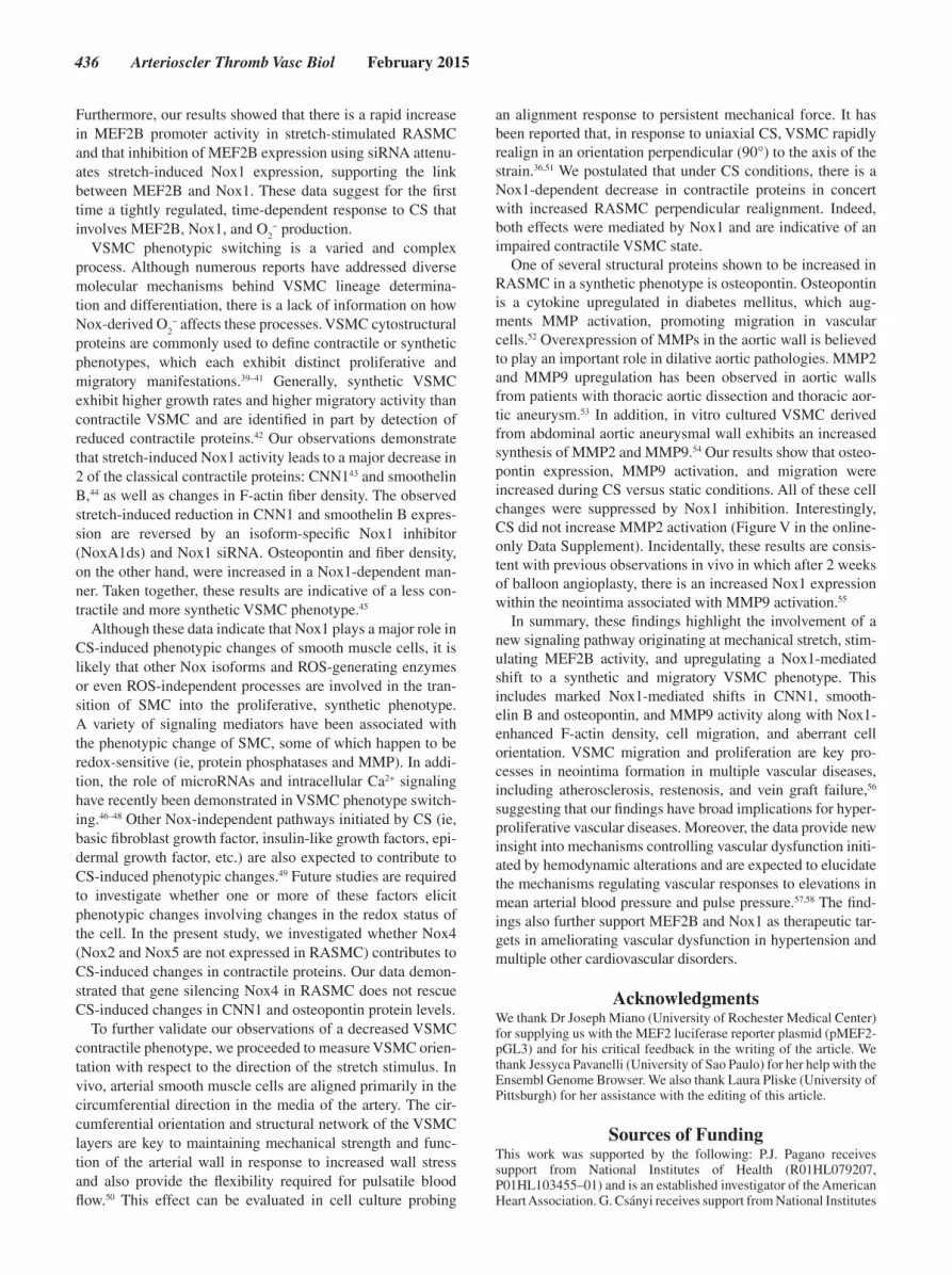
Nox1 Mediates an Increase in RASMC Synthetic Phenotype Markers in Response to Uniaxial CSTo further assess the role of Nox1 in stretch-induced synthetic phenotype, osteopontin protein expression was measured after 24 hours of CS. CS increased osteopontin protein lev-els by 1.5-fold versus static conditions, which was inhibited by NoxA1ds-pretreatment and Nox1 gene silencing but not scrambled controls (Figure 4A and 4B).
Nox1 Increases MMP9 Activity and Stimulates Migration in Response to CSIncreased MMP expression contributes to the dedifferen-tiation process and plays a role in the migration of synthetic SMC.24,25 Thus, we determined whether Nox1 participates in CS-induced MMP activity and VSMC migration. CS stimulated MMP9 activity by ≈2.5-fold versus static control (Figure 4C). NoxA1ds, but not Scr control, treatment reduced MMP9 activity to static levels (Figure 4C). Finally, CS induced a significant increase in RASMC migration compared
0 .9
Sta t ic 1hr 3hr 6hr 12hr 24hr 48hr
1 .2
1 .5
1 .8
βA
B
C
Figure 1. Uniaxial cyclic stretch induces NADPH oxidase isoform 1 (Nox1) expression and activity. Rat aortic vascular smooth muscle cells (RASMCs) were subjected to a time course of cyclic stretch. A, Nox1 mRNA was measured by qPCR (n=5). B, Nox1 protein was measured by Western blot (n=6). C, RASMC were incubated with Nox1 inhibitor (NoxA1ds) or scrambled (Scr) peptide (10 μmol/L) and subjected to 24 hours cyclic stretch. SOD-inhibitable O2
− was measured by cyto-chrome c reduction (n=9). Data are expressed as means±SEM. *P<0.05 vs static control.

Rodríguez et al MEF2B, Nox, and Vascular Smooth Muscle Phenotype 433
with static, which was reduced after pharmacological inhibi-tion of Nox1 (Figure 4D and 4E).
Finally, we tested whether CS induces changes in the archi-tecture of F-actin network in SMC. CS significantly increased the density of actin filaments in SMC in a Nox1-dependent manner (Figure 4F). In contrast, quantitative analysis of fiber thickness showed no significant actin thickening in response to CS.
DiscussionThe present study illustrates for the first time that uniaxial stretch-induced phenotypic transitioning of vascular smooth muscle cells from a contractile to synthetic phenotype, as detected by changes in cytoskeletal proteins, F-actin density, MMP9 activity, cell migration, and cell orientation, is medi-ated by an early increase in MEF2B transcription and protein levels, upregulation of Nox1 expression, and increased Nox1-derived O
2− production. These novel findings indicate that
MEF2B to Nox1 signaling causing alterations in cytoskeletal proteins and MMP9 activation are pivotal for the synthetic and hyperproliferative/promigratory VSMCs in response to CS.
Blood vessels are continuously subjected to hemody-namic mechanical forces, including CS and shear stress, and these forces are highly dependent on the fluid dynamics of the blood, in particular, flow and viscosity.26 Increases in any of these conditions concomitantly lead to increased CS and shear stress. Under physiological conditions, as in early vas-cular development, blood pressure and thus mechanical stress in the arterial wall regulate critical parameters of vascular function and maintain the balance between blood supply and
tissue oxygen demand.27 In contrast to these physiological processes, sustained or chronic elevations in blood pressure and flow lead to phenotypic changes of the vascular wall and vascular remodeling.28
NADPH oxidases (Noxes) are well established as major sources of ROS in the vasculature, as well as significant contributors to vascular pathologies, including neointima formation.13,29–32 A previous study demonstrated that p22phox–expressing smooth muscle cells in the neointima, that have greater capability to produce ROS, are positive for SMemb but not for SM2, suggesting that ROS-producing cells could possess a synthetic rather than a contractile phenotype.33 The manner and means by which MEF-induced Nox-derived ROS induces a molecular shift away from contractile pro-teins and activity to a highly proliferative and synthetic phe-notype are unknown.
We observed that uniaxial CS (10%, 1 Hz, conditions mim-icking hypertension in humans)9 increased Nox1 mRNA, protein expression, and activity in smooth muscle cells. Our findings illustrate an early upregulation of Nox1 mRNA at 3 to 4 hours of stretch followed by increased protein expres-sion at 12 to 24 hours. A previous report suggested that Nox1 can be upregulated in smooth muscle cells under in vitro CS conditions.34 Our data confirm this finding and go further in demonstrating a rise in Nox1 mRNA, protein expression, and specific activity in response to CS. On establishing optimal conditions for Nox1 upregulation, we measured O
2− produc-
tion (via cyt c reduction assay) and observed a robust increase in SOD-inhibitable O
2− production after 24 hours of CS.
Previous reports suggested that CS induces O2
− production12,35
Figure 2. Uniaxial cyclic stretch induces myocyte-enhanced factor 2 (MEF2B) promoter activity, MEF2B protein expression, and MEF2B-dependent NADPH oxidase isoform 1 (Nox1) expression. A, Rat aortic vascular smooth muscle cells (RASMCs) were subjected to a time course of cyclic stretch, and MEF2B promoter activity was mea-sured by dual firefly luciferase reporter assay, using MEF2B firefly luciferase reporter and a control reporter expressing Renilla luciferase (n=6). B, RASMCs were subjected to a time course of cyclic stretch, and MEF2B protein expression was investigated by Western blot (n=4). C, RASMCs were pretreated with MEF2B siRNA or scrambled (Scr) siRNA (48 hours), and subjected to 24 hours cyclic stretch. Nox1 protein expression was mea-sured by Western blot (n=6). Data are expressed as means±SEM. *P<0.05 vs vehicle or static condition.

434 Arterioscler Thromb Vasc Biol February 2015
or implicated a CS-induced O2
− effect on transduction path-ways using broad-range flavoprotein inhibitor DPI36 or apocy-nin.37 Nevertheless, those data pointed to a role for O
2− in CS.
Our findings are the first to our knowledge to identify the func-tional involvement of Nox1, or any Nox for that matter, in the stretch response of VSMC (using 2 approaches: gene silenc-ing and pharmacological inhibition using an isoform-specific Nox1 inhibitor, NoxA1ds).22 Previous work in our laboratory confirmed siRNA efficacy to inhibit Nox1 by 60% to 70% in
this assay.38 Moreover, NoxA1ds was able to completely blunt Nox1 activity in RASMC. Taken together, these data strongly support that mimicking hypertensive conditions via CS causes an increase in Nox1 expression and Nox1-derived O
2− produc-
tion. The time frame of our results suggest a tightly regulated signaling role for Nox1.
Accumulating evidence suggests that the family of MEF2 transcription factors is involved in VSMC differentiation and disease.3,17,19 It has been proposed that Nox1 promoter region
Figure 3. NADPH oxidase isoform 1 (Nox1) mediates a decreased contractile phenotype in response to uniaxial cyclic stretch. Rat aortic vascular smooth muscle cells (RASMCs) were subjected for 24 hours to stretch or static conditions. A and B, Calponin 1 (CNN1) protein expression measured by Western blot (n=4). C and D, Smoothelin B protein expression measured by Western blot (n=4). E, Cell nuclear perpendicular alignment vs the direction of the stretch vector represented by radial histograms (n=5, over 130 cells). F, Summary of the average alignment angles (n=5 over 130 cells). A, C, E, and F, Cells were pretreated with NADPH oxidase 1 inhibitor (NoxA1ds) or scram-bled peptide (10 μmol/L). B and D, Cells were pretreated with Nox1 siRNA or scrambled (Scr) siRNA. Data are expressed as means±SEM. *P<0.05 vs static control.

Rodríguez et al MEF2B, Nox, and Vascular Smooth Muscle Phenotype 435
Figure 4. NADPH oxidase isoform 1 (Nox1)–mediated increase in synthetic phenotype in response to uniaxial cyclic stretch. Rat aortic vas-cular smooth muscle cells (RASMCs) were subjected for 24 hours to stretch or static conditions. A and B, Osteopontin (OPN) was measured by Western blot (n=4). C, Extracellular matrix metalloproteinase 9 (MMP9) activity was measured by zymography (n=5). D and E, Cell migra-tion was measured by wound healing assay (n=8). F, Uniaxial cyclic stretch increases actin fiber density via Nox1. Fluorescence microscopy images of phalloidin-stained RASMC. Fiber density was assessed using ImageJ software and with the aid of the Hessian matrix plug-in. The graph depicts mean values of 8 cells from 4 independent experiments. The green bars represent 20 μm. A, C, D, E, and F, Cells were treated with NADPH oxidase 1 inhibitor (NoxA1ds) or scrambled (Scr) peptide (10 μmol/L). B, Cells were pretreated with Nox1 siRNA or scrambled siRNA (5 pmol/mL) for 48 hours. Data are expressed as means±SEM. *P<0.05 vs static control. #P<0.05 vs stretch scrmb.
possesses a cis-regulatory element that is a consensus site for MEF2B binding.21 To corroborate the existence of this MEF2B regulatory element in the Nox1 gene, we evaluated the Nox1 promoter region using Ensembl Genome Browser
and found a consensus sequence for MEF2B binding located at −438 bp upstream of the Nox1 transcription initiation codon (Figure VI in the online-only Data Supplement), dem-onstrating a putative association between MEF2B and Nox1.
436 Arterioscler Thromb Vasc Biol February 2015
Furthermore, our results showed that there is a rapid increase in MEF2B promoter activity in stretch-stimulated RASMC and that inhibition of MEF2B expression using siRNA attenu-ates stretch-induced Nox1 expression, supporting the link between MEF2B and Nox1. These data suggest for the first time a tightly regulated, time-dependent response to CS that involves MEF2B, Nox1, and O
2− production.
VSMC phenotypic switching is a varied and complex process. Although numerous reports have addressed diverse molecular mechanisms behind VSMC lineage determina-tion and differentiation, there is a lack of information on how Nox-derived O
2− affects these processes. VSMC cytostructural
proteins are commonly used to define contractile or synthetic phenotypes, which each exhibit distinct proliferative and migratory manifestations.39–41 Generally, synthetic VSMC exhibit higher growth rates and higher migratory activity than contractile VSMC and are identified in part by detection of reduced contractile proteins.42 Our observations demonstrate that stretch-induced Nox1 activity leads to a major decrease in 2 of the classical contractile proteins: CNN143 and smoothelin B,44 as well as changes in F-actin fiber density. The observed stretch-induced reduction in CNN1 and smoothelin B expres-sion are reversed by an isoform-specific Nox1 inhibitor (NoxA1ds) and Nox1 siRNA. Osteopontin and fiber density, on the other hand, were increased in a Nox1-dependent man-ner. Taken together, these results are indicative of a less con-tractile and more synthetic VSMC phenotype.45
Although these data indicate that Nox1 plays a major role in CS-induced phenotypic changes of smooth muscle cells, it is likely that other Nox isoforms and ROS-generating enzymes or even ROS-independent processes are involved in the tran-sition of SMC into the proliferative, synthetic phenotype. A variety of signaling mediators have been associated with the phenotypic change of SMC, some of which happen to be redox-sensitive (ie, protein phosphatases and MMP). In addi-tion, the role of microRNAs and intracellular Ca2+ signaling have recently been demonstrated in VSMC phenotype switch-ing.46–48 Other Nox-independent pathways initiated by CS (ie, basic fibroblast growth factor, insulin-like growth factors, epi-dermal growth factor, etc.) are also expected to contribute to CS-induced phenotypic changes.49 Future studies are required to investigate whether one or more of these factors elicit phenotypic changes involving changes in the redox status of the cell. In the present study, we investigated whether Nox4 (Nox2 and Nox5 are not expressed in RASMC) contributes to CS-induced changes in contractile proteins. Our data demon-strated that gene silencing Nox4 in RASMC does not rescue CS-induced changes in CNN1 and osteopontin protein levels.
To further validate our observations of a decreased VSMC contractile phenotype, we proceeded to measure VSMC orien-tation with respect to the direction of the stretch stimulus. In vivo, arterial smooth muscle cells are aligned primarily in the circumferential direction in the media of the artery. The cir-cumferential orientation and structural network of the VSMC layers are key to maintaining mechanical strength and func-tion of the arterial wall in response to increased wall stress and also provide the flexibility required for pulsatile blood flow.50 This effect can be evaluated in cell culture probing
an alignment response to persistent mechanical force. It has been reported that, in response to uniaxial CS, VSMC rapidly realign in an orientation perpendicular (90°) to the axis of the strain.36,51 We postulated that under CS conditions, there is a Nox1-dependent decrease in contractile proteins in concert with increased RASMC perpendicular realignment. Indeed, both effects were mediated by Nox1 and are indicative of an impaired contractile VSMC state.
One of several structural proteins shown to be increased in RASMC in a synthetic phenotype is osteopontin. Osteopontin is a cytokine upregulated in diabetes mellitus, which aug-ments MMP activation, promoting migration in vascular cells.52 Overexpression of MMPs in the aortic wall is believed to play an important role in dilative aortic pathologies. MMP2 and MMP9 upregulation has been observed in aortic walls from patients with thoracic aortic dissection and thoracic aor-tic aneurysm.53 In addition, in vitro cultured VSMC derived from abdominal aortic aneurysmal wall exhibits an increased synthesis of MMP2 and MMP9.54 Our results show that osteo-pontin expression, MMP9 activation, and migration were increased during CS versus static conditions. All of these cell changes were suppressed by Nox1 inhibition. Interestingly, CS did not increase MMP2 activation (Figure V in the online-only Data Supplement). Incidentally, these results are consis-tent with previous observations in vivo in which after 2 weeks of balloon angioplasty, there is an increased Nox1 expression within the neointima associated with MMP9 activation.55
In summary, these findings highlight the involvement of a new signaling pathway originating at mechanical stretch, stim-ulating MEF2B activity, and upregulating a Nox1-mediated shift to a synthetic and migratory VSMC phenotype. This includes marked Nox1-mediated shifts in CNN1, smooth-elin B and osteopontin, and MMP9 activity along with Nox1-enhanced F-actin density, cell migration, and aberrant cell orientation. VSMC migration and proliferation are key pro-cesses in neointima formation in multiple vascular diseases, including atherosclerosis, restenosis, and vein graft failure,56 suggesting that our findings have broad implications for hyper-proliferative vascular diseases. Moreover, the data provide new insight into mechanisms controlling vascular dysfunction initi-ated by hemodynamic alterations and are expected to elucidate the mechanisms regulating vascular responses to elevations in mean arterial blood pressure and pulse pressure.57,58 The find-ings also further support MEF2B and Nox1 as therapeutic tar-gets in ameliorating vascular dysfunction in hypertension and multiple other cardiovascular disorders.
AcknowledgmentsWe thank Dr Joseph Miano (University of Rochester Medical Center) for supplying us with the MEF2 luciferase reporter plasmid (pMEF2-pGL3) and for his critical feedback in the writing of the article. We thank Jessyca Pavanelli (University of Sao Paulo) for her help with the Ensembl Genome Browser. We also thank Laura Pliske (University of Pittsburgh) for her assistance with the editing of this article.
Sources of FundingThis work was supported by the following: P.J. Pagano receives support from National Institutes of Health (R01HL079207, P01HL103455–01) and is an established investigator of the American Heart Association. G. Csányi receives support from National Institutes

Rodríguez et al MEF2B, Nox, and Vascular Smooth Muscle Phenotype 437
of Health (K99HL114648). All Vascular Medicine Institute investiga-tors receive support from the Institute for Transfusion Medicine and the Hemophilia Center of Western Pennsylvania, PA.
DisclosuresNone.
References 1. Fisher SA. Vascular smooth muscle phenotypic diversity and
function. Physiol Genomics. 2010;42A:169–187. doi: 10.1152/physiolgenomics.00111.2010.
2. Ogut O, Brozovich FV. Regulation of force in vascular smooth muscle. J Mol Cell Cardiol. 2003;35:347–355.
3. Owens GK, Kumar MS, Wamhoff BR. Molecular regulation of vascular smooth muscle cell differentiation in development and disease. Physiol Rev. 2004;84:767–801. doi: 10.1152/physrev.00041.2003.
4. Miano JM. Mammalian smooth muscle differentiation: origins, markers and transcriptional control. Results Probl Cell Differ. 2002;38:39–59.
5. Spin JM, Maegdefessel L, Tsao PS. Vascular smooth muscle cell phe-notypic plasticity: focus on chromatin remodelling. Cardiovasc Res. 2012;95:147–155. doi: 10.1093/cvr/cvs098.
6. Alexander MR, Owens GK. Epigenetic control of smooth muscle cell differentiation and phenotypic switching in vascular develop-ment and disease. Annu Rev Physiol. 2012;74:13–40. doi: 10.1146/annurev-physiol-012110-142315.
7. Gomez D, Owens GK. Smooth muscle cell phenotypic switching in ath-erosclerosis. Cardiovasc Res. 2012;95:156–164. doi: 10.1093/cvr/cvs115.
8. Davis-Dusenbery BN, Wu C, Hata A. Micromanaging vascular smooth muscle cell differentiation and phenotypic modulation. Arterioscler Thromb Vasc Biol. 2011;31:2370–2377. doi: 10.1161/ATVBAHA.111.226670.
9. Maul TM, Chew DW, Nieponice A, Vorp DA. Mechanical stimuli differ-entially control stem cell behavior: morphology, proliferation, and differ-entiation. Biomech Model Mechanobiol. 2011;10:939–953. doi: 10.1007/s10237-010-0285-8.
10. Shyu KG. Cellular and molecular effects of mechanical stretch on vascu-lar cells and cardiac myocytes. Clin Sci (Lond). 2009;116:377–389. doi: 10.1042/CS20080163.
11. Shaw A, Xu Q. Biomechanical stress-induced signaling in smooth muscle cells: an update. Curr Vasc Pharmacol. 2003;1:41–58.
12. Zhu JH, Chen CL, Flavahan S, Harr J, Su B, Flavahan NA. Cyclic stretch stimulates vascular smooth muscle cell alignment by redox-dependent activation of Notch3. Am J Physiol Heart Circ Physiol. 2011;300:H1770–H1780. doi: 10.1152/ajpheart.00535.2010.
13. Al Ghouleh I, Khoo NK, Knaus UG, et al. Oxidases and peroxidases in cardiovascular and lung disease: new concepts in reactive oxygen spe-cies signaling. Free Radic Biol Med. 2011;51:1271–1288. doi: 10.1016/j.freeradbiomed.2011.06.011.
14. Szocs K, Lassegue B, Sorescu D, Hilenski LL, Valppu L, Couse TL, Wilcox JN, Quinn MT, Lambeth JD, Griendling KK. Upregulation of nox-based nad(p)h oxidases in restenosis after carotid injury. Arterioscler Thromb Vasc Biol. 2002;22:21–27.
15. Lassègue B, San Martín A, Griendling KK. Biochemistry, physiology, and pathophysiology of NADPH oxidases in the cardiovascular system. Circ Res. 2012;110:1364–1390. doi: 10.1161/CIRCRESAHA.111.243972.
16. Katoh Y, Molkentin JD, Dave V, Olson EN, Periasamy M. MEF2B is a component of a smooth muscle-specific complex that binds an A/T-rich element important for smooth muscle myosin heavy chain gene expres-sion. J Biol Chem. 1998;273:1511–1518.
17. Black BL, Olson EN. Transcriptional control of muscle development by myocyte enhancer factor-2 (MEF2) proteins. Annu Rev Cell Dev Biol. 1998;14:167–196. doi: 10.1146/annurev.cellbio.14.1.167.
18. Potthoff MJ, Olson EN. MEF2: a central regulator of diverse develop-mental programs. Development. 2007;134:4131–4140. doi: 10.1242/dev.008367.
19. Firulli AB, Miano JM, Bi W, Johnson AD, Casscells W, Olson EN, Schwarz JJ. Myocyte enhancer binding factor-2 expression and activity in vascular smooth muscle cells. Association with the activated phenotype. Circ Res. 1996;78:196–204.
20. Martin JF, Miano JM, Hustad CM, Copeland NG, Jenkins NA, Olson EN. A Mef2 gene that generates a muscle-specific isoform via alternative mRNA splicing. Mol Cell Biol. 1994;14:1647–1656.
21. Katsuyama M, Ozgur Cevik M, Arakawa N, Kakehi T, Nishinaka T, Iwata K, Ibi M, Matsuno K, Yabe-Nishimura C. Myocyte enhancer factor 2B is
involved in the inducible expression of NOX1/NADPH oxidase, a vas-cular superoxide-producing enzyme. FEBS J. 2007;274:5128–5136. doi: 10.1111/j.1742-4658.2007.06034.x.
22. Ranayhossaini DJ, Rodriguez AI, Sahoo S, Chen BB, Mallampalli RK, Kelley EE, Csanyi G, Gladwin MT, Romero G, Pagano PJ. Selective reca-pitulation of conserved and nonconserved regions of putative NOXA1 protein activation domain confers isoform-specific inhibition of Nox1 oxidase and attenuation of endothelial cell migration. J Biol Chem. 2013;288:36437–36450. doi: 10.1074/jbc.M113.521344.
23. Al Ghouleh I, Frazziano G, Rodriguez AI, Csányi G, Maniar S, St Croix CM, Kelley EE, Egaña LA, Song GJ, Bisello A, Lee YJ, Pagano PJ. Aquaporin 1, Nox1, and Ask1 mediate oxidant-induced smooth muscle cell hypertrophy. Cardiovasc Res. 2013;97:134–142. doi: 10.1093/cvr/cvs295.
24. Filippov S, Koenig GC, Chun TH, Hotary KB, Ota I, Bugge TH, Roberts JD, Fay WP, Birkedal-Hansen H, Holmbeck K, Sabeh F, Allen ED, Weiss SJ. MT1-matrix metalloproteinase directs arterial wall invasion and neointima formation by vascular smooth muscle cells. J Exp Med. 2005;202:663–671. doi: 10.1084/jem.20050607.
25. Lehti K, Rose NF, Valavaara S, Weiss SJ, Keski-Oja J. MT1-MMP pro-motes vascular smooth muscle dedifferentiation through LRP1 process-ing. J Cell Sci. 2009;122(Pt 1):126–135. doi: 10.1242/jcs.035279.
26. Kirby BJ. Micro- and Nanoscale Fluid Mechanics: Transport in Microfluidic Devices. Cambridge University Press: Cambridge, United Kingdom; 2010.
27. Mongardon N, Dyson A, Singer M. Pharmacological optimization of tis-sue perfusion. Br J Anaesth. 2009;103:82–88. doi: 10.1093/bja/aep135.
28. Levy BI, Schiffrin EL, Mourad JJ, Agostini D, Vicaut E, Safar ME, Struijker-Boudier HA. Impaired tissue perfusion: a pathology common to hypertension, obesity, and diabetes mellitus. Circulation. 2008;118:968–976. doi: 10.1161/CIRCULATIONAHA.107.763730.
29. Ardanaz N, Pagano PJ. Hydrogen peroxide as a paracrine vascular mediator: regulation and signaling leading to dysfunction. Exp Biol Med (Maywood). 2006;231:237–251.
30. Cascino T, Csanyi G, Al Ghouleh I, Montezano AC, Touyz RM, Haurani MJ, Pagano PJ. Adventitia-derived hydrogen peroxide impairs relaxation of the rat carotid artery via smooth muscle cell p38 mitogen-activated protein kinase. Antioxid Redox Signal. 2011;15:1507–1515. doi: 10.1089/ars.2010.3631.
31. Lassègue B, Clempus RE. Vascular NAD(P)H oxidases: specific features, expression, and regulation. Am J Physiol Regul Integr Comp Physiol. 2003;285:R277–R297. doi: 10.1152/ajpregu.00758.2002.
32. Sedeek M, Hébert RL, Kennedy CR, Burns KD, Touyz RM. Molecular mechanisms of hypertension: role of Nox family NADPH oxidases. Curr Opin Nephrol Hypertens. 2009;18:122–127. doi: 10.1097/MNH.0b013e32832923c3.
33. Azumi H, Inoue N, Takeshita S, Rikitake Y, Kawashima S, Hayashi Y, Itoh H, Yokoyama M. Expression of NADH/NADPH oxidase p22phox in human coronary arteries. Circulation. 1999;100:1494–1498.
34. Ohmine T, Miwa Y, Takahashi-Yanaga F, Morimoto S, Maehara Y, Sasaguri T. The involvement of aldosterone in cyclic stretch-mediated activation of NADPH oxidase in vascular smooth muscle cells. Hypertens Res. 2009;32:690–699. doi: 10.1038/hr.2009.76.
35. Wang BW, Chang H, Shyu KG. Regulation of resistin by cyclic mechani-cal stretch in cultured rat vascular smooth muscle cells. Clin Sci (Lond). 2010;118:221–230.
36. Chen Q, Li W, Quan Z, Sumpio BE. Modulation of vascular smooth mus-cle cell alignment by cyclic strain is dependent on reactive oxygen species and P38 mitogen-activated protein kinase. J Vasc Surg. 2003;37:660–668. doi: 10.1067/mva.2003.95.
37. Paravicini TM, Montezano AC, Yusuf H, Touyz RM. Activation of vascu-lar p38MAPK by mechanical stretch is independent of c-Src and NADPH oxidase: influence of hypertension and angiotensin II. J Am Soc Hypertens. 2012;6:169–178. doi: 10.1016/j.jash.2012.01.002.
38. Csányi G, Yao M, Rodríguez AI, Al Ghouleh I, Sharifi-Sanjani M, Frazziano G, Huang X, Kelley EE, Isenberg JS, Pagano PJ. Thrombospondin-1 regu-lates blood flow via CD47 receptor-mediated activation of NADPH oxi-dase 1. Arterioscler Thromb Vasc Biol. 2012;32:2966–2973. doi: 10.1161/ATVBAHA.112.300031.
39. Wang L, Zhang J, Fu W, Guo D, Jiang J, Wang Y. Association of smooth muscle cell phenotypes with extracellular matrix disorders in thoracic aor-tic dissection. J Vasc Surg. 2012;56:1698–709, 1709.e1. doi: 10.1016/j.jvs.2012.05.084.
40. Wang L, Zheng J, Du Y, Huang Y, Li J, Liu B, Liu CJ, Zhu Y, Gao Y, Xu Q, Kong W, Wang X. Cartilage oligomeric matrix protein maintains the contractile phenotype of vascular smooth muscle cells by interacting

438 Arterioscler Thromb Vasc Biol February 2015
with alpha(7)beta(1) integrin. Circ Res. 2010;106:514–525. doi: 10.1161/CIRCRESAHA.109.202762.
41. Tsai MC, Chen L, Zhou J, Tang Z, Hsu TF, Wang Y, Shih YT, Peng HH, Wang N, Guan Y, Chien S, Chiu JJ. Shear stress induces synthetic-to-contractile phenotypic modulation in smooth muscle cells via peroxisome proliferator-activated receptor alpha/delta activations by prostacyclin released by sheared endothelial cells. Circ Res. 2009;105:471–480. doi: 10.1161/CIRCRESAHA.109.193656.
42. Rensen SS, Doevendans PA, van Eys GJ. Regulation and characteris-tics of vascular smooth muscle cell phenotypic diversity. Neth Heart J. 2007;15:100–108.
43. Nobrega MA, Shiozawa M, Koike G, Jacob HJ, Miano JM. Gene struc-ture and chromosomal mapping of the rat smooth muscle calponin gene. Mamm Genome. 2000;11:115–119.
44. Rensen SS, Niessen PM, Long X, Doevendans PA, Miano JM, van Eys GJ. Contribution of serum response factor and myocardin to transcrip-tional regulation of smoothelins. Cardiovasc Res. 2006;70:136–145. doi: 10.1016/j.cardiores.2005.12.018.
45. Christen T, Bochaton-Piallat ML, Neuville P, Rensen S, Redard M, van Eys G, Gabbiani G. Cultured porcine coronary artery smooth muscle cells. A new model with advanced differentiation. Circ Res. 1999;85: 99–107.
46. Rangrez AY, Massy ZA, Metzinger-Le Meuth V, Metzinger L. miR-143 and miR-145: molecular keys to switch the phenotype of vascular smooth muscle cells. Circ Cardiovasc Genet. 2011;4:197–205. doi: 10.1161/CIRCGENETICS.110.958702.
47. Boettger T, Beetz N, Kostin S, Schneider J, Krüger M, Hein L, Braun T. Acquisition of the contractile phenotype by murine arterial smooth muscle cells depends on the Mir143/145 gene cluster. J Clin Invest. 2009;119:2634–2647. doi: 10.1172/JCI38864.
48. Matchkov VV, Kudryavtseva O, Aalkjaer C. Intracellular Ca²+ signalling and phenotype of vascular smooth muscle cells. Basic Clin Pharmacol Toxicol. 2012;110:42–48. doi: 10.1111/j.1742-7843.2011.00818.x.
49. Beamish JA, He P, Kottke-Marchant K, Marchant RE. Molecular regula-tion of contractile smooth muscle cell phenotype: implications for vas-cular tissue engineering. Tissue Eng Part B Rev. 2010;16:467–491. doi: 10.1089/ten.TEB.2009.0630.
50. Jacobsen JC, Holstein-Rathlou NH. A life under pressure: circumfer-ential stress in the microvascular wall. Basic Clin Pharmacol Toxicol. 2012;110:26–34. doi: 10.1111/j.1742-7843.2011.00796.x.
51. Liu B, Qu MJ, Qin KR, Li H, Li ZK, Shen BR, Jiang ZL. Role of cyclic strain frequency in regulating the alignment of vascular smooth muscle cells in vitro. Biophys J. 2008;94:1497–1507. doi: 10.1529/biophysj.106.098574.
52. Lai CF, Seshadri V, Huang K, Shao JS, Cai J, Vattikuti R, Schumacher A, Loewy AP, Denhardt DT, Rittling SR, Towler DA. An osteopontin-NADPH oxidase signaling cascade promotes pro-matrix metalloproteinase 9 acti-vation in aortic mesenchymal cells. Circ Res. 2006;98:1479–1489. doi: 10.1161/01.RES.0000227550.00426.60.
53. Koullias GJ, Ravichandran P, Korkolis DP, Rimm DL, Elefteriades JA. Increased tissue microarray matrix metalloproteinase expres-sion favors proteolysis in thoracic aortic aneurysms and dissections. Ann Thorac Surg. 2004;78:2106–10; discussion 2110. doi: 10.1016/j.athoracsur.2004.05.088.
54. Patel MI, Melrose J, Ghosh P, Appleberg M. Increased synthesis of matrix metalloproteinases by aortic smooth muscle cells is implicated in the etiopathogenesis of abdominal aortic aneurysms. J Vasc Surg. 1996;24:82–92.
55. Xu S, Shriver AS, Jagadeesha DK, Chamseddine AH, Szőcs K, Weintraub NL, Griendling KK, Bhalla RC, Miller FJ Jr. Increased expression of Nox1 in neointimal smooth muscle cells promotes activation of matrix metallo-proteinase-9. J Vasc Res. 2012;49:242–248. doi: 10.1159/000332958.
56. Singh NK, Kundumani-Sridharan V, Kumar S, Verma SK, Kotla S, Mukai H, Heckle MR, Rao GN. Protein kinase N1 is a novel substrate of NFATc1-mediated cyclin D1-CDK6 activity and modulates vascular smooth muscle cell division and migration leading to inward blood vessel wall remodeling. J Biol Chem. 2012;287:36291–36304. doi: 10.1074/jbc.M112.361220.
57. Chae CU, Lee RT, Rifai N, Ridker PM. Blood pressure and inflammation in apparently healthy men. Hypertension. 2001;38:399–403.
58. Liu S, Li Y, Zhang Z, Xie F, Xu Q, Huang X, Huang J, Li C. α1-Adrenergic receptors mediate combined signals initiated by mechanical stretch stress and norepinephrine leading to accelerated mouse vein graft atherosclerosis. J Vasc Surg. 2013;57:1645–56, 1656.e1. doi: 10.1016/j.jvs.2012.09.061.
This is the first study to establish a link between MEF2B and Nox1 and identify both factors as mediators of vascular smooth muscle cell phe-notypic changes during mechanical stretch. Our findings illustrate that mechanical stretch stimulates MEF2B transcription, leading to Nox1 upregulation and increased reactive species production. The data also demonstrate that under pathological cyclic stretch, Nox1 mediates a vascular smooth muscle cell contractile to synthetic phenotypic switch manifested by alterations in key cytoskeletal proteins, matrix metal-loproteinase 9 activation, disorientation of cell alignment, and augmented cell migration. Furthermore, the data demonstrate the feasibility of a novel isoform-specific inhibitor to ameliorate these phenotypic changes and provide additional proof of its viability as a therapeutic agent in vascular disease (see appendix Ranayhossaini et al).22
Significance
Related Documents