Cellular & Molecular Immunology 317 Review Volume 6 Number 5 October 2009 Modulation of Neuroimmune Responses on Glia in the Central Nervous System: Implication in Therapeutic Intervention against Neuroinflammation Raymond Chuen-Chung Chang 1, 2, 3, 4 , Kin Chiu 1 , Yuen-Shan Ho 1 and Kwok-Fai So 1, 2, 3 It has long been known that the brain is an immunologically privileged site in normal conditions. Although the cascade of immune responses can occur as long as there is a neuronal injury or a potent immune stimulation, how the brain keeps glial cells in a quiescent state is still unclear. Increasing efforts have been made by several laboratories to elucidate how repression of immune responses is achieved in the neuronal environment. The suppression factors include neurotransmitters, neurohormones, neurotrophic factors, anti-inflammatory factors, and cell-cell contact via adhesion molecules or CD200 receptor. This review discusses how these factors affect the cascade of cerebral immune responses because no single factor listed above can fully account for the immune suppression. While several factors contribute to the suppression of immune responses, activation of glial cells and their production of pro-inflammatory factors do occur as long as there is a neuronal injury, suggesting that some neuronal components facilitate immune responses. This review also discusses which signals initiate or augment cerebral immune responses so that stimulatory signals override the suppressive signals. Increasing lines of evidence have demonstrated that immune responses in the brain are not always detrimental to neurons. Attempt to simply clear off inflammatory factors in the CNS may not be appropriate for neurons in neurological disorders. Appropriate control of immune cells in the CNS may be beneficial to neurons or even neuroregeneration. Therefore, understanding the mechanisms underlying immune suppression may help us to reshape pharmacological interventions against inflammation in many neurological disorders. Cellular & Molecular Immunology. 2009;6(5): 317-326. Key Words: neural cell adhesion molecule, neurotrophins, potassium ions, microglia, neuroinflammation Introduction It is now well-known that immune responses or inflammation in the central nervous system (CNS) play important roles in both chronic and acute neurological disorders. Excessive production of pro-inflammatory factors such as tumor necrosis factor (TNF-), interleukin-1 (IL-1), nitric oxide (NO), and superoxide complicate the pathogenesis of neurodegenerative diseases (1). They are important factors determining the fate of neurons and/or contributing to the expansion of injury volume. Although many lines of evidence show the detrimental effects of immune responses in the CNS, increasing lines of new findings demonstrate that immune responses can be beneficial to neurons as long as they occur at the right time and to an appropriate extent (2-5). How can we know when is the appropriate time for immune responses in the CNS to occur after brain injury, so that they are beneficial but not detrimental to neurons? Before we can answer this question, we have to understand how immune responses in the CNS are regulated and modulated by the micro-environment in the CNS, and of how some neuronal components stimulate immune responses to override the repressive signals. Historically, it has been considered that there are few 1 Laboratory of Neurodegenerative Diseases, Department of Anatomy, LKS Faculty of Medicine, University of Hong Kong, Pokfulam, Hong Kong SAR, China; 2 Research Centre of Heart, Brain, Hormone and Healthy Aging, LKS Faculty of Medicine, University of Hong Kong, Pokfulam, Hong Kong SAR, China; 3 State Key Laboratory for Brain and Cognitive Sciences, University of Hong Kong, Pokfulam, Hong Kong SAR, China; Received Sep 28, 2009. Accepted Oct 7, 2009. © 2009 Chinese Society of Immunology and University of Science & Technology of China 4 Correspondence to: Dr. Raymond Chuen-Chung Chang, Department of Anatomy, LKS Faculty of Medicine, The University of Hong Kong, Rm. L1-49 Laboratory Block, Faculty of Medicine Building, 21 Sassoon Road, Pokfulam, Hong Kong SAR, China. Tel: +852-2819-9127; Fax: +852-2817- 0857; E-mail: [email protected]

Welcome message from author
This document is posted to help you gain knowledge. Please leave a comment to let me know what you think about it! Share it to your friends and learn new things together.
Transcript

Cellular & Molecular Immunology 317
Review
Volume 6 Number 5 October 2009
Modulation of Neuroimmune Responses on Glia in the Central Nervous System: Implication in Therapeutic Intervention against Neuroinflammation Raymond Chuen-Chung Chang1, 2, 3, 4, Kin Chiu1, Yuen-Shan Ho1 and Kwok-Fai So1, 2, 3
It has long been known that the brain is an immunologically privileged site in normal conditions. Although the cascade of immune responses can occur as long as there is a neuronal injury or a potent immune stimulation, how the brain keeps glial cells in a quiescent state is still unclear. Increasing efforts have been made by several laboratories to elucidate how repression of immune responses is achieved in the neuronal environment. The suppression factors include neurotransmitters, neurohormones, neurotrophic factors, anti-inflammatory factors, and cell-cell contact via adhesion molecules or CD200 receptor. This review discusses how these factors affect the cascade of cerebral immune responses because no single factor listed above can fully account for the immune suppression. While several factors contribute to the suppression of immune responses, activation of glial cells and their production of pro-inflammatory factors do occur as long as there is a neuronal injury, suggesting that some neuronal components facilitate immune responses. This review also discusses which signals initiate or augment cerebral immune responses so that stimulatory signals override the suppressive signals. Increasing lines of evidence have demonstrated that immune responses in the brain are not always detrimental to neurons. Attempt to simply clear off inflammatory factors in the CNS may not be appropriate for neurons in neurological disorders. Appropriate control of immune cells in the CNS may be beneficial to neurons or even neuroregeneration. Therefore, understanding the mechanisms underlying immune suppression may help us to reshape pharmacological interventions against inflammation in many neurological disorders. Cellular & Molecular Immunology. 2009;6(5): 317-326. Key Words: neural cell adhesion molecule, neurotrophins, potassium ions, microglia, neuroinflammation Introduction It is now well-known that immune responses or inflammation in the central nervous system (CNS) play important roles in both chronic and acute neurological disorders. Excessive production of pro-inflammatory factors such as tumor necrosis factor (TNF-), interleukin-1 (IL-1), nitric oxide (NO), and superoxide complicate the pathogenesis of
neurodegenerative diseases (1). They are important factors determining the fate of neurons and/or contributing to the expansion of injury volume. Although many lines of evidence show the detrimental effects of immune responses in the CNS, increasing lines of new findings demonstrate that immune responses can be beneficial to neurons as long as they occur at the right time and to an appropriate extent (2-5). How can we know when is the appropriate time for immune responses in the CNS to occur after brain injury, so that they are beneficial but not detrimental to neurons? Before we can answer this question, we have to understand how immune responses in the CNS are regulated and modulated by the micro-environment in the CNS, and of how some neuronal components stimulate immune responses to override the repressive signals.
Historically, it has been considered that there are few
1Laboratory of Neurodegenerative Diseases, Department of Anatomy, LKS Faculty of Medicine, University of Hong Kong, Pokfulam, Hong Kong SAR, China;
2Research Centre of Heart, Brain, Hormone and Healthy Aging, LKS Faculty of Medicine, University of Hong Kong, Pokfulam, Hong Kong SAR, China;
3State Key Laboratory for Brain and Cognitive Sciences, University of Hong Kong, Pokfulam, Hong Kong SAR, China;
Received Sep 28, 2009. Accepted Oct 7, 2009. © 2009 Chinese Society of Immunology and University of Science & Technology of China
4Correspondence to: Dr. Raymond Chuen-Chung Chang, Department of Anatomy, LKS Faculty of Medicine, The University of Hong Kong, Rm. L1-49 Laboratory Block, Faculty of Medicine Building, 21 Sassoon Road, Pokfulam, Hong Kong SAR, China. Tel: +852-2819-9127; Fax: +852-2817-0857; E-mail: [email protected]

318 Modulation of Glia for Neuroinflammation
Volume 6 Number 5 October 2009
immune responses in the brain, stating the fact that the brain actively suppresses the occurrence of immune responses. The term ‘immune privilege’ to describe the ignorance of the immune system in the CNS is still partly correct for the normal brain (6). The reasons for ‘immune privilege’ in the CNS are as follow: firstly, the presence of the blood-brain barrier excludes most of the immune system components although activated lymphocytes can infiltrate into the brain (7); secondly, the ability of antigen presentation as well as the expression of the major histocompatibility complex (MHC), an essential molecule for the presentation of antigen to T-lymphocytes, is suppressed in the CNS except in the presence of a potent immune stimulant (8); thirdly, in spite of the infiltration of T lymphocytes into the brain, they eventually undergo apoptosis so that very few T lymphocytes can be found in the brain (9); and fourthly, the production of pro-inflammatory cytokines such as TNF- and IL-1 is also limited so that the complete cycle for activation of infiltrating macrophages and lymphocytes cannot be easily accomplished (10). Taking all these factors into consideration, a question of how immune responses in the brain are modulated by the CNS microenvironment receives increasing attention.
The CNS requires a quiescent environment without any immune responses. Reactions of immune responses or the production of cytokines can disturb the normal functions of neurons including signal transmission between neurons (11, 12). For example, IL-1 and TNF- have been reported to induce fever. Even normal sleep pattern can be affected by the level of TNF-α in the brain (13). TNF- can also affect the uptake or release of neurotransmitters or amino acids (14). These examples indicate that cytokines can disturb normal neuronal functions. To ensure normal neuronal functions or neuronal communication, immune responses and production of cytokines have to be controlled. In this review, we discuss how unique neuronal environment reduces or minimizes the occurrence of immune responses. No single neuronal factor is fully responsible for immune suppression in the CNS. This review will discuss how soluble factors as well as cell-cell contacts exert immune suppression.
Neuronal environment not only suppresses immune responses, but also, in some conditions such as ischemia and traumatic brain injury, facilitates immune responses. In fact, numerous reports have shown the activation of microglia after brain injury. In this review, we also discuss how neuronal environment or components facilitate immune responses and to override suppressive signals in case of brain injury. This article is to review different studies of how unique neuronal environment modulates immune responses in the CNS. An understanding of all the repressive mechanisms can definitely help us to redesign the therapeutic interventions for neurodegenerative diseases, because suppression of immune responses is not always beneficial to neurons in degenerative diseases. Suppressive signals for cerebral immune responses Perry and his co-workers had observed long ago that there
are differences between the inflammatory responses in the CNS and peripheral systems (10). According to these authors, if the bacterial endotoxin lipopolysaccharide (LPS) (20-200 ng/ml) was injected into the hippocampus, activation of microglia could be observed only after 48 h. The delayed responses of microglia are not because microglia has different metabolic properties to macrophages. It has been reported that recruitment of macrophages to injured optic nerve but not sciatic nerve is limited by resident factor (15). Also, when macrophages are incubated with optic nerve in vitro, production of nitric oxide is significantly reduced (16). In addition, only few neutrophils infiltrate into the CNS parenchyma. Apart from this study, Dusart and Schwab showed the restricted inflammatory reaction after dorsal hemisection of rat spinal cord (17). Furthermore, another study showed the injured optic nerve and sciatic nerve also had differential effects on macrophages and microglia (16). All these findings demonstrate that the environment or some factors produced by the cellular elements in the CNS have immune suppressive effects. Neurotransmitters and neurohormones A unique aspect of the environment in the CNS is the abundance of neurotransmitters released by neurons. All brain cells are continuously exposed to neurotransmitters. Therefore, how neurotransmitters affect cerebral immune responses has long been investigated. Early studies from Frohman and co-workers have observed that norepinephrine (NE) could suppress interferon-γ (IFN-γ)-induced MHC class II expression on astrocytes (18). Subsequently, Feinstein’s group has further proved that NE can significantly reduce production of nitric oxide and IL-1β by rat cortical microglia (19). Frohman’s group also found that another neurotransmitter vasoactive intestinal peptide (VIP) could also inhibit the expression of MHC class II antigens on astrocytes (20). It was then proposed that these two neurotransmitters stimulate the synthesis of intracellular cyclic adenosine monophosphate (cAMP) resulting in the inhibition of MHC class II expression (21). In spite of the fact that the concentrations used in these studies are quite high, a report using a drug propentofylline to increase intracellular cAMP has proved the immune suppressive effects of cAMP on microglia (22). Not only NE or VIP, but also glutamate has been demonstrated to suppress MHC class II expression on astroglia (23). Regardless of the non- physiological concentrations used in these experimental models, the series of studies suggest that neurotransmitters are able to modify immune responses in the brain because the expression of MHC class II antigens is important for T lymphocytes to initiate cell-mediated immune responses. Recently, Hong’s laboratory showed that very low concentrations of dynorphin (10-14 - 10-12 M) are sufficient to reduce LPS-stimulated activation of microglia, and in turn the production of pro-inflammatory factors such as NO, superoxide and TNF- in vivo and in vitro studies (24-26). Furthermore, opioid receptor antagonist can be a biological

Cellular & Molecular Immunology 319
Volume 6 Number 5 October 2009
target to reduce free-radicals production (27). Besides neurotransmitters, neurohormones can also exert
immune suppressive effects on microglial cells. For examples, -melanocyte-stimulating hormone has been shown to inhibit the production of TNF- and NO by IFN-- and - amyloid-stimulated microglia (28). Recently, several laboratories have investigated the effects of estrogen on microglial cells. It has been shown that application of 17-estradiol to N9 microglial cells could reduce LPS- and phorbol ester-stimulated superoxide production and phago- cytic activity (29). Other laboratories also showed that 17-estradiol could reduce morphological changes of isolated microglial cells during the activation processes (30). In addition, 17-estradiol could reduce LPS-stimulated NO, prostaglandin-E2 and metalloproteinase-9 production (30). Although the receptors for estrogen, ER and ER, were found to be expressed on microglial cells and effects of 17-estradiol on microglial cells have been shown to be estrogen receptor-mediated because the antagonist ICI 182,780 could block its effects (29, 30), estrogen was also shown to have other effects such as induction of apolipoprotein E (apoE) in brain glia (31). Since apoE was also shown to be an immune suppressant, the immune modulation effects of estrogen on microglial cells may not be limited to the signaling events associated with its receptors.
Taken all the evidence from neurotransmitters and neurohormones together, a wide array of neurotransmitters and hormones can suppress cerebral immune responses on both microglia and astrocytes. A recent report has shown that venlafaxine, an antidepressant for blocking NE-serotonin reuptake system, elicits anti-inflammatory by reducing IL-6 and IFN-γ in a microglia-astrocytes co-cultures (32). This is an example of therapeutic intervention by utilizing our knowledge in modulating immune responses of glial cells. Neurotrophic factors Apart from neurotransmitters, the brain is very rich in neurotrophic factors because both neurons and glial cells synthesize various neurotrophic factors. Neumann and co-workers were among the first to discover that neurons could express MHC class I antigens provided that electrical activity was suppressed by tetrodotoxin (TTX) (33). Furthermore, they found that the expressions of MHC class II on astroglial and microglial cells are inhibited as long as neurons maintain their electrical activity (34). While blockage of neuronal activity with TTX enhanced the expression of MHC class II antigens on microglial cells stimulated with IFN-, direct application of TTX to isolated microglial cells did not affect their MHC class II expression. In contrast, application of neurotrophins or neutralization of neurotrophins with antibodies could modulate the MHC class II expression on isolated microglial cells (8). Therefore, the electrical activity might alter the production of neurotrophins, which in turn affects the expression of MHC class II antigens on microglial cells. Besides MHC class II expression, the expressions of CD40 and the co-stimulatory factor B7-1 on microglial cells for T lymphocytes were found to be reduced
by nerve growth factor (NGF) (35). Recently, trans- endothelial migration of monocytes was also shown to be inhibited in the presence of NGF (36).
Among the different neurotrophins, only NGF and neurotrophin-3, but not brain-derived neurotrophic factor (BDNF), were found to effectively inhibit IFN--induced MHC class II expression on microglial cells (8). Since the low-affinity receptor p75 for neurotrophins is expressed on microglial cells, it was postulated that cellular signaling events associated with p75 mediate the immune suppressive effects of NGF or neurotrophin-3 (8).
Although a p75 signaling pathway has been hypothesized, another report showed different results. Apart from the expression of MHC class II antigens, neurotrophins were also found to reduce LPS-stimulated NO production by microglial cells (37). In this report, only BDNF but, to a less extend, neurotrophin-3 could reduce LPS-stimulated NO production while NGF and neurotrophin-4 did not. Interestingly, these authors demonstrated the presence of TrkA, TrkB, TrkC, and p75 receptors on isolated microglial cells, while the TrkA receptor was not found on microglial cells by Neumann’s laboratory. Taking all these results together, it is possible that the signaling events associated with TrkA or p75 are different and affect only certain types of immune activity of microglial cells, so that different results were obtained from the two laboratories. Anti-inflammatory cytokines and factors normally expressed in the brain One way to exert constant immune suppression is the production of anti-inflammatory cytokines. Early studies demonstrated the production of transforming growth factor- (TGF-) (38-40), an anti-inflammatory cytokine, by gliomas. Microglial cells can express TGF- to function as autocrine or paracrine signals to inhibit the proliferation of the astroglial and microglial cells themselves (41, 42). Consistent with this notion, it has been considered that TGF- is one of the soluble factors produced by astrocytes in a constitutive and inducible manner to reduce the immune responsiveness of microglial cells to LPS (43). When microglial cells were co-cultured with astrocytes, the responsiveness of microglial cells to LPS was reduced in term of the level of inducible NO synthase. From this line of studies, interactions between astroglial and microglial cells already establish the first line of control for cerebral immune responses. Apart from its anti-inflammatory functions, TGF- was also found to induce apoptosis of microglial cells to further deplete the number of microglial cells (44). The presence of anti-inflammatory cytokines such as TGF- functions as a kind of policeman patrolling the neuronal environment to restrict the occurrence of cerebral immune responses. Since both astroglial and microglial cells can produce TGF- to different extends, TGF- may serve as an auto-regulator and may help the brain recover from immune responses after injury. In this notion, if TGF- is so potent as an immune suppressant, can we over express TGF- so that immune responses cannot occur in the brain? Unfortunately, the answer to this question is quite

320 Modulation of Glia for Neuroinflammation
Volume 6 Number 5 October 2009
disappointing. It has recently been shown that chronic overproduction of TGF-1 by astroglial cells can promote the deposition of -amyloid, which can in turn stimulate glial immune activities (45). Therefore, although TGF- exerts immune suppression, its side effects may activate glial immune responses. The use of TGF- as a pharmaceutical agent against cerebral immune responses is still waiting for further studies.
Besides TGF-, another anti-inflammatory factor in the brain is apoE. ApoE has also received increasing attention because of its involvement in AD (46). It has been shown that the production of TNF-, IL-1, IL-6 and NO by glial cells prepared from apoE-deficient mice was significantly higher than that from wild-type mice (47, 48). These results suggested that constitutively expressed apoE or apoE- mimetic peptide was able to repress glial immune responses by inhibiting the production of TNF- and NO (49).
Albeit not anti-inflammatory cytokines, there is increasing interest in studying the effect of chemokines. Among the various chemokines, fractalkine has been shown to reduce LPS-stimulated activation of microglia (50). Fractalkine, a CX3CL1 chemokine, is produced by neurons (51). The protein and the mRNA for its receptor CX3CR1 have been shown to be expressed in microglial cells (51, 52), and astroglial cells (53). Although inhibitory effects of fractalkine on TNF- production have been reported, fractalkine can also induce migration of monocytes across the endothelial cells (54). However, CX3CL1 has recently been found as a mediator for initiation and maintenance of pain hypersensitivity (55), rendering a doubt of whether it can be used as therapeutic agent to suppress glial immune responses. Cell-cell contact via adhesion molecules While there are reports showing how soluble factors including neurotransmitters, neurohormones, neurotrophic factors or anti-inflammatory factors restrict cerebral immune responses, several laboratories have started to explore cell-cell contact as another mode of immune suppression. McMillian and co-workers (56, 57) were amongst the first to notice that the presence of neuronal cells can reduce the activity of glial cells, such as immunoreactivity of glial fibrillary acidic protein (GFAP), a marker of reactive astrocytes in inflammatory responses. These results led this group to further examine whether neurons also suppress immune responses of glial cells. In an in vitro experiment using mixed glial cultures and neuron-glia mixed cultures with the stimulation by LPS, the production of pro-inflammatory factors such as NO and TNF by neuron-glia mixed cultures was significantly lower than that by glial cells (58, 59). By comparing the dose- response curve and time course of NO production, glial cells in neuron-glia mixed cultures were less responsive to LPS than when they were cultured alone (58). From the studies of neurotransmitters to pro-inflammatory factors, the effects of neurons seem to require cell-cell contact between neurons and glial cells. Among all of the different proteins expressed
on the membranes of neurons and glial cells, neural cell adhesion molecule (NCAM or CD56 on natural killer cells) is one of the proteins abundantly expressed on both types of cells. NCAM has been considered as an important adhesion molecule facilitating neuronal differentiation and neurite outgrowth (60). Neurons and astroglial cells make contact via NCAM-NCAM homophilic binding resulting in neurite outgrowth. Interestingly, homophilic binding of NCAMs can inhibit the proliferation of astroglial cells in cultures without inducing the death of astroglial cells (61) and in animals after stab wound injury (62). This series of findings are physiologically relevant because the proliferation of glial cells, also termed reactive gliosis, after brain injury is restricted if exogenous NCAMs are applied to the injured area. By using this concept, we applied soluble NCAM to the mixed glial cells stimulated with LPS. Consequently, the production of NO was also significantly reduced (58, 63).
NCAM has two fibronectin domains and five immuno- globulin (Ig) domains. It has been found that the third Ig domain is important for mediating the effects of NCAM on neurons and glial cells (62, 64). NCAM-mediated inhibition of astroglial proliferation is blocked by the glucocorticoid receptor antagonist RU-486 and is considered to be mediated via the glucocorticoid receptor pathway (64, 65). By applying an antibody which can bind to the third Ig domain and function as an NCAM (66) to mixed glial cultures stimulated with LPS, the production of NO was also significantly reduced (63). However, an antibody binding to other regions of NCAM did not affect NO production. This study showed that inhibitory effects of NCAM on glia are likely to be mediated via the homophilic binding between the third Ig domains. The study demonstrates that interactions between neurons and glial cells via NCAM-NCAM interactions can restrict the responsiveness of glial cells to endotoxin and probably other immune stimuli. Indeed, recently a couple reports using an NCAM-mimetic peptide targeting the fibronectin domain instead of the third Ig domain have shown anti-inflammatory effects (67, 68). The underlying mechanisms of this anti-inflammatory peptide are related to another important molecule, CD200.
Besides NCAM being reported to mediate suppression of immune responses on glia, the functions of CD200 (former name as OX2) have received increasing attention for its roles in suppressing activation of microglia. It has been firstly reported that expression of CD200 receptor on macrophage lineage can modulate their activation state (69). It was then found that neurons in the brain as well as retina express its ligand CD200, through which can down-regulate activation of microglia (70-72). All these reports suggest that interaction between glia (astrocytes and microglia) and neurons via CD200-CD200R elicits suppressive effects on glial inflammatory responses. More studies are on the way to investigate how to manipulate their expression. A very recent report has shown that the expression of CD200 is decreased in IL-4-deficient mice (73). Since IL-4 is an anti- inflammatory cytokines, the expression of CD200 to maintain microglia in quiescent state may be related to the production profile and level of anti-inflammatory cytokines.

Cellular & Molecular Immunology 321
Volume 6 Number 5 October 2009
Unique CNS environment facilitates immune responses In the normal brain, immune responses are difficult to induce or the production of immune responses is relatively less than in peripheral systems because of the various mechanisms of immune suppression discussed above. However, once neuronal injury such as brain trauma and ischemia have occurred, activation of glial cells to produce pro- inflammatory factors and transformation of resting glia to reactive glia become prominent (74, 75). Therefore, one may ask the questions of why and how some neuronal components exert stimulatory signals to override the suppressive signals. The answer is still not clear. However, several neuronal components that can facilitate the stimulation of glial cells have been demonstrated over the last two decades, suggesting that immune stimulatory signals can override the immune suppressive environment in the CNS. The stimulatory factors we discuss here are different from those of pro-inflammatory factors/cytokines. Production of pro-inflammatory factors/cytokines is stimulated or induced by activation signals from injured neurons or from potent immune stimulants. Increase in extracellular potassium ions In certain types of neurological disorders, such as ischemic and traumatic brain injuries, the pathophysiological changes are often complicated by an increase in extracellular potassium (K+) ions. Consequently, K+ ions repeatedly trigger transient depolarizations across the cerebral cortex called spreading depression (SD). Koistinaho and co-workers (76) have suggested that the number of waves of SD is directly proportional to the amount of neuronal death. Therefore, SD after brain injury has been suggested to be a key factor expanding neuronal injuries (77).
In these kinds of brain injuries, activation of microglial cells and their production of TNF often occur at early time (74, 75). This phenomenon is in contrast to what we have discussed about immune suppression by neuronal environment. Therefore, we evaluate what signals facilitate activation of microglial cells. To answer this question, we have to first understand the physiological properties of microglial cells and their responsiveness to environmental changes. It has been reported that microglial cells express inwardly rectifying potassium channels that make them sensitive to changes of extracellular K+ ions (78). Upon activation by mitogen or immune stimulant such as LPS, microglial cells then express outwardly rectifying potassium channels (79, 80). With this physiological property, it is possible that an increase in extracellular K+ ions can augment activation of immune responses by glial cells. An early study has shown that injection of 4 M KCl into the cortex induced cortical SD. The SD induced by KCl was sufficient to activate microglial cells and resulted in morphological changes, an index for activation of microglial cells (81). Later, a report showed that an increase in the concentration of K+ ions in the culture medium significantly augmented
LPS-stimulated NO and TNF- production by mixed glial cells or isolated microglial cells (82). Consequently, death of neurons was also markedly increased. Not only activation of microglial cells, but an increase in GFAP staining, a marker for astroglial cells, was also found after SD (83). These findings are in agreement with the hypothesis (77) that SD contributes to the expansion of brain injury after ischemic and traumatic brain injuries. Therefore, it may serve as a stimulatory factor that can override the repression of cerebral immune responses by the neuronal environment. According to this notion, activation of microglial cells or the cascade of cerebral immune responses can occur without encountering lesion-induced injury. This concept may be applied to other types of injuries such as facial nerve injury because it is still unclear how the loss of retrograde transport activates microglial cells. Facial nerve transection does not result in disruption of membrane integrity but microglial cells are somehow activated (84). Perhaps, it is possible that changes of the extracellular environment such as the concentration of K+ ions activate microglial cells. ATP and adenosine Apart from K+ ions, it has been reported that microglia express purinoreceptors (P1, P2x and P2y) (85, 86) that can bind ATP and adenosine. Binding of ATP or adenosine on microglial cells results in depolarization and an increase in intracellular calcium ions (80, 87). The cellular signaling pathways of ATP and adenosine are similar to those observed for high concentrations of K+ ions. Therefore, it is suggested that ATP and adenosine are immune stimulatory factors in brain injury. High levels of ATP and adenosine in the extracellular space are released by injured neurons and other cells (84). Therefore, most of the studies employed high concentrations of ATP or adenosine (88, 89). Not only microglial cells, but peripheral macrophages were also found to be stimulated by ATP (88) because they carry ATP-activated Ca2+-permeable channels (90). Also, activation of purinoreceptors can stimulate NF-AT which can in turn activate microglial cells (91). A very recent report shows that ATP can interact with adenosine A2A receptor to retract the processes so that microglia can become amoeboid activated state (92). While many reports have demonstrated that ATP and adenosine are immune stimulants, a report has shown that they could also be immune suppressants (93). It should be noted that ATP can also induce outward currents in microglial cells (85), which renders microglial cells less sensitive to the changes of extracellular K+ ions. Therefore, it is also possible that ATP and adenosine function as immune suppressants depending on the exposure time. Pharmacological approach to modulate glial immune responses It has long been considered that inflammatory responses in the CNS are detrimental to neurons. Indeed, most of the situations of inflammatory responses in neurological disorders are bad to neurons. Therefore, use of immune
322 Modulation of Glia for Neuroinflammation
Volume 6 Number 5 October 2009
suppressive drugs is helpful. However, increasing lines of evidence have demonstrated that appropriate activation of the body immunity can exhibit neuroprotection in different types of neurons (94, 95). Whether microglia elicit detrimental or beneficial effects to neurons may depend on the activation state of microglia. Conventional stimulation of microglia/ macrophages by classical pathogens such as bacterial endotoxin LPS or zymogen can be neurodestructive, because massive production of free-radicals triggered by these stimuli can induce both apoptosis and necrosis of neurons. Besides free-radicals, high levels of pro- inflammatory cytokines such as TNF can also induce neurodegeneration. This kind of stimulation can eventually result in activation-induced cell
death depleting the pool of these innate immune cells in the CNS. Therefore, conventional stimulation of microglia/ macrophages not only produces cytotoxic pro-inflammatory factors, but also depletes the pool of the innate immune cells to elicit possible neuroprotective effects.
In contrast to conventional stimulation, activation of microglia/macrophages can be modulated by the cytokines secreted by infiltrated T-lymphocytes, or the local CNS environment, which is named as restricted (appropriate) stimulation. Cytokines released from CD4+/CD25+ regulatory T cells or Th2 cells such as IL-4, IL-10 or TGF-β can markedly modulate the activation state of microglia/ macrophages. These cytokines have been considered to be immune suppressive and can be produced by Th2 cells or CD4+/CD25+ regulatory T cells (96, 97). IL-4 has been shown to suppress the expression of class II of major histocompatibility complex (MHC class II), co-stimulatory factor B7 and CD40 on microglia, which can impair the activation of Th1 cells. However, IL-4 can stimulate the proliferation of microglia and intracellular signaling of STAT6 in microglia/macrophages, which may modulate immune responsiveness (98). Apart from Th2 cells, cytokines such as IL-2 or IFN-γ can be secreted from Th1 cells and have significant implication in various neurodegenerative diseases (99).
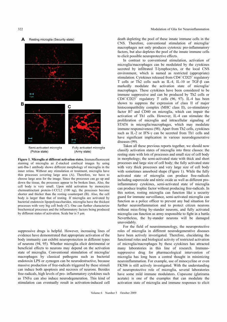
Taken all these previous reports together, we should now classify activation states of microglia into three classes: the resting state with lots of processes and small size of cell body in morphology; the semi-activated state with thick and short processes and large size of cell body; the fully activated state with very thick processes and very large size of cell body with sometimes amoeboid shape (Figure 1). While the fully activated state of microglia can produce free-radicals including superoxide and nitric oxide as well as different pro- inflammatory cytokines, semi-activated state of microglia can produce trophic factor without producing free-radicals. In this notion, resting microglia can function like a security guard for immune surveillance, semi-activated microglia can function as a police officer to prevent any bad situation for further neuroinflammation and to protect citizen neurons without miss-firing by-stander neurons, and fully activated microglia can function as army responsible to fight in a battle. Nevertheless, the by-stander neurons will be damaged unavoidably.
For the field of neuroimmunology, the neuroprotective roles of microglia in different neurodegenerative diseases have been actively investigated. Therefore, elucidating the functional roles and biological activity of restricted activation of microglia/macrophages by these cytokines has attracted many laboratories in this line of research. Immuno- suppressive drug for pharmacological intervention of microglia has long been a central thought in minimizing neuroinflammation. For example, use of minocycline or even FK506 is still actively investigated. With the understanding of neuroprotective role of microglia, several laboratories have some mild immune modulators. Copaxone (glatirama acetate) is one of the examples that can modulate the activation state of microglia and immune responses to elicit
A
BC
Figure 1. Microglia at different activation states. Immunofluorescentstaining of microglia as Z-stacked confocal images by using anti-Iba-1 antibody shows different morphology of microglia in the inner retina. Without any stimulation or treatment, microglia have thin processes covering large area (A). Therefore, we have to choose large area for the image. Since the processes can go up and down the tissue, the processes appear to be broken lines. Also, the cell body is very small. Upon mild activation by monocytes chemoattractant protein-1/CCL2 (100 ng), the processes become shorter and thicker than the resting counterpart (B). Also, the cell body is larger than that of resting. If microglia are activated by bacterial endotoxin lipopolysaccharides, microglia have the thickest processes with very big cell body (C). One can further characterize biochemical processes and the inflammatory factors being produced by different states of activation. Scale bar is 5 µm.

Cellular & Molecular Immunology 323
Volume 6 Number 5 October 2009
neuroprotection (100). There are several reports showing that it can attenuate activation of microglia to reduce neuro- inflammation in Alzheimer’s disease, macular degeneration and glaucoma (100-102). It was originally thought that it can modulate regulatory T lymphocytes. Now it has been shown to modulate microglia to relieve the stress from neuro- inflammation. Besides, the use of anti-inflammatory cytokines such as IL-10 is actively under investigation. While IL-10 can reduce production of pro-inflammatory cytokines and retune microglia to police-state, direct interactions between IL-10 and neurons to safeguard neurons may complicate our understanding of its anti-inflammatory role. In addition to the above pharmacological intervention, our laboratory has investigated the role of herbal medicine to mildly modulate microglia. One of the examples is the use of Wolfberry polysaccharides (103). We have shown that polysaccharides from Wolfberry can retune microglia into police officer mode to provide neuroprotection to neurons in glaucoma, neurodegeneration in the retina. This line of research is just at the beginning of exploration. Concluding remarks It has long been known that immune responses in the brain are difficult to induce and the production of pro- inflammatory factors is less than that in the peripheral systems. However, it is still unclear which kinds of signals or factors contribute to immune suppression. This review provides information about what has been found in this line of research. Essentially, no single factor is fully responsible for immune suppression. Rather, soluble factors as well as cell-cell contact via adhesion molecules or CD200R together lead to repression of immune responses. Immune suppressive effects of neurotrophins are an attracting idea. However, it has been reported that production of neurotrophins in experimental brain trauma such as CNTF requires the stimulation of IL-1β as there is no up-regulation of CNTF in genetically deficient for IL-1β mice (104). In addition, chronic overproduction of TGF-β1 by astroglial cells in TGF-β1 transgenic mice promotes the deposition of β-amyloid peptides, which can in turn stimulate microglial immune activities (45). These lines of results may question about the possibility of using soluble factor as pharmaco- logical intervention of neuroimmune responses. To further investigate how neuronal environment affects the immune responses, we have to explore more on how cell-cell contacts and the intracellular signaling for suppression of microglia so that neuronal communications are normal in a peaceful environment.
Although cerebral immune responses are highly restricted, activation of glial cells to initiate the cascade of immune responses is possible once neuronal injury occurs or in the presence of stimulatory signals which override the repressive signals. Inhibitory and stimulatory signals are actively competing with each other. Interactions between astroglia and microglia represent the first line of repressive control. However, they can also work together to initiate the cascade
of cerebral immune responses. A burst of immune responses can become uncontrollable and kill the neighboring neurons. In normal conditions, neurons and their secreted products exert immune suppression by cell-cell contact or the production of soluble immune suppressive factors.
While cerebral immune responses have long been considered to be detrimental factors contributing to the expansion of brain injury, increasing lines of evidence have shown that appropriate immune responses can also be beneficial, protect neurons and promote neuroregeneration (2, 4, 5). Therefore, it is essential to further understand which factors are involved in the repression of immune responses and how they are regulated. Reduction of immune responses simply by blocking the production of all kinds of inflammatory factors may not be a good therapeutic strategy. Appropriate manipulation of the cascade of immune responses may be beneficial to neurons. Like glutamate which is a part of neurotransmission in the brain, components in the cerebral immune responses also try to protect us from foreign antigens. A better understanding of all immune components can definitely help us to design better therapeutic interventions against many neurological disorders. Acknowledgements The work done by this laboratory has been or is currently supported by The Glaucoma Foundation, USA; American Health Assistant Foundation, USA; HKU Alzheimer’s Disease Research Network under Strategic Theme Research on Healthy Aging; University Strategic Research Theme on Drug Discovery; Research Fund for the Control of Infectious Diseases (09080822) from Food and Health Bureau of Hong Kong SAR Government; General Research Fund (761609M & 755206M) from Research Grant Council; National Science Foundation of China - Research Grant Council of Hong Kong Joint Research Scheme (N_HKU 707/07M); and HKU Seed Funding for Basic Research (200811159082). References
1. Simi A, Tsakiri N, Wang P, Rothwell NJ. Interleukin-1 and inflammatory neurodegeneration. Biochem Soc Trans. 2007;35: 1122-1126.
2. Schwartz M, Kipnis J. Protective autoimmunity and neuroprotection in inflammatory and noninflammatory neurodegenerative diseases. J Neurol Sci. 2005;233:163-166.
3. Crutcher KA, Gendelman HE, Kipnis J, et al. Debate: "is increasing neuroinflammation beneficial for neural repair?". J Neuroimmune Pharmacol. 2006;1:195-211.
4. Lucas SM, Rothwell NJ, Gibson RM. The role of inflammation in CNS injury and disease. Br J Pharmacol. 2006;147(Suppl 1):S232-240.
5. Schwartz M, London A. Immune maintenance in glaucoma: boosting the body's own neuroprotective potential. J Ocul Biol Dis Infor. 2009;2:73-77.
6. Barker CF, Billingham RE. Immunologically privileged sites. Adv Immunol. 1977;25:1-54.

324 Modulation of Glia for Neuroinflammation
Volume 6 Number 5 October 2009
7. Hickey WF, Hsu BL, Kimura H. T-lymphocyte entry into the central nervous system. J Neurosci Res. 1991;28:254-260.
8. Neumann H, Misgeld T, Matsumuro K, Wekerle H. Neurotrophins inhibit major histocompatibility class II inducibility of microglia: involvement of the p75 neurotrophin receptor. Proc Natl Acad Sci U S A. 1998;95:5779-5784.
9. Bauer J, Wekerle H, Lassmann H. Apoptosis in brain-specific autoimmune disease. Curr Opin Immunol. 1995;7:839-843.
10. Perry VH, Andersson PB, Gordon S. Macrophages and inflammation in the central nervous system. Trends Neurosci. 1993;16:268-273.
11. Bianchi M, Sacerdote P, Panerai AE. Cytokines and cognitive function in mice. Biol Signals Recept. 1998;7:45-54.
12. Wilson CJ, Finch CE, Cohen HJ. Cytokines and cognition – the case for a head-to-toe inflammatory paradigm. J Am Geriatr Soc. 2002;50:2041-2056.
13. Krueger JM, Obal FJ, Fang J, Kubota T, Taishi P. The role of cytokines in physiological sleep regulation. Ann N Y Acad Sci. 2001;933;211-221.
14. Chang RCC, Stadlin A, Tsang D. Effects of tumor necrosis factor alpha on taurine uptake in cultured rat astrocytes. Neurochem Int. 2001b;38:249-254.
15. Hirschberg DL, Schwartz M. Macrophage recruitment to acutely injured central nervous system is inhibited by a resident factor: a basis for an immune-brain barrier. J Neuroimmunol. 1995;61:89-96.
16. Zeev-Brann AB, Lazarov-Spiegler O, Brenner T, Schwartz M. Differential effects of central and peripheral nerves on macrophages and microglia. Glia. 1998;23:181-190.
17. Dusart I, Schwab ME. Secondary cell death and the inflammatory reaction after dorsal hemisection of the rat spinal cord. Eur J Neurosci. 1994;6:712-724.
18. Frohman EM, Vayuvegula B, Gupta S, van den Noort S. Norepinephrine inhibits γ-interferon-induced major histocompatibility class II (Ia) antigen expression on cultured astrocytes via β-2-adrenergic signal transduction mechanisms. Proc Natl Acad Sci U S A. 1988b;85:1292-1296.
19. Dello Russo C, Boullerne AI, Gavrilyuk V, Feinstein DL. Inhibition of microglial inflammatory responses by norepinephrine: effects on nitric oxide and interleukin-1β production. J Neuroinflamm. 2004;1:9.
20. Frohman EM, Frohman TC, Vayuvegula B, Gupta S, van den Noort S. Vasoactive intestinal polypeptide inhibits the expression of the MHC class II antigens on astrocytes. J Neurol Sci. 1988a;88:339-346.
21. Kim WK, Kan Y, Ganea D, Hart RP, Gozes I, Jonakait GM. Vasoactive intestinal peptide and pituitary adenylyl cyclase- activating polypeptide inhibit tumor necrosis factor-α production in injured spinal cord and in activated microglia via a cAMP-dependent pathway. J Neurosci. 2000;20:3622-3630.
22. Si Q, Nakamura Y, Ogata T, Kataoka K, Schubert P. Differential regulation of microglial activation by propentofylline via cAMP signaling. Brain Res. 1998;812:97-104.
23. Lee SC, Collins M, Vanguri P, Shin ML. Glutamate differentially inhibits the expression of class II MHC antigens on astrocytes and microglia. J Immunol. 1992;148:3391-3397.
24. Kong LY, McMillian MK, Hudson PM, Jin L, Hong JS. Inhibition of lipopolysaccharide-induced nitric oxide and cytokine production by ultralow concentrations of dynorphins in mixed glia cultures. J Pharmacol Exp Ther. 1997;280:61-66.
25. Kong LY, Jeohn G, Hudson PM, Du L, Liu B, Hong JS. Reduction of lipopolysaccharide-induced neurotoxicity in mouse mixed cortical neuron/glia cultures by ultralow concentrations of dynorphins. J Biomed Sci. 2000;7:241-247.
26. Liu B, Qin L, Yang SN, Wilson BC, Liu Y, Hong JS. Femtomolar concentrations of dynorphins protect rat mesencephalic dopaminergic neurons against inflammatory damage. J Pharmacol Exp Ther. 2001;298:1133-1141.
27. Chang RCC, Rota C, Glover RE, Mason RP, Hong JS. A novel effect of an opioid receptor antagonist, naloxone, on the production of reactive oxygen species by microglia: a study by electron paramagnetic resonance spectroscopy. Brain Res. 2000;854:224-229.
28. Galimberti D, Baron P, Meda L, et al. α-MSH peptides inhibit production of nitric oxide and tumor necrosis factor-α by microglial cells activated with β-amyloid and interferon γ. Biochem Biophys Res Commun. 1999;263:251-256.
29. Bruce-Keller AJ, Keeling JL, Keller JN, Huang FF. Camondola S, Mattson MP. Antiinflammatory effects of estrogen on microglial activation. Endocrinology. 1999;141:3646-3656.
30. Vegeto E, Bonincontro C, Pollio G, et al. Estrogen prevents the lipopolysaccharide-induced inflammatory response in microglia. J Neurosci. 2001;21:1809-1818.
31. Stone DJ, Rozovsky I, Morgan TE, Anderson CP, Hajian H, Finch CE. Astrocytes and microglia respond to estrogen with increased apoE mRNA in vivo and in vitro. Exp Neurol. 1997;143:313-318.
32. Vollmar P, Haghikia A, Dermietzel R, Faustmann PM. Venlafaxine exhibits an anti-inflammatory effect in an inflammatory co-culture model. Int J Neuropsychopharmacol. 2008;11:111-117.
33. Neumann H., Cavalie A, Jenne DE, Wekerle H. Induction of MHC class I genes in neurons. Science. 1995;269:549-552.
34. Neumann H, Boucraut J, Hahnel C, Misgeld T, Wekerle H. Neuronal control of MHC class II inducibility in rat astrocytes and microglia. Eur J Neurosci. 1996;8:2582-2590.
35. Wei R, Jonakait GM. Neurotrophins and the anti-inflammatory agents interleukin-4 (IL-4), IL-10, IL-11 and transforming growth factor-beta1 (TGF-1) down-regulate T cell costimulatory molecules B7 and CD40 on cultured rat microglia. J Neuroimmunol. 1999;95:8-18.
36. Flugel A, Matsumuro K, Neumann H, et al. Anti-inflammatory activity of nerve growth factor in experimental autoimmune encephalomyelitis: inhibition of monocyte transendothelial migration. Eur J Immunol. 2001;31:11-22.
37. Nakajima K, Kikuchi Y, Ikoma E. et al. Neurotrophins regulate the function of cultured microglia. Glia. 1998;24:272-289.
38. Bodmer S, Strommer K, Frei K, et al. Immunosuppression and transforming growth factor-beta in glioblastoma. Preferential production of transforming growth factor-beta 2. J Immunol. 1989;143:3222-3229.
39. Kuppner MC, Hamou MF, Sawamura Y, Bodmer S, de Tribolet N. Inhibition of lymphocyte function by glioblastoma-derived transforming growth factor beta 2. J Neurosurg. 1989;71:211- 217.
40. Fontana A, Bodmer S, Frei K, Malipiero U, Siepl C. Expression of TGF-2 in human glioblastoma: a role in resistance to immune rejection? Ciba Found Symp. 1991;157:232-238.
41. Lindholm D, Castren E, Kiefer R, Zafra F, Thoenen H. Transforming growth factor-beta 1 in the rat brain: increase after injury and inhibition of astrocyte proliferation. J Cell Biol. 1992;117:395-400.
42. Jones LL, Kreutzberg GW, Raivich G. Transforming growth factor beta's 1, 2 and 3 inhibit proliferation of ramified microglia on an astrocyte monolayer. Brain Res. 1998;795:301- 306.
43. Qian L, Wei SJ, Zhang D, et al. Potent anti-inflammatory and neuroprotective effects of TGF-1 are mediated through the

Cellular & Molecular Immunology 325
Volume 6 Number 5 October 2009
inhibition of ERK and p47phox-Ser345 phosphorylation and translocation in microglia. J Immunol. 2008;181:660-668.
44. Xiao BG, Bai XF, Zhang GX, Link H. Transforming growth factor-beta1 induces apoptosis of rat microglia without relation to bcl-2 oncoprotein expression. Neurosci Lett. 1997;226:71-74.
45. Wyss-Coray T, Lin C, Sanan DA, Mucke L, Masliah E. Chronic overproduction of transforming growth factor-beta1 by astrocytes promotes Alzheimer's disease-like microvascular degeneration in transgenic mice. Am J Pathol. 2000;156:139- 150.
46. Barger SW, Harmon AD. Microglial activation by Alzheimer amyloid precursor protein and modulation by apolipoprotein E. Nature. 1997;388:878-881.
47. Laskowitz DT, Goel S, Bennett ER, Matthew WD. Apolipoprotein E suppresses glial cell secretion of TNF alpha. J Neuroimmunol. 1997;76:70-74.
48. Lynch JR, Morgan D, Mance J, Matthew WD, Laskowitz DT. Apolipoprotein E modulates glial activation and the endogenous central nervous system inflammatory response. J Neuroimmunol. 2001;114:107-113.
49. Laskowitz DT, Thekdi AD, Thekdi SD, et al. Downregulation of microglial activation by apolipoprotein E and apoE-mimetic peptides. Exp Neurol. 2001;167:74-85.
50. Lyons A, Lynch AM, Downer EJ, et al. Fractalkine-induced activation of the phosphatidylinositol-3 kinase pathway attentuates microglial activation in vivo and in vitro. J Neurochem. 2009;110:1547-1556.
51. Harrison JK, Jiang Y, Chen S, et al. Role for neuronally derived fractalkine in mediating interactions between neurons and CX3CR1-expressing microglia. Proc Natl Acad Sci U S A. 1998;95:10896-10901.
52. Nishiyori A, Minami M, Ohtani Y, et al. Localization of fractalkine and CX3CR1 mRNAs in rat brain: does fractalkine play a role in signaling from neuron to microglia? FEBS Lett. 1998;429:167-172.
53. Maciejewski-Lenoir D, Chen S, Feng L, Maki R, Bacon KB. Characterization of fractalkine in rat brain cells: migratory and activation signals for CX3CR-1-expressing microglia. J Immunol. 1999;163:1628-1635.
54. Tong N, Perry SW, Zhang Q, et al. Neuronal fractalkine expression in HIV-1 encephalitis: roles for macrophage recruitment and neuroprotection in the central nervous system. J Immunol. 2000;164:1333-1339.
55. Gosselin RD, Dansereau MA, Pohl M, et al. Chemokine network in the nervous system: a new target for pain relief. Curr Med Chem. 2008;15:2866-2875.
56. McMillian MK, Thai L, Hong JS, O'Callaghan JP, Pennypacker KR. Brain injury in a dish: a model for reactive gliosis. Trends Neurosci. 1994;17:138-142.
57. McMillian MK, Pennypacker KR, Thai L, et al. Dexamethasone and forskolin synergistically increase [Met5] enkephalin accumulation in mixed brain cell cultures. Brain Res. 1996; 730:67-74.
58. Chang RCC, Hudson P, Wilson B, Haddon L, Hong JS. Influence of neurons on lipopolysaccharide-stimulated production of nitric oxide and tumor necrosis factor-alpha by cultured glia. Brain Res. 2000a;853:236-244.
59. Chang RCC, Chen W, Hudson P, Wilson B, Han DS, Hong JS. Neurons reduce glial responses to lipopolysaccharide (LPS) and prevent injury of microglial cells from over-activation by LPS. J Neurochem. 2001a;76:1042-1049.
60. Edelman GM. Cell adhesion molecule expression and the regulation of morphogenesis. Cold Spring Harb Symp Quant Biol. 1985;50:877-889.
61. Sporns O, Edelman GM, Crossin KL. The neural cell adhesion molecule (N-CAM) inhibits proliferation in primary cultures of rat astrocytes. Proc Natl Acad Sci U S A. 1995;92:542-546.
62. Krushel LA, Sporns O, Cunningham BA, Crossin KL, Edelman GM. Neural cell adhesion molecule (N-CAM) inhibits astrocyte proliferation after injury to different regions of the adult rat brain. Proc Natl Acad Sci U S A. 1995;92:4323-4327.
63. Chang RCC, Hudson P, Wilson B, et al. Immune modulatory effects of neural cell adhesion molecules on lipopolysaccharide- induced nitric oxide production by cultured glia. Brain Res Mol Brain Res. 2000b;81:197-201.
64. Krushel LA, Tai MH, Cunningham BA, Edelman GM, Crossin KL. Neural cell adhesion molecule (N-CAM) domains and intracellular signaling pathways involved in the inhibition of astrocyte proliferation. Proc Natl Acad Sci U S A. 1998;95: 2592-2596.
65. Crossin KL, Tai MH, Krushel LA, Mauro VP, Edelman GM. Glucocorticoid receptor pathways are involved in the inhibition of astrocyte proliferation. Proc Natl Acad Sci U S A. 1997;94: 2687-2692.
66. Ackley RL, Madison RD, Archibald SJ, Hemperly JJ. Monoclonal antibody interaction with the third immuno- globulin-like domain of N-CAM Is sufficient to cause cell migration. Mol Cell Neurosci. 1997;10:117-129.
67. Downer EJ, Cowley TR, Lyons A, et al. A novel anti- inflammatory role of NCAM-derived mimetic peptide, FGL. Neurobiol Aging. 2008; in press.
68. Downer EJ, Cowley TR, Cox F, et al. A synthetic NCAM- derived mimetic peptide, FGL, exerts anti-inflammatory properties via IGF-1 and interferon-gamma modulation. J Neurochem. 2009;109:1516-1525.
69. Hoek RM, Ruuls SR, Murphy CA, et al. Down-regulation of the macrophage lineage through interaction with OX2 (CD200). Science. 2000;290:1768-1771.
70. Dick AD, Broderick C, Forrester JV, Wright GJ. Distribution of OX2 antigen and OX2 receptor within retina. Invest Ophthalmol Vis Sci. 2001;42:170-176.
71. Chitnis T, Imitola J, Wang Y, et al. Elevated neuronal expression of CD200 protects Wlds mice from inflammation-mediated neurodegeneration. Am J Pathol. 2007;170:1695-1712.
72. Koning N, Swaab DF, Hoek RM, Huitinga I. Distribution of the immune inhibitory molecules CD200 and CD200R in the normal central nervous system and multiple sclerosis lesions suggests neuron-glia and glia-glia interactions. J Neuropathol Exp Neurol. 2009;68:159-67.
73. Lyons A, McQuillan K, Deighan BF, et al. Decreased neuronal CD200 expression in IL-4-deficient mice results in increased neuroinflammation in response to lipopolysaccharide. Brain Behav Immun. 2009; in press.
74. Fan, L, Young PR, Barone FC, Feuerstein GZ, Smith DH, McIntosh TK. Experimental brain injury induces differential expression of tumor necrosis factor-alpha mRNA in the CNS. Brain Res Mol Brain Res. 1996;36:287-291.
75. Rostworowski M, Balasingam V, Chabot S, Owens T, Yong VW. Astrogliosis in the neonatal and adult murine brain post-trauma: elevation of inflammatory cytokines and the lack of requirement for endogenous interferon-γ. J Neurosci. 1997;17:3664-3674.
76. Koistinaho J, Pasonen S, Yrjanheikki J, Chan PH. Spreading depression-induced gene expression is regulated by plasma glucose. Stroke. 1999;30:114-119.
77. Hossmann KA. Periinfarct depolarizations. Cerebrovasc. Brain Metab Rev. 1996;8:195-208.
78. Eder C. Ion channels in microglia (brain macrophages). Am J Physiol. 1998;275:C327-C342.
326 Modulation of Glia for Neuroinflammation
Volume 6 Number 5 October 2009
79. Norenberg W, Gebicke-Haerter PJ, Illes P. Inflammatory stimuli induce a new K+ outward current in cultured rat microglia. Neurosci Lett. 1992;147:171-174.
80. Illes P, Norenberg W, Gebicke-Haerter PJ. Molecular mechanisms of microglial activation. B. Voltage- and purinoceptor-operated channels in microglia. Neurochem Int. 1996;29:13-24.
81. Gehrmann J, Mies G, Bonnekoh P, et al. Microglial reaction in the rat cerebral cortex induced by cortical spreading depression. Brain Pathol. 1993;3:11-17.
82. Chang RCC, Hudson PM, Wilson BC, Liu B, Abel H, Hong JS. High concentrations of extracellular potassium enhance bacterial endotoxin lipopolysaccharide-induced neurotoxicity in glia-neuron mixed cultures. Neuroscience. 2000c;97:757- 764.
83. Kraig RP, Dong LM, Thisted R, Jaeger CB. Spreading depression increases immunohistochemical staining of glial fibrillary acidic protein. J Neurosci. 1991;11:2187-2198.
84. Kreutzberg GW. Microglia, the first line of defense in brain pathologies. Arzneimittelforschung 1995;45:357-360.
85. Langosch JM, Gebicke-Haerter PJ, Norenberg W, Illes P. Characterization and transduction mechanisms of purinoceptors in activated rat microglia. Br J Pharmacol. 1994;113:29-34.
86. Wang X, Franciosi S, Bae JH, Kim SU, McLarnon JG. Expression of P2y and P2x receptors in cultured human microglia. Proc West Pharmacol Soc. 1999;42:79-81.
87. McLarnon JG, Zhang L, Goghari V, et al. Effects of ATP and elevated K+ on K+ currents and intracellular Ca2+ in human microglia. Neuroscience. 1999;91:343-352.
88. Ferrari D, Chiozzi P, Falzoni S, Hanau S, Di Virgilio F. Purinergic modulation of interleukin-1 beta release from microglial cells stimulated with bacterial endotoxin. J Exp Med. 1997;185:579-582.
89. Hide I, Tanaka M, Inoue A, et al. Extracellular ATP triggers tumor necrosis factor- release from rat microglia. J Neurochem. 2000;75:965-972.
90. Naumov AP, Kuryshev YA, Kaznacheyeva EV, Mozhayeva GN. ATP-activated Ca2+-permeable channels in rat peritoneal macrophages. FEBS Lett. 1992;313:285-287.
91. Ferrari D, Stroh C, Schulze-Osthoff K. P2X7/P2Z purinoreceptor- mediated activation of transcription factor NFAT in microglial cells. J Biol Chem. 1999;274:13205-13210.
92. Orr AG, Orr AL, Li XJ, Gross RE, Traynelis SF. Adenosine A2A receptor mediates microglial process retraction. Nature Neurosci. 2009;12:872-878.
93. Wollmer MA, Lucius R, Wilms H, Held-Feindt J, Sievers J, Mentlein R. ATP and adenosine induce ramification of microglia in vitro. J Neuroimmunol. 2001;115:19-27.
94. Schwartz M, Kipnis J. A common vaccine for fighting neurodegenerative disorders: recharging immunity for homeostasis. Trends Pharmacol Sci. 2004;25:407-412.
95. Shaked I, Porat Z, Gersner R, Kipnis J, Schwartz M. Early activation of microglia as antigen-presenting cells correlates with T cell-mediated protection and repair of the injured central nervous system. J Neuroimmunol. 2004;146:84-93.
96. Kipnis J, Mizrahi T, Hauben E, Shaked I, Shevach E, Schwartz M. Neuroprotective autoimmunity: naturally occurring CD4+
CD25+ regulatory T cells suppress the ability to withstand injury to the central nervous system. Proc Natl Acad Sci U S A. 2002; 99:15620-15625.
97. Kipnis J, Avidan H, Caspi RR, Schwartz M. Dual effect of CD4+CD25+ regulatory T cells in neurodegeneration: a dialogue with microglia. Proc Natl Acad Sci U S A. 2004;101(Suppl 2): 14663-14669.
98. Nguyen VT, Benveniste EN. IL-4-activated STAT-6 inhibits IFN-γ-induced CD40 gene expression in macrophages/ microglia. J Immunol. 2000;165:6235-6243.
99. Sholl-Franco A, Figueiredo KGA, de Arauji EG. Interleukin-2 and interleukin-4 increase the survival of retinal ganglion cells in culture. Neuroreport. 2001;12:109-112.
100. Schwartz M. Modulating the immune system: a vaccine for glaucoma? Can J Ophthalmol. 2007;42:439-441.
101. Butovsky O, Koronyo-Hamaoui M, Kunis G, et al. Glatiramer acetate fights against Alzheimer's disease by inducing dendritic- like microglia expressing insulin-like growth factor 1. Proc Natl Acad Sci U S A. 2006;103:11784-11789.
102. Landa G, Butovsky O, Shoshani J, Schwartz M, Pollack A. Weekly vaccination with Copaxone (glatiramer acetate) as a potential therapy for dry age-related macular degeneration. Curr Eye Res. 2008;33:1011-1013.
103. Chiu K, Chan HC, Yeung SC, et al. Modulation of microglia by Wolfberry on the survival of retinal ganglion cells in a rat ocular hypertension model. J Ocular Biol Dis Informatics. 2009;2:47- 56.
104. Herx LM, Rivest S, Yong VW. Central nervous system-initiated inflammation and neurotrophism in trauma: IL-1 is required for the production of ciliary neurotrophic factor. J Immunol. 2000;165:2232-2239.
Related Documents