Malin Sandström TRITA-NA-E04014 Models of CaMKII Activation

Welcome message from author
This document is posted to help you gain knowledge. Please leave a comment to let me know what you think about it! Share it to your friends and learn new things together.
Transcript

Malin Sandström
TRITA-NA-E04014
Models of CaMKII Activation

NADA
Numerisk analys och datalogi Department of Numerical AnalysisKTH and Computer Science100 44 Stockholm Royal Institute of Technology
SE-100 44 Stockholm, Sweden
Malin Sandström
TRITA-NA-E04014
Master’s Thesis in Biomedical Engineering (20 credits)at the School of Engineering Physics,
Royal Institute of Technology year 2004Supervisor at Nada was Jeanette Hellgren-Kotaleski
Examiner was Anders Lansner
Models of CaMKII Activation
Abstract
Models of CaMKII activationCalcium/calmodulin-dependent Kinase Type II (CaMKII) is a Ca2+-activatedenzyme that is highly abundant in the brain. It is central to the regulation ofglutamatergic synapses and necessary for the induction of long-term potentia-tion (LTP), an activity-dependent strengthening of synapses that is thought tobe the basis for some forms of learning and memory. CaMKII is activated byCa2+ elevations and its key property is its ability to remain persistently active,in the absence of increased [Ca2+], for long periods of time. Existing computa-tional models of CaMKII activation are surprisingly diverse and there is littlegeneral consensus on which processes should be included or how detailed themodelling needs to be.
This study focuses on properties of CaMKII activation models. Eight modelsare reviewed, three of these are implemented and tested against experimentaldata. The models are compared with respect to general and specific modelfeatures and biological realism. Finally, the influence of some common modelassumptions are discussed and a general outline for a realistic model of CaMKIIactivation is proposed.
Sammanfattning
Modeller av CaMKII-aktiveringCalcium/calmodulin-beroende kinas typ II (CaMKII) ar ett Ca2+-aktiverat en-zym som ar narvarande i stora mangder i hjarnan. Det ar centralt for reglereringav glutamaterga synapser och nodvandigt for induktion av ”long-term potenti-ation” (LTP), en aktivitetsberoende okning av synapsstyrka som betraktas somgrunden for vissa typer av inlarning och minne. CaMKII aktiveras av hojdaCa2+-nivaer och dess viktigaste egenskap ar formagan att forbli aktiv, aven iavsaknad av Ca2+, under langa tidsperioder. Existerande berakningsmodellerfor CaMKII-aktivering ar forvanansvart olika och samstammigheten om vilkaprocesser som bor inkluderas, samt hur detaljerad modelleringen bor vara, ardalig.
Denna studie fokuserar pa egenskaper hos modeller av aktivering av CaMKII.Atta modeller gas igenom varav tre implementeras och testas mot experimentelladata. Generella och specifika drag hos modellerna samt overensstammelse medden biologiska verkligheten jamfors. Slutligen diskuteras effekterna av nagravanliga modellantaganden och ett utkast for en realistisk modell av CaMKII-aktivering foreslas.

Preface
This report describes a Master’s project in Computational Neuroscience, per-formed in the SANS group at the Department of Numerical Analysis and Com-puter Science (NADA) at the Royal Institute of Technology (KTH) in Stock-holm. Dr. Jeanette Hellgren-Kotaleski was supervisor and Prof. Anders Lansnerwas examiner.
The project consisted of three parts: a literature study on synaptic plastic-ity and models of CaMKII activation, implementation and testing of some ofthe models and a comparison of all the studied models on grounds of generalmodel structure and biological realism.
This report has three parts. First, a rather extensive background section onenzyme kinetics, synaptic plasticity and the properties of the substances in-cluded in the CaMKII pathway. Second, a general overview of a number ofmodels of CaMKII activation and third, a study and comparison of these mod-els on the grounds of general model properties and biological realism. Also, alist of abbreviations and an overview of model characteristics are given in theappendices. A reader unfamiliar with basic enzyme kinetics and/or the CaMKIIpathway is recommended to read the background chapter before the rest of thereport.
All implementation and simulation has been performed with MATLAB 6.5.
Acknowledgement
I would like to thank the following people who have all, in one way or another,contributed to this work:
My supervisor Jeanette Hellgren for her encouragement and support.My roommates Fredrik Edin, Vincente Charcos Llorens, Lotta Eriksson and An-ders Sandberg who have been very welcoming and friendly, providing me with anice environment to work in.All the other nice people at SANS.Ausra Saudargiene for her interest in my work and her questions, and for pro-viding me with matlab code on NMDA channels.Pal Westermark for his helpful comments on the sections 1.1.2-1.1.4.Ola Westin for his LaTeX tips.Last, but not least, I want to thank Rune Westin for support and encourage-ment, and the occasional loan of his computer.

Contents
1 Background 11.1 Enzymes: kinetics and basic concepts . . . . . . . . . . . . . . . . 2
1.1.1 Enzymes are biochemical catalysts . . . . . . . . . . . . . 21.1.2 The free energy governs all biochemical reactions . . . . . 21.1.3 The Michaelis-Menten model of enzyme kinetics . . . . . 21.1.4 Cooperativity is modeled by the Hill equation . . . . . . . 4
1.2 Synaptic plasticity . . . . . . . . . . . . . . . . . . . . . . . . . . 41.3 Dendritic spines . . . . . . . . . . . . . . . . . . . . . . . . . . . . 5
1.3.1 The Post-Synaptic Density (PSD) . . . . . . . . . . . . . 51.4 Glutamate receptors . . . . . . . . . . . . . . . . . . . . . . . . . 5
1.4.1 NMDA receptors . . . . . . . . . . . . . . . . . . . . . . . 51.4.2 AMPA receptors . . . . . . . . . . . . . . . . . . . . . . . 6
1.5 The Ca2+ ion - an intracellular messenger . . . . . . . . . . . . . 71.5.1 Ca2+ regulation in general . . . . . . . . . . . . . . . . . . 71.5.2 Ca2+ regulation in dendritic spines . . . . . . . . . . . . . 81.5.3 Ca2+ thresholds for induction of LTP and LTD . . . . . . 8
1.6 Calmodulin . . . . . . . . . . . . . . . . . . . . . . . . . . . . . . 81.6.1 Integration of Ca2+ signals by CaM . . . . . . . . . . . . 91.6.2 Ca2+ binding to CaM is affected by Mg2+ . . . . . . . . . 91.6.3 Ca2+ binding to CaM is affected by CaM-binding peptides 9
1.7 Kinases and phosphatases . . . . . . . . . . . . . . . . . . . . . . 91.7.1 CaMKII . . . . . . . . . . . . . . . . . . . . . . . . . . . . 101.7.2 Calcineurin (PP2B) . . . . . . . . . . . . . . . . . . . . . 141.7.3 Protein Phosphatase 1 (PP1) . . . . . . . . . . . . . . . . 16
1.8 Reactions included or excluded in models . . . . . . . . . . . . . 161.9 Producing realistic LTP/LTD curves . . . . . . . . . . . . . . . . 16
2 Models 192.1 D’Alcantara et al. . . . . . . . . . . . . . . . . . . . . . . . . . . 19
2.1.1 Aim . . . . . . . . . . . . . . . . . . . . . . . . . . . . . . 202.1.2 Input signal . . . . . . . . . . . . . . . . . . . . . . . . . . 202.1.3 The model . . . . . . . . . . . . . . . . . . . . . . . . . . 202.1.4 Results of d’Alcantara et al. . . . . . . . . . . . . . . . . . 20
2.2 Coomber . . . . . . . . . . . . . . . . . . . . . . . . . . . . . . . . 21
2.2.1 Aim . . . . . . . . . . . . . . . . . . . . . . . . . . . . . . 212.2.2 Input signal . . . . . . . . . . . . . . . . . . . . . . . . . . 212.2.3 The model . . . . . . . . . . . . . . . . . . . . . . . . . . 212.2.4 Coomber’s results . . . . . . . . . . . . . . . . . . . . . . 21
2.3 Dupont et al. . . . . . . . . . . . . . . . . . . . . . . . . . . . . . 222.3.1 Aim . . . . . . . . . . . . . . . . . . . . . . . . . . . . . . 222.3.2 Input signal . . . . . . . . . . . . . . . . . . . . . . . . . . 222.3.3 The model . . . . . . . . . . . . . . . . . . . . . . . . . . 222.3.4 Results of Dupont et al. . . . . . . . . . . . . . . . . . . . 23
2.4 Holmes . . . . . . . . . . . . . . . . . . . . . . . . . . . . . . . . 232.4.1 Aim . . . . . . . . . . . . . . . . . . . . . . . . . . . . . . 232.4.2 Input signal . . . . . . . . . . . . . . . . . . . . . . . . . . 232.4.3 The model . . . . . . . . . . . . . . . . . . . . . . . . . . 232.4.4 Holmes’ results . . . . . . . . . . . . . . . . . . . . . . . . 26
2.5 Kubota and Bower . . . . . . . . . . . . . . . . . . . . . . . . . . 262.5.1 Aim . . . . . . . . . . . . . . . . . . . . . . . . . . . . . . 262.5.2 Input signal . . . . . . . . . . . . . . . . . . . . . . . . . . 262.5.3 The model . . . . . . . . . . . . . . . . . . . . . . . . . . 262.5.4 Results of Kubota and Bower . . . . . . . . . . . . . . . . 27
2.6 Lundh . . . . . . . . . . . . . . . . . . . . . . . . . . . . . . . . . 282.6.1 Aim . . . . . . . . . . . . . . . . . . . . . . . . . . . . . . 282.6.2 Input signal . . . . . . . . . . . . . . . . . . . . . . . . . . 282.6.3 The model . . . . . . . . . . . . . . . . . . . . . . . . . . 28
2.7 Okamoto and Ichikawa . . . . . . . . . . . . . . . . . . . . . . . . 282.7.1 Aim . . . . . . . . . . . . . . . . . . . . . . . . . . . . . . 282.7.2 Input signal . . . . . . . . . . . . . . . . . . . . . . . . . . 282.7.3 The model . . . . . . . . . . . . . . . . . . . . . . . . . . 282.7.4 Results of Okamoto and Ichikawa . . . . . . . . . . . . . . 30
2.8 Zhabotinsky . . . . . . . . . . . . . . . . . . . . . . . . . . . . . . 302.8.1 Aim . . . . . . . . . . . . . . . . . . . . . . . . . . . . . . 302.8.2 Input signal . . . . . . . . . . . . . . . . . . . . . . . . . . 302.8.3 The model . . . . . . . . . . . . . . . . . . . . . . . . . . 312.8.4 Phosphorylation . . . . . . . . . . . . . . . . . . . . . . . 312.8.5 Dephosphorylation . . . . . . . . . . . . . . . . . . . . . . 322.8.6 Zhabotinsky’s results . . . . . . . . . . . . . . . . . . . . . 33
2.9 Lisman and Zhabotinsky . . . . . . . . . . . . . . . . . . . . . . . 332.9.1 Aim . . . . . . . . . . . . . . . . . . . . . . . . . . . . . . 332.9.2 Input signal . . . . . . . . . . . . . . . . . . . . . . . . . . 332.9.3 The model . . . . . . . . . . . . . . . . . . . . . . . . . . 332.9.4 AMPAR addition to the membrane . . . . . . . . . . . . . 34
3 Comparison of models 353.1 Ca2+ and CaM dynamics . . . . . . . . . . . . . . . . . . . . . . 35
3.1.1 A simple test of correction of Holmes’ CaM constants . . 373.2 Trapping of CaM* and capping of CaMKII . . . . . . . . . . . . 403.3 Feature span . . . . . . . . . . . . . . . . . . . . . . . . . . . . . 40

3.4 Implementation . . . . . . . . . . . . . . . . . . . . . . . . . . . . 433.5 A simple test . . . . . . . . . . . . . . . . . . . . . . . . . . . . . 44
4 Comparisons with experimental data 474.1 Dupont et al . . . . . . . . . . . . . . . . . . . . . . . . . . . . . 47
4.1.1 Implementation . . . . . . . . . . . . . . . . . . . . . . . . 474.1.2 Comparison with experimental data . . . . . . . . . . . . 47
4.2 Holmes . . . . . . . . . . . . . . . . . . . . . . . . . . . . . . . . 484.2.1 Implementation . . . . . . . . . . . . . . . . . . . . . . . . 484.2.2 Comparison with experimental data . . . . . . . . . . . . 48
4.3 Zhabotinsky . . . . . . . . . . . . . . . . . . . . . . . . . . . . . . 514.3.1 Implementation . . . . . . . . . . . . . . . . . . . . . . . . 514.3.2 Comparison with experimental data . . . . . . . . . . . . 51
5 Model assumptions revisited 575.1 The well-stirred cell assumption . . . . . . . . . . . . . . . . . . . 575.2 Simplified Ca2+ binding to CaM . . . . . . . . . . . . . . . . . . 575.3 Diffusion of CaM and CaM* . . . . . . . . . . . . . . . . . . . . . 58
5.3.1 Diffusion of CaM . . . . . . . . . . . . . . . . . . . . . . . 585.3.2 Diffusion of CaM* . . . . . . . . . . . . . . . . . . . . . . 58
5.4 CaM levels . . . . . . . . . . . . . . . . . . . . . . . . . . . . . . 595.4.1 CaM distribution in the spine . . . . . . . . . . . . . . . . 595.4.2 Limiting CaM . . . . . . . . . . . . . . . . . . . . . . . . . 59
5.5 Modelling CaMKII configurations . . . . . . . . . . . . . . . . . . 605.5.1 Simplifications . . . . . . . . . . . . . . . . . . . . . . . . 60
5.6 CaMKII capping . . . . . . . . . . . . . . . . . . . . . . . . . . . 605.6.1 The role of capping in frequency detection . . . . . . . . . 605.6.2 The maximal level of capping . . . . . . . . . . . . . . . . 61
5.7 CaMKII isoforms behave differently . . . . . . . . . . . . . . . . 615.8 Dual CaN regulation . . . . . . . . . . . . . . . . . . . . . . . . . 615.9 ”Constant” and/or ”unlimited”? . . . . . . . . . . . . . . . . . . 62
5.9.1 CaM . . . . . . . . . . . . . . . . . . . . . . . . . . . . . . 625.9.2 ATP . . . . . . . . . . . . . . . . . . . . . . . . . . . . . . 625.9.3 PKA . . . . . . . . . . . . . . . . . . . . . . . . . . . . . . 625.9.4 CaN . . . . . . . . . . . . . . . . . . . . . . . . . . . . . . 62
5.10 Stochastic versus deterministic . . . . . . . . . . . . . . . . . . . 63
6 Discussion 656.1 Model diversity and unknown mechanisms . . . . . . . . . . . . . 656.2 The implemented models . . . . . . . . . . . . . . . . . . . . . . 656.3 Replication of experimental data . . . . . . . . . . . . . . . . . . 666.4 The role of CaMKII isoforms . . . . . . . . . . . . . . . . . . . . 666.5 My recommendations for a model . . . . . . . . . . . . . . . . . . 66
Bibliography 69

Appendices 75Abbreviations . . . . . . . . . . . . . . . . . . . . . . . . . . . . . . . . 75Overview of models . . . . . . . . . . . . . . . . . . . . . . . . . . . . . 77
Chapter 1
Background
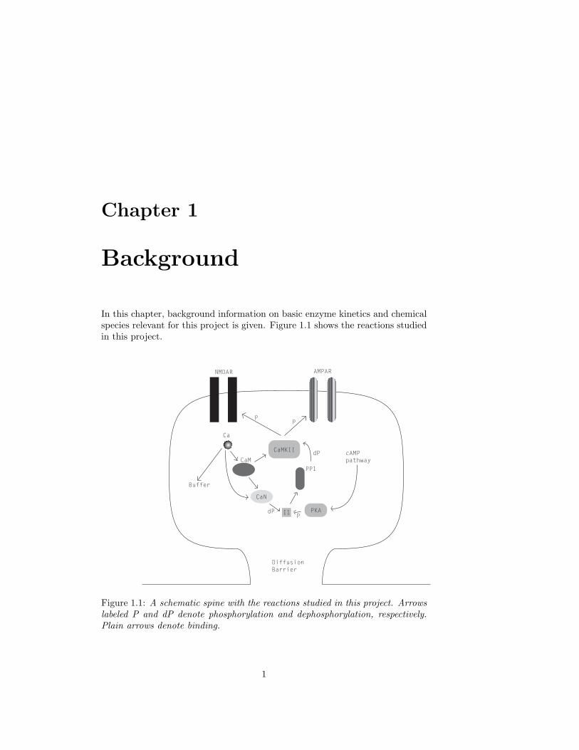
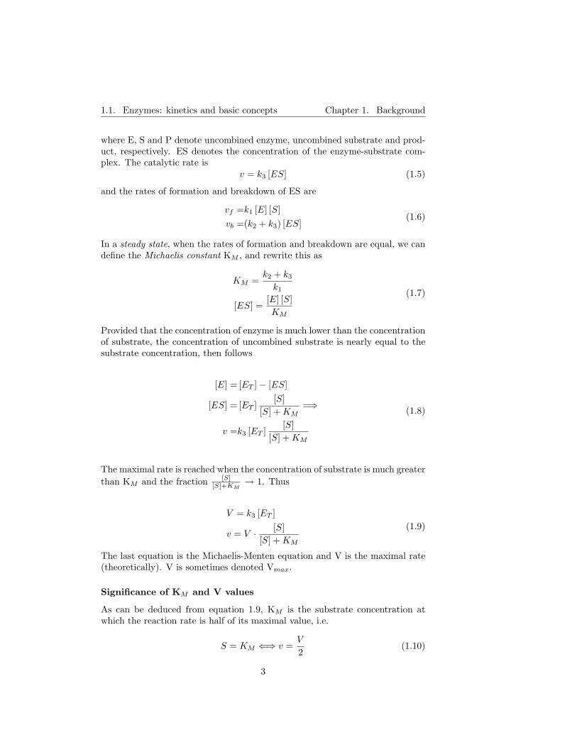
In this chapter, background information on basic enzyme kinetics and chemicalspecies relevant for this project is given. Figure 1.1 shows the reactions studiedin this project.
Ca
CaM
CaMKII
CaN
I1 PKA
PP1
NMDAR AMPAR
PP
dP
PdP
Buffer
cAMPpathway
DiffusionBarrier
Figure 1.1: A schematic spine with the reactions studied in this project. Arrowslabeled P and dP denote phosphorylation and dephosphorylation, respectively.Plain arrows denote binding.
1
1.1. Enzymes: kinetics and basic concepts Chapter 1. Background
1.1 Enzymes: kinetics and basic concepts
Readers who are unfamiliar with basic enzyme kinetics are recommended to readeither this section or the enzyme kinetics chapter(s) in a standard biochemistrytextbook (such as Stryers Biochemistry, 4th ed.) before continuing to latersections and chapters in the report. All equations in sections 1.1.1-1.1.3 areadapted from Stryers Biochemistry, 4th ed [53].
1.1.1 Enzymes are biochemical catalysts
Enzymes are the catalysts of biological systems, i.e. they increase the rate ofbiochemical reactions. They are specific both in which reactions they catalyzeand in their choice of substrates. Many enzymes are regulated, for exampleby phosphorylation, which is the reversible attachment of phosphoryl groups tospecific serine (Ser) and threonine (Thr) residues. Phosphorylation is catalyzedby kinases and dephosphorylation is catalyzed by phosphatases (see section 1.7).
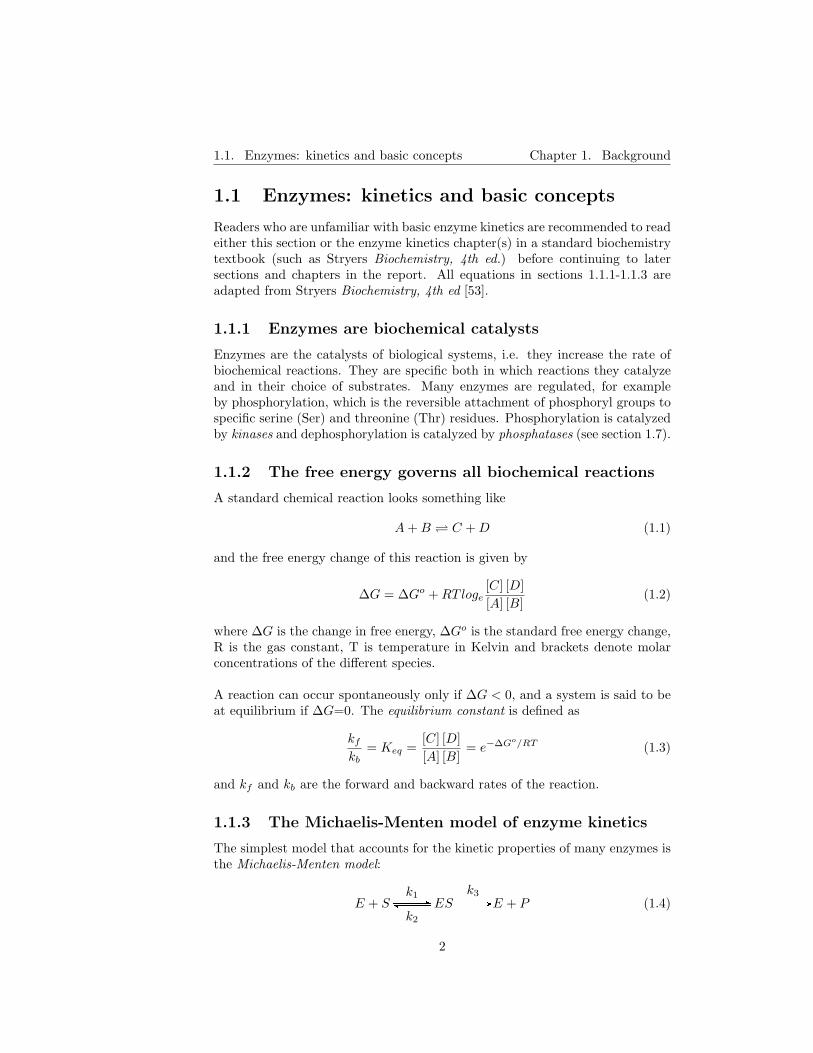
1.1.2 The free energy governs all biochemical reactions
A standard chemical reaction looks something like
A + B C + D (1.1)
and the free energy change of this reaction is given by
∆G = ∆Go + RTloge[C] [D][A] [B]
(1.2)
where ∆G is the change in free energy, ∆Go is the standard free energy change,R is the gas constant, T is temperature in Kelvin and brackets denote molarconcentrations of the different species.
A reaction can occur spontaneously only if ∆G < 0, and a system is said to beat equilibrium if ∆G=0. The equilibrium constant is defined as
kf
kb= Keq =
[C] [D][A] [B]
= e−∆Go/RT (1.3)
and kf and kb are the forward and backward rates of the reaction.
1.1.3 The Michaelis-Menten model of enzyme kinetics
The simplest model that accounts for the kinetic properties of many enzymes isthe Michaelis-Menten model:
E + Sk1
GGGGGGBFGGGGGGk2
ESk3
GGGGGGAE + P (1.4)
2
1.1. Enzymes: kinetics and basic concepts Chapter 1. Background
where E, S and P denote uncombined enzyme, uncombined substrate and prod-uct, respectively. ES denotes the concentration of the enzyme-substrate com-plex. The catalytic rate is
v = k3 [ES] (1.5)
and the rates of formation and breakdown of ES are
vf =k1 [E] [S]vb =(k2 + k3) [ES]
(1.6)
In a steady state, when the rates of formation and breakdown are equal, we candefine the Michaelis constant KM , and rewrite this as
KM =k2 + k3
k1
[ES] =[E] [S]KM
(1.7)
Provided that the concentration of enzyme is much lower than the concentrationof substrate, the concentration of uncombined substrate is nearly equal to thesubstrate concentration, then follows
[E] = [ET ]− [ES]
[ES] = [ET ][S]
[S] + KM=⇒
v =k3 [ET ][S]
[S] + KM
(1.8)
The maximal rate is reached when the concentration of substrate is much greaterthan KM and the fraction [S]
[S]+KM→ 1. Thus
V = k3 [ET ]
v = V · [S][S] + KM
(1.9)
The last equation is the Michaelis-Menten equation and V is the maximal rate(theoretically). V is sometimes denoted Vmax.
Significance of KM and V values
As can be deduced from equation 1.9, KM is the substrate concentration atwhich the reaction rate is half of its maximal value, i.e.
S = KM ⇐⇒ v =V
2(1.10)
3
1.2. Synaptic plasticity Chapter 1. Background
If the rate of product formation, k3, is much lower than the rate of enzyme-substrate complex breakdown, k2, KM is equal to the dissociation constant ofthe complex. Thus, under these conditions KM is a measure of the strengh ofthe ES complex: a high KM then indicates weak binding and vice versa.
1.1.4 Cooperativity is modeled by the Hill equation
Cooperativity enables the enzymes to change their behaviour in response tomoderate (or even small) changes in substrate concentration. This enables morefine-tuned regulation of the enzyme-catalyzed process and does not demand ashuge amounts of substrate for maximum reaction velocity as would otherwisebe necessary.
When an enzyme can bind more than one substrate, each substrate facilitatingthe binding of the next, one says that the enzyme exhibits cooperativity towardsthe substrate. Equation 1.4 then generalizes to
E+n ·SK1
GGGGGGGBFGGGGGGGES1+(n−1) ·SK2
GGGGGGGBFGGGGGGG . . .Kn
GGGGGGGBFGGGGGGGESn
k3GGGGGGAE+n ·P (1.11)
Following a similar procedure to equations 1.5-1.9 and taking the limit (assumingthat the intermediary states ES1 - ESn−1 do not exist), one obtains the Hillequation. Now, n does not need to be an integer and the notation is changed toh
v = V · [S]h
[S]h + Sh0.5
(1.12)
We see that V and KM retain their meaning (although here, KM is exchangedfor S0.5), and we obtain a new characteristic variable: h, the Hill number (oftendenoted nH). The larger the Hill number, the steeper the concentration-fluxcurve will become. As h →∞, the curve becomes a step function. If h=1, theHill equation reduces to the Michaelis-Menten equation.
1.2 Synaptic plasticity
Long-lasting and activity-dependent changes in synaptic strength are consideredas key properties of neurons to explain cellular and molechular mechanisms sup-porting the formation and storage of memories. So-called bidirectional synapticplasticity is considered to be governed by calcium influx through NMDA recep-tors. The calcium signal then triggers a signal transduction cascade, involvingcalcium-dependent protein kinases and phosphatases (among others calmodulindependent kinase type II (CaMKII) and calcineurin (CaN)), that induces LTP(long-term potentiation, an increase in synaptic strength) or LTD (long-termdepression, a decrease in synaptic strength). There is strong evidence that LTPinvolves a postsynaptic process [41], which selectively enhances AMPA receptor-mediated transmission.
4

1.3. Dendritic spines Chapter 1. Background
1.3 Dendritic spines
Dendritic spines are small protrusions from dendrites of various types of neurons,consisting of a head, with a volume of ∼0.01-1 µm3 , connected to its parentdendrite via a thin (∼ 0.1 µm) neck [38]. Most spines recieve an excitatorysynapse on their head. The neck effectively limits diffusional exchange betweenthe spine head and the dendrite (see section 1.5.2). The spine contains a wealthof different substances, and spine morphology is highly variable even on thesame dendrite [45]. A simple illustration of a spine and the reactions studied inthis project can be found in figure 1.1.
1.3.1 The Post-Synaptic Density (PSD)
The PSD is a scaffolding organelle located at the postsynaptic membrane. Itcontains a wealth of proteins important for signal transmission, among themscaffolding proteins, glutamate receptors, kinases and phosphatases. Many ofthese proteins are known to organize themselves into complexes that are thoughtto be important for synapse function, see [21],[29] and [45].
1.4 Glutamate receptors
The glutamate receptors can be divided into two categories: the ionotropic re-ceptors that directly gate channels and the metabotropic receptors that gatechannels indirectly, through second messengers.
The ionotropic glutamate receptors belong to a separate genetic family of ligand-gated channels. Two branches of the glutamate receptor-gene family, closely re-lated to each other, include the AMPA and kainate receptors. A more distantlyrelated branch of the family codes for the NMDA type of receptors [52]. Theglutamate-gated channels are all multimeric proteins, thought to be composedof four subunits built up by three transmembrane α-helices each.
1.4.1 NMDA receptors
NMDA receptors (NMDARs) are heteromeric ion channels, composed of NR1and NR2 subunit proteins. The NR2 subunit has four subtypes (NR2A-NR2D),each of which gives the receptor distinct functional properties [9].
NMDARs are detectors of coincident activity
The NMDA receptor channel is unique in that it is permeable not only to Na+
and K+, but also to Ca2+, and that its opening depends on both membranevoltage and the presence of the transmitter. Thus, the NMDAR can act asa detector of coincident activity in the pre- and postsynaptic cells [52]. Thechannels open efficiently only when glutamate is released from the presynapticterminal (i.e. when the presynaptic cell is active) and the postsynaptic cell is
5

1.4. Glutamate receptors Chapter 1. Background
strongly depolarized.
At membrane resting potential, the channel is blocked by Mg2+. When themembrane is depolarized, Mg2+ is expelled from the channel by electrostatic re-pulsion. Channel opening produces a rise in [Ca2+] that is largely restricted tothe dendritic spine onto which the active synapse terminates. The depolariza-tion level that is needed to activate NMDARs usually requires the summation ofmultiple synaptic inputs. LTP induction is considered to depend on activationof NMDA receptors.
Proteins interacting with NMDA receptors
There are many substances that bind to and/or interact with NMDA receptorsin different ways, either directly or via scaffolding proteins. Lists of these can befound in the articles by Husi et al. [21] and Tashiro and Yuste [45]. Interactingproteins include CaM, CaMKII, CaN and PP1. The NMDAR subunit NR2Bcan be phosphorylated by CaMKII (see section 1.7.1) and this phosphorylationpractically inhibits further CaMKII binding.
1.4.2 AMPA receptors
The AMPA receptor (AMPAR) is a heteromer composed of multiple subtypes ofsubunit proteins (GluR1-GluR4). In the adult hippocampus, receptors consist-ing of GluR1/GluR2 and GluR2/GluR3 are predominant [18]. The function ofthe AMPAR is regulated by the composition of individual receptors and/or thephosphorylation and dephosphorylation states of individual subunit proteins. Ofparticular interest are the residues serine 831 (Ser831) and serine 845 (Ser845) onthe GluR1 subunit, as they can be phosphorylated by CaMKII/protein kinaseC and protein kinase A, respectively.
Phosphorylation of AMPARs in LTP and LTD
The induction of LTP specifically increases phosphorylation of S-831 which in-creases the single channel conductance of GluR1 AMPARs [18]. The inductionof LTD is accompanied by a decrease in the phosphorylation of Ser845, whichappears to be phosphorylated at resting potential. Phosphorylation of Ser845
increases the ”open time” of the AMPAR [9] and is required for receptor incor-poration in the membrane [18].
AMPA receptor trafficking
AMPA receptor subunits are synthesized and assembled in the rough endoplas-mic reticulum (ER) and then inserted into the plasma membrane after crossingthe Golgi. AMPARs undergo a constant trafficking between the plasma mem-brane and intracellular compartments. This process plays a key role in theregulation of the synaptic levels of AMPA receptors. Activation of CaMKIIcauses translocation of the GluR1 subunit to dendritic spines and synapses [17].
6
1.5. The Ca2+ ion - an intracellular messenger Chapter 1. Background
AMPA receptor internalization can be influenced by both AMPA and NMDAreceptors, but the nature of the stimuli determines the exact fate of the inter-nalized AMPARs. Upon NMDAR activation, AMPA receptors are transportedrapidly from the membrane and can be recycled (i.e. rapidly reinserted) [17],but after AMPAR activation, the receptors are probably transported away anddegraded [18].
NMDAR-dependent AMPAR trafficking is regulated by PKA and accompaniedby GluR1 phosphorylation and dephosphorylation at a PKA site [17]. PKA alsoexerts a differential modulatory effect on intracellular AMPA receptor traffick-ing upon AMPA- and NMDA-induced internalization. NMDA receptor inducedAMPA receptor trafficking is accompanied by dephosphorylation followed byrephosphorylation of GluR1 AMPA receptor subunits at Ser845 by PKA. Incontrast, PKA is without effect on AMPA-induced AMPA receptor cycling [18].
Proteins interacting with AMPA receptors
AMPA receptor localization at the postsynaptic membrane of excitatory synapsesis dynamically regulated. Targeting of receptors to synapses is thought to bemediated through interaction of AMPA receptor subunits with scaffolding pro-teins. For a list of some directly interacting proteins and further details, seeGomes et al. [18] and Tashiro and Yuste [45].
1.5 The Ca2+ ion - an intracellular messenger
The resting level of Ca2+ in a cell is typically somewhat lower than 100nM,which is several orders of magnitude less than the extracellular Ca2+ level.Thus, a rise in [Ca2+] (Ca2+ concentration) – achieved by the opening of calciumchannels, or release from intracellular stores – can be used as a signal. Calciumconcentration regulates many processes in cells, on many different timescales:for instance synaptic transmission and muscle contraction in skeletal and cardiacmuscles. Prolonged exposure to high levels of Ca2+ is toxic and can end in celldeath (excitotoxicity).
1.5.1 Ca2+ regulation in general
Calcium enters the cell via several kinds of calcium channels and via NMDAchannels. It can be stored in and released from intracellular stores such asbuffers and the sarcoplasmic reticulum (SR), and is removed from the cell by aCa2+/H2+ ATPase, a Ca2+ pump driven by ATP hydrolysis, and a 3Na+/Ca2+
exchanger driven by the electrochemical Na+ gradient across the membrane [37].Calcium concentration can also be directly and indirectly influenced by itselfthrough processes as Ca2+ pump regulation, calcium-induced-calcium releasefrom the SR and phosphorylation of NMDA receptors by CaMKII (see below).
7

1.6. Calmodulin Chapter 1. Background
1.5.2 Ca2+ regulation in dendritic spines
Calcium is known to be crucial for the induction of LTP and LTD. In these cases,NMDA receptors are considered to be the important Ca2+ sources. Sabatini etal. [38] have shown that spines are specialized in several ways:
- Spines have low Ca2+ buffer capacity (20 units→ 5% of the Ca2+ enteringthe spine remains unbound [19])
- Ca2+ diffusion across the spine neck is negligible
- The spine head functions as a separate compartment, allowing localizedCa2+ buildup (which is crucial for synaptic plasticity)
- The kinetics of NMDAR opening primarily determines the time courseof synaptically evoked [Ca2+] transients. NMDAR-mediated transients inunperturbed spines reach ∼12 µM at depolarizing potentials.
- Spine Ca2+ returns to resting levels with a time constant of ∼12 ms.
- Ca2+ resting level in the spines is low; [Ca2+]0 = 70 ± 29 nM at restingpotential.
- Clearance of Ca2+ from the cytoplasm is too rapid to allow Ca2+ accu-mulation during low-frequency stimulation (LFS)
- The 3Na+/Ca2+ exchanger is probably responsible for most of the Ca2+
efflux from spines
A discussion of Ca2+ measurement in spines and commentary on the Sabatiniresults can be found in the article by Helmchen [19].
1.5.3 Ca2+ thresholds for induction of LTP and LTD
Cormier et al. [12] found that the [Ca2+] needed to induce plasticity was ∼ 180nM, while LTD was induced in the range of 180-500 nM and LTP was inducedwhen [Ca2+]> ∼540 nM.
1.6 Calmodulin
Many proteins are regulated by calmodulin (CaM), which is a small proteinthat can bind up to four calcium ions. It consists of two structurally similardomains, each containing a pair of helix-loop-helix calcium-binding motifs (alsocalled EF-hands). However, the affinities of all Ca2+ binding sites are not equal;CaM has two high-affinity and two low-affinity sites (the Ca2+-binding sites inthe C-terminal end have a tenfold higher affinity (Kd ≈ 0.2 µM) than thosein the N-terminal end (Kd ≈ 2 µM) [48]). Positive cooperativity is observedbetween sites within a domain.
8

1.7. Kinases and phosphatases Chapter 1. Background
Mostly, CaM-binding proteins bind CaM with four Ca2+ ions (CaM fully loadedwith Ca2+ will in this report be denoted CaM*) and become activated, but thereare also cases where CaM is bound without Ca2+ and activates the protein whenCa2+ is bound. Yet another variety are the proteins that bind calcium-freecalmodulin and release it when it binds calcium [33]. In many enzymes that areactivated by CaM, an autoinhibitory part overlapping the CaM binding site isdislocated upon the binding of CaM, leading to enzyme activation.
1.6.1 Integration of Ca2+ signals by CaM
In rat sensory neurons, Millikan et al. [33] found that CaM was able to ”inte-grate” Ca2+ signals in the sense that
- CaM activation peaked after the maximum of the Ca2+ signal, with max-imum activation a half second after the calcium influx ended
- CaM activation did not immediately fall to baseline when the Ca2+ leveldid, but fell to a slowly decaying plateau persisting for tens of seconds
However, in this case Ca2+ remained in the cytosol for a couple of seconds (i.e.Ca2+ clearance was not as rapid as in spines).
1.6.2 Ca2+ binding to CaM is affected by Mg2+
Mg2+ binds to CaM and competes with Ca2+ binding [31], but the affinityfor Mg2+ is at least thousandfold lower [48]. Both Mg2+ and CaM-bindingproteins affect the affinity of CaM for Ca2+, as reported by Stemmer and Klee[42]. Moderate (i.e. millimolar) Mg2+ concentrations increase the affinity whilehigh concentrations decrease the affinity.
1.6.3 Ca2+ binding to CaM is affected by CaM-bindingpeptides
The presence of Mg2+ and CaM binding peptides (short protein fragments con-taining the CaM-binding sequence of CaN subunit A) increases the affinity forCa2+ in a complex way, but in general by at least 10-fold for each of the fourbinding sites [42].
1.7 Kinases and phosphatases
The human genome encodes ∼ 500 kinases and ∼ 150 phosphatases [10]. Theyregulate protein function by phosphorylation and dephosphorylation, respec-tively (addition or removal of a phosphoryl group). Two special subclasses areserine-threonine kinases and phosphatases, who phosphorylate and dephospho-rylate serine and threonine residues. CaMKII is the dominant kinase in thehippocampal area; it is present in 20 times higher concentration than the otherkinases [26].
9

1.7. Kinases and phosphatases Chapter 1. Background
1.7.1 CaMKII
CaMKII in the PSD
CaMKII (calmodulin-dependent kinase type II) is one of the major constituentsof the post-synaptic density (PSD) [41] and is thought to have both structuraland functional importance. It is activated by binding of CaM* and its keyproperty is autophosphorylation, the ability of its subunits to phosphorylateneighbouring subunits.
Initially, CaMKII is bound to F-actin [28] distributed in the cytoplasm, butit translocates to the PSD upon binding of CaM* [40]. The half-maximal timefor translation ranges from around 20 s (α-CaMKII) to 280 s (β-CaMKII) andtranslation time can thus be regulated in the neurons by the ratio of α- to β-CaMKII. PSD-bound CaMKII is nearly immobile during glutamate stimulation[40].
Structure
CaMKII has 8-12 nearly identical subunits, arranged in two hexameric rings.Each subunit consists of a catalytic domain, an autoinhibitory domain, a vari-able segment and a self-association domain. The catalytic domain contains theATP- and substrate-binding sites, as well as sites for interaction with anchoringproteins.
CaMKII states
CaMKII subunits can be said to have four different states of activation, as wellas an inactive ground state: bound to CaM*, trapped, autonomous and capped.See figure 1.2.
- The activation in the bound state lasts only as long as CaM* is bound.
- The trapped state, or CaM* trapping on CaMKII subunits, is an effectof the thousandfold decreased off rate of CaM* after phosphorylation atThr286/287 by a neighbouring subunit that is already active.
- An autonomous CaMKII subunit is phosphorylated at Thr286/287 (by aneighbouring subunit) and has in addition lost its bound CaM* (i.e. theactivity is still high, but has become CaM*-independent).
- Capped CaMKII, finally, is also phosphorylated at Thr305/306. Having atrapped CaM* prevents capping, and capping prevents rebinding of CaM*.
Capping of CaMKII appears to be both an intrasubunit and an intersubunit re-action, and capped CaMKII has less catalytic activity than autonomous CaMKIIor no catalytic activity, according to some [11],[26]. Kubota and Bower claimthat capped CaMKII has the same activity as autonomous CaMKII [25]. Holmesclaims [20] that the activity of autonomous and capped subunits is about 40 %of the activity of bound and trapped subunits.
10

1.7. Kinases and phosphatases Chapter 1. Background
CIIsIj
C // CIIsBjoo
P // CIIsTjoo // CIIsA
jCoo
P //
D
ii CIIsCj
Doo
Figure 1.2: CaMKII states. CIIsXj denotes CamKII subunit in state X, C indi-
cates calcium/calmodulin dependent transitions, P denotes phosphorylation andD denotes dephosphorylation.
CaMKII isoforms
There are four genes for different CaMKII isoforms: α, β, γ and δ. Additionalisoforms can also be generated by alternative splicing of all four isoforms. Theα and β isoforms are the predominant isoforms in the brain, and the enzymecan contain both α and β subunits.
β-CaMKII has a larger affinity for CaM than α-CaMKII (CaM50for autophos-phorylation is 15nM for β-CaMKII and 130 nM for α-CaMKII [7]) and has arelatively larger activation at all frequencies [13]. Also, regulation of α-CaMKIItranscription levels is strongly influenced by NMDAR activity, while β-CaMKIItranscription levels are insensitive to NMDAR activity and sensitive to AMPARactivity [47]. For other CaMKII interactions with NMDA, see section 1.7.1
It is thought that the cell fine-tunes its response to changing Ca2+ signallinglevels by regulating the relative amount of α-CaMKII to β-CaMKII. The α/βratio has been shown to change as most ∼5-fold after 24 hours as a response tochanging intensities of Ca2+ signalling [47].
Under resting conditions, β-CaMKII but not α-CaMKII are bound to F-actin([28] and [40], respectively) distributed in the cytoplasm.
Activation and function
Under basal conditions, the kinase has almost no catalytic activity since theautoinhibitory domain of each subunit inhibits its own catalytic domain. Eachsubunit can be activated by the binding of a single CaM* to that subunit. Thissimple form of activation lasts only as long as the increase in [Ca2+].
When the increase in [Ca2+] is sufficient to cause CaM* to bind simultaneouslyto two neighbouring units, one subunit can phosphorylate the other at residuethreonine 286 (Thr286, for α-CaMKII) or threonine 287 (β-CaMKII). The oc-curence of two neighbouring subunits with bound CaM* is sparse, so this is aslow process. Further phosphorylation only requires binding of one CaM* (toa neighbouring site to one already phosphorylated) and proceeds much faster.Autophosphorylation of CaMKII by itself shows a strong dependence on [Ca2+],
11

1.7. Kinases and phosphatases Chapter 1. Background
with a Hill coefficient close to 5 (with no phosphatases present [6]).
Immobilized CaMKII has been shown to reach 60-80 percent autonomous ac-tivity, and have a half-maximal activation at a CaM concentration of 25-80 nM(CaM50 for pure β-CaMKII and pure α-CaMKII, respectively) [13]. Brocke etal. report CaM50 values of 15 nM and 130 nM, respectively [7].
CaMKII in the PSD is dephosphorylated by PP1
An autonomous subunit remains active even after [Ca2+] levels return to base-line. Thus, phosphorylation results in persistent activity; a biochemical ”mem-ory trace” of the previous [Ca2+] elevation. Its activity will remain until it isdephosphorylated. In the PSD, CAMKII is dephosphorylated only by PP1 [29].When CaMKII autophosphorylation is balanced by PP1 dephosphorylation, ithas an even stronger dependence on [Ca2+], with a Hill coefficient close to 8 [6],compared to a Hill coefficient of 5 without PP1.
CaMKII frequency sensitivity
De Koninck and Schulman [13] have shown experimentally that CaMKII acti-vation in itself is dependent on the frequency of stimulation, i.e. that oscillationfrequency and duration is translated to different levels of activity. They im-mobilized CaMKII inside PVC tubing and subjected it to pulses of Ca2+ andCaM of varying amplitude. No phosphatases were present. Activation at 1 Hz(with pulses of 100 nM CaM and saturating amounts of Ca2+ and ATP, 500µM and 250 µM respectively) produced only ∼ 6-7 % activity after 100 pulses,while stimulation at 4 Hz produced ∼ 60 % activation after the same numberof pulses. Naturally, also the pulse length had influence on the sensitivity, withthe frequency response shifted to lower frequencies with longer pulse lengths.
The frequency sensitivity of β-CaMKII has also been demonstrated to be modu-lated by alternative splicing [3]. As the splice variants are differentially expressedamong individual neurons, this provides cells with a possibility to modulate theirsensitivity to oscillating Ca2+ signals.
Frequency sensitivity has also been replicated by several models, by amongothers Coomber [11], Dupont et al. [16] and Kubota and Bower [25].
CaMKII translocation to the PSD
Shen and Meyer [40] have shown that, upon Ca2+ influx through NMDA re-ceptors and subsequent binding of CaM, β-CaMKII rapidly dissociates fromF-actin and translocates to the PSD (where it is thought to increase synapticstrength by phosphorylating PSD ion channels and signaling proteins). CaMbinding but not autophosphorylation was needed for translation to occur, al-though autophosphorylation plays an indirect part as the affinity of CaMKII
12

1.7. Kinases and phosphatases Chapter 1. Background
for CaM is increased hundred-fold upon autophosphorylation. This is thoughtto prolong the association of CaMKII with the PSD. Dosemeci et al. [15] haveshown that accumulation of CaMKII in the PSD is reversed within 30 minutesupon removal of glutamate and Ca2+.
Binding of phosphorylated CaMKII to the NMDAR
After it is exposed by binding of CaM*, the catalytic domain of a CaMKIIsubunit can also bind to the NMDA receptor subunit NR2B with high affinity.This will cause the CaMKII subunit to remain active also after releasing CaM*.Phosphorylation of the NR2B subunit residue Ser1303 by CaMKII (with a verylow turnover rate, ∼ 10 min−1) inhibits almost all CaMKII/NR2B interaction.The NR2B·CaMKII complex has a dissociation time of several minutes [43].
Phosphatase activity may, paradoxically, increase CaMKII/NR2B interactionif Ser1303 is more dephosphorylated than CaMKII. The identity of the phos-phatase that dephosphorylates Ser1303 is not known [43].
A structural role for CaMKII in the PSD
It has been proposed by Lisman and Zhabotinsky [29] that CaMKII binding tothe NMDA receptor organizes a structural process that leads to the incorpora-tion of AMPA-receptor-binding proteins into the PSD, and to the subsequentanchoring of additional AMPA receptors, accumulating AMPARs in the post-synaptic membrane.
It is further suggested that these scaffolding proteins create an isolated andlimited pool of PP1 in the PSD and prevent PP2A, which is able to dephospho-rylate CaMKII in solution but apparently not in the PSD [28], from dephos-phorylating CaMKII. This would allow the local phosphatase pool to becomesaturated by highly phosphorylated kinase.
Saturation of the local phosphatase pool would provide a solution to the ”pro-tein turnover problem”; keeping the highly phosphorylated state although partof the highly phosphorylated kinase pool is replaced by newly transcribed, un-phosphorylated protein. It would also allow bistability, which is thought to bea property required for synaptic plasticity [27],[28],[29],[51]. Bradshaw et al. [6]have shown that the CaMKII-PP1 system indeed is bistable.
CaMKII phosphorylation of AMPARs
Besides the role CAMKII has in AMPAR trafficking, it is also known thatactivated CaMKII phosphorylates GluR1 subunits of already existing AMPARson the Ser831 residue [2]. This increases their singel-channel conductance (whichhas the possible range of 9 to 28 pS, depending on the conductance state of thechannel) by 74 ± 6 % [14]. Presence of CaMKII does not increase the mean
13

1.7. Kinases and phosphatases Chapter 1. Background
open probability of the channel [14]. An LTP induction protocol produces amean increase in single channel conductance of 84 ± 20 % [4].
CaMKII regulation by Zn2+
According to Lengyel et al. [26], α-CaMKII can during presence of Zn2+ beconverted into a form wholly or partly unable to respond to CaM*. It can alsobe autophosphorylated (on Thr286, Thr306 and Ser279) by Zn2+, although notas efficiently as by CaM*. Releasable Zn2+ is present in mossy fiber terminalsin the hippocampal CA3 region. Thus, CaM* activation is, strictly speaking,not the only activation mechanism for CaMKII (but by far the most usual andrelevant one).
CaMKII clustering
In intact adult brain, CaMKII molecules containing α-CaMKII have been shownto self-organize into clusters upon prolonged [Ca2+] elevation (after 1-2 minutes).It is considered to be a mechanism for protection against excessive protein phos-phorylation and is reversible [44]. This would give a large decrease in effectiveCaMKII concentration and also, as the possibility of CaMKII to bind CaMremains, a large decrease in the concentration of free CaM (due to continuedtrapping).
Experimental behaviour of CaMKII and modelling this behaviour
According to Bradshaw et al. [6], the CaMKII-PP1 system is an ultrasensi-tive switch, i.e. is cooperative to Ca2+. They examine this cooperativity byletting α-CaMKII, in a solution of CaM, ATP and differing amounts of Ca2+,autophosphorylate for 5 minutes. The resulting autonomy, for different [Ca2+],is fitted to the Hill equation. See figure 1.3. They also investigate the resultingautonomy with the same procedure when PP1 is included.
1.7.2 Calcineurin (PP2B)
Structure
Calcineurin (CaN, or PP2B) is a so-called serine/threonine phosphatase; a pro-tein phosphatase that dephosphorylates serine and threonine residues on pro-teins. It consists of two subunits, called Calcineurin A and Calcineurin B, thatare inseparable unless the protein is denatured. Calcineurin A binds one CaM*and Calcineurin B can bind up to four Ca2+ ions; it contains four EF-hands(Ca2+-binding motifs) and its calcium binding parts are structurally similar tothose found in calmodulin. Thus, CaN can be activated by calcium in two dif-ferent ways; partly by binding of CaM*, partly by binding of Ca2+ directly tothe B subunit [24], [42].
14

1.7. Kinases and phosphatases Chapter 1. Background
10−2
10−1
100
101
102
0
10
20
30
40
50
60
70
80
90
100%
Max
Aut
onom
y
[Ca2+] (microM)
CaMKII autophosphorylation, 1 microM of CaMKII
Figure 1.3: CaMKII autophosphorylation with 1 µM CaMKII and 50 µM CaM.The Hill number is 4.9, the half-maximal Ca2+ value is 1.4. All values takenfrom Bradshaw et al.
Activation and function
CaN activation by CaM* is highly cooperative; it has a Hill coefficient of 2.8-3.1,indicating that binding to at least three of the four Ca2+ binding sites of CaMis necessary for activation [42]. Calcium binding directly to CaN B decreasesthe Km (the half-maximal concentration) without changing the Vmax (the max-imum catalytic rate) of CaN, while CaM* binding increases the Vmax but notthe KM . In the presence of Ca2+ but not CaM, CaN has an activity that is∼10% of the maximal activity obtained when both are present.
CaN is involved in regulation of PP1 activity (see section 1.7.3).
CaN in LTD and LTP
Using a fluorescent indicator for CaN activity to study the role of CaN in LTD-induction in neurons of rat visual cortex, Yasuda et al. [50] found that low-frequency stimulation (LFS) during at least 3 minutes is necessary to activateCaN, and that θ-burst stimulation (five θ-bursts of 100Hz stimuli, this is knownto induce LTP according to [50]) fails to significantly activate CaN. They alsoconfirm that not only the Ca2+ concentration but also the period during which itis in the optimal range has consequences for the activation of CaN. Jouvenceauet al. [22] found that CaN is involved in depotentiation of LTP, but not in LTD.
15
1.8. Reactions included or excluded in models Chapter 1. Background
1.7.3 Protein Phosphatase 1 (PP1)
PP1 is a member of the same family of serine/threonine phosphatases as PP2B(CaN). It interacts with over 50 regulatory subunits of target proteins in differentparts of the body and it is thought that the large diversity of PP1 function isdue to the interaction with these targets [10]. There are many inhibitors of PP1;for instance Inhibitor-1 (I-1) and DARPP-32 (DARPP-32 is an isoform of I-1highly expressed in part of the brain). Tables of PP1 interactors and inhibitorscan be found in Cohen [10].
PP1 regulation in the PSD
PP1 is the only phosphatase that dephosphorylates CaMKII in the PSD [29]. I-1and DARPP-32 are both converted to PP1 inhibitors through phosphorylationby PKA (PKA activation is caused by cAMP), and in the PSD, PP1 and PKAare kept in close proximity by the A-Kinase-anchoring protein Yotiao. Phos-phorylated I-1/DARPP-32 is dephosphorylated – thereby reactivating PP1 – byCaN. For a full list of PP1 interactors, see Cohen [10].
1.8 Reactions included or excluded in models
Models of CaMKII activation differ by the reactions that are included. All in-clude Ca binding to CaM and subsequent CaM* binding to CaMKII, but withdifferent levels of detail. Some include PP1 or/and PP1-regulators such as I1and CaN. All possible reactions involving Ca2+, CaM, CaMKII and CaN (exceptfor Ca2+ binding directly to CaN) can be seen in figure 2.2, with rate constantstaken from [20].
Some models include PP1, others only contain dephosphorylation rates. CaMKIIactivation states (trapping, autonomy, capping) might be explicitly included orsimplified to one ’activated’ state. Some models also include activation of PKA,which actually is activated by another pathway (the cAMP patway). Often,CaM binding of Ca2+ is simplified to one step, dependent on the fourth powerof [Ca2+]. Ca2+ kinetics in the spine are also often considerably simplified.
For a full discussion of model characteristics, see chapters 3 and 5. For a con-densed overview of models, see the tables in appendix B.
1.9 Producing realistic LTP/LTD curves
Castellani et al. [9] found, with a more abstract calcium-dependent model ofLTP/LTD, that the following conditions were necessary for realistic LTP/LTDcurves:
- That the activity level of phosphatases rises at lower calcium concentrationthan the activity level of kinases
16
1.9. Producing realistic LTP/LTD curves Chapter 1. Background
- That, at high calcium concentration, the activity level of kinases is higherthan the activity level of phosphatases
These conditions would give a threshold calcium level for LTP/LTD induction.However, Castellani et al. did not study Ca2+-dependent inhibition of phos-phatase.
17

1.9. Producing realistic LTP/LTD curves Chapter 1. Background
18
Chapter 2
Models
In this chapter I will give an overview of all models I have studied, and pointout some relevant results from the respective articles. Results from my simula-tions with the models by Dupont et al. [16], Holmes [20] and Zhabotinsky [51]will be presented in chapter 4. There are also tables with overviews of modelcharacteristics in the appendices.
Rationale for the choice of models to study
There are many models that, in some way, include CaMKII activation. Thismade it necessary to chose only some of the models, and to try to find good cri-teria for which models to choose. The limited availability of time and computerresources made it necessary to choose ”small” models of CaMKII activation, i.e.models that contain a limited number of chemical species. This was the mainreson not to include the well-known model by Bhalla and Iyengar [5]. Also, Ihave tried to use recently published models – the oldest model I have studiedwas published in 1998. One of the reasons for this is that new and more accurateanalysis methods are continuously developed, making new and more accuratedata available which makes it possible to make better approximations for modelparameters such as rate constants. The third concern was the level of detailin the CaMKII activation process. CaMKII has several activation states, butmany models (especially models where CaMKII activation is not the focus) onlycontain one active state. I have chosen to include both more and less detailedmodels, and to implement two more detailed models (Holmes [20] and Dupontet al. [16]) and one more abstract model (Zhabotinsky [51]) for comparison.
2.1 D’Alcantara et al.
The article by d’Alcantara et al. [1] introduces one full model, with interdepen-dent activation and inhibitory pathways together with two simplified models. Iwill here only present the full model.
19

2.1. D’Alcantara et al. Models
2.1.1 Aim
The aim of d’Alcantara et al. is to examine if a ”realistic model using reportedparameter values” can simulate the activity of either LTD or LTP, as expressedby the activity levels of AMPARs.
2.1.2 Input signal
The input signal is Ca2+ concentration, given directly (i.e. not simulated as aninflux from NMDA channels or voltage-gated calcium channels (VGCC:s)).
2.1.3 The model
The model includes Ca binding to CaM (a Hill equation with nH=4), CaMKIIactivation by CaM* and CaMKII autophosphorylation on Thr286 (only thetrapped state), CaMKII dephosphorylation by PP1, CaN activation by CaM*,DARPP-32/I1 phosphorylation by PKA and dephosphorylation by CaN, PP1inactivation by phosphorylated DARPP-32/I1 and AMPAR phosphorylationby CaMKII and PKA (of the GluR1 subunit at Ser831 and Ser845) and dephos-phorylation by PP1 and CaN. The model has three AMPAR states: the fullydephosphorylated, the naive state with only Ser845 phosphorylated, and thefully phosphorylated state.
LTP is in this model assumed to correspond to phosphorylation of Ser831, whileLTD is assumed to correspond to Ser845 dephosphorylation. AMPAR ”activ-ity” is a general variable, and thus not dependent on whether insertion of newAMPARs or an increase in AMPAR conductance is assumed to be the relevantAMPAR strengthening mechanism.
2.1.4 Results of d’Alcantara et al.
The steady-state configuration of AMPARs in different states was found to de-pend on Ca2+ concentration, or rather the relative increases in phosphatase andkinase activity.
The indirect modulation of PKA by Ca2+ (via the cAMP pathway) was found tohave no effect on AMPAR depression at low Ca2+ concentrations, but amplifiedthe amplitude of the AMPAR potentiation occurring at higher Ca2+ concentra-tions (the cAMP pathway was not included; PKA modulation was estimated byletting PKA phosphorylation rates depend linearly on CaM* concentration).
20

2.2. Coomber Models
2.2 Coomber
2.2.1 Aim
Coomber’s aim [11] is to construct and investigate a detailed model of CaMKIIbased on experimental findings and concepts from earlier modelling studies.
2.2.2 Input signal
The input signal is fluctuations in [Ca2+] caused by presynaptic spikes (thereference level of Ca2+ is set to zero for convenience). The time evolution of[Ca2+] is specified as a dual-exponential function, with the time to peak ∼10ms and time to decay to baseline ∼1 s. Peak amplitude is 1 µM.
2.2.3 The model
Coomber [11] simplifies his model by reducing the number of subunits to four,and grouping rotationally symmetric configurations as one configuration. Eachmodeled CaMKII subunit can have twelve different states; inactive, bound toCaM*, phosphorylated and bound to CaM* (trapping), phosphorylated (au-tonomous) and three capped states ( phosphorylated on Thr286 and phosphory-lated on Thr305 or/and Thr306 or just phosphorylated at Thr305/306). Subunitsthat are only phosphorylated at Thr305/306 are considered inhibited. The fivelast states consist of a phosphorylated state with a bound molecule of phos-phatase (i.e. subunits that will become dephosphorylated).
Dephosphorylation by a CaMKII-specific phosphatase is included. Nearest-neighbour phospshorylation is assumed to be unidirectional. Coomber alsoassumes a ”reference concentration” of basal Ca2+, equal to zero. The concen-tration of ATP is assumed to be much larger than the concentration of CaMKIIand CaMKII-specific phosphatase (1mM; as far as I know, this is the only modelincluding an explicit concentration of ATP).
2.2.4 Coomber’s results
The model exhibited frequency sensitivity; high-frequency stimulation (50-100Hz) was needed for a high level (>70% ) of kinase activity, while low-frequency(<2 Hz) stimuli resulted in minimal activity. Generally, HFS promoted ki-nase activation and LFS promoted kinase inhibition. This was not dependenton a limiting concentration of CaM, but due to the preferential stimulation ofphosphatase activity. The system exhibited a frequency stimulation thresholdfor switching behaviour (ie. persistent CaMKII activity in the absence of Ca2+).
The influence of the strength of CaM trapping by a phosphorylated subunit wasexamined by varying the off rate of CaM from the subunit and simulating 100Ca2+ spikes. The largest difference was seen at low Ca2+ spike frequency, and
21

2.3. Dupont et al. Models
the fraction of trapped subunits converged to ∼0.7 with increasing frequency.The fraction of inhibited subunits peaked at ∼1 Hz, the maximum value was0.4-0.5 depending on the off rate.
Capping
In simulations, Coomber found that the maximal level of CaMKII inhibitioncould be as high as 50 percent (with LFS), and that the inactivation could last aslong as 20 minutes (with HFS). These levels were influenced by the assumed rateof dephosphorylation, but the result that LFS selectively autophosphorylatesinhibitory sites was conserved. In both cases this could have a substantial effecton subsequent CaMKII activation; prior LFS could prevent the induction of LTPby HFS. The amount of inhibition of CaMKII could also affect the thresholdfor induction of LTP.
2.3 Dupont et al.
2.3.1 Aim
The aim of Dupont et al. is to construct a simple model that captures thefrequency sensitivity of CaMKII to Ca2+ signals as previously reported by DeKoninck and Schulman [13].
2.3.2 Input signal
The input signal is Ca2+ concentration, given directly.
2.3.3 The model
Dupont et al. [16] have developed a simple model, without dephosphorylation,that captures the frequency sensitivity of CaMKII. It is a deterministic modelwith fixed CaM concentration (i.e. a fixed, inexhaustible CaM pool) and fiveCaMKII states (inactive, bound, ”phosphorylated”, trapped and autonomous –capping is not included). The non-standard ”phosphorylated” state is a trappedstate, but with only CaM bound to the trapping subunit. The number orlocation of CamKII subunits is not specified; instead of neighbour-dependentautophosphorylation Dupont et al. use a nonlinear (empirically based) rate ofphosphorylation based on the ratio of active subunits:
VA = KA · ((cBWB)(cBWB + cP WP + cT WT + cAWA))
KA = K ′A · (a · T + b · T 2 + c · T 3)
(2.1)
where cX is the relative activity of state X, WX is the amount of CaMKII instate X, K′
A is the phenomenological rate constant, a-c are constants that wereadjusted for a good fit to experimental data from De Koninck and Schulman[13] and T is the fraction of CaMKII in any of the active states. The activities
22
2.4. Holmes Models
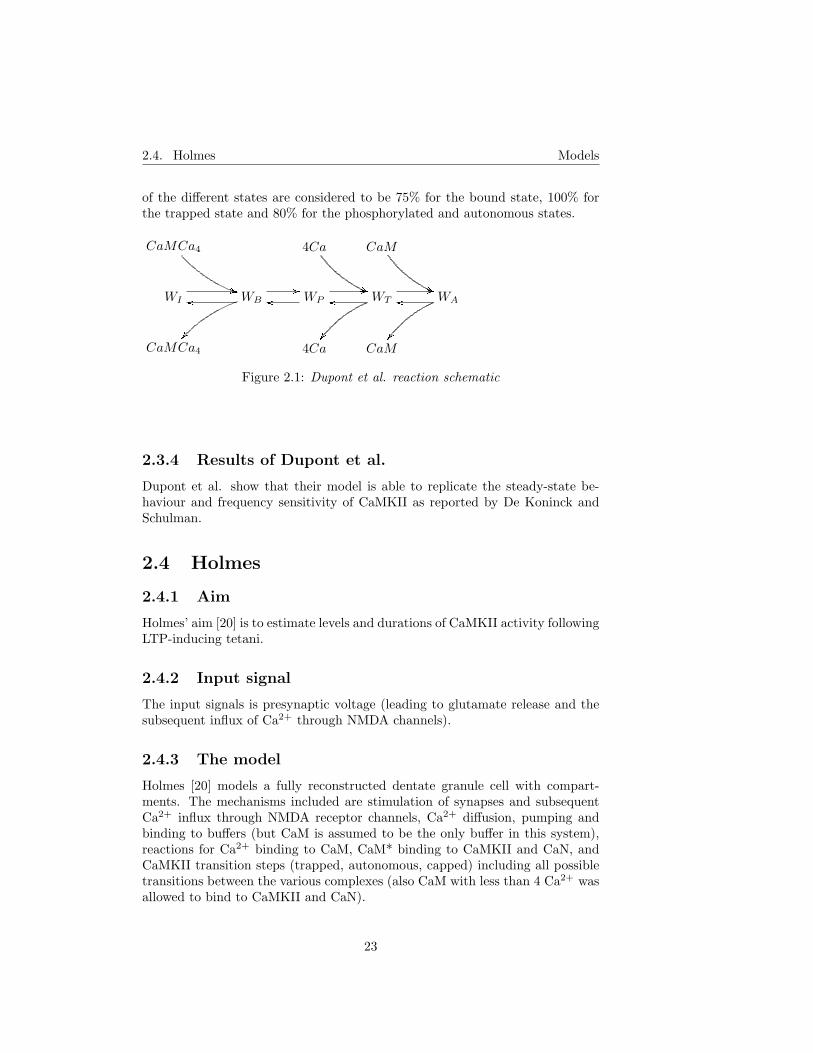
of the different states are considered to be 75% for the bound state, 100% forthe trapped state and 80% for the phosphorylated and autonomous states.
CaMCa4
))
4Ca
((
CaM
((WI
//WB
//
��
oo WPoo//WToo
//
��
WAoo
��CaMCa4 4Ca CaM
Figure 2.1: Dupont et al. reaction schematic
2.3.4 Results of Dupont et al.
Dupont et al. show that their model is able to replicate the steady-state be-haviour and frequency sensitivity of CaMKII as reported by De Koninck andSchulman.
2.4 Holmes
2.4.1 Aim
Holmes’ aim [20] is to estimate levels and durations of CaMKII activity followingLTP-inducing tetani.
2.4.2 Input signal
The input signals is presynaptic voltage (leading to glutamate release and thesubsequent influx of Ca2+ through NMDA channels).
2.4.3 The model
Holmes [20] models a fully reconstructed dentate granule cell with compart-ments. The mechanisms included are stimulation of synapses and subsequentCa2+ influx through NMDA receptor channels, Ca2+ diffusion, pumping andbinding to buffers (but CaM is assumed to be the only buffer in this system),reactions for Ca2+ binding to CaM, CaM* binding to CaMKII and CaN, andCaMKII transition steps (trapped, autonomous, capped) including all possibletransitions between the various complexes (also CaM with less than 4 Ca2+ wasallowed to bind to CaMKII and CaN).
23

2.4. Holmes Models
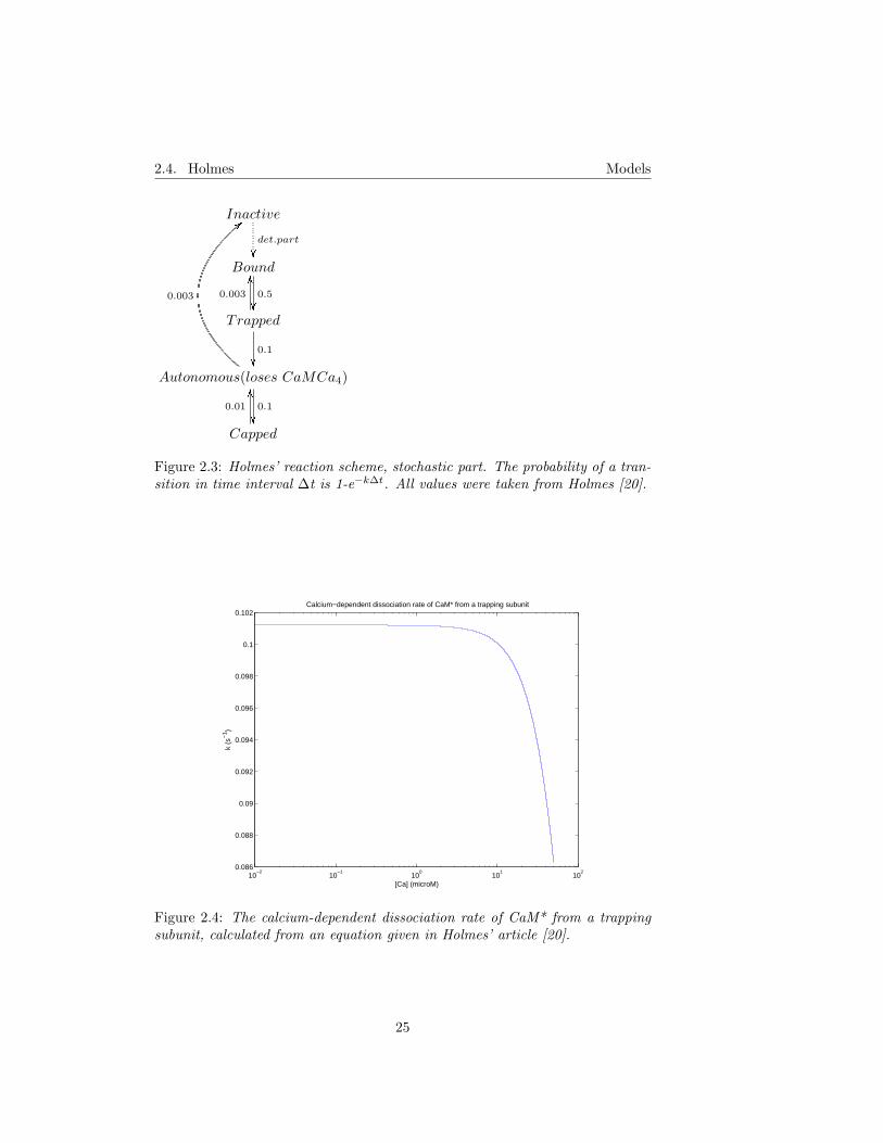
Note that there is a large difference between the values for Ca2+-CaM inter-action that Holmes uses and the value commonly used in the Hill equation.CaM* dissociation from a trapping subunit is dependent on [Ca2+] (see figure2.4). CaM and CaM-Cax diffusion is also included. The model is split into twoparts: one deterministic (for all reactions except for CaMKII transition steps)and one stochastic. Autonomous CaMKII rebinding of CaM* and subsequenttransition back to the trapped state is modeled as a deterministic process. Seefigures 2.2 and 2.3 for details.
Dendritic spine part of the model
The spine head in the model has four spine-head compartments, four spine-neck compartments and twelve dendritic compartments. The dendritic spineis assumed to be a long-thin spine with cylindrical dimensions of 0.55 x 0.55µm for the head and 0.1 x 0.73 µm for the neck. CaMKII is considered to berestricted to the outer 50 nm of the spine head (corresponding to PSD-boundCaMKII).
CaMKII
��
Ca
))��uu
CaN
��CaMKII − CaM
2700 //
OO
4
��
CaM0.2
oo0.2 //
1
��
CaN − CaM5000
oo
OO
100
��CaMKII − CaMCa
2700 //20
OO
100
��
CaMCa5 //
0.8oo
20
OO
33.3
��
CaN − CaMCa3750
oo
60
OO
100
��CaMKII − CaMCa2
20
OO
100
��
2250 //CaMCa2
10 //2
oo
20
OO
5
��
CaN − CaMCa2
10
OO
100
��
1250oo
CaMKII − CaMCa3
2
OO
100
��
22.5 //CaMCa3
100 //100
oo
500
OO
100
��
CaN − CaMCa3
8
OO
100
��
10oo
CaMKII − CaMCa4
100
OO
4.5 //CaMCa4
500
OO
100oo
100 //CaN − CaMCa4
30
OO
0.6oo
Autonomous
33.3
��Trapped
Figure 2.2: Holmes’ reaction scheme, deterministic part. All values (units areµM and s) were taken from Holmes [20]
24
2.4. Holmes Models
Inactive
det.part
��Bound
0.5
��Trapped
0.003
OO
0.1
��Autonomous(loses CaMCa4)
0.1
��
0.003
<<
Capped
0.01
OO
Figure 2.3: Holmes’ reaction scheme, stochastic part. The probability of a tran-sition in time interval ∆t is 1-e−k∆t. All values were taken from Holmes [20].
10−2
10−1
100
101
102
0.086
0.088
0.09
0.092
0.094
0.096
0.098
0.1
0.102
[Ca] (microM)
k (s
−1 )
Calcium−dependent dissociation rate of CaM* from a trapping subunit
Figure 2.4: The calcium-dependent dissociation rate of CaM* from a trappingsubunit, calculated from an equation given in Holmes’ article [20].
25

2.5. Kubota and Bower Models
2.4.4 Holmes’ results
The level of CaM trapping was found to be strongly dependent on the frequencyof the input and the magnitude of the calcium signal. Total activity of CaMKIIdecayed slowly, and decay was further slowed if the activity was induced with arepeated tetanus rather than a single one. When NMDA receptor desensitiza-tion was included and intervals between tetani were short, the maximal activityand number of bound subunits were significantly altered.
Disallowing the binding of Ca to all CaMKII-CaMCaX and CaN-CaMCaX wasshown to increase the total activation approximately twofold, compared to the”normal” case.
2.5 Kubota and Bower
2.5.1 Aim
Kubota and Bower [25] aim to construct a detailed biophysical model basedsolely on the kinetics of purified enzymes and first show that the model replicatesthe frequency sensitivity of CaMKII as reported by De Koninck and Schulman,secondly investigate which the mechanisms responsible for this sensitivity are.
2.5.2 Input signal
The input signal is Ca2+ concentration, given directly.
2.5.3 The model
The model of Kubota and Bower [25] includes Ca2+ binding to CaM, CaMKIIactivation by CaM*, CaMKII phosphorylation on Thr286 and subsequent CaM*trapping, CaMKII autophosphorylation, CaMKII capping. The associationrates of CaM from CaMKII were modeled as functions of [Ca2+].
Ca2+ binding to CaM is modeled by the Adair-Klotz equation [25] that consid-ers the cooperation between sites:
CaM∗ = CaMT ·0.2 · Ca + 0.32 · Ca2 + 0.0336 · Ca3 + 0.00196 · Ca4
4 + 0.8 · Ca + 0.64 · Ca2 + 0.0448 · Ca3 + 0.00196 · Ca4
(2.2)
where CaM* is CaMCa4 concentration, CaMT is total CaM concentration andCa is Ca concentration.
26

2.5. Kubota and Bower Models
In the model, the number of CaMKII subunits is four (to reduce model complex-ity, just as in Coomber’s model). CaMKII states are modeled deterministically,with terms of the kind P i1i2i3i4
j1j2j3j4(i=0,1,2, j=0,1), where the superscripts denote
the phosphorylation state of the first through fourth subunit (i=0,1,2; 1 denotesphosphorylation on Thr286 and 2 denotes phosphorylation on Thr305/306). Thecorresponding subscripts denote if CaM* is bound (=1) or not (=0). The equa-tions are on the form
d
dtP 0001
0000 =kn1 · P 00011000 + kn1 · P 0001
0100 + kn1 · P 00010010 + kn10 · P 0001
0001−
− (CaM∗ · k1 + CaM∗ · k1 + CaM∗ · k1 + CaM∗ · k10) · P 00010000−
− k3 · P 00010000
(2.3)
and configurations with the same rotational symmetry are contained in the sameterm. Interautophosphorylation is assumed to be unidirectional. Capped statesare considered to be active. Dephosphorylation of CaMKII is assumed to beperformed solely by PP1 and to follow the scheme
PP1 + Sp GGGBFGGG PP1SpGGGAPP1 + S (2.4)
i.e. the Michaelis-Menten model, where Sp denotes phosphorylated CaMKIIsubunit and S denotes dephosphorylated CaMKII subunit.
2.5.4 Results of Kubota and Bower
Kubota and Bower found that their model was able to reproduce both the basalenzyme kinetics of CaMKII, in comparison with data from Brocke et al. [7],and the experimentally established frequency sensitivity of CaMKII as reportedby De Koninck and Schulman [13].
Frequency sensitivity
Frequency sensitivity was simulated both in the absence of phosphatase, as inthe experiment by De Koninck and Schulman, and with PP1. No qualitativedifference was observed, but overall phosphorylation was slightly reduced in thepresence of phosphatase. Frequency sensitivity could be modeled with as littleas 2 subunits. Kubota and Bower also claim that the phenomenon ”frequencysensitivity” should be split in two parts; transient and asymptotic frequencysensitivity. Transient frequency sensitivity is predicted to be an intrinsic prop-erty of CaMKII, while asymptotic frequency sensitivity is seen to depend on theinterplay between CaMKII and phosphatase. See Kubota and Bower [25] fordetails.
27
2.6. Lundh Models
2.6 Lundh
2.6.1 Aim
Lundh’s aim [30] is to study the Ca2+-dependence of phosphorylation of synapsinI and its consequences for short-term facilitation.
2.6.2 Input signal
The input signal is Ca2+ influx governed by Hodgkin-Huxley dynamics.
2.6.3 The model
The model by Lundh [30] includes concentrations of synapsin I, cAMP, CaMand Ca2+, and focuses on phosphorylation of synapsin I, not CaMKII activa-tion. Some Ca2+ dynamics are included (a Ca2+ current and an ATP-drivenCa2+ pump, but not Ca2+ buffering). Adenylyl cyclase (AC), phosphodiesterase(PDE) and CaMKII are present as rate constants (i.e. for instance CaM trap-ping by CaMKII is not considered). Ca2+ binding to CaM is modeled as afour-step process. The cytosolic CaM concentration is 10 µM.
As the goal of this model was to study phosphorylation of synapsin and itsconsequences for short-term facilitation (rather than CaMKII activation), theresults and the equations will not be included here. The interested reader isreferred to Lundh [30]. However, it is noteworthy that Lundh is the only oneexcept for Holmes who has an explicit four-step model of Ca2+ binding to CaM.
2.7 Okamoto and Ichikawa
2.7.1 Aim
The aim of Okamoto and Ichikawa [34] is to investigate the functional impor-tance of CaMKII trapping of CaM* in the neuronal structure (several neigh-bouring spines are modelled, as well as their mother dendrite).
2.7.2 Input signal
The input signal is Ca2+, given directly for each spine.
2.7.3 The model
The model includes Ca2+, Ca2+ binding to CaM and CaM* binding to CaMKII,phosphorylation and dephosphorylation of CaMKII on Thr286/287. Capping isnot included. Buffering of Ca2+ and CaM* (i.e. binding to other proteins thanCaM or CaMKII, respectively) is included but not discussed. CaMKII is con-sidered to have 10 subunits.
28

2.7. Okamoto and Ichikawa Models
Phosphorylation is modeled as
K(n)G(n)
GGGGGGGGGAK(n + 1)
G(n) = kaγ2(N − n)(N − n− 1) + kbγ2n(N − n)
γ =[Ca2+/CaM − S]∑Nn=0(N − n)[K(n)]
(2.5)
where K(n) denotes CaMKII holoenzyme of N subunits, of which n are phos-phorylated (and N −n are dephosphorylated). [Ca2+/CaM-S] denotes the con-centration of dephosphorylated CaMKII subunit with bound CaM*.
Dephosphorylation is modeled as
K(n)R(n)
GGGGGGGGGAK(n− 1)
R(n) =nVD
KD +∑N
n=0 n[K(n)]
(2.6)
This gives the following set of dynamical equations
d
dt[K(n)] = G(n− 1)K(n− 1)− (G(n) + R(n))[K(n)]+
+ R(n + 1)K(n + 1)
d
dt[Ca2+/CaM − S] = −
N∑n=0
(G(n)−R(n))[K(n)] + k2[Ca2+/CaM ][S]−
− k−2[Ca2+/CaM − S]
d
dtE = −
N∑n=0
(G(n)−R(n))[K(n)]−
− ks((E − [Ca2+/CaM − S]− [Ca2+/CaM −B])− [CaM ]d)d
dt[CaM ]d = −kd([CaM ]d − (E − [Ca2+/CaM − S]− [Ca2+/CaM −B]))
(2.7)
29
2.8. Zhabotinsky Models
and algebraical equations
E = [Ca2+/CaM − S] + [Ca2+/CaM −B] + [Ca2+/CaM ] + [CaM ]
[S] =N∑
n=0
(N − n)[K(n)]− [Ca2+/CaM − S]
[Ca2+/CaM ] =−a +
√a2 − 4b
2
a = [Ca2+/CaM − S] + K3 + CB −[Ca2+]4E
K41 + [Ca2+]4
b =( [Ca2+]4E
K41 + [Ca2+]4
− [Ca2+/CaM − S])K3
[Ca2+/CaM −B] =[Ca2+/CaM ]CB
K3 + [Ca2+/CaM ]
(2.8)
where E is the total concentration of non-trapped CaM, B denotes other CaM-binding proteins and S denotes (unphosphorylated) CaMKII subunit.
Although it is not immediately apparent, the CaMKII part of this model isquite similar to the CaMKII part of Zhabotinsky’s model (see section 2.8).
2.7.4 Results of Okamoto and Ichikawa
The model by Okamoto and Ichikawa is the only model containing more than onespine, of the models I have studied. The model assumes diffusional transporta-tion of CaM between spines and the parent dendrite. Okamoto and Ichikawashow that CaMKII trapping of CaM* can lead to a ”winner-take-all” competi-tion between spines for CaM, as trapping increases the affinity for CaM* andthereby promotes further trapping.
2.8 Zhabotinsky
2.8.1 Aim
Zhabotinsky’s aim [51] is to investigate if the CaMKII-PP1 system can operateas a bistable switch, i.e. to investigate if CaMKII autophosphorylation can bebistable in a wide range of [Ca2+] and whether such bistability can play a rolein LTP.
2.8.2 Input signal
The input signal consists of simple Ca2+ pulses given by an exponential equationwith given maximal amplitude and time constant.
30

2.8. Zhabotinsky Models
2.8.3 The model
Zhabotinsky’s model [51] contains concentrations of CaMKII – unactivated orphosphorylated on from one to ten subunits, giving a total of eleven differentCaMKII concentrations – PP1 and inhibitor-1. PKA and CaN are included asrate constants in some reactions (see below) and are thus treated as unlimited.CaM is implicitly included in the Ca2+ activation of CaMKII. The fraction ofCaMKII subunits bound to Ca2+/CaM is given by
F =([Ca2+]/KH1)4
1 + ([Ca2+]/KH1)4(2.9)
in contrast to Holmes, who treats all the CaMCaX steps explicitly. The per-subunit rates ν1 of the first phosphorylation step (which requires binding ofCaM* at two neighbouring sites) and ν2 of the following steps are, consequently:
ν1 = 10k1P0 · F 2 (2.10)
ν2 = k1 · F (2.11)
where P0 is the concentration of unactivated (unphosphorylated) CaMKII.
2.8.4 Phosphorylation
Zhabotinsky claims that phosphorylated subunits are phosphorylated at ran-dom, resulting in a random distribution of phosphorylated and unphosphory-lated subunits in the holoenzyme, and assumes that all distinguishable config-urations of subunits in the holoenzyme with a given number of phosphorylatedsubunits exist with equal probabilities. The effective number of autophospho-rylating pairs, w is given by
wi = w10−i =∑i
1 jmj∑i1 mj
(2.12)
31

2.8. Zhabotinsky Models
and ν2 is multiplied with the corresponding w to give the total rate of phospho-rylation, Vi = ν3wiPi:
dP0
dt= −ν1 + ν3P1
dP1
dt= ν1 − ν3P1 − ν2 + 2ν3P2
dP2
dt= ν2P1 − 2ν3P2 − 1.8ν2P2 + 3ν3P3
dP3
dt= 1.8ν2P2 − 3ν3P3 − 2.3ν2P3 + 4ν3P4
dP4
dt= 2.3ν2P3 − 4ν3P4 − 2.7ν2P4 + 5ν3P5
dP5
dt= 2.7ν2P4 − 5ν3P5 − 2.8ν2P5 + 6ν3P6
dP6
dt= 2.8ν2P5 − 6ν3P6 − 2.7ν2P6 + 7ν3P7
dP7
dt= 2.7ν2P6 − 7ν3P7 − 2.3ν2P7 + 8ν3P8
dP8
dt= 2.3ν2P7 − 8ν3P8 − 1.8ν2P8 + 9ν3P9
dP9
dt= 1.8ν2P8 − 9ν3P9 − ν2P9 + 10ν3P10
dP10
dt= ν2P9 − ν310P10
(2.13)
where Pi is the concentration of holoenzymes with i phosphorylated subunits,ν3 is the rate of dephosphorylation, and the total rate of dephosphorylation ofholoenzymes with i phosphorylated subunits is V−i = ν3iPi.
2.8.5 Dephosphorylation
PP1 is deactivated by binding to phosphorylated inhibitor-1. The per-subunitrate of dephosphorylation is given by
ν3 =k2ep
KM +∑10
1 iPi
(2.14)
where ep is the concentration of PP1 not bound to phosphorylated inhibitor-1(I1P). Inhibitor-1 is phosphorylated by PKA and dephosphorylated by activatedCaN. Zhabotinsky has CaN activation modeled as a Hill equation with nH = 3(opposed to Holmes who has the whole range of CaNCaM to CaNCaMCa4 butwho does not really use CaN for anything). The interaction between PP1 and
32
2.9. Lisman and Zhabotinsky Models
inhibitor-1 is expressed as:
dep
dt= −k3Iep + k4(ep0 − ep)
dI
dt= −k3Iep + k4(ep0 − ep) + νPKAI0 −
νCaN ([Ca2+]/KH2)3)I1 + ([Ca2+]/KH2)3)
(2.15)
where ep0 is the total concentration of PP1, I is the concentration of free I1P andI0 is the concentration of free inhibitor-1 which is treated as constant (whichmeans that I may well exceed I0).
2.8.6 Zhabotinsky’s results
Zhabotinsky shows that, if the total concentration of CaMKII subunits is signif-icantly higher than the phosphatase Michaelis constant, two stable steady statesof CaMKII autophosphorylation can be found over a wide range of intracellularCa2+ concentrations. Also, he finds that the levels of CaMKII necessary toproduce this bistability are in the same range as the values found in the post-synaptic density.
However, Zhabotinsky’s model does not include diffusion or CaM binding dy-namics and he treats the supply of CaM as unlimited. Several other authorshave pointed out that the level of CaM might be important, see Okamoto andIchikawa [34] and further discussion in section 5.4.
2.9 Lisman and Zhabotinsky
The Lisman-Zhabotinsky model [29] is built on Zhabotinsky’s model (see theprevious section), no relevant new simulations are performed.
2.9.1 Aim
The aim of Lisman and Zhabotinsky is to use Zhabotinsky’s model as a frame-work for a hypothesis of the mechanism of AMPAR addition to the membrane.
2.9.2 Input signal
The input signal is the same as in Zhabotinsky’s model (in the previous section,section 2.8).
2.9.3 The model
It is proposed that phosphorylated CaMKII enhances transmission through anassembly process that leads to addition of AMPARs to the membrane. CaMKIIis thought to be dephosphorylated only by PP1, and only the PP1 that is heldin the PSD by scaffolding proteins. This restriction means that the relevant
33
2.9. Lisman and Zhabotinsky Models
biochemical compartment is the volume of the PSD within which the concen-tration of CaMKII subunits is 100-200 µM [29] (this is roughly equivalent toHolmes – see section 2.4 – who has CaMKII restricted to the outer 50 nm of thespinehead, equivalent to a concentration of CaMKII subunits of 110 µM). Thiswould make it easier to saturate the local phosphatase pool and thus make iteasier to keep CaMKII in an activated state even at basal [Ca2+].
2.9.4 AMPAR addition to the membrane
Phosphorylated CaMKII is known to bind to the NMDA receptor and α-actinin(an actin-binding protein) binds to α-CaMKII. Another actin-binding protein,called protein 4.1, is known to be in a complex with the protein SAP97. Bothare major binding partners for the GluR1 subunits of AMPARs and form siteswhere AMPARs can anchor (see Lisman and Zhabotinsky [29] and referencestherein). The key link, proposed by Lisman and Zhabotinsky, is that actinfilaments could bind to α-actinin and form a binding site for the protein 4.1,allowing actin filaments to link the NMDAR-CAMKII-α-actinin complex tothe AMPAR-SAP97-4.1 complex. Lisman and Zhabotinsky [29] also list someexperimental evidence for this link.
About the role of AMPAR insertion
Benke et al. [4] found that the increase in AMPAR single channel conductanceresulting from phosphorylation could be sufficient to explain the enhanced AM-PAR efficacy, without the need for AMPAR insertion, and that if new-insertedAMPARs exist, they have to be in the high-conductance state [4],[14].
34

Chapter 3
Comparison of models
In this chapter I will discuss some important general model features and relatethese to the studied models.
3.1 Ca2+ and CaM dynamics
Surprisingly few models contain a multi-step binding scheme for Ca2+ to CaM.Since binding dynamics of CaM surely will influence transient CaMKII acti-vation, at least LFS and/or low-amplitude Ca2+ signals should not be mod-eled without full CaM dynamics. However, the rate constants that are used inHolmes’ model are strongly deviant from the corresponding value used in theHill equation (KHill is ∼ 9 times larger than the equivalent value in Holmesmodel, see below) and the values reported by Vetter and Leclerc [48], see section1.6. An example of CaM dynamics (rate constants were taken from Holmes [20])with a 4Hz Ca2+ signal is given in figure 3.1. The Ca2+ signal in all simulationspresented in this chapter is a square pulse with a duration of 200 ms and anamplitude of 1 µM.
The dynamics of Holmes’ four-step CaM model will also be considerably al-tered by the presence of CaM-binding proteins. CaM dynamics with the sameconditions as in figure 3.1, but with CaN included, are plotted in figures 3.2(3-fold more CaN than CaM) and 3.3 (3-fold less CaN than CaM). Notice es-pecially the interesting difference in the dynamics of CaMCa3 when the CaNconcentration is altered (a close-up is given in figure 3.4). For reference, theconcentration of CaMCa4 with the same 4Hz signal when one uses simplifiedbinding (the Hill equation or the Adair-Klotz equation) is given in figure 3.5.The Adair-Klotz equation takes cooperativity of Ca2+ binding to CaM intoconsideration but still gives the same kind of simplified dynamics as the Hillequation. Note the relatively large difference (∼ 5-fold) between the Hill equa-tion and the Adair-Klotz equation.
35

3.1. Ca2+ and CaM dynamics Comparison of models
0 1 2 30
0.1
0.2
0.3
0.4
0.5
t (s)
[CaM
Ca]
(uM
)
0 1 2 30
0.1
0.2
0.3
0.4
0.5
0.6
0.7
t (s)
[CaM
Ca 2] (
uM)
0 1 2 30
1
2
3
4
5
6
7x 10
−3
t (s)
[CaM
Ca 3] (
uM)
0 1 2 30
0.2
0.4
0.6
0.8
1
1.2
1.4x 10
−3
t (s)
[CaM
Ca 4] (
uM)
Figure 3.1: The dynamics of all CaMCax states with a 4Hz Ca2+ signal of 1µM Ca2+ in 200 ms. Total CaM concentration was 10 µM. All rate constantswere taken from Holmes [20].
As is evident from the previous figures (3.1-3.4), the presence of CaM-bindingprotein alters both shape and amplitude of the resulting CaMCax concentra-tions in Holmes’model. The frequency of the input also has some influence: infigure 3.6 a comparison is given between CaMCax concentrations in the presenceof much and little CaN (30 µM and 3.3 µM respectively, in comparison to 10µM of CaM) with the same Ca2+ signal but a frequency of 1 Hz.
There are some experimental arguments for and against the usefulness of theHill equation: Burgoyne et al. [8] found, in measurements of CaM behaviourat different [Ca2+], that CaM bound no distinguishable amount of Ca2+ belowCa2+ concentrations of ∼200 nM (i.e. no Ca2+ is bound at resting levels, sothere should not be any CaM* present at all, contrary to the Hill equation ap-proach), and that the half-maximal occupancy occurred at ∼1 µM. This is inaccordance with the value commonly used, but ∼ 9 times larger than the result-ing value of
( ∏4i=1
kb
kf
)1/4 for Holmes’ model (the exact value is 8.8). Burgoyneet al. also calculate that in order to have complete loading of CaM by Ca2+
within 1 ms, [Ca2+] has to rise above 10 µM. Thus, the lower [Ca2+], the lessrapid is the loading of CaM by Ca2+. If substantial ”integration” of Ca2+ byCaM (section 1.6.1) is evident, simplified binding will likewise not account forthis.
36

3.1. Ca2+ and CaM dynamics Comparison of models
0 1 2 30
0.05
0.1
0.15
0.2
0.25
0.3
0.35
t (s)
[CaM
Ca]
(uM
)
0 1 2 30
0.1
0.2
0.3
0.4
0.5
t (s)
[CaM
Ca 2] (
uM)
0 1 2 30
1
2
3
4
5
6x 10
−3
t (s)
[CaM
Ca 3] (
uM)
0 1 2 30
2
4
6x 10
−4
t (s)
[CaM
Ca 4] (
uM)
Figure 3.2: The dynamics of all CaMCax states with a 4Hz Ca2+ signal of 1µM Ca2+ in 200 ms, in the presence of a CaM-binding protein (CaN). TotalCaM concentration was 10 µM and total CaN concentration was 30 µM. Allrate constants were taken from Holmes [20], thus all CaMCax can bind to CaN.
3.1.1 A simple test of correction of Holmes’ CaM con-stants
The simplest way to examine the effects of the difference of the Ca2+-CaM stepsis to multiply the forward rate constants with the fourth root of the difference,which is approximately 8.8. The corresponding result to figure 3.1 is shown infigure 3.7. Note that the difference between CaMCa4 in Holmes’ model andCaMCa4 from the Hill equation is much smaller (but the level of CaMCa4 isstill 5-fold lower than with the Hill equation) and that the agreement with theAdair-Klotz equation is rather good. Also, the general ”shape” of the CaMCa4-curve is more ”Hill-like” than with Holmes’ original model.
To continue the comparison, also the forward rate constants for Ca2+ bind-ing to CaN-CaMCax were multiplied with the same correcting factor (to satisfythe demand for thermodynamic equilibrium) and simulations corresponding tofigures 3.2 and 3.3 were performed. The results are shown in figures 3.8 and 3.9,respectively. It can be seen that the differences between the states with moreand less CaM-binding proteins are more pronounced, but that the shape of thecurve in the four-step case still is approximately like that of the Hill equation.Note that the level of available CaM is drastically lowered in the case of limitingCaM. The behaviours of the other CaMCax are also altered, but this is hard
37

3.1. Ca2+ and CaM dynamics Comparison of models
0 1 2 30
0.1
0.2
0.3
0.4
t (s)
[CaM
Ca]
(uM
)
0 1 2 30
0.1
0.2
0.3
0.4
0.5
0.6
0.7
t (s)
[CaM
Ca 2] (
uM)
0 1 2 30
1
2
3
4
5x 10
−3
t (s)
[CaM
Ca 3] (
uM)
0 1 2 30
0.2
0.4
0.6
0.8
1x 10
−3
t (s)
[CaM
Ca 4] (
uM)
Figure 3.3: The dynamics of all CaMCax states with a 4Hz Ca2+ signal of 1µM Ca2+ in 200 ms, in the presence of a CaM-binding protein (CaN). TotalCaM concentration was 10 µM and total CaN concentration was 3.3 µM. Allrate constants were taken from Holmes [20], thus all CaMCax can bind to CaN.
0.9 1 1.1 1.2 1.3 1.4 1.5 1.6 1.7 1.8
2
2.5
3
3.5
4
4.5
x 10−3
t (s)
[CaM
Ca 3] (
uM)
Figure 3.4: Close-up of [CaMCa3] from the previous figure
38

3.1. Ca2+ and CaM dynamics Comparison of models
0 0.5 1 1.5 2 2.5 30
0.5
1
1.5
2
2.5
3
3.5
4
4.5
5
t (s)
[CaM
Ca 4] (
uM)
Simplified binding of Ca2+ to CaM
Figure 3.5: The concentration of CaMCa4 given by the Hill equation (solid lines)and the Adair-Klotz equation (dotted lines), with the same conditions as in theprevious two figures.
to interpret since there will be effects from Holmes’ assumption that CaN canbind all CaMCax.
39

3.2. Trapping of CaM* and capping of CaMKII Comparison of models
0 1 2 30
0.05
0.1
0.15
0.2
0.25
0.3
0.35
t (s)
[CaM
Ca]
(uM
)
0 1 2 30
0.1
0.2
0.3
0.4
0.5
t (s)
[CaM
Ca 2] (
uM)
0 1 2 30
0.5
1
1.5
2
2.5
3
3.5x 10
−3
t (s)
[CaM
Ca 3] (
uM)
0 1 2 30
2
4
6x 10
−4
t (s)
[CaM
Ca 4] (
uM)
Figure 3.6: The dynamics of CaMCax in the presence of 30 µM and 3.3 µM CaN(dotted and solid lines, respectively). Total [CaM] was 10 µM, the amplitude ofthe Ca2+ signal was 1 µM, duration 200 ms and frequency 1 Hz.
3.2 Trapping of CaM* and capping of CaMKII
It has been speculated that trapping of CaM* has the functional role of depriv-ing the other CaM-dependent pathways of CaM. Thus, trapping can for instancelower the level of PP1 activity by preventing activation of CaN (that dephos-phorylates the PP1 inhibitor I1P). Trapping also seems to protect CaMKIIsubunits from capping. Capping, on the other hand, prevents previously au-tonomous subunits from reuptake of CaM* (i.e. ensures that CaM* becomesavailable to the other CaM pathways). If any other CaM-dependent reactionsthan CaMKII activation are included in a model (for instance PP1 regulation),then both trapping and capping should be included. None of the studied mod-els includes both capping and full PP1 regulation. I consider the lack of allCaMKII activation states to be one of the most important deficiencies of theZhabotinsky model.
3.3 Feature span
Since Ca2+ is involved in so many cellular processes, it is hard to cut out anumber of pathways and claim that they are a self-sufficient system. Never-theless, I think most of the studied models contain too few processes. If oneconsiders CaMKII and PP1 to be a system, one should also add the PP1 regu-lation pathway that competes with CaMKII both for CaM* and for the level of
40
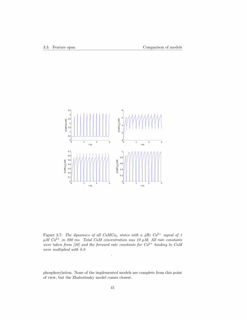
3.3. Feature span Comparison of models
0 1 2 30
0.5
1
1.5
2
2.5
3
3.5
t (s)
[CaM
Ca]
(uM
)
0 1 2 30
2
4
6
8
t (s)
[CaM
Ca 2] (
uM)
0 1 2 30
0.1
0.2
0.3
0.4
0.5
0.6
0.7
t (s)
[CaM
Ca 3] (
uM)
0 1 2 30
0.2
0.4
0.6
0.8
1
t (s)
[CaM
Ca 4] (
uM)
Figure 3.7: The dynamics of all CaMCax states with a 4Hz Ca2+ signal of 1µM Ca2+ in 200 ms. Total CaM concentration was 10 µM. All rate constantswere taken from [20] and the forward rate constants for Ca2+ binding to CaMwere multiplied with 8.8
.
phosphorylation. None of the implemented models are complete from this pointof view, but the Zhabotinsky model comes closest.
41

3.3. Feature span Comparison of models
0 1 2 30
0.05
0.1
0.15
0.2
0.25
0.3
0.35
t (s)
[CaM
Ca]
(uM
)
0 1 2 30
0.2
0.4
0.6
0.8
t (s)
[CaM
Ca 2] (
uM)
0 1 2 30
0.005
0.01
0.015
0.02
t (s)
[CaM
Ca 3] (
uM)
0 1 2 30
1
2
3
4
5
6
7x 10
−3
t (s)
[CaM
Ca 4] (
uM)
Figure 3.8: The dynamics of all CaMCax states with a 4Hz Ca2+ signal of 1 µMCa2+ in 200 ms. Total CaM concentration was 10 µM and total CaN concen-tration was 30 µM. All rate constants were taken from [20] and the forward rateconstants for Ca2+ binding to bound and unbound CaM were multiplied with 8.8
.
42

3.4. Implementation Comparison of models
0 1 2 30
0.5
1
1.5
2
2.5
t (s)
[CaM
Ca]
(uM
)
0 1 2 30
1
2
3
4
5
t (s)
[CaM
Ca 2] (
uM)
0 1 2 30
0.1
0.2
0.3
0.4
0.5
t (s)
[CaM
Ca 3] (
uM)
0 1 2 30
0.1
0.2
0.3
0.4
0.5
0.6
0.7
t (s)
[CaM
Ca 4] (
uM)
Figure 3.9: The dynamics of all CaMCax states with a 4Hz Ca2+ signal of 1 µMCa2+ in 200 ms. Total CaM concentration was 10 µM and total CaN concentra-tion was 3.3 µM. All rate constants were taken from [20] and the forward rateconstants for Ca2+ binding to bound and unbound CaM were multiplied with 8.8
.
3.4 Implementation
I have found that in general, published models are hard to implement sinceimportant rate constants often are missing, or units are not given. Simple de-
43
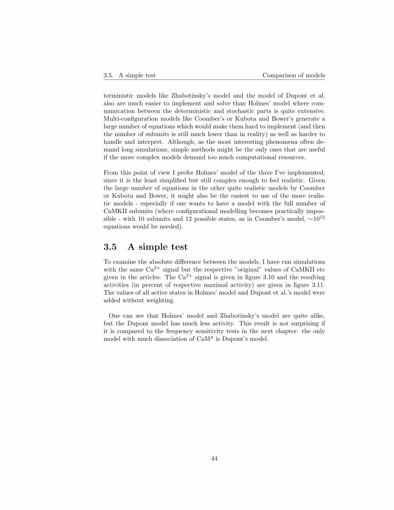
3.5. A simple test Comparison of models
terministic models like Zhabotinsky’s model and the model of Dupont et al.also are much easier to implement and solve than Holmes’ model where com-munication between the deterministic and stochastic parts is quite extensive.Multi-configuration models like Coomber’s or Kubota and Bower’s generate alarge number of equations which would make them hard to implement (and thenthe number of subunits is still much lower than in reality) as well as harder tohandle and interpret. Although, as the most interesting phenomena often de-mand long simulations, simple methods might be the only ones that are usefulif the more complex models demand too much computational resources.
From this point of view I prefer Holmes’ model of the three I’ve implemented,since it is the least simplified but still complex enough to feel realistic. Giventhe large number of equations in the other quite realistic models by Coomberor Kubota and Bower, it might also be the easiest to use of the more realis-tic models - especially if one wants to have a model with the full number ofCaMKII subunits (where configurational modelling becomes practically impos-sible - with 10 subunits and 12 possible states, as in Coomber’s model, ∼1012
equations would be needed).
3.5 A simple test
To examine the absolute difference between the models, I have run simulationswith the same Ca2+ signal but the respective ”original” values of CaMKII etcgiven in the articles. The Ca2+ signal is given in figure 3.10 and the resultingactivities (in percent of respective maximal activity) are given in figure 3.11.The values of all active states in Holmes’ model and Dupont et al.’s model wereadded without weighting.
One can see that Holmes’ model and Zhabotinsky’s model are quite alike,but the Dupont model has much less activity. This result is not surprising ifit is compared to the frequency sensitivity tests in the next chapter: the onlymodel with much dissociation of CaM* is Dupont’s model.
44
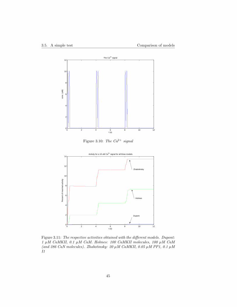
3.5. A simple test Comparison of models
0 2 4 6 8 10 120
2
4
6
8
10
12
t (s)
conc
. (uM
)
The Ca2+ signal
Figure 3.10: The Ca2+ signal
0 2 4 6 8 10 120
2
4
6
8
10
12
14
t (s)
Per
cent
of m
axim
al a
ctiv
ity
Activity for a 10 uM Ca2+ signal for all three models
Zhabotinsky
Holmes
Dupont
Figure 3.11: The respective activities obtained with the different models. Dupont:1 µM CaMKII, 0.1 µM CaM. Holmes: 100 CaMKII molecules, 100 µM CaM(and 286 CaN molecules). Zhabotinsky: 20 µM CaMKII, 0.05 µM PP1, 0.1 µMI1
45

3.5. A simple test Comparison of models
46
Chapter 4
Comparisons withexperimental data
In this chapter, results from simulations with the models of Dupont et al. [16],Holmes [20] and Zhabotinsky [51] are reported. Comparisons with experimentalresults from Bradshaw et al. [6] and De Koninck and Schulman [13] are made.
4.1 Dupont et al
4.1.1 Implementation
I implemented this model in Matlab 6.5. A fourth-order Runge-Kutta methodwas used to integrate the equations. The stability varied considerably withCa concentration (a thousandfold decrease in the timestep length was requiredwhen the Ca concentration was increased from 5 to 50 µM).
4.1.2 Comparison with experimental data
The Dupont model was explicitly developed to embody the frequency sensitivitycharacteristics reported by De Koninck and Schulman [13]. A thorough compar-ison with these data can be found in [16]. I have tested to run the model withthe same 1 Hz and 4 Hz stimuli described in the article (which was also usedby De Koninck and Schulman). The resulting frequency sensitivity is clearlyevident (see figure 4.1).
I thought it would be interesting to examine if the model’s behaviour agreedalso with data from Bradshaw et al. [6], so I made steady-state simulationswith 50 µM CaM, varying Ca2+ concentrations and 1 µM CaMKII. All relativeactivities were considered equal to 1. Values of constants are given in the figurelegend of figure 4.2. If one assumes that the ”phosphorylated state” WP isequal to the ”autonomy” measured by Bradshaw et al., the agreement is rather
47
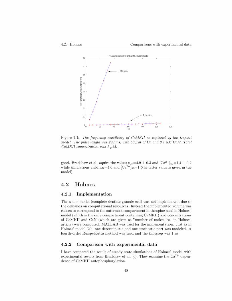
4.2. Holmes Comparisons with experimental data
0 20 40 60 80 100 1200
0.1
0.2
0.3
0.4
0.5
0.6
0.7
0.8Frequency sensitivity of CaMKII, Dupont model
t (s)
conc
of p
hosp
h. C
aMK
II (m
icro
M)
4Hz stim.
1 Hz stim.
Figure 4.1: The frequency sensitivity of CaMKII as captured by the Dupontmodel. The pulse length was 200 ms, with 50 µM of Ca and 0.1 µM CaM. TotalCaMKII concentration was 1 µM.
good. Bradshaw et al. aquire the values nH=4.9 ± 0.3 and [Ca2+]50=1.4 ± 0.2while simulations yield nH=4.0 and [Ca2+]50=1 (the latter value is given in themodel).
4.2 Holmes
4.2.1 Implementation
The whole model (complete dentate granule cell) was not implemented, due tothe demands on computational resources. Instead the implemented volume waschosen to correspond to the outermost compartment in the spine head in Holmes’model (which is the only compartment containing CaMKII) and concentrationsof CaMKII and CaN (which are given as ”number of molecules” in Holmes’article) were computed. MATLAB was used for the implementation. Just as inHolmes’ model [20], one deterministic and one stochastic part was modeled. Afourth-order Runge-Kutta method was used and the timestep was 1 µs.
4.2.2 Comparison with experimental data
I have compared the result of steady state simulations of Holmes’ model withexperimental results from Bradshaw et al. [6]. They examine the Ca2+ depen-dence of CaMKII autophosphorylation.
48

4.2. Holmes Comparisons with experimental data
10−1
100
101
102
0
10
20
30
40
50
60
70
80
90
100Comparison btw the Dupont model and Bradshaw data
[Ca2+] (microM)
perc
ent a
ctiv
atio
n
Figure 4.2: Comparison of steady-state phosphorylation of Dupont’smodel with experimental data from Bradshaw et al. (dotted). Concen-trations: CaMKII=1 µM,CaM=50 µM. Experimental values were nH=4.9 ±0.3 (Hill number) and Cah=1.4 ± 0.2 (half-maximal Ca-value). Simulation re-sults correspond to Cah=1 (given in model) and nH=4.0. The values a=0.0036,b=2.013, c=-1.0156 and K′
A=0.45 (taken from Dupont et al. [16]) were used.
I have performed steady state simulations of Holmes’ model with Ca2+ concen-trations ranging from 0.1 to 50 µM, with 100 µM CaM and 10.97 µM CaMKII(which is equivalent to 100 10-subunit CaMKII molecules with the volume equalto the volume of the outermost compartment Holmes uses). The closest cor-responding values in Bradshaw et al. [6] are 50 µM CaM and 5 µM CaMKII.Bradshaw et al. recorded their results after 5 minutes, so I ran the simulationsfor the same time. Regrettably, I do not have the resources to run multiplesimulations and average the results; the results presented below are from singlesimulations.
Bradshaw et al. performed their experiments at 0◦C to avoid capping, whileHolmes’ model includes capping. Thus, I modified the model so that it no longerincluded capping. No other rate constants were adjusted.
For comparison with the experimental values I use the equation
A = Amax[Ca2+]nH /([Ca2+]nH + Ca2+1/2
nH ) (4.1)
49
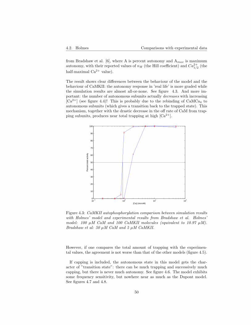
4.2. Holmes Comparisons with experimental data
from Bradshaw et al. [6], where A is percent autonomy and Amax is maximumautonomy, with their reported values of nH (the Hill coefficient) and Ca2+
1/2 (thehalf-maximal Ca2+ value).
The result shows clear differences between the behaviour of the model and thebehaviour of CaMKII: the autonomy response in ’real life’ is more graded whilethe simulation results are almost all-or-none. See figure 4.3. And more im-portant: the number of autonomous subunits actually decreases with increasing[Ca2+] (see figure 4.4)! This is probably due to the rebinding of CaMCa4 toautonomous subunits (which gives a transition back to the trapped state). Thismechanism, together with the drastic decrease in the off rate of CaM from trap-ping subunits, produces near total trapping at high [Ca2+].
10−1
100
101
102
0
10
20
30
40
50
60
70
80
90
100
[Ca] (microM)
Per
cent
tota
l act
ivity
Figure 4.3: CaMKII autophosphorylation comparison between simulation resultswith Holmes’ model and experimental results from Bradshaw et al. Holmes’model: 100 µM CaM and 100 CaMKII molecules (equivalent to 10.97 µM).Bradshaw et al: 50 µM CaM and 5 µM CaMKII.
However, if one compares the total amount of trapping with the experimen-tal values, the agreement is not worse than that of the other models (figure 4.5).
If capping is included, the autonomous state in this model gets the char-acter of ”transition state”: there can be much trapping and successively muchcapping, but there is never much autonomy. See figure 4.6. The model exhibitssome frequency sensitivity, but nowhere near as much as the Dupont model.See figures 4.7 and 4.8.
50
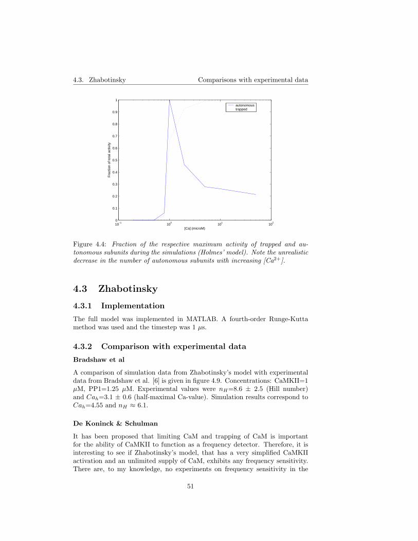
4.3. Zhabotinsky Comparisons with experimental data
10−1
100
101
102
0
0.1
0.2
0.3
0.4
0.5
0.6
0.7
0.8
0.9
1
[Ca] (microM)
Fra
ctio
n of
tota
l act
ivity
autonomoustrapped
Figure 4.4: Fraction of the respective maximum activity of trapped and au-tonomous subunits during the simulations (Holmes’ model). Note the unrealisticdecrease in the number of autonomous subunits with increasing [Ca2+].
4.3 Zhabotinsky
4.3.1 Implementation
The full model was implemented in MATLAB. A fourth-order Runge-Kuttamethod was used and the timestep was 1 µs.
4.3.2 Comparison with experimental data
Bradshaw et al
A comparison of simulation data from Zhabotinsky’s model with experimentaldata from Bradshaw et al. [6] is given in figure 4.9. Concentrations: CaMKII=1µM, PP1=1.25 µM. Experimental values were nH=8.6 ± 2.5 (Hill number)and Cah=3.1 ± 0.6 (half-maximal Ca-value). Simulation results correspond toCah=4.55 and nH ≈ 6.1.
De Koninck & Schulman
It has been proposed that limiting CaM and trapping of CaM is importantfor the ability of CaMKII to function as a frequency detector. Therefore, it isinteresting to see if Zhabotinsky’s model, that has a very simplified CaMKIIactivation and an unlimited supply of CaM, exhibits any frequency sensitivity.There are, to my knowledge, no experiments on frequency sensitivity in the
51

4.3. Zhabotinsky Comparisons with experimental data
10−1
100
101
102
0
10
20
30
40
50
60
70
80
90
100
[Ca] (microM)
Per
cent
of m
axim
al tr
appi
ng
Figure 4.5: Total amount of trapped subunits (Holmes’ model) during the sim-ulations, compared to the experimental ’percent max. autonomy’ of Bradshawet al. Experimental values were chosen within the boundaries of the standarddeviation to give as good agreement as possible ([Ca]50=1.3, nH=5.8).
presence of PP1, but the model by Kubota and Bower exhibited a frequencysensitivity that only changed a little when PP1 was added.
The Zhabotinsky model gives yet another opportunity: to investigate if andhow the frequency sensitivity is changed when inhibition of PP1 is added. Fig-ures 4.10 and 4.11 show the frequency sensitivity with I1. There is practicallyno difference between the two cases, i.e. frequency sensitivity with and withoutI1, at least not at the relatively low concentrations I have examined (data notshown).
52
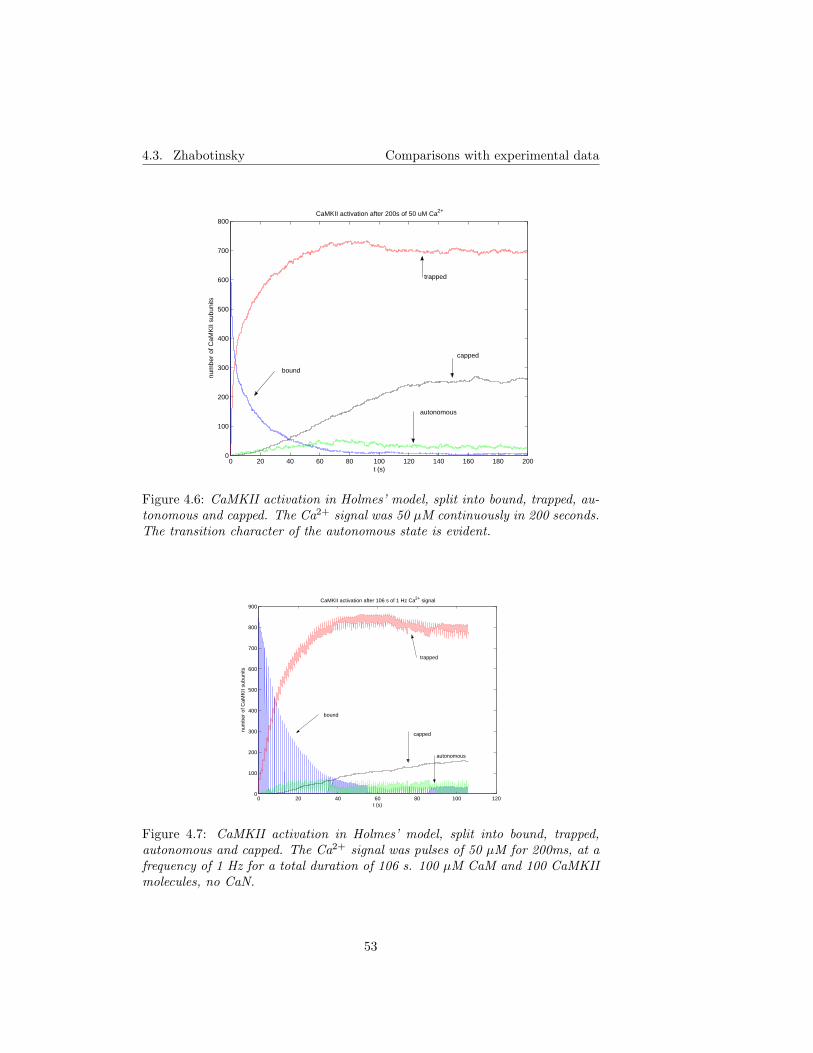
4.3. Zhabotinsky Comparisons with experimental data
0 20 40 60 80 100 120 140 160 180 2000
100
200
300
400
500
600
700
800
t (s)
num
ber
of C
aMK
II su
buni
tsCaMKII activation after 200s of 50 uM Ca2+
bound
trapped
autonomous
capped
Figure 4.6: CaMKII activation in Holmes’ model, split into bound, trapped, au-tonomous and capped. The Ca2+ signal was 50 µM continuously in 200 seconds.The transition character of the autonomous state is evident.
0 20 40 60 80 100 1200
100
200
300
400
500
600
700
800
900
t (s)
num
ber
of C
aMK
II su
buni
ts
CaMKII activation after 106 s of 1 Hz Ca2+ signal
bound
trapped
autonomous
capped
Figure 4.7: CaMKII activation in Holmes’ model, split into bound, trapped,autonomous and capped. The Ca2+ signal was pulses of 50 µM for 200ms, at afrequency of 1 Hz for a total duration of 106 s. 100 µM CaM and 100 CaMKIImolecules, no CaN.
53

4.3. Zhabotinsky Comparisons with experimental data
0 5 10 15 20 25 30 350
100
200
300
400
500
600
700
800
900
1000
t (s)
num
ber
of C
aMK
II su
buni
ts
CaMKII activation after 30 s of 4 Hz Ca2+ signal
bound
trapped
autonomous capped
Figure 4.8: CaMKII activation in Holmes’ model, split into bound, trapped,autonomous and capped. The Ca2+ signal was pulses of 50 µM for 200ms, at afrequency of 4 Hz for a total duration of 30 s. 100 µM CaM and 100 CaMKIImolecules, no CaN.
10−1
100
101
102
0
10
20
30
40
50
60
70
80
90
100Comparison of CaMKII activation −− Zhabotinsky’s model vs. experimental values
[Ca2+] (microM)
% o
f max
imum
act
ivat
ion
Zhabotinsky Experimental
Figure 4.9: Comparison of steady-state phosphorylation of Zhabotinsky’s modelwith experimental data from Bradshaw et al. [6]. Concentrations: CaMKII=1µM, PP1=1.25 µM. Experimental values were nH=8.6 ± 2.5 (Hill number)and Cah=3.1 ± 0.6 (half-maximal Ca-value). Simulation results correspond toCah=4.55 and nH ≈ 6.1 .
54

4.3. Zhabotinsky Comparisons with experimental data
0 20 40 60 80 100 1200
2
4
6
8
10
12
14
16
18
20
t (s)
conc
. of f
ully
act
ive
CaM
KII
(uM
)
Frequency sensitivity of Zhabotinsky’s model with I1 and PP1
4 Hz
1 Hz
Figure 4.10: Frequency sensitivity of Zhabotinsky’s model with I1 regulation ofPP1 activity. Only the concentration of fully active CaMKII is shown (1 Hzand 4 Hz pulses of 50 µM Ca2+, 20 µM CaMKII, 0.05 µM PP1 and 0.1 µMI1).
0 20 40 60 80 100 1200
10
20
30
40
50
60
70
80
90
100
t (s)
perc
ent o
f max
imal
act
ivity
Frequency sensitivity of Zhabotinsky’s model with I1 and PP1
4 Hz 1 Hz
Figure 4.11: Frequency sensitivity of Zhabotinsky’s model with I1 regulation ofPP1 activity. The total weighted sum of CaMKII activity is shown, in percentof maximal activity (1 Hz and 4 Hz pulses of 50 µM Ca2+, 20 µM CaMKII,0.05 µM PP1 and 0.1 µM I1).
55

4.3. Zhabotinsky Comparisons with experimental data
56

Chapter 5
Model assumptionsrevisited
This chapter contains specific discussion on model features, assumptions andtheir consequences. A general concluding discussion on modelling of CaMKIIactivation can be found in chapter 6.
5.1 The well-stirred cell assumption
Assuming that all involved species are equally distributed in the modeled vol-ume (i.e. that there are no spatial variations in concentration) is referred to asthe well-stirred cell assumption. This means that diffusion-related effects areneglected, as well as spatially related differences in concentration. The validityof the assumption differs from situation to situation, but in situations wherediffusion is important (for instance in larger volumes), the assumption is gener-ally not valid, likewise when the number of molecules is small.
One way to model processes where diffusion is included is to use compartmentalmodelling, where the simulated volume is divided into relevant subvolumes andthe diffusional exchange between neighbouring subvolumes is computed for eachtimestep (A relevant subvolume could in the current context be the PSD, whereamong others CaMKII, NMDARs and AMPARs are anchored). A general dis-cussion of modelling of complex biological systems can be found in Weng etal. [49].
5.2 Simplified Ca2+ binding to CaM
Holmes is the only one who includes all CaM-Ca binding steps in a CaMKII ac-tivation model (he even goes as far as allowing all different CaMCaX (X=0,...,4)to bind to both CaMKII and CaN). The standard way otherwise seems to be
57

5.3. Diffusion of CaM and CaM* Model assumptions revisited
to assume that the binding of Ca2+ to CaM is always at equilibrium, and use asimplified binding scheme where
CaMCa4 = CaMTot ·Ca4
Ca4 + [Ca2+]41/2
(5.1)
This is the Hill equation, with nH=4.
This does not completely reproduce CaM dynamics (see sections 1.6 and 3.1);the simulated concentration and dynamics of CaM* will precisely follow those ofthe Ca2+ signal, as shown in the previous chapter, section 3.1. In steady-statesimulations this might not be so important but otherwise it will have effects onfor instance the level of trapping.
5.3 Diffusion of CaM and CaM*
In cells, almost all CaM* is bound. The Holmes model includes diffusion ofCaM*, and CaM diffusion between spines is the key assumption in the Okamoto-Ichikawa model. As even Ca2+ is safely sequestered in the spine (at least for∼1 s [38]), how valid are these assumptions?
5.3.1 Diffusion of CaM
According to Kim et al. [23], diffusion of CaM is slowed down by a factor of2.2 when [Ca2+] rises above resting levels (giving a τD of 1.6 ms as opposedto τD=0.73 ms at resting levels), and at least part of this slowing is due tointeractions with CaMKII.
These experiments were carried out in intact cells (HEK-293 type) with a con-centration of CaMKII in the nanomolar range. Given the large number ofcalmodulin-binding species in the spine, and the fact that especially CaMKII ispresent in much larger concentration (∼10 µM in the PSD) than in the exper-iment, there would likely be a much more pronounced slowing effect on CaMdiffusion when Ca2+ levels rise. Thus, it is questionable if CaM diffusion betweenspines in reality is fast enough to allow the kind of winner-take-all competitionbetween spines for CaM that Okamoto and Ichikawa describe.
5.3.2 Diffusion of CaM*
According to Persechini and Stemmer [36], the mean distance of diffusion forCaMCax, before it loses all bound Ca2+, is ∼ 0.1 µm in prescence of excesstargets. Diffusion of CaMCaX can therefore not be an important Ca2+-bearingprocess.
58

5.4. CaM levels Model assumptions revisited
The short diffusion distance of CaM* means that there can be – dependingon the distribution of CaM and targets of CaM – transient local elevations ofCaM* (also depending on if only CaMCa4 or all CaMCax are bound by targets).Different pathways of CaM* can then, at least indirectly, influence each other.
5.4 CaM levels
Persechini and Cronk [35] have examined the relation between concentrations ofCa2+ and CaM* in intact human embryonic kidney 293 (HEK-293) cells. Theyfound that the concentration of CaM-binding proteins was ∼2-fold higher thanthe concentration of CaM, and that the maximal level of CaM* was 45 nM.HEK 293 cells have been found to be considerably similar to neurons and cellsof neuronal lineage [39] so these results may be relevant also for neurons. Also,the level of Ca2+ required to produce half-maximal concentration of free CaM*was 20-fold less in the cell than the concentration required to half-saturate pureCaM in vitro, and below a [Ca2+] of 0.2 µM there was no evident CaM activation[35], [8].
5.4.1 CaM distribution in the spine
As is illustrated by the results of Okamoto and Ichikawa [34], CaM will mostlikely not be evenly distributed in the cell. Instead, the concentration of CaMwill be highest where the binding of CaM is most efficient (i.e. where the mostCaM is bound compared to the amount of CaM that is released).
5.4.2 Limiting CaM
According to Persechini and Stemmer [36], CaM concentration in the cell is lim-iting, i.e., CaM concentration is significantly below the total concentration of itstargets. This would make the assumption in the Zhabotinsky model [51], thatthe CaM concentration is unlimited and constant, improper. The Zhabotinskymodel, though, better captures the experimental behaviour of the CaMKII-PP1system (examined in [6]) but on the other hand, in that experiment there are nocompeting CaM pathways or targets. Persechini and Stemmer also found thatthe behaviours of CaM binding systems with and without excess CaM can bequalitatively different.
If the CaM concentration is limiting, CaM trapping provides a mechanism todetect the frequency of calcium spikes [11]. Limiting CaM also means that theα/β ratio of CaMKII heteromers becomes important, since autophosphorylationis dependent on CaM affinity. Intersubunit phosphorylation of β-CaMKII thencan become dependent on CaM* binding to its lower-affinity α-CaMKII neigh-bours and vice versa (see [7] for further discussion). Also, the as yet unresolvedissue of uni- or bidirectional intersubunit phosphorylation might not be as triv-
59

5.5. Modelling CaMKII configurations Model assumptions revisited
ial as it has been thought to be (by, among others, Holmes [20]) if the CaMaffinity of neighbours is different and the neighbours are unevenly distributed.
5.5 Modelling CaMKII configurations
Given that a CaMKII molecule consists of 8-12 subunits of at least two possibleisoforms and that each of these subunits will be in one of five different states,the number of possible configurations to model is somewhat daunting from acomputational point of view. Mainly, there are two ways to choose between:stochastic (using, for instance, a matrix with numbers indicating the state ofeach subunit) or deterministic (using one concentration for each configurationincluded in the model).
5.5.1 Simplifications
Often, modellers prefer a deterministic approach and this will cause the modelto become increasingly complex (and computationally heavy) with each stateincluded in the model. The most obvious simplification is to take advantageof the fact that the CaMKII molecule is rotationally symmetric and adjust therate constants for each configuration. Also, the existence of isoforms is usuallyignored (see discussion in section 5.7). Another simplification is to assume thatthe number of CaMKII subunits is constant (often, it is assumed to be equal to10), and yet another, used by for instance Coomber [11] and Kubota and Bower[25], is to reduce the number of subunits. Models have also been simplifiedby using one ”activated” CaMKII state instead of several different states (forinstance Zhabotinsky [51]).
5.6 CaMKII capping
Only some models ([11],[20],[25]) include CaMKII capping, phosphorylation onThr305/306, that inhibits CaM* binding and leads to an autonomous form withdecreased [26] or inhibited [11] activity (according to Kubota and Bower [25]capped subunits are active, but they only cite unpublished data). There seems tobe no general agreement on how active capped subunits are, or the importanceof capping. Based on Coomber’s finding, that prior LFS could prevent LTPinduction by HFS by giving rise to substantial amounts of capping, cappingprobably should be included in a model that studies both LTP and LTD.
5.6.1 The role of capping in frequency detection
Coomber claims that capping promotes the ability of CaMKII to act as a fre-quency detector. The reason is that the large time constant of CaM* dissociationfrom CaMKII prevents the discrimination of more fine-grained LFS, and thatcapping would allow synaptic activity to be encoded as a balance between activeand inhibited subunits.
60

5.7. CaMKII isoforms behave differently Model assumptions revisited
5.6.2 The maximal level of capping
The maximal possible level of capping can be influenced by the assumed phos-phorylation direction(s). If one assumes, as Coomber does, that capping isdue to phosphorylation (on Thr305/306) by a neighbouring active (non-capped)subunit, this clearly places a geometric constraint on the maximal level of cap-ping: every subunit that becomes inhibited must have an active neighbour. Asphosphorylation is random, for a ten-subunit enzyme, the minimum maximallevel of capping is 60 % if bidirectional phosphorylation is allowed, or 50 % ifphosphorylation is unidirectional. If phosphorylation occured in a completelyorderly fashion, where subunit nr 2 phosphorylates subunit nr 1 and then itselfgets phosphorylated by subunit nr 3 and so on, the maximal levels would be 100or 90 % , respectively. Both of these cases are naturally unlikely; the maximalobserved level will probably lie between these two extremes.
5.7 CaMKII isoforms behave differently
The properties of α-CaMKII homomers have generally been much more studiedthan the properties of β homomers or α/β heteromers. The result of this isthat many models might incorporate only α-CaMKII characteristics which donot fully correspond to CaMKII behaviour in real life.
As can be seen in section 1.7.1, α-CaMKII and β-CaMKII have rather differentaffinities for CaM, which means that all CaMKII molecules will not have ex-actly the same behaviour towards CaM and that these differences can be ratherlarge. Also, the α/β ratio determines how fast translocation to the PSD occurs[40]. α/β ratios differ between parts of the brain; for example in rat forebrain,CaMKII is present in α-CaMKII homomers and α/β heteromers in the ratio 2:1(∼ 50% α homomers and 50 % α/β heteromers [7]).
5.8 Dual CaN regulation
As mentioned in section 1.7.2, calcineurin is regulated both by Ca2+ and CaM*.The regulatory subunit of CaN, Calcineurin B, can bind up to four Ca2+ ionsdirectly, and binding of Ca2+ partly stimulates CaN (to ∼10% of its maximalphosphatase activity [42]). This fact is not included in any model I have studiedso far. Partial stimulation could be expected to have some effect on PP1 activitywhen CaM* is limiting during high levels of Ca2+, for instance when CaMKIItrapping of CaM is substantial.
61
5.9. ”Constant” and/or ”unlimited”? Model assumptions revisited
5.9 ”Constant” and/or ”unlimited”?
5.9.1 CaM
As mentioned before, CaM is probably limiting in the cell. It is unclear ifdiffusion of CaM can be so efficient that CaM can be counted as unlimited inthe dendritic spine, but recalling the number of CaM-binding species in the cell,it seems implausible. Limiting CaM also provides the CaM-CaMKII pathwaywith qualities that are considered to be crucial or at least very important, andenables crosstalk (through competition for CaM) between the CaMKII pathwayand other pathways. Thus, a realistic model would need a limiting concentrationof CaM. Allowing the CaM concentration to be constant would, at least duringprolonged stimuli, have the same effect as allowing it to be non-limiting. Seealso sections 5.3, 5.4 and 5.4.2.
5.9.2 ATP
ATP is the phosphate group donor. Most models (except Coomber [11]) sim-ply assume an unlimited supply of ATP, and experimental studies have ATPincluded in the buffer in unlimiting quantities. I have not found any articles onthe subject.
5.9.3 PKA
PKA is activated by another pathway than the CaMKII pathway, the cAMPpathway. Production of cAMP is regulated by adenylyl cyclase (AC) and phos-phodiesterase (PDE), and there is an enrichment of Ca2+ sensitive AC:s inregions exposed to high free intracellular Ca2+ concentrations by NMDA chan-nels and VGCC’s [46]. Thus, Ca2+ dynamics in the spine certainly will influencePKA activity, thereby influencing PP1 regulation. Setting the PKA activity toan arbitrary constant value will skew PP1 activation, how much is unclear. Inmost of the models studied here, PP1 activity is crucial for LTD induction andhas some effects also on LTP induction. The extent of variation in PKA ac-tivation should at least be examined before a constant value of PKA can beassigned.
5.9.4 CaN
The Zhabotinsky model assumes a Hill equation (with nH=3) for activation ofCaN. However, using this assumption one will probably (especially in combi-nation with Hill equation activation for CaM) overestimate CaN activation (inthe same way as CaM activation is overestimated, see chapter 3 section 3.1 andfigures therein), especially for low-frequency stimuli (which are considered toactivate CaN over CaMKII and thereby, according to some, induce LTD). Seesection 3.1. To overestimate CaN activity in the range of frequencies where itis considered to be most important - thereby underestimating PP1 inhibition -could be a serious disadvantage in the study of synaptic plasticity.
62

5.10. Stochastic versus deterministic Model assumptions revisited
5.10 Stochastic versus deterministic
At physiological concentrations, especially in as small volumes as a dendriticspine, the actual concentrations might be equivalent with a small number ofmolecules. The approximation of concentration as a continuous variable losescredibility, and stochastic simulations (Markov processes) will be needed. Ex-actly where to draw the line depends, of course, on how much error one iswilling to accept. On the other hand, stochastic simulations tend to demandmore computational resources and, additionally, should be run several timesand the results averaged, which furter increases the workload. For simulationsof phenomena that have long time-scales, this is a serious disadvantage andmight even make the choice of a stochastic model impossible. A compromisemight be to use a model that has both deterministic and stochastic parts, suchas Holmes’ model [20].
63

5.10. Stochastic versus deterministic Model assumptions revisited
64

Chapter 6
Discussion
This chapter contains some concluding general discussion on modelling of CaMKIIactivity and comparison with experimental results. Specific discussion on modelfeatures can be found in chapter 5.
6.1 Model diversity and unknown mechanisms
Models of CaMKII activation are surprisingly diverse, and there is disagreementon the importance of several features of the CaMKII pathway, such as trappingand limiting CaM concentration. The mechanism(s) that induce the change insynaptic plasticity during and after synaptic activity are not well known, butmany models seem too simple to give accurate predicitions of the mechanismsof synaptic plasticity. Further work with more complex models will be needed.
6.2 The implemented models
Considering that synaptic plasticity is a complicated phenomenon, it is maybenot surprising that I, among the implemented models, prefer Holmes’ model.Its main advantages are the detailed CaM-CaMKII-CaN interaction and thestochastic CaMKII activation part (which can be very intuitively modelled as amatrix of subunits). Nevertheless, the lack of regulation of dephosphorylation isthe model’s most serious drawback. However, this lack is easier to improve thanit would be to add three other CaMKII activation states to Zhabotinsky’s model(then one would end up with something like Coomber’s model or Kubota’s andBowers’ model, which would lead to a definitive need to diminish the number ofmodeled subunits). Using a model with too few subunits, even though it may beshown to have the correct qualitative behaviour, could lead to subtle effects onthe levels of trapping and capping (which are neighbour-dependent). To use amodel without capping, regardless of if capped subunits are considered active ornot, will definitely have effect on both trapping levels and autonomy levels andprobably also on the availability of CaM to other pathways. Using too simplified
65
6.3. Replication of experimental data Discussion
Ca2+ signals and kinetics will probably both overestimate and underestimatethe level of activation of CaMKII, depending on the circumstances.
6.3 Replication of experimental data
All of the implemented models exhibit some of the experimentally establishedCaMKII frequency sensitivity. There are large differences, though, between theamount of frequency sensitivity: only the Dupont model shows a large differencebetween the 1 Hz and 4 Hz signals. As the models have some differences in howCaMKII activity is defined and what activation states are included, comparisonwith the experimental values of Bradshaw et al. [6] is not straightforward. Itcan be seen, though, that even a simple model can show qualitative agreementwith experimental data. It would have been interesting to have PP1 includedin Holmes’ model and be able to examine how well the models behaviour wouldagree with data on the CaMKII-PP1 system. As it is, it is hard to relate thefixed dephosphorylation rate in Holmes’ model to a concentration of PP1.
The fact that a good model should be based on enzyme kinetics at body temper-ature (instead of the 0 ◦C that Bradshaw et al. performed their experiment at)also makes interpretation of their results in comparison to the simulation resultsdifficult. (Of course, one would expect that all reactions would run slower atlower temperature, which would make more simulation activity – compared tothe experimental results – probable.) There is also very little discussion on thegeneral effect on the system of lowering the temperature so much.
6.4 The role of CaMKII isoforms
To the best of my knowledge no models, so far, have included isoforms ofCaMKII. As α- and β-CaMKII have very different affinities for CaM (see sec-tion 1.7.1), interesting effects on for instance trapping (which is neighbour-dependent) could be expected in heteroenzymes (CaMKII molecules consistingof both α and β subunits), as the states of low-affinity subunits become depen-dent on the states of high-affinity subunits.
6.5 My recommendations for a model
I would like to present my conclusion as a recommendation of model featuresto include, grouped after priority:
66

6.5. My recommendations for a model Discussion
Definitely
• Membrane voltageNeeded for implementation of NMDARs, and eventually for VGCC’s
• Ca2+ influx through NMDA channelsSince there is a general consensus on NMDA channels being the importantsource of Ca2+
• Fast Ca2+ clearance and low endogenous buffer levelsAs reported by Sabatini et al. [38] for spines
• Four-step Ca2+ binding to CaMBecause the dynamics might be too simplified otherwise, which will risk anapparent under- and overestimation of the Ca2+ available to the system.The best way might be to try both a four-step model and a Hill activationmodel to see if the difference is important. Another possibility is to use atwo-step model based on the fact that CaM has two low-affinity and twohigh-affinity binding sites for Ca2+
• CaN, I1 and PP1 (with active PKA)As inhibition of PP1 is Ca2+-activated and a competing process for CaM*
• Stochastic CaMKII activation steps (as in Holmes’ model [20])The easiest way to implement neighbour-dependent reactions for a fullholoenzyme
• Capping includedCapping seems to alter the nature of the autonomous state according tosimulations with Holmes’ model
Examine the effects and add if necessary
• Activation of PKABecause PKA is activated by another, at least partly Ca2+-dependent path-way
• NMDAR interaction with CaMKII (including phosphorylation)Alters the NMDAR activity level and allows CaMKII to be active withoutCaM*, interaction between NMDAR and CaMKII is slow and may lowerthe effective level of CaMKII for the rest of the substrates
Further possible extensions
• Compartmental modelTo investigate the effects of diffusion
• Diffusion of Ca2+, CaM and CaM*To see how large the role is for Ca2+-dependent activation outside theimmediate vicinity of the Ca2+ release
67

6.5. My recommendations for a model Discussion
• Diffusion of CaMKII in combination with NMDAR interaction and AM-PAR phosphorylationSince AMPARs and NMDARs are kept close by scaffolding proteins, thelikelihood for AMPAR phosphorylation after CaMKII dissociation fromNMDARs might be an important addition
• Diffusion of remaining substancesTo see if there are any effects
• Voltage-gated Ca2+ channelsAs these Ca2+ channels also are activated by depolarizations and the ad-ditional Ca2+ might be important near threshold levels
• Ca2+ release from internal storesTo investigate if this contribution is important
• Phosphorylation of AMPARsAs an ”output” variable, to compare to other more abstract models, andas a target for CaMKII activity
• Mg2+ interaction with CaMAs Mg2+ also is important for NMDAR function
• CaMKII isoformsWhich may have substantial influence on trapping
68
Bibliography
Articles
[1] d’Alcantara, P., Schiffmann, S. & Swillens, S. (2003) Bidirectional synapticplasticity as a consequence of interdependent Ca2+-controlled phosphory-lation and dephosphorylation pathways Eur. J. Neurosci. 17, pp 2521-2528
[2] Barria, A., Muller, D., Derkach, V., Griffith, L. & Soderling, T. (1997)Regulatory phosphorylation of AMPA-type glutamate receptors by CaM-KII during long-term potentiation Science 276, pp 2042-2045
[3] Bayer, U., De Koninck, P. & Schulman, H. (2002) Alternative splicing mod-ulates the frequency-dependent response of CaMKII to Ca2+ oscillationsEMBO J. 21:14, pp 3590-3597
[4] Benke, T., Luthi, A., Isaac, J. & Collingridge, G. (1998) Modulation ofAMPA receptor unitary conductance by synaptic activity Nature 393, pp793-797
[5] Bhalla, U. & Iyengar, R. (1999) Emergent properties of networks of biolog-ical signalling pathways Science 283, pp 381-387
[6] Bradshaw, J., Kubota, Y., Meyer, T. & Schulman, H. (2003) An ultrasen-sitive Ca2+/calmodulin-dependent protein kinase II-protein phosphatase 1switch facilitates specificity in postsynaptic calcium signalling Proc. Natl.Acad. Sci. 100:18, pp 10512-10517
[7] Brocke, L., Chiang, L., Wagner, P. & Schulman, H. (1999) Functionalimplications of the subunit composition of neuronal CaM kinase II J. Biol.Chem. 274:32, pp 22713-22722
[8] Burgoyne, R. & Clague, M. (2003) Calcium and calmodulin in membranefusion Bioch. et Bioph. Acta 1641, pp 137-143
[9] Castellani, G., Quinlan, E., Cooper, L. & Shouval, H. (2001) A biophysi-cal model of bidirectional synaptic plasticity: Dependence on AMPA andNMDA receptors Proc. Natl. Acad. Sci. 98:22, pp 12772-12777
69

Bibliography Bibliography
[10] Cohen, P. (2002) Protein phosphatase 1 – targeted in many directions J.Cell Science 115, pp 241-256
[11] Coomber, C. (1998) Site-selective autophosphorylation ofCa2+/calmodulin-dependent protein kinase II as a synaptic encodingmechanism Neural Comp. 10:7, pp 1653-1678
[12] Cormier, R., Greenwood, A. & Connor, J. (2001) Bidirectional synapticplasticity correlated with the magnitude of dendritic calcium transientsabove a threshold J. Neurophysiol. 85, pp 399-406
[13] De Koninck, P. & Schulman, H. (1998) Sensitivity of CaM kinase II to thefrequency of Ca2+ oscillations Science 279, pp 227-230
[14] Derkach,V., Barria,A. & Soderling, T. (1999) Ca2+/calmodulin-kinase II enhances channel conductance of α-amino-3-hydroxy-5-metyl-4-isoxazolepropionate type glutamate receptors Proc. Natl. Acad. Sci 96, pp3269-3274
[15] Dosemeci, A., Tao-Cheng, J., Vinade, L., Winters, C., Pozzo-Miller, L. &Reese, T. (2001) Glutamate-induced transient modification of the postsy-naptic density Proc. Natl. Acad. Sci. 98:18, pp 10428-10432
[16] Dupont, G., Houart, G. & De Koninck, P. (2003) Sensitivity of CaM kinaseII to the frequency of Ca2+ oscillations: A simple model Cell Calcium 34,pp 485-497
[17] Ehlers, M. (2000) Reinsertion or degradation of AMPA receptors deter-mined by activity-dependent endocytic sorting Neuron 28, pp 511-525
[18] Gomes, A., Correia, S., Carvalho,A. & Duarte, C. (2003) Regulation ofAMPA receptor activity, synaptic targeting and recycling: Role in synapticplasticity Neurochem. Res. 28:10, pp 1459-1473
[19] Helmchen, F. (2002) Raising the speed limit - fast Ca2+ handling in den-dritic spines Trends Neurosci. 25:9, pp 438-441
[20] Holmes, W. R. (2000) Models of calmodulin trapping and CaM kinase IIactivation in a dendritic spine J. Comp. Neurosci. 8, pp 65-85
[21] Husi, H., Ward, M., Choudhary, J., Blackstock, W. & Grant, S. (2000) Pro-teomic analysis of NMDA receptor-adhesion protein signalling complexesNature Neurosci. 3:7, pp 661-669
[22] Jouvenceau, A., Billard, J., Haditsch, U., Mansuy, I. & Dutar, P. (2003)Different phosphatase-dependent mechansims mediate long-term depres-sion and depotentiation of long-term potentiation in mouse hippocampalCA1 area Eur. J. Neurosci. 18, pp 1279-1285
70
Bibliography Bibliography
[23] Kim, S., Heinze, K., Waxham, N. & Scwille, P. (2004) Intracellular calmod-ulin availability acessed with two-photon cross-correlation Proc. Natl. Acad.Soc 101:1, pp 105-110
[24] Klee, C., Ren, H. & Wang, X. (1998) Regulation of the calmodulin-stimulated protein phosphatase, calcineurin J. Biol. Chem 273:22, pp13367-13370
[25] Kubota, Y. & Bower, J. (2001) Transient versus asymptotic dynamics ofCaM kinase II: Possible roles of phosphatase J. Comp. Neurosci. 11, pp263-279
[26] Lengyel, I., Fieuw-Makaroff, S., Hall, A., Sim, A., Rostas, J. &Dunkley, P. (2000) Modulation of the phosphorylation and activity ofcalcium/calmodulin-dependent protein kinase II by zinc J. Neurochem. 75,pp 594-605
[27] Lisman, J. & McIntyre, C. (2001) Synaptic plasticity: A molecular memoryswitch Curr. Biol. 11, pp 788-791
[28] Lisman, J., Schulman, H. & Cline, H. The molecular basis of CaMKIIfunction in synaptic and behavioural memory (2002) Nature Rev. Neurosci.3, pp 175-190
[29] Lisman, J. & Zhabotinsky, A. (2001) A model of synaptic memory: ACaMKII/PP1 switch that potentiates transmission by organizing an AMPAreceptor anchoring assembly Neuron 31, pp 191-201
[30] Lundh, D. (1998) A kinetic model on calcium residues and facilitation BrainRes. Bulletin 45:6, pp 589-597
[31] Masino, L., Martin, S. & Bayley, P. (2000) Ligand binding and thermody-namic stability of a multidomain protein, calmodulin Protein Science 9, pp1519-1529
[32] Michelson, S. & Schulman, H. (1994) CaM Kinase: A model for its activa-tion and dynamics J. Theor. Biol. 171, pp 281-290
[33] Millikan, J., Carter, T., Horne, J., Tzortzopoulos, A., Torok, K. & Bolsover,S. (2002) Integration of calcium signals by calmodulin in rat sensory neu-rons Eur. J. Neurosci. 15, pp 661-670
[34] Okamoto, H. & Ichikawa, K. (2000) A model for molecular mechanisms ofsynaptic competition for a finite resource Biosystems 55(1-3), pp 65-71
[35] Persechini,A. & Cronk, B. (1999) The relationship between the free con-centrations of Ca2+ and Ca2+-calmodulin in intact cells J. Biol. Chem 11,pp 6827-6830
[36] Persechini, A. & Stemmer, P. (2002) Calmodulin is a limiting factor in thecell Trends Cardiovasc. Med. 12, pp 32-37
71
Bibliography Bibliography
[37] Rasmussen, H., Barret, P., Smallwood, J., Bollag, W. & Isales, C. (1990)Calcium ion as intracellular messenger and cellular toxin Env. Health Persp.84, pp 17-25
[38] Sabatini, B., Oertner, T. & Svoboda, K. (2002) The life cycle of Ca2+ ionsin dendritic spines Neuron 33, pp 439-452
[39] Shaw, G., Morse, S., Ararat, M. & Graham, F. (2002) Preferential trans-formation of human neuronal cells by human adenoviruses and the originof HEK 293 cells FASEB J. express article 10.1096/fj.01-0995-fje
[40] Shen, K. & Meyer, T. (1999) Dynamic control of CaMKII translocationand localization in hippocampal neurons by NMDA receptor stimulationScience 284, pp 162-166
[41] Soderling, T. & Derkach, V. (2000) Postsynaptic protein phosphorylationand LTP Trends Neurosci. 23:2, pp 75-80
[42] Stemmer, P & Klee, C. (1994) Dual calcium ion regulation of calcineurinby calmodulin and calcineurin B. Biochemistry 33, pp 6859-6866
[43] Strack, S., McNeill, R. & Colbran, R. (2000) Mechanism and regulationof calcium/calmodulin-dependent protein kinase II targeting to the NR2Bsubunit of the N-Methyl-D-aspartate receptor J. Biol. Chem 275:31, pp23798-23806
[44] Tao-Cheng, J., Vinade, L., Smith, C., Winters, C., Ward, R., Brightman,M., Reese, T. & Dosemeci, A. (2001) Sustained elevation of calcium in-duces Ca2+/calmodulin-dependent kinase II clusters in hippocampal neu-rons Neuroscience 106:1, pp 69-78
[45] Tashiro, A. & Yuste, R. (2003) Structure and molecular organization ofdendritic spines Histology and Histopathology 18, pp 617-634
[46] Tasken, K. & Aandahl, E. (2004) Localized effects of cAMP mediated bydistinct routes of protein kinase A Physiol. Rev. 84, pp 137-167
[47] Thiagarajan, T., Piedras-Renteria, E. & Tsien, R. (2002) α- and β-CaMKII: Inverse regulation by neuronal activity and opposing effects onsynaptic strength Neuron 36, pp 1103-1114
[48] Vetter, S. & Leclerc, E. (2003) Novel aspects of calmodulin target recogni-tion and activation Eur. J. Biochem. 270, pp 404-414
[49] Weng, G., Bhalla, S. & Iyengar, R. (1999) Complexity in biological signalingsystems Science 284, pp 92-96
[50] Yasuda, H., Higashi, H., Kudo, Y., Inoue, T., Hata, Y., Mikoshiba,K. & Tsumoto, T. (2003) Imaging of calcineurin activated by long-termdepression-inducing synaptic inputs in living neurons of rat visual cortexJ. Neurosci. 17, pp 287-297
72
Bibliography Bibliography
[51] Zhabotinsky, A. (2000) Bistability in the Ca2+/calmodulin-dependent pro-tein kinase-phosphatase system Biophys. J. 79, pp 2211-2221
Books
[52] Kandel, E., Schwartz, J. & Jessel, T. Principles of neural science, interna-tional edition, fourth edition. McGraw-Hill ISBN 0-07-112000-9
[53] Stryer, L. (1995)Biochemistry, fourth edition WH Freeman and CO ISBN0-7167-3687-X (out of print)
73

Bibliography Bibliography
74
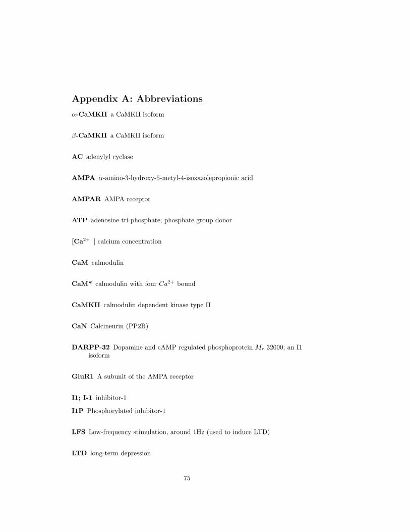
Appendix A: Abbreviations
α-CaMKII a CaMKII isoform
β-CaMKII a CaMKII isoform
AC adenylyl cyclase
AMPA α-amino-3-hydroxy-5-metyl-4-isoxazolepropionic acid
AMPAR AMPA receptor
ATP adenosine-tri-phosphate; phosphate group donor
[Ca2+ ] calcium concentration
CaM calmodulin
CaM* calmodulin with four Ca2+ bound
CaMKII calmodulin dependent kinase type II
CaN Calcineurin (PP2B)
DARPP-32 Dopamine and cAMP regulated phosphoprotein Mr 32000; an I1isoform
GluR1 A subunit of the AMPA receptor
I1; I-1 inhibitor-1
I1P Phosphorylated inhibitor-1
LFS Low-frequency stimulation, around 1Hz (used to induce LTD)
LTD long-term depression
75

Abbreviations Abbreviations
LTP long-term potentiation
NMDA N-methyl-D-aspartate
NMDAR NMDA receptor
PDE phosphodiesterase
PKA protein kinase A
PKC protein kinase C
PP1 protein phosphatase 1
PP2B protein phosphatase 2B (calcineurin)
PSD the post-synaptic density
Ser serine residue
SR the sarcoplasmic reticulum (a Ca2+ storage organelle)
STDP spike-timing dependent plasticity (bidirectional plasticity)
Thr threonine residue
VGCC voltage-gated Ca2+ channel
76

Overview of models
Appendix B: Overview of models
Implemented models
Dupont Holmes Zhabotinsky
model typewell-stirred X - Xcompartmental - X -deterministic X - Xdet./stoch. hybrid - X -
Ca2+/CaMsimple Ca2+ bind-ing
X - X
Adair-Klotz-equation
- - -
Ca-dep. dissocia-tion rates
- - -
variable CaM con-centration
- X -
CaMKIIone CaMKII activa-tion state
- - X
capping - X -stochastic CaMKIIactivation
- X -
explicit neighbourphosphorylation
- X X
PP1PP1 concentration - - XInhibitor conc. - - XCaN conc. - X -CaN dephosph ofI1/DARPP-32
- - X
PKAPKA phosph. - - XcAMP conc. - - -
77
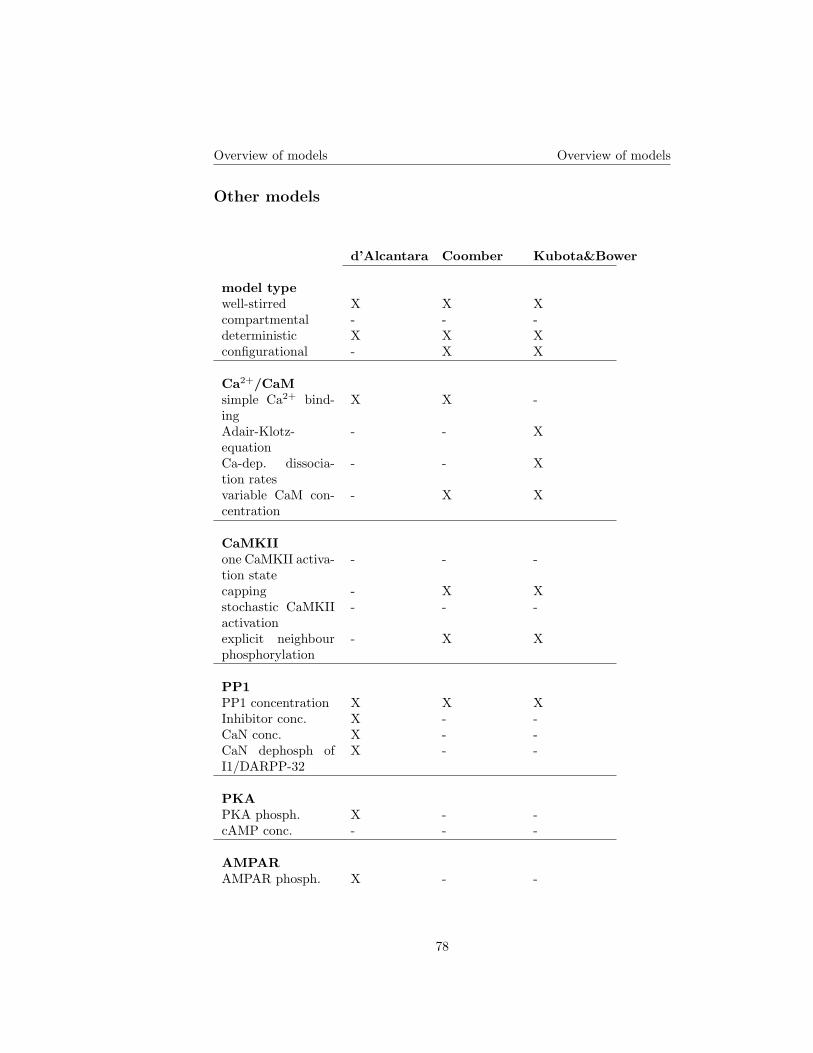
Overview of models Overview of models
Other models
d’Alcantara Coomber Kubota&Bower
model typewell-stirred X X Xcompartmental - - -deterministic X X Xconfigurational - X X
Ca2+/CaMsimple Ca2+ bind-ing
X X -
Adair-Klotz-equation
- - X
Ca-dep. dissocia-tion rates
- - X
variable CaM con-centration
- X X
CaMKIIone CaMKII activa-tion state
- - -
capping - X Xstochastic CaMKIIactivation
- - -
explicit neighbourphosphorylation
- X X
PP1PP1 concentration X X XInhibitor conc. X - -CaN conc. X - -CaN dephosph ofI1/DARPP-32
X - -
PKAPKA phosph. X - -cAMP conc. - - -
AMPARAMPAR phosph. X - -
78

Overview of models Overview of models
Lundh Okamoto&Ichikawa
model typewell-stirred - -compartmental X Xdeterministic X Xconfigurational - -
Ca2+/CaMsimple Ca2+ bind-ing
- X
Adair-Klotz-equation
- -
Ca-dep. dissocia-tion rates
- -
variable CaM con-centration
X X
CaMKIIone CaMKII activa-tion state
X -
capping - -stochastic CaMKIIactivation
- -
explicit neighbourphosphorylation
- -
PP1PP1 concentration - -Inhibitor conc. - -CaN conc. - -CaN dephosph ofI1/DARPP-32
- -
PKAPKA phosph. - -cAMP conc. X -
AMPARAMPAR phosph. - -
79
Related Documents