The Journal of Clinical Investigation | November 2003 | Volume 112 | Number 9 1351 Introduction Mitochondria are essential intracellular organelles required for oxidative phosphorylation. One of the unique aspects of mitochondria is the dual genetic con- trol of mitochondrial biosynthesis. Mitochondria con- tain their own genome, mitochondrial DNA (mtDNA), which is a small (16.5 kb), self-replicating DNA mole- cule present in multiple copies in the mitochondrial matrix. This genome encodes 13 essential proteins of the mitochondrial respiratory chain as well as 2 ribo- somal RNA (rRNA) and 22 transfer RNA (tRNA) genes required for intramitochondrial synthesis of proteins. All the other mitochondrial proteins are nuclear encod- ed and transported into the mitochondria. Inherited abnormalities of the mitochondrial genome are now increasingly recognized as important causes of disease (1). These mutations can affect all copies of the mito- chondrial genome within a cell — termed homoplasmy — or there may be a mixture of mutated and wild-type genomes in the same cell — heteroplasmy (2). Since mitochondria contain multiple mtDNA copies and cells may contain hundreds or thousands of mito- chondria, in the presence of heteroplasmy high levels of mutated mtDNA usually must be present within an individual cell before a defect of oxidative phosphory- lation is apparent (3). In addition to inherited mtDNA mutations, sporadic mtDNA mutations causing disease are common, and it has been noted for some time that mtDNA mutations occur with aging in postmitotic tissues (4–7). Both these events suggest that mtDNA mutation is a fairly Mitochondrial DNA mutations in human colonic crypt stem cells Robert W. Taylor, 1 Martin J. Barron, 1 Gillian M. Borthwick, 1 Amy Gospel, 1 Patrick F. Chinnery, 1 David C. Samuels, 2 Geoffrey A. Taylor, 3 Stefan M. Plusa, 4 Stephanie J. Needham, 5 Laura C. Greaves, 1,6 Thomas B.L. Kirkwood, 6 and Douglass M. Turnbull 1 1 Department of Neurology, and 2 Department of Mathematics, The Medical School, University of Newcastle upon Tyne, Newcastle upon Tyne, United Kingdom 3 Medical Research Council Development Centre for Brain Ageing, University of Newcastle upon Tyne, Newcastle upon Tyne, United Kingdom 4 Department of Surgery, 5 Department of Pathology, and 6 Department of Gerontology, The Medical School, University of Newcastle upon Tyne, Newcastle upon Tyne, United Kingdom The mitochondrial genome encodes 13 essential subunits of the respiratory chain and has remark- able genetics based on uniparental inheritance. Within human populations, the mitochondrial genome has a high rate of sequence divergence with multiple polymorphic variants and thus has played a major role in examining the evolutionary history of our species. In recent years it has also become apparent that pathogenic mitochondrial DNA (mtDNA) mutations play an important role in neurological and other diseases. Patients harbor many different mtDNA mutations, some of which are mtDNA mutations, some of which are inherited, but others that seem to be sporadic. It has also been suggested that mtDNA mutations play a role in aging and cancer, but the evidence for a causative role in these conditions is less clear. The accumulated data would suggest, however, that mtDNA mutations occur on a frequent basis. In this article we describe a new phenomenon: the accu- mulation of mtDNA mutations in human colonic crypt stem cells that result in a significant bio- chemical defect in their progeny. These studies have important consequences not only for under- standing of the finding of mtDNA mutations in aging tissues and tumors, but also for determining the frequency of mtDNA mutations within a cell. J. Clin. Invest. 112:1351–1360 (2003). doi: 10.1172/JCI200319435. Received for publication July 9, 2003, and accepted in revised form September 9, 2003. Address correspondence to: D.M. Turnbull, Mitochondrial Research Group, School of Neurology, Neurobiology and Psychiatry, The Medical School, University of Newcastle upon Tyne, Newcastle upon Tyne NE2 4HH, United Kingdom. Phone: 44-191-2228334; Fax: 44-191-2228553; E-mail: [email protected]. David C. Samuels’ present address is: Virginia Bioinformatics Institute, Virginia Polytechnical Institute and State University, Blacksburg, Virginia 24061, USA. Robert W. Taylor and Martin J. Barron contributed equally to this work. Conflict of interest: The authors have declared that no conflict of interest exists. Nonstandard abbreviations used: mitochondrial DNA (mtDNA); ribosomal RNA (rRNA); transfer RNA (tRNA); succinate dehydrogenase (SDH). See the related Commentary beginning on page 1312.

Welcome message from author
This document is posted to help you gain knowledge. Please leave a comment to let me know what you think about it! Share it to your friends and learn new things together.
Transcript
The Journal of Clinical Investigation | November 2003 | Volume 112 | Number 9 1351
IntroductionMitochondria are essential intracellular organellesrequired for oxidative phosphorylation. One of theunique aspects of mitochondria is the dual genetic con-trol of mitochondrial biosynthesis. Mitochondria con-tain their own genome, mitochondrial DNA (mtDNA),which is a small (16.5 kb), self-replicating DNA mole-
cule present in multiple copies in the mitochondrialmatrix. This genome encodes 13 essential proteins ofthe mitochondrial respiratory chain as well as 2 ribo-somal RNA (rRNA) and 22 transfer RNA (tRNA) genesrequired for intramitochondrial synthesis of proteins.All the other mitochondrial proteins are nuclear encod-ed and transported into the mitochondria. Inheritedabnormalities of the mitochondrial genome are nowincreasingly recognized as important causes of disease(1). These mutations can affect all copies of the mito-chondrial genome within a cell — termed homoplasmy— or there may be a mixture of mutated and wild-typegenomes in the same cell — heteroplasmy (2). Sincemitochondria contain multiple mtDNA copies andcells may contain hundreds or thousands of mito-chondria, in the presence of heteroplasmy high levelsof mutated mtDNA usually must be present within anindividual cell before a defect of oxidative phosphory-lation is apparent (3).
In addition to inherited mtDNA mutations, sporadicmtDNA mutations causing disease are common, and ithas been noted for some time that mtDNA mutationsoccur with aging in postmitotic tissues (4–7). Boththese events suggest that mtDNA mutation is a fairly
Mitochondrial DNA mutations in human colonic crypt stem cells
Robert W. Taylor,1 Martin J. Barron,1 Gillian M. Borthwick,1 Amy Gospel,1
Patrick F. Chinnery,1 David C. Samuels,2 Geoffrey A. Taylor,3 Stefan M. Plusa,4
Stephanie J. Needham,5 Laura C. Greaves,1,6 Thomas B.L. Kirkwood,6
and Douglass M. Turnbull1
1Department of Neurology, and2Department of Mathematics, The Medical School, University of Newcastle upon Tyne, Newcastle upon Tyne, United Kingdom
3Medical Research Council Development Centre for Brain Ageing, University of Newcastle upon Tyne, Newcastle upon Tyne,United Kingdom
4Department of Surgery,5Department of Pathology, and6Department of Gerontology, The Medical School, University of Newcastle upon Tyne, Newcastle upon Tyne, United Kingdom
The mitochondrial genome encodes 13 essential subunits of the respiratory chain and has remark-able genetics based on uniparental inheritance. Within human populations, the mitochondrialgenome has a high rate of sequence divergence with multiple polymorphic variants and thus hasplayed a major role in examining the evolutionary history of our species. In recent years it has alsobecome apparent that pathogenic mitochondrial DNA (mtDNA) mutations play an important rolein neurological and other diseases. Patients harbor many different mtDNA mutations, some of whichare mtDNA mutations, some of which are inherited, but others that seem to be sporadic. It has alsobeen suggested that mtDNA mutations play a role in aging and cancer, but the evidence for acausative role in these conditions is less clear. The accumulated data would suggest, however, thatmtDNA mutations occur on a frequent basis. In this article we describe a new phenomenon: the accu-mulation of mtDNA mutations in human colonic crypt stem cells that result in a significant bio-chemical defect in their progeny. These studies have important consequences not only for under-standing of the finding of mtDNA mutations in aging tissues and tumors, but also for determiningthe frequency of mtDNA mutations within a cell.
J. Clin. Invest. 112:1351–1360 (2003). doi: 10.1172/JCI200319435.
Received for publication July 9, 2003, and accepted in revised formSeptember 9, 2003.
Address correspondence to: D.M. Turnbull, MitochondrialResearch Group, School of Neurology, Neurobiology andPsychiatry, The Medical School, University of Newcastle uponTyne, Newcastle upon Tyne NE2 4HH, United Kingdom. Phone: 44-191-2228334; Fax: 44-191-2228553; E-mail: [email protected] C. Samuels’ present address is: Virginia BioinformaticsInstitute, Virginia Polytechnical Institute and State University,Blacksburg, Virginia 24061, USA.Robert W. Taylor and Martin J. Barron contributed equally to thiswork.Conflict of interest: The authors have declared that no conflict ofinterest exists.Nonstandard abbreviations used: mitochondrial DNA(mtDNA); ribosomal RNA (rRNA); transfer RNA (tRNA);succinate dehydrogenase (SDH).
See the related Commentary beginning on page 1312.
1352 The Journal of Clinical Investigation | November 2003 | Volume 112 | Number 9
frequent event, a view supported by the high sequencedivergence within the human population. It is suspect-ed that the high mutation rate is due to an environ-ment in which there is a high production of free radi-cals by the respiratory chain, compounded by limitedrepair of this genome compared with nuclear DNA.Even if mutations occur at high frequency, the mecha-nism by which these mutations accumulate to high lev-els in individual cells remains uncertain, but mathe-matical models would favor clonal expansion byrandom genetic drift (8, 9).
Previous work on somatic mtDNA mutations has con-centrated on the observations in postmitotic tissues,such as muscle and the CNS, since these tissues are pre-dominantly involved in patients with mtDNA disease.Even in these tissues, systematic studies of the natureand incidence of mutations is limited. A small numberof reports cite the accumulation of mtDNA mutationsin replicative cells (6, 10). An important question in mito-chondrial genetics is whether there is evidence ofmtDNA mutations in stem cells, and if so, do thesemutations accumulate to levels sufficient to result in abiochemical defect. If so, this could have profound impli-cations for our understanding of the potential impor-tance of these mutations in aging and cancer. To investi-gate the existence of mtDNA mutations in stem cells, wedecided to study colonic crypt stem cells. Colonic cryptstem cells are present at the crypt base, and thus all thecells within the crypt must be derived from these stemcells. Thus, we would not have to isolate individual stemcells to look for mtDNA mutations, but could in theorystudy their progeny. While there is uncertainty about thenumber of stem cells within human colonic crypts, stud-ies in mice suggest that crypts are maintained by smallnumbers of stem cells (11). For example, in chimericmouse strains a single crypt often shows evidence ofclonality in adult mice, suggesting that the majority ofcells within an individual crypt are the progeny of a sin-gle stem cell (12). Studies in humans are much more dif-ficult because strategies that directly mark cell fates, suchas aggregation chimeras and mutagenesis, are impracti-cal in humans (13); however, current evidence would sup-port that the situation is similar to mice.
MethodsPatients and colonic samples. Colons were collected from16 patients with no family history of colon cancer (agerange: 50–80 years) undergoing resection for colonictumors. Each sample of normal colonic mucosa wascollected at a distance of at least 12 cm from the edgeof the tumor. We also obtained colonoscopy biopsiesfrom 12 individuals undergoing investigation ofaltered bowel function in which no pathology wasfound (age range: 36–78 years).
Histochemistry. Colon samples were mounted for sec-tioning and frozen in isopentane previously cooled to–160°C in liquid nitrogen. Standard H&E histologywas performed on 12-µm sections. A dual histochemi-cal assay was used to determine the respiratory activity
of the crypts. Cryostat sections (12 µm) were first incu-bated in cytochrome c oxidase medium (100 µMcytochrome c, 4 mM diaminobenzidine tetrahydro-chloride, and 20 µg/ml catalase in 0.2 M phosphatebuffer, pH 7.0) at 37°C for 50 minutes. Sections werethen washed in PBS (3 × 5 minutes) and incubated insuccinate dehydrogenase (SDH) medium (130 mMsodium succinate, 200 µM phenazine methosulphate,1 mM sodium azide, 1.5 mM nitroblue tetrazolium in0.2 M phosphate buffer, pH 7.0) at 37°C for 40 min-utes. Sections were washed in PBS, pH 7.4 (3 × 5 min-utes), dehydrated in a graded ethanol series (70%, 95%,2 × 100%), cleared in Histoclear (National Diagnostics,Atlanta, Georgia, USA), and mounted in DPX (BDHLaboratory Supplies, Poole, United Kingdom).
The percentage of cytochrome c oxidase–deficientcrypts was calculated from transverse sections countedat several different levels, and approximately 100 cryptswere evaluated at each level. Only crypts that were morethan 50% cytochrome c oxidase deficient were included.
Immunocytochemistry. Immunocytochemistry was per-formed on 10-µm cryostat sections air-dried at roomtemperature for 1 hour. The sections were fixed in 4%paraformaldehyde in 0.1 M phosphate buffer for 10minutes, followed by rinsing in distilled water and thenPBS containing 0.1% Triton X-100 for 10 minutes. Thesections were further permeabilized in a graded (70%,95%, 100% vol/vol) methanol series over a period of 1hour. Endogenous peroxidase activity was quenched atthis stage by the addition of hydrogen peroxide (0.3%vol/vol) to the 95% (vol/vol) methanol. The sectionswere rinsed in PBS, then incubated with anti–cytochrome c oxidase subunit Ab (anti-subunit I, 2µg/ml, anti-subunit II and IV, 5 µg/ml; MolecularProbes Inc., Eugene, Oregon, USA) diluted in 4% BSAin PBS for 1 hour at room temperature. Followingthree washes in PBS, the sections were incubated witha peroxidase-conjugated Ab (rabbit anti-mouse Ig’s, 50µg/ml; DAKO Ltd., Ely, United Kingdom) for 1 hour atroom temperature. Following washing in PBS, peroxi-dase activity was detected by incubating in 1.4 mM 3,3′-diaminobenzadine tetrahydrochloride and 0.01%(vol/vol) hydrogen peroxide in 0.1 M phosphate buffer,pH 7.4, for 5 minutes at room temperature.
Three-dimensional reconstruction of colonic crypts. Three-dimensional reconstruction of crypts was performedusing KS300 3D software (Imaging Associates Ltd.,Thame, United Kingdom) on digital images of 59 seri-al sections (8 µm) reacted for cytochrome c oxidase andSDH histochemistry.
Isolation of total DNA from individual and partial crypts.Fresh frozen sections (20 µm) of colon were mountedon polyethylenenaphthalate slides (Leica MicrosystemsUK Ltd., Milton Keynes, United Kingdom). Sampleswere subjected to dual histochemistry as describedabove and air-dried after dehydration. Single and par-tial crypts were cut into sterile 0.5-ml PCR tubes usinga Leica laser microdissection system. Following cen-trifugation (7,000 g for 10 minutes), the crypt was lysed
The Journal of Clinical Investigation | November 2003 | Volume 112 | Number 9 1353
in 10 µl of cell lysis buffer (50 mM Tris-HCl, pH 8.5, 1 mM EDTA, 0.5% Tween-20, 200 ng/ml proteinase K)at 55°C for 2 hours and then 95°C for 10 minutes todenature the proteinase K.
Real-time PCR analysis of mtDNA. A quantitative real-time PCR approach employing fluorogenic probes wasused to quantify the amount of mtDNA in two differ-ent regions of the mitochondrial genome, one that israrely deleted in patients and aging (ND1 gene) andone that is frequently deleted (ND4 gene). PCR primersand fluorogenic probes (Applied Biosystems, War-rington, United Kingdom) for regions of ND1 (forwardprimer, L3485–3504; reverse primer, H3553–3532;probe, L3506–3529), and ND4 (forward primer,L12087–12109; reverse primer, H12170–12140; probe,L12111–12138) were synthesized, and 5 µl of DNAlysate from a single crypt was amplified separatelywith each of the ND1 and ND4 primer/probe combi-nations as described previously (14).
Sequencing of mtDNA from individual colonic crypts. Theentire sequence of the mitochondrial genome frommicrodissected crypts was determined using the single-cell lysate (see above) as the DNA template and a two-stage amplification protocol essentially as describedpreviously (15). The primary PCR reactions involvedamplification of the mitochondrial genome in 11 frag-ments of approximately 2 kb using a series of overlap-ping primer pairs. These initial large PCR reactionproducts decrease the risk of amplifying pseudogeneswhen extracting DNA from small quantities of DNA(16). All PCR amplifications were performed in a 50 µlvolume containing 1× PCR buffer (10 mM Tris-HCl,pH 8.3, 1.5 mM MgCl2, 50 mM KCl, 0.001% wt/vol gel-atin), 0.2 mM dNTPs, 0.6 µM primers, 1 U AmpliTaqGold DNA polymerase (Applied Biosystems), and 1 µlsingle-cell lysate. Reaction conditions were 94°C for 12minutes and 38 cycles of 94°C for 1 minute, 60°C for1 minute, and 72°C for 2 minutes. The final extensionwas for 8 minutes.
The secondary PCR reactions involved amplificationof the primary PCR products with 28 primer pairsspecifically to generate overlapping fragments ofbetween 600–700 bp that span the entire sequence ofthe human mitochondrial genome (15). To facilitatethe direct sequencing of PCR-amplified products, allprimer pairs are tagged with M13 sequence anddesigned to anneal optimally at 58°C. All reactionsproceeded for 30 cycles and used 2 µl of primary reac-tion product as DNA template. Samples were subse-quently purified to remove unincorporated primersand were directly sequenced using BigDye terminatorcycle-sequencing chemistries (Applied Biosystems) onan ABI 377 automated DNA sequencer. The sequencesobtained were compared with the revised Cambridgereference sequence using Sequence Navigator and Fac-tura software (Applied Biosystems).
Individual mutations were confirmed in all instancesby repeating the second-round PCR and resequencingthe product. For some products, the mutation was con-
firmed by restriction fragment–length polymorphismanalysis using restriction enzymes that specifically rec-ognized the mutation. Finally, to ensure mutationswere not introduced during PCR, we microdissectedtwo separate segments of the same cytochrome c oxi-dase–deficient crypt. Sequencing of the entire mito-chondrial genome from both partial crypts revealednot only the presence of an identical mtDNA mutation,but no other detectable sequence changes.
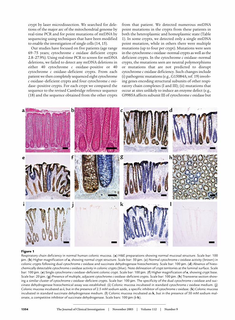
ResultsRespiratory chain deficiency in colonic crypts and crypt stemcells. Cells deficient in cytochrome c oxidase but withnormal SDH activity are excellent biomarkers ofmtDNA defects (17). We wished to determine if suchcells were present in colonic epithelium and preparedfrozen sections of normal colonic epithelium. Themucosal anatomy was preserved with our procedure forcollection and mounting of sections, as shown by thecrypt morphology using H&E (Figure 1, a and b). Somecrypts from every patient we examined were cyto-chrome c oxidase deficient (Figure 1, c–h). To ensurethat our techniques were completely specific, we per-formed control experiments on colonic epitheliumusing specific inhibitors of mitochondrial enzymeactivity (sodium azide for cytochrome c oxidase andsodium malonate for SDH) (Figure 1, i–l). These exper-iments confirm that the reaction is specific and thatour observations reflect a genuine decrease incytochrome c oxidase activity. We found that thesecytochrome c oxidase–deficient crypts were distributedthroughout the colon because we examined samplesobtained from both right and left hemicolectomy.
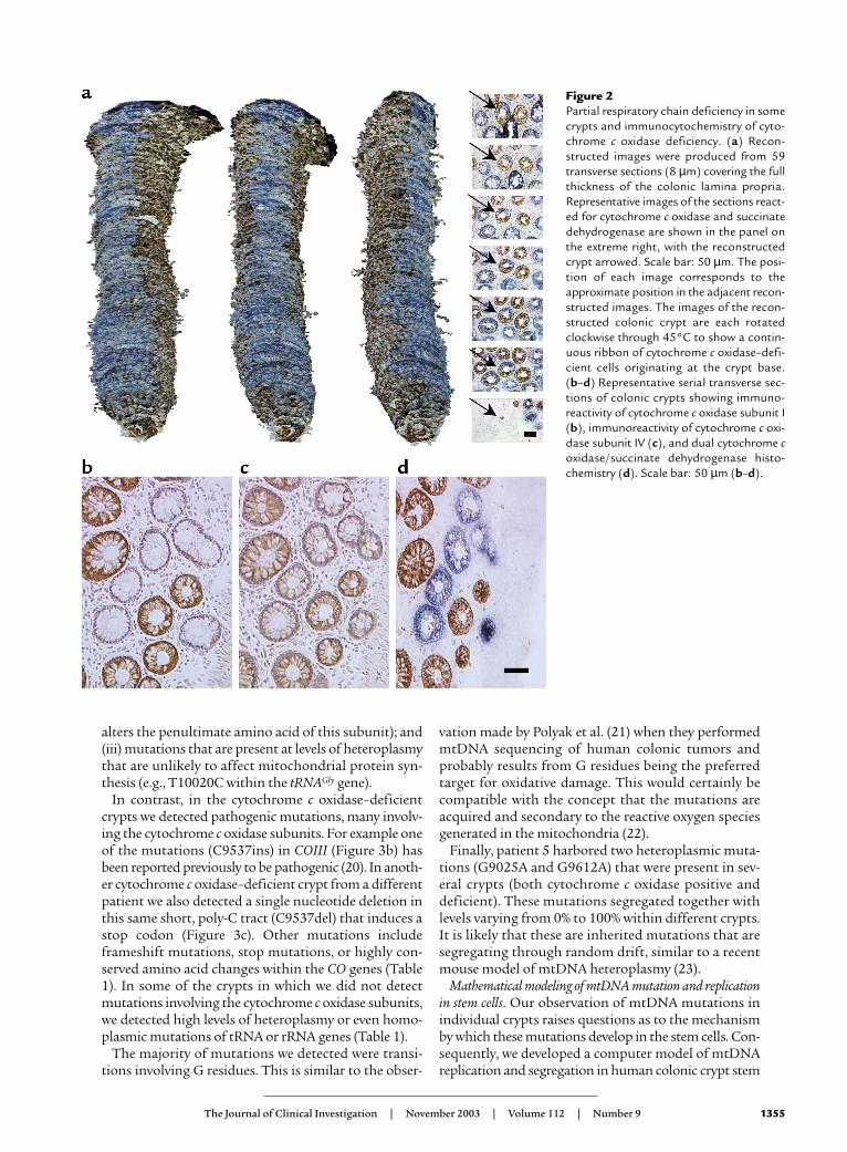
In addition to detecting whole crypts showing cyto-chrome c oxidase deficiency, we observed crypts show-ing a deficiency of this enzyme in only part of the crypt.To investigate the phenomenon of partial cytochromec oxidase deficiency further, we serially sectioned (8 µm)colonic mucosa transversely and performed 3D recon-struction of the crypts following dual cytochrome c oxi-dase/SDH histochemistry (Figure 2a). In these cryptsthere is a ribbon of cytochrome c oxidase–deficient cellswithin an otherwise normal crypt that is entirely com-patible with the view that there are multiple stem cellsin some crypts and that what we are observing reflectsinvolvement of one stem cell and its progeny.
We also studied the expression of cytochrome c oxi-dase subunits I (Figure 2b) and II (both mtDNA encod-ed) and subunit IV (nuclear encoded) (Figure 2c) innine patients. In all patients there was an absence ofimmunoreactivity against subunits I (Figure 2b) and IIin cytochrome c oxidase–deficient crypts. Immuno-reactivity against subunit IV was reduced in all cyto-chrome c oxidase–deficient crypts (Figure 2c), but notdecreased to the level seen with subunits I and II.
Mitochondrial genome analysis. To establish if thecytochrome c oxidase deficiency, observed in the coloniccrypts was due to mtDNA mutations we identified indi-vidual crypts on longitudinal sections and isolated the
1354 The Journal of Clinical Investigation | November 2003 | Volume 112 | Number 9
crypt by laser microdissection. We searched for dele-tions of the major arc of the mitochondrial genome byreal-time PCR and for point mutations of mtDNA bysequencing using techniques that have been modifiedto enable the investigation of single cells (14, 15).
Our studies have focused on five patients (age range69–75 years; cytochrome c oxidase deficient crypts2.8–27.9%). Using real-time PCR to screen for mtDNAdeletions, we failed to detect any mtDNA deletions ineither 40 cytochrome c oxidase–positive or 40cytochrome c oxidase–deficient crypts. From eachpatient we then completely sequenced eight cytochromec oxidase–deficient crypts and four cytochrome c oxi-dase–positive crypts. For each crypt we compared thesequence to the revised Cambridge reference sequence(18) and the sequence obtained from the other crypts
from that patient. We detected numerous mtDNApoint mutations in the crypts from these patients inboth the heteroplasmic and homoplasmic state (Table1). In some crypts, we detected only a single mtDNApoint mutation, while in others there were multiplemutations (up to four per crypt). Mutations were seenin the cytochrome c oxidase–normal crypts as well as thedeficient crypts. In the cytochrome c oxidase–normalcrypts, the mutations seen are neutral polymorphismsor mutations that are not predicted to disruptcytochrome c oxidase deficiency. Such changes include(i) pathogenic mutations (e.g., G15084A; ref. 19) involv-ing genes encoding structural subunits of other respi-ratory chain complexes (I and III); (ii) mutations thatoccur at sites unlikely to induce an enzyme defect (e.g.,G9985A affects subunit III of cytochrome c oxidase but
Figure 1Respiratory chain deficiency in normal human colonic mucosa. (a) H&E preparations showing normal mucosal structure. Scale bar: 100µm. (b) Higher magnification of a, showing normal crypt structure. Scale bar: 50 µm. (c) Normal cytochrome c oxidase activity (brown) incolonic crypts following dual cytochrome c oxidase and succinate dehydrogenase histochemistry. Scale bar: 100 µm. (d) Absence of histo-chemically detectable cytochrome c oxidase activity in colonic crypts (blue). Note delineation of crypt territories at the luminal surface. Scalebar: 100 µm. (e) Single cytochrome c oxidase–deficient colonic crypt. Scale bar: 100 µm. (f) Higher magnification of e, showing crypt base.Scale bar: 20 µm. (g) Presence of multiple, adjacent cytochrome c oxidase–deficient crypts. Scale bar: 100 µm. (h) Transverse section show-ing a similar cluster of cytochrome c oxidase–deficient crypts. Scale bar: 100 µm. The specificity of the dual cytochrome c oxidase and suc-cinate dehydrogenase histochemical assay was established. (i) Colonic mucosa incubated in standard cytochrome c oxidase medium. (j)Colonic mucosa incubated as i, but in the presence of 2.5 mM sodium azide, a specific inhibitor of cytochrome c oxidase. (k) Colonic mucosaincubated in standard succinate dehydrogenase medium. (l) Colonic mucosa incubated as k, but in the presence of 50 mM sodium mal-onate, a competitive inhibitor of succinate dehydrogenase. Scale bars: 100 µm (i–k).
The Journal of Clinical Investigation | November 2003 | Volume 112 | Number 9 1355
alters the penultimate amino acid of this subunit); and(iii) mutations that are present at levels of heteroplasmythat are unlikely to affect mitochondrial protein syn-thesis (e.g., T10020C within the tRNAGly gene).
In contrast, in the cytochrome c oxidase–deficientcrypts we detected pathogenic mutations, many involv-ing the cytochrome c oxidase subunits. For example oneof the mutations (C9537ins) in COIII (Figure 3b) hasbeen reported previously to be pathogenic (20). In anoth-er cytochrome c oxidase–deficient crypt from a differentpatient we also detected a single nucleotide deletion inthis same short, poly-C tract (C9537del) that induces astop codon (Figure 3c). Other mutations includeframeshift mutations, stop mutations, or highly con-served amino acid changes within the CO genes (Table1). In some of the crypts in which we did not detectmutations involving the cytochrome c oxidase subunits,we detected high levels of heteroplasmy or even homo-plasmic mutations of tRNA or rRNA genes (Table 1).
The majority of mutations we detected were transi-tions involving G residues. This is similar to the obser-
vation made by Polyak et al. (21) when they performedmtDNA sequencing of human colonic tumors andprobably results from G residues being the preferredtarget for oxidative damage. This would certainly becompatible with the concept that the mutations areacquired and secondary to the reactive oxygen speciesgenerated in the mitochondria (22).
Finally, patient 5 harbored two heteroplasmic muta-tions (G9025A and G9612A) that were present in sev-eral crypts (both cytochrome c oxidase positive anddeficient). These mutations segregated together withlevels varying from 0% to 100% within different crypts.It is likely that these are inherited mutations that aresegregating through random drift, similar to a recentmouse model of mtDNA heteroplasmy (23).
Mathematical modeling of mtDNA mutation and replicationin stem cells. Our observation of mtDNA mutations inindividual crypts raises questions as to the mechanismby which these mutations develop in the stem cells. Con-sequently, we developed a computer model of mtDNAreplication and segregation in human colonic crypt stem
Figure 2Partial respiratory chain deficiency in somecrypts and immunocytochemistry of cyto-chrome c oxidase deficiency. (a) Recon-structed images were produced from 59transverse sections (8 µm) covering the fullthickness of the colonic lamina propria.Representative images of the sections react-ed for cytochrome c oxidase and succinatedehydrogenase are shown in the panel onthe extreme right, with the reconstructedcrypt arrowed. Scale bar: 50 µm. The posi-tion of each image corresponds to theapproximate position in the adjacent recon-structed images. The images of the recon-structed colonic crypt are each rotatedclockwise through 45°C to show a contin-uous ribbon of cytochrome c oxidase–defi-cient cells originating at the crypt base.(b–d) Representative serial transverse sec-tions of colonic crypts showing immuno-reactivity of cytochrome c oxidase subunit I(b), immunoreactivity of cytochrome c oxi-dase subunit IV (c), and dual cytochrome coxidase/succinate dehydrogenase histo-chemistry (d). Scale bar: 50 µm (b–d).
1356 The Journal of Clinical Investigation | November 2003 | Volume 112 | Number 9
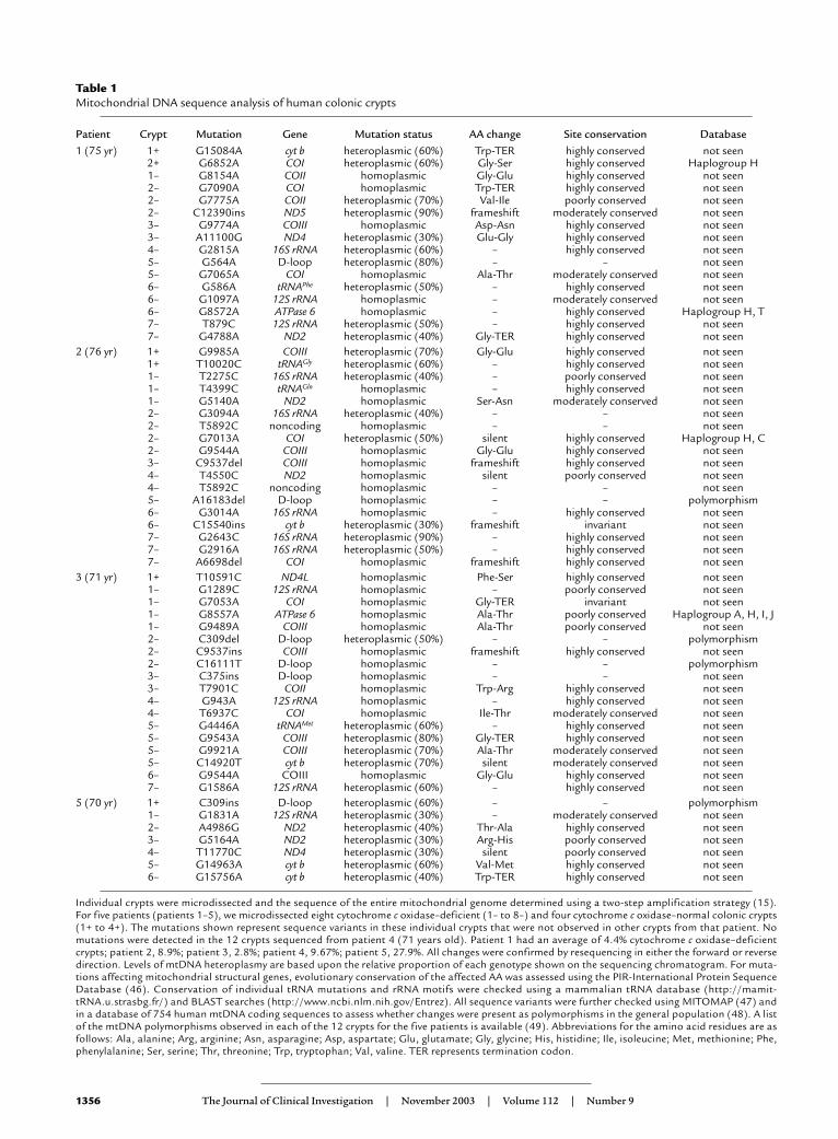
Table 1Mitochondrial DNA sequence analysis of human colonic crypts
Patient Crypt Mutation Gene Mutation status AA change Site conservation Database1 (75 yr) 1+ G15084A cyt b heteroplasmic (60%) Trp-TER highly conserved not seen
2+ G6852A COI heteroplasmic (60%) Gly-Ser highly conserved Haplogroup H1– G8154A COII homoplasmic Gly-Glu highly conserved not seen2– G7090A COI homoplasmic Trp-TER highly conserved not seen2– G7775A COII heteroplasmic (70%) Val-Ile poorly conserved not seen2– C12390ins ND5 heteroplasmic (90%) frameshift moderately conserved not seen3– G9774A COIII homoplasmic Asp-Asn highly conserved not seen3– A11100G ND4 heteroplasmic (30%) Glu-Gly highly conserved not seen4– G2815A 16S rRNA heteroplasmic (60%) – highly conserved not seen5– G564A D-loop heteroplasmic (80%) – – not seen5– G7065A COI homoplasmic Ala-Thr moderately conserved not seen6– G586A tRNAPhe heteroplasmic (50%) – highly conserved not seen6– G1097A 12S rRNA homoplasmic – moderately conserved not seen6– G8572A ATPase 6 homoplasmic – highly conserved Haplogroup H, T7– T879C 12S rRNA heteroplasmic (50%) – highly conserved not seen7– G4788A ND2 heteroplasmic (40%) Gly-TER highly conserved not seen
2 (76 yr) 1+ G9985A COIII heteroplasmic (70%) Gly-Glu highly conserved not seen1+ T10020C tRNAGly heteroplasmic (60%) – highly conserved not seen1– T2275C 16S rRNA heteroplasmic (40%) – poorly conserved not seen1– T4399C tRNAGln homoplasmic – highly conserved not seen1– G5140A ND2 homoplasmic Ser-Asn moderately conserved not seen2– G3094A 16S rRNA heteroplasmic (40%) – – not seen2– T5892C noncoding homoplasmic – – not seen2– G7013A COI heteroplasmic (50%) silent highly conserved Haplogroup H, C2– G9544A COIII homoplasmic Gly-Glu highly conserved not seen3– C9537del COIII homoplasmic frameshift highly conserved not seen4– T4550C ND2 homoplasmic silent poorly conserved not seen4– T5892C noncoding homoplasmic – – not seen5– A16183del D-loop homoplasmic – – polymorphism6– G3014A 16S rRNA homoplasmic – highly conserved not seen6– C15540ins cyt b heteroplasmic (30%) frameshift invariant not seen7– G2643C 16S rRNA heteroplasmic (90%) – highly conserved not seen7– G2916A 16S rRNA heteroplasmic (50%) – highly conserved not seen7– A6698del COI homoplasmic frameshift highly conserved not seen
3 (71 yr) 1+ T10591C ND4L homoplasmic Phe-Ser highly conserved not seen1– G1289C 12S rRNA homoplasmic – poorly conserved not seen1– G7053A COI homoplasmic Gly-TER invariant not seen1– G8557A ATPase 6 homoplasmic Ala-Thr poorly conserved Haplogroup A, H, I, J1– G9489A COIII homoplasmic Ala-Thr poorly conserved not seen2– C309del D-loop heteroplasmic (50%) – – polymorphism2– C9537ins COIII homoplasmic frameshift highly conserved not seen2– C16111T D-loop homoplasmic – – polymorphism3– C375ins D-loop homoplasmic – – not seen3– T7901C COII homoplasmic Trp-Arg highly conserved not seen4– G943A 12S rRNA homoplasmic – highly conserved not seen4– T6937C COI homoplasmic Ile-Thr moderately conserved not seen5– G4446A tRNAMet heteroplasmic (60%) – highly conserved not seen5– G9543A COIII heteroplasmic (80%) Gly-TER highly conserved not seen5– G9921A COIII heteroplasmic (70%) Ala-Thr moderately conserved not seen5– C14920T cyt b heteroplasmic (70%) silent moderately conserved not seen6– G9544A COIII homoplasmic Gly-Glu highly conserved not seen7– G1586A 12S rRNA heteroplasmic (60%) – highly conserved not seen
5 (70 yr) 1+ C309ins D-loop heteroplasmic (60%) – – polymorphism1– G1831A 12S rRNA heteroplasmic (30%) – moderately conserved not seen2– A4986G ND2 heteroplasmic (40%) Thr-Ala highly conserved not seen3– G5164A ND2 heteroplasmic (30%) Arg-His poorly conserved not seen4– T11770C ND4 heteroplasmic (30%) silent poorly conserved not seen5– G14963A cyt b heteroplasmic (60%) Val-Met highly conserved not seen6– G15756A cyt b heteroplasmic (40%) Trp-TER highly conserved not seen
Individual crypts were microdissected and the sequence of the entire mitochondrial genome determined using a two-step amplification strategy (15).For five patients (patients 1–5), we microdissected eight cytochrome c oxidase–deficient (1– to 8–) and four cytochrome c oxidase–normal colonic crypts(1+ to 4+). The mutations shown represent sequence variants in these individual crypts that were not observed in other crypts from that patient. Nomutations were detected in the 12 crypts sequenced from patient 4 (71 years old). Patient 1 had an average of 4.4% cytochrome c oxidase–deficientcrypts; patient 2, 8.9%; patient 3, 2.8%; patient 4, 9.67%; patient 5, 27.9%. All changes were confirmed by resequencing in either the forward or reversedirection. Levels of mtDNA heteroplasmy are based upon the relative proportion of each genotype shown on the sequencing chromatogram. For muta-tions affecting mitochondrial structural genes, evolutionary conservation of the affected AA was assessed using the PIR-International Protein SequenceDatabase (46). Conservation of individual tRNA mutations and rRNA motifs were checked using a mammalian tRNA database (http://mamit-tRNA.u.strasbg.fr/) and BLAST searches (http://www.ncbi.nlm.nih.gov/Entrez). All sequence variants were further checked using MITOMAP (47) andin a database of 754 human mtDNA coding sequences to assess whether changes were present as polymorphisms in the general population (48). A listof the mtDNA polymorphisms observed in each of the 12 crypts for the five patients is available (49). Abbreviations for the amino acid residues are asfollows: Ala, alanine; Arg, arginine; Asn, asparagine; Asp, aspartate; Glu, glutamate; Gly, glycine; His, histidine; Ile, isoleucine; Met, methionine; Phe,phenylalanine; Ser, serine; Thr, threonine; Trp, tryptophan; Val, valine. TER represents termination codon.
The Journal of Clinical Investigation | November 2003 | Volume 112 | Number 9 1357
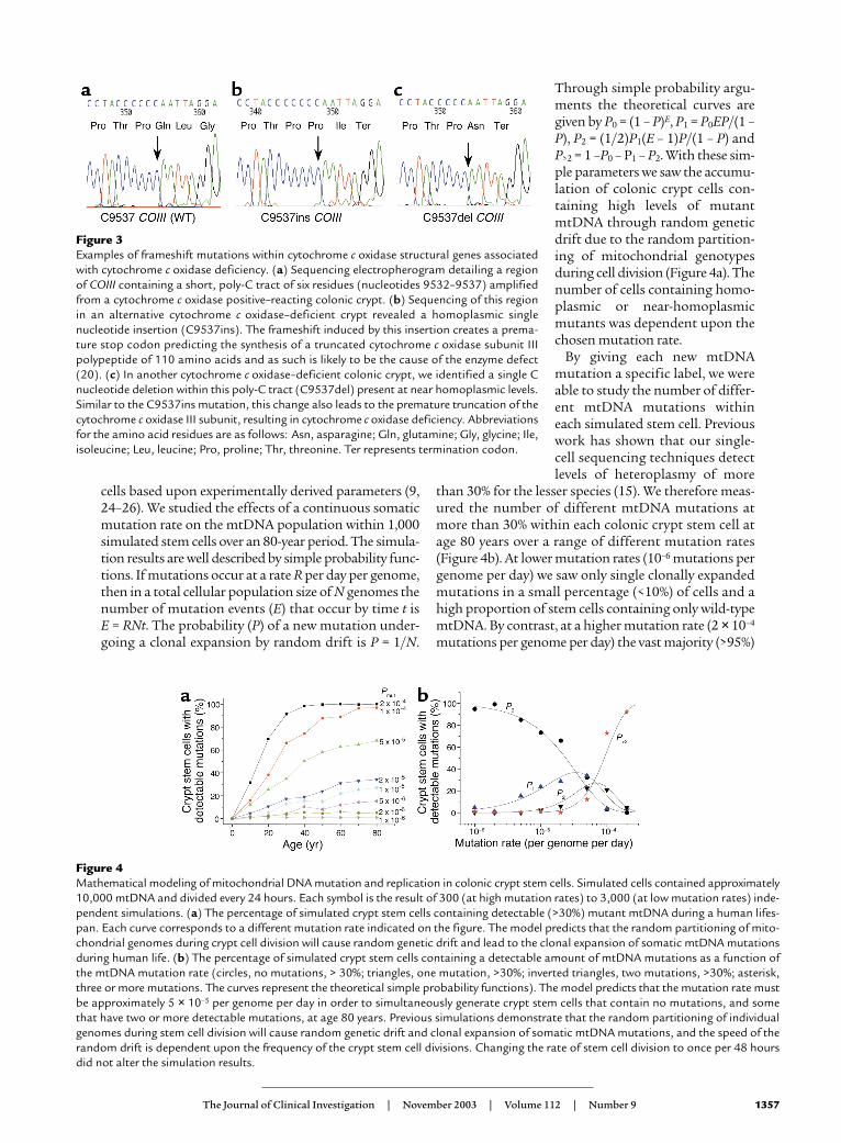
cells based upon experimentally derived parameters (9,24–26). We studied the effects of a continuous somaticmutation rate on the mtDNA population within 1,000simulated stem cells over an 80-year period. The simula-tion results are well described by simple probability func-tions. If mutations occur at a rate R per day per genome,then in a total cellular population size of N genomes thenumber of mutation events (E) that occur by time t is E = RNt. The probability (P) of a new mutation under-going a clonal expansion by random drift is P = 1/N.
Through simple probability argu-ments the theoretical curves aregiven by P0 = (1 – P)E, P1 = P0EP/(1 –P), P2 = (1/2)P1(E – 1)P/(1 – P) andP>2 = 1 –P0 – P1 – P2. With these sim-ple parameters we saw the accumu-lation of colonic crypt cells con-taining high levels of mutantmtDNA through random geneticdrift due to the random partition-ing of mitochondrial genotypesduring cell division (Figure 4a). Thenumber of cells containing homo-plasmic or near-homoplasmicmutants was dependent upon thechosen mutation rate.
By giving each new mtDNAmutation a specific label, we wereable to study the number of differ-ent mtDNA mutations withineach simulated stem cell. Previouswork has shown that our single-cell sequencing techniques detectlevels of heteroplasmy of more
than 30% for the lesser species (15). We therefore meas-ured the number of different mtDNA mutations atmore than 30% within each colonic crypt stem cell atage 80 years over a range of different mutation rates(Figure 4b). At lower mutation rates (10–6 mutations pergenome per day) we saw only single clonally expandedmutations in a small percentage (<10%) of cells and ahigh proportion of stem cells containing only wild-typemtDNA. By contrast, at a higher mutation rate (2 × 10⁻⁴
mutations per genome per day) the vast majority (>95%)
Figure 3Examples of frameshift mutations within cytochrome c oxidase structural genes associatedwith cytochrome c oxidase deficiency. (a) Sequencing electropherogram detailing a regionof COIII containing a short, poly-C tract of six residues (nucleotides 9532–9537) amplifiedfrom a cytochrome c oxidase positive–reacting colonic crypt. (b) Sequencing of this regionin an alternative cytochrome c oxidase–deficient crypt revealed a homoplasmic singlenucleotide insertion (C9537ins). The frameshift induced by this insertion creates a prema-ture stop codon predicting the synthesis of a truncated cytochrome c oxidase subunit IIIpolypeptide of 110 amino acids and as such is likely to be the cause of the enzyme defect(20). (c) In another cytochrome c oxidase–deficient colonic crypt, we identified a single Cnucleotide deletion within this poly-C tract (C9537del) present at near homoplasmic levels.Similar to the C9537ins mutation, this change also leads to the premature truncation of thecytochrome c oxidase III subunit, resulting in cytochrome c oxidase deficiency. Abbreviationsfor the amino acid residues are as follows: Asn, asparagine; Gln, glutamine; Gly, glycine; Ile,isoleucine; Leu, leucine; Pro, proline; Thr, threonine. Ter represents termination codon.
Figure 4Mathematical modeling of mitochondrial DNA mutation and replication in colonic crypt stem cells. Simulated cells contained approximately10,000 mtDNA and divided every 24 hours. Each symbol is the result of 300 (at high mutation rates) to 3,000 (at low mutation rates) inde-pendent simulations. (a) The percentage of simulated crypt stem cells containing detectable (>30%) mutant mtDNA during a human lifes-pan. Each curve corresponds to a different mutation rate indicated on the figure. The model predicts that the random partitioning of mito-chondrial genomes during crypt cell division will cause random genetic drift and lead to the clonal expansion of somatic mtDNA mutationsduring human life. (b) The percentage of simulated crypt stem cells containing a detectable amount of mtDNA mutations as a function ofthe mtDNA mutation rate (circles, no mutations, > 30%; triangles, one mutation, >30%; inverted triangles, two mutations, >30%; asterisk,three or more mutations. The curves represent the theoretical simple probability functions). The model predicts that the mutation rate mustbe approximately 5 × 10⁻⁵ per genome per day in order to simultaneously generate crypt stem cells that contain no mutations, and somethat have two or more detectable mutations, at age 80 years. Previous simulations demonstrate that the random partitioning of individualgenomes during stem cell division will cause random genetic drift and clonal expansion of somatic mtDNA mutations, and the speed of therandom drift is dependent upon the frequency of the crypt stem cell divisions. Changing the rate of stem cell division to once per 48 hoursdid not alter the simulation results.
1358 The Journal of Clinical Investigation | November 2003 | Volume 112 | Number 9
of stem cells contained three or more clonally expand-ed mutant species, and very few contained only wild-type mtDNA. Only at intermediate mutation rates (5 × 10⁻⁵ mutations per genome per day) did we see bothsingle clonally expanded mtDNA mutations and mul-tiple clonally expanded species affecting 10–30% of thesimulated crypt stem cells and a similar number of cellscontaining only wild-type mtDNA. This has importantimplications for our understanding of somatic mtDNAmutation. Our data provide the first estimate of thesomatic mtDNA mutation rate in humans in vivo ofapproximately 5 × 10⁻⁵ mutations per genome per day,corresponding to a rate of 1.10 mutations per site permillion years. This is strikingly similar to the pedigree-derived mtDNA substitution rates (27) that are consis-tently higher than mutation rates derived from phylo-genetic analysis (28). Moreover, the rate we have derivedfrom the study of colonic crypts is similar to the ratemeasured for mtDNA (2 × 10⁻⁸ mutations per site percell division) in cultured cells in vitro (29) and substan-tially greater than that of nuclear DNA (6 × 10–11 muta-tions per site per cell division).
DiscussionWe hypothesized that mtDNA mutations may accumu-late in stem cells in view of a high mutation rate in themitochondrial genome and the possibility of accumula-tion of these mutations by a combination of relaxedreplication and random segregation between daughtercells. Our investigations concentrated on colonic cryptstem cells because the progeny of these stem cells can beclearly identified and thus detecting the mutations andthe resulting biochemical deficiency would be easierthan for many other stem cells. Our investigations show
that these stem cells do indeed accumulate mtDNAmutations to high levels and if the mutations involve animportant base in either a structural or RNA gene thenthey can result in a biochemical defect. We have no rea-son to believe that these findings will be limited tocolonic crypt stem cells and thus believe that our obser-vations have implications for other stem cells.
Interestingly, in the cytochrome c oxidase–deficientcrypts we detected predominantly mutations in the pro-tein-encoding genes, which are very different from themutation spectrum we see in patients with mtDNA dis-ease. In our experience of investigating patients withinherited mtDNA defects, the number of mutationsinvolving RNA genes is much greater than the muta-tions involving structural genes. This may indicate thatthe more severe phenotype associated with subunitmutations is less compatible with transmission throughthe germline, an observation in keeping with the rela-tively rare transmission of mtDNA deletions (30).
In some cytochrome c oxidase–deficient crypts we didnot detect mtDNA point mutations, or, if they werepresent, they were likely to be polymorphic variantsrather than pathogenic mutations. This was especiallytrue of patients 4 and 5, the patients who had the high-est number of cytochrome c oxidase–deficient crypts.
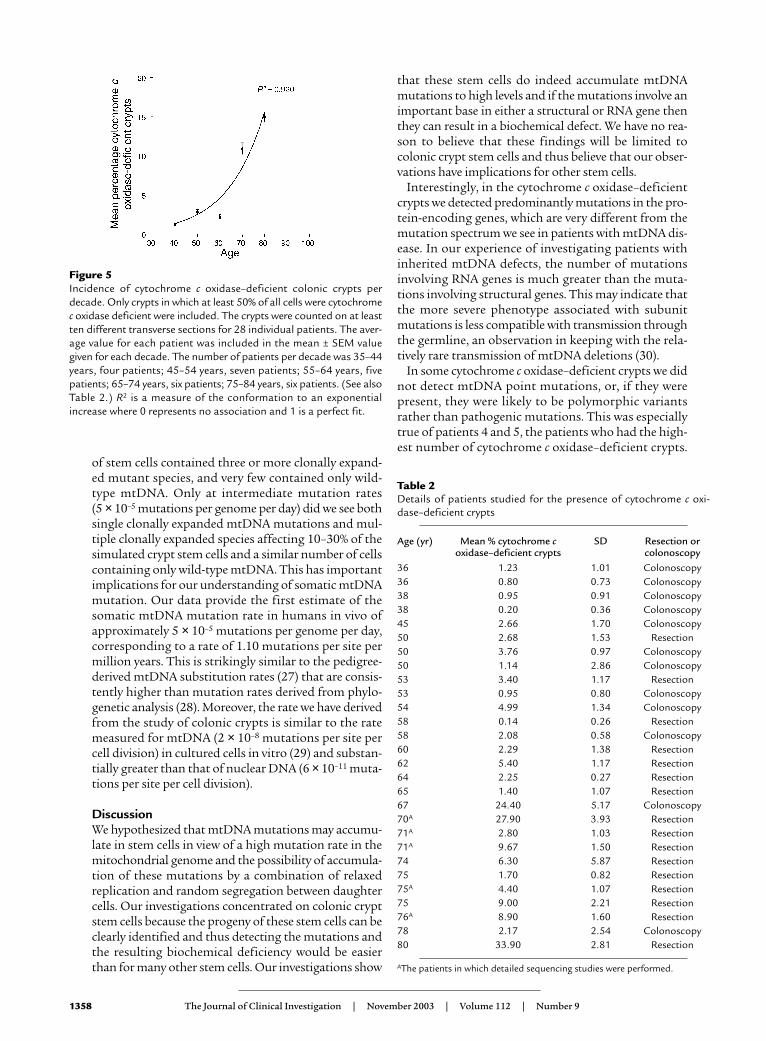
Figure 5Incidence of cytochrome c oxidase–deficient colonic crypts perdecade. Only crypts in which at least 50% of all cells were cytochromec oxidase deficient were included. The crypts were counted on at leastten different transverse sections for 28 individual patients. The aver-age value for each patient was included in the mean ± SEM valuegiven for each decade. The number of patients per decade was 35–44years, four patients; 45–54 years, seven patients; 55–64 years, fivepatients; 65–74 years, six patients; 75–84 years, six patients. (See alsoTable 2.) R2 is a measure of the conformation to an exponentialincrease where 0 represents no association and 1 is a perfect fit.
Table 2Details of patients studied for the presence of cytochrome c oxi-dase–deficient crypts
Age (yr) Mean % cytochrome c SD Resection or oxidase–deficient crypts colonoscopy
36 1.23 1.01 Colonoscopy36 0.80 0.73 Colonoscopy38 0.95 0.91 Colonoscopy38 0.20 0.36 Colonoscopy45 2.66 1.70 Colonoscopy50 2.68 1.53 Resection50 3.76 0.97 Colonoscopy50 1.14 2.86 Colonoscopy53 3.40 1.17 Resection53 0.95 0.80 Colonoscopy54 4.99 1.34 Colonoscopy58 0.14 0.26 Resection58 2.08 0.58 Colonoscopy60 2.29 1.38 Resection62 5.40 1.17 Resection64 2.25 0.27 Resection65 1.40 1.07 Resection67 24.40 5.17 Colonoscopy70A 27.90 3.93 Resection71A 2.80 1.03 Resection71A 9.67 1.50 Resection74 6.30 5.87 Resection75 1.70 0.82 Resection75A 4.40 1.07 Resection75 9.00 2.21 Resection76A 8.90 1.60 Resection78 2.17 2.54 Colonoscopy80 33.90 2.81 Resection
AThe patients in which detailed sequencing studies were performed.
The Journal of Clinical Investigation | November 2003 | Volume 112 | Number 9 1359
There are several possibilities to explain the cyto-chrome c oxidase deficiency in these crypts, includingmtDNA deletions that were not detected by our real-time assay or multiple mtDNA point mutations pres-ent below the limit of detection by automated sequenc-ing. A further possibility would be mutations ofnuclear genes. Our immunocytochemical studiesrevealed an absence of cytochrome c oxidase subunits Iand II (encoded by the mitochondrial genome) andpartial deficiency of subunit IV (encoded by the nucleargenome). There was no difference in the immunocyto-chemical reactions between those patients with highlevels of mtDNA mutations and those (patient 4 and 5)in which the mtDNA mutations were not detected.These results could imply involvement of either themitochondrial or nuclear genome. Nuclear gene muta-tions causing systemic cytochrome c oxidase deficien-cy are increasingly being recognized and may involve anuclear-encoded cytochrome c oxidase structural sub-unit (although no mutations have been described todate) (31), a cytochrome c oxidase assembly factor, or aprotein involved in mtDNA maintenance (32–37). If,indeed, the cytochrome c oxidase deficiency is due to anuclear gene defect, it is uncertain as to whether thisinvolves a single gene or several different genes, witheach deficient crypt due to a new mutation. It is recog-nized that nuclear gene defects can cause a mosaic pic-ture of cytochrome c oxidase deficiency and thereforethe involvement of different crypts does not differenti-ate between the possibilities of a mitochondrial andnuclear gene involvement (38). We suspect that anuclear gene defect causing either mtDNA depletion ormultiple deletions would have been detected by ourreal-time assay, which not only detects deletions butalso quantifies mtDNA copy number (14). We failed,however, to detect any difference in copy numberbetween cytochrome c oxidase–deficient and cyto-chrome c oxidase–normal crypts in any of our patients.Another possibility is that the observation of lowcytochrome c oxidase activity relates to a downregula-tion of the respiratory chain activity in response to thecolonic tumor. This we think is unlikely because thecytochrome c oxidase–deficient crypts were alsoobserved in patients without tumors, and they occur inthe presence of normal SDH activity (Figure 1). Final-ly, if nuclear genetic defects (or altered regulation) arethe cause of the cytochrome c oxidase deficiency inpatient 4 and 5, what then could be the contribution ofthe nuclear genome to the cytochrome c oxidase–defi-cient crypts in our other patients? At this stage we canonly speculate that we believe that the mtDNA defectis pathogenic in some crypts, but further studies arerequired to determine if the mutations are sufficientalone to generate the biochemical defect.
What then is the significance of our observations formitochondrial genetics and stem cell biology? Ourobservations highlight how frequently these mutationsoccur and, when present, that they can accumulate tohigh levels, sufficient, we believe, to cause a biochemical
defect. The observed mutation load in these stem cells ishigh; we sequenced the complete mitochondrial genomein 60 crypts and detected a total of 59 different mtDNApoint mutations. In addition, there is likely to have beena significant number of mtDNA mutations presentwithin these stem cells that were not detected. Auto-mated fluorescent-sequencing techniques and real-timePCR will only detect moderate levels of a heteroplasmicchange (>25% and >40%, respectively) (14, 39), and low-level heteroplasmy would be missed. If a significantnumber of stem cells accumulate mutations, it becomesclear that within an individual there will be a mixture ofmutations, which may influence tissue function.
Our studies have shown that mtDNA mutations accu-mulate in cytochrome c oxidase–deficient crypts, andthese crypts show an age-related increase in their number(Figure 5 and Table 2). These studies also represent thefirst description of pathogenic mtDNA mutations instem cells with human aging. These observations supporta role for mtDNA mutations in the aging process, whichuntil now has been based mainly on an age-related accu-mulation of mtDNA mutations in postmitotic cells. Cur-rent understanding indicates that aging is caused byaccumulation of stochastic molecular and cellular dam-age (40, 41), and there is evidence from studies in micethat functional impairment of intestinal stem cells occurswith age and is associated with an altered DNA damageresponse (42). The heterogeneous nature of such sto-chastic damage at the cellular level makes its detectionmore difficult and is fully consistent with the diversity wehave observed. While our observations are in the colon,we believe they raise important questions about the widerrole of mtDNA mutations in stem cell aging.
Our findings are also relevant to reports of mtDNAmutations in colon (21) and other forms of cancer (43).The presence of mtDNA mutations within the coloniccrypt stem cells suggests that the mutations are pres-ent prior to the development of the colon cancer. Thus,mtDNA mutations in other cancers suggest that thesemutations might also originate in the stem cells inthese tissues. It is as yet uncertain whether the mtDNAmutation and its associated biochemical defect areinvolved in the development of malignancy. There is aprecedent, however, in that defects of oxidative metab-olism caused by nuclear mutations may lead to thedevelopment of tumors (44, 45).
AcknowledgmentsWe are grateful to N. Howell for access to the MitoKordatabase and J. Elson for help with interpretation ofsequence analysis. We would also like to thank R.N.Lightowlers, J. Mathers, E. Williams, and J. Burn for help-ful discussions and provision of normal colonic materi-al. These studies were supported by the Wellcome Trust,the Medical Research Council, and Cancer Research UK.A. Gospel was in receipt of a bursary from the PPP Foun-dation. Finally, we are grateful for the insightful com-ments of the anonymous reviewers of this manuscript,some of which have contributed to the discussion.
1360 The Journal of Clinical Investigation | November 2003 | Volume 112 | Number 9
1. Chinnery, P.F., et al. 2000. The epidemiology of pathogenic mitochon-drial DNA mutations. Ann. Neurol. 48:188–193.
2. Lightowlers, R.N., Chinnery, P.F., Turnbull, D.M., and Howell, N. 1997.Mammalian mitochondrial genetics: heredity, heteroplasmy and disease.Trends Genet. 13:450–455.
3. Sciacco, M., Bonilla, E., Schon, E.A., DiMauro, S., and Moraes, C.T. 1994.Distribution of wild-type and common deletion forms of mtDNA innormal and respiration-deficient muscle fibers from patients with mito-chondrial myopathy. Hum. Mol. Genet. 3:13–19.
4. Brierley, E.J., Johnson, M.A., Lightowlers, R.N., James, O.F., and Turn-bull, D.M. 1998. Role of mitochondrial DNA mutations in humanaging: implications for the central nervous system and muscle. Ann. Neu-rol. 43:217–223.
5. Cortopassi, G., and Arnheim, N. 1990. Detection of specific mitochondr-ial DNA deletion in tissues of older humans. Nucleic Acids Res. 275:169–180.
6. Michikawa, Y., Mazzucchelli, F., Bresolin, N., Scarlato, G., and Attardi,G. 1999. Aging-dependent large accumulation of point mutations in thehuman mtDNA control region for replication. Science. 286:774–779.
7. Murdock, D.G., Christacos, N.C., and Wallace, D.C. 2000. The age-relat-ed accumulation of a mitochondrial DNA control region mutation inmuscle, but not brain, detected by a sensitive PNA-directed PCR clamp-ing based method. Nucleic Acids Res. 28:4350–4355.
8. Coller, H.A., et al. 2001. High frequency of homoplasmic mitochondri-al DNA mutations in human tumors can be explained without selection.Nat. Genet. 28:147–150.
9. Chinnery, P.F., and Samuels, D.C. 1999. Relaxed replication of mtDNA:a model with implications for the expression of disease. Am. J. Hum.Genet. 64:1158–1165.
10. Nekhaeva, E., et al. 2002. Clonally expanded mtDNA point mutationsare abundant in individual cells of human tissues. Proc. Natl. Acad. Sci. U. S. A. 99:5521–5526.
11. Bach, S.P., Renehan, A.G., and Potten, C.S. 2000. Stem cells: the intes-tinal stem cell as a paradigm. Carcinogenesis. 21:469–476.
12. Schmidt, G.H., Winton, D.J., and Ponder, B.A. 1988. Development of thepattern of cell renewal in the crypt-villus unit of chimaeric mouse smallintestine. Development. 103:785–790.
13. Yatabe, Y., Tavare, S., and Shibata, D. 2001. Investigating stem cells inhuman colon by using methylation patterns. Proc. Natl. Acad. Sci. U. S. A.98:10839–10844.
14. He, L., et al. 2002. Detection and quantification of mitochondrial DNAdeletions in individual cells by real-time PCR. Nucleic Acids Res. [serialonline]. 30:e68. http://nar.oupjournals.org/cgi/content/full/30/14/e68.
15. Taylor, R.W., Taylor, G.A., Durham, S.E., and Turnbull, D.M. 2001. Thedetermination of complete human mitochondrial DNA sequences insingle cells: implications for the study of somatic mitochondrial DNApoint mutations. Nucleic Acids Res. [serial online]. 29:e74. http://nar.oupjournals.org/cgi/content/full/29/15/e74.
16. Hirano, M., et al. 1997. Apparent mtDNA hetzeroplasmy in Alzheimer’sdisease patients and normals due to PCR amplification of nuclear-embed-ded mtDNA pseudogenes. Proc. Natl. Acad. Sci. U. S. A. 94:14894–14899.
17. Johnson, M.A., Bindoff, L.A., and Turnbull, D.M. 1993. Cytochrome coxidase activity in single muscle fibers: assay techniques and diagnosticapplications. Ann. Neurol. 33:28–35.
18. Andrews, R.M., et al. 1999. Reanalysis and revision of the Cambridge Refer-ence Sequence for human mitochondrial DNA. Nat. Genet. 23:147. (Letter)
19. Andreu, A.L., et al. 1999. Exercise intolerance due to mutations in thecytochrome b gene of mitochondrial DNA. N. Engl. J. Med. 341:1037–1044.
20. Tiranti, V., et al. 2000. A novel frameshift mutation of the mtDNA COIIIgene leads to impaired assembly of cytochrome c oxidase in a patientaffected by Leigh-like syndrome. Hum. Mol. Genet. 9:2733–2742.
21. Polyak, K., et al. 1998. Somatic mutations of the mitochondrial genomein human colorectal tumours. Nat. Genet. 20:291–293.
22. Cadenas, E., and Davies, K.J. 2000. Mitochondrial free radical generation,oxidative stress, and aging(1). Free Radic. Biol. Med. 29:222–230.
23. Jenuth, J.P., Peterson, A.C., and Shoubridge, E.A. 1997. Tissue-specificselection for different mtDNA genotypes in heteroplasmic mice. Nat.Genet. 16:93–95.
24. Shadel, G.S., and Clayton, D.A. 1997. Mitochondrial DNA maintenancein vertebrates. Annu. Rev. Biochem. 66:409–435.
25. Gross, N.J., Getz, G.S., and Rabinowitz, M. 1969. Apparent turnover ofmitochondrial deoxyribonucleic acid and mitochondrial phospholipidsin the tissues of the rat. J. Biol. Chem. 244:1552–1562.
26. Elson, J.L., Samuels, D.C., Turnbull, D.M., and Chinnery, P.F. 2001. Ran-dom intracellular drift explains the clonal expansion of mitochondrialDNA mutations with age. Am. J. Hum. Genet. 68:802–806.
27. Howell, N., Kubacka, I., and Mackey, D.A. 1996. How rapidly does thehuman mitochondrial genome evolve? Am. J. Hum. Genet. 59:501–509.
28. Heyer, E., et al. 2001. Phylogenetic and familial estimates of mitochon-drial substitution rates: study of control region mutations in deep-root-ing pedigrees. Am. J. Hum. Genet. 69:1113–1126.
29. Khrapko, K., et al. 1997. Mitochondrial mutational spectra in humancells and tissues. Proc. Natl. Acad. Sci. U. S. A. 94:13798–13803.
30. Shanske, S., et al. 2002. Identical mitochondrial DNA deletion in awoman with ocular myopathy and in her son with Pearson syndrome.Am. J. Hum. Genet. 71:679–683.
31. Adams, P.L., Lightowlers, R.N., and Turnbull, D.M. 1997. Molecularanalysis of cytochrome c oxidase deficiency in Leigh’s syndrome. Ann.Neurol. 41:268–270.
32. Papadopoulou, L.C., et al. 1999. Fatal infantile cardioencephalomyopa-thy with COX deficiency and mutations in SCO2, a COX assembly gene.Nat. Genet. 23:333–337.
33. Tiranti, V., et al. 1998. Mutations of SURF-1 in Leigh disease associatedwith cytochrome c oxidase deficiency. Am. J. Hum. Genet. 63:1609–1621.
34. Zhu, Z., et al. 1998. SURF1, encoding a factor involved in the biogenesisof cytochrome c oxidase, is mutated in Leigh syndrome. Nat. Genet.20:337–343.
35. Mootha, V.K., et al. 2003. Identification of a gene causing humancytochrome c oxidase deficiency by integrative genomics. Proc. Natl. Acad.Sci. U. S. A. 100:605–610.
36. Antonicka, H., et al. 2003. Mutations in COX15 produce a defect in themitochondrial heme biosynthetic pathway, causing early-onset fatalhypertrophic cardiomyopathy. Am. J. Hum. Genet. 72:101–114.
37. Valnot, I., et al. 2000. Mutations of the SCO1 gene in mitochondrialcytochrome c oxidase deficiency with neonatal-onset hepatic failure andencephalopathy. Am. J. Hum. Genet. 67:1104–1109.
38. Rahman, S., et al. 2000. Cytochrome oxidase immunohistochemistry:clues for genetic mechanisms. Brain. 123:591–600.
39. Herrnstadt, C., et al. 2002. A high frequency of mtDNA polymorphismsin HeLa cell sublines. Mutat. Res. 501:19–28.
40. Kirkwood, T.B., and Austad, S.N. 2000. Why do we age? Nature.408:233–238.
41. Herndon, L.A., et al. 2002. Stochastic and genetic factors influence tis-sue-specific decline in ageing C. elegans. Nature. 419:808–814.
42. Martin, K., Potten, C.S., and Kirkwood, T.B. 2000. Age-related changesin irradiation-induced apoptosis and expression of p21 and p53 in cryptstem cells of murine intestine. Ann. N. Y. Acad. Sci. 908:315–318.
43. Fliss, M.S., et al. 2000. Facile detection of mitochondrial DNA mutationsin tumors and bodily fluids. Science. 287:2017–2019.
44. Neumann, H.P., et al. 2002. Germ-line mutations in nonsyndromicpheochromocytoma. N. Engl. J. Med. 346:1459–1466.
45. Tomlinson, I.P., et al. 2002. Germline mutations in FH predispose todominantly inherited uterine fibroids, skin leiomyomata and papillaryrenal cell cancer. Nat. Genet. 30:406–410.
46. Wu, C.H., et al. 2002. The Protein Information Resource: an integratedpublic resource of functional annotation of proteins. Nucleic Acids Res.30:35–37.
47. MITOMAP: A human mitochondrial genome database. http://www.mit-omap.org.
48. Herrnstadt, C., et al. 2002. Reduced-median-network analysis ofcomplete mitochondrial DNA coding-region sequences for the majorAfrican, Asian, and European haplogroups. Am. J. Hum. Genet.70:1152–1171.
49. Taylor, R.W., et al. 2003. mtDNA polymorphisms in patient colonic crypts.http://www.ncl.ac.uk/nnp/nnp/research/mnrg/mtdnaage/colon.htm.
Related Documents