REVIEW ARTICLE published: 31 July 2014 doi: 10.3389/fphys.2014.00282 Mitochondrial and cellular mechanisms for managing lipid excess Miguel A. Aon*, Niraj Bhatt and Sonia C. Cortassa Division of Cardiology, Department of Medicine, Johns Hopkins University School of Medicine, Baltimore, MD, USA Edited by: Paolo Bernardi, University of Padova, Italy Reviewed by: Nina Kaludercic, National Research Council of Italy, Italy Christian Frezza, Hutchison/MRC Research Institute, UK Paolo Bernardi, University of Padova, Italy *Correspondence: Miguel A. Aon, Division of Cardiology, Department of Medicine, Johns Hopkins University School of Medicine, 720 Rutland Avenue, Ross Bldg. 1059, Baltimore, MD 21205, USA e-mail: [email protected] Current scientific debates center on the impact of lipids and mitochondrial function on diverse aspects of human health, nutrition and disease, among them the association of lipotoxicity with the onset of insulin resistance in skeletal muscle, and with heart dysfunction in obesity and diabetes. Mitochondria play a fundamental role in aging and in prevalent acute or chronic diseases. Lipids are main mitochondrial fuels however these molecules can also behave as uncouplers and inhibitors of oxidative phosphorylation. Knowledge about the functional composition of these contradictory effects and their impact on mitochondrial-cellular energetics/redox status is incomplete. Cells store fatty acids (FAs) as triacylglycerol and package them into cytoplasmic lipid droplets (LDs). New emerging data shows the LD as a highly dynamic storage pool of FAs that can be used for energy reserve. Lipid excess packaging into LDs can be seen as an adaptive response to fulfilling energy supply without hindering mitochondrial or cellular redox status and keeping low concentration of lipotoxic intermediates. Herein we review the mechanisms of action and utilization of lipids by mitochondria reported in liver, heart and skeletal muscle under relevant physiological situations, e.g., exercise. We report on perilipins, a family of proteins that associate with LDs in response to loading of cells with lipids. Evidence showing that in addition to physical contact, mitochondria and LDs exhibit metabolic interactions is presented and discussed. A hypothetical model of channeled lipid utilization by mitochondria is proposed. Direct delivery and channeled processing of lipids in mitochondria could represent a reliable and efficient way to maintain reactive oxygen species (ROS) within levels compatible with signaling while ensuring robust and reliable energy supply. Keywords: palmitoyl CoA, lipid droplet, perilipin, beta-oxidation, redox environment, energetics, reactive oxygen species Discovery consists of seeing what everybody has seen and thinking what nobody has thought. Albert Szent-Gyorgyi INTRODUCTION The role of lipids in human health, nutrition, and disease is taking center stage. Several circumstances including hotly debated issues concur to explain this unusual interest. Among them, pressing societal and biomedical issues concerning the epidemic propor- tions of obesity and related diseases in the United States and its increasing prevalence worldwide. Higher food consumption, decline in physical activity and a progressively aging population are among the social and behavioral roots of this phenomenon. Biologically, it adopts the form of a so-called “metabolic syn- drome,” a set of comorbidities including upper body obesity, insulin resistance, dyslipidemia, and hypertension that increase the risk for developing type 2 diabetes, coronary artery disease, and stroke (Kok and Brindley, 2012; Schilling and Mann, 2012). Functional impairments associated with increased circulat- ing levels of lipids and their induced metabolic alterations in fatty acids (FAs) utilization and intracellular signaling, have been broadly termed lipotoxicity (Wende et al., 2012). Current scien- tific debates concern the association of lipotoxicity with the onset of insulin resistance in skeletal muscle, and with heart dysfunction in obese and diabetic patients. Mitochondrial function is closely associated with the mount- ing attention on lipids. One obvious reason is that mitochondria are the main site of lipid degradation. However, the major driving force underlying this association is the fundamental role played by mitochondrial dysfunction in aging and acute or chronic disease conditions such as metabolic disorders (obesity, diabetes), cancer, inflammatory disorders, neurodegeneration, and cardiovascular disease (Akar et al., 2005; Aon et al., 2009; Bugger and Abel, 2010; Camara et al., 2011; Martinez-Outschoorn et al., 2012; Wallace, 2012; Helguera et al., 2013; Cortassa et al., 2014; Rossignol and Frye, 2014). Cells protect themselves from lipotoxicity or death (Bernardi et al., 2002; Penzo et al., 2002) by either oxidizing FAs or seques- tering them as triacylglycerol (TAG) within lipid droplets (LDs) (Greenberg et al., 2011; Walther and Farese, 2012)(Figure 1). The ability to store TAG in LDs is evolutionarily conserved and observed in yeast, plants, invertebrates, and vertebrates www.frontiersin.org July 2014 | Volume 5 | Article 282 | 1

Welcome message from author
This document is posted to help you gain knowledge. Please leave a comment to let me know what you think about it! Share it to your friends and learn new things together.
Transcript
REVIEW ARTICLEpublished: 31 July 2014
doi: 10.3389/fphys.2014.00282
Mitochondrial and cellular mechanisms for managing lipidexcessMiguel A. Aon*, Niraj Bhatt and Sonia C. Cortassa
Division of Cardiology, Department of Medicine, Johns Hopkins University School of Medicine, Baltimore, MD, USA
Edited by:
Paolo Bernardi, University ofPadova, Italy
Reviewed by:
Nina Kaludercic, National ResearchCouncil of Italy, ItalyChristian Frezza, Hutchison/MRCResearch Institute, UKPaolo Bernardi, University ofPadova, Italy
*Correspondence:
Miguel A. Aon, Division ofCardiology, Department ofMedicine, Johns Hopkins UniversitySchool of Medicine, 720 RutlandAvenue, Ross Bldg. 1059, Baltimore,MD 21205, USAe-mail: [email protected]
Current scientific debates center on the impact of lipids and mitochondrial function ondiverse aspects of human health, nutrition and disease, among them the associationof lipotoxicity with the onset of insulin resistance in skeletal muscle, and with heartdysfunction in obesity and diabetes. Mitochondria play a fundamental role in aging and inprevalent acute or chronic diseases. Lipids are main mitochondrial fuels however thesemolecules can also behave as uncouplers and inhibitors of oxidative phosphorylation.Knowledge about the functional composition of these contradictory effects and theirimpact on mitochondrial-cellular energetics/redox status is incomplete. Cells store fattyacids (FAs) as triacylglycerol and package them into cytoplasmic lipid droplets (LDs). Newemerging data shows the LD as a highly dynamic storage pool of FAs that can be used forenergy reserve. Lipid excess packaging into LDs can be seen as an adaptive responseto fulfilling energy supply without hindering mitochondrial or cellular redox status andkeeping low concentration of lipotoxic intermediates. Herein we review the mechanismsof action and utilization of lipids by mitochondria reported in liver, heart and skeletalmuscle under relevant physiological situations, e.g., exercise. We report on perilipins, afamily of proteins that associate with LDs in response to loading of cells with lipids.Evidence showing that in addition to physical contact, mitochondria and LDs exhibitmetabolic interactions is presented and discussed. A hypothetical model of channeled lipidutilization by mitochondria is proposed. Direct delivery and channeled processing of lipidsin mitochondria could represent a reliable and efficient way to maintain reactive oxygenspecies (ROS) within levels compatible with signaling while ensuring robust and reliableenergy supply.
Keywords: palmitoyl CoA, lipid droplet, perilipin, beta-oxidation, redox environment, energetics, reactive oxygen
species
Discovery consists of seeing what everybody has seen and thinkingwhat nobody has thought.
Albert Szent-Gyorgyi
INTRODUCTIONThe role of lipids in human health, nutrition, and disease is takingcenter stage. Several circumstances including hotly debated issuesconcur to explain this unusual interest. Among them, pressingsocietal and biomedical issues concerning the epidemic propor-tions of obesity and related diseases in the United States andits increasing prevalence worldwide. Higher food consumption,decline in physical activity and a progressively aging populationare among the social and behavioral roots of this phenomenon.Biologically, it adopts the form of a so-called “metabolic syn-drome,” a set of comorbidities including upper body obesity,insulin resistance, dyslipidemia, and hypertension that increasethe risk for developing type 2 diabetes, coronary artery disease,and stroke (Kok and Brindley, 2012; Schilling and Mann, 2012).
Functional impairments associated with increased circulat-ing levels of lipids and their induced metabolic alterations infatty acids (FAs) utilization and intracellular signaling, have been
broadly termed lipotoxicity (Wende et al., 2012). Current scien-tific debates concern the association of lipotoxicity with the onsetof insulin resistance in skeletal muscle, and with heart dysfunctionin obese and diabetic patients.
Mitochondrial function is closely associated with the mount-ing attention on lipids. One obvious reason is that mitochondriaare the main site of lipid degradation. However, the major drivingforce underlying this association is the fundamental role played bymitochondrial dysfunction in aging and acute or chronic diseaseconditions such as metabolic disorders (obesity, diabetes), cancer,inflammatory disorders, neurodegeneration, and cardiovasculardisease (Akar et al., 2005; Aon et al., 2009; Bugger and Abel, 2010;Camara et al., 2011; Martinez-Outschoorn et al., 2012; Wallace,2012; Helguera et al., 2013; Cortassa et al., 2014; Rossignol andFrye, 2014).
Cells protect themselves from lipotoxicity or death (Bernardiet al., 2002; Penzo et al., 2002) by either oxidizing FAs or seques-tering them as triacylglycerol (TAG) within lipid droplets (LDs)(Greenberg et al., 2011; Walther and Farese, 2012) (Figure 1).The ability to store TAG in LDs is evolutionarily conservedand observed in yeast, plants, invertebrates, and vertebrates
www.frontiersin.org July 2014 | Volume 5 | Article 282 | 1
Aon et al. Mitochondrial function and lipid excess
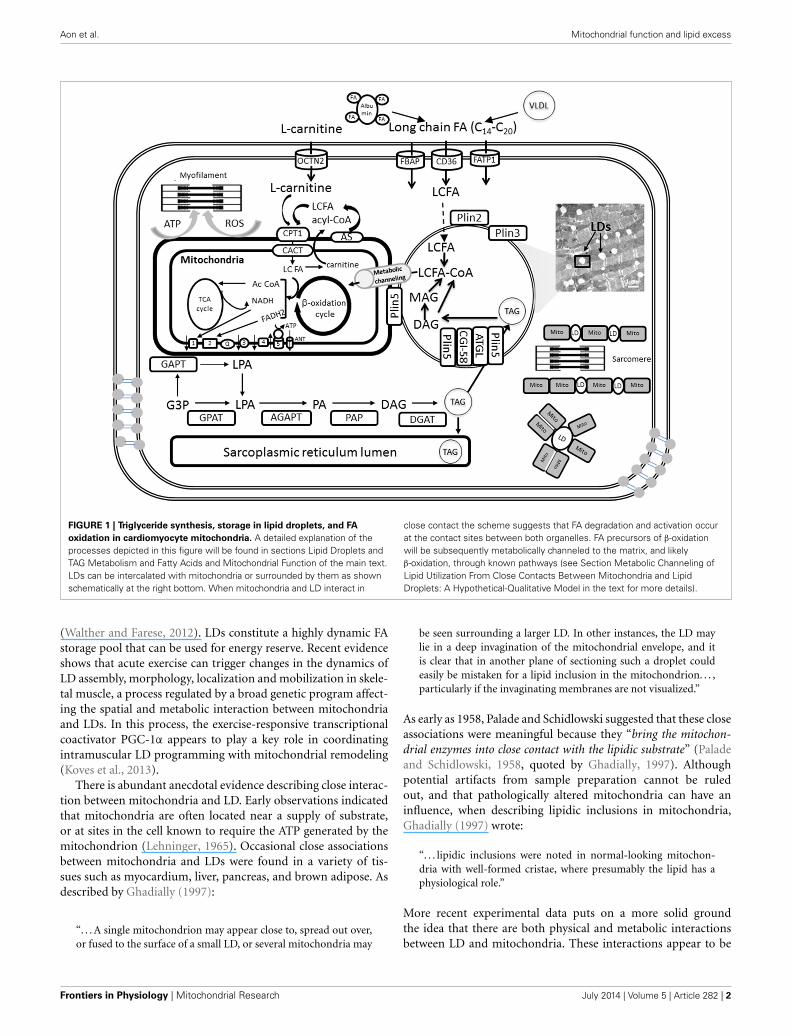
FIGURE 1 | Triglyceride synthesis, storage in lipid droplets, and FA
oxidation in cardiomyocyte mitochondria. A detailed explanation of theprocesses depicted in this figure will be found in sections Lipid Droplets andTAG Metabolism and Fatty Acids and Mitochondrial Function of the main text.LDs can be intercalated with mitochondria or surrounded by them as shownschematically at the right bottom. When mitochondria and LD interact in
close contact the scheme suggests that FA degradation and activation occurat the contact sites between both organelles. FA precursors of β-oxidationwill be subsequently metabolically channeled to the matrix, and likelyβ-oxidation, through known pathways (see Section Metabolic Channeling ofLipid Utilization From Close Contacts Between Mitochondria and LipidDroplets: A Hypothetical-Qualitative Model in the text for more details).
(Walther and Farese, 2012). LDs constitute a highly dynamic FAstorage pool that can be used for energy reserve. Recent evidenceshows that acute exercise can trigger changes in the dynamics ofLD assembly, morphology, localization and mobilization in skele-tal muscle, a process regulated by a broad genetic program affect-ing the spatial and metabolic interaction between mitochondriaand LDs. In this process, the exercise-responsive transcriptionalcoactivator PGC-1α appears to play a key role in coordinatingintramuscular LD programming with mitochondrial remodeling(Koves et al., 2013).
There is abundant anecdotal evidence describing close interac-tion between mitochondria and LD. Early observations indicatedthat mitochondria are often located near a supply of substrate,or at sites in the cell known to require the ATP generated by themitochondrion (Lehninger, 1965). Occasional close associationsbetween mitochondria and LDs were found in a variety of tis-sues such as myocardium, liver, pancreas, and brown adipose. Asdescribed by Ghadially (1997):
“. . . A single mitochondrion may appear close to, spread out over,or fused to the surface of a small LD, or several mitochondria may
be seen surrounding a larger LD. In other instances, the LD maylie in a deep invagination of the mitochondrial envelope, and itis clear that in another plane of sectioning such a droplet couldeasily be mistaken for a lipid inclusion in the mitochondrion. . . ,particularly if the invaginating membranes are not visualized.”
As early as 1958, Palade and Schidlowski suggested that these closeassociations were meaningful because they “bring the mitochon-drial enzymes into close contact with the lipidic substrate” (Paladeand Schidlowski, 1958, quoted by Ghadially, 1997). Althoughpotential artifacts from sample preparation cannot be ruledout, and that pathologically altered mitochondria can have aninfluence, when describing lipidic inclusions in mitochondria,Ghadially (1997) wrote:
“. . . lipidic inclusions were noted in normal-looking mitochon-dria with well-formed cristae, where presumably the lipid has aphysiological role.”
More recent experimental data puts on a more solid groundthe idea that there are both physical and metabolic interactionsbetween LD and mitochondria. These interactions appear to be
Frontiers in Physiology | Mitochondrial Research July 2014 | Volume 5 | Article 282 | 2

Aon et al. Mitochondrial function and lipid excess
modulated by relevant physiological situations such as fastingand exercise training. Available evidence also shows that proteinslocated in the LD surface closely interact with enzymes of thelypolytic cascade modulating FA acid efflux from the droplet.
LIPID DROPLETS AND TAG METABOLISMTAG is the major form of energy storage that with sterol estersserve as reservoirs of membrane lipid components (Waltherand Farese, 2009). In cardiomyocytes TAGs are synthesized byacyltransferases and phosphatases at the sarcoplasmic reticu-lum and mitochondrial membrane and then packaged into LDs(Walther and Farese, 2009; Singh and Cuervo, 2012; Kienesbergeret al., 2013). TAG synthesis is initiated by glycerol-3-phosphateacyltransferases (GPAT) at the mitochondrial and sarcoplas-mic reticulum membrane and then completed at the sarcoplas-mic reticulum by sn-1-acyl-glycerol-3-phosphate acyltransferase(AGPAT), phosphatidic acid phosphatase (PAP), and sn-1,2-diacylglycerol acyltransferase (DGAT) reactions (Kienesbergeret al., 2013) (Figure 1). Newly formed TAGs are packaged intocytoplasmic LDs. Thus, lipids are not stored as FAs but as TAGs(triglycerides) produced by a series of esterification reactionsthat combine three FA molecules with glycerol 3-phosphate; forexample, the TAG for palmitate is tripalmitin.
LDs are considered dynamic cellular organelles rather thansimple lipid storage depots that, relatively recently, have beenimplicated in many biological processes (Walther and Farese,2009, 2012; Greenberg and Coleman, 2011; Singh and Cuervo,2012). LDs size varies from a diameter of 0.1 μm in yeast to over100 μm in a white adipocyte. LDs consist of a single protein-decorated phospholipid monolayer that delimits their hydropho-bic core from the rest of the cell (Fujimoto and Parton, 2011).The hydrophobic core contains neutral lipids, most notably TAGand sterol esters. The adipose tissue LD has a core predominantlyformed by TAG whereas in most cells cholesterol and TAG sharethe nuclear core of the LD (Singh and Cuervo, 2012). LDs areprominent in many types of mammalian cells, with adipocytesbeing the most highly specialized for lipid and energy storage. LDsinteract with the endoplasmic reticulum and the mitochondria—the two organelles that have been proposed as sites of formationof the autophagosome limiting membrane (Fujimoto et al., 2008;Murphy et al., 2009; Singh and Cuervo, 2012). Such contactzones are also sites of active lipid synthesis enriched in AcylCoA:diacylglycerol acyltransferase 2 (DGAT2), the major enzymecatalyzing TAG synthesis (Cases et al., 2001; Walther and Farese,2009).
TAG stored in LDs is catabolized by the sequential hydroly-sis of ester bonds between FAs and the glycerol backbone. TAGhydrolysis is a tightly regulated process that involves a complexinteraction between lipases and regulatory proteins (Lass et al.,2011). TAG catabolism is performed by a cascade of lipolyticreactions that is initiated by adipose triglyceride lipase (ATGL)producing diacylglycerol (DAG). Hormone-sensitive lipase (HSL)and monoacylglycerol lipase (MGL) complete the lipolytic cas-cade by sequentially hydrolyzing DAG and monoacylglycerol(MAG), respectively, (Figure 1). MAG lipase (MGL) performs thefinal step in TAG catabolism by hydrolyzing MAGs to glyceroland FAs (Kienesberger et al., 2013). The rate of lipolysis can be
dramatically stimulated by adrenergic hormones via activation ofprotein kinase A (PKA). PKA phosphorylates perilipin and HSLand causes a complex set of events leading to TAG hydrolysis.
The FAs released during TAG catabolism are mainly used forβ-oxidation and subsequent ATP synthesis via OxPhos in mito-chondria (Figure 1; see below: Fatty Acids and MitochondrialFunction). In oxidative tissues such as the heart, TAG-derived FAsare utilized as an energy source, but they also serve as signalingmolecules as well as building blocks for membranes and complexlipids.
Hepatocytes, heart and skeletal myocytes, adrenocortical cells,enterocytes, and macrophages may all contain large amountsof LDs. Excessive LD accumulation is a hallmark of T2DM,obesity, atherosclerosis, hepatic steatosis, and other metabolicdiseases. However, in certain organs like skeletal muscle, intramy-ocellular triacylglycerol (IMTG) accumulation is not strictly apathologic phenomenon (see below: Mitochondria, Lipids andInsulin Resistance). Lipid content is elevated in red comparedwith white skeletal muscles and increases in response to habit-ual exercise in both oxidative and glycolytic fibers. The “athleteparadox” consists of IMTG accumulation observed in endurance-trained athletes that retain insulin sensitivity irrespective of thefact that in some cases IMTGs exceed those measured in seden-tary obese or T2DM obese patients (Goodpaster et al., 2001; vanLoon et al., 2003; Shaw et al., 2010; Egan and Zierath, 2013; Koveset al., 2013). As with aerobic exercise, both muscle glycogen andIMTG contribute to energy provision during resistance exercise(Koopman et al., 2006).
MITOCHONDRIA AND PERILIPINSThe protein family of perilipins (Plin) is associated withLDs. As scaffolding proteins perilipins affect the spatial andmetabolic interactions between LD and mitochondria (Figure 1).Development of tissue lipotoxicity and dysfunction is linked toalterations in LD biogenesis and regulation of TAG hydrolysis(Wang and Sztalryd, 2011). Since in response to lipid loadingof cells perilipins associate with LDs the role of these proteins isunder intense scrutiny.
The Plin protein family, or PAT for perilipin/ADRP/TIP47, isconstituted by Plin1 to Plin5, and droplets may contain variouscombinations of them (Greenberg et al., 2011). Plin1 is the mostabundant PAT protein in adipocytes and Plin2 in the liver, whereit has been linked to hepatic steatosis. Whereas Plin1 and 4 arelimited to adipose tissue, Plin2 and 3 are ubiquitous. Plin1 and 2are always found in an LD-bound state whereas Plin3 to 5 can beeither LD-bound or free in the cytoplasm.
Genetic manipulations aiming at ablating perilipins to inferabout their physiological roles and impact on fat depositionhave been performed. Plin1-null mice are lean and develop sys-temic insulin resistance as they grow older. Plin1-null adipocytesexhibited enhanced rates of constitutive (unstimulated) lipol-ysis and reduced catecholamine-stimulated lipolysis (Tanseyet al., 2001). Together, these data suggested that Plin1 proteinenhances catecholamine-stimulated lipolysis and, importantly,that a reduction in Plin1 protein expression is associated withincreased constitutive lipolysis, which can promote systemicinsulin resistance (Greenberg et al., 2011).
www.frontiersin.org July 2014 | Volume 5 | Article 282 | 3

Aon et al. Mitochondrial function and lipid excess
Plin5 is found primarily in oxidative tissues, e.g. skeletal andheart muscles, liver (Bickel et al., 2009). Plin5 knockout micelacked detectable LDs in the heart and had significantly reducedmyocardial TAG content, an effect that was rescued by lipase inhi-bition (Kuramoto et al., 2012). The excessive TAG catabolismexhibited by Plin5-deficient hearts was paralleled by increasedFA oxidation (FAO) and enhanced ROS levels that led to an age-dependent decline in heart function. Thus, it was suggested thatuncontrolled lipolysis and defective TAG storage impair cardiacfunction through chronic mitochondrial FA overload. Plin5 mayregulate LD degradation and the flux of lipolysis-derived FAsto mitochondria for energy production (Figure 1) (Kienesbergeret al., 2013). Plin5 overexpression in cardiac muscle produceda robust increase in LDs resulting in cardiac steatosis but with-out major consequences for heart function. This data indicatedthat Plin5 plays a critical role in droplet formation and stabiliza-tion via its regulatory role of lipolysis in vivo (Wang et al., 2013).Interestingly, mitochondria in heart tissue from the Plin5 overex-pressor appeared to always be distributed in tight clusters aroundLDs exhibiting a significant increase in size without changes innumber as revealed by morphometric analysis (Wang et al., 2013).In skeletal muscle, Plin5 overexpression increased IMCL contentwithout hindering insulin mediated glucose uptake while pro-moting the expression of genes involved in mitochondrial FAOand fat catabolism (Bosma et al., 2013).
In liver, down-modulation of Plin2 promotes a reduction inhepatic steatosis and increases insulin sensitivity, but a reductionin both Plin2 and Plin3 causes insulin resistance (Greenberg et al.,2011). In the heart, Plin2 does not promote the interaction ofmitochondria with LDs, but increased TAG accumulation associ-ated with reduced presence of ATGL in LD and decreased lipolysis(Wang et al., 2011). As the first enzyme from the lipolytic cascade(Zimmermann et al., 2004), the constitutive activity of ATGL ispredominantly responsible for basal levels of lipolysis (Greenberget al., 2011). ATGL overexpression in a cardiomyocyte-specificmanner decreased myocardial TAG and lipotoxic intermediatesaccumulation in type 1 diabetic mice (Pulinilkunnil et al., 2013).This resulted in decreased reliance on FAO, and preserved contentof respiratory complexes as well as cardiac function during earlystages of diabetes.
Overall, the reported data indicate that reduced expression ofperilipins may promote both lipolysis and fat oxidation, result-ing in reduced intracellular TAG and adipose mass. On the otherhand, excessive lypolysis and defective lipid storage may pro-mote insulin resistance and impaired cardiac function throughchronic mitochondrial FA overload. Consequently, lipid storageand utilization appears to be a tightly regulated cellular process.
FATTY ACIDS AND MITOCHONDRIAL FUNCTIONPreservation of the intracellular redox environment (RE) is cru-cial for vital functions such as division, differentiation, contractilework and survival amongst others (Schafer and Buettner, 2001;Aon et al., 2007, 2009; Brown et al., 2010; Fisher-Wellman andNeufer, 2012; Jeong et al., 2012; Lloyd et al., 2012; Muoio andNeufer, 2012; Aggarwal and Makielski, 2013). Mitochondria aremain drivers of the intracellular RE (Aon et al., 2010, 2012;Stanley et al., 2011; Tocchetti et al., 2012; Fisher-Wellman et al.,
2013; Kembro et al., 2013) and together with peroxisomes consti-tute the main subcellular compartments where lipid degradationoccurs. Yet, the impact of lipids on mitochondrial redox statusand ROS emission, and their links to energetics are not fullyelucidated.
FAs are main metabolic fuels in heart and skeletal muscle,and β-oxidation represents their main degradation pathway. Therate of β-oxidation is led by demand since an increase in workrate and ATP utilization leads to faster oxidative phosphorylation(OxPhos) and tricarboxylic acid (TCA) cycle activity. In turn, thedecrease in NADH and acetyl-CoA (AcCoA) levels leads to anincrease of the β-oxidation flux (Neely et al., 1969; Oram et al.,1973; Eaton et al., 1996a; Eaton, 2002; Lopaschuk et al., 2010).
Lipids are supplied in the form of albumin-bound FAssecreted from adipose tissue or by catabolism of very low densitylipoprotein (VLDL) complex by coronary vascular endotheliallipoprotein lipases (Figure 1). Long chain FA (LCFA) trans-port requires carrier proteins in the sarcolemma (FATP1, fattyacid transporter protein 1; FABP, plasma membrane-associatedfatty acid-binding protein; LCFAT, long-chain fatty acid trans-porter; OCTN2, plasma membrane sodium-dependent carnitinetransporter; FAT/CD36, fatty acid translocase CD36) and themitochondria (CPT1, carnitine palmitoyltransferase 1; CACT,carnitine:acylcarnitine translocase).
Upon entry into the cell, LCFA first gets activated by form-ing thioesters with coenzyme A (CoA), LCFA-CoA, and is eitheroxidized in the mitochondria via β-oxidation or forms TAG byesterification (Figure 1). Subsequently TAGs can be stored in theform of LD. Long-chain FAs are activated on the mitochondrialouter membrane by the long-chain acyl-CoA synthetase but themitochondrial inner membrane is not permeable to these acyl-CoAs. CPT1 catalyzes the conversion of long-chain acyl CoA tolong-chain acylcarnitine, which is subsequently shuttled into themitochondria (Lopaschuk et al., 2010). Control at the level ofCPT1 activity appears to be important in heart and skeletal mus-cle β-oxidation flux (Awan and Saggerson, 1993; Lopaschuk et al.,1994; Zammit, 1999; Eaton, 2002).
After its formation by CPT1, the long-chain acylcarnitine istranslocated across the inner mitochondrial membrane by CACTthat involves the exchange of carnitine for acylcarnitine. CACThas extremely high activity in most cell types with active β-oxidation (Ramsay and Tubbs, 1976; Noel et al., 1985; Eaton,2002). CACT is a critical step in the translocation of FA moietiesinto the mitochondria, as evidenced by the development of car-diomyopathies and irregular heartbeats in individuals with CACTdeficiencies (Lopaschuk et al., 1994, 2010).
In the matrix, acylcarnitine is converted back to acyl CoAand catabolized via β-oxidation. The β-oxidation of activated FAsoccurs within the mitochondrial matrix and is catalyzed by thesequential action of four enzyme families (acyl-CoA dehydroge-nase, enoyl-CoA hydratase, 3-hydroxyacyl-CoA dehydrogenase,and 3-ketoacyl- CoA thiolase), with acyl-CoA dehydrogenaseexhibiting different substrate specificity for short-, medium-,long- and very long-chain acyl-CoAs (Kunau et al., 1995; Eatonet al., 1996a; Kerner and Hoppel, 2000). The end product of eachcycle of β-oxidation is AcCoA, shortening the LCFA by 2 car-bons. Ac CoA then enters the TCA cycle for complete oxidation
Frontiers in Physiology | Mitochondrial Research July 2014 | Volume 5 | Article 282 | 4

Aon et al. Mitochondrial function and lipid excess
rendering reducing equivalents in the form of the electron donorsNADH and FADH2 leading to ATP synthesis via OxPhos in therespiratory chain (Figure 1). Ultimately, ATP is utilized by thecontractile machinery to transduce chemical energy into mechan-ical work. ROS may also affect contractile performance via signal-ing or redox modification of sensitive cysteines from, e.g., myosinheavy chain (Canton et al., 2011; Steinberg, 2013).
Besides their metabolic role in the provision of energy, long-chain free FAs exert diverse effects on cellular membranes andon the catalytic activities of many enzymes (Loskovich et al.,2005). FAs play the dual role of uncouplers and inhibitors of mito-chondrial respiration (Wojtczak and Schonfeld, 1993) through aprotonophoric effect on the inner membrane, and an inhibitoryaction on the electron transfer chain (Schonfeld and Reiser, 2006;Schonfeld and Wojtczak, 2007, 2008). Additionally, FAs havethe potential to drastically alter mitochondrial membranes per-meability through opening of the permeability transition pore(Scorrano et al., 2001; Bernardi et al., 2002; Penzo et al., 2002,2004). Excluded from these effects are the acyl-CoAs that donot exert protonophoric activity and do not uncouple OxPhosbecause they are unable to cross the inner mitochondrial mem-brane (Wojtczak, 1976).
Free FAs can act as specific complex I-directed inhibitors(Loskovich et al., 2005; Schonfeld and Wojtczak, 2008), andlong-chain acyl-CoAs are known inhibitors of ANT (Pande andBlanchaer, 1971; Lerner et al., 1972; Wojtczak, 1976). The inhibi-tion is of a competitive character (Duszynski and Wojtczak, 1975)and strongly depends on the carbon chain length of the fatty acylmoiety (Morel et al., 1974). Further evidence that FAs, in theiranionic form, can be substrates for transport by ANT was givenby their inhibitory effect on ATP and ADP exchanges (Wojtczakand Zaluska, 1967; Schonfeld et al., 1996; Klingenberg, 2008).According to the FA cycling model (Skulachev, 1991) undissoci-ated FA molecules can undergo a spontaneous flip-flop from theouter to the inner leaflet of the inner mitochondrial membranewhere they release protons because of the alkaline milieu of thematrix. Then, in the form of anions, they are transported backto the external leaflet by ANT; one proton is transferred from theexternal space to the matrix compartment per molecule of theFA per cycle. In this manner, FAs can lead to energy dissipationthrough a selective protonophoric action mediated by couplingof transmembrane movement of the fatty acyl anion (via theANT, uncoupling proteins, UCPs, and/or other inner membranecarriers). These events result in dissipative proton cycling thatdecreases the proton motive force thereby affecting respiration,ATP synthesis, and ion homeostasis.
Palmitoyl CoA inhibits the ANT independently from β-oxidation, according to more recent evidence obtained in isolatedmitochondria from rat liver (Ciapaite et al., 2005) and guineapig heart (Aon and Cortassa, unpublished) respiring on G/M. Inthe case of liver mitochondria it was shown that the ANT inhi-bition induced changes in intra- and extra-mitochondrial ATPconcentrations and ��m. This interference with the ANT carrierincreased ��m and the reduction level of coenzyme Q (Bakkeret al., 2000) both expected to promote the formation of ROS.Studies further showed that the PCoA-elicited concentration-dependent H2O2 formation can be explained by its effect on ��m
that in the presence of 5 μM PCoA showed a 13 mV increase(Ciapaite et al., 2006). The specific action of PCoA on the ANTin the liver (Ciapaite et al., 2006), is in contrast with an apparentmulti target effect in the heart (Aon and Cortassa, unpublished).These differences may be given by intrinsic functional differencesdue to species (rat, guinea pig) or organ specificity, e.g., liverand heart mitochondria. Differences may also be linked to thepresence of distinct FA transporters (FATPs or SLC27As) or FAbinding proteins (FABPs).
MITOCHONDRIA, LIPIDS, AND INSULIN RESISTANCEThe shift from intermediate values of RE, corresponding to ROSlevels compatible with signaling functions (Aon et al., 2010;Cortassa et al., 2014), toward either more reducing or oxidizingconditions is a topic of great potential importance and interestwith implications for insulin signaling. Indeed, the associationbetween lipotoxicity and the onset of insulin resistance in skele-tal muscle is a hotly debated subject (Muoio and Neufer, 2012).One side posits that it is due to dysfunctional mitochondria withintrinsic deficiencies in OxPhos and deficits in fat oxidation.These impairments impinge on insulin signaling by diverting FAsaway from oxidation and toward production of DAGs, ceramideand other toxic lipid species (Lowell and Shulman, 2005; Roden,2005). The other side of the debate notes that this idea is incom-patible with the principles of bioenergetics because mitochondrialrespiration is governed by energy demand; intracellular lipids willaccumulate whenever FAs supply exceeds the energy needs of thecell. Consequently, they suggest that the etiology of muscle insulinresistance is grounded on the fundamental principles that gov-ern cellular and mitochondrial bioenergetics and the redox stressthat is placed on the respiratory system when energy supply per-sistently outpaces energy demand (Muoio and Neufer, 2012). Inagreement with this idea other authors have emphasized that thematching between increased FA availability and oxidative capacitydistinguishes the increase in IMTG following endurance trainingfrom obesity/diabetic conditions. Chronic exercise training canelicit high oxidative capacity conferred by higher mitochondrialcontent but not mitochondrial function. Under these conditions,lipid infusion in endurance-trained athletes is able to reduceinsulin sensitivity only by 29% as compared to 63% in untrainedsubjects (Phielix et al., 2012).
Whereas in exercise training IMTG reflects an increasedreliance on fats as substrate, in obesity/diabetes will implyaccumulation of lipid metabolites [long chain fatty acyl-CoA(LCFA-CoA), DAG, and ceramide] that are responsible for theimpairment in insulin action rather than the IMTG pool con-tained in LDs (Schrauwen et al., 2010; Fisher-Wellman andNeufer, 2012). Apparently, increased concentrations of intramus-cular LCFA-CoA and DAG activate PKC, which appears to induceimpairments in insulin signaling via serine phosphorylation ofthe insulin receptor substrate-1. In a model of diet-induced obe-sity, accumulation of acylcarnitines, as products of incompleteβ-oxidation, was shown in skeletal muscle (Koves et al., 2008).These findings led to the idea of a mitochondria-derived sig-nal that couples incomplete β-oxidation with insulin resistance.Chronic elevations of incomplete oxidation intermediates of FAsand branched-chain amino acids (Newgard, 2012) might foster
www.frontiersin.org July 2014 | Volume 5 | Article 282 | 5

Aon et al. Mitochondrial function and lipid excess
a mitochondrial microenvironment that is conducive to higherH2O2 release from mitochondria with potential to modulateinsulin signaling (Fisher-Wellman and Neufer, 2012; Muoio andNeufer, 2012).
The debate about the role of mitochondrial and lipidmetabolism at the origin of insulin resistance is highly relevantfor the diabetic heart because of its heavy dependence on fats forfunction (Holloway et al., 2009, 2011). The debate centered on themitochondrial load-oxidative potential in skeletal muscle, is alsorelevant for the heart where function is led by energy demand.In fact, lipid accumulation in the heart is largely seen as a mis-match between supply and demand, i.e., lipids amass when supplyoutpaces demand.
A fundamentally important question still heavily debated iswhether or not a shift in substrate preference toward fat oxi-dation lowers disease risk (Muoio and Neufer, 2012). FAs andglucose are the two major fuels driving heart contraction. Intype 2 diabetes and obesity FAO is increased (Lopaschuk, 2002;Carley and Severson, 2005) but our knowledge about the com-bined effects of hyperglycemia, a hallmark of diabetes, and highFA availability, on metabolism, redox/ROS balance and theirimpact on heart function is incomplete. Although the healthyheart is flexible regarding fuel selection, in the metabolicallychallenged diabetic heart by high levels of glucose and fat, thefactors contributing to dysfunction and which are beneficial asenergy source or redox donors are still unclear. Existing com-pelling evidence indicates that substrate-driven redox status playsa critical role in cardiac contractile performance in type 2 diabeteswhere cellular/mitochondrial redox and energetics are altered(see below: Mitochondrial, Cellular and Organ Mechanisms forManaging Lipid Affluence) (Anderson et al., 2009a; Tocchetti et al.,2012). Overall, there is no disputing that lipid oxidation con-fers a metabolic advantage during starvation and exercise, but therole of fuel selection per se in defending against metabolic diseaseneeds further investigation.
MITOCHONDRIAL, CELLULAR, AND ORGAN MECHANISMSFOR MANAGING LIPID AFFLUENCEAs important fuels of cellular function it is very well known howFAs are degraded by mitochondria. Yet, the mechanisms by whichmitochondria manage lipid excess are largely unknown. The roleof β-oxidation per se as an underlying cause of obesity-associatedglucose intolerance remains a topic of active research and debate(Fisher-Wellman and Neufer, 2012; Muoio and Neufer, 2012).Furthermore, mitochondria play a central role in the develop-ment of diabetes and obesity complications (Bugger and Abel,2010; Sivitz and Yorek, 2010) and their energetic/redox dysfunc-tion is directly involved in the redox imbalance exhibited by theheart (Tocchetti et al., 2012; Frasier et al., 2013) and skeletalmuscle (Anderson et al., 2009a).
Mitochondria and lipid oxidation play a predominant role asdrivers of the intracellular RE. FAs are a major source of cellu-lar ATP which, in the heart, is synthesized up to two thirds viareducing equivalents (e.g., 24 NADH, 8 FADH2 for palmitate)derived from β-oxidation in mitochondria. The higher energeticbudget provided by the saturated FA palmitate (three times higherthan from glucose when ATP/mol substrate is considered) in the
form of reducing power provides electrons to antioxidant sys-tems and the mitochondria respiratory/energetic machinery. Inagreement with the prominent role of lipids on the intracellu-lar redox status, it was shown that Palm determined a transitionfrom oxidized-to-reduced cellular redox status in cardiomyocytesfrom type-2 diabetic (db/db) hearts abating ROS levels drasti-cally (Tocchetti et al., 2012). This effect was coupled to a markedGSH rise both in wild type and db/db myocytes. As a conse-quence of its favorable effect on cellular redox balance, Palmsignificantly improved isoproterenol-induced contractile reservein db/db cardiomyocytes (Tocchetti et al., 2012).
Keeping a proper cellular/mitochondrial RE is vital for optimalexcitation-contraction (EC) coupling as well as energy supply inthe heart (Burgoyne et al., 2012; Christians and Benjamin, 2012;Nickel et al., 2013, 2014). Intracellular redox balance affects Ca2+handling by interfering with a wide range of proteins implicatedin EC coupling (Fauconnier et al., 2007) including the SR Ca2+release channels [the ryanodine receptors], the SR Ca2+ pumps,and the sarcolemmal Na+/Ca2+ exchanger (Zima and Blatter,2006; Dedkova and Blatter, 2008). Also unknown is whether themechanisms utilized by mitochondria to deal with lipid excessdiffer between organs. Important examples are the skeletal andcardiac muscles where β-oxidation predominates due to their lackof de novo lipogenesis (Eaton, 2002). Certainly, the organ’s func-tional specificity plays a role. As a matter of fact, skeletal muscleis the largest glycogen storage organ (∼4-fold the capacity ofthe liver) thus critical for glycemic control as the predominant(∼80%) site of glucose disposal under insulin-stimulated con-ditions (DeFronzo et al., 1981; Egan and Zierath, 2013). On theother hand, the heart carries out its pump function transducingthe chemical energy stored in FAs and glucose into mechani-cal and electrical energy. At rest, the heart cycles about 6 kg ofATP every day while beating about 100,000 times (Neubauer,2007). Mitochondria provide the bulk of the ATP needed forcardiac muscle contraction (about two thirds) and sarcolemmaland sarcoplasmic ion transport (one third), responsible for theCa2+ transients and electrical activity in cardiac cells (Solaini andHarris, 2005; Cortassa et al., 2009; Nickel et al., 2013).
The far higher amounts of O2 processed by the heart ona specific basis with respect to, e.g., brain and skeletal muscle(Rolfe and Brown, 1997), and its continuous activity, make thisorgan susceptible to oxidative damage (Burgoyne et al., 2012;Christians and Benjamin, 2012). As a matter of fact, myocardialfunction and the ability of the heart to tolerate stress decline withage (Lakatta and Sollott, 2002). Although the mechanisms con-tributing to age-related alterations in myocardial function are notfully understood, mitochondrial dysfunction, oxidative stress andthe accumulation of oxidant-induced damage are major factors(Fannin et al., 1999; Suh et al., 2003; Judge et al., 2005).
Defects in mitochondrial FA β-oxidation lead to several well-known metabolic disorders, such as Reye syndrome, cardiomy-opathy and sudden infant death syndrome (Roe and Ding, 2001;Yang et al., 2001). The maintenance of high levels of mitochon-drial β-oxidation could reduce the excessive fat accumulation andstorage leading to human obesity. Lipid overload involving TAGaccumulation in non-adipose tissues characterizes disorders suchas hyperlipidemia and lipodystrophies, heart dysfunction, liver
Frontiers in Physiology | Mitochondrial Research July 2014 | Volume 5 | Article 282 | 6
Aon et al. Mitochondrial function and lipid excess
disease, in both humans and in animal models of obesity anddiabetes.
It is becoming increasingly clear that adequate regulation ofTAG metabolism in different organs is critical for both energymetabolism and function. Liver and heart respond to the mas-sive influx of lipids from blood by up regulating LD biogenesis, asa mechanism of defense against the toxicity of FAs, which uponesterification get converted into TAG and stored into LD (Lasset al., 2011). Failure to do so in the liver originates pathogenicconditions such as steatosis and steatohepatitis (Greenberg et al.,2011). The lipid excess situation is also relevant for heart functionin T2DM where FAs are preferred fuels (Lopaschuk et al., 2010).However, under acute, non-chronic, conditions FAs can exhibitadvantageous actions, especially in the heart under diabetic con-ditions (Tocchetti et al., 2012). Cellular TAG accumulation in LDsmay be beneficial rather than detrimental because it diverts FAsfrom pathways leading to cytotoxicity thus serving as a bufferagainst lipotoxicity (Listenberger et al., 2003).
From the examples and arguments above, it is clear that lipidshave a considerable impact on many cellular processes, includ-ing mitochondria. This impact influences the functional outcomeof several organs such as the liver, skeletal and cardiac muscles.Deregulation of lipid metabolism produces overload that is atthe origin or as an aggravating consequence of many diseases.Consequently, the fundamental as well as practical importance ofunraveling the mechanisms by which mitochondria handle lipidsexcess cannot be overstated. First, at the most basic level, we donot know enough about lipids action on mitochondrial ener-getic and redox functions. Lipids can act both as uncouplers andinhibitors of OxPhos (Wojtczak and Schonfeld, 1993; Bernardiet al., 2002), and the consequences of these contradictory effectson mitochondrial energetic, redox and signaling functions are juststarting to be unraveled (Schonfeld and Wojtczak, 2008). Second,besides being the main site of lipid degradation, mitochondriamay be actively modulating the balance between lipid storage andutilization.
In the following sections we explore some of the new emergingmechanisms of lipid storage and utilization by mitochondria atthe organelle, cellular and organ level in different physiologicalsettings.
CLOSE CONTACT MITOCHONDRIA-LIPID DROPLETRegular exercise and physical activity are considered cornerstonesin the prevention, management, and treatment of numerouschronic conditions, including hypertension, coronary heart dis-ease, obesity, T2DM, and age-related muscle wasting (sarcopenia)(Haskell et al., 2007; Colberg et al., 2010; Egan and Zierath,2013).
Exercise training enhances mitochondrial biogenesis and per-formance in skeletal muscle (Irrcher et al., 2003), but not in theheart (Li et al., 2011). Whether the same is true in T2DM heartsis unclear. In electron micrographs LDs can be easily detected intype 2 diabetic (db/db) (Boudina et al., 2007) or ob/ob (Ge et al.,2012) but not in WT mice hearts. In cells LDs can be readily visu-alized using the fluorescent FA analog (dodecanoic acid) BODIPYthat labels neutral lipids in cytoplasmic droplets (Walther andFarese, 2012).
The occurrence of close contact between mitochondria and LDin the heart is remarkable because of its dependence on mito-chondrial energetics preferentially fueled by FAs. More notewor-thy though is the fact that these close contacts occur in the T2DMheart, where the dependence on fat fueling is even more promi-nent (Lopaschuk, 2002; Bugger and Abel, 2010). Interestingly,Plin5 overexpression in heart tissue rendered tight mitochondrialclusters around LDs with mitochondria significantly larger butnot higher in number (Wang et al., 2013). The same authors pro-posed that Plin5 could play a regulatory role in the FA flux fromLDs to mitochondria under conditions of increased cellular FAinflux (Wang and Sztalryd, 2011). These data also suggest thatPlin5 with its role of favoring LD accumulation may act to keepthe intracellular levels of FA metabolites (e.g., DAG, ceramide)below lipotoxic amounts (see below: Metabolic Channeling ofLipid Utilization From Close Contacts Between Mitochondria andLipid Droplets: A Hypothetical-Qualitative Model).
In skeletal muscle IMTG accumulates and is actively utilizedduring exercise (Shaw et al., 2010; Egan and Zierath, 2013; Koveset al., 2013). Endurance exercise training increases mitochon-drial content (by size not numbers) for men and women buthealthy active women have higher IMTG accumulation com-pared with men due to greater number rather than size of LDs(Tarnopolsky et al., 2007). Interestingly, this study also reportedan increase in the physical contact between mitochondria andIMTGs following endurance exercise training. Rates of wholebody fat oxidation and IMTG utilization are determined by fac-tors such as diet, intensity and duration of exercise, and fitness.During acute exercise, the contribution of various metabolicpathways to energy provision is determined by the relative inten-sity and absolute power output of the exercise bout (Egan andZierath, 2013). The rate of ATP demand and energy expendi-ture is determined by the absolute power output whereas therelative exercise intensity influences the relative contributions ofcarbohydrate oxidation and lipid sources, and circulating (extra-muscular) and intramuscular fuel stores, to energy provision. Asexercise intensity increases, muscle utilization of circulating freeFAs slightly declines, whereas utilization of circulating glucoseincreases progressively up to near-maximal intensities (van Loonet al., 2001).
IMTG breakdown occurs primarily via HSL and ATGL (Wattand Spriet, 2010). Although IMTGs constitute only a smallfraction (∼1–2%) of whole-body lipid stores they represent animportant fuel source during prolonged (>90 min) but moder-ate intensity exercise. IMTGs can provide ∼25% of total energyhowever their contribution decreases at either higher or lowerintensities of exercise (Romijn et al., 1993; van Loon et al.,2001). Maximal rates of fat oxidation occur at moderate exer-cise intensities (∼60% VO2 max) (Shaw et al., 2010; Egan andZierath, 2013). At low-to-moderate exercise intensity, the pri-mary substrates fueling skeletal muscle are glucose, derived fromhepatic glycogenolysis (or gluconeogenesis) or oral ingestion,and free FAs released by adipose tissue lipolysis. Prolongedexercise (>60 min) at a fixed intensity increases the energycontribution from lipid oxidation (Egan and Zierath, 2013).IMTG stores can be reduced by ∼60% following exercise, pre-dominantly in type I muscle fibers (van Loon et al., 2003;
www.frontiersin.org July 2014 | Volume 5 | Article 282 | 7

Aon et al. Mitochondrial function and lipid excess
Stellingwerff et al., 2007; Shaw et al., 2010; Egan and Zierath,2013).
Lipophagy, i.e., the turnover of LDs by autophagy, may occurdue to random sequestration of cytosolic material by “in bulk”autophagy. However, when lipophagy is activated in response to alipid challenge or prolonged starvation, a switch toward the pref-erential sequestration of LD seems to happen, supporting somelevel of selectivity in this process (Singh et al., 2009). We suggestthat this may also be the case for close contacts mitochondria-LD,and that energy demand may be a main elicitor of the interac-tion between these two organelles. Consonant with this idea, ithas been proposed that LDs assembly in skeletal muscle underexercise training would improve the management of high FAinflux enabling a more precisely regulated trafficking of substrateto and from IMTG thus contributing to optimal mitochondrialperformance and metabolic flexibility (Koves et al., 2013).
LIPOTOXICITY AND LD ACCUMULATION DYNAMICSIn pathologic states lipotoxicity may occur over time, despite TAGaccumulation, when either the cellular capacity for TAG storageis exceeded or when triglyceride pools are hydrolyzed, resultingin increased cellular free FA levels. Thus, the duration and extentof lipid overload may determine if a cell is protected or damaged.Whether mitochondrial energy/redox status can alter the balanceLD formation and utilization in the short-term is a question thathas not been hitherto addressed.
Studies performed with non-invasive spectroscopic techniqueshave shown elevated IMCL triglyceride content in the left ventri-cle (i.e., LV steatosis) of obese and T2DM patients (McGavocket al., 2007; Rijzewijk et al., 2008) but its association with earlydiastolic dysfunction leading to subsequent systolic dysfunctionremains controversial (Anderson et al., 2009b; Lopaschuk et al.,2010). Again, lipids through accumulation of triglycerides are atthe center of the controversy. In skeletal (Liu et al., 2007) and car-diac (Ussher et al., 2009) muscle, IMCL accumulation as a resultof diet-induced obesity is not at all pathogenic, but may even beprotective against obesity-associated maladies.
Previous reports have linked ROS-mediated mitochondrialdysfunction to DAG and ceramide, two main products of lipiddegradation (Coen and Goodpaster, 2012). Lipid channeling tomitochondria may represent a mechanism by which concentra-tion build-up of these intermediaries is avoided, especially underhigh energy demand. Based on these premises, we suggest thattemporary lipid storage in LDs does not necessarily representpathophysiological behavior. On the contrary, it may embody anadaptive response, at least in the short-term thus representingan adaptive strategy of lipids utilization ensuring energy sup-ply without affecting neither mitochondrial nor cellular redoxstatus.
REDOX OPTIMIZED ROS BALANCE AND MITOCHONDRIALREDOX AND ENERGETICSLipid metabolites can damage the respiratory chain leading toimpaired energetic transition in mitochondria through their dualeffect as uncouplers and inhibitors (Wojtczak and Schonfeld,1993). Impairment of the key state 4→3 energetic transition canoccur via inhibition of ANT or ATPsynthase thereby producing
a continuous release of ROS irrespective of ADP addition(Tocchetti et al., 2012).
Mitochondria are a main source of ROS but can also be theirtarget. The RE is a major driving force of the crucial energy-redox link of mitochondrial function (Cortassa et al., 2014).The mitochondrial RE depends on the intrinsic redox potentialand instantaneous reducing capacity of this organelle as well asits response to the cytoplasmic redox status (Aon et al., 2010;Kembro et al., 2013). In this context, Redox-Optimized ROSBalance (R-ORB) provides a useful conceptual framework torationalize many results described in the present review. One ofthe main R-ORB postulates is that ROS efflux from mitochon-dria will attain a minimum at intermediate values of RE, whenVO2 reaches a maximum following ADP stimulation (Figure 2)(Cortassa et al., 2014). Under state 3 respiration, glutathioneand thioredoxin systems are essential for minimizing ROS releasefrom mitochondria (Aon et al., 2010, 2012; Stanley et al., 2011;Kudin et al., 2012; Cortassa et al., 2014). In excess, lipid precur-sors of β-oxidation can promote mitochondrial uncoupling andoxidized redox status (Aon and Cortassa, unpublished). In moreoxidized RE, away from the optimum (intermediate) RE compati-ble with minimal ROS, antioxidant systems become overwhelmedleading to pathological ROS overflow (Aon et al., 2010; Cortassaet al., 2014).
Mitochondria function in more oxidative environments inchronic diseases (Tocchetti et al., 2012). Thus, it becomes funda-mental to understand how oxidative stress influences the depen-dence of ROS emission on respiration (Cortassa et al., 2014).When oxidant challenged, mitochondria displayed H2O2 emis-sion levels 2-fold higher than controls, and exhibited lower res-piration (Figure 2). Oxidative stress shifted redox balance towardthe more oxidized range where the sensitivity of the ROS effluxto the RE decreases more drastically in state 4 than in state 3respiration. A 50% decrease in reduced glutathione (GSH) wasmainly responsible for the shift of the RE to a more oxidized state(Cortassa et al., 2014).
METABOLIC CHANNELING OF LIPID UTILIZATION FROMCLOSE CONTACTS BETWEEN MITOCHONDRIA AND LIPIDDROPLETS: A HYPOTHETICAL-QUALITATIVE MODELRecent evidence supports physical and metabolic interactionsbetween LDs and mitochondria mediated by the scaffoldingprotein Plin 5 (Wang and Sztalryd, 2011; Wang et al., 2011;Koves et al., 2013). Wang and collaborators observed that Plin5-overexpressing cells show decreased LD hydrolysis and palmitateβ-oxidation when compared with controls. Instead, palmitateincreasingly incorporated into TAGs under basal conditionswhereas in protein kinase A-stimulated state LD hydrolysis inhibi-tion was removed and FAs released for β-oxidation. These resultssuggested that Plin5 regulates LD hydrolysis and controls localFA flux to protect mitochondria against excessive exposure to FA(Wang and Sztalryd, 2011). All these observations are in agree-ment with the relatively recent realization that the LD proteome ishighly dynamic and more complex than previously thought. TheLD proteome contains key components of the fat mobilizationsystem and proteins that suggest LD interactions with a variety ofcell organelles, including the mitochondria (Beller et al., 2010).
Frontiers in Physiology | Mitochondrial Research July 2014 | Volume 5 | Article 282 | 8

Aon et al. Mitochondrial function and lipid excess
FIGURE 2 | Redox-Optimized ROS Balance and the effect of oxidative
stress on mitochondrial respiration, H2O2 emission, and the RE. R-ORBpostulates that ROS levels (as the net result of production and scavenging)depend on the intra-cellular and -mitochondrial redox environment (RE). Italso proposes that there is a minimum level of ROS emission whenmitochondria maximize their energetic output. Under high energy demand,and despite large respiratory rates, ROS emission levels will be kept to aminimum by ROS scavenging systems (Stanley et al., 2011; Aon et al.,2012). Oxidative stress can happen at either extreme of RE, either highlyreduced or highly oxidized, but governed by different mechanisms (Aonet al., 2010; Kembro et al., 2014). The plot displays schematically thesummary of the response of respiration (black traces) and ROS emission instressed mitochondria (gray traces) plus further addition of the uncouplerFCCP (dashed-dotted line). Continuous lines correspond to the absence ofstress whereas dashed lines belong to mitochondria under stressedconditions (Cortassa et al., 2014). Black arrows indicate the direction ofchange in VO2 and ROS elicited by stress. Notice the shift toward moreoxidized RE in the curves corresponding to stressful conditions. The thickgray arrow pointing to the left denotes pathological conditions arising, e.g.,from chronic diseases, where severe stress will affect both energetic (e.g.,��m, ADP consumption) and redox [e.g., NAD(P)H, GSH, Trx] functionsthus increased mitochondrial ROS emission and higher cytoplasmic ROSlevels. Reprinted from Cortassa et al. (2014).
Based on the premise of metabolic links extending beyondphysical contact between mitochondria and LDs, we propose amodel of metabolic channeling for lipid utilization by mitochon-dria. According to our model, metabolic channeling represents away mitochondria can manage lipid affluence in an energeticallyand redox-controlled fashion. Qualitatively, the lipid utilizationchanneling model postulates that after TAG degradation, lipidsare directly delivered for activation, transport and β-oxidationfrom the LD to the mitochondrion at the contact site (Figure 1).The model also proposes that β-oxidation may also happenmetabolically channeled through the enzymatic components ofthe lipid degradation pathway organized as a multienzyme com-plex (Eaton, 2002).
From a structural standpoint, the model is based on directand close contact between LDs and mitochondria involvingtheir recruitment and surrounding of the LD. The modelalso postulates membrane fusion-mediated reorganization of
intra-mitochondrial membrane and molecular components(Walther and Farese, 2009) as well as lipids segregation within thedroplet (Fujimoto and Parton, 2011).
Biochemically, the pathway of long-chain FAO to AcCoA isone of the longest unbranched pathways in metabolism, contain-ing 27 intermediates between palmitoyl-CoA and AcCoA (Eaton,2002). That the enzymes of β-oxidation may be organized into amultienzyme complex was suggested long ago. In these biomolec-ular assemblies, sequential catalytic reactions proceed via transferof the intermediates between individual component enzymes,precluding their diffusion into the bulk aqueous medium, thus“metabolically channeled” (Welch, 1977; Sumegi et al., 1991).
An earlier proposal of metabolic channeling in β-oxidationwas based on the detection of low concentrations of intermedi-ates (Garland et al., 1965) and the observation that β-oxidationintermediates that accumulate behaved more like products thanintermediates (Stewart et al., 1973; Stanley and Tubbs, 1974,1975; Eaton et al., 1994, 1996a,b, 1999). This led to the “leakyhosepipe” model for the control of β-oxidation flux (Stewart et al.,1973; Stanley and Tubbs, 1974, 1975) in which channeling of asmall, quickly turning-over pool of intermediates was implied(see Eaton, 2002 for a review).
Some aspects of the structural basis for a channeling mecha-nism in β-oxidation have been described (Ishikawa et al., 2004).Evidence in support of a multifunctional FAO complex withinmitochondria, physically associated with respiratory chain super-complexes that favor metabolic channeling, has been recentlyreported (Wang et al., 2010). Functionally, the direct delivery oflipids at contact sites, and their channeled processing will avoidelevation of their concentration, thus ruling out the potentialinhibitory as well as uncoupling action of FAs (Wojtczak andSchonfeld, 1993). The latter will ensure a reliable and efficientenergy supply.
CONCLUDING REMARKSMitochondria, cells and organs have developed mechanisms thatallow managing heavy influx of FAs within functionally reliablelimits. The LD as a dynamic storage of FAs can also be seenas a protective mechanism employed by cells to avoid excessiveintracellular concentration of FAs thus hindering their poten-tial deleterious effects on mitochondrial function. The tightand reciprocal regulation of lipid storage and utilization is evi-denced by genetic manipulation of perilipins indicating that theirreduced expression leads to increased lipid oxidation and reducedaccumulation of intracellular fat and adipose mass. On the otherhand, however, excessive lipolysis and defective lipid storage pro-motes insulin resistance through mitochondrial FA overload andROS overflow.
Preservation of the intracellular RE is crucial for vital func-tions. Mitochondria play a decisive role as the organelle thatspecifically handles the highest amounts of oxygen processed bythe organism thus prone not only to be the source but also thetarget of oxidative stress. Mitochondrial function needs to sus-tain energy supply reliably while releasing ROS levels compatiblewith signaling. However, lipids can derail both of these criticalfunctions. Consequently, the hypothetical lipid utilization chan-neling model we are proposing herein satisfies the fundamentals
www.frontiersin.org July 2014 | Volume 5 | Article 282 | 9

Aon et al. Mitochondrial function and lipid excess
of cellular and mitochondrial energetics and redox. In principle,diversion of excess lipids to LDs can be an effective cytoplasmicmechanism for “sequestering” FAs thereby helping to keep lowconcentration of lipotoxic intermediates resulting from lipid oxi-dation. Functionally, direct delivery and channeled processing oflipids in mitochondria could represent a reliable and efficient wayto ensure energy supply and redox control. Such a mechanismwould avoid exceeding the lipid storage capacity thus becomingcrucial for skeletal muscle or heart subjected to high workload,and therefore, heavy influx of FAs.
ACKNOWLEDGMENTSThis work was supported by National Institutes of Health grantsR01-HL091923 (Miguel A. Aon) and R21HL106054 (Sonia C.Cortassa).
REFERENCESAggarwal, N. T., and Makielski, J. C. (2013). Redox control of cardiac excitability.
Antioxid. Redox Signal. 18, 432–468. doi: 10.1089/ars.2011.4234Akar, F. G., Aon, M. A., Tomaselli, G. F., and O’Rourke, B. (2005). The mitochon-
drial origin of postischemic arrhythmias. J. Clin. Invest. 115, 3527–3535. doi:10.1172/JCI25371
Anderson, E. J., Kypson, A. P., Rodriguez, E., Anderson, C. A., Lehr, E. J.,and Neufer, P. D. (2009b). Substrate-specific derangements in mitochondrialmetabolism and redox balance in the atrium of the type 2 diabetic human heart.J. Am. Coll. Cardiol. 54, 1891–1898. doi: 10.1016/j.jacc.2009.07.031
Anderson, E. J., Lustig, M. E., Boyle, K. E., Woodlief, T. L., Kane, D. A., Lin, C. T.,et al. (2009a). Mitochondrial H2O2 emission and cellular redox state link excessfat intake to insulin resistance in both rodents and humans. J. Clin. Invest. 119,573–581. doi: 10.1172/JCI37048
Aon, M. A., Cortassa, S., Akar, F. G., Brown, D. A., Zhou, L., and O’Rourke, B.(2009). From mitochondrial dynamics to arrhythmias. Int. J. Biochem. Cell Biol.41, 1940–1948. doi: 10.1016/j.biocel.2009.02.016
Aon, M. A., Cortassa, S., Maack, C., and O’Rourke, B. (2007). Sequential openingof mitochondrial ion channels as a function of glutathione redox thiol status.J. Biol. Chem. 282, 21889–21900. doi: 10.1074/jbc.M702841200
Aon, M. A., Cortassa, S., and O’Rourke, B. (2010). Redox-optimized ROSbalance: a unifying hypothesis. Biochim. Biophys. Acta 1797, 865–877. doi:10.1016/j.bbabio.2010.02.016
Aon, M. A., Stanley, B. A., Sivakumaran, V., Kembro, J. M., O’Rourke, B., Paolocci,N., et al. (2012). Glutathione/thioredoxin systems modulate mitochondrialH2O2 emission: an experimental-computational study. J. Gen. Physiol. 139,479–491. doi: 10.1085/jgp.201210772
Awan, M. M., and Saggerson, E. D. (1993). Malonyl-CoA metabolism in cardiacmyocytes and its relevance to the control of fatty acid oxidation. Biochem. J.295(pt 1), 61–66.
Bakker, S. J., IJzerman, R. G., Teerlink, T., Westerhoff, H. V., Gans, R. O., andHeine, R. J. (2000). Cytosolic triglycerides and oxidative stress in central obe-sity: the missing link between excessive atherosclerosis, endothelial dysfunction,and beta-cell failure? Atherosclerosis 148, 17–21. doi: 10.1016/S0021-9150(99)00329-9
Beller, M., Thiel, K., Thul, P. J., and Jackle, H. (2010). Lipid droplets:a dynamic organelle moves into focus. FEBS Lett. 584, 2176–2182. doi:10.1016/j.febslet.2010.03.022
Bernardi, P., Penzo, D., and Wojtczak, L. (2002). Mitochondrial energy dissipationby fatty acids. Mechanisms and implications for cell death. Vitam. Horm. 65,97–126. doi: 10.1016/S0083-6729(02)65061-7
Bickel, P. E., Tansey, J. T., and Welte, M. A. (2009). PAT proteins, an ancient familyof lipid droplet proteins that regulate cellular lipid stores. Biochim. Biophys. Acta1791, 419–440. doi: 10.1016/j.bbalip.2009.04.002
Bosma, M., Sparks, L. M., Hooiveld, G. J., Jorgensen, J. A., Houten, S. M.,Schrauwen, P., et al. (2013). Overexpression of PLIN5 in skeletal muscle pro-motes oxidative gene expression and intramyocellular lipid content withoutcompromising insulin sensitivity. Biochim. Biophys. Acta 1831, 844–852. doi:10.1016/j.bbalip.2013.01.007
Boudina, S., Sena, S., Theobald, H., Sheng, X., Wright, J. J., Hu, X. X., et al. (2007).Mitochondrial energetics in the heart in obesity-related diabetes: direct evidencefor increased uncoupled respiration and activation of uncoupling proteins.Diabetes 56, 2457–2466. doi: 10.2337/db07-0481
Brown, D. A., Aon, M. A., Frasier, C. R., Sloan, R. C., Maloney, A. H., Anderson, E.J., et al. (2010). Cardiac arrhythmias induced by glutathione oxidation can beinhibited by preventing mitochondrial depolarization. J. Mol. Cell. Cardiol. 48,673–679. doi: 10.1016/j.yjmcc.2009.11.011
Bugger, H., and Abel, E. D. (2010). Mitochondria in the diabetic heart. Cardiovasc.Res. 88, 229–240. doi: 10.1093/cvr/cvq239
Burgoyne, J. R., Mongue-Din, H., Eaton, P., and Shah, A. M. (2012). Redox signal-ing in cardiac physiology and pathology. Circ. Res. 111, 1091–1106. doi: 10.1161/CIRCRESAHA.111.255216
Camara, A. K., Bienengraeber, M., and Stowe, D. F. (2011). Mitochondrialapproaches to protect against cardiac ischemia and reperfusion injury. Front.Physiol. 2:13. doi: 10.3389/fphys.2011.00013
Canton, M., Menazza, S., Sheeran, F. L., Polverino de Laureto, P., Di Lisa, F., andPepe, S. (2011). Oxidation of myofibrillar proteins in human heart failure. J. Am.Coll. Cardiol. 57, 300–309. doi: 10.1016/j.jacc.2010.06.058
Carley, A. N., and Severson, D. L. (2005). Fatty acid metabolism is enhanced in type2 diabetic hearts. Biochim. Biophys. Acta 1734, 112–126. doi: 10.1016/j.bbalip.2005.03.005
Cases, S., Stone, S. J., Zhou, P., Yen, E., Tow, B., Lardizabal, K. D., et al. (2001).Cloning of DGAT2, a second mammalian diacylglycerol acyltransferase, andrelated family members. J. Biol. Chem. 276, 38870–38876. doi: 10.1074/jbc.M106219200
Christians, E. S., and Benjamin, I. J. (2012). Proteostasis and REDOX state inthe heart. Am. J. Physiol. Heart Circ. Physiol. 302, H24–H37. doi: 10.1152/ajp-heart.00903.2011
Ciapaite, J., Bakker, S. J., Diamant, M., van Eikenhorst, G., Heine, R. J., Westerhoff,H. V., et al. (2006). Metabolic control of mitochondrial properties by adeninenucleotide translocator determines palmitoyl-CoA effects. Implications for amechanism linking obesity and type 2 diabetes. FEBS J. 273, 5288–5302. doi:10.1111/j.1742-4658.2006.05523.x
Ciapaite, J., Van Eikenhorst, G., Bakker, S. J., Diamant, M., Heine, R. J., Wagner, M.J., et al. (2005). Modular kinetic analysis of the adenine nucleotide translocator-mediated effects of palmitoyl-CoA on the oxidative phosphorylation in iso-lated rat liver mitochondria. Diabetes 54, 944–951. doi: 10.2337/diabetes.54.4.944
Coen, P. M., and Goodpaster, B. H. (2012). Role of intramyocelluar lipids in humanhealth. Trends Endocrinol. Metab. 23, 391–398. doi: 10.1016/j.tem.2012.05.009
Colberg, S. R., Albright, A. L., Blissmer, B. J., Braun, B., Chasan-Taber, L., Fernhall,B., et al. (2010). Exercise and type 2 diabetes: American College of SportsMedicine and the American Diabetes Association: joint position statement.Exercise and type 2 diabetes. Med. Sci. Sports Exerc. 42, 2282–2303. doi:10.1249/MSS.0b013e3181eeb61c
Cortassa, S., O’Rourke, B., and Aon, M. A. (2014). Redox-Optimized ROS Balanceand the relationship between mitochondrial respiration and ROS. Biochim.Biophys. Acta 1837, 287–295. doi: 10.1016/j.bbabio.2013.11.007
Cortassa, S., O’Rourke, B., Winslow, R. L., and Aon, M. A. (2009). Control andregulation of mitochondrial energetics in an integrated model of cardiomyocytefunction. Biophys. J. 96, 2466–2478. doi: 10.1016/j.bpj.2008.12.3893
Dedkova, E. N., and Blatter, L. A. (2008). Mitochondrial Ca2+ and the heart. CellCalcium 44, 77–91. doi: 10.1016/j.ceca.2007.11.002
DeFronzo, R. A., Jacot, E., Jequier, E., Maeder, E., Wahren, J., and Felber, J. P. (1981).The effect of insulin on the disposal of intravenous glucose. Results from indi-rect calorimetry and hepatic and femoral venous catheterization. Diabetes 30,1000–1007. doi: 10.2337/diab.30.12.1000
Duszynski, J., and Wojtczak, L. (1975). Effect of metal cations on the inhibitionof adenine nucleotide translocation by acyl-CoA. FEBS Lett. 50, 74–78. doi:10.1016/0014-5793(75)81044-1
Eaton, S. (2002). Control of mitochondrial beta-oxidation flux. Prog. Lipid Res. 41,197–239. doi: 10.1016/S0163-7827(01)00024-8
Eaton, S., Bartlett, K., and Pourfarzam, M. (1996a). Mammalian mitochondrial
beta-oxidation. Biochem. J. 320(pt 2), 345–357.Eaton, S., Bartlett, K., and Pourfarzam, M. (1999). Intermediates of myocar-
dial mitochondrial beta-oxidation: possible channelling of NADH and of CoAesters. Biochim. Biophys. Acta 1437, 402–408. doi: 10.1016/S1388-1981(99)00027-X
Frontiers in Physiology | Mitochondrial Research July 2014 | Volume 5 | Article 282 | 10
Aon et al. Mitochondrial function and lipid excess
Eaton, S., Pourfarzam, M., and Bartlett, K. (1996b). The effect of respiratory chainimpairment of beta-oxidation in rat heart mitochondria. Biochem. J. 319(pt 2),633–640.
Eaton, S., Turnbull, D. M., and Bartlett, K. (1994). Redox control of beta-oxidationin rat liver mitochondria. Eur. J. Biochem. 220, 671–681. doi: 10.1111/j.1432-1033.1994.tb18668.x
Egan, B., and Zierath, J. R. (2013). Exercise metabolism and the molecu-lar regulation of skeletal muscle adaptation. Cell Metab. 17, 162–184. doi:10.1016/j.cmet.2012.12.012
Fannin, S. W., Lesnefsky, E. J., Slabe, T. J., Hassan, M. O., and Hoppel, C. L. (1999).Aging selectively decreases oxidative capacity in rat heart interfibrillar mito-chondria. Arch. Biochem. Biophys. 372, 399–407. doi: 10.1006/abbi.1999.1508
Fauconnier, J., Andersson, D. C., Zhang, S. J., Lanner, J. T., Wibom, R., Katz, A.,et al. (2007). Effects of palmitate on Ca(2+) handling in adult control and ob/obcardiomyocytes: impact of mitochondrial reactive oxygen species. Diabetes 56,1136–1142. doi: 10.2337/db06-0739
Fisher-Wellman, K. H., Mattox, T. A., Thayne, K., Katunga, L. A., La Favor, J. D.,Neufer, P. D., et al. (2013). Novel role for thioredoxin reductase-2 in mitochon-drial redox adaptations to obesogenic diet and exercise in heart and skeletalmuscle. J. Physiol. 591, 3471–3486. doi: 10.1113/jphysiol.2013.254193
Fisher-Wellman, K. H., and Neufer, P. D. (2012). Linking mitochondrial bioen-ergetics to insulin resistance via redox biology. Trends Endocrinol. Metab. 23,142–153. doi: 10.1016/j.tem.2011.12.008
Frasier, C. R., Moukdar, F., Patel, H. D., Sloan, R. C., Stewart, L. M., Alleman, R. J.,et al. (2013). Redox-dependent increases in glutathione reductase and exercisepreconditioning: role of NADPH oxidase and mitochondria. Cardiovasc. Res.98, 47–55. doi: 10.1093/cvr/cvt009
Fujimoto, T., Ohsaki, Y., Cheng, J., Suzuki, M., and Shinohara, Y. (2008). Lipiddroplets: a classic organelle with new outfits. Histochem. Cell Biol. 130, 263–279.doi: 10.1007/s00418-008-0449-0
Fujimoto, T., and Parton, R. G. (2011). Not just fat: the structure and function ofthe lipid droplet. Cold Spring Harb. Perspect. Biol. 3, 1–17. doi: 10.1101/cshper-spect.a004838
Garland, P. B., Shepherd, D., and Yates, D. W. (1965). Steady-state concentrationsof coenzyme A, acetyl-coenzyme A and long-chain fatty acyl-coenzyme A inrat-liver mitochondria oxidizing palmitate. Biochem. J. 97, 587–594.
Ge, F., Hu, C., Hyodo, E., Arai, K., Zhou, S., Lobdell, H. 4th., et al. (2012).Cardiomyocyte triglyceride accumulation and reduced ventricular function inmice with obesity reflect increased long chain fatty acid uptake and de novo fattyacid synthesis. J. Obes. 2012:205648. doi: 10.1155/2012/205648
Ghadially, F. N. (1997). Ultrastructural Pathology of the Cell and Matrix. Boston,MA: Butterworth-Heinemann.
Goodpaster, B. H., He, J., Watkins, S., and Kelley, D. E. (2001). Skeletal muscle lipidcontent and insulin resistance: evidence for a paradox in endurance-trainedathletes. J. Clin. Endocrinol. Metab. 86, 5755–5761. doi: 10.1210/jcem.86.12.8075
Greenberg, A. S., and Coleman, R. A. (2011). Expanding roles for lipid droplets.Trends Endocrinol. Metab. 22, 195–196. doi: 10.1016/j.tem.2011.04.002
Greenberg, A. S., Coleman, R. A., Kraemer, F. B., McManaman, J. L., Obin, M. S.,Puri, V., et al. (2011). The role of lipid droplets in metabolic disease in rodentsand humans. J. Clin. Invest. 121, 2102–2110. doi: 10.1172/JCI46069
Haskell, W. L., Lee, I. M., Pate, R. R., Powell, K. E., Blair, S. N., Franklin, B. A.,et al. (2007). Physical activity and public health: updated recommendation foradults from the American College of Sports Medicine and the American HeartAssociation. Med. Sci. Sports Exerc. 39, 1423–1434. doi: 10.1249/mss.0b013e3180616b27
Helguera, P., Seiglie, J., Rodriguez, J., Hanna, M., Helguera, G., and Busciglio, J.(2013). Adaptive downregulation of mitochondrial function in down syndrome.Cell Metab. 17, 132–140. doi: 10.1016/j.cmet.2012.12.005
Holloway, G. P., Benton, C. R., Mullen, K. L., Yoshida, Y., Snook, L. A., Han, X.X., et al. (2009). In obese rat muscle transport of palmitate is increased andis channeled to triacylglycerol storage despite an increase in mitochondrialpalmitate oxidation. Am. J. Physiol. Endocrinol. Metab. 296, E738–E747. doi:10.1152/ajpendo.90896.2008
Holloway, G. P., Snook, L. A., Harris, R. J., Glatz, J. F., Luiken, J. J., and Bonen,A. (2011). In obese Zucker rats, lipids accumulate in the heart despite nor-mal mitochondrial content, morphology and long-chain fatty acid oxidation.J. Physiol. 589, 169–180. doi: 10.1113/jphysiol.2010.198663
Irrcher, I., Adhihetty, P. J., Joseph, A. M., Ljubicic, V., and Hood, D. A. (2003).Regulation of mitochondrial biogenesis in muscle by endurance exercise. SportsMed. 33, 783–793. doi: 10.2165/00007256-200333110-00001
Ishikawa, M., Tsuchiya, D., Oyama, T., Tsunaka, Y., and Morikawa, K. (2004).Structural basis for channelling mechanism of a fatty acid beta-oxidationmultienzyme complex. EMBO J. 23, 2745–2754. doi: 10.1038/sj.emboj.7600298
Jeong, E. M., Liu, M., Sturdy, M., Gao, G., Varghese, S. T., Sovari, A. A., et al. (2012).Metabolic stress, reactive oxygen species, and arrhythmia. J. Mol. Cell. Cardiol.52, 454–463. doi: 10.1016/j.yjmcc.2011.09.018
Judge, S., Jang, Y. M., Smith, A., Hagen, T., and Leeuwenburgh, C. (2005). Age-associated increases in oxidative stress and antioxidant enzyme activities incardiac interfibrillar mitochondria: implications for the mitochondrial theoryof aging. FASEB J. 19, 419–421. doi: 10.1096/fj.04-2622fje
Kembro, J. M., Aon, M. A., Winslow, R. L., O’Rourke, B., and Cortassa, S. (2013).Integrating mitochondrial energetics, redox and ROS metabolic networks: atwo-compartment model. Biophys. J. 104, 332–343. doi: 10.1016/j.bpj.2012.11.3808
Kembro, J. M., Cortassa, S., and Aon, M. A. (2014). “Mitochondrial reactive oxygenspecies and arrhythmias,” in Systems Biology of Free Radicals and Antioxidants,ed I. Laher (Berlin-Heidelberg: Springer-Verlag), 1047–1076.
Kerner, J., and Hoppel, C. (2000). Fatty acid import into mitochondria. Biochim.Biophys. Acta 1486, 1–17. doi: 10.1016/S1388-1981(00)00044-5
Kienesberger, P. C., Pulinilkunnil, T., Nagendran, J., and Dyck, J. R. (2013).Myocardial triacylglycerol metabolism. J. Mol. Cell. Cardiol. 55, 101–110. doi:10.1016/j.yjmcc.2012.06.018
Klingenberg, M. (2008). The ADP and ATP transport in mitochondria and itscarrier. Biochim. Biophys. Acta 1778, 1978–2021. doi: 10.1016/j.bbamem.2008.04.011
Kok, B. P., and Brindley, D. N. (2012). Myocardial fatty acid metabolism and lipo-toxicity in the setting of insulin resistance. Heart Fail. Clin. 8, 643–661. doi:10.1016/j.hfc.2012.06.008
Koopman, R., Manders, R. J., Jonkers, R. A., Hul, G. B., Kuipers, H., and van Loon,L. J. (2006). Intramyocellular lipid and glycogen content are reduced followingresistance exercise in untrained healthy males. Eur. J. Appl. Physiol. 96, 525–534.doi: 10.1007/s00421-005-0118-0
Koves, T. R., Sparks, L. M., Kovalik, J. P., Mosedale, M., Arumugam, R., DeBalsi,K. L., et al. (2013). PPARgamma coactivator-1alpha contributes to exercise-induced regulation of intramuscular lipid droplet programming in mice andhumans. J. Lipid Res. 54, 522–534. doi: 10.1194/jlr.P028910
Koves, T. R., Ussher, J. R., Noland, R. C., Slentz, D., Mosedale, M., Ilkayeva,O., et al. (2008). Mitochondrial overload and incomplete fatty acid oxida-tion contribute to skeletal muscle insulin resistance. Cell Metab. 7, 45–56. doi:10.1016/j.cmet.2007.10.013
Kudin, A. P., Augustynek, B., Lehmann, A. K., Kovacs, R., and Kunz, W. S. (2012).The contribution of thioredoxin-2 reductase and glutathione peroxidase toH(2)O(2) detoxification of rat brain mitochondria. Biochim. Biophys. Acta 1817,1901–1906. doi: 10.1016/j.bbabio.2012.02.023
Kunau, W. H., Dommes, V., and Schulz, H. (1995). beta-oxidation of fatty acidsin mitochondria, peroxisomes, and bacteria: a century of continued progress.Prog. Lipid Res. 34, 267–342. doi: 10.1016/0163-7827(95)00011-9
Kuramoto, K., Okamura, T., Yamaguchi, T., Nakamura, T. Y., Wakabayashi, S.,Morinaga, H., et al. (2012). Perilipin 5, a lipid droplet-binding protein, protectsheart from oxidative burden by sequestering fatty acid from excessive oxidation.J. Biol. Chem. 287, 23852–23863. doi: 10.1074/jbc.M111.328708
Lakatta, E. G., and Sollott, S. J. (2002). Perspectives on mammalian cardiovascularaging: humans to molecules. Comp. Biochem. Physiol. A Mol. Integr. Physiol. 132,699–721. doi: 10.1016/S1095-6433(02)00124-1
Lass, A., Zimmermann, R., Oberer, M., and Zechner, R. (2011). Lipolysis—a highlyregulated multi-enzyme complex mediates the catabolism of cellular fat stores.Prog. Lipid Res. 50, 14–27. doi: 10.1016/j.plipres.2010.10.004
Lehninger, A. L. (1965). The Mitochondrion. Molecular Basis of Structure andFunction. New York, NY: W.A. Benjamin.
Lerner, E., Shug, A. L., Elson, C., and Shrago, E. (1972). Reversible inhibition ofadenine nucleotide translocation by long chain fatty acyl coenzyme A estersin liver mitochondria of diabetic and hibernating animals. J. Biol. Chem. 247,1513–1519.
Li, L., Muhlfeld, C., Niemann, B., Pan, R., Li, R., Hilfiker-Kleiner, D., et al. (2011).Mitochondrial biogenesis and PGC-1alpha deacetylation by chronic treadmillexercise: differential response in cardiac and skeletal muscle. Basic Res. Cardiol.106, 1221–1234. doi: 10.1007/s00395-011-0213-9
Listenberger, L. L., Han, X., Lewis, S. E., Cases, S., Farese, R. V. Jr., Ory, D.S., et al. (2003). Triglyceride accumulation protects against fatty acid-induced
www.frontiersin.org July 2014 | Volume 5 | Article 282 | 11

Aon et al. Mitochondrial function and lipid excess
lipotoxicity. Proc. Natl. Acad. Sci. U.S.A. 100, 3077–3082. doi: 10.1073/pnas.0630588100
Liu, L., Zhang, Y., Chen, N., Shi, X., Tsang, B., and Yu, Y. H. (2007). Upregulationof myocellular DGAT1 augments triglyceride synthesis in skeletal muscle andprotects against fat-induced insulin resistance. J. Clin. Invest. 117, 1679–1689.doi: 10.1172/JCI30565
Lloyd, D., Cortassa, S., O’Rourke, B., and Aon, M. A. (2012). What yeastand cardiomyocytes share: ultradian oscillatory redox mechanisms of cel-lular coherence and survival. Integr. Biol. (Camb.) 4, 65–74. doi: 10.1039/c1ib00124h
Lopaschuk, G. D. (2002). Metabolic abnormalities in the diabetic heart. Heart Fail.Rev. 7, 149–159. doi: 10.1023/A:1015328625394
Lopaschuk, G. D., Belke, D. D., Gamble, J., Itoi, T., and Schonekess, B. O. (1994).Regulation of fatty acid oxidation in the mammalian heart in health anddisease. Biochim. Biophys. Acta 1213, 263–276. doi: 10.1016/0005-2760(94)00082-4
Lopaschuk, G. D., Ussher, J. R., Folmes, C. D., Jaswal, J. S., and Stanley, W. C.(2010). Myocardial fatty acid metabolism in health and disease. Physiol. Rev.90, 207–258. doi: 10.1152/physrev.00015.2009
Loskovich, M. V., Grivennikova, V. G., Cecchini, G., and Vinogradov, A. D. (2005).Inhibitory effect of palmitate on the mitochondrial NADH:ubiquinone oxi-doreductase (complex I) as related to the active-de-active enzyme transition.Biochem. J. 387, 677–683. doi: 10.1042/BJ20041703
Lowell, B. B., and Shulman, G. I. (2005). Mitochondrial dysfunction and type 2diabetes. Science 307, 384–387. doi: 10.1126/science.1104343
Martinez-Outschoorn, U. E., Sotgia, F., and Lisanti, M. P. (2012). Power surge:supporting cells “fuel” cancer cell mitochondria. Cell Metab. 15, 4–5. doi:10.1016/j.cmet.2011.12.011
McGavock, J. M., Lingvay, I., Zib, I., Tillery, T., Salas, N., Unger, R., et al.(2007). Cardiac steatosis in diabetes mellitus: a 1H-magnetic resonance spec-troscopy study. Circulation 116, 1170–1175. doi: 10.1161/CIRCULATIONAHA.106.645614
Morel, F., Lauquin, G., Lunardi, J., Duszynski, J., and Vignais, P. V. (1974). Anappraisal of the functional significance of the inhibitory effect of long chainacyl-CoAs on mitochondrial transports. FEBS Lett. 39, 133–138. doi: 10.1016/0014-5793(74)80035-9
Muoio, D. M., and Neufer, P. D. (2012). Lipid-induced mitochondrial stress andinsulin action in muscle. Cell Metab. 15, 595–605. doi: 10.1016/j.cmet.2012.04.010
Murphy, S., Martin, S., and Parton, R. G. (2009). Lipid droplet-organelle inter-actions; sharing the fats. Biochim. Biophys. Acta 1791, 441–447. doi: 10.1016/j.bbalip.2008.07.004
Neely, J. R., Bowman, R. H., and Morgan, H. E. (1969). Effects of ventricularpressure development and palmitate on glucose transport. Am. J. Physiol. 216,804–811.
Neubauer, S. (2007). The failing heart–an engine out of fuel. N.Engl. J. Med. 356,1140–1151. doi: 10.1056/NEJMra063052
Newgard, C. B. (2012). Interplay between lipids and branched-chain amino acids indevelopment of insulin resistance. Cell Metab. 15, 606–614. doi: 10.1016/j.cmet.2012.01.024
Nickel, A., Kohlhaas, M., and Maack, C. (2014). Mitochondrial reactive oxy-gen species production and elimination. J. Mol. Cell. Cardiol. 73, 26–33. doi:10.1016/j.yjmcc.2014.03.011
Nickel, A., Loffler, J., and Maack, C. (2013). Myocardial energetics in heart failure.Basic Res. Cardiol. 108:358. doi: 10.1007/s00395-013-0358-9
Noel, H., Goswami, T., and Pande, S. V. (1985). Solubilization and reconstitutionof rat liver mitochondrial carnitine acylcarnitine translocase. Biochemistry 24,4504–4509. doi: 10.1021/bi00338a003
Oram, J. F., Bennetch, S. L., and Neely, J. R. (1973). Regulation of fatty acidutilization in isolated perfused rat hearts. J. Biol. Chem. 248, 5299–5309.
Palade, G. E., and Schidlowski, G. (1958). Functional association of mitochondriaand lipid inclusions. Anat. Rec. 130:352.
Pande, S. V., and Blanchaer, M. C. (1971). Reversible inhibition of mitochondrialadenosine diphosphate phosphorylation by long chain acyl coenzyme A esters.J. Biol. Chem. 246, 402–411.
Penzo, D., Petronilli, V., Angelin, A., Cusan, C., Colonna, R., Scorrano, L., et al.(2004). Arachidonic acid released by phospholipase A2 activation triggersCa2+-dependent apoptosis through the mitochondrial pathway. J. Biol. Chem.279, 25219–25225. doi: 10.1074/jbc.M310381200
Penzo, D., Tagliapietra, C., Colonna, R., Petronilli, V., and Bernardi, P. (2002).Effects of fatty acids on mitochondria: implications for cell death. Biochim.Biophys. Acta 1555, 160–165. doi: 10.1016/S0005-2728(02)00272-4
Phielix, E., Meex, R., Ouwens, D. M., Sparks, L., Hoeks, J., Schaart, G., et al.(2012). High oxidative capacity due to chronic exercise training attenuates lipid-induced insulin resistance. Diabetes 61, 2472–2478. doi: 10.2337/db11-1832
Pulinilkunnil, T., Kienesberger, P. C., Nagendran, J., Waller, T. J., Young, M. E.,Kershaw, E. E., et al. (2013). Myocardial adipose triglyceride lipase overexpres-sion protects diabetic mice from the development of lipotoxic cardiomyopathy.Diabetes 62, 1464–1477. doi: 10.2337/db12-0927
Ramsay, R. R., and Tubbs, P. K. (1976). The effects of temperature and someinhibitors on the carnitine exchange system of heart mitochondria. Eur. J.Biochem. 69, 299–303. doi: 10.1111/j.1432-1033.1976.tb10886.x
Rijzewijk, L. J., van der Meer, R. W., Smit, J. W., Diamant, M., Bax, J. J., Hammer,S., et al. (2008). Myocardial steatosis is an independent predictor of diastolicdysfunction in type 2 diabetes mellitus. J. Am. Coll. Cardiol. 52, 1793–1799. doi:10.1016/j.jacc.2008.07.062
Roden, M. (2005). Muscle triglycerides and mitochondrial function: possiblemechanisms for the development of type 2 diabetes. Int. J. Obes. (Lond.)29(Suppl. 2), S111–S115. doi: 10.1038/sj.ijo.0803102
Roe, C. R., and Ding, J. H. (2001). “Mitochondrial fatty acid oxidation disorders,”in The Metabolic and Molecular Bases of Inherited Disease, eds C. R. Scriver, A.L. Beaudet, W. S. Sly, and D. Valle (New York, NY: McGraw-Hill), 2297–2326.
Rolfe, D. F., and Brown, G. C. (1997). Cellular energy utilization and molec-ular origin of standard metabolic rate in mammals. Physiol. Rev. 77,731–758.
Romijn, J. A., Coyle, E. F., Sidossis, L. S., Gastaldelli, A., Horowitz, J. F., Endert,E., et al. (1993). Regulation of endogenous fat and carbohydrate metabolism inrelation to exercise intensity and duration. Am. J. Physiol. 265, E380–E391.
Rossignol, D. A., and Frye, R. E. (2014). Evidence linking oxidative stress, mito-chondrial dysfunction, and inflammation in the brain of individuals withautism. Front. Physiol. 5:150. doi: 10.3389/fphys.2014.00150
Schafer, F. Q., and Buettner, G. R. (2001). Redox environment of the cell as viewedthrough the redox state of the glutathione disulfide/glutathione couple. FreeRadic. Biol. Med. 30, 1191–1212. doi: 10.1016/S0891-5849(01)00480-4
Schilling, J. D., and Mann, D. L. (2012). Diabetic cardiomyopathy: bench tobedside. Heart Fail. Clin. 8, 619–631. doi: 10.1016/j.hfc.2012.06.007
Schonfeld, P., and Reiser, G. (2006). Rotenone-like action of the branched-chainphytanic acid induces oxidative stress in mitochondria. J. Biol. Chem. 281,7136–7142. doi: 10.1074/jbc.M513198200
Schonfeld, P., Schild, L., and Bohnensack, R. (1996). Expression of the ADP/ATPcarrier and expansion of the mitochondrial (ATP + ADP) pool contributeto postnatal maturation of the rat heart. Eur. J. Biochem. 241, 895–900. doi:10.1111/j.1432-1033.1996.00895.x
Schonfeld, P., and Wojtczak, L. (2007). Fatty acids decrease mitochondrial gener-ation of reactive oxygen species at the reverse electron transport but increaseit at the forward transport. Biochim. Biophys. Acta 1767, 1032–1040. doi:10.1016/j.bbabio.2007.04.005
Schonfeld, P., and Wojtczak, L. (2008). Fatty acids as modulators of the cellularproduction of reactive oxygen species. Free Radic. Biol. Med. 45, 231–241. doi:10.1016/j.freeradbiomed.2008.04.029
Schrauwen, P., Schrauwen-Hinderling, V., Hoeks, J., and Hesselink, M. K. (2010).Mitochondrial dysfunction and lipotoxicity. Biochim. Biophys. Acta 1801,266–271. doi: 10.1016/j.bbalip.2009.09.011
Scorrano, L., Penzo, D., Petronilli, V., Pagano, F., and Bernardi, P. (2001).Arachidonic acid causes cell death through the mitochondrial permeabilitytransition. Implications for tumor necrosis factor-alpha apoptotic signaling.J. Biol. Chem. 276, 12035–12040. doi: 10.1074/jbc.M010603200
Shaw, C. S., Clark, J., and Wagenmakers, A. J. (2010). The effect of exercise andnutrition on intramuscular fat metabolism and insulin sensitivity. Annu. Rev.Nutr. 30, 13–34. doi: 10.1146/annurev.nutr.012809.104817
Singh, R., and Cuervo, A. M. (2012). Lipophagy: connecting autophagy and lipidmetabolism. Int. J. Cell Biol. 2012:282041. doi: 10.1155/2012/282041
Singh, R., Kaushik, S., Wang, Y., Xiang, Y., Novak, I., Komatsu, M., et al.(2009). Autophagy regulates lipid metabolism. Nature 458, 1131–1135. doi:10.1038/nature07976
Sivitz, W. I., and Yorek, M. A. (2010). Mitochondrial dysfunction in diabetes: frommolecular mechanisms to functional significance and therapeutic opportuni-ties. Antioxid. Redox Signal. 12, 537–577. doi: 10.1089/ars.2009.2531
Frontiers in Physiology | Mitochondrial Research July 2014 | Volume 5 | Article 282 | 12

Aon et al. Mitochondrial function and lipid excess
Skulachev, V. P. (1991). Fatty acid circuit as a physiological mechanism of uncou-pling of oxidative phosphorylation. FEBS Lett. 294, 158–162. doi: 10.1016/0014-5793(91)80658-P
Solaini, G., and Harris, D. A. (2005). Biochemical dysfunction in heart mito-chondria exposed to ischaemia and reperfusion. Biochem. J. 390, 377–394. doi:10.1042/BJ20042006
Stanley, B. A., Sivakumaran, V., Shi, S., McDonald, I., Lloyd, D., Watson, W. H., et al.(2011). Thioredoxin reductase-2 is essential for keeping low levels of H(2)O(2)emission from isolated heart mitochondria. J. Biol. Chem. 286, 33669–33677.doi: 10.1074/jbc.M111.284612
Stanley, K. K., and Tubbs, P. K. (1974). The occurrence of intermediates inmitochondrial fatty acid oxidation. FEBS Lett. 39, 325–328. doi: 10.1016/0014-5793(74)80141-9
Stanley, K. K., and Tubbs, P. K. (1975). The role of intermediates in mitochondrialfatty acid oxidation. Biochem. J. 150, 77–88.
Steinberg, S. F. (2013). Oxidative stress and sarcomeric proteins. Circ. Res. 112,393–405. doi: 10.1161/CIRCRESAHA.111.300496
Stellingwerff, T., Boon, H., Jonkers, R. A., Senden, J. M., Spriet, L. L.,Koopman, R., et al. (2007). Significant intramyocellular lipid use duringprolonged cycling in endurance-trained males as assessed by three differ-ent methodologies. Am. J. Physiol. Endocrinol. Metab. 292, E1715–E1723. doi:10.1152/ajpendo.00678.2006
Stewart, H. B., Tubbs, P. K., and Stanley, K. K. (1973). Intermediates in fatty acidoxidation. Biochem. J. 132, 61–76.
Suh, J. H., Heath, S. H., and Hagen, T. M. (2003). Two subpopulations of mito-chondria in the aging rat heart display heterogenous levels of oxidative stress.Free Radic. Biol. Med. 35, 1064–1072. doi: 10.1016/S0891-5849(03)00468-4
Sumegi, B., Sherry, A. D., Malloy, C. R., Evans, C., and Srere, P. A. (1991). Is theretight channelling in the tricarboxylic acid cycle metabolon? Biochem. Soc. Trans.19, 1002–1005.
Tansey, J. T., Sztalryd, C., Gruia-Gray, J., Roush, D. L., Zee, J. V., Gavrilova,O., et al. (2001). Perilipin ablation results in a lean mouse with aber-rant adipocyte lipolysis, enhanced leptin production, and resistance to diet-induced obesity. Proc. Natl. Acad. Sci. U.S.A. 98, 6494–6499. doi: 10.1073/pnas.101042998
Tarnopolsky, M. A., Rennie, C. D., Robertshaw, H. A., Fedak-Tarnopolsky, S.N., Devries, M. C., and Hamadeh, M. J. (2007). Influence of enduranceexercise training and sex on intramyocellular lipid and mitochondrialultrastructure, substrate use, and mitochondrial enzyme activity. Am. J.Physiol. Regul. Integr. Comp. Physiol. 292, R1271–R1278. doi: 10.1152/ajpregu.00472.2006
Tocchetti, C. G., Caceres, V., Stanley, B. A., Xie, C., Shi, S., Watson, W. H., et al.(2012). GSH or palmitate preserves mitochondrial energetic/redox balance, pre-venting mechanical dysfunction in metabolically challenged myocytes/heartsfrom type 2 diabetic mice. Diabetes 61, 3094–3105. doi: 10.2337/db12-0072
Ussher, J. R., Koves, T. R., Jaswal, J. S., Zhang, L., Ilkayeva, O., Dyck, J. R.,et al. (2009). Insulin-stimulated cardiac glucose oxidation is increased in high-fat diet-induced obese mice lacking malonyl CoA decarboxylase. Diabetes 58,1766–1775. doi: 10.2337/db09-0011
van Loon, L. J., Greenhaff, P. L., Constantin-Teodosiu, D., Saris, W. H., andWagenmakers, A. J. (2001). The effects of increasing exercise intensity onmuscle fuel utilisation in humans. J. Physiol. 536, 295–304. doi: 10.1111/j.1469-7793.2001.00295.x
van Loon, L. J., Koopman, R., Stegen, J. H., Wagenmakers, A. J., Keizer, H. A., andSaris, W. H. (2003). Intramyocellular lipids form an important substrate sourceduring moderate intensity exercise in endurance-trained males in a fasted state.J. Physiol. 553, 611–625. doi: 10.1113/jphysiol.2003.052431
Wallace, D. C. (2012). Mitochondria and cancer. Nat. Rev. Cancer 12, 685–698. doi:10.1038/nrc3365
Walther, T. C., and Farese, R. V. Jr. (2009). The life of lipid droplets. Biochim.Biophys. Acta 1791, 459–466. doi: 10.1016/j.bbalip.2008.10.009
Walther, T. C., and Farese, R. V. Jr. (2012). Lipid droplets and cellular lipidmetabolism. Annu. Rev. Biochem. 81, 687–714. doi: 10.1146/annurev-biochem-061009-102430
Wang, H., Sreenevasan, U., Hu, H., Saladino, A., Polster, B. M., Lund, L. M.,et al. (2011). Perilipin 5, a lipid droplet-associated protein, provides physicaland metabolic linkage to mitochondria. J. Lipid Res. 52, 2159–2168. doi:10.1194/jlr.M017939
Wang, H., Sreenivasan, U., Gong, D. W., O’Connell, K. A., Dabkowski, E. R.,Hecker, P. A., et al. (2013). Cardiomyocyte-specific perilipin 5 overexpressionleads to myocardial steatosis and modest cardiac dysfunction. J. Lipid Res. 54,953–965. doi: 10.1194/jlr.M032466
Wang, H., and Sztalryd, C. (2011). Oxidative tissue: perilipin 5 links storage withthe furnace. Trends Endocrinol. Metab. 22, 197–203. doi: 10.1016/j.tem.2011.03.008
Wang, Y., Mohsen, A. W., Mihalik, S. J., Goetzman, E. S., and Vockley, J. (2010).Evidence for physical association of mitochondrial fatty acid oxidation andoxidative phosphorylation complexes. J. Biol. Chem. 285, 29834–29841. doi:10.1074/jbc.M110.139493
Watt, M. J., and Spriet, L. L. (2010). Triacylglycerol lipases and metabolic con-trol: implications for health and disease. Am. J. Physiol. Endocrinol. Metab. 299,E162–E168. doi: 10.1152/ajpendo.00698.2009
Welch, G. R. (1977). On the role of organized multienzyme systems in cellularmetabolism: a general synthesis. Prog. Biophys. Mol. Biol. 32, 103–191. doi:10.1016/0079-6107(78)90019-6
Wende, A. R., Symons, J. D., and Abel, E. D. (2012). Mechanisms of lipotoxicityin the cardiovascular system. Curr. Hypertens. Rep. 14, 517–531. doi: 10.1007/s11906-012-0307-2
Wojtczak, L. (1976). Effect of long-chain fatty acids and acyl-CoA on mitochondrialpermeability, transport, and energy-coupling processes. J. Bioenerg. Biomembr.8, 293–311.
Wojtczak, L., and Schonfeld, P. (1993). Effect of fatty acids on energy coupling pro-cesses in mitochondria. Biochim. Biophys. Acta 1183, 41–57. doi: 10.1016/0005-2728(93)90004-Y
Wojtczak, L., and Zaluska, H. (1967). The inhibition of translocation of adeninenucleotides through mitochondrial membranes by oleate. Biochem. Biophys.Res. Commun. 28, 76–81. doi: 10.1016/0006-291X(67)90409-3
Yang, B. Z., Mallory, J. M., Roe, D. S., Brivet, M., Strobel, G. D., Jones, K. M.,et al. (2001). Carnitine/acylcarnitine translocase deficiency (neonatal pheno-type): successful prenatal and postmortem diagnosis associated with a novelmutation in a single family. Mol. Genet. Metab. 73, 64–70. doi: 10.1006/mgme.2001.3162
Zammit, V. A. (1999). Carnitine acyltransferases: functional significance of subcel-lular distribution and membrane topology. Prog. Lipid Res. 38, 199–224. doi:10.1016/S0163-7827(99)00002-8
Zima, A. V., and Blatter, L. A. (2006). Redox regulation of cardiac calcium channelsand transporters. Cardiovasc. Res. 71, 310–321. doi: 10.1016/j.cardiores.2006.02.019
Zimmermann, R., Strauss, J. G., Haemmerle, G., Schoiswohl, G., Birner-Gruenberger, R., Riederer, M., et al. (2004). Fat mobilization in adipose tissue ispromoted by adipose triglyceride lipase. Science 306, 1383–1386. doi: 10.1126/science.1100747
Conflict of Interest Statement: The authors declare that the research was con-ducted in the absence of any commercial or financial relationships that could beconstrued as a potential conflict of interest.
Received: 07 April 2014; accepted: 10 July 2014; published online: 31 July 2014.Citation: Aon MA, Bhatt N and Cortassa SC (2014) Mitochondrial and cellularmechanisms for managing lipid excess. Front. Physiol. 5:282. doi: 10.3389/fphys.2014.00282This article was submitted to Mitochondrial Research, a section of the journal Frontiersin Physiology.Copyright © 2014 Aon, Bhatt and Cortassa. This is an open-access article distributedunder the terms of the Creative Commons Attribution License (CC BY). The use, dis-tribution or reproduction in other forums is permitted, provided the original author(s)or licensor are credited and that the original publication in this journal is cited, inaccordance with accepted academic practice. No use, distribution or reproduction ispermitted which does not comply with these terms.
www.frontiersin.org July 2014 | Volume 5 | Article 282 | 13
Related Documents


![[7] Tunable pH-Sensitive Liposomes Hafez et al 2004.pdf · anionic lipid in molar excess over a monovalent cationic lipid containing a ... layer phases are favored at acidic pH values](https://static.cupdf.com/doc/110x72/5b94925a09d3f22b0a8d86af/7-tunable-ph-sensitive-hafez-et-al-2004pdf-anionic-lipid-in-molar-excess.jpg)