Stem Cells & Regenerative Medicine Series Editor Kursad Turksen [email protected] For other titles published in this series, go to http://www.springer.com/series/7896

Welcome message from author
This document is posted to help you gain knowledge. Please leave a comment to let me know what you think about it! Share it to your friends and learn new things together.
Transcript

Stem Cells & Regenerative Medicine
Series EditorKursad [email protected]
For other titles published in this series, go tohttp://www.springer.com/series/7896

Krishnarao Appasani
Raghu K. AppasaniEditors
Stem Cells & Regenerative Medicine
From Molecular Embryology to Tissue Engineering
Foreword bySir John B. Gordon
EditorsKrishnarao AppasaniGeneExpressions Systems, IncWaltham, MA [email protected]
Raghu K. AppasaniStudent ScienceBoston, [email protected]
ISBN 978-1-60761-859-1 e-ISBN 978-1-60761-860-7DOI 10.1007/978-1-60761-860-7Springer New York Dordrecht Heidelberg London
Library of Congress Control Number: 2010938600
© Springer Science+Business Media, LLC 2011All rights reserved. This work may not be translated or copied in whole or in part without the written permission of the publisher (Humana Press, c/o Springer Science+Business Media, LLC, 233 Spring Street, New York, NY 10013, USA), except for brief excerpts in connection with reviews or scholarly analysis. Use in connection with any form of information storage and retrieval, electronic adaptation, computer software, or by similar or dissimilar methodology now known or hereafter developed is forbidden.The use in this publication of trade names, trademarks, service marks, and similar terms, even if they are not identified as such, is not to be taken as an expression of opinion as to whether or not they are subject to proprietary rights.
Printed on acid-free paper
Humana Press is part of Springer Science+Business Media (www.springer.com)
v
Preface
Embryology is a branch of biology that has an immediate bearing on the problem of “life.” Life cannot be fully accounted for without an understanding of its dynamic nature, which expresses itself in the incessant production of new organisms in the process of ontogenetic development. Therefore, embryology is defined as the sci-ence of the development of an embryo from the fertilization of the ovum to the fetus stage. Teaching of embryology has long been an established feature at universities throughout the world, both for students in biology and students in medical sciences. During the twentieth century most of this science has been overshadowed by exper-imental-based genetics and cell biology, which have turned classical embryology into “developmental biology.” Several universities are now teaching developmental biology instead of embryology as a course in biology programs. Significant contri-butions made in the twenty-first century in the fields of molecular biology, biochem-istry, and genomics, integrated with embryology and developmental biology, provide an understanding of the molecular portrait of a “developmental cell.” This integrated approach to development is incorporated in the present book as “stem cell biology,” a new sister branch of embryology/developmental biology that emphasizes the study of self-renewal, differentiation, pluripotency, and nuclear programming, which are characteristics of stem cells. In a broad sense, stem cell biology is nothing more than an understanding of embryology and development together at the molecular level using engineering, imaging, and cell culture principles. With such a wide scope, this book can only be an introduction to stem cell biology.
Stem Cells and Regenerative Medicine: From Embryology to Tissue Engineering is mainly intended for readers in the biotechnology and molecular medicine fields. Although quite a number of books already exist covering stem cells, this book dif-fers, in that is the first text completely devoted to the basic developmental, cellular, and molecular biologic aspects of stem cells and their clinical applications in tissue engineering and regenerative medicine. We took serious consideration in choosing the chapters and sections in this book to maintain the theme of Molecular Embryology to Tissue Engineering.
This book focuses on the basic biology of embryonic and cancer cells and their key involvement in self-renewal, muscle repair, epigenetic processes, and therapeu-tic applications. Significant contributions, such as nuclear reprogramming–induced pluripotency, and stem cell culture techniques using novel biomaterials, are also

vi Preface
covered. This text consists of 36 chapters, grouped into six parts. Most of the chap-ters are written by experts in the field from academia and industry. The goal is to have this book serve as a reference for graduate students, post-docs, and teachers and as an explanatory analysis for executives and scientists in biotech and pharma-ceutical companies. Our hope is that this volume will provide a prologue to the field for both newcomers and those already active in the field.
The term “stem cell” appeared in the scientific literature as early as 1868 in the work of the eminent German biologist Ernst Haeckel. Haeckel, a supporter of Darwinian evolution, developed a number of phylogenetic trees to represent the evolution of organisms from common ancestors and called these trees Stammbaume (“stem trees”). In this context, he used the term Stammzelle (“stem cell”) to describe the ancestor unicellular organism from which he presumed all multicellular organ-isms evolved. He referred to fertilized egg as the source that gives rise to all cells of the organism. Later, in 1887, Theodor Boveri and Valentin Hacker identified the earliest germ cells in animal embryos. In 1892, Valentin Hacker described stem cells as the cells that later in development produce oocytes in the gonads. Thus, in these early studies, the term stem cell referred to what we call the “germline line-age,” “primordial germ cells,” and “germline stem cells.” In 1896, Edmund Wilson, an embryologist, reviewed the finding of these German scientists in his book The Cell in Development and Inheritance, which was published in English and became an inspirational work for a generation of embryologists and geneticists, especially in United States. Given his wide readership, Wilson is generally credited as having coined the term “stem cell.”
Nuclear programming is the process that instructs specialized adult cells to form early pluripotent stem cells. Pluripotent stem cells posses the capacity to become any type of mature cell in the body and therefore have great potential for experi-mental and therapeutic purposes. Using the concept of “cellular reprogramming,” Briggs and King in 1952 produced normal tadpoles by transplanting nuclei from blastula cells to enucleated eggs in the frog Rana pipens. However, transplanting nuclei from differentiated cells was achieved by John Gurdon in 1962 in the African clawed toad, Xenopus laevis, which is now known as the classic nuclear transfer experiment. It took more than another decade (1975) for Gurdon to succeed in producing healthy and sexually mature fertile frogs with functional muscle, beat-ing hearts, well-differentiated eyes, and all of the other organs. This experiment provided the first clear evidence that cell specialization does not involve irreversible inactivation in the genes required for development of an animal. This conceptual framework led to the start of the field of nuclear reprogramming, and Gurdon became known as the “father” of nuclear reprogramming (cloning). It took almost another 10 years to clone an adult sheep, Dolly (in 1996), by Kevin Campbell and Ian Wilmut of the Roslin Institute in Edinburgh, Scotland. This experiment dra-matically extended Gurdon’s concept from frogs to mammals. The Dolly-related work of somatic cell nuclear transfer was further extended to produce monkeys, cows, dogs, mice, and other animals. These remarkable contributions stimulated other researchers to think about using nuclear transfer to generate pluripotent human embryonic stem cells for cell replacement therapy.
viiPreface
The road to embryonic stem cells and beyond began in the 1960s with the work of Leroy Stevens from the Jackson Labs, Bar Harbor, Maine, who discovered embryonal carcinoma cells while studying testicular carcinomas. Later Stevens and colleagues demonstrated that these embryonal carcinoma cells are indeed pluripo-tent stem cells. In the mid-1970s, Gail Martin’s postdoctoral work with Martin Evans at the University of Cambridge led her to develop new in vitro clonal culture methods of embryoid cells. In the early 1980s, Martin, then at the University of California at San Francisco, and Martin Evans and Matthew Kaufman of the University of Cambridge independently isolated stem cells from mouse embryos and coined the term “embryonic stem cells.” It took almost 10 years for Jamie Thompson of the University of Wisconsin to culture monkey embryonic stem cells and subsequently human embryonic stem cells in 1999. Thompson’s work pro-pelled the activity of stem cell research and cell propagation technologies in general.
There are two routes to producing a living animal: (1) injection of a somatic cell nucleus into an enucleated egg (nuclear reprogramming) and (2) use of an embryo to produce embryonic stem cells. In a quite astonishing discovery, Kazutoshi Takahashi and Shinya Yamanaka of Kyoto University in Japan in 2006 for the first time turned adult mouse skin fibroblast cells into pluripotent cells. This break-through of inducing fribroblasts was achieved by stable transfection of only four transcription factors (Oct4, Sox2, Klf4, and c-Myc), and these are now referred to as induced pluripotent stem (iPS) cells. The discovery of iPS cells turned the field of nuclear reprogramming upside down. This work was extended and further con-firmed by several groups that generated iPS cells from individuals with various neurodegenerative diseases, raising the hope of cell replacement therapy and mak-ing personalized medicine a reality. A section of this book with six chapters details the concepts behind nuclear reprogramming and induced pluripotent stem cells.
In 1868, Ernst Neumann suggested that hematopoiesis occurs in bone marrow. He used the term “stem cell” to refer to the common precursor of the blood system in 1912. The debate about the existence of a common hematopoietic stem cell con-tinued for several decades until definitive evidence was provided in 1961 by two Canadian scientists, James Till and Ernest McCulloch. Blood and the system that forms it, known as the hematopoietic system, consists of many cell types with spe-cialized functions (some of these include red blood cells, platelets, granulocytes, macrophages, B and T lymphocytes, etc). Generally, the hematopoietic system is destroyed by radiation and chemotherapeutic agents that kill dividing cancer cells. In order to quantitatively assess the radiation sensitivity of normal bone marrow cells, a colony-forming unit assay was developed by Till and McCulloch, who coined the term “pluripotent hematopoietic stem cells” (HSCs). Today, we know that the best locations for HSCs are bone marrow, umbilical cord blood, and embry-onic stem cells. In 1959, for the first time, Edward Donnall Thomas of the University of Washington used HSCs for treating leukemia and lymphomas through bone marrow transplantation. The efficient expansion of HSCs in culture remains one of the major research themes of stem cell biology. Combined applications of genomics, proteomics, and gene therapy approaches will further help to widen the

viii Preface
horizon for clinical applications. According to Irving Weissman of Stanford University Medical School, the progeny produced from hematopoietic stem cells exhibits properties that include self-renewal, differentiation, migration, and apopto-sis. A few chapters in the third part of this book highlight of the use of HSCs for bone marrow cell therapy, heart transplantation, and cell replacement therapy for neurologic disorders.
The term “tissue engineering” was first used by Eugene Bell of MIT in 1984, and later was also used extensively by Wolter and Meyer in 1984. Tissue engineer-ing combines cells, engineering, and materials methods with suitable biochemical and physiochemical factors to improve or replace biologic functions. In other words, it deals with the repair or replacement of portions of or whole tissues such as bone, blood vessels, bladder, skin, and artificial organs. According to Robert Langer and Joseph Vacanti, it “applies the principles of engineering and life sci-ences toward the development of biological substitutes that restore, maintain, or improve tissue function or a whole organ.” Powerful developments in the multidis-ciplinary field of tissue engineering have yielded a novel set of tissue replacement parts and implementation strategies. Scientific advances in biomaterials, stem cells, growth and differentiation factors, and biomimetic environments have created unique opportunities to fabricate tissues in the laboratory from combinations of engineered extracellular matrices (“scaffolds”), cells, and biologically active mol-ecules. A section of this book with five chapters highlights recent developments in biomaterials, three-dimensional culture systems, lab-on-a-chip concepts, and microtechnologies used in attempts to understand stem cell biology.
Regenerative medicine is a new branch of medicine that attempts to change the course of chronic disease, in many instances regenerating failing organ systems lost due to age, disease, damage, or congenital defects. The term “regenerative medi-cine” was first referred to in 1992 by Leland Kaiser and then popularly used by William Haselstine of Human Genome Sciences. The term regenerative medicine is often used synonymously with tissue engineering, although those involved in regenerative medicine place more emphasis on the use of stem cells to treat diseases using cell therapies or transplantation methods. This field holds the promise of regenerating damaged tissues and organs in the body by stimulating previously irreparable organs to heal themselves. Regenerative medicine also empowers scien-tists to grow tissues and organs in the laboratory and safely implant them when the body cannot heal itself. A section in this book is entirely devoted to describing the use of stem cells in muscle repair and treating cardiac and urologic diseases.
Gurdon has spent much of his career deciphering the molecules and mechanisms that an egg uses to “rejuvenate” nuclei. We know a lot about nuclear transfer, but the question remains of how to regulate and control the most efficient way to repro-gram the nucleus. Although both Gurdon’s (nuclear reprogramming) and Yamanaka’s (iPS) technologies can generate living animals, we do not know the molecular mechanisms underlying these two strategies. The potential of iPS cell technology in biology and medicine is enormous; however, it is still in its infancy, and there are many challenges to overcome before various applications can be used successfully. We still need to understand the components of oocytes or eggs used to depress

ixPreface
somatic gene expression and discover the direct cell-type switches by over-expressed transcription factors. It is also important to identify the basis for the stability of the differentiated state of cells, which will help us to understand how egg-reprogramming factors operate. Finally, mapping of the “embryome” is a necessity, and is looks as though it will become available soon, which will help us to understand the intricacies and epigenetic imprints of embryos.
Many people have contributed to making our involvement in this project possi-ble. We thank our teachers for their excellent teaching, guidance, and mentorship, which helped us to bring this educational enterprise. We are extremely thankful to all of the contributors to this book, without whose commitment this book would not have been possible. Many people have had a hand in the preparation of this book. Each chapter has been passed back and forth between the authors for criticism and revision; hence each chapter represents a joint composition. We thank our readers, who have made our hours putting together this volume worth it. We are indebted to the staff of Springer Science + Business Media (Humana Press), and in particular Mindy Okura-Marszycki and Vindra Dass for their generosity in giving time and effort throughout the editing of this book. This book is dedicated to memory of my late friend, Prof. Xiangzhong (Jerry) Yang of the University of Connecticut, Storrs, who was the first to clone a cow (Amy, the calf) and a strong proponent of stem cell research here in US and China. This book is also dedicated to memory of my late friend, Prof. C.M. Habibullah of the Deccan College of Medical Sciences, India, who was a strong supporter of stem cell research in India. We especially thank Prof. John Gurdon, a researcher of great distinction, for his kindness and support in writ-ing the Foreword to this book. Last, but not least, we thank Shyamala Appasani for her understanding and cooperation during the development of this book.
This book is the first joint project of father and son. A portion of the royalties will be contributed to the Dr. Appasani Foundation, a nonprofit organization devoted to bringing social change through the education of youth in developing nations.
Waltham, MA Krishnarao AppasaniBoston, MA Raghu K. Appasani

xi
I am very grateful to Krishnarao and Raghu Appasani for preparing this volume on the massively expanding fields of stem cells and regenerative medicine and for inviting me to offer a few introductory comments.
The prospect of being able to rejuvenate cell types of almost any kind from easily accessible cells of an adult makes it realistic to envisage cell replacement. Most important, this possibility would provide a patient with new cells of their own genetic type, thereby avoiding the necessity of immunosuppression, as would be required for any cells derived from any other individual, except an identical twin. The great interest in this field has been enormously stimulated by the recent discov-ery of induced pluripotency stem cells but has depended on several much earlier discoveries, most notably on that of embryonic stem cells.
There has been something of a tidal wave of interest in stem cells and regenera-tive medicine as researchers all over the world become active in it. Almost every day there are new papers published on various aspects of pluripotency, and it would be hard, even for those intimately involved in experimental work of this kind, to keep up to date with every advance. It is therefore very valuable to have a volume of 36 contributions summarizing the current status of progress in the various fields that contribute to regenerative medicine. Krishnarao and Raghu Appasani have assembled the contributing chapters into six main areas, ranging from stem cell biology through tissue engineering and therapeutic possibilities. The component chapters will be valuable not only to those who are experimentally active in an aspect of regenerative medicine, but also to those concerned with potential thera-peutic applications. This volume also contains a valuable historical perspective by the Appasanis explaining key events in the development of this field over the last 150 years.
Although there is great enthusiasm for the eventual therapeutic value of work in this field for human health, scientists are very cautious about the time scale of human benefit. Bone marrow cells have been of great clinical value for a number of years. However, there is a long way to go before the brain and heart, to take two examples, can benefit from laboratory-created stem cells. It is indeed remarkable that beating heart cells or dopamine-secreting brain cells can now be derived from human skin and can be proliferated in the laboratory. However, substantial advances will be needed for it to be possible to integrate these laboratory-grown cells into
Foreword
xii Foreword
organs or tissues of living individuals and to arrange for these new cells to continue their newly acquired activity once transplanted into a patient. It is unlikely that a complex organ, often consisting of many different cell types, will soon be able to be constructed in the laboratory. The number of cells required for human therapy is also of concern, since a human heart or brain consists of more than 1 million mil-lion (1012) cells. On the other hand, some cells make their contribution by secret-ing products or by providing critical neural connections, and even 10,000 cells of one kind could be valuable, as, for example, in the retina of the eye. I believe there is a cautious optimism in this field. It is generally true that once scientists find out how to achieve a desired result to a small extent, it is only a question of time before this advance is made to work enormously more efficiently.
My last comment concerns the reliability and safety of stem cells in regenerative medicine. There is understandable concern that any stem cells used for therapeutic purposes should be completely free of potential cancer cells or potentially harmful viruses. However, I submit that a situation might be reached where, even though one patient may suffer, more than 99.9% of other patients may derive enormous benefit. I hope that the fear of an occasional harmful replacement cell will not dis-courage continuing attempts to derive replacement cells that could be of enormous therapeutic value for a great number of other patients.
Cambridge, UK John. B. Gurdon

xiii
Part I Stem Cell Biology
Introduction to Stem Cells and Regenerative Medicine .............................. 3Krishnarao Appasani and Raghu K. Appasani
Embryonic Stem Cells: Discovery, Development, and Current Trends ..... 19Elias Theodorou and Michael Snyder
Bmi1 in Self-Renewal and Homeostasis of Pancreas ................................... 45Eugenio Sangiorgi and Mario Capecchi
Cancer Stem Cells in Solid Tumors ............................................................... 59Elodie du Potet, Lauren Cameron, Nagy A. Habib, and Natasa Levicar
Adipose-Derived Stem Cells and Skeletal Muscle Repair ........................... 77Claude A. Dechesne, Didier F. Pisani, Sébastien Goudenege, and Christian Dani
Regeneration of Sensory Cells of Adult Mammalian Inner Ear ................ 89Dongguang Wei and Ebenezer N. Yamoah
Stem Cells and Their Use in Skeletal Tissue Repair .................................... 103Laura Baumgartner, Vuk Savkovic, Susanne Trettner, Colette Martin, and Nicole I. zur Nieden
Part II Epigenetic and microRNA Regulation in Stem Cells
Epigenetic Identity in Cancer Stem Cells ..................................................... 127Maria Ouzounova, Hector Hernandez-Vargas, and Zdenko Herceg
Function of MicroRNA-145 in Human Embryonic Stem Cell Pluripotency ................................................................................... 141Na Xu, and Kenneth S. Kosik
Contents
xiv Contents
Mesenchymal Stem Cells for Liver Regeneration ........................................ 155Tom K. Kuo, Yueh-Hsin Ping, and Oscar K. Lee
The Role of Time-Lapse Microscopy in Stem Cell Research and Therapy .................................................................................................... 181Kevin E. Loewke and Renee A. Reijo Pera
Part III Stem Cells for Therapeutic Applications
Therapeutic Applications of Mesenchymal Stem/Multipotent Stromal Cells ................................................................................................... 195Weian Zhao, Debanjan Sarkar, James Ankrum, Sean Hall, Weili Loh, Wei Suong Teo, and Jeffrey M. Karp
Gastrointestinal Stem Cells ............................................................................ 219N. Parveen, Aleem A. Khan, M. Aejaz Habeeb, and C. M. Habibullah
Lung Epithelial Stem Cells ............................................................................. 227Magnus Karl Magnusson, Olafur Baldursson, and Thorarinn Gudjonsson
Placental-Derived Stem Cells: Potential Clinical Applications .................. 243Sean Murphy, Euan Wallace, and Graham Jenkin
Bone Marrow Cell Therapy for Acute Myocardial Infarction: A Clinical Trial Review ................................................................................... 265Franca S. Angeli and Yerem Yeghiazarians
Stem Cell Transplantation to the Heart ........................................................ 279Michael J. Mann
Adult Neural Progenitor Cells and Cell Replacement Therapy for Huntington Disease ................................................................... 299Bronwen Connor
Migration of Transplanted Neural Stem Cells in Experimental Models of Neurodegenerative Diseases ......................................................... 315Nathaniel W. Hartman, Laura B. Grabel, and Janice R. Naegele
Prospects for Neural Stem Cell Therapy of Alzheimer Disease ................. 337Thorsten Gorba, Sarah Harper, and P. Joseph Mee
Part IV Nuclear Reprogramming and Induced Pluripotent Stem Cells
Nuclear Transfer Embryonic Stem Cells as a New Tool for Basic Biology ..................................................................................... 351Sayaka Wakayama, Eiji Mizutani, and Teruhiko Wakayama
xvContents
Pluripotent Stem Cells in Reproductive Medicine: Formation of the Human Germ Line in Vitro............................................... 371Sofia Gkountela, Anne Lindgren, and Amander T. Clark
Prospects for Induced Pluripotent Stem Cell Therapy for Diabetes .......... 387Robert J. Drummond, James A. Ross, and P. Joseph Mee
Keratinocyte-Induced Pluripotent Stem Cells: From Hair to Where? ..................................................................................... 399Trond Aasen and Juan Carlos Izpisúa Belmonte
Generation and Characterization of Induced Pluripotent Stem Cells from Pig......................................................................................... 413Toshihiko Ezashi, Bhanu Prakash V. L. Telugu, and R. Michael Roberts
Induced Pluripotent Stem Cells: On the Road Toward Clinical Applications ..................................................................................................... 427Fanyi Zeng and Qi Zhou
Direct Reprogramming of Human Neural Stem Cells by the Single Transcription Factor OCT4 ........................................................................... 439Jeong Beom Kim, Holm Zaehres, and Hans R. Schöler
Part V Tissue Engineering
Stem Cells and Biomaterials: The Tissue Engineering Approach ............. 451Stefania Antonini, Angelo Vescovi, and Fabrizio Gelain
Microtechnology for Stem Cell Culture ........................................................ 465Elena Serena, Elisa Cimetta, Camilla Luni, and Nicola Elvassore
Using Lab-on-a-Chip Technologies for Stem Cell Biology .......................... 483Kshitiz Gupta, Deok-Ho Kim, David Ellison, Christopher Smith, and Andre Levchenko
The Development of Small Molecules and Growth Supplements to Control the Differentiation of Stem Cells and the Formation of Neural Tissues ............................................................ 499Victoria B. Christie, Daniel J. Maltman, Andy Whiting, Todd B. Marder, and Stefan A. Przyborski
Long-Term Propagation of Neural Stem Cells: Focus on Three-Dimensional Culture Systems and Mitogenic Factors ................ 515Rikke K. Andersen, Jens Zimmer, and Morten Meyer

xvi Contents
Part VI Regenerative Medicine
Stem Cells and Regenerative Medicine in Urology ...................................... 541Anthony Atala
Muscle-Derived Stem Cells: A Model for Stem Cell Therapy in Regenerative Medicine ............................................................................... 565Burhan Gharaibeh, Lauren Drowley, and Johnny Huard
Regenerative Strategies for Cardiac Disease ................................................ 579Xiaojing Huang, James Oh, and Sean M. Wu
Collecting, Processing, Banking, and Using Cord Blood Stem Cells for Regenerative Medicine .......................................................... 595David T. Harris
Index ................................................................................................................. 615
xvii
Contributors
Trond Aasen, PhD Institut de Recerca Hospital Vall d’Hebron, 08035 Barcelona, Spain and Pathology Department Fundació Institut de Recerca Hospital Vall d’ Hebron, 08035 Barcelona, Spain
Rikke K. Andersen Department of Anatomy and Neurobiology Institute of Medical Biology, University of Southern Denmark, Odense C, DK-5000, Denmark
Franca S. Angeli, MDDivision of Cardiology, University of California at San Francisco Medical Center, 505 Parnassus Avenue, San Francisco, CA 94143-0124, USA
James Ankrum Harvard-MIT Division of Health Sciences and Technology, Department of Medicine Brigham and Women’s Hospital, Harvard Medical School, and Harvard Stem Cell Institute, 65 Landsdowne Street PRB325, Cambridge, MA 02139, USA
Stefania Antonini Center for Nanomedicine and Tissue Engineering and Department of Biotechnology and Biosciences, University of Milan-Bicocca, A.O. Ospedale Niguarda Ca’ Granda, Milan 20126, Italy
Krishnarao Appasani, PhD, MBA Gene Expression Systems, Inc., P.O. Box 540170, Waltham, MA 02454, USA
Anthony Atala, MD Wake Forest Institute for Regenerative Medicine and Department of Urology Wake Forest University School of Medicine, Winston-Salem, NC 27157, USA
xviii Contributors
Olafur BaldurssonDepartment of Pulmonary Medicine, Landspitali University Hospital, Reykjavik, Iceland
Laura BaumgartnerFraunhofer Institute for Cell Therapy and Immunology, Perlickstrasse 1, Leipzig, 04103, Germany
Juan Carlos Izpisúa Belmonte, PhDCenter of Regenerative Medicine in Barcelona, Dr. Aiguader 88, Barcelona, 08003, Spain and Gene Expression Laboratory, Salk Institute for Biological Stududies, 10010 North Torrey Pines Rd., La Jolla, CA 92037, USA
Lauren Cameron Department of Surgery, Imperial College of London, Hammersmith Campus, Du Cane Road, London W12 ONN, UK
Mario R. Capecchi, PhDDistinguished Professor of Human Genetics & Biology Investigator, Howard Hughes Medical Institute, University of Utah School of Medicine, 15 North 2030 East, Room 5440, Salt Lake City, UT 84112, USA
Victoria B. ChristieSchool of Biological and Biomedical Sciences, Durham University and Reinnervate Limited, Durham, DH1 3LE, UK
Elisa CimettaDepartment of Chemical Engineering, University of Padua, Via Marzolo 9, Padova, 35131, Italy
Amander T. Clark, PhDDepartment of Molecular Cell and Developmental Biology, University of California at Los Angeles, Los Angeles, CA 90095, USA
Bronwen Connor, PhDDepartment of Pharmacology and Clinical Pharmacology, Centre for Brain Research, FMHS, University of Auckland, Private Bag 92019, Auckland, New Zealand
Christian Dani, PhDInstitute of Developmental Biology and Cancer, University of Nice Sophia-Antipolis, CNRS, UMR 6543, Nice, France

xixContributors
Claude A. Dechesne Institute of Developmental Biology and Cancer, University of Nice Sophia-Antipolis, CNRS, UMR 6543, Nice, France
Lauren Drowley, PhD Stem Cell Research Center, University of Pittsburgh, Bridgeside Point 2, Suite 206, 450 Technology Drive, Pittsburgh, PA 15219, USA
Robert J. DrummondTissue Injury and Repair Group, School of Clinical Sciences and Community Health, University of Edinburgh Room FU501, Chancellors Building, 49 Little France Crescent, Edinburgh, Scotland, UK
Elodie du PotetDepartment of Surgery , Imperial College of London, Hammersmith Campus, Du Cane Road, London W12 ONN, UK
David EllisonDepartment of Biomedical Engineering, Johns Hopkins University Clark Hall, 3400 N. Charles Street, Baltimore, MD 21218, USA
Nicola Elvassore, PhDDepartment of Chemical Engineering, University of Padua and Venetian Institute of Molecular Medicine, Via Marzolo 9, 35131, Padova, Italy
Toshihiko Ezashi, PhDDivision of Animal Sciences, University of Missouri-Columbia, Christopher S. Bond Life Sciences Center, 1201 E. Rollins Street, Columbia, MO 65211-7310, USA
Fabrizio Gelain, PhDCenter for Nanomedicine and Tissue Engineering, and Department of Biotechnology and Biosciences, University of Milan-Bicocca, A.O. Ospedale Niguarda Ca’ Granda, 20126 Milan, Italy
Burhan Gharaibeh, PhDStem Cell Research Center, University of Pittsburgh Bridgeside Point 2, Suite 206, 450 Technology Drive, Pittsburgh, PA 15219, USA
Sofia Gkountela, PhDDepartment of Molecular Cell and Developmental Biology, Eli and Edythe Broad Center of Regenerative Medicine and Stem Cell Research, Molecular Biology Institute and Jonsson Comprehensive Cancer Center , College of Letters and Science, University of California Los Angeles, Los Angeles, CA 90095, USA
xx Contributors
Thorsten GorbaStem Cell Sciences UK Ltd., Minerva Building 250, Babraham Research Campus, Cambridge, CB22 3AT, UK
Sébastien GoudenegeInstitute of Developmental Biology and Cancer, University of Nice Sophia-Antipolis, CNRS, UMR6543, Nice, France
Laura B. Grabel, PhD, Lauren B. Dachs Professor of Science in Society, Department of Biology, Hall-Atwater Labs, Wesleyan University, Middletown, CT 06459, USA
Thorarinn Gudjonsson, PhDStem Cell Research Unit, Dapartment of Anatomy, Faculty of Medicine, University of Iceland and Department of Laboratory Hematology, Landspitali – University Hospital, Reykjavik, Iceland
Kshitiz GuptaDepartment of Biomedical Engineering, Johns Hopkins University, Clark Hall, 3400 N. Charles Street, Baltimore, MD 21218, USA
John Gurdon, DPhil, FRSEmeritus Professor, Gurdon Institute, University of Cambridge, Cambridge, UK
M. Aejaz HabeebCentre for Liver Research and Diagnostics, Deccan College of Medical Sciences, Kanchanbagh, Hyderabad 500058, A.P., India
Nagy A. Habib, MBCh, PhD, FRCSDepartment of Surgery, Imperial College of London, Hammersmith Campus, Du Cane Road, London W12 ONN, UK
C. M. Habibullah, MD (deceased)Centre for Liver Research and Diagnostics, Deccan College of Medical Sciences, Kanchanbagh, Hyderabad 500058, A.P. India
Sean HallDivision of Newborn Medicine and Pulmonary and Critical Care, Brigham and Women’s Hospital, Harvard Medical School, 75 Francis Street, Boston, MA 02115, USA
Sarah HarperStem Cell Sciences UK Ltd., Minerva Building 250, Babraham Research Campus, Cambridge, CB22 3AT, UK
xxiContributors
David T. Harris, PhDDepartment of Immunobiology Organization, University of Arizona, 1656 E. Mabel, MRB 221, Tucson, AZ 85724, USA
Nathaniel W. HartmanDepartment of Biology Wesleyan University, Middletown, CT 06459, USA
Zdenko Herceg, PhDEpigenetics Group Leader, International Agency for Research on Cancer, 150 cours Albert-Thomas Lyon, Cedex 08, 69372, France
Xiaojing HuangDivision of Cardiology, Cardiovascular Research Center, Massachusetts General Hospital, Harvard Medical School CPZN 3224, Simches Building, 185 Cambridge Street, Boston, MA 02114 USA
Johnny Huard, PhDDepartment of Orthopaedic Surgery and Molecular Genetics and Biochemistry, and Stem Cell Research Center, University of Pittsburgh, Pittsburgh, PA 15219, USA
Graham Jenkin, PhDThe Ritchie Center, Monash Institute of Medical Research, Faculty of Medicine, Nursing and Health Sciences, Monash University, Clayton, VIC 3168, Australia
Jeffrey M. Karp, MD, PhDDepartment of Medicine, Harvard-MIT Division of Health Sciences and Technology, Brigham and Women’s Hospital, Harvard Medical School and Harvard Stem Cell Institute, 65 Landsdowne Street, Cambridge, MA 02139, USA
Aleem A. KhanCentre for Liver Research and Diagnostics, Deccan College of Medical Sciences, Kanchanbagh, Hyderabad 500058, A.P. India
Deok-Ho Kim, PhDDepartment of Biomedical Engineering, Johns Hopkins University, 207 Clark Hall, 3400 N. Charles Street, Baltimore, MD 21218, USA
Jeong Beom KimDepartment of Cell and Developmental Biology, Max Planck Institute for Molecular Biomedicine, Röntgenstrasse 20, 48149 Münster, NRW, Germany

xxii Contributors
Kenneth S. Kosik, MDDepartment of Molecular, Cellular and Developmental Biology, Neuroscience Research Institute, University of California at Santa Barbara, Santa Barbara, CA 93106, USA
Tom K. KuoStem Cell Research Center, National Yang-Ming University, Taipei, Taiwan
Oscar K. Lee, MD, PhDDepartment of Medical Research and Education, Taipei Veterans General Hospital, Institute of Clinical Medicine, National Yang-Ming University, Taipei, Taiwan
Andre Levchenko, PhDDepartment of Biomedical Engineering, Johns Hopkins University, 208 Clark Hall, 3400 N. Charles Street, Baltimore, MD 21218, USA
Natasa LevicarDepartment of Surgery, Imperial College of London, Hammersmith Campus, Du Cane Road, London, W12 ONN, UK
Anne Lindgren, PhDDepartment of Molecular Cell and Developmental Biology, College of Letters and Science, University of California at Los Angeles, Eli and Edythe Broad Center of Regenerative Medicine and Stem Cell Research, Molecular Biology Institute and Jonsson Comprehensive Cancer Center, Los Angeles, CA 90095, USA
Kevin E. LoewkeDepartment of Mechanical Engineering, Stanford University, Auxogyn, Inc., 1490 O’Brien Drive, Menlo Park, CA 94025, USA
Weili LohHarvard-MIT Division of Heath Sciences and Technology, Department of Medicine, Brigham and Women’s Hospital, Harvard Medical School and Harvard Stem Cell Institute, 65 Landsdowne Street PRB325, Cambridge, MA 02139, USA
Camilla LuniDepartment of Chemical Engineering, University of Padua, Via Marzolo 9, Padova, 35131, Italy
Magnus Karl MagnussonStem Cell Research Unit, Biomedical Center, University of Iceland, Department of Laboratory Hematology, Landspitali University Hospital, Reykjavik, Iceland
xxiiiContributors
Daniel J. MaltmanSchool of Biological and Biomedical Sciences, Durham University and Reinnervate Limited, Durham, DH1 3LE, UK
Michael J. Mann, MDDivision of Cardiothoracic Surgery, Director, Cardiothoracic Translational Research Laboratory, University of California at Director, San Francisco, CA, USA
Todd B. MarderDepartment of Chemistry, Durham University, Durham, DH1 3LE, UK
Colette MartinDepartment of Cell Biology and Neuroscience and Stem Cell Center, University of California Riverside, Riverside, CA 92521, USA
P. Joseph Mee, PhD Stem Cell Sciences PLC, Minerva Building 250, Babraham Research Campus, Cambridge, CB22 3AT, UK
Morten Meyer, PhDDepartment of Neurobiology Research, Institute of Molecular Medicine, University of Southern Denmark, J.B. Winslows Vej 21, DK-5000 Odense C, Denmark
Sean Murphy, PhDFaculty of Medicine, Nursing Health Sciences, Monash Immunology and Stem Cell Laboratories, Monash University, Wellington Road, Clayton, Victoria 3800, Australia
Eiji MizutaniLaboratory for Genomic Reprogramming, RIKEN Center for Developmental Biology 2-2-3 Minatojima-minamimachi, Chuo-ku, Kobe 650-0047, Japan
Janice R. Naegele, PhDDepartment of Biology, Hall-Atwater Labs, Wesleyan University, Middletown, CT 06459, USA
James OhDivision of Cardiology, Cardiovascular Research Center, Massachusetts General Hospital, Harvard Medical School CPZN 3224, Simches Building, 185 Cambridge Street, Boston, MA 02114, USA
xxiv Contributors
Maria OuzounovaEpigenetics Group, International Agency for Research on Cancer, 150 cours Albert-Thomas, Lyon cedex 08, 69372 France
Yueh-Hsin Ping, PhDStem Cell Research Center and Institute of Pharmacology, National Yang-Ming University, Taipei, Taiwan
Didier F. PisaniInstitute of Developmental Biology and Cancer, University of Nice Sophia-Antipolis, CNRS, UMR6543, Nice, France
N. ParveenCentre for Liver Research and Diagnostics, Deccan College of Medical Sciences, Kanchanbagh, Hyderabad, 500058, A.P., India
Stefan A. Przyborski, PhDSchool of Biological and Biomedical Sciences, Durham University, Durham, DH1 3LE, UK and Reinnervate Limited, Durham, DH1 3HP, UK
Renee A. Reijo Pera, PhDProfessor, Center for Human Embryonic Stem Cell Research and Education, Department of Obstetrics and Gynecology, Institute for Stem Cell Biology and Regenerative Medicine, Stanford University School of Medicine, 1050 Arastradero Road, Palo Alto, CA 94304-5542, USA
Michael R. Roberts, PhDEmeritus Professor, Department of Animal Sciences, 240h, Christopher S. Bond Life Sciences Center, University of Missouri-Columbia, 1201 E. Rollins Street, Columbia, MO 65211-7310, USA
James A. RossTissue Injury and Repair Group, School of Clinical Sciences and Community Health, University of Edinburgh, Room FU501, Chancellors Building, 49 Little France Crescent, Edinburgh, Scotland, UK
Eugenio Sangiorgi, MDIstituto di Genetica Medica, Università Cattolica del Sacro Cuore, Largo F. Vito 1, 00168, Roma, Italy
Debanjan SarkarHarvard-MIT Division of Heath Sciences and Technology, Department of Medicine, Brigham and Women’s Hospital, Harvard Medical School and Harvard Stem Cell Institute, 65 Landsdowne Street PRB325, Cambridge, MA 02139, USA
xxvContributors
Vuk SavkovicFraunhofer Institute for Cell Therapy and Immunology, Perlickstrasse 1, Leipzig, 04103, Germany
Hans R. Schöler, PhD Department of Cell and Developmental Biology, Max Planck Institute for Molecular Biomedicine, Röntgenstrasse 20, 48149 Münster, NRW, Germany
Elena SerenaDepartment of Chemical Engineering, University of Padua and Venetian Institute of Molecular Medicine, Via Marzolo 9, Padova, 35131, Italy
Christopher SmithDepartment of Biomedical Engineering, Johns Hopkins University, Clark Hall, 3400 N. Charles Street, Baltimore, MD 21218, USA
Michael Snyder, PhDDepartment of Genetics, Stanford University School of Medicine, MC: 5120, 300 Pasteur Dr., M-344, Stanford, CA 94305-2200, USA
Bhanu Prakash Telugu, PhDDepartment of Animal Sciences, 240h, Christopher S. Bond Life Sciences Center, University of Missouri-Columbia, 1201 E. Rollins Street, Columbia, MO 65211-7310, USA
Wei Suong TeoDepartment of Medicine, Harvard-MIT Division of Health Sciences and Technology, Brigham and Women’s Hospital, Harvard Medical School and Harvard Stem Cell Institute, 65 Landsdowne Street PRB325, Cambridge, MA 02139, USA
Elias TheodorouMolecular, Cellular and Developmental Biology Department, Yale University, P.O. Box 208103, New Haven, CT 06511, USA
Susanne TrettnerFraunhofer Institute for Cell Therapy and Immunology, Perlickstrasse 1, Leipzig 04103, Germany
Hector Hernandez VargasEpigenetics Group Leader, International Agency for Research on Cancer, 150 cours Albert-Thomas, Lyon cedex 08, 69372, France
xxvi Contributors
Angelo VescoviCenter for Nanomedicine and Tissue Engineering and Department of Biotechnology and Biosciences, University of Milan-Bicocca A.O. Ospedale Niguarda Ca’ Granda, Milan 20126, Italy
Sayaka WakayamaLaboratory for Genomic Reprogramming, RIKEN Center for Developmental Biology, 2-2-3 Minatojima-minamimachi, Chuo-ku, Kobe 650-0047, Japan
Teruhiko Wakayama, PhDLaboratory for Genomic Reprogramming, RIKEN Center for Developmental Biology, 2-2-3 Minatojima-minamimachi, Chuo-ku, Kobe 650-0047, Japan
Euan Wallace, MDFaculty of Medicine, Nursing and Health Sciences, Department of Obstetrics and Gynaecology, Monash Institute of Medical Research, Monash University, Wellington Road, Clayton, Victoria 3800, Australia
Dongguang Wei, PhDDepartment of Anesthesiology and Pain Medicine, Center for Neuroscience, University of California at Davis, 1544 Newton Court, Davis, CA 95618, USA
Andy WhitingDepartment of Chemistry, Durham University, Durham, DH1 3LE, UK
Sean M. Wu, MD, PhDCardiovascular Research Center, Division of Cardiology, Massachusetts General Hospital, Harvard Stem Cell Institute, Simches Building, CPZN 3224 185 Cambridge Street, Boston, MA 02114, USA
Na Xu, PhDDepartment of Molecular, Cellular and Developmental Biology, Neuroscience Research Institute, University of California at Santa Barbara, Santa Barbara, CA 93106, USA
Ebenezer N. Yamoah, PhDDepartment of Anesthesiology and Pain Medicine, Center for Neuroscience, Program in Communication Science, University of California at Davis, 1544 Newton Court, Davis, CA 95618, USA
Yerem Yeghiazarians, MDDivision of Cardiology, Cardiac Stem Cell Translational Development Program, University of California at San Francisco Medical Center, 505 Parnassus Avenue, San Francisco, CA 94143-0124, USA
xxviiContributors
Holm ZaehresDepartment of Cell and Developmental Biology, Max Planck Institute for Molecular Biomedicine, Röntgenstrasse 20, 48149 Münster, NRW, Germany
Fanyi Zeng, MD, PhDShanghai Institute of Medical Genetics, Shanghai Jiao Tong University School of Medicine, Shanghai, 200040, P.R. China
Weian Zhao, PhDDepartment of Medicine, Harvard-MIT Division of Heath Sciences and Technology, Brigham and Women’s Hospital, Harvard Medical School and Harvard Stem Cell Institute, 65 Landsdowne Street, Cambridge, MA 02139, USA
Qi ZhouShanghai Institute of Medical Genetics, Shanghai Stem Cell Institute, Shanghai Jiao Tong University School of Medicine, 280 S. ChongQing Road, Bldg 5, Room 707, Shanghai 200025, China
Jens ZimmerDepartment of Anatomy and Neurobiology, Institute of Medical Biology, University of Southern Denmark, Odense C, DK-5000, Denmark
Nicole I. zur Nieden, PhDDepartment of Cell Biology, and Neuroscience and Stem Cell Center, University of California Riverside, 1113 Biological Sciences Building, Riverside, CA 92521, USA and Fraunhofer Institute for Cell Therapy and Immunology, Perlickstrasse 1, 04103 Leipzig, Germany

1
1 Micro-technology for stem cell culture
Elena Serena1,2
, Elisa Cimetta1, Camilla Luni
1, Nicola Elvassore
1,2*
(1) Department of Chemical Engineering, University of Padua, Via Marzolo 9, I-35131 Padova,
Italy
(2) Venetian Institute of Molecular Medicine, Via Orus 2, I-35131 Padova, Italy
(*) correspondence to: Nicola Elvassore, Ph.D.
Department of Chemical Engineering, University of Padua, Via Marzolo 9, I-35131 Padova, Italy
Phone: +390498275469; Fax: +390498275461
Email: [email protected]
Chapter in Book: Stem Cells & Regenerative Medicine
Editor: Dr. Krishna Appasani

2
Abstract
The advances in stem cell research of the last decades have been also sustained by the progresses
made in the development of novel technologies aimed at being coupled with biological systems. In
parallel, mimicking in vitro the stem cell niche is becoming crucial for both basic stem cell research
and for the development of innovative therapies based on stem cells.
Innovative microscale-technologies can contribute to quantitative understanding of how phenomena
at the microscale can determine the stem cell behavior, increasing the fidelity in controlling the
culture conditions and the throughput of the data, while reducing times and costs. In particular,
micro-technologies must be designed and developed in order to capture the complexity of cell-
substrate, cell-cell and cell-soluble environment interactions considering both the characteristic time
and length scales of biological phenomena. While acknowledging the advantages of applying these
technologies to stem cell culture, this chapter is focused on the relevant issues related to the control
and mimicking of the microenvironmental cues of the stem cell niche such as the substrate
properties, the cell topology, the soluble environment and the electro-physiology.
Key words (4-5): stem cells, niche, micro-technology, patterning, microfluidics.
3
1.1 Introduction
The nowadays scientific community is becoming more and more aware of the importance of the
concept of stem cell niche and how to accurately control its temporal evolution in vitro1. Innovative
technology can enhance our capability to gain new insight into stem cell research and, in particular,
to contribute to quantitative understanding of how phenomena at the microscale can determine the
stem cell behavior at higher level of complexity2. To elucidate the potentiality of using novel micro-
technology in stem cell research, fundamental principles of biological and cellular stem cell
functions have also been analyzed with reference to their characteristic time and length scales.
Cells in their native tissues reside in an extremely complex environment constituted of other cell
types, extracellular matrix proteins (ECM), and structural templates guiding their orientation and
maintaining the three-dimensional (3D) architecture. In addition, it must also be considered the
intricate network of short and long range communications between single cells and/or tissues that
occurs through a vast cascade of signaling pathways and regulates their behavior3-7
. Such
complexity of cellular functions and their response to internal events and environmental
perturbations or stimuli, develop in both time and space with characteristic scales spanning over
orders of magnitude8,9
.
Figure 1A shows a simple schematization of the components of the in vitro cell microenvironment
constituting the so-called niche which is mainly given by: i) substrate, ii) soluble environment and
iii) electrical properties. The cells adhere to a specific substrate, which is defined by a set of
chemico-physical properties, such as chemical composition, stiffness or surface topology. The cells
are immersed in a soluble environment composed by a complex ensemble of factors that can be
either produced by the cells themselves, endogenous factors, or by other cells in the surroundings,
exogenous factors. In particular, the temporal evolution of composition of both endogenous and
exogenous factors within the microenvironment surrounding the cell is strictly related to cell local
density and cell topology landscape. Finally, another important component of the stem cell niche is
4
given by the electro-physiological stimuli which are particularly relevant for electro-excitable cells.
Figure 1B shows approximate ranges for both space and time scales of some relevant biological
phenomena. In this sight, it is evident how biological phenomena cannot be simply considered as
the sum of "building blocks" whose final structure recapitulates the function or behavior of a cell or
tissue. Each "level" is tightly interconnected with all the others and phenomena occurring on
smaller scales influence also the large-scale ones. For example, in the case of electrically excitable
cells or tissues, intracellular events triggered by small ion dynamics can affect the whole single-cell
or even cell cluster behavior in terms of contractile properties and/or gene transcription10
.
Conventional stem cell cultures are typically performed in Petri dishes, multiwell plates or T-flasks
in a volume ranges from some microliters to several milliliters. While they are simple and widely-
used devices, the culture conditions in these systems show some intrinsic limitations in resembling
both the temporal evolution and the constitutive characteristics of the stem cell niche. First of all,
medium is stagnant over the cells, and the transport of metabolites, gases and signaling molecules
from the bulk to the cell surface is slowly driven only by diffusion, producing a time-dependent
environment. Thus, the culture system inherent properties can be accurately defined only at the
beginning whereas its temporal evolution pattern is hardly controllable and, often, unpredictable.
Moreover, the batch-wise operation associated with the periodic medium change does not allow
neither stationary culture conditions nor the generation of precise temporal patterns of biochemical
stimulation.
On the other hand, the ability of controlling cellular local environment surrounding it is not
straightforward in conventional systems. For instance, the stiffness of cell culture surface (typically
made of glass or polystyrene) which was found extremely relevant in stem cell defferentation11
,
cannot be properly tuned, even when surface functionalization technique is used.
Cell density and cell topology landscape are key factors in stem cell differentiation and
proliferation, but they are hardly controlled in a Petri dish12
. In fact, after manual seeding, cells are
spread according to a random two-dimensional spatial distribution, which is scarcely repeatable and
5
produces uncontrollable cell-cell interactions; this aspect is particularly relevant when cell co-
culture is required to have a given cell-cell pattern. Finally, electrical stimulation is totally neglected
in conventional cultures.
These observations lead to the impelling necessity to develop novel technologies for studying stem
cell biology under controllable conditions, able to capture the complexity of the native environment
and, at the same time, able to meet the demand for increased throughput13
. To this aim,
miniaturization of the culture systems is an important step towards accurate control of the cultured
stem cells and tissues14-16
. Microscale technologies are of paramount importance in successfully
controlling stem cell cultures, allowing for the precise definition of the local microenvironment
surrounding the cells, and responding to the above-mentioned specific requirements for accurately
mimicking the natural niche encountered in vivo. In addition, a major advantage introduced by
microscale technologies resides in the feasibility of producing parallel high-throughput systems and
simultaneously acquiring vast amounts of information at affordable operative costs17,18
.
In perspective, these technologies have a wide field of application that includes:
- basic science knowledge on normal or pathological stem cell behavior, both at the cellular
and molecular level. Phenomena such as stem cell activation, proliferation, differentiation,
and cell cycle progression can be investigated in a defined microenvironment by systematic
experimental campaigns;
- the precise control over stem cell differentiation, that enables the obtainment of cell
constructs as reliable in vitro models for the development and screening of new therapeutic
strategies and drugs;
- the transfer of biological knowledge for production of clinical grade stem cells on larger
scale. Once the critical operative parameters have been identified, the repeatability of the
experimental conditions and the safety of the process will increase;
- the development of small-scale highly-reliable diagnostic technology.
6
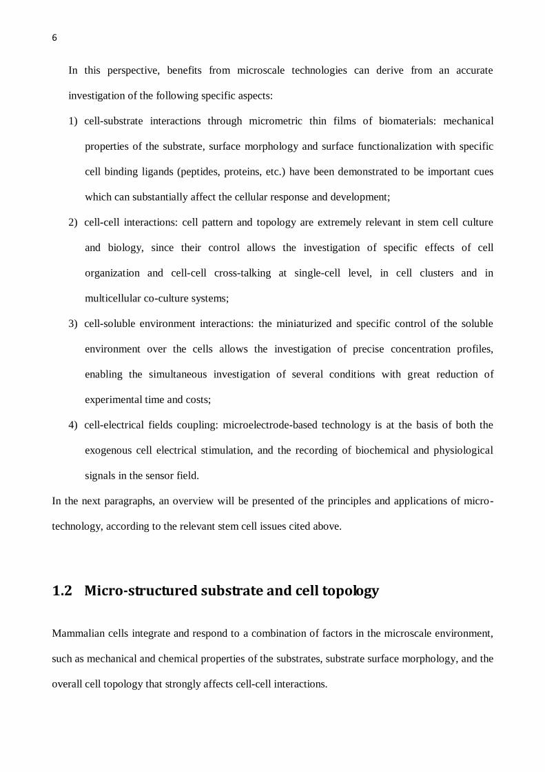
In this perspective, benefits from microscale technologies can derive from an accurate
investigation of the following specific aspects:
1) cell-substrate interactions through micrometric thin films of biomaterials: mechanical
properties of the substrate, surface morphology and surface functionalization with specific
cell binding ligands (peptides, proteins, etc.) have been demonstrated to be important cues
which can substantially affect the cellular response and development;
2) cell-cell interactions: cell pattern and topology are extremely relevant in stem cell culture
and biology, since their control allows the investigation of specific effects of cell
organization and cell-cell cross-talking at single-cell level, in cell clusters and in
multicellular co-culture systems;
3) cell-soluble environment interactions: the miniaturized and specific control of the soluble
environment over the cells allows the investigation of precise concentration profiles,
enabling the simultaneous investigation of several conditions with great reduction of
experimental time and costs;
4) cell-electrical fields coupling: microelectrode-based technology is at the basis of both the
exogenous cell electrical stimulation, and the recording of biochemical and physiological
signals in the sensor field.
In the next paragraphs, an overview will be presented of the principles and applications of micro-
technology, according to the relevant stem cell issues cited above.
1.2 Micro-structured substrate and cell topology
Mammalian cells integrate and respond to a combination of factors in the microscale environment,
such as mechanical and chemical properties of the substrates, substrate surface morphology, and the
overall cell topology that strongly affects cell-cell interactions.

7
Substrate mechanical properties can be properly designed by physical or chemical deposition of
micrometric thin layers of soft biomaterials. Among different biomaterials, hydrogels have recently
captured attention in the field of tissue engineering because of their high water content,
biocompatibility and elastic properties, resembling those of native tissues. It is worth to underline
that the elastic properties can be finely controlled and modulated by changing the amount of
polymer and crosslinker ratio during the production process. The stiffness of the hydrogels affects
the cell behavior at the cytoskeletal level and the transcriptional activity of mesenchimal stem
cells19-21
. Hydrogels, such as poly(ethylene glycol) (PEG) and derivatives, have been widely used as
substrates for cell culture or for encapsulating living cells22,23
. Their non fouling surface hinders cell
adhesion, thus allowing the formation and suspension cultures of a cell type such as embryoid
bodies (EB), cells aggregates derived from embryonic stem cells, which initiate differentiation and
recapitulate embryonic development to a limited extent24
. On the other hand, in order to enhance
and promote cell attachment, a wide variety of copolymers, composed of a synthetic backbone and
grafted biomolecules (or vice versa), such as fibrinogen25
, hyaluronic acid26
, or short peptides
sequences such as RGD27
, were developed. These biomimetic substrates were proposed and tested
to reproduce biological cues required for stem cell differentiation.
Besides cell-substrate interactions, the control of cell topology is of paramount importance for
studying stem cell differentiation, which requires a particular cell template, and for controlling cell-
cell interactions. For example, this latter aspect could be extremely relevant when a specific cell
pattern of two different cell types is required in a co-culture systems. To this aim, two different
methods which are based on different principles, were widely developed and applied: i) “physical”
method based on microstructured surface morphology; ii) “biochemical” method based on
micropatterning of functionalized surface.
In the first case, the topologic control over cell culture can be achieved through the use of substrates
(generally synthetic polymeric membranes) whose surface carries a three-dimensional morphology:
nanopatterned gratings, microgrooves and microtextures28-30
. Cell adhesion is thus guided by the
8
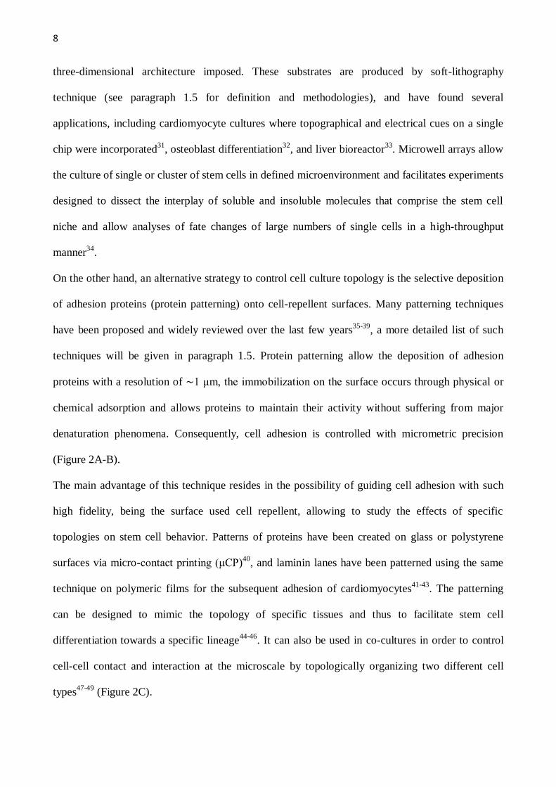
three-dimensional architecture imposed. These substrates are produced by soft-lithography
technique (see paragraph 1.5 for definition and methodologies), and have found several
applications, including cardiomyocyte cultures where topographical and electrical cues on a single
chip were incorporated31
, osteoblast differentiation32
, and liver bioreactor33
. Microwell arrays allow
the culture of single or cluster of stem cells in defined microenvironment and facilitates experiments
designed to dissect the interplay of soluble and insoluble molecules that comprise the stem cell
niche and allow analyses of fate changes of large numbers of single cells in a high-throughput
manner34
.
On the other hand, an alternative strategy to control cell culture topology is the selective deposition
of adhesion proteins (protein patterning) onto cell-repellent surfaces. Many patterning techniques
have been proposed and widely reviewed over the last few years35-39
, a more detailed list of such
techniques will be given in paragraph 1.5. Protein patterning allow the deposition of adhesion
proteins with a resolution of ∼1 μm, the immobilization on the surface occurs through physical or
chemical adsorption and allows proteins to maintain their activity without suffering from major
denaturation phenomena. Consequently, cell adhesion is controlled with micrometric precision
(Figure 2A-B).
The main advantage of this technique resides in the possibility of guiding cell adhesion with such
high fidelity, being the surface used cell repellent, allowing to study the effects of specific
topologies on stem cell behavior. Patterns of proteins have been created on glass or polystyrene
surfaces via micro-contact printing (μCP)40
, and laminin lanes have been patterned using the same
technique on polymeric films for the subsequent adhesion of cardiomyocytes41-43
. The patterning
can be designed to mimic the topology of specific tissues and thus to facilitate stem cell
differentiation towards a specific lineage44-46
. It can also be used in co-cultures in order to control
cell-cell contact and interaction at the microscale by topologically organizing two different cell
types47-49
(Figure 2C).

9
As frequently argued, performing stem cell cultures onto surfaces, 2D systems, do not mimic with
high fidelity the natural 3D architecture of in vivo tissues. In this sight, Albrecht and colleagues
explored the role of microscale organization of tissues in three-dimensions through the integration
of co-culture and topological cell organization in a 3D model50
, and they demonstrated that
chondrocyte biosynthesis specifically modulates microscale organization.
Another application of micropatterning aims at studying, in a highthroughput fashion, the effect of
the extracellular matrix (ECM) on stem cell behavior (Figure 2D). Microarray platforms of ECM
proteins were developed for screening their effects on stem cells in a combinatorial and parallel
fashion. A conventional DNA robotic spotter has been modified for the immobilization of specific
ECM components (collagen I, collagen III, collagen IV, laminin and fibronectin) or combinations of
these molecules on hydrogel surfaces51
. The developed array was used for studying the commitment
of mouse ES cells towards an early hepatic fate, leading to the identification of several
combinations of ECM components that synergistically impact ES cell differentiation and hepatocyte
function. This platform was further developed to analyze the interactions between ECM
components and soluble growth factors on stem cell fate, resulting in a multiwell microarray
platform that allows 1200 simultaneous experiments on 240 unique signaling microenvironments.
These studies were consistent with results previously reported in the literature, confirming the
reliability of the platform. Similarly, a microarray of biomaterials has been developed and used for
the high-throughput and rapid synthesis and screening of combinatorial libraries of
photopolymerizable biomaterials to identify compositions that influence human ES cell attachment,
growth and differentiation52
. Over 1700 human ES cell-material interactions were simultaneously
characterized and a multitude of unexpected materials effects were identified, offering new levels of
control over human embryonic stem cell behavior.
10
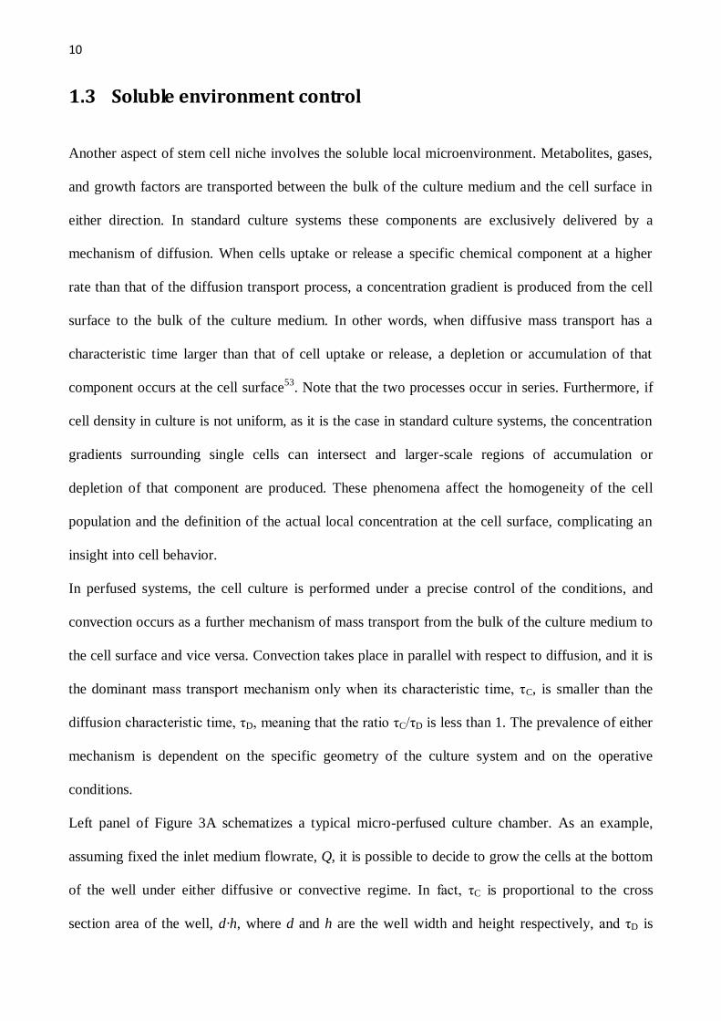
1.3 Soluble environment control
Another aspect of stem cell niche involves the soluble local microenvironment. Metabolites, gases,
and growth factors are transported between the bulk of the culture medium and the cell surface in
either direction. In standard culture systems these components are exclusively delivered by a
mechanism of diffusion. When cells uptake or release a specific chemical component at a higher
rate than that of the diffusion transport process, a concentration gradient is produced from the cell
surface to the bulk of the culture medium. In other words, when diffusive mass transport has a
characteristic time larger than that of cell uptake or release, a depletion or accumulation of that
component occurs at the cell surface53
. Note that the two processes occur in series. Furthermore, if
cell density in culture is not uniform, as it is the case in standard culture systems, the concentration
gradients surrounding single cells can intersect and larger-scale regions of accumulation or
depletion of that component are produced. These phenomena affect the homogeneity of the cell
population and the definition of the actual local concentration at the cell surface, complicating an
insight into cell behavior.
In perfused systems, the cell culture is performed under a precise control of the conditions, and
convection occurs as a further mechanism of mass transport from the bulk of the culture medium to
the cell surface and vice versa. Convection takes place in parallel with respect to diffusion, and it is
the dominant mass transport mechanism only when its characteristic time, τC, is smaller than the
diffusion characteristic time, τD, meaning that the ratio τC/τD is less than 1. The prevalence of either
mechanism is dependent on the specific geometry of the culture system and on the operative
conditions.
Left panel of Figure 3A schematizes a typical micro-perfused culture chamber. As an example,
assuming fixed the inlet medium flowrate, Q, it is possible to decide to grow the cells at the bottom
of the well under either diffusive or convective regime. In fact, τC is proportional to the cross
section area of the well, d·h, where d and h are the well width and height respectively, and τD is
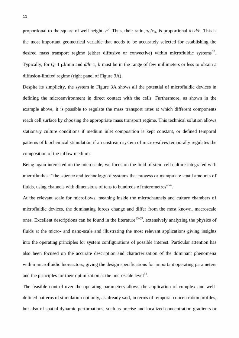
11
proportional to the square of well height, h2. Thus, their ratio, τC/τD, is proportional to d/h. This is
the most important geometrical variable that needs to be accurately selected for establishing the
desired mass transport regime (either diffusive or convective) within microfluidic systems53
.
Typically, for Q=1 l/min and d/h=1, h must be in the range of few millimeters or less to obtain a
diffusion-limited regime (right panel of Figure 3A).
Despite its simplicity, the system in Figure 3A shows all the potential of microfluidic devices in
defining the microenvironment in direct contact with the cells. Furthermore, as shown in the
example above, it is possible to regulate the mass transport rates at which different components
reach cell surface by choosing the appropriate mass transport regime. This technical solution allows
stationary culture conditions if medium inlet composition is kept constant, or defined temporal
patterns of biochemical stimulation if an upstream system of micro-valves temporally regulates the
composition of the inflow medium.
Being again interested on the microscale, we focus on the field of stem cell culture integrated with
microfluidics: “the science and technology of systems that process or manipulate small amounts of
fluids, using channels with dimensions of tens to hundreds of micrometres”54
.
At the relevant scale for microflows, meaning inside the microchannels and culture chambers of
microfluidic devices, the dominating forces change and differ from the most known, macroscale
ones. Excellent descriptions can be found in the literature55-59
, extensively analyzing the physics of
fluids at the micro- and nano-scale and illustrating the most relevant applications giving insights
into the operating principles for system configurations of possible interest. Particular attention has
also been focused on the accurate description and characterization of the dominant phenomena
within microfluidic bioreactors, giving the design specifications for important operating parameters
and the principles for their optimization at the microscale level53
.
The feasible control over the operating parameters allows the application of complex and well-
defined patterns of stimulation not only, as already said, in terms of temporal concentration profiles,
but also of spatial dynamic perturbations, such as precise and localized concentration gradients or
12
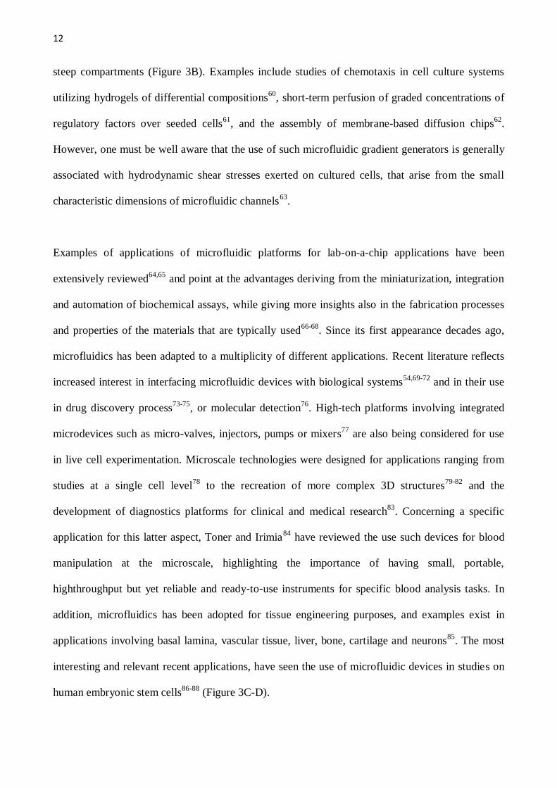
steep compartments (Figure 3B). Examples include studies of chemotaxis in cell culture systems
utilizing hydrogels of differential compositions60
, short-term perfusion of graded concentrations of
regulatory factors over seeded cells61
, and the assembly of membrane-based diffusion chips62
.
However, one must be well aware that the use of such microfluidic gradient generators is generally
associated with hydrodynamic shear stresses exerted on cultured cells, that arise from the small
characteristic dimensions of microfluidic channels63
.
Examples of applications of microfluidic platforms for lab-on-a-chip applications have been
extensively reviewed64,65
and point at the advantages deriving from the miniaturization, integration
and automation of biochemical assays, while giving more insights also in the fabrication processes
and properties of the materials that are typically used66-68
. Since its first appearance decades ago,
microfluidics has been adapted to a multiplicity of different applications. Recent literature reflects
increased interest in interfacing microfluidic devices with biological systems54,69-72
and in their use
in drug discovery process73-75
, or molecular detection76
. High-tech platforms involving integrated
microdevices such as micro-valves, injectors, pumps or mixers77
are also being considered for use
in live cell experimentation. Microscale technologies were designed for applications ranging from
studies at a single cell level78
to the recreation of more complex 3D structures79-82
and the
development of diagnostics platforms for clinical and medical research83
. Concerning a specific
application for this latter aspect, Toner and Irimia84
have reviewed the use such devices for blood
manipulation at the microscale, highlighting the importance of having small, portable,
highthroughput but yet reliable and ready-to-use instruments for specific blood analysis tasks. In
addition, microfluidics has been adopted for tissue engineering purposes, and examples exist in
applications involving basal lamina, vascular tissue, liver, bone, cartilage and neurons85
. The most
interesting and relevant recent applications, have seen the use of microfluidic devices in studies on
human embryonic stem cells86-88
(Figure 3C-D).

13
Research is being conducted to improve the cell culture soluble environment also at larger scales
(hundreds of milliliters) using suspension bioreactors where stirring is mechanically provided to
improve the transport of metabolites, gases, and growth factors89-91
. These devices have been used
to culture stem cells that can grow in suspension as single cells or as cell clusters, such as
hematopoietic stem cells92-95
, neural stem cells96
, or embryonic stem cells97
. The potential of these
bioreactors has also been extended to other types of cells that are suspended after encapsulation in
gelatin microspheres98
or inside porous polymeric microcarriers99-101
. These large-scale devices
have currently been limited in their use because of the high costs and the large number of cells
needed91
. In perspective, scaling down these systems to stirred microliter bioreactors, taking
advantages from knowledge of microfluidic stirring mechanisms already studied for other
applications, such as buoyancy-driven thermoconvection and electrokinetic flow102,103
, would be
extremely useful.
1.4 Electrical stimulation and recording
Endogenous electric fields play a key role in several biological processes (cell differentiation,
movement and growth). They are present in developing and regenerating animal tissues, both in the
extracellular space or in cell cytoplasm, and have magnitude that ranges from few to several
hundreds of mV/mm104-106
. Electrical stimulation and recording of excitable tissues (brain, muscles,
peripheral nerves or sensory system) are the subjects of interest of electrophysiological research and
clinical functional electrical stimulation107
. Consequently, the integration of electrical stimulation
devices with stem cell cultures could be a powerful tool for studying the effects of an exogenous
electric field on stem cell biology and give important insight into cell physiology.
The exposure of stem cell to neuro-physiological stimulation can be achieved through co-culture
with neuronal tissue108,109
, or the modulation of ion channel expression110
or finally the generation

14
of an electric field between electrodes, the latter being the most efficacious, as it allows the fine
control of crucial parameters such as amplitude, duration and frequency of the imposed pulse111
.
The need for increasing highthroughput characteristics of the experimentations, has led to the
development of miniaturized and arrayed platforms allowing for the generation of highthroughput
data112
. Among these systems, microelectrode arrays (MEA) are the most used and have been
proven to be a valuable tool for detecting and measuring extracellular potentials for excitable cell
types such as cardiomyocytes and neurons31,113-117
(Figure 4A). In general, microelectrodes allow
chemical measurements in spatial and temporal domains that were previously inaccessible118
.
In addition, microelectrodes platforms can be used as biosensors for recording signals released from
the cells themselves, thus allowing to monitor and record changes in defined physiological and
biochemical signals for applications ranging from pharmacology, cell biology, toxicology, and drug
discovery119,120
(Figure 4B). Several examples exist in the literature, illustrating basic rules dictating
their operative performances121
and demonstrating the ease of use in effective signal
recording122,123
. The most advanced systems integrate nanostructured materials, such as carbon
nanotubes, within the sensing elements, thus greatly improving the performance of the
devices124,125
.
1.5 Micro-technology overview
As well described in some of the above cited works55,58,59
, a multiplicity of techniques have been
applied in order to gain micrometric control over culture systems.
One of the first techniques ever applied to fabricate microelectromechanical systems (MEMS) is
silicon or glass micromachining. However, even if this method is still extensively used for
applications involving strong solvents or high temperatures, its use for biologically inspired studies
is limited due to its high costs, difficult realization, and limited compatibility of the materials (i.e.
silicon is not transparent).

15
To date, lithographic techniques, and especially photolithography, are the most commonly used to
fabricate microscale features on glass or silicon substrates to be subsequently used as masters for
obtaining the final devices. Since the early ’90’90, when Whitesides and others initiated the
exploration of its use in the field of biological studies36,126-128
, soft lithography has emerged among
all lithographic techniques and has been exploited for its many advantages such as the ease of
application, low cost, high versatility and properties of the usable materials. Soft lithography is
commonly used to create structures on surfaces for controlling cell-substrate interactions. It
basically covers a group of techniques with the common feature that at some stage of the process an
elastomeric (soft) material is used to create the desired structures. Such elastomeric devices
(typically in poly(dimethyl siloxane), PDMS) can be obtained through replica molding on the above
mentioned microstructured silicon masters. Photolithography and PDMS replica molding are the
leading techniques for the production of microfluidic platforms and microbioreactors.
Similarly micro-contact printing (µCP), first described by Whitesides in 1993129
, is the technique
for micropatterned substrates production, and consists in using a microstructured elastomeric stamp
with relief features to transfer an “inked” material (adhesion proteins in the case of cell culture)
onto a substrate. Similarly to µCP, other techniques such as microtransfer molding, micromolding
in capillaries and solvent-assisted micromolding use microstructured PDMS replica masters to
transfer its features on other materials. Alternatively, microscale features could be created on
supporting materials via selective chemical vapor deposition, wet or dry etching processes, or more
advanced micromachining processes such as bulk and surface micromachining, microstereo
lithography or finally laser mediated ablations. We again refer to specific publications for a more
detailed description of all these techniques59
.
16
Figure captions
Figure 1. Stem cell niche. A. Schematic representation of the stem cell niche, where
matrix/substrate interactions, cell-cell contact and soluble factors direct cell fate. B. Length and
time scales for some relevant biological phenomena.
Figure 2. Micropatterning and microstructured cultures. A. Examples of µCP of laminin and
resulting mouse satellite cell culture in circular dots (A1) of 300µm diameter and parallel lanes (A1)
75µm wide and 125µm spaced .Scale bars, 300µm. B. Human embryonic stem cell colonies
patterned at 200µm, 400µm, and 800µm diameters. Scale bars, 250µm. [PERMISSION]. C. Phase
contrast images of hepatocyte-fibroblast co-cultures. Scale bars, 200µm. [PERMISSION]. D.
Schematic representation of the layout of the extracellular matrix array; bright field of hepatocytes
cultured onto the array; immunofluorescence of albumin (a marker of differentiated hepatocyte
function). Scale bar, 1mm; inset scale bar, 500µm. [PERMISSION]
Figure 3. Soluble environment control. A1. A schematic representation of a typical culture chamber
with the microfluidic channels for fluid perfusion. The characteristic dimensions are its width d and
height h (distance between the bottom of the chamber and the microfluidic channels). A2. Graph
exemplifying the results of computational modeling of the averaged properties of the system and in
particular of the diffusion and convection time constants as a function of medium flow rate and
culture chamber geometry. Results are presented for two chemical species representative of a small
metabolite (higher diffusion coefficient D) and of a larger growth factor (lower D). The existence of
two distinct mass transport regimes is evident, one dominated by molecular diffusion, and the other
17
by convection. B. Two examples of microfluidic platforms capable of recreating defined and
compartmentalized concentration patterns on adherent cell populations within a microchannel. The
microbioreactors designs and images of their validation are presented. C. An image of a
microbioreactor array platform constituted of 3 independent rows, each composed of 4 individual
culture chambers (4mm diameter) [PERMISSION] that was used for the culture of hESC colonies
(image on the right). D. Results of a long-term culture of hESC colonies within a microfluidic
channel [PERMISSION] .
Figure 4. Electrical stimulation and recording. A. ES cell-derived neurons at different time points
after initiating the differentiation. Phase-contrast images of ES cell-derived neurons on
microelectrode arrays (MEAs) at 7 (left) or 28 days (right) after initiating the differentiation.
[PERMISSION]. B. an example of an integrated multi-electrodes biosensor array is reported.
specifically, it allows detections of glucose, lactate, glutamine, and glutamate. [PERMISSION]

18
1.6 References
1. K. Powell, (2005), It's the ecology, stupid!, Nature, 435, 268-270. 2. D. Kaplan, R. T. Moon and G. Vunjak-Novakovic, (2005), It takes a village to grow a tissue, Nat
Biotechnol, 23, 1237-1239. 3. C. M. Metallo, J. C. Mohr, C. J. Detzel, et al., (2007), Engineering the Stem Cell Microenvironment,
Biotechnology Progress, 23, 18-23. 4. D. E. Discher, D. J. Mooney and P. W. Zandstra, (2009), Growth Factors, Matrices, and Forces
Combine and Control Stem Cells, Science, 324, 1673-1677. 5. A. L. Barabási and Z. N. Oltvai, (2004), Network biology: understanding the cell's functional
organization, Nature Reviews Genetics, 5, 101-113. 6. L. H. Hartwell, J. J. Hopfield, S. Leibler, et al., (1999), From molecular to modular cell biology,
Nature, 402. 7. Z. N. Oltvai and A. L. Barabási, (2002), Life’s Complexity Pyramid, Science, 298, 763-764. 8. M. Cervinka, Z. Cervinkova and E. Rudolf, (2008), The role of time-lapse fluorescent microscopy in
the characterization of toxic effects in cell populations cultivated in vitro, Toxicology in Vitro, 22, 1382-1386
9. N. Jamshidi and B. Ø. Palsson, (2008), Top-Down Analysis of Temporal Hierarchy in Biochemical Reaction Networks, PLoS Computational Biology, 4.
10. T. Eschenhagen and W. H. Zimmermann, (2005), Engineering Myocardial Tissue, Circulation Research, 97, 1220-1231.
11. D. E. Discher, P. Janmey and Y.-L. Wang, (2005), Tissue Cells Feel and Respond to the Stiffness of Their Substrate, Science, 310, 1139-1143.
12. C. L. Bauwens, R. Peerani, S. Niebruegge, et al., (2008), Control of human embryonic stem cell colony and aggregate size heterogeneity influences differentiation trajectories, Stem Cells, 26, 2300-2310.
13. L. G. Griffith and M. A. Swartz, (2006), Capturing complex 3D tissue physiology in vitro, Nature Reviews Molecular Cell Biology, 7, 211-224.
14. A. Folch and M. Toner, (2000), Microengineering of cellular interactions, Annu. Rev. Biomed. Eng., 2, 227–256.
15. A. Khademhosseini, R. Langer, J. Borenstein, et al., (2006), Microscale technologies for tissue engineering and biology, PNAS, 103, 2480–2487.
16. D. B. Weibel, W. R. DiLuzio and G. M. Whitesides, (2007), Microfabrication meets microbiology, Nature Reviews - Microbiology, 209-218.
17. D. S. Chen and M. M. Davis, (2006), Molecular and functional analysis using live cell microarrays., Curr Opin Chem Biol 10, 28-34.
18. A. Kozarova, S. Petrinac, A. Ali, et al., (2006), Array of informatics: applications in modern research, J Proteome Res, 5, 1051-1059.
19. A. Engler, L. Bacakova, C. Newman, et al., (2004), Substrate compliance versus ligand density in cell on gel responses, Biophysical Journal, 86, 617-628.
20. S. R. Peyton and A. J. Putnam, (2005), Extracellular matrix rigidity governs smooth muscle cell motility in a biphasic fashion, J Cell Physiol 204, 198-209.
21. A. J. Engler, S. Sen, H. L. Sweeney, et al., (2006), Matrix Elasticity Directs Stem Cell Lineage Specification, Cell, 126, 677-689.
22. K. Britton-Keys, F. M. Andreopoulos and N. Peppas, (1998), Poly(ethylene glycol) star polymer hydrogels, Macromolecules 31, 8149-8156.
23. S. Lin-Gibson, R. L. Jones, N. R. Washburn, et al., (2005), Structure-property relationships of photopolymerizable poly(ethylene glycol) dimethacrylate hydrogels, Macromolecules 38, 2897-2902.

19
24. M. J. Yang, C. H. Chen, P. J. Lin, et al., (2007), Novel method of forming human embryoid bodies in a polystyrene dish surface-coated with a temperature-responsive methylcellulose hydrogel, Biomacromolecules, 8, 2746-2752
25. L. Almany and D. Seliktar, (2005), Biosynthetic hydrogel scaffolds made from fibrinogen and polyethylene glycol for 3D cell cultures, Biomat 26, 2467-2477.
26. J. B. Leach, K. A. Bivens, C. W. Patrick, et al., (2003), Photocrosslinked hyaluronic acid hydrogels: natural, biodegradable tissue engineering scaffolds, Biotechnology & Bioengineering, 82, 578-575.
27. D. Hern and J. A. Hubbell, (1998), Incorporation of adhesion peptides into nonadhesive hydrogels useful for tissue resurfacing, J Biomed Mater Res, 39, 266-276.
28. D. Motlagh, S. E. Senyo, T. A. Desai, et al., (2003), Microtextured substrata alter gene expression, protein localization and the shape of cardiac myocytes, Biomaterials 24, 2463 2476.
29. R. B. Vernon, M. D. Gooden, S. L. Lara, et al., (2005), Microgrooved fibrillar collagen membranes as scaffolds for cell support and alignment, Biomaterials, 26, 3131-3140.
30. E. K. F. Yim, R. M. Reano, S. W. Pang, et al., (2005 ), Nanopattern-induced changes in morphology and motility of smooth muscle cells, Biomaterials, 26, 5405-5413.
31. H. T. H. Au, B. Cui, Z. E. Chu, et al., (2009), Cell culture chips for simultaneous application of topographical and electrical cues enhance phenotype of cardiomyocytes, Lab on a Chip, 9, 564-575.
32. G. Kirmizidis and M. A. Birch, (2009), Microfabricated grooved substrates influence cell-cell communication and osteoblast differentiation in vitro, Tissue Engineering Part A, 16, 1427-1436.
33. J. Park, Y. Li, F. Berthiaume, et al., (2008), Radial flow hepatocyte bioreactor using stacked microfabricated grooved substrates, Biotechnology & Bioengineering, 99, 455-467.
34. M. P. Lutolf, R. Doyonnas, K. Havenstrite, et al., (2009), Perturbation of single hematopoietic stem cell fates in artificial niches. , Integrative Biology, 1, 59-69.
35. D. Falconnet, G. Csucs, H. M. Grandin, et al., (2006), Surface engineering approaches to micropattern surfaces for cell-based assays, Biomaterials, 27, 3044-3063.
36. Y. Xia and G. M. Whitesides, (1998), Soft lithography, Angew Chem Int Ed, 37, 550-575. 37. J. M. Karp, Y. Yeo, W. Geng, et al., (2006), A photolithographic method to create cellular
micropatterns, Biomaterials, 27, 4755-4764. 38. S. Rohr, D. M. Scholly and A. G. Kleber, (1991), Patterned growth of neonatal rat heart cells in
culture. Morphological and electrophysiological characterization, Circulation Research, 68, 114-130. 39. K. Y. Suh, J. Seong, A. Khademhosseini, et al., (2004), A simple soft lithographic route to fabrication
of poly(ethylene glycol) microstructures for protein and cell patterning, Biomaterials, 25, 557-563. 40. S. A. Ruiz and C. S. Chen, (2007), Microcontact printing: A tool to pattern, Soft Matter 3, 1–11. 41. T. C. McDevitt, J. C. Angello, M. L. Whitney, et al., (2002), In vitro generation of differentiated
cardiac myofibers on micropatterned laminin surfaces, Journal of biomedical materials research, 60, 472-479.
42. T. C. McDevitt, K. A. Woodhouse, S. D. Hauschka, et al., (2003), Spatially organized layers of cardiomyocytes on biodegradable polyurethane films for myocardial repair, Journal of Biomedical Material Reseasch, 66A, 586-595.
43. E. Cimetta, S. Pizzato, S. Bollini, et al., (2009), Production of arrays of cardiac and skeletal muscle myofibers by micropatterning techniques on a soft substrate, Biomedical Microdevices, 11.
44. A. J. Engler, M. A. Griffin, S. Sen, et al., (2004), Myotubes differentiate optimally on substrates with tissue-like stiffness: pathological implications for soft or stiff microenvironments, The Journal of Cell Biology, 166, 877-887.
45. P. Camelliti, J. O. Gallagher, P. Kohl, et al., (2006), Micropatterned cell cultures on elastic membranes as an in vitro model of myocardium, Nature Protocols, 1, 1379-1391.
46. P. Krsko, T. E. McCann, T. T. Thach, et al., (2009), Length-scale mediated adhesion and directed growth of neural cells by surface-patterned poly(ethylene glycol) hydrogels, Biomaterials, 30, 721-729.
47. K. Kikuchi, K. Sumaru, J. I. Edahiro, et al., (2009), Stepwise Assembly of Micropatterned Co-cultures Using Photoresponsive Culture Surfaces and Its Application to Hepatic Tissue Arrays, Biotechnology & Bioengineering, 103, 552-561.
20
48. E. E. Hui and S. N. Bhatia, (2007), Microscale Control of Cell Contact and Spacing via Three-Component Surface Patterning, Langmuir, 23, 4103-4107.
49. A. Revzin, P. Rajagopalan, A. W. Tilles, et al., (2004), Designing a Hepatocellular Microenvironment with Protein Microarraying and Poly(ethylene glycol) Photolithography, Langmuir, 20, 2999-3005.
50. D. R. Albrecht, G. H. Underhill, T. B. Wassermann, et al., (2006), Probing the role of multicellular organization in three-dimensional microenvironments, Nat Methods, 3, 369-375.
51. C. J. Flaim, S. Chien and S. N. Bhatia, (2005), An extracellular matrix microarray for probing cellular differentiation, Nature Methods, 2, 119-125.
52. D. G. Anderson, S. Levenberg and R. Langer, (2004), Nanoliter-scale synthesis of arrayed biomaterials and application to human embryonic stem cells, Nature Biotechnology, 22, 863-866.
53. E. Cimetta, E. Figallo, C. Cannizzaro, et al., (2009), Micro-bioreactor arrays for controlling cellular environments: Design principles for human embryonic stem cell applications, Methods, 47, 81-89.
54. G. M. Whitesides, (2006), The origins and the future of microfluidics, Nature, 442, 368-373. 55. D. J. Beebe, G. A. Mensing and G. MWalker, (2002), Physics and applications of microfluidics in
biology, Annu. Rev. Biomed. Eng., 4, 261–286. 56. T. M. Squires and S. R. Quake, (2005), Microfluidics: Fluid physics at the nanoliter scale, Reviews of
Modern Physics, 77, 977-1016. 57. H. A. Stone and S. Kim, (2001), Microfluidics: Basic Issues, Applications, and Challenges, AIChE
Journal, 47, 1250-1254. 58. J. Berthier and P. Silberzan, (2005), Microfluidics for Biotechnology, Artech House Publishers. 59. N.-T. Nguyen and S. Wereley, (2002), Fundamentals and Applications of Microfluidics, Artech House
Publishers. 60. J. A. Burdick, A. Khademhosseini and R. Langer, (2004), Fabrication of gradient hydrogels using a
microfluidics/photopolymerization process, Langmuir 20, 5153-5156. 61. W. Saadi, S. J. Wang, F. Lin, et al., (2006), A parallel-gradient microfluidic chamber for quantitative
analysis of breast cancer cell chemotaxis, Biomed Microdevices, 8, 109–118. 62. J. Diao, L. Young, S. Kim, et al., (2006), A three-channel microfluidic device for generating static
linear gradients and its application to the quantitative analysis of bacterial chemotaxis, Lab on a Chip, 6, 381–388.
63. Y. Chisti, (2001), Hydrodynamic Damage to Animal Cells, Critical Reviews in Biotechnology, 21, 67-110.
64. D. N. Breslauer, P. J. Lee and L. P. Lee, (2006), Microfluidics-based systems biology, Mol. BioSyst., 2, 97-112.
65. S. Haeberle and R. Zengerle, (2007), Microfluidic platforms for lab-on-a-chip applications, Lab on a Chip, 7, 1094–1110.
66. J. C. McDonald, D. C. Duffy, J. R. Anderson, et al., (2000), Fabrication of microfluidic systems in poly(dimethylsiloxane), Electrophoresis, 21, 27-40
67. S. K. Sia and G. M. Whitesides, (2003), Microfluidic devices fabricated in poly(dimethylsiloxane) for biological studies, Electrophoresis, 24, 3563–3576.
68. G. M. Whitesides, E. Ostuni, S. Takayama, et al., (2001), Soft lithography in biology and biochemistry, Annual Review of Biomedical Engineering, 3, 335–373.
69. L. Kim, Y.-C. Toh, J. Voldman, et al., (2007), A practical guide to microfluidic perfusion culture of adherent mammalian cells, Lab on a Chip, 7, 681–694.
70. D. Amarie, J. A. Glazier and S. C. Jacobson, (2007), Compact Microfluidic Structures for Generating Spatial and Temporal Gradients, Analytical Chemistry.
71. N. L. Jeon, S. K. W. Dertinger, D. T. Chiu, et al., (2000), Generation of Solution and Surface Gradients Using Microfluidic Systems, Langmuir, 16, 8311-8316.
72. T. M. Keenan and A. Folch, (2007), Biomolecular gradients in cell culture systems, Lab on a Chip, 8, 34–57.
73. S. R. Khetani and S. N. Bhatia, (2008), Microscale culture of human liver cells for drug development, Nature Biotechnology, 21, 120-126.
74. P. S. Dittrich and A. Manz, (2006), Lab-on-a-chip: microfluidics in drug discovery, Nature Reviews - Drug Discovery, 5, 210-218.
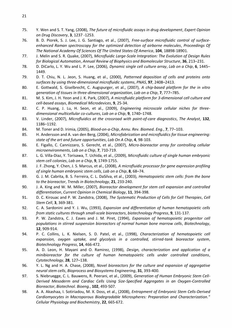
21
75. Y. Wen and S. T. Yang, (2008), The future of microfluidic assays in drug development, Expert Opinion on Drug Discovery, 3, 1237 -1253.
76. B. D. Piorek, S. J. Lee, J. G. Santiago, et al., (2007), Free-surface microfluidic control of surface-enhanced Raman spectroscopy for the optimized detection of airborne molecules, Proceedings Of The National Academy Of Sciences Of The United States Of America, 104, 18898-18901.
77. J. Melin and S. R. Quake, (2007), Microfluidic Large-Scale Integration: The Evolution of Design Rules for Biological Automation, Annual Review of Biophysics and Biomolecular Structure, 36, 213–231.
78. D. DiCarlo, L. Y. Wu and L. P. Lee, (2006), Dynamic single cell culture array, Lab on a Chip, 6, 1445–1449.
79. D. T. Chiu, N. L. Jeon, S. Huang, et al., (2000), Patterned deposition of cells and proteins onto surfaces by using three-dimensional microfluidic systems, PNAS, 97, 2408–2413.
80. E. Gottwald, S. Giselbrecht, C. Augspurger, et al., (2007), A chip-based platform for the in vitro generation of tissues in three-dimensional organization, Lab on a Chip, 7, 777–785.
81. M. S. Kim, J. H. Yeon and J. K. Park, (2007), A microfluidic platform for 3-dimensional cell culture and cell-based assays, Biomedical Microdevices, 9, 25-34.
82. C. P. Huang, J. Lu, H. Seon, et al., (2009), Engineering microscale cellular niches for three-dimensional multicellular co-cultures, Lab on a Chip, 9, 1740–1748.
83. V. Linder, (2007), Microfluidics at the crossroad with point-of-care diagnostics, The Analyst, 132, 1186–1192.
84. M. Toner and D. Irimia, (2005), Blood-on-a-Chip, Annu. Rev. Biomed. Eng., 7, 77–103. 85. H. Andersson and A. van den Berg, (2004), Microfabrication and microfluidics for tissue engineering:
state of the art and future opportunities, Lab On A Chip, 4, 98-103. 86. E. Figallo, C. Cannizzaro, S. Gerecht, et al., (2007), Micro-bioreactor array for controlling cellular
microenvironments, Lab on a Chip, 7, 710-719. 87. L. G. Villa-Diaz, Y. Torisawa, T. Uchida, et al., (2009), Microfluidic culture of single human embryonic
stem cell colonies, Lab on a Chip, 9, 1749-1755. 88. J. F. Zhong, Y. Chen, J. S. Marcus, et al., (2008), A microfluidic processor for gene expression profiling
of single human embryonic stem cells, Lab on a Chip, 8, 68–74. 89. G. J. M. Cabrita, B. S. Ferreira, C. L. DaSilva, et al., (2003), Hematopoietic stem cells: from the bone
to the bioreactor, Trends in Biotechnology, 21, 233-240. 90. J. A. King and W. M. Miller, (2007), Bioreactor development for stem cell expansion and controlled
differentiation, Current Opinion in Chemical Biology, 11, 394-398. 91. D. C. Kirouac and P. W. Zandstra, (2008), The Systematic Production of Cells for Cell Therapies, Cell
Stem Cell, 3, 369-381. 92. C. A. Sardonini and Y. J. Wu, (1993), Expansion and differentiation of human hematopoietic cells
from static cultures through small-scale bioreactors, biotechnology Progress, 9, 131-137. 93. P. W. Zandstra, C. J. Eaves and J. M. Piret, (1994), Expansion of hematopoietic progenitor cell
populations in stirred suspension bioreactors of normal human bone marrow cells, Biotechnology, 12, 909-914.
94. P. C. Collins, L. K. Nielsen, S. D. Patel, et al., (1998), Characterization of hematopoietic cell expansion, oxygen uptake, and glycolysis in a controlled, stirred-tank bioreactor system, Biotechnology Progress, 14, 466-472.
95. A. D. Leon, H. Mayani and O. Ramirez, (1998), Design, characterization and application of a minibioreactor for the culture of human hematopoietic cells under controlled conditions, Cytotechnology, 28, 127–138.
96. Y. L. Ng and H. A. Chase, (2008), Novel bioreactors for the culture and expansion of aggregative neural stem cells, Bioprocess and Biosystems Engineering, 31, 393-400.
97. S. Niebruegge, C. L. Bauwens, R. Peerani, et al., (2009), Generation of Human Embryonic Stem Cell-Derived Mesoderm and Cardiac Cells Using Size-Specified Aggregates in an Oxygen-Controlled Bioreactor, Biotechnol. Bioeng., 102, 493-507.
98. A. A. Akashaa, I. Sotiriadou, M. X. Doss, et al., (2008), Entrapment of Embryonic Stem Cells-Derived Cardiomyocytes in Macroporous Biodegradable Microspheres: Preparation and Characterization." Cellular Physiology and Biochemistry, 22, 665-672.
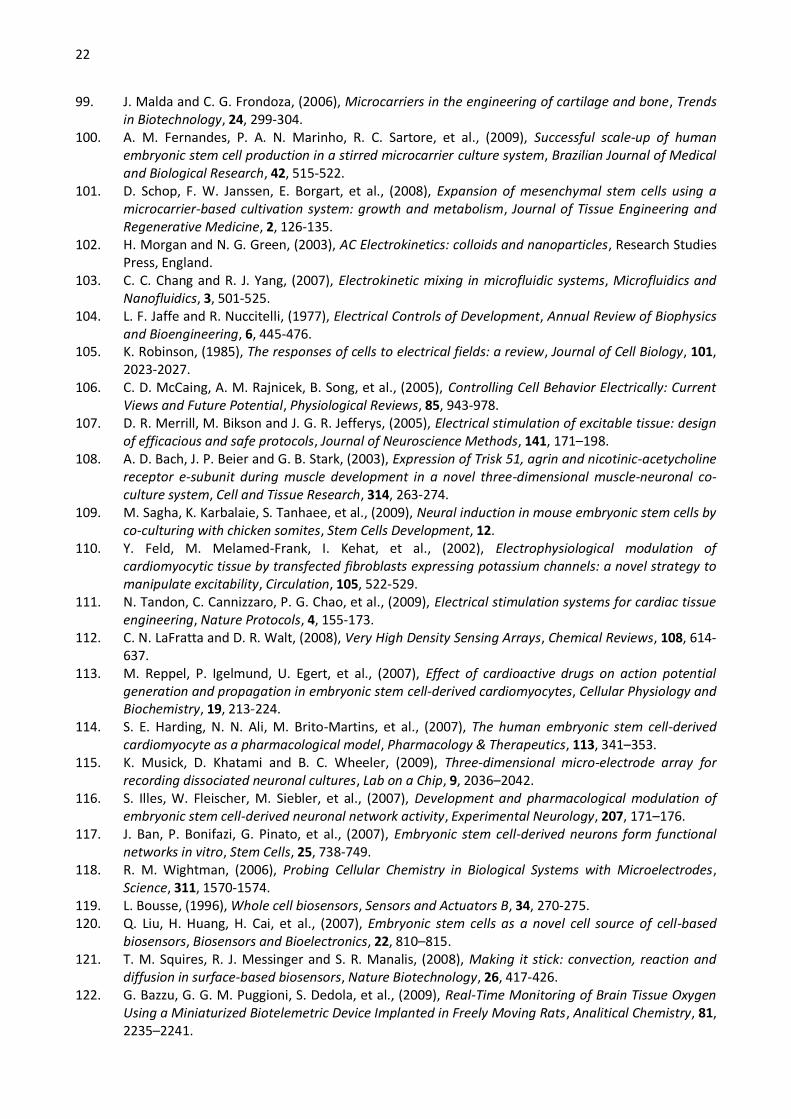
22
99. J. Malda and C. G. Frondoza, (2006), Microcarriers in the engineering of cartilage and bone, Trends in Biotechnology, 24, 299-304.
100. A. M. Fernandes, P. A. N. Marinho, R. C. Sartore, et al., (2009), Successful scale-up of human embryonic stem cell production in a stirred microcarrier culture system, Brazilian Journal of Medical and Biological Research, 42, 515-522.
101. D. Schop, F. W. Janssen, E. Borgart, et al., (2008), Expansion of mesenchymal stem cells using a microcarrier-based cultivation system: growth and metabolism, Journal of Tissue Engineering and Regenerative Medicine, 2, 126-135.
102. H. Morgan and N. G. Green, (2003), AC Electrokinetics: colloids and nanoparticles, Research Studies Press, England.
103. C. C. Chang and R. J. Yang, (2007), Electrokinetic mixing in microfluidic systems, Microfluidics and Nanofluidics, 3, 501-525.
104. L. F. Jaffe and R. Nuccitelli, (1977), Electrical Controls of Development, Annual Review of Biophysics and Bioengineering, 6, 445-476.
105. K. Robinson, (1985), The responses of cells to electrical fields: a review, Journal of Cell Biology, 101, 2023-2027.
106. C. D. McCaing, A. M. Rajnicek, B. Song, et al., (2005), Controlling Cell Behavior Electrically: Current Views and Future Potential, Physiological Reviews, 85, 943-978.
107. D. R. Merrill, M. Bikson and J. G. R. Jefferys, (2005), Electrical stimulation of excitable tissue: design of efficacious and safe protocols, Journal of Neuroscience Methods, 141, 171–198.
108. A. D. Bach, J. P. Beier and G. B. Stark, (2003), Expression of Trisk 51, agrin and nicotinic-acetycholine receptor e-subunit during muscle development in a novel three-dimensional muscle-neuronal co-culture system, Cell and Tissue Research, 314, 263-274.
109. M. Sagha, K. Karbalaie, S. Tanhaee, et al., (2009), Neural induction in mouse embryonic stem cells by co-culturing with chicken somites, Stem Cells Development, 12.
110. Y. Feld, M. Melamed-Frank, I. Kehat, et al., (2002), Electrophysiological modulation of cardiomyocytic tissue by transfected fibroblasts expressing potassium channels: a novel strategy to manipulate excitability, Circulation, 105, 522-529.
111. N. Tandon, C. Cannizzaro, P. G. Chao, et al., (2009), Electrical stimulation systems for cardiac tissue engineering, Nature Protocols, 4, 155-173.
112. C. N. LaFratta and D. R. Walt, (2008), Very High Density Sensing Arrays, Chemical Reviews, 108, 614-637.
113. M. Reppel, P. Igelmund, U. Egert, et al., (2007), Effect of cardioactive drugs on action potential generation and propagation in embryonic stem cell-derived cardiomyocytes, Cellular Physiology and Biochemistry, 19, 213-224.
114. S. E. Harding, N. N. Ali, M. Brito-Martins, et al., (2007), The human embryonic stem cell-derived cardiomyocyte as a pharmacological model, Pharmacology & Therapeutics, 113, 341–353.
115. K. Musick, D. Khatami and B. C. Wheeler, (2009), Three-dimensional micro-electrode array for recording dissociated neuronal cultures, Lab on a Chip, 9, 2036–2042.
116. S. Illes, W. Fleischer, M. Siebler, et al., (2007), Development and pharmacological modulation of embryonic stem cell-derived neuronal network activity, Experimental Neurology, 207, 171–176.
117. J. Ban, P. Bonifazi, G. Pinato, et al., (2007), Embryonic stem cell-derived neurons form functional networks in vitro, Stem Cells, 25, 738-749.
118. R. M. Wightman, (2006), Probing Cellular Chemistry in Biological Systems with Microelectrodes, Science, 311, 1570-1574.
119. L. Bousse, (1996), Whole cell biosensors, Sensors and Actuators B, 34, 270-275. 120. Q. Liu, H. Huang, H. Cai, et al., (2007), Embryonic stem cells as a novel cell source of cell-based
biosensors, Biosensors and Bioelectronics, 22, 810–815. 121. T. M. Squires, R. J. Messinger and S. R. Manalis, (2008), Making it stick: convection, reaction and
diffusion in surface-based biosensors, Nature Biotechnology, 26, 417-426. 122. G. Bazzu, G. G. M. Puggioni, S. Dedola, et al., (2009), Real-Time Monitoring of Brain Tissue Oxygen
Using a Miniaturized Biotelemetric Device Implanted in Freely Moving Rats, Analitical Chemistry, 81, 2235–2241.

23
123. I. Moser, G. Jobst and G. A. Urban, (2002), Biosensor arrays for simultaneous measurement of glucose, lactate, glutamate, and glutamine, Biosensors & Bioelectronics, 17, 297–302.
124. W. R. Yang, P. Thordarson, J. J. Gooding, et al., (2007), Carbon nanotubes for biological and biomedical applications, Nanotechnology, 18, 12.
125. N. W. S. Kam, E. Jan and N. A. Kotov, (2009), Electrical Stimulation of Neural Stem Cells Mediated by Humanized Carbon Nanotube Composite Made with Extracellular Matrix Protein, Nano Letters, 9, 273-278.
126. D. Duffy, J. McDonald, O. Schueller, et al., (1998), Rapid prototyping of microfluidic systems in poly (dimethyl siloxane), Analytical Chemistry, 70, 4974–4984.
127. C. S. Chen, M. Mrksich, S. Huang, et al., (1997), Geometric Control of Cell Life and Death, Science, 276, 1425-1428.
128. S. R. Quake and A. Scherer, (2000), From Micro- to Nanofabrication with Soft Materials, Science, 290, 1536 -1540.
129. A. Kumar and G. M. Whitesides, (1993), Features of gold having micrometer to centimeter dimensions can be formed through a combination of stamping with an elastomeric stamp and an alkanethiol ink followed by chemical etching, Applied Physics Letters, 63, 2002-2004.

Figure 1

Figure 2

Figure 3
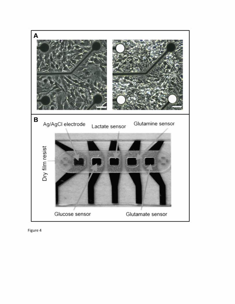
Figure 4
Related Documents